
Submitted:
26 August 2026
Posted:
31 August 2026
You are already at the latest version
Abstract
Background: Sialolithiasis is a multifactorial biomineralization disorder in which local salivary, biological, and potentially environmental factors may influence stone formation and maturation. The relationship between drinking-water mineralization and the chemical composition of salivary stones remains poorly understood. This study aimed to characterize the composition of sialoliths in an Armenian population and to explore whether long-term regional drinking-water hardness is associated with variation in their mineralization profile. Methods: Fifty submandibular sialoliths obtained from 50 patients were analyzed using atomic absorption spectroscopy, optical emission spectroscopy, flame-emission analysis, and photocolorimetric methods. Elemental concentrations were reported as oxide-equivalent weight percentages, and the organic fraction was quantified separately. The molar Ca/P ratio was calculated from CaO- and P₂O₅-equivalent concentrations. Regional drinking-water hardness data for 2018–2026 were obtained from official Armenian water-supply and regulatory sources and expressed in mmol/L. Associations with patient age, sex, geographic region, and long-term regional water hardness were evaluated using non-parametric statistical methods, with correction for multiple comparisons. Results: Calcium was the predominant individual inorganic constituent (CaO-equivalent: 31.38 ± 2.35 wt%), accompanied by P₂O₅ (15.83 ± 2.78 wt%) and a substantial organic fraction (32.09 ± 4.26 wt%). The mean molar Ca/P ratio was 2.62 ± 0.71. CaO and P₂O₅ concentrations were not significantly correlated, indicating considerable heterogeneity in calcium–phosphorus stoichiometry among stones. No significant associations were observed between major compositional parameters and patient age or sex. In contrast, CaO-equivalent content differed significantly among geographic regions (Kruskal–Wallis H = 19.88, p = 0.0058; Holm-adjusted p = 0.0409). Long-term drinking-water hardness ranged from approximately 1.04 to 9.52 mmol/L across the eight regions. At the patient-record level, greater regional water hardness was positively associated with CaO content (Spearman ρ = 0.374, p = 0.0075) and the molar Ca/P ratio (ρ = 0.364, p = 0.0093); however, these associations were attenuated after correction for multiple testing and were not statistically significant when the eight regions were analyzed as independent ecological units. Conclusions: Armenian submandibular sialoliths are chemically heterogeneous composite structures characterized by a calcium- and phosphorus-rich inorganic component and a substantial organic fraction. The significant geographic variability in calcium content and the exploratory positive relationship with regional drinking-water hardness suggest that environmental mineral exposure may contribute to variation in the biomineralization phenotype of sialoliths. However, the ecological nature of the exposure assessment and the absence of significant region-level associations preclude causal inference. Larger prospective studies integrating individual water exposure, salivary biochemistry, and phase-sensitive mineralogical analyses are warranted.
Keywords:
sialolithiasis
; salivary stones
; biomineralization
; drinking-water hardness
; calcium
; phosphorus
; Ca/P ratio
; elemental analysis
1. Introduction
Sialolithiasis is characterized by the formation of calcified deposits, known as sialoliths or salivary calculi, within the ductal system or parenchyma of the major salivary glands [1,2]. It is among the most common non-neoplastic disorders of the salivary glands and predominantly affects the submandibular gland, whereas the parotid and sublingual glands are involved less frequently [2,3]. Obstruction of salivary outflow may cause recurrent meal-related pain and swelling, secondary bacterial infection, and progressive chronic sialadenitis [1,2].
Sialolith formation is generally considered a multifactorial biomineralization process rather than the result of a single metabolic abnormality. Proposed mechanisms include mineral deposition around an organic nidus composed of mucus, desquamated epithelial cells, microorganisms, foreign material, or inflammatory products; aggregation and calcification of pre-existing sialomicroliths; and mineralization within a highly viscous mucoepithelial matrix [3,4,5,6,7,8,9]. More recent evidence suggests that neutrophil extracellular traps generated during ductal inflammation may provide a framework for the nucleation, retention, and growth of calcium-containing crystals [10]. Reduced salivary flow, altered salivary pH, microbial activity, ductal anatomy, and changes in the concentrations of calcium, phosphate, magnesium, citrate, and phytate may further promote supersaturation and precipitation of poorly soluble calcium salts [5,11,12].
Mineralogical studies have consistently demonstrated that calcium-phosphate phases constitute the principal inorganic framework of most salivary stones. Hydroxyapatite, carbonate apatite, whitlockite, octacalcium phosphate, and, less frequently, brushite have been identified in sialoliths using X-ray diffraction, Fourier-transform infrared spectroscopy, electron microscopy, and energy-dispersive spectroscopic techniques [3,13,14,15,16,17,18,19]. These phases may represent different stages of stone maturation or reflect variation in the local physicochemical environment. Early deposits may contain amorphous or poorly crystalline calcium phosphates, which can progressively transform into more stable apatite-related structures during layer-by-layer growth [16,17,18,20]. Magnesium, sodium, potassium, silicon, iron, aluminum, and other trace elements may be incorporated through ionic substitution, adsorption onto crystal surfaces, or entrapment within successive mineral layers, thereby modifying crystal stability and maturation without necessarily constituting the primary mineral phase [3,17,20,21,22].
The elemental and mineral composition of sialoliths may also be influenced by systemic, dietary, salivary, and environmental factors. Increased salivary calcium concentrations and decreased concentrations of magnesium and citrate have been reported in patients with sialolithiasis, supporting a local biochemical environment favorable to calcium-phosphate precipitation [11]. Systemic metabolic conditions, including dyslipidemia and diabetes, have also been investigated as potential risk factors [6]. Nevertheless, the contribution of long-term environmental mineral exposure, particularly through drinking water, remains uncertain.
Water hardness is primarily determined by dissolved calcium and magnesium salts and varies according to the geological and hydrological characteristics of the water source [23,24]. Long-term consumption of water with different mineral profiles could theoretically influence systemic mineral intake, salivary electrolyte composition, and the physicochemical conditions associated with biomineralization. However, epidemiological findings have been inconsistent. Schrøder et al. reported associations between regional concentrations of calcium, magnesium, and hydrogen carbonate in drinking water and hospital-admitted sialolithiasis in Denmark [23]. In contrast, Sherman and McGurk found no significant relationship between drinking-water hardness and the occurrence of salivary calculi in England [24]. These contrasting findings suggest that the relationship may depend on population-specific environmental, geological, dietary, and biological factors. Consequently, it remains unclear whether long-term regional water hardness is associated with measurable variation in the calcium-, magnesium-, phosphate-, or carbonate-related composition of sialoliths. Examination of stone composition in relation to environmental mineral exposure may offer additional insight into the conditions that modulate sialolith growth and maturation, even if such exposure is not sufficient to initiate stone formation independently.
Armenia provides a particulary informative setting for investigating this relationship because its regions differ markedly in altitude, geology, groundwater characteristics, and drinking-water mineralization [25]. In a recent multicenter epidemiological study of salivary-gland disorders in the Armenian adult population, Misakyan et al., characterized the regional epidemiology of salivary-gland pathologies, including sialolithiasis [26]. These previously established regional epidemiological data provide an opportunity to examine whether geographic variation in sialolithiasis is associated with long-term differences in drinking-water hardness. Despite this environmental heterogeneity, population-specific data on the inorganic composition of Armenian sialoliths remain limited, and no previous study has evaluated their composition in relation to long-term regional drinking-water hardness. Combining patient-derived sialolith analyses with multi-year regional water-quality measurements may therefore provide new information regarding the possible environmental modulation of salivary stone biomineralization.
The analytical techniques applied in this study—atomic absorption spectroscopy, optical emission spectroscopy and photocolorimetric methods—permit quantitative assessment of major and trace inorganic constituents. Because these methods characterize elemental or ion-related composition rather than directly identifying crystalline phases, compositional findings are reported using geochemical oxide-equivalent notation and interpreted without claiming direct mineral-phase identification.
The aim of this study was to investigate whether long-term regional drinking-water hardness is associated with sialolithiasis and with the inorganic composition of sialoliths in the Armenian population.
2. Materials and Methods
2.1. Study Design and Setting
This analytical observational study was conducted at the Department of ENT and Maxillofacial Surgery, Heratsi No. 1 University Hospital, Yerevan State Medical University, Armenia. Salivary stone specimens were collected between July 2023 and August 2025 from patients undergoing surgical treatment for submandibular sialolithiasis. The laboratory analyses were performed at the Department of Analytical Research/Chemical Laboratory, Institute of Geological Sciences, National Academy of Sciences of the Republic of Armenia.
The study combined patient-level compositional analysis of sialolith specimens with region-level environmental data on drinking-water hardness. The environmental component was designed as an exploratory ecological analysis evaluating whether regional variation in long-term water hardness was associated with variation in the inorganic composition of salivary stones.
Regional drinking-water hardness measurements for 2018–2026 were provided by Veolia Jur CJSC: the State Water Committee of the Republic of Armenia. Hardness values were expressed in mmol/L. Annual measurements were compiled for each region included in the analysis, and the mean value across the available study period was calculated to represent long-term regional drinking-water hardness.
2.1.1. Sample Size Determination
An a priori sample-size calculation was performed for the primary specimen-level analysis evaluating the association between regional drinking-water hardness and sialolith composition. Assuming a moderate correlation coefficient of r = 0.40, a two-sided significance level of \alpha = 0.05, and a statistical power of 80%, the minimum required sample size was estimated to be 47 specimens. A total of 50 sialoliths obtained from 50 patients were ultimately included, exceeding the calculated minimum sample size.
2.1.2. Geographic Classification
Patients were classified according to their recorded region of residence in Armenia. Regional assignment was based on the patient’s place of habitual residence rather than the location of the treating hospital. The geographic categories represented in the study included Yerevan and the participating administrative regions of Armenia.
Because the number of included patients differed among regions, regional analyses were considered exploratory. The larger number of patients from Yerevan reflects the population distribution and referral pattern of Heratsi No. 1 University Hospital, which is located in the capital and serves a substantial proportion of patients residing in Yerevan.
2.1.3. Sialolith Specimen Collection
This analytical study was conducted between July 2023 and August 2025. During this period, forty-five sialolith specimens were collected at the Department of ENT and Maxillofacial Surgery of Heratsi #1 University Hospital. The sialoliths were obtained via sialolithotomy, which was carried out by making an incision over the duct to expose the sialolith or sialadenectomy. The patient’s sex, age, diet and region of residence in Armenia were considered. Given the pronounced geographical heterogeneity of Armenia, which includes both mountainous and lowland regions, as well as significant regional differences in water hardness, a comparative analysis of the inorganic components of salivary stones was performed according to the region of residence of each patient. Analysis of residence data indicated that patients who underwent salivary stone removal procedures resided in nine distinct regions of the country, which were numerically labeled as follows: Yerevan (I), Ararat (II), Gegharkunik (III), Armavir (IV), Meghri (V), Kotayq (VI), Ijevan (VII), and Lori (VIII).
After extraction, the stones were carefully washed in distilled water to remove surface contaminants, air-dried at room temperature and transferred to the Laboratory of Chemistry of the Institute of Geological Sciences (National Academy of Sciences of the Republic of Armenia).
The analysis of sialoliths, or salivary stones, involves various physicochemical techniques that can be classified into qualitative and quantitative methods, with each providing valuable insights into their inorganic composition. Atomic absorption spectroscopy (AAS), optical emission spectroscopy (OES) and photocolorimetric methods were implemented in this study.
2.1.4. Atomic Absorption Method
Atomic absorption spectroscopy (AAS) was used for the quantitative determination of elemental composition in sialolith samples. Analyses were performed using an AAC-1N atomic absorption spectrometer (Karl Zeiss, Germany). The principle of atomic absorption spectroscopy is based on measuring the absorption of light by atoms of a specific element. When light passes through a gas atomic vapor containing the element of interest, some of the light is absorbed by the atoms, and the extent of this absorption is directly proportional to the concentration of the element in the sample.
Prior to analysis, the dried specimens (sialoliths) were mechanically ground to a fine, homogeneous powder using an agate mortar and pestle in order to minimize contamination and ensure representative elemental analysis. Prior to spectroscopic analysis, powdered samples were stored in clean, airtight containers under dry conditions.
Concentrations of investigated elements were measured using element-specific lamps and calibrated against certified standard solutions, which enables quantitative results to be obtained. Sample preparation was performed using an acid digestion and fusion procedure. The powdered samples were treated with high-purity nitric acid, sulfuric acid (d = 1.84), hydrochloric acid (d = 1.19), and hydrofluoric acid (40% solution) to ensure complete dissolution of mineral components. In addition, a flux consisting of sodium carbonate and borax in a 2:1 ratio was used during the fusion process. Analytical-grade reagents used during preparation included calcium carbonate, magnesium carbonate, lanthanum chloride, silicon dioxide, titanium dioxide, ammonium fluoride, potassium chloride, and sodium chloride. Metallic aluminum, iron, and manganese (99.99% purity) were used as elemental reference materials. All reagents used were of analytical or higher purity grade. Calibration was carried out using certified single-element standard solutions (1000 mg/L) for Ca, Mg, Na, K, Fe, Mn, and other relevant elements, traceable to international reference materials. Multi-point calibration curves (three to five concentration levels) were constructed for each element, with reagent blanks included. Linearity was confirmed by correlation coefficients ≥ 0.995.
The AAS method not only enables the assessment of quantitative characteristics but also helps determine various physicochemical properties of salivary stones. This can be particularly useful for disease diagnosis and for obtaining a deeper understanding of the composition of salivary stones, which, in turn, may contribute to the development of effective treatments and preventive methods.
2.1.5. Optical Emission Spectroscopy
Optical emission spectroscopy (OES) was conducting using a DFS-8 spectrometer. Finely powdered sialolith samples were analyzed under high-energy excitation conditions, and emitted spectra were recorded at element-specific wavelengths. The intensity of the light that was emitted at specific wavelengths was directly related to the concentrations of the corresponding elements in the sample. Elemental concentrations were determined by comparison with calibration standards. OES was calibrated using multi-element standards prepared from certified stock solutions with known elemental composition. Instrument stability and analytical precision were verified through repeated measurements and quality-control checks. All measurements were performed under standardized operating conditions to ensure reproducibility. This method is beneficial for studying the mineral composition of salivary stones, aiding in understanding their formation and potential treatment strategies.
Photocolorimetric analysis was performed using a KFK-2 device for the qualitative assessment of calcium, magnesium, phosphorus, sulfur, manganese, and titanium. Sodium and potassium concentrations were additionally measured using flame photometry with the AAC-1N device operating in emission mode. Instrument calibration was carried out using certified reference materials with known elemental composition. These standards were used to construct calibration curves and ensure the accuracy of quantitative measurements. Each set of reference materials was accompanied by a certificate specifying the concentrations of the analyzed elements. To ensure analytical reliability, measurements were performed in duplicate, and the obtained values were averaged. Quality control procedures included calibration verification using reference standards and periodic instrument stability checks during the analytical process.
Central to this method is the Beer‒Lambert law, which posits that the absorbance of light by a solution is directly proportional to the concentration of the absorbing species and the length of the light path through the solution. To initiate the analysis, the sample is typically dissolved in an appropriate solvent. Depending on the analyte in question, specific reagents may be added to facilitate the formation of colored complexes. The introduction of these reagents often results in the production of colored compounds. The intensity of the resulting color is closely related to the concentration of the analyte. The color intensity is quantified using a colorimeter or a spectrophotometer. These instruments direct light of a specific wavelength through the solution and assess the amount of light that is absorbed by the sample. By comparing the observed color intensity of the sample against that of standard solutions (which contain known concentrations), one can qualitatively infer the presence of certain substances. If the color of the sample aligns with that of a reference standard, the presence of the corresponding analyte is confirmed. Although the photocolorimetric method is effective for qualitative assessments, quantitative results that are obtained via the photocolorimetric method may lack precision without a proper calibration process. Additionally, the presence of other substances in the sample may reduce with the accuracy of the results.
The above mentioned analytical techniques used in this study—atomic absorption spectroscopy, optical emission spectroscopy, and photocolorimetric analysis—primarily determine elemental concentrations. These techniques provide reliable elemental quantification but do not directly determine crystallographic mineral phases. In accordance with common practice in geochemical and mineralogical analyses, the measured elemental concentrations were expressed as oxide-equivalent forms (e.g., CaO, MgO, Na₂O, Fe₂O₃) for standardized reporting and comparison. These oxide expressions do not indicate the direct presence of discrete oxide minerals in the biological samples, but rather represent conventional stoichiometric conversions used to report elemental composition.
2.1.6. Expression of Analytical Results
Elemental results were recalculated and reported as oxide-equivalent weight percentages, including SiO₂, Al₂O₃, TiO₂, Fe₂O₃, CaO, MgO, SO₃, P₂O₅, MnO, Na₂O, and K₂O. Carbonate, water, and phosphate were reported using the laboratory’s corresponding compositional notation.
The use of oxide notation represents a standard geochemical normalization convention. Thus, a value reported as CaO denotes the total measured calcium concentration expressed as an equivalent mass fraction of CaO; it does not indicate that free calcium oxide or “quicklime” was present in the biological specimen. Similarly, P₂O₅ and the other reported oxides represent normalized reporting units rather than direct identification of discrete molecular or crystalline compounds.
All compositional results were expressed as weight percentage (wt%). The conversion factors used to transform elemental concentrations into oxide-equivalent values should be documented in the analytical protocol or supplementary material.
2.2. Ethical Considerations
The study was conducted in accordance with relevant ethical standards and was approved by the Yerevan State Medical University Ethics Committee (IRB No. 5-7/2025, 15.10.2025). Written informed consent was obtained from all participants and/or their legal guardians, as applicable.
All patient identifiers were removed before analysis. Clinical and demographic information was used exclusively for the purposes of the present study.
2.3. Statistical Analysis
Statistical analyses were performed using SPSS version 16.0 (SPSS Inc., Chicago, IL, USA) and pvalue.io (R-based statistical interface). Data were summarized using means with standard deviations (SD) or medians with interquartile ranges (IQR), as appropriate. The distribution of each compositional parameter was assessed using graphical methods and the Shapiro–Wilk test. Nonparametric tests were applied due to non-normal data distribution. The Mann–Whitney U test was used to compare inorganic component concentrations between sexes, and the Kruskal–Wallis test was used for comparisons among regions of residence. Because several regions contained relatively few specimens and group sizes were unequal, regional comparisons were explicitly considered exploratory. Where an overall regional difference was identified, post hoc pairwise comparisons were adjusted for multiple testing Correlations between mineral components were assessed using Spearman or Pearson correlation coefficients, as appropriate. The primary environmental analysis assessed the association between long-term regional mean water hardness and regional sialolith composition. Temporal variability in water hardness from 2018 to 2026 was summarized for each region using the mean, standard deviation, minimum, maximum, and coefficient of variation. If temporal variability was minimal, the nine-year regional mean was used in the primary analysis. All statistical tests were two-sided. A p value ≤ 0.05 was considered statistically significant.
3. Results
3.1. Study Population
A total of 50 submandibular sialoliths obtained from 50 patients were included in the analysis. The study population comprised 33 men (66.0%) and 17 women (34.0%). Mean patient age was 48.2 ± 10.8 years, with a median of 48.5 years and a range of 28–66 years.
Patients represented eight geographic regions of Armenia. Yerevan accounted for the largest proportion of specimens (n = 18, 36.0%), followed by Ararat (n = 7, 14.0%), Armavir (n = 6, 12.0%), Gegharkunik and Kotayq (n = 5 each, 10.0%), Lori (n = 4, 8.0%), Meghri (n = 3, 6.0%), and Ijevan (n = 2, 4.0%).

Table 1.
Demographic and geographic characteristics of the study population.
| Characteristic | Value |
| Patients/sialoliths, n | 50 |
| Age, years, mean ± SD | 48.2 ± 10.8 |
| Age, median (range) | 48.5 (28–66) |
| Male, n (%) | 33 (66.0%) |
| Female, n (%) | 17 (34.0%) |
| Yerevan | 18 (36.0%) |
| Ararat | 7 (14.0%) |
| Armavir | 6 (12.0%) |
| Gegharkunik | 5 (10.0%) |
| Kotayq | 5 (10.0%) |
| Lori | 4 (8.0%) |
| Meghri | 3 (6.0%) |
| Ijevan | 2 (4.0%) |
3.2. Compositional Characteristics of the Sialoliths
The analyzed sialoliths demonstrated a calcium-rich inorganic component accompanied by a substantial organic fraction. Calcium expressed as CaO equivalent was the predominant single inorganic component, with a mean concentration of 31.38 ± 2.35 wt% and a median of 31.30 wt%. P₂O₅ averaged 15.83 ± 2.78 wt%, while the mean organic-matter content was 32.09 ± 4.26 wt%. (Table 2)
Among the other major components, SO₃ averaged 9.49 ± 3.55 wt%, CO₃ 5.19 ± 1.11 wt%, MgO 2.15 ± 1.09 wt%, and SiO₂ 2.36 ± 1.18 wt%. (Table 2; Figure 1). Sodium, potassium, iron, aluminum, titanium, and manganese were present at substantially lower concentrations.
The broad distribution of the organic fraction, from 20.08 to 39.69 wt%, together with variability in the inorganic components, demonstrated considerable compositional heterogeneity among individual sialoliths.
3.3. Calcium–Phosphorus Relationship
The molar Ca/P ratio, calculated from CaO- and P₂O₅-equivalent concentrations after stoichiometric conversion, had a mean value of 2.62 ± 0.71 and ranged from 1.93 to 5.03.
CaO and P₂O₅ concentrations were not significantly correlated (Pearson r = −0.048, p = 0.742; Spearman ρ = −0.160, p = 0.267), indicating that calcium and phosphorus contents did not vary in a fixed proportional relationship across individual stones.
The calculated Ca/P ratio should therefore be interpreted as a compositional index and not as direct identification of a crystalline calcium-phosphate phase.
3.4. Associations with Patient Sex and Age
No statistically significant differences in the principal compositional variables were observed between male and female patients. Median CaO concentrations were 31.3 wt% in men and 31.6 wt% in women (Mann–Whitney U = 289.5, p = 0.862). Similarly, no sex-related differences were detected for MgO, CO₃, SO₃, P₂O₅, organic matter, or the molar Ca/P ratio (all p > 0.18).
Patient age was not significantly associated with CaO (Spearman ρ = −0.108, p = 0.455), MgO (ρ = 0.050, p = 0.732), CO₃ (ρ = −0.141, p = 0.329), SO₃ (ρ = −0.015, p = 0.918), P₂O₅ (ρ = −0.075, p = 0.603), organic matter (ρ = 0.202, p = 0.160), or the Ca/P ratio (ρ = 0.004, p = 0.977).
3.5. Regional Drinking-Water Hardness
Long-term mean drinking-water hardness demonstrated substantial geographic variability across the eight represented Armenian regions, ranging from approximately 1.04 mmol/L in Lori to 9.52 mmol/L in Armavir, representing more than a nine fold regional gradient. (Table 3)
The magnitude of regional variation in drinking-water hardness is illustrated in Figure 2.
3.6. Regional Variation in Sialolith Composition
A statistically significant overall difference in CaO-equivalent content was observed among the eight regions (Kruskal–Wallis H = 19.88, p = 0.0058). Importantly, this association remained statistically significant after Holm correction for the seven prespecified compositional comparisons (adjusted p = 0.041).
Mean CaO content was highest in Armavir (34.34 wt%), followed by Meghri (32.97 wt%) and Kotayq (32.15 wt%), whereas the lowest values were observed in Ijevan (28.85 wt%) and Lori (28.95 wt%). Although the molar Ca/P ratio also differed across regions in the unadjusted analysis (H = 16.50, p = 0.0209), this association did not remain significant after correction for multiple comparisons (Holm-adjusted p = 0.126) (Table 4).
No statistically significant regional differences were observed for MgO, CO₃, SO₃, P₂O₅, or organic-matter content.
Pairwise post hoc comparisons of CaO did not remain significant after Holm adjustment, reflecting the small and unequal sample sizes within individual regions. Consequently, the overall regional CaO effect should be emphasized rather than individual region-to-region contrasts.
Figure 3 illustrates the distribution of CaO-equivalent content across regions ordered according to increasing drinking-water hardness.
3.7. Association Between Regional Drinking-Water Hardness and Sialolith Composition
When each specimen was assigned the long-term mean drinking-water hardness corresponding to the patient’s region of residence, higher water hardness was positively associated with CaO-equivalent content (Spearman ρ = 0.374, p = 0.0075). A similar positive association was observed for the molar Ca/P ratio (ρ = 0.364, p = 0.0093).
However, these patient-record analyses do not represent 50 independent environmental exposure measurements because patients residing in the same region share the same regional water-hardness value. Following Holm correction across the seven compositional outcomes, the associations with CaO and Ca/P were attenuated to adjusted p = 0.0526 and p = 0.0559, respectively.
No significant patient-record associations were observed between regional water hardness and MgO, CO₃, SO₃, P₂O₅, or organic-matter content.
To account for the ecological nature of the exposure, an additional analysis was therefore conducted using the eight regions as independent analytical units. The correlation between mean regional water hardness and mean regional CaO remained positive (Spearman ρ = 0.357), but was not statistically significant (p = 0.385). The regional Ca/P relationship was similarly positive but non-significant (ρ = 0.405, p = 0.320). No statistically significant regional-level associations were identified for the remaining compositional variables. (Table 5).
The region-level relationship between long-term drinking-water hardness and calcium content is shown in Figure 4.
Collectively, these analyses demonstrate significant geographic heterogeneity in the calcium content of Armenian sialoliths and identify an exploratory positive relationship between regional drinking-water hardness and stone calcium enrichment. However, because the association was not statistically significant when the eight geographic regions were treated as independent exposure units, the findings do not establish an independent or causal effect of drinking-water hardness on sialolith composition.
Discussion
Salivary stones, which are also called sialoliths, are most frequently located in the submandibular duct and salivary gland (72–95%) and less frequently located in the parotid duct and gland (4–28%) [13,27]. The etiopathogenesis of salivary stones is not completely understood [23]. There are three main theories: agglomeration of sialomicroliths, calcification of a mucus plug and an altered biochemical composition of the saliva [2,27]. Su et al. [11] reported that the saliva of patients with salivary stones is supersaturated with calcium and unsatiated with citrate, phytate and magnesium. Salivary stasis or decreased salivary flow is assumed to contribute to the precipitation of calcium. Factors such as pH variation, the presence of bacteria, reduced salivary flow, and increased calcium concentration are considered to decrease the solubility of calcium phosphates in saliva, which contributes to their precipitation [11,12,14,15,16]. The variability in crystalline phases reported across studies—including hydroxyapatite, carbonate apatite, whitlockite, octacalcium phosphate, and brushite—likely reflects differences in the stage of mineral transformation, local ionic composition, and salivary microenvironment rather than fundamental differences in stone composition [3,13,16,17].
The present study provides an integrated assessment of the chemical composition of submandibular sialoliths and regional drinking-water hardness in an Armenian population. Three principal findings emerged. First, the analyzed stones demonstrated a calcium- and phosphorus-rich inorganic component accompanied by a substantial organic fraction, with considerable interindividual compositional heterogeneity. Second, calcium content differed significantly across geographic regions, whereas no consistent associations were identified with patient age or sex. Third, greater regional drinking-water hardness showed an exploratory positive relationship with the calcium content and molar Ca/P ratio of sialoliths at the patient-record level; however, these associations were attenuated after correction for multiple comparisons and were not statistically significant when the eight geographic regions were treated as independent environmental exposure units.
4.1. Sialolith Composition and Biomineralization
Calcium was the predominant individual inorganic constituent in the present series, with a mean CaO-equivalent content of 31.38 wt%, while P₂O₅ averaged 15.83 wt%. These findings are consistent with the well-established importance of calcium-phosphate mineralization in sialolith formation. Previous mineralogical investigations using X-ray diffraction, Fourier-transform infrared spectroscopy, scanning electron microscopy, and related techniques have identified hydroxyapatite, carbonate apatite, whitlockite, octacalcium phosphate, brushite, and other calcium-phosphate phases in salivary calculi [3,13,14,15,16,17,18,19]. The present findings therefore support a calcium–phosphorus-dominant compositional framework, although the analytical techniques employed in this study quantify elemental or oxide-equivalent composition and do not directly identify individual crystalline phases. This distinction is essential when comparing the present results with phase-sensitive mineralogical studies.
An important finding arising from clarification of the original laboratory records was the substantial organic component of the stones. Organic matter represented approximately one-third of total stone mass on average (32.09 ± 4.26 wt%), with individual values ranging from 20.08 to 39.69 wt%. This finding reinforces the concept that sialoliths are not simply inorganic precipitates but composite biological structures in which mineral deposition occurs within and around an organic matrix. Proposed components of this matrix include mucins, desquamated epithelial cells, cellular debris, microorganisms, inflammatory products, lipids, and extracellular macromolecules [3,8,9]. More recently, neutrophil extracellular traps have been proposed as a potential scaffold for crystal nucleation and progressive mineral accretion [10].
The coexistence of a substantial organic fraction with calcium- and phosphorus-rich inorganic material is therefore compatible with a multistep biomineralization process in which organic matrix formation, local inflammation, ionic supersaturation, nucleation, and subsequent mineral deposition interact over time. The considerable between-stone variability observed in both organic and inorganic constituents further suggests that sialolith development is unlikely to represent a chemically uniform process.
The calculated molar Ca/P ratio also varied substantially among specimens, with a mean of 2.62 ± 0.71 and a range of 1.93–5.03. Moreover, CaO and P₂O₅ concentrations were not significantly correlated. These observations indicate that calcium and phosphorus were not incorporated into the stones in a fixed stoichiometric relationship. Importantly, the Ca/P ratio derived from bulk chemical measurements should not be interpreted as direct evidence for a specific calcium-phosphate crystal phase. Stoichiometric hydroxyapatite has a theoretical Ca/P molar ratio of approximately 1.67, whereas the ratios observed in the present study were generally higher. Such divergence may reflect the presence of calcium in carbonate-, sulfate-, or other calcium-containing components; heterogeneous mixtures of mineral phases; variable organic content; and differences in stone maturation. Direct determination of these possibilities would require phase-sensitive methods such as X-ray diffraction, FTIR, Raman spectroscopy, or complementary microstructural analysis [2,13,21,28,29].
In addition to calcium and phosphorus, the stones contained measurable amounts of magnesium, carbonate-related components, sulfur-related components, silicon, sodium, potassium, iron, aluminum, titanium, and manganese. Anthropogenic activities can negatively impact both surface and groundwater, which are inherently interconnected [26]. The lower concentrations and variable detection of several trace constituents are consistent with the heterogeneous nature of biological mineralization. Previous studies have likewise demonstrated that salivary stones contain numerous minor and trace elements in addition to their predominant calcium-phosphate component [3,14,16,17,18,19,20,21,22].
Magnesium is particularly relevant because it may modify calcium-phosphate crystallization. Previous salivary studies have reported increased calcium together with reduced magnesium and citrate concentrations in patients with sialolithiasis, suggesting that the balance among crystallization-promoting and crystallization-inhibiting ions may be more important than the absolute concentration of a single constituent [11]. In the present study, MgO content did not differ significantly among regions and showed no association with drinking-water hardness. This finding argues against a simple direct transfer of the mineral composition of regional drinking water into the chemical composition of sialoliths. Schrøder et al.[23] demonstrated a significant association between the incidence of sialolithiasis and regional drinking water concentrations of calcium, magnesium, and hydrogen carbonate in Denmark, as well as correlations between water mineral content and salivary ion composition. In contrast, Sherman and McGurk [24] reported no significant association between water hardness and salivary calculi incidence in an English population, indicating that environmental and dietary factors may exert population-specific effects. Therefore, while regional drinking water composition may contribute to the mineral profile of sialoliths, further studies incorporating direct water hardness measurements are required to confirm this relationship in the Armenian population.
Most of the stones that were examined by Kraaij et al., in a Netherland population contained phosphate (88.4%), calcium (87.0%) and magnesium (68.1%). Carbonate and oxalate were present in approximately one-third of the stones. Ammonium, cystine and urate were rarely detected (<3%) [13]. The biochemical composition of the salivary stones that were collected during the second period was determined using Fourier-transform infrared spectroscopy (FT-IR). Nearly all the salivary stones contained carbonate apatite (98.9%), and struvite was present in approximately half of the stones (43.7%) [2,13].
Sialoliths vary in terms of structure and typically contain a round core that is surrounded by thin inorganic and organic layers that are composed of mucin, proteins, carbohydrates, lipids, bacterial cells, and mineral constituents, especially apatites [29,31]. Preliminary studies that were carried out by Czaplewska et al., revealed that sialoliths contain significant amounts of bacterial proteins in addition to human proteins [29]. The results of recent investigations in European and Asian population into the inorganic makeup of sialoliths underscore the predominance of calcium phosphate minerals, especially hydroxyapatite and whitlockite, and reveal significant spatial variation in trace elements across different stone layers [3,15,27,29,30,31].
Sodnom-Ish et al. [3] conducted TEM/EDS analyses on 22 sialoliths in Seoul National University and reported that the Ca/P ratio varied across zones and was highest in the compact zone (~1.77), followed by the core (~1.39), and lowest in the peripheral multilayer zone (~0.87), suggesting mineral maturation and layer-specific growth dynamics. They reported a layer-specific elemental distribution in which hydroxyapatite layers contained major amounts of Ca, C, O, and P across all zones, whereas trace elements such as Na, Si, and Mg were found in more than two regions. Notably, Cu and Zn appeared exclusively in the peripheral region, and the content of Mg was highest in the core [3].
Barrueco et al., in their study performed in spain population with 100 calculi obtained reported that the main crystalline phases that were identified by XRD in 137 studied sialoliths were octacalcium phosphate (OCP) Ca8H2(PO4)6·5H2O, HAP Ca5(PO4)3(OH), and whitlockite (WHL) Ca3(PO4)2 or Ca9Mg(PO4)·6PO3OH. Brushite (DCPD) CaHPO4·2H2O was detected in only a small proportion of the analyzed sialoliths [14].
The type and number of substitutions affect the degree of variability of inorganic crystals [21]. Findings by Yuan et al., revealed that the primary location where HAP substitution occurred in submandibular stones was the (PO4)3- position [17]. Im et al., similarly reported that calcium phosphate minerals containing magnesium, such as whitlockite, developed in the initial stage and gradually transformed into crystallites composed of hydroxyapatite during the growth of crystallites [18].
Sabot et al., in their study of 74 sialoliths X-Ray diffractometry and SEM evaluation, revealed that approximately 99% of sialoliths are constituted of calcium phosphates, under carbonated forms [19]. The authors have noted, that no specimen contains hydroxyapatite; but they are composed of carbonate apatites with irregular microcrystallized forms [19]. So, the previous studies from Europe, Asia, and North America have similarly reported that salivary calculi are predominantly composed of calcium-phosphate minerals, most commonly hydroxyapatite or related apatite phases. The compositional profile observed in the present Armenian cohort is therefore consistent with previously reported biomineralization patterns of sialolithiasis.
Labis et al., performed sialolith (n=5) X-ray fluorescence (XRF) analysis, and the results revealed the presence of P, Ca (basic), Fe, Zn, Se, Br, and Sr in the surface layers of the studied samples [22]. The presence of metal-containing particles was demonstrated using STEM and microanalysis methods. The formations consisted mainly of Ca and contained chemical elements such as Mg, Na, O, F and P. Carbon and Cu were present in the substrate and copper mesh [22]. This finding is somewhat similar to that of the present study, where in addition to basic elements such as calcium, magnum, phosphorus, sodium, and potassium, elements such as silicon, aluminum, iron, titanium, and manganese were found. In addition to the major inorganic constituents, low concentrations of trace elements such as aluminum and titanium are unlikely to serve as primary nucleation drivers; rather, they may be incorporated into the mineral matrix through isomorphic substitution, adsorption onto crystal surfaces, or entrapment during layer-by-layer stone growth. Their sporadic occurrence and minimal quantitative contribution suggest an incidental or modulatory role, potentially reflecting environmental exposure, dietary intake, or local salivary conditions.
One potential source of titanium and aluminum ions in body fluids (blood, saliva) is the corrosion or wear of dental implants and other titanium-based medical constructions, which has been shown to result in ion release and subsequent incorporation into surrounding tissues and biological structures. Recent evidence indicates that such released ions may disseminate beyond the immediate implant site and interact with biological fluids, potentially contributing to trace-element incorporation in calcified deposits [32,33]. Nevertheless, the absence of implant-related clinical data in the present cohort precludes definitive conclusions, and further targeted investigations are required to clarify the relationship between medical metal structures/components and trace-element incorporation in sialoliths. Another hypothesis of potential source of trace elements such silicon, aluminum, iron, titanium in sialoliths could be there high level in rocks minerals of Armenia and thus their inclusion in mountain water composition.
Yuan et al., reported that C, O, Ca and P composed the majority of the elements in five sialoliths and accounted for 98.77%, 99.37%, 99.18%, 99.17% and 98.83% on average, respectively, whereas the contents of Na, Mg, silicon (Si), sulfur (S), chlorine (Cl) and potassium (K) were minimal [17]. A structural analysis suggested that the sample stones were composed mainly of HAP. Furthermore, a composition analysis revealed that the submandibular stones still contained C, Na, Mg, Si, S, Cl and K, in addition to the basic elements of HAP crystals [14]. The sex of the patient had no significant effect on the biochemical composition of salivary stones in the present study, which is in accordance with the findings of most related studies [1,5,6,13,27]. Although age-related variations in salivary stone composition have been suggested in some studies, the present dataset did not demonstrate a clear association between patient age and the elemental composition of the stones [13,14]. Further studies with larger cohorts may help clarify potential age-related patterns. The observed age distribution is consistent with previous reports indicating that sialolithiasis most commonly affects middle-aged and older adults [6,13,14].
4.2. Geographic Variation in Calcium Content
One of the most notable findings of the present study was significant geographic heterogeneity in CaO-equivalent content. The overall difference among the eight regions remained significant after correction for multiple comparisons (Kruskal–Wallis H = 19.88; unadjusted p = 0.0058; Holm-adjusted p = 0.0409). In contrast, no significant regional heterogeneity was identified for MgO, P₂O₅, CO₃, SO₃, or organic-matter content.
This apparent specificity of the regional effect to calcium is potentially relevant to the biology of sialolith formation. Calcium concentration and local calcium-phosphate supersaturation are central to mineral nucleation and growth, while the incorporation of other components may depend on local salivary chemistry, organic matrix characteristics, inflammatory activity, and stone maturation [5,11,23]. However, the absence of significant adjusted pairwise differences between individual regions indicates that the observed geographic effect should be interpreted as overall heterogeneity rather than evidence that any particular region independently confers a distinct compositional phenotype.
Geographic variability may reflect multiple correlated exposures, including drinking-water mineralization, local geology, dietary mineral intake, climate, socioeconomic factors, and differences in long-term residence or water-source use. The regional signal observed in the present study therefore provided the rationale for examining drinking-water hardness as one measurable environmental exposure rather than attributing geographic variation to water hardness alone.
4.3. Drinking-Water Hardness and Sialolith Composition
The environmental component represents an important extension of previous work on sialolithiasis. Long-term mean drinking-water hardness varied markedly among the represented Armenian regions, from approximately 1.04 mmol/L in Lori to 9.52 mmol/L in Armavir. This more than ninefold gradient provides a useful natural setting for exploring whether environmental mineral exposure is related to salivary stone composition.
When the regional mean hardness value was assigned to each patient’s stone according to region of residence, water hardness showed a positive association with CaO-equivalent content (Spearman ρ = 0.374, p = 0.0075) and with the molar Ca/P ratio (ρ = 0.364, p = 0.0093). However, these associations became borderline after Holm correction for multiple testing (adjusted p = 0.0526 and 0.0559, respectively). Furthermore, because all individuals residing within a given region share the same regional exposure estimate, these observations cannot be regarded as 50 independent measurements of water hardness.
For this reason, a more conservative ecological analysis was conducted using the eight regions as independent units. The direction of the association between mean regional water hardness and mean regional CaO remained positive (ρ = 0.357), as did the association with Ca/P (ρ = 0.405), but neither reached statistical significance. This distinction is central to interpretation of the findings. The data identify a possible environmental signal linking harder regional drinking water with greater calcium enrichment of sialoliths, but they do not establish an independent effect of water hardness and should not be interpreted as evidence of causation.
Previous epidemiological evidence regarding drinking water and sialolithiasis has been inconsistent. Schrøder et al. reported associations between calcium, magnesium, and hydrogen carbonate concentrations in drinking water and hospital-admitted sialolithiasis in Denmark [12], whereas Sherman and McGurk found no significant association between water hardness and salivary calculi in England [24]. These apparently divergent findings may reflect differences in water composition, geology, diet, population characteristics, exposure assessment, and study design.
The present investigation addresses a somewhat different question. Rather than considering only whether harder water is associated with the occurrence of sialolithiasis, we examined whether long-term regional water hardness is associated with the chemical composition of stones once they have formed. An environmental mineral exposure could conceivably modify crystal growth or mineral incorporation without necessarily being a sufficient determinant of disease initiation.
At the same time, water hardness is a composite environmental measure dominated principally by dissolved calcium and magnesium and does not represent the actual amount of these minerals absorbed by an individual. Gastrointestinal absorption, dietary intake, renal regulation, systemic mineral homeostasis, salivary gland transport, salivary flow, pH, citrate concentration, and other local factors intervene between drinking-water exposure and the physicochemical environment within salivary glands [26]. Therefore, even a genuine environmental contribution would be expected to be indirect and biologically modulated rather than a simple concentration-dependent transfer from water to stone.
4.4. Organic Matter and Environmental Exposure
The identification of organic matter as approximately one-third of total stone composition provides an additional perspective on the environmental analysis. Regional drinking-water hardness was not associated with organic-matter content either in the patient-record analysis or in the region-level analysis. Thus, the exploratory water-hardness signal observed for calcium was not accompanied by a generalized shift in the relative organic component of the stones.
This finding may suggest that environmental mineralization, if involved, is more closely related to the inorganic accretion phase than to formation of the organic matrix itself. However, this interpretation remains hypothetical. The present analytical approach quantified the bulk organic fraction but did not characterize its molecular composition. Proteomic, lipidomic, microbiological, or histochemical studies would be required to determine whether the nature of the organic matrix differs between stones formed under different environmental or clinical conditions [8,9,34].
4.5. Clinical and Biological Implications
The present findings support a model of sialolithiasis as a multifactorial biomineralization disorder in which organic matrix formation and inorganic mineral deposition are interdependent but potentially influenced by different biological and environmental determinants. The substantial organic fraction supports the importance of local ductal and inflammatory processes, whereas geographic variability in calcium content raises the possibility that environmental mineral exposure may modify the subsequent mineralization phenotype.
These observations should not currently alter clinical management. In particular, the data do not justify recommending changes in drinking-water consumption, calcium restriction, or other dietary interventions for patients with sialolithiasis. Calcium homeostasis is tightly regulated, and the present ecological associations are insufficient to establish that harder drinking water increases either the risk of stone formation or recurrence.
Nevertheless, the findings generate a testable hypothesis for future studies. Prospective investigations could combine individual-level water-consumption histories with direct measurements of calcium, magnesium, bicarbonate and other ions in household drinking water; dietary mineral intake; serum mineral metabolism; and paired saliva and sialolith composition. Such an approach could determine whether environmental exposure is reflected in salivary chemistry and, ultimately, in stone biomineralization.
4.6. Strengths and Limitations
A principal strength of this study is the integration of patient-derived compositional data with long-term environmental measurements. Fifty submandibular sialoliths were analyzed using complementary chemical analytical techniques, while drinking-water hardness was evaluated using official regional measurements spanning 2018–2026. The broad regional hardness gradient in Armenia provided a distinctive environmental setting in which to explore possible relationships between water mineralization and sialolith composition. Another strength is the explicit separation of patient-record and region-level analyses, reducing the risk of overinterpreting repeated regional exposure values as independent individual measurements.
Several limitations should nevertheless be considered.
First, the study included 50 sialoliths, and the number of specimens was small and unequal across individual regions. The ecological analysis was consequently based on only eight independent geographic units, resulting in limited statistical power for detecting moderate region-level associations. The absence of statistical significance in the regional water-hardness analysis should therefore not be interpreted as evidence of absence of an association.
Second, drinking-water hardness was assessed at the regional rather than individual level. Information on patients’ lifetime residential history, duration of residence, household water source, bottled-water consumption, filtration practices, daily water intake, and occupational mobility was unavailable. Exposure misclassification is therefore possible, and ecological associations cannot be directly extrapolated to individual-level exposure.
Third, water hardness represents an aggregate measure and does not distinguish the contributions of calcium, magnesium, bicarbonate, sulfate, and other dissolved constituents. Future studies should analyze the complete ionic composition of drinking water and evaluate individual mineral species separately.
Fourth, detailed stone-related clinical variables, particularly stone size, weight, precise location within Wharton’s duct or gland, duration of symptoms, recurrence, and degree of associated sialadenitis, were not consistently available. These factors could potentially influence stone maturation and chemical composition.
Fifth, although the analytical methods used in this study provide quantitative information on major and trace constituents, they do not directly identify crystalline mineral phases. Consequently, oxide-equivalent measurements and calculated Ca/P ratios cannot establish the presence or relative abundance of hydroxyapatite, carbonate apatite, whitlockite, brushite, or other specific calcium-phosphate phases. Future investigations combining quantitative elemental analysis with X-ray diffraction, FTIR or Raman spectroscopy, scanning electron microscopy, and energy-dispersive spectroscopy would provide a more complete structural and mineralogical characterization.
Sixth, the organic fraction was quantified as bulk organic matter but was not molecularly characterized. Its biological origin and relative contributions from proteins, mucins, lipids, microorganisms, cellular debris, and inflammatory products therefore remain unknown.
Finally, the observational and partially ecological design precludes causal inference. Residual confounding by dietary mineral intake, salivary flow and pH, systemic metabolic conditions, medication use, smoking, oral health, socioeconomic factors, and other environmental exposures cannot be excluded.
Despite these limitations, the present study provides a novel framework integrating environmental mineral exposure with the chemical phenotype of sialoliths and generates hypotheses that can be tested in larger, prospectively characterized cohorts.
5. Conclusions
This study demonstrates that submandibular sialoliths from an Armenian population are chemically heterogeneous composite structures characterized by a calcium- and phosphorus-rich inorganic component and a substantial organic fraction. Calcium content showed significant geographic heterogeneity independent of age and sex, suggesting that regional factors may contribute to variation in the biomineralization phenotype of salivary stones.
Long-term regional drinking-water hardness showed an exploratory positive relationship with sialolith calcium content and the molar Ca/P ratio at the patient-record level; however, these associations were attenuated after correction for multiple comparisons and were not statistically significant when the eight regions were analyzed as independent environmental units. Accordingly, the present findings do not establish drinking-water hardness as a causal determinant of sialolithiasis or stone composition, but they identify a biologically plausible environmental signal that warrants further investigation.
Future prospective studies integrating individual-level water exposure, detailed drinking-water mineral composition, dietary assessment, salivary biochemistry, clinical stone characteristics, and phase-sensitive mineralogical techniques are required to determine whether environmental mineral exposure contributes to the initiation, growth, or maturation of salivary calculi.
Author Contributions
Conceptualization, A.P. and M.M.; methodology, A.P. and M.M.; software, M.O.; validation, A.B., A.M. and A.M.; formal analysis, A.P.; investigation, M.O.; A.S., D.A.; resources, M.M.; data curation, A.P., A.S, D.A; writing original draft preparation, A.P.; writing—review and editing, A.M.; visualization, A.B.; supervision, A.M.; project administration, A.M. All authors have read and agreed to the published version of the manuscript.
Funding
This research received no external funding.
Data availability statement
Data are available from the corresponding author upon reasonable request.:
Acknowledgments
The authors gratefully acknowledge Veolia Jur CJSC, Armenia, for providing the regional drinking-water hardness data used in the environmental component of this study. The authors also appreciate the organization’s assistance in facilitating access to these data.
Consent to participate: Informed consent was obtained from all subjects and/or their legal guardian(s).
Consent to publish: Not required for this type of study.
Conflicts of Interest
The authors declare no conflicts of interest.
Institutional Review Board Statment: This research was conducted in accordance with relevant ethical standards, and the study protocol was approved by the Yerevan State Medical University Ethics Committee [IRB №5-7/2025].
Abbreviations
The following abbreviations are used in this manuscript:
HAP Hydroxyapatite
AAS Atomic absorption method
OES Optical emission spectroscopy
References
- Hemminki, K.; Hemminki, O.; Koskinen, A.I.M.; Försti, A.; Sundquist, K.; Sundquist, J.; Li, X. Familial risks in and between stone diseases: sialolithiasis, urolithiasis and cholelithiasis in the population of Sweden. BMC Nephrol. 2018, 19(1), 158. [Google Scholar] [CrossRef] [PubMed]
- Kraaij, S.; de Visscher, J.G.A.M.; Apperloo, R.C.; Nazmi, K.; Bikker, F.J.; Brand, H.S. Lactoferrin and the development of salivary stones: a pilot study. BioMetals 2023, 36(3), 657–665. [Google Scholar] [CrossRef] [PubMed]
- Sodnom-Ish, B.; Eo, M.Y.; Cho, Y.J.; Seo, M.H.; Yang, H.C.; Kim, M.K.; Myoung, H.; Lee, S.K.; Kim, S.M. Identification of biological components for sialolith formation organized in circular multi-layers. Sci. Rep. 2023, 13(1), 12277. [Google Scholar] [CrossRef] [PubMed]
- Park, J.; Jung, S.Y.; Kim, H.Y.; Lee, K.E.; Go, Y.J.; Kim, H.S.; Yoon, S.Y.; Kwon, C.O.; Park, Y.S. Microbiomic association between the saliva and salivary stone in patients with sialolithiasis. Sci. Rep. 2024, 14, 9184. [Google Scholar] [CrossRef] [PubMed]
- Grases, F.; Santiago, C.; Simonet, B.M.; Costa-Bauzá, A. Sialolithiasis: mechanism of calculi formation and etiologic factors. Clin. Chim. Acta 2003, 334(1-2), 131–136. [Google Scholar] [CrossRef] [PubMed]
- Jonas, E.; Gillman, L.; Masri, D.; Rosenfeld, E.; Chaushu, G.; Avishai, G. Systemic risk factors contributing to sialolithiasis: a big-data retrospective analysis. Clin. Oral Investig. 2025, 29(2), 119. [Google Scholar] [CrossRef] [PubMed]
- Lommen, J.; Schorn, L.; Roth, B.; Naujoks, C.; Handschel, J.; Holtmann, H.; Kübler, N.R.; Sproll, C. Sialolithiasis: retrospective analysis of the effect of an escalating treatment algorithm on patient-perceived health-related quality of life. Head Face Med. 2021, 17(1), 8. [Google Scholar] [CrossRef] [PubMed]
- Szalma, J.; Böddi, K.; Lempel, E.; Sieroslawska, A.F.; Szabó, Z.; Harfouche, R.; Olasz, L.; Takátsy, A.; Guttman, A. Proteomic and scanning electron microscopic analysis of submandibular sialoliths. Clin. Oral Investig. 2013, 17(7), 1709–1717. [Google Scholar] [CrossRef] [PubMed]
- Musiał, N.; Bogucka, A.; Tretiakow, D.; Skorek, A.; Ryl, J.; Czaplewska, P. Proteomic analysis of sialoliths from calcified, lipid and mixed groups as a source of potential biomarkers of deposit formation in the salivary glands. Clin. Proteom. 2023, 20(1), 11. [Google Scholar] [CrossRef] [PubMed]
- Schapher, M.; Koch, M.; Weidner, D.; et al. Neutrophil extracellular traps promote the development and growth of human salivary stones. Cells 2020, 76(6), 2139. [Google Scholar] [CrossRef] [PubMed]
- Su, Y.X.; Zhang, K.; Ke, Z.F.; Zheng, G.S.; Chu, M.; Liao, G.Q. Increased calcium and decreased magnesium and citrate concentrations of submandibular/sublingual saliva in sialolithiasis. Arch. Oral Biol. 2010, 55(1), 15–20. [Google Scholar] [CrossRef] [PubMed]
- Schrøder, S.; Homøe, P.; Wagner, N.; Vataire, A.L.; Madsen, H.E.L.; Bardow, A. Does drinking water influence hospital-admitted sialolithiasis on an epidemiological level in Denmark? BMJ Open 2015, 3;5(4), e007385. [Google Scholar] [CrossRef] [PubMed]
- Kraaij, S.; Brand, H.S.; van der Meij, E.H.; de Visscher, J.G. Biochemical composition of salivary stones in relation to stone- and patient-related factors. Med. Oral Patol. Oral Cir. Bucal 2018, 23(5), e540–e544. [Google Scholar] [CrossRef] [PubMed]
- Barrueco, A.S.; Cornejo, M.V.L.A.; Rueda, I.A.; Andrés, S.L.; Galán, F.G.; Tapia, G.D.; Fernández, I.M.; Español, C.C.; Aubá, J.M.V. Sialolithiasis: mineralogical composition, crystalline structure, calculus site, and epidemiological features. Br. J. Oral Maxillofac. Surg. 2022, 60(10), 1385–1390. [Google Scholar] [CrossRef] [PubMed]
- Teymoortash, A.; Buck, P.; Jepsen, H.; Werner, J.A. Sialolith crystals localized intraglandularly and in the Wharton’s duct of the human submandibular gland: an X-ray diffraction analysis. Arch. Oral Biol. 2003, 48(3), 233–236. [Google Scholar] [CrossRef] [PubMed]
- Barrueco, Á.S.; Cornejo, M.V.L.A.; Sanzen-Baker, W.A.; López-Andrés, S.; Tapia, G.D.; Rueda, I.A.; Coello, J.M.S.; Español, C.C.; Aubá, J.M.V. Evolutionary conformation model of salivary gland lithiasis. Front Oral Health 2025, 5; 6, 1610977. [Google Scholar] [CrossRef] [PubMed]
- Yuan, Z.; Cai, W.X.; Tao, Q. The roles of heteromorphic crystals and organic compounds in the formation of the submandibular stones. Heliyon 2022, 14;8(12), e12329. [Google Scholar] [CrossRef] [PubMed]
- Im, Y.G.; Kook, M.S.; Kim, B.G.; Kim, J.H.; Park, Y.J.; Song, H.J. Characterization of a submandibular gland sialolith: micromorphology, crystalline structure, and chemical compositions. Oral Surg. Oral Med. Oral Pathol. Oral Radiol. 2017, 124(1), e13–e20. [Google Scholar] [CrossRef] [PubMed]
- Sabot, J.F.; Gustin, M.P.; Delahougue, K.; Faure, F.; Machon, C.; Hartmann, D.J. Analytical investigation of salivary calculi, by mid-infrared spectroscopy. Analyst 2012, 137, 2095. [Google Scholar] [CrossRef] [PubMed]
- Ibsen, C.J.S.; Chernyshov, D.; Birkedal, H. Apatite formation from amorphous calcium phosphate and mixed amorphous calcium phosphate/amorphous calcium carbonate. Chemistry 2016, 22;22(35), 12347–12357. [Google Scholar] [CrossRef] [PubMed]
- Cacciotti, I. Multisubstituted hydroxyapatite powders and coatings: the influence of the codoping on the hydroxyapatite performances. Int. J. Appl. Ceram. Technol. 2019, 16(5), 1864–1884. [Google Scholar] [CrossRef]
- Labis, V.; Gaiduk, I.; Bazikyan, E.; Khmelenin, D.; Zhigalina, O.; Dyachkova, I.; Zolotov, D.; Asadchikov, V.; Kravtsov, I.; Polyakov, N.; Solovyev, A.; Prusakov, K.; Basmanov, D.; Kozlov, I.G. The Role of Metal Nanoparticles in the Pathogenesis of Stone Formation. Int. J. Mol. Sci. 2024, 25(17), 9609. [Google Scholar] [CrossRef] [PubMed] [PubMed Central]
- Schrøder, S.A.; Homøe, P.; Wagner, N.; Bardow, A. Does saliva composition affect the formation of sialolithiasis? J. Laryngol. Otol. 2017, 131(2), 162–167. [Google Scholar] [CrossRef] [PubMed]
- Sherman, J.A.; McGurk, M. Lack of correlation between water hardness and salivary calculi in England. Br. J. Oral Maxillofac. Surg. 2000, 38(1), 50–53. [Google Scholar] [CrossRef] [PubMed]
- Misakyan, M.; Poghosyan, A.Y.; Poghosyan, Y.; Papikyan, A.S.; Sepyan, A.; Badalyan, A.; Sargsyan, S.; Karapetyan, G.K.; Tadevosyan, A.; Bablumyan, A.Y.; Minasyan, A.; Muradyan, A.A. The Epidemiology of Salivary Glands Pathologies in the Adult Population of Armenia: A Retrospective Multicenter Cohort Study. J. Maxillofac. Oral Surg. 2026. [Google Scholar] [CrossRef]
- Poghosyan, S.; Muradyan, S.; Khachatryan, B.; Ter-Zaqaryan, S.; Tshantshapanyan, A.; Keshishyan, A.; Tadevosyan, N. The concentration of synthetic surfactants in drinking and surface water in Armenia and the apparent non-association with certain diseases. J. Environ. Sci. Health Part A 2023, 58(1), 24–30. [Google Scholar] [CrossRef] [PubMed]
- Stelmach, R.; Pawłowski, M.; Klimek, L.; Janas, A. Biochemical structure, symptoms, location and treatment of sialoliths. J. Dent. Sci. 2016, 11(3), 299–303. [Google Scholar] [CrossRef] [PubMed]
- Tretiakow, D.; Skorek, A.; Wysocka, J.; Darowicki, K.; Ryl, J. Classification of submandibular salivary stones based on ultrastructural studies. Oral Dis. 2021, 27(7), 1711–1719. [Google Scholar] [CrossRef] [PubMed]
- Czaplewska, P.; Bogucka, A.E.; Musiał, N.; Tretiakow, D.; Skorek, A.; Stodulski, D. Trial proteomic qualitative and quantitative analysis of the protein matrix of submandibular sialoliths. Molecules 2021, 26(21), 6725. [Google Scholar] [CrossRef] [PubMed]
- Qu, L.Y.; Zheng, D.N.; Ling, X.T.; Liu, G.Q.; Xu, X.Y.; Liu, D.G. Microarchitecture and crystalline composition: a comprehensive exploration of salivary gland stones. Oral Dis. 2025, 31(6), 1735–1744. [Google Scholar] [CrossRef] [PubMed]
- Rakesh, N.; Kantharaj, Y.D.B.; Agarwal, M.; Agarwal, K. Ultrastructural and elemental analysis of sialoliths and their comparison with nephroliths. J. Investig. Clin. Dent. 2014, 5(1), 32–37. [Google Scholar] [CrossRef] [PubMed]
- Swalsky, A.; Noumbissi, S.S.; Wiedemann, T.G. The systemic and local interactions related to titanium implant corrosion and hypersensitivity reactions: a narrative review of the literature. Int. J. Implant Dent. 2024, 10(1), 58. [Google Scholar] [CrossRef] [PubMed] [PubMed Central]
- Mangal, M.; Grover, V.; Sood, S.; Jain, A. Metal ion release of titanium and titanium alloy dental implants with dissimilar abutment materials: A systematic review of in vitro studies. J. Indian Soc. Periodontol. 2025, 29(3), 230–240. [Google Scholar] [CrossRef] [PubMed] [PubMed Central]
- Busso, C.S.; Guidry, J.J.; Gonzalez, J.J.; Zorba, V.; Son, L.S.; Winsauer, P.J.; Walvekar, R.R. A comprehensive analysis of sialolith proteins and the clinical implications. Clin. Proteom. 2020, 31;17, 12. [Google Scholar] [CrossRef] [PubMed]
Figure 1.
Distribution of major sialolith components.
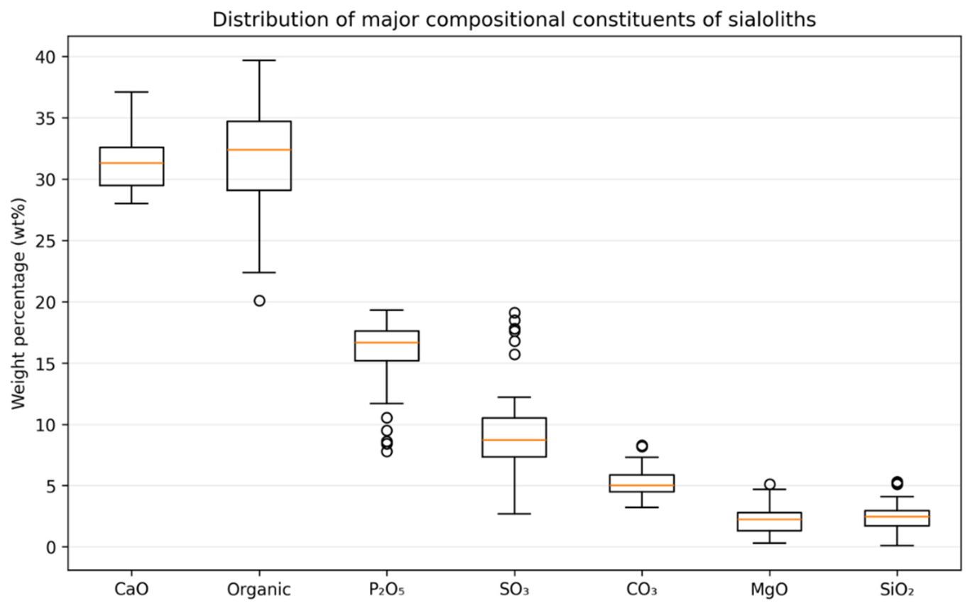
Figure 2.
Regional drinking-water hardness, 2018-2026.
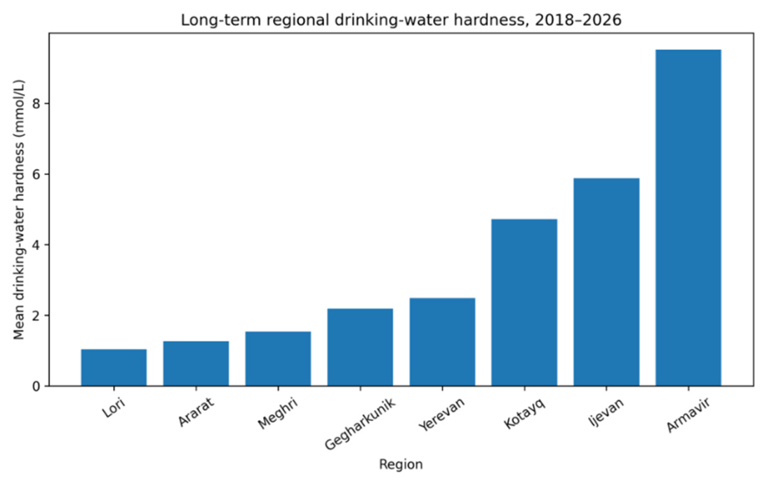
Figure 3.
Regional distribution of sialolith CaO content.
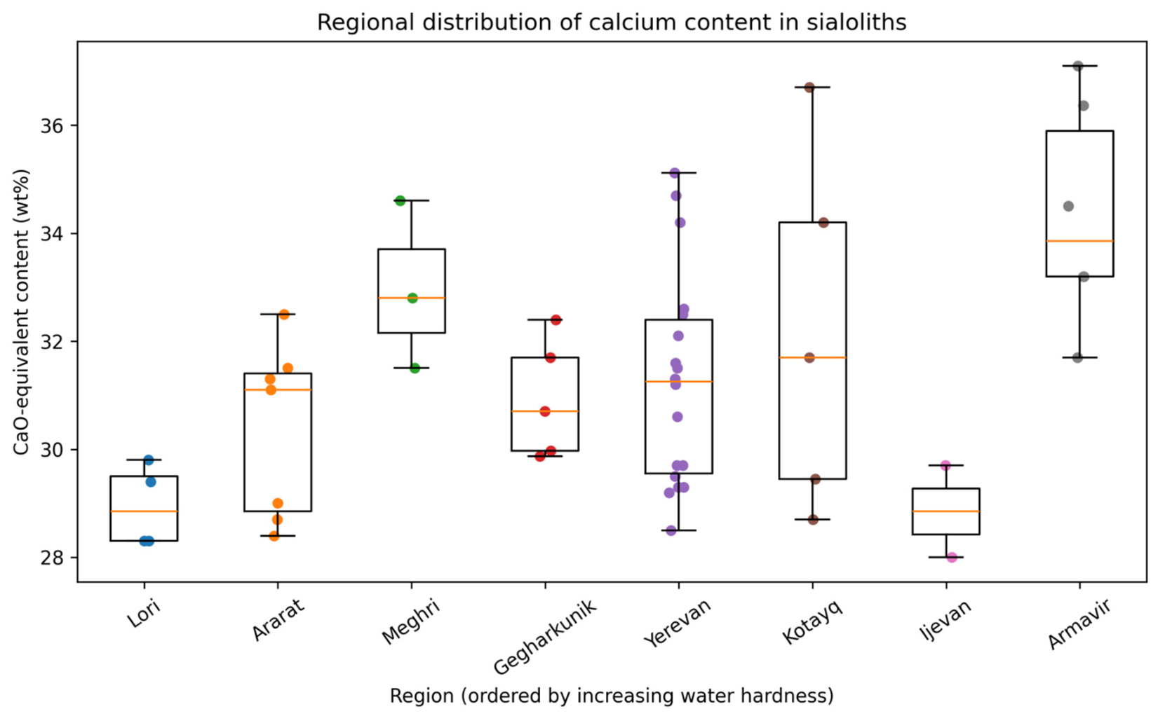
Figure 4.
Regional water hardness versus mean sialolith CaO content.
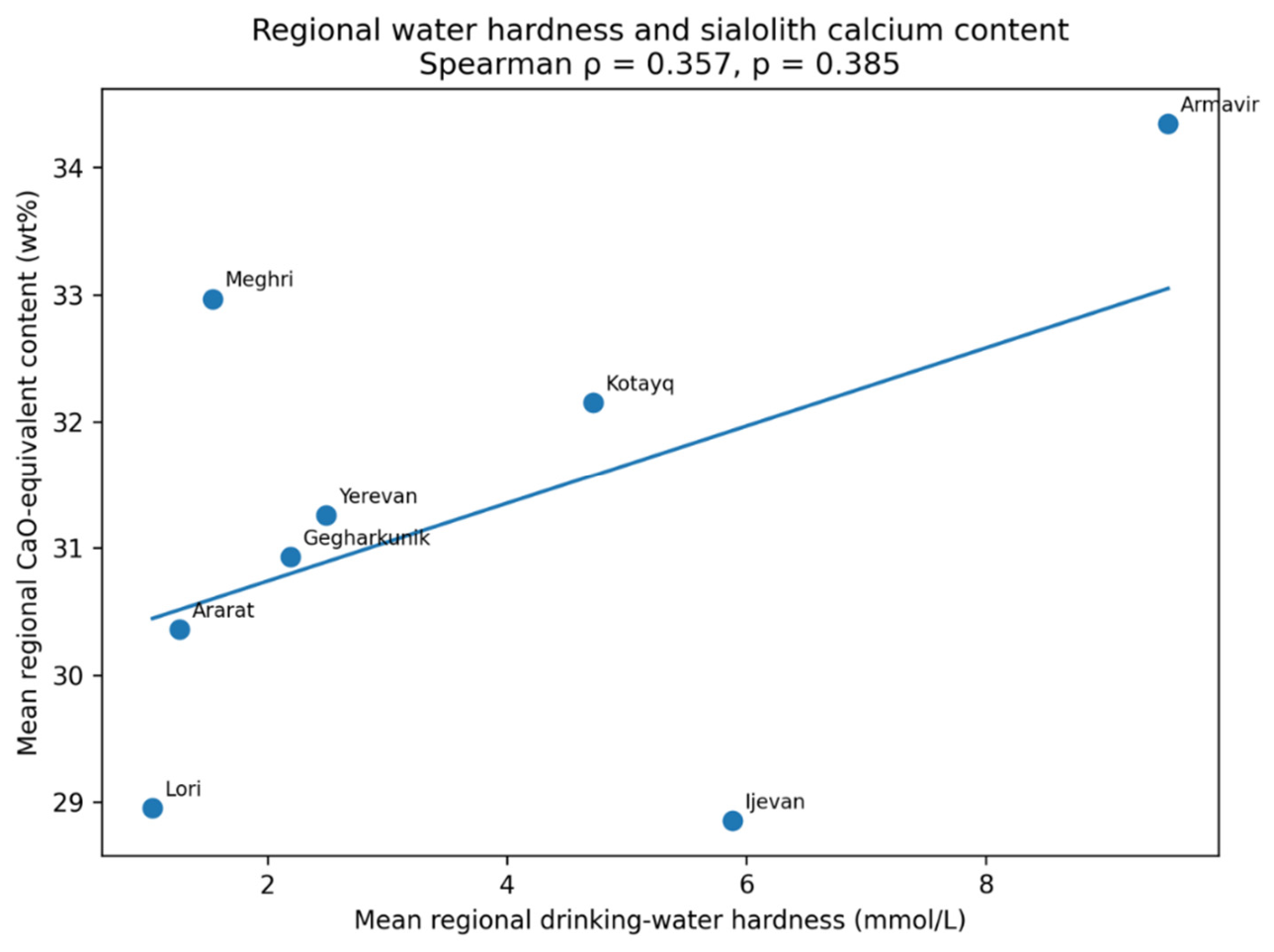
Table 2.
Composition of the analyzed sialoliths (n=50).
| Component | n | Mean ± SD | Median [IQR] | Range |
| SiO₂, wt% | 50 | 2.364 ± 1.175 | 2.450 [1.723–2.950] | 0.100–5.300 |
| Al₂O₃, wt% | 44 | 0.073 ± 0.059 | 0.060 [0.030–0.100] | 0–0.300 |
| TiO₂, wt% | 37 | 0.031 ± 0.043 | 0.020 [0.005–0.030] | 0–0.200 |
| Fe₂O₃, wt% | 50 | 0.099 ± 0.128 | 0.050 [0.020–0.098] | 0.001–0.600 |
| CaO, wt% | 50 | 31.379 ± 2.348 | 31.300 [29.463–32.575] | 28.000–37.100 |
| MgO, wt% | 50 | 2.152 ± 1.090 | 2.250 [1.325–2.788] | 0.300–5.100 |
| CO₃, wt% | 50 | 5.191 ± 1.109 | 5.000 [4.500–5.875] | 3.200–8.300 |
| SO₃, wt% | 50 | 9.493 ± 3.553 | 8.715 [7.325–10.500] | 2.700–19.100 |
| P₂O₅, wt% | 50 | 15.833 ± 2.779 | 16.650 [15.200–17.600] | 7.800–19.300 |
| MnO, wt% | 6 | 0.014 ± 0.006 | 0.015 [0.010–0.020] | 0.006–0.020 |
| Na₂O, wt% | 50 | 0.258 ± 0.113 | 0.255 [0.200–0.330] | 0.050–0.620 |
| K₂O, wt% | 38 | 0.056 ± 0.050 | 0.035 [0.020–0.100] | 0.010–0.200 |
| H₂O, wt% | 50 | 0.927 ± 0.294 | 0.900 [0.700–1.200] | 0.380–1.400 |
| Organic matter, wt% | 50 | 32.094 ± 4.257 | 32.380 [29.083–34.707] | 20.080–39.690 |
| Molar Ca/P ratio | 50 | 2.624 ± 0.709 | 2.453 [2.175–2.728] | 1.934–5.033 |
*Oxide values represent oxide-equivalent weight percentages derived from elemental measurements according to the analytical reporting convention described in the Methods. Organic matter represents the laboratory-reported organic fraction. *Variation in n for selected trace constituents reflects non-detectable or unavailable measurements.
Table 3.
Long-term regional drinking-water hardness and principal sialolith characteristics.
| Region | n | Hardness, mmol/L | CaO, wt% | P₂O₅, wt% | Organic matter, wt% | Mean molar Ca/P |
| Lori | 4 | 1.04 | 28.95 | 17.98 | 31.00 | 2.04 |
| Ararat | 7 | 1.26 | 30.36 | 16.40 | 30.62 | 2.42 |
| Meghri | 3 | 1.54 | 32.97 | 15.70 | 32.40 | 2.66 |
| Gegharkunik | 5 | 2.19 | 30.93 | 16.58 | 34.48 | 2.38 |
| Yerevan | 18 | 2.49 | 31.26 | 14.70 | 32.38 | 2.94 |
| Kotayq | 5 | 4.72 | 32.15 | 15.96 | 30.71 | 2.55 |
| Ijevan | 2 | 5.89 | 28.85 | 16.80 | 37.05 | 2.18 |
| Armavir | 6 | 9.52 | 34.34 | 16.15 | 31.03 | 2.70 |
*Compositional values are regional means.
Table 4.
Regional, age, and sex associations with major compositional variables.
| Outcome | Kruskal–Wallis H | Regional p | Holm-adjusted p | Age ρ | Age p | Sex p |
| CaO | 19.875 | 0.0058 | 0.0409 | −0.108 | 0.455 | 0.862 |
| MgO | 5.657 | 0.580 | 1.000 | 0.050 | 0.732 | 0.186 |
| CO₃ | 12.343 | 0.0898 | 0.449 | −0.141 | 0.329 | 0.486 |
| SO₃ | 9.150 | 0.242 | 0.968 | −0.015 | 0.918 | 0.854 |
| P₂O₅ | 7.292 | 0.399 | 1.000 | −0.075 | 0.603 | 1.000 |
| Organic matter | 5.983 | 0.542 | 1.000 | 0.202 | 0.160 | 0.735 |
| Ca/P ratio | 16.500 | 0.0209 | 0.1255 | 0.004 | 0.977 | 0.759 |
Table 5.
Associations between drinking-water hardness and sialolith composition.
| Outcome | Patient-record ρ | p | Holm-adjusted p | Regional-level ρ (n=8) | p |
| CaO | 0.374 | 0.0075 | 0.0526 | 0.357 | 0.385 |
| MgO | −0.071 | 0.625 | 1.000 | −0.048 | 0.911 |
| CO₃ | 0.093 | 0.522 | 1.000 | −0.095 | 0.823 |
| SO₃ | −0.232 | 0.105 | 0.500 | −0.500 | 0.207 |
| P₂O₅ | −0.235 | 0.100 | 0.500 | −0.238 | 0.570 |
| Organic matter | −0.004 | 0.977 | 1.000 | 0.333 | 0.420 |
| Ca/P ratio | 0.364 | 0.0093 | 0.0559 | 0.405 | 0.320 |
Disclaimer/Publisher’s Note: The statements, opinions and data contained in all publications are solely those of the individual author(s) and contributor(s) and not of MDPI and/or the editor(s). MDPI and/or the editor(s) disclaim responsibility for any injury to people or property resulting from any ideas, methods, instructions or products referred to in the content. |
© 2026 by the authors. Licensee MDPI, Basel, Switzerland. This article is an open access article distributed under the terms and conditions of the Creative Commons Attribution (CC BY) license (http://creativecommons.org/licenses/by/4.0/).
Copyright: This open access article is published under a Creative Commons CC BY 4.0 license, which permit the free download, distribution, and reuse, provided that the author and preprint are cited in any reuse.