
Submitted:
25 August 2026
Posted:
27 August 2026
You are already at the latest version
Abstract
The deep-sea harbors an exceptional proportion of the ocean’s unexplored biological and chemical diversity, while the extreme conditions of hydrothermal vent ecosystems have imposed selective pressures that have driven the evolution of biochemical and structural adaptations with no clear counterpart among shallow-water organisms. This review focuses on one such organism, the vent mussel Bathymodiolus azoricus, dominant across Mid-Atlantic Ridge hydrothermal fields, as a source of biomedically relevant molecules that goes beyond its well-known role as a physiological adaptation model organism. We address this potential through two complementary lines of evidence, one rooted in immunology and the other in biomaterials research, which have largely been treated as separate fields in the literature but which we argue form part of a unified narrative. First, we review Bathymodiolus azoricus as a model organism for the study of innate immunity, focusing on its hemocyte biology, phagocytic and signaling responses, and the transcriptional interplay between host immune genes and its chemosynthetic endosymbionts. Although this body of work, conducted predominantly by Bettencourt and colleagues over the past fifteen years, has significantly advanced our understanding of host-symbiont interactions in hydrothermal vent mussels, it has only rarely been connected to the biomaterials literature on the same species. Second, we examine the byssal adhesive proteins secreted by the mussel foot, which are rich in the catecholic amino acid DOPA, as templates for the development of medical bioadhesives. Particular attention is given to the possibility that the metal-rich hydrothermal vent environment has selected for Fe3+-binding chemistries with higher affinity than those found in shallow-water mytilids. This constitutes a testable B. azoricus-specific hypothesis, rather than a generic restatement of established principles of mussel adhesion biology. We further extend this discussion to shell-derived biomaterials, focusing on nacre and calcium carbonate biomineralization as potential precursors for calcium phosphate-based bone-graft materials. We also highlight mussel byssal collagen, a structurally important yet underexploited protein whose biomedical potential has only recently begun to attract attention. Collectively, these observations suggest that the vent-adapted physiology of B. azoricus not only supports its value as a model system for extremophile biology but also identifies it as a promising and underutilized source of structural biomaterials and immune-active molecules. Testing this hypothesis will require addressing several outstanding experimental challenges, including recombinant production of foot-derived adhesive proteins, completion of the species’ immune-gene catalogue, and systematic structural and functional comparisons with shallow-water relatives.
Keywords:
Bathymodiolus azoricus
; hydrothermal vent
; innate immunity
; mussel adhesive proteins
; bioadhesives
; byssal collagen
; nacre
; biomineralization
; deep-sea biomaterials
1. Introduction
The oceans cover more than 70% of the Earth’s surface and harbor an immense diversity of life. Over millions of years of evolution, marine organisms have developed adaptations that enable survival in environments characterized by extreme physical and chemical conditions, among which deep-sea hydrothermal vents are among the most inhospitable. Hydrothermal vents occur worldwide in association with volcanic and tectonic activity and support chemosynthesis-based ecosystems shaped by high hydrostatic pressure, steep thermal gradients, and often toxic concentrations of sulfur compounds, methane, and heavy metals [1,2]. These systems sustain diverse communities of endemic invertebrates adapted to such physicochemical extremes.
Among the macrofauna inhabiting deep-sea hydrothermal vents, the mussel Bathymodiolus azoricus is the dominant species along much of the Mid-Atlantic Ridge (MAR), where it typically forms dense aggregations. Its successful colonization of sulfide and methane-rich habitats is enabled primarily by a dual endosymbiosis with sulfur-oxidizing and methane-oxidizing bacteria housed within specialized gill epithelial cells [3,4]. While this symbiosis is considered the principal mechanism compensating for the host’s limited metabolic capabilities in such environments [5], survival under extreme pressure, permanent darkness, and elevated concentrations of toxic metals also requires macromolecules and biopolymers possessing specialized biochemical and structural properties [6,7]. These environmental pressures have driven the evolution of molecular adaptations, including chaperone systems, metabolic enzymes, immune effectors, and tissue-repair factors, capable of maintaining function under conditions that would compromise their shallow-water counterparts.
The biological uniqueness of these molecules has attracted increasing interest for the development of novel pharmaceuticals, cosmeceuticals, and biomaterials, particularly in regenerative medicine, tissue engineering, and wound healing [8,9]. Hydrothermal vent animals and their associated microbial communities therefore represent a largely untapped source of marine-derived compounds with potential relevance to human health [10,11,12].
In 2010, Bettencourt and colleagues published the first high-throughput transcriptome of a deep-sea hydrothermal vent animal, based on the gill tissue of B. azoricus [10]. The resulting DeepSeaVent database was originally developed to address a specific biological question: how does the immune system of an animal simultaneously defend itself against pathogens while maintaining a permanent and beneficial bacterial symbiosis? This immunological motivation has often been overlooked as the dataset became increasingly cited as a resource for biomolecular and biomaterials discovery.
In this review, we purposely reconnect these two perspectives. First, we examine the immune biology of B. azoricus, focusing on the mechanisms that govern host defense and symbiont tolerance. We then revisit the transcriptomic resources generated from this species and the biomaterial opportunities they revealed, including byssal adhesive proteins, shell-derived biominerals, and mussel collagen. We conclude by advancing what we consider the central, testable hypothesis of this review: that the vent-adapted chemistry underlying these biological systems, particularly mechanisms involved in metal binding and metal homeostasis, confers distinctive functional properties on B. azoricus-derived biomaterials and bioactive molecules. If correct, these materials should not be viewed merely as additional examples of biomolecules already characterized from shallow-water mussels, but rather as mechanistically distinct adaptations shaped by the unique selective pressures of the hydrothermal vent environment.
2. Bathymodiolus Azoricus as a Model Organism for Innate Immunity
Before considering B. azoricus as a source of structural biomaterials, we first examine its biomedical relevance in its own right. The same characteristic that makes B. azoricus an attractive source of biomaterials, namely its permanent, high-density coexistence with bacterial symbionts under conditions that would be pathogenic in most other biological contexts, also makes it an unusually informative model for the study of innate immunity. Rather than representing a speculative extension of the literature, this perspective draws upon a substantial and largely self-contained body of research conducted by Bettencourt and colleagues over the past fifteen years, which has yet to be fully integrated into the biomaterials field.
The interaction between microorganisms and host defense mechanisms is central to the survival of marine bivalves, which rely on cellular and humoral immune responses rather than adaptive immunity to control naturally occurring pathogens. In B. azoricus, the cellular constituents of the hemolymph and extrapallial fluid include three hemocyte types, among which granulocytes are the most abundant and represent the principal phagocytic cell population. These cells were further characterized through fluorescent WGA-lectin staining of surface carbohydrate epitopes [21]. Exposure of hemocytes to microbial elicitors, including zymosan, glucan, Bacillus subtilis, and Vibrio parahaemolyticus, activates MAPK signaling pathways, with ERK, p38, and JNK all implicated by Western blot analysis. In addition, the fluorescent Ca2+ indicator Fura-2 AM demonstrated hemocyte activation following exposure to laminarin or live V. parahaemolyticus [21]. The antimicrobial peptide gene mytilin, expressed in hemocytes underlying the gill epithelium, was upregulated following exposure of live mussels to Vibrio, providing direct evidence that a specific immune effector can be induced under environmentally relevant infection conditions [21].
This line of research was subsequently extended through a series of Vibrio challenge experiments using Vibrio diabolicus, a bacterial species originally isolated from the hydrothermal vent polychaete Alvinella pompejana and therefore an ecologically relevant model pathogen for vent-associated fauna. Across acclimatization and infection-challenge experiments, immune genes involved in pattern recognition (including Toll-like receptor signaling), signal transduction, and effector responses exhibited time-dependent expression patterns linked to the degree of endosymbiont colonization. Comparisons between mussels collected from the Menez Gwen and Lucky Strike vent fields, which differ in their geochemical characteristics, revealed site-specific and tissue-specific variation in the expression of genes including Carcinolectin, Serpin-2, SRCR, IRGs, RTK, TLR2, NF-κB, HSP70, and ferritin [52,53,54].
Endosymbiont genes, including ALDH, CA, CBB, MeDH, MMO, and SOXB, were likewise upregulated following Vibrio challenge, indicating that host immune activation and symbiont transcriptional activity are interconnected rather than independent processes. This coordinated response distinguishes the B. azoricus system from conventional non-symbiotic bivalve immune models and supports the hypothesis that an actively responding endosymbiont community may contribute to host defense and physiological resilience [7].
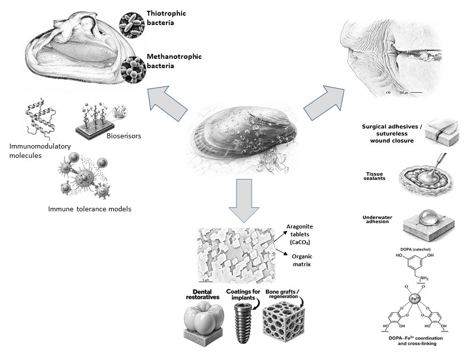
Taken together, this body of work establishes B. azoricus as a valuable model for investigating innate immunity and stress responses in deep-sea hydrothermal vent animals, as proposed in our earlier review [7]. From the perspective of biomedical discovery, the immune components characterized in this system, including pattern-recognition receptors capable of detecting LPS and other pathogen-associated molecular patterns (PAMPs), antimicrobial peptides such as mytilin, cytokine-like signaling molecules, and apoptosis regulators functioning in the context of chronic microbial exposure, represent molecular classes of considerable interest for antimicrobial discovery, biosensor development, and the study of host-microbiome interactions relevant to chronic inflammatory disorders.
Whether antimicrobial peptides, pattern-recognition receptors, or other immune effectors from B. azoricus possess functional properties that differ from those of their shallow-water homologues remains an open question. Such a possibility is biologically plausible given the species’ long-term coexistence with dense and metabolically active bacterial symbionts. However, like the proposed metal-binding specialization of its adhesive proteins, this hypothesis requires direct experimental validation.
3. Beyond the Transcriptome: Bathymodiolus Azoricus as a Potential Source of Biomaterials
The 2010 gill transcriptome of B. azoricus was the first tissue-level transcriptional analysis of a deep-sea hydrothermal vent animal generated using next-generation sequencing technologies [10]. It produced a searchable catalogue of genes that enabled expression profiling by quantitative PCR in a non-model organism and provided the most comprehensive sequence resource then available for a vent species, including numerous genes putatively involved in immune and inflammatory processes [7,10]. Although originally developed to expand the representation of innate-immunity genes in vent mussels, the transcriptome also identified coding sequences for proteins associated with cell adhesion, tissue repair, biomineralization, and host-microbiome interactions. As a result, the dataset proved valuable not only for immunological research but also as an early resource for the discovery of biomolecules with potential biotechnological and biomedical applications.
The production of such compounds by B. azoricus is plausibly linked to adaptive advantage, reflecting the biochemical diversity and structural complexity that arise from the distinctive chemical environment of hydrothermal vents and, potentially, from ecological pressures such as predation. Investigating deep-sea vent mussels as a source of marine-derived compounds therefore provides an opportunity to uncover chemical and biological innovations associated with the evolutionary adaptations of hydrothermal vent fauna.
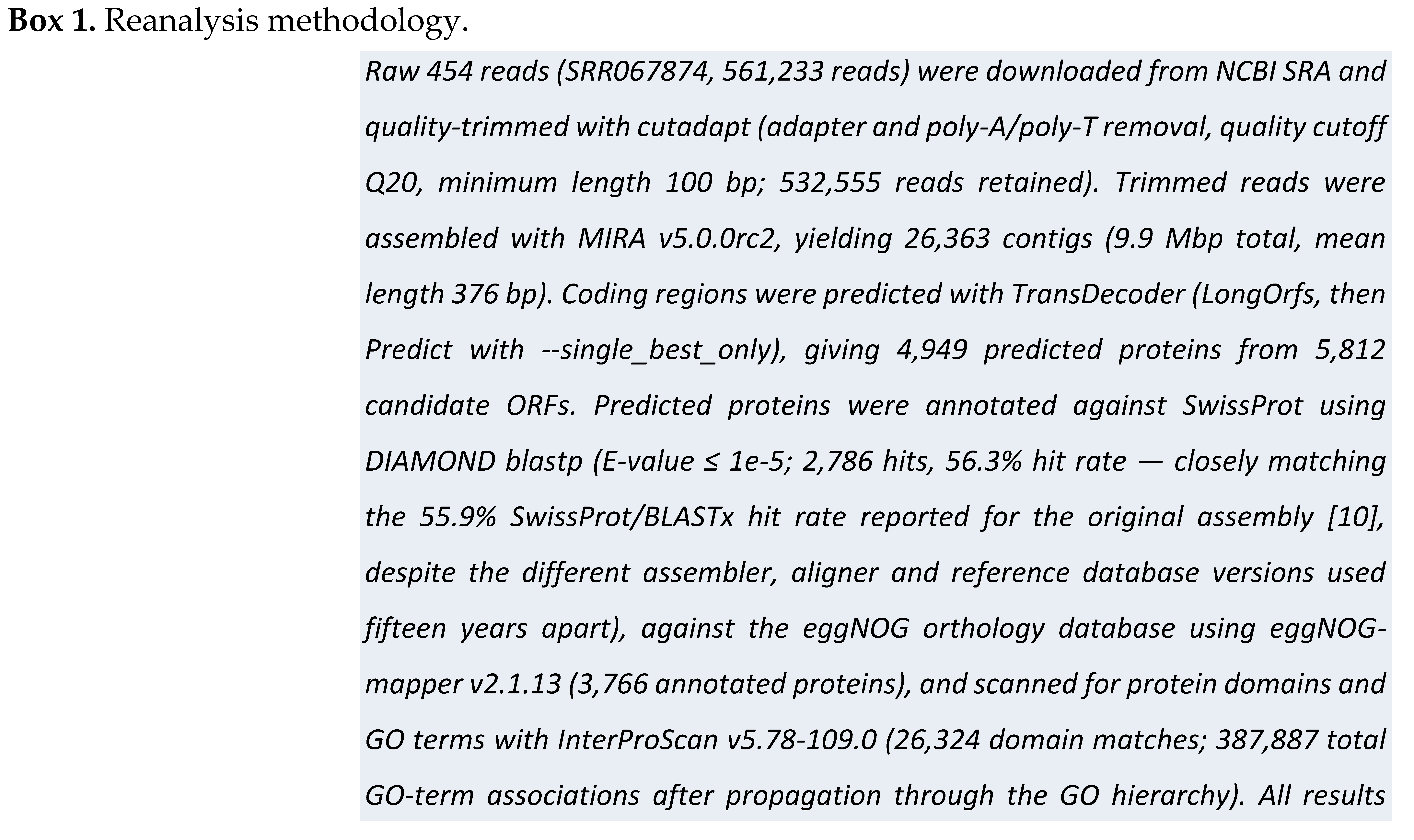
The DeepSeaVent web portal originally developed to query this dataset [10] is no longer accessible, as the server hosting its custom browser and BLAST interface has been discontinued. The underlying sequencing data, however, remain available through the NCBI Sequence Read Archive (SRA accession SRR067874). For the present review, we performed an independent reanalysis of the raw reads, described in Box 1, to recover gene-level information that was previously accessible through the original portal.
In addition, a second and independent gill transcriptome was generated by Bettencourt and colleagues in 2024 using Illumina sequencing. This dataset is incorporated throughout the comparative analyses presented below (Table 1) and serves as an independent cross-platform validation of the candidate genes discussed in this review. The dataset has been submitted to the NCBI Sequence Read Archive under BioProject PRJNA1508204 (SRA accession SRR40024054) and is awaiting public release following NCBI’s standard processing procedures.
Researchers have long looked to marine mussels as sources of novel adhesive systems and nature-inspired models for bioadhesion because of their remarkable ability to bond strongly to submerged surfaces. Attachment is mediated by the byssus, a thread-like structure terminating in adhesive plaques that are secreted by specialized glands in the foot. Mussel adhesive proteins (MAPs) function under constantly changing environmental conditions and on a wide variety of substrates in saline, high-energy habitats [13,14,15,16]. Through a highly coordinated secretion and self-assembly process, these proteins form the fibrous architecture of the byssus.
The mussel foot is a large muscular and glandular organ containing a deep longitudinal groove that folds near its distal end into a cup-shaped structure used to engage solid surfaces. At the base of this groove, the byssus pit secretes a viscous proteinaceous exudate that hardens upon contact with seawater, producing tough and elastic byssal threads capable of maintaining attachment even under strong hydrodynamic forces.
Byssal threads and adhesive plaques comprise multiple structural and adhesive proteins that are synthesized, secreted, and molded by the foot into a durable attachment apparatus [18]. This mechanism enables mussels to colonize a wide range of habitats, including B. azoricus, which remains firmly attached to hard substrata at hydrothermal vent sites in much the same way that shallow-water mytilids occupy intertidal environments. Because of their exceptional wet-adhesion performance, marine mussels have become one of the most extensively studied model systems in bioadhesion research and have inspired decades of work on water-compatible adhesives for biomedical and industrial applications [13,17]. Consequently, mussel adhesive proteins remain among the most promising targets for biomimetic materials research [17].
Mussel foot proteins (Mfps) share a distinctive biochemical feature: a high content of 3,4-dihydroxyphenyl-L-alanine (DOPA), a catecholic amino acid generated through post-translational modification of tyrosine residues. Mfps are generally characterized by high isoelectric points and considerable sequence diversity. Secreted in liquid form by specialized glands in the foot, they undergo polymerization through catechol-mediated cross-linking reactions that drive the formation and hardening of byssal threads and adhesive plaques [34]. DOPA plays a central role in underwater adhesion, contributing both to interfacial binding and to the cohesion and solidification of the adhesive matrix [34]. As a result, catechol-based adhesion chemistry has become a major source of inspiration for the development of bioadhesive materials for a wide range of biomedical applications [34].
Like its shallow-water relatives in the genus Mytilus, B. azoricus belongs to the family Mytilidae and retains the characteristic byssal attachment system of marine mussels. However, this species functions within the distinctive physicochemical environment of hydrothermal vents. In addition to possessing large ctenidia colonized by methanotrophic and thiotrophic bacterial endosymbionts that contribute substantially to host nutrition [3,4,5], B. azoricus remains firmly attached to rocky substrata in habitats shaped by hydrothermal venting, tectonic activity, and episodic crustal fissuring. These environmental conditions may impose mechanical and chemical challenges not typically encountered by shallow-water mussels, potentially placing unique selective pressures on byssal attachment systems and their underlying adhesive proteins. As in intertidal mytilids, mechanical damage, predation, or habitat disturbance are also likely to require efficient shell and mantle repair mechanisms.
Wound healing is a complex biological process that restores the integrity and barrier function of injured tissues [9]. In bivalve molluscs, the shell serves essential structural and protective functions [20]. Shell formation is controlled by the mantle, a tissue that encloses the animal and comprises connective, neural, muscular, glandular, and epithelial components involved in biomineralization [20]. Following mantle injury, genes encoding shell-forming proteins are activated as part of a coordinated biomineralization program. Transcriptomic studies aimed at identifying biomineralization-related genes have been conducted in several bivalve species, including Pecten maximus, Mytilus edulis, and Crassostrea gigas [20]. Beyond its role in shell formation, the mantle is a multifunctional organ involved in nutrient storage, sensory perception, and the regulation of particle transport toward the gills, while its muscular and neural elements contribute to mantle movement and environmental responsiveness [36,37].
Wound repair in bivalves involves the rapid recruitment of hemocytes to the site of injury. These cells help prevent hemolymph loss by forming a provisional clot that seals the wound and provides a scaffold for subsequent epithelial regeneration. In the tellinid bivalve Scrobicularia plana, wounds resulting from siphonal amputation healed rapidly, with connective tissue accumulating around the lesion within hours and epidermal cells completely covering the wound site within 72 h [38]. A similar pattern of rapid siphon regeneration was reported in the donacid bivalve Donax serra [39], suggesting that this regenerative capacity is widespread among bivalve lineages rather than being restricted to a single taxonomic group. In addition to biomineralization genes, successful wound healing requires the coordinated expression of genes involved in extracellular matrix reconstruction and tissue repair, including growth factors, cytokines, and cell-adhesion proteins that regulate cell-cell and cell-matrix interactions.
In B. azoricus, shell damage may arise from predation as well as from physical disturbances associated with hydrothermal vent activity, including tectonic and eruptive events. Such injuries are expected to trigger regenerative responses involving both shell deposition and repair of the underlying mantle tissue [40,41]. Consequently, genes associated with biomineralization, extracellular matrix remodeling, and tissue regeneration may represent an additional source of biomolecules with potential biomedical relevance.
Phenoloxidase (PO) activity and melanin deposition were observed around sites of shell injury in preliminary B. azoricus experiments. Phenoloxidases are tyrosinase-type enzymes involved in pigment production, innate immunity, wound healing, and exoskeleton or shell hardening. PO catalyzes the oxidation of phenolic compounds to quinones, which subsequently polymerize into insoluble melanin. In insects, PO activity is closely associated with coagulation and melanin deposition at wound sites, where it helps limit hemolymph loss and initiate tissue repair. The prophenoloxidase-activating (proPO) system is widely distributed across invertebrate phyla, including crustaceans, in which it plays a central role in clotting and immune defense [42,43].
Many tyrosinase genes in marine bivalves are expressed at relatively high levels in the mantle, the tissue responsible for shell formation. Comparative analyses of tyrosinase expression across mantle regions in the closely related pearl oysters Pinctada maxima and P. margaritifera have shown that recently diverged orthologous tyrosinase genes can exhibit markedly different expression patterns. The expansion of this gene family, together with its integration into mantle regulatory networks, is consistent with the view that shell formation is supported by a rapidly evolving transcriptomic repertoire [44]. Comparable mantle transcriptome resources are now available for Pecten maximus, Mytilus edulis, and Crassostrea gigas [20].
Taken together, these observations suggest that tyrosinase-mediated pathways involved in shell repair and biomineralization warrant investigation in B. azoricus, where injury responses may be shaped by the distinctive environmental pressures of hydrothermal vent ecosystems.
Initial gene-expression work following mechanical abrasion of the B. azoricus shell identified candidate genes potentially involved in wound repair through searches of the 2010 DeepSeaVent database. That database has since been discontinued (see Box 1); Table 1 instead reports an independent reanalysis of the same underlying sequencing data.
A complementary and more targeted comparison is possible for the small subset of candidates that were not merely identified bioinformatically in the present reanalysis, but had already been experimentally validated by RT-PCR/qPCR in the original study: integrin, EGF, and aggrecan (Table 2). Two of these three validated findings hold up cleanly across both independent reanalyses. Aggrecan is the exception: it could not be reproduced in the shallower 2010 reanalysis — an apparent regression most plausibly explained by that reanalysis’s smaller assembly relative to the originally published dataset, rather than a genuine absence — and is recovered directly in the deeper 2024 reanalysis, resolving rather than undermining the original RT-PCR-confirmed finding.
Wound healing itself involves a well-characterized sequence of cellular events aimed at repairing damaged tissue and restoring its integrity [23]. These events involve cell recruitment, mediator and growth factors, and cytokines, and typically proceed through three overlapping stages — inflammation, new-tissue formation via extracellular matrix reconstruction, and tissue remodeling [23]. Health status and other physiological conditions can inhibit or delay this process; in such cases, wound dressings, tissue engineering approaches and bioactive molecules are employed to accelerate healing at different stages of regeneration.
4. The Mollusc Shell as a Biomaterial: Nacre and Calcium-Phosphate Conversion
The mollusc shell is formed by biomineralization and consists mainly of calcium carbonate (CaCO3) deposited within a small but functionally critical organic matrix, giving it potential as a raw material for material-science and nanotechnology applications. Biomineralization proceeds through the deposition of calcium-carbonate crystals within a well-organized organic scaffold of macromolecules, proteins, and polysaccharides rich in carboxylate, phosphate, and sulfate groups [45].
The organic–inorganic composite produced by some molluscs as an inner shell layer — nacre, also the material of pearls — is strong, resistant, and iridescent, with a distinctive hierarchical structure and notable mechanical properties. Nacre and nacre-derived materials interact favorably with bone [45], making them attractive natural candidates for bone-graft substitutes with osteogenic activity on human osteoblast cell lines [35]. Zhang et al. (2017) describe nacre as a natural, multi-use, and clinically timely biomaterial for bone-graft substitution [45].
Bivalve shell CaCO3 can itself be converted into calcium-phosphate phases — for example monetite (dicalcium phosphate) — through relatively simple hydrothermal or wet-chemical exchange reactions, using discarded mussel shell as the starting material rather than mined mineral sources. Macha et al. (2013) demonstrated this conversion using shells of the shallow-water mussel Mytilus galloprovincialis [46], and the same route is a natural fit for B. azoricus shell material, which is otherwise a processing by-product of any aquarium or field study on the species.
Calcium-phosphate phases such as monetite and hydroxyapatite are already the mineral component of choice in bone-graft and dental-repair biomaterials because of their compositional similarity to bone mineral. Whether B. azoricus shell, formed under the distinct trace-metal and pH conditions of hydrothermal vents, yields calcium-phosphate materials with altered crystallinity, trace-metal composition, or resorption characteristics relative to shallow-water mussel shells remains, to our knowledge, an unexplored question.
Structural evidence from the shell itself supports the broader hypothesis that B. azoricus shell material possesses characteristics distinct from those of shallow-water mussels. Environmental regulation of shell biomineralization is not unique to deep-sea species: in the pearl oyster Pinctada margaritifera, temperature and food availability directly influence both shell growth rate and the mantle expression of genes encoding shell matrix proteins [47]. The nacre ultrastructure of B. azoricus has been characterized directly, including its behavior under depressurization, which differs from any shallow-water congener [48]. This physical distinctiveness strengthens the view that vent-specific environmental conditions leave measurable signatures on shell structure and composition. Such effects may extend beyond calcium-phosphate conversion chemistry to influence the stability, morphology, and functional properties of the native CaCO3-based shell material itself [51].
Direct evidence for B. azoricus shell biology, although still limited, indicates that shell formation is influenced by the hydrothermal-vent environment in which it occurs.Trace-metal analysis of B. azoricus shells has shown that they incorporate micro-essential metals sourced from the surrounding hydrothermal fluid [49] — independent evidence, in a second tissue, for the same vent-chemistry-leaves-a-measurable-signature argument this review makes for the foot adhesive proteins above, and a further point in favor of treating metal handling as a recurring theme in this species’ biology rather than a foot-specific peculiarity.
Shell regeneration itself is not purely a mantle-secretory process in this species. Following induced shell damage, B. azoricus mounts a hemocyte response that delivers CaCO3 directly to the site of repair, with mantle-to-shell calcium transfer measurably sensitive to hydrostatic pressure [50,51].
These are the same cell types, and largely the same immune mechanisms, discussed above in the context of innate immunity. Shell repair in this species therefore appears to be, at least in part, an immunological process rather than an entirely separate biomineralization pathway. Preliminary evidence of phenoloxidase activity and melanin deposition around sites of shell injury, documented in this review’s own experiments (see Figure 3 above), is consistent with this: the same tyrosinase/phenoloxidase chemistry implicated in wound-healing clot formation elsewhere in this review also participates in shell hardening and repair more broadly, and the injury response captured in those images likely reflects this same hemocyte-mediated mechanism at the shell surface rather than a distinct process.
The clearest evidence that this species’ shell biology is itself vent-adapted, rather than simply built by a vent-dwelling animal, comes from direct structural characterization. SEM analysis of B. azoricus nacre across three geochemically distinct Mid-Atlantic Ridge vent sites found nacre micro-morphology to be largely conserved despite substantial differences in local pressure and fluid chemistry, with the modest variation observed more plausibly attributable to differences in Ca2+ availability than to pressure itself [48]. More strikingly, the same study found that decompression is actively corrosive to this animal’s shell: mussels held at atmospheric pressure showed measurable nacre dissolution within a single day, worsening substantially by day 12, while mussels maintained at 20 bar remained largely protected over the same period. Beyond its implications for the sampling and maintenance of vent specimens, this finding demonstrates a direct, testable link between this species’ vent-adapted physiology and the structural integrity of its shell — the same kind of pressure-dependence this review’s central hypothesis proposes for the foot adhesive proteins, here shown directly for a different structural tissue.
Taken together, these findings support the view that shell formation and repair in B. azoricus are themselves components of a vent-adapted phenotype. This influence is evident in the incorporation of hydrothermal-vent trace metals into the shell, in the participation of immune-associated hemocytes in shell regeneration, and in the pressure-dependent stability of the nacre structure. However, an important knowledge gap remains. Unlike the gill and foot, the mantle of B. azoricus has not yet been characterized transcriptomically, leaving the molecular basis of shell-matrix production, calcium transport, and regenerative responses unresolved. A mantle-tissue transcriptome would therefore provide a critical foundation for identifying the genes and pathways that link biomineralization, immunity, and environmental adaptation in this species.
5. Mussel-Derived Materials in Wound Healing: What Is Actually Mussel-Specific
Marine organisms have been widely investigated as sources of wound-healing materials, and a substantial literature describes the biomedical applications of chitin, chitosan, alginate, hyaluronic acid, and marine-derived collagen obtained from a broad range of taxa [9]. A comprehensive review of this field is beyond the scope of the present article and would risk shifting the focus away from B. azoricus toward a more general survey of marine biomaterials. Moreover, the wound-healing properties of materials such as chitosan and alginate are not specific to mussels and have already been extensively covered elsewhere. Instead, we focus on the components of this literature that are directly relevant to B. azoricus and mussel-inspired biomaterials, namely byssal adhesive proteins, mussel collagen, and the catechol-functionalized bioadhesives derived from these systems.
We also avoid restricting the discussion to cutaneous wound healing. Although an extensive literature exists on mussel-inspired hydrogels and dressings for skin repair [63], the translational impact of mussel-derived adhesive chemistry extends well beyond topical applications. Reviews of clinical bioadhesives identify major opportunities in internal and surgical settings, including applications involving the brain, eye, heart, liver, and other soft tissues [62]. The biological processes discussed throughout this review, including hemocyte recruitment, extracellular-matrix remodeling, and phenoloxidase-mediated clotting, are relevant not only to skin repair but also to internal tissue regeneration and surgical wound closure. Consequently, the biomaterials highlighted below are considered primarily in the context of surgical sealants and tissue adhesives [63], where much of the current mussel-inspired bioadhesives literature is focused.
6. Novel Bioadhesives Inspired by Mussel Foot Adhesive Proteins
Closing injured tissue during surgery is a critical step in restoring structure and function. Traditional closure methods — sutures, wires, staples — are time-consuming, may cause further tissue damage, increase infection risk, and are difficult to apply in some interventions [62]. High-strength surgical sealants and nature-inspired bioadhesives are a promising alternative, particularly where medical adhesives can promote tissue regeneration and reduce surgery time [9,27].
Proteins from the mussel foot have been extensively investigated for their underwater adhesive bonding and resistance to mechanical stress, making them a source of inspiration for new biomedical bioadhesives. Many researchers have exploited DOPA as a cross-linking mediator in synthetic polymer-based hydrogels capable of both cohesive hardening and adhesive bonding to different surfaces [27], and hydrogel systems using DOPA-containing recombinant MAPs continue to attract attention for their combination of adhesion and cohesion in wet environments [27]. Recombinant protein technology remains the method of choice for overcoming the difficulty of purifying sufficient natural MAP from mussel tissue, though heterologous expression — particularly in bacterial systems — is often limited by insoluble or misfolded protein, incorrect codon usage, and the absence of post-translational modifications such as phosphorylation and glycosylation, all of which matter for a protein whose function depends on precise secondary structure and aromatic/cationic residue placement [83]. Among previously reported recombinant MAPs, rfp-1 has been considered the most suitable candidate for hydrogel formulation, given its high DOPA content and good aqueous solubility; rfp-5 and rfp-3 have also been tested for adhesive properties relevant to injectable, wet-resistant bioadhesives for sutureless wound closure [24,30].
The mechanism and structure–function relationships of mussel adhesion have been studied extensively over the past two decades, informing the design of synthetic biomimetic adhesives [34]. The strong adhesive performance of MAPs makes them attractive for water-resistant, biomedically relevant adhesives [16], and their biocompatibility and biodegradability further support their development, particularly given the environmental cost of harvesting large numbers of wild mussels to obtain sufficient purified protein.
Tissue-engineered hydrogels and tissue adhesives have evolved toward in situ cross-linkable, polymer-based systems requiring fewer cross-linkers than earlier formulations [26,27]. Synthesizing biocompatible, biodegradable hydrogel tissue adhesives has nonetheless proven difficult, since most cross-linked hydrogels with high water content lack adhesive properties toward tissue and mucosal layers [60]. Fibrin glue — made from thrombin, fibrinogen concentrates and sometimes factor XIII and antifibrinolytic agents — remains a widely used biological tissue adhesive with useful haemostatic properties, but carries a risk of viral/prion contamination and comparatively low tissue-bonding strength [61]. Cyanoacrylates, a family of fast-acting synthetic adhesives used across industrial, medical and household contexts, offer strong adhesion and rapid curing on contact with moisture but generate degradation products that are not always suitable for medical use [59]. New synthetic, polymer-based analogues of MAPs have consequently been developed as bioadhesives for medical and tissue-engineering applications, combining biocompatibility and biodegradability with the potential for large-scale production [30].
Despite many studies and several commercial surgical adhesives, no single bioadhesive currently satisfies medical and surgical requirements simultaneously — biocompatibility, biodegradability, reliable hemostatic action, strong wet adhesion, and good cosmetic outcome remain difficult to combine in one formulation. Recombinant MAPs hold potential for targeted modulation of skin or implant surfaces to improve cell adhesion, proliferation and differentiation, and functionalized coatings of this kind could also serve as platforms for immobilizing inorganic nanoparticles, extracellular-matrix molecules, nucleic acids and therapeutic drugs on target surfaces [30].
Given their strong and versatile adhesive properties, recombinant MAPs represent promising candidates for the development of tissue adhesives capable of rapidly sealing damaged tissues while simultaneously promoting cell adhesion, proliferation, and differentiation. Ion-mediated DOPA cross-linking, particularly through metal chelation and coordination, contributes to the cohesion, self-healing behavior, and mechanical resilience of mussel-inspired adhesive systems [31].
This brings us to the central, B. azoricus-specific hypothesis advanced in this review. Because hydrothermal vent ecosystems are characterized by elevated concentrations of dissolved metals, and because DOPA-mediated adhesion depends critically on metal-catechol interactions, it is plausible that adhesive proteins from B. azoricus have evolved Fe3+-binding properties that differ from those of shallow-water mytilids. Such differences could arise through variation in tyrosine content, DOPA abundance, protein architecture, or the affinity of catechol-containing domains for metal ions. If so, recombinant B. azoricus MAPs might form metal-mediated cross-links more efficiently than homologous proteins from shallow-water species, potentially conferring enhanced adhesion, cohesion, or self-healing performance.
This hypothesis also creates opportunities for future protein engineering. For example, B. azoricus MAPs could be combined with cell-interactive motifs such as RGD peptides or other extracellular-matrix-derived sequences to generate multifunctional bioadhesives that not only adhere strongly to wet tissues but also promote fibroblast migration, cell attachment, and extracellular-matrix deposition during repair. We emphasize, however, that this remains a hypothesis rather than an experimental result. To our knowledge, no study has yet directly compared Fe3+-binding affinity, metal-mediated cross-linking behavior, or adhesive performance between B. azoricus and shallow-water Mytilus MAPs. Such comparative analyses constitute the most direct test of the vent-adaptation hypothesis proposed here.
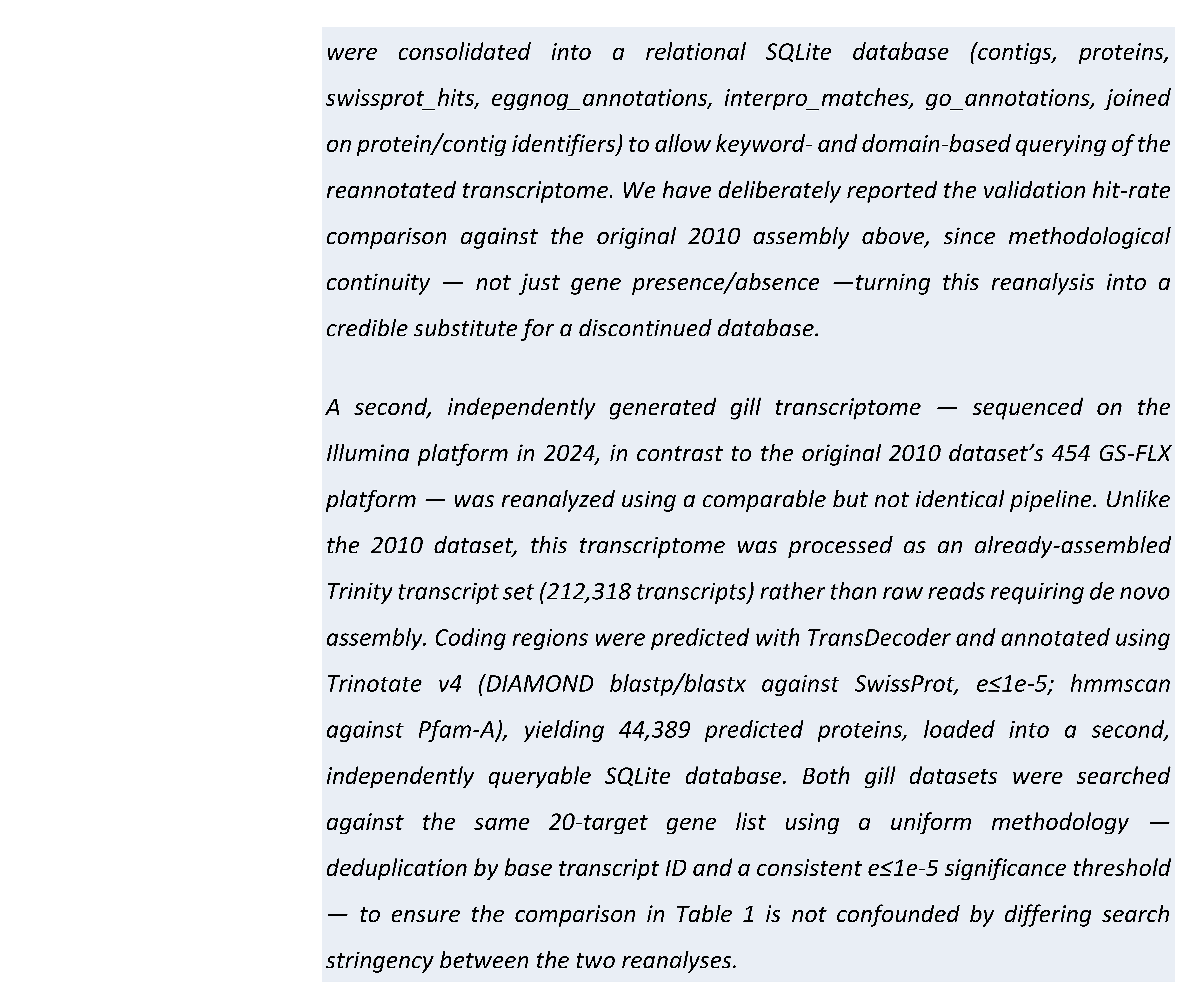
While at the 3B’s Research Group, our group has undertaken a de novo transcriptome sequencing analysis of B. azoricus foot tissue (sample N1188; StabVida project RNQ201711000080), aimed at identifying vent-mussel adhesive proteins — foot mussel proteins (fmps) — expressed at the time of RNA extraction. This dataset was first analyzed between 2017–2019, when fmp-2 emerged as the most abundant and best-supported homolog in preliminary searches against Mytilus sp. sequences (57% identity, 65% positives over 231 residues, e=2×10−65, to foot protein 2 from Mytilus californianus; a hand-built alignment at the time also highlighted the protein’s characteristic tandem EGF-like repeat architecture — reproduced above in Figure 4). That original analysis was lost along with the local server it was run on, though the underlying raw sequencing data survived. Applying the same reanalysis workflow used for the gill transcriptome (Box 2) to this surviving assembly, we have now independently reproduced the original finding: transcript TRINITY_DN360524 shows 59.3% identity (e=1.0×10⁻³⁰) to Mfp-2 (UniProt Q25464), with the aligned region an almost exact match to the repeat unit identified in the 2017 analysis (…NGKCVLNGYGGYKCKCVGGYTGSRCEKNPC…, differing by only 1–2 residues); a second transcript, TRINITY_DN351523, shows the same repeat with a single substitution (52.6% identity, e=1.2×10−44). Both transcripts fell below the default minimum length threshold used by the ORF-prediction step of the annotation pipeline and were only recovered by translating directly from the raw nucleotide alignment coordinates — a methodological note we consider worth recording for anyone repeating this kind of search on short, tandem-repeat adhesive proteins, where standard gene-prediction defaults are tuned for typical globular proteins rather than short secreted peptides.
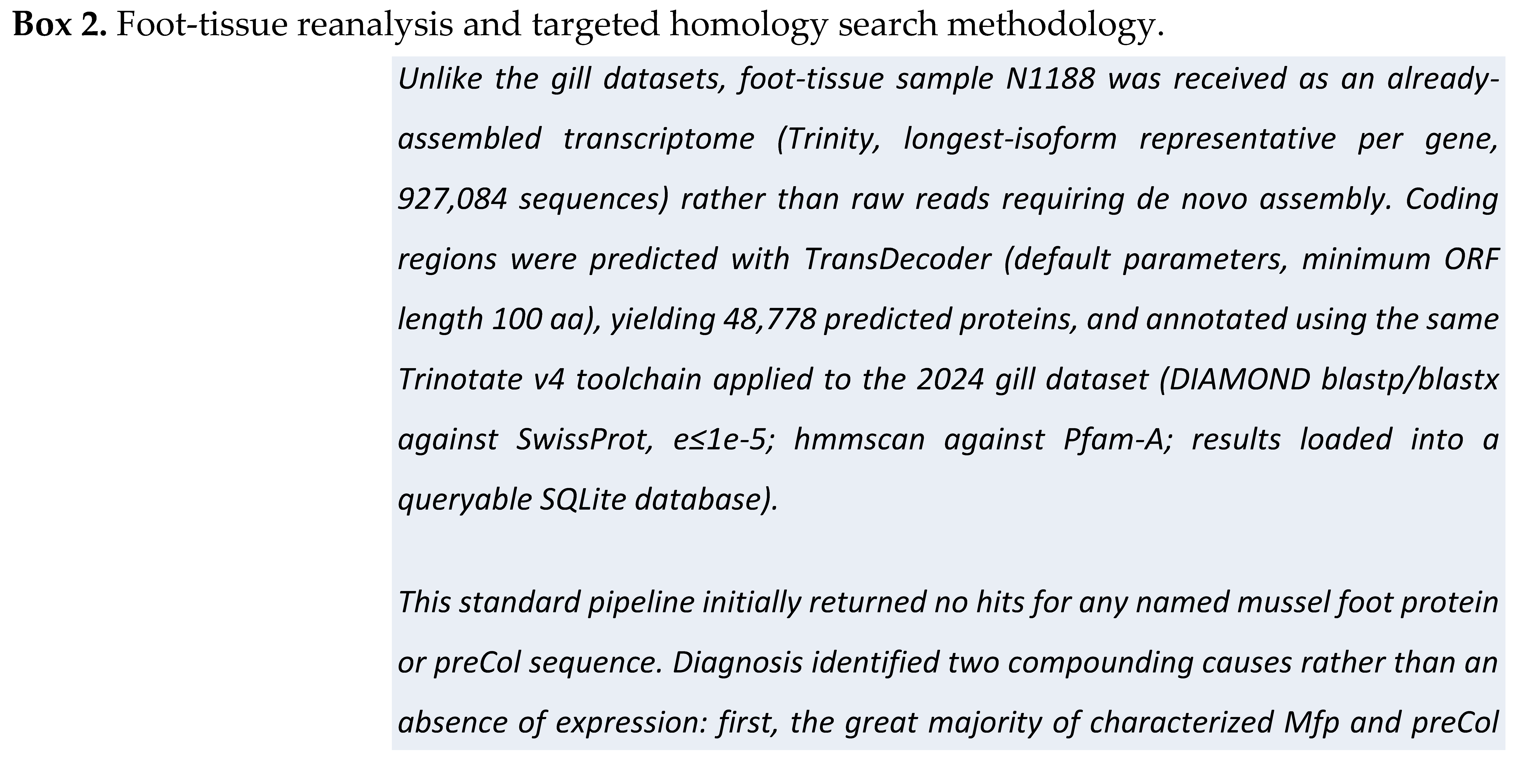
One further methodological caution from this reanalysis: a third transcript, TRINITY_DN380823, initially returned the strongest domain-level BLAST and Pfam score of any candidate and appeared, by that measure alone, to be the best fmp-2 match. On inspecting its full length (2,237 residues, far larger than fmp-2’s 431 residues), it proved to be an unrelated Notch-family receptor sharing only the ancestral EGF-repeat module with fmp-2, not a true ortholog. We report this here for the first time in B. azoricus foot tissue, because it illustrates a general point relevant to any modular, repeat-domain-containing protein family: a strong domain-level homology score is not, by itself, sufficient evidence of full-protein identity, and a length verification-check against the true reference protein is a necessary control step that a purely score-ranked search would not surface. This foot-tissue dataset remains distinct from the gill-tissue reanalyzes reported in Table 1; the raw sequencing reads have been deposited in the NCBI Sequence Read Archive under BioProject PRJNA1508337 (SRA accession SRR40036597), following the same workflow already applied to the gill datasets, with public release pending NCBI’s standard processing.
Mfp-2 is of particular interest because it is the most abundant protein in the byssal plaque, accounting for more than 25% of plaque dry mass. It is a ~45 kDa protein composed of 11 epidermal growth factor (EGF)-like repeats, each stabilized by three disulfide bonds. Significant concentrations of calcium and iron have been detected within byssal plaques, suggesting that Mfps interact with these ions in ways that influence adhesive performance and structural integrity [31]. The abundance of Mfp-2, its localization within the plaque, its interactions with collagenous scaffolding and associated matrix proteins, and its reversible Fe3+-binding capacity collectively make it an attractive template for the development of mussel-inspired bioadhesives.
New biomimetic adhesives could, in principle, be engineered through recombinant production of catechol-functionalized mussel foot proteins such as Mfp-2, combined with collagen- and extracellular matrix (ECM)-derived components. For example, recombinant Mfps fused to an RGD peptide sequence have been shown to enhance cell adhesion and proliferation under serum-free conditions [28,33]. Efficient cell attachment to extracellular scaffolds is fundamental to tissue engineering, while ECM-mediated interactions regulate cell adhesion, proliferation, spreading, viability, and differentiation. Beyond RGD, which primarily targets integrin-mediated adhesion, fusion constructs incorporating peptide motifs derived from fibronectin, laminin, or collagen provide additional opportunities to introduce cell-signaling functionality into biomaterials [22]. Combined with recombinant Mfps, these ECM-derived peptides could be immobilized within or conjugated to polymers, hydrogels, and nanofibrous scaffolds through catechol-based functionalization strategies [22].
7. Mussel Byssal Collagen as an Underexploited Structural Biomaterial
Most research on mussel-derived biomaterials, including the topics discussed in the preceding sections, has focused on the non-collagenous adhesive proteins of the byssal plaque. This emphasis risks overlooking an important component of the byssus itself: collagen. Members of the preCol family form the structural core of the byssal thread and are largely responsible for its characteristic combination of stiffness, extensibility, and mechanical resilience. Increasingly, byssal collagen is being investigated as a marine collagen source in its own right, distinct from the fish- and bovine-derived collagens that currently dominate biomedical applications.
Rodríguez et al. (2017) extracted collagen directly from the byssus of the shallow-water mussel Mytilus chilensis using a pepsin-assisted enzymatic method, characterized its physicochemical properties, and proposed byssal collagen as a novel marine collagen source with potential industrial applications [55]. Notably, the byssus is generally regarded as a processing by-product, making it an environmentally and ethically attractive source of collagen in any future B. azoricus aquaculture or specimen-collection framework. The structural behaviour of byssal collagen has also been investigated at the molecular level. Suhre et al. (2014) identified a collagen-binding matrix protein involved in organizing the preCol collagen core during thread assembly [56], while Priemel et al. (2017), using in situ Raman microspectroscopy, demonstrated that byssal collagen precursors self-assemble extremely rapidly into a hierarchical structure. Alignment of the semi-crystalline collagen core was shown to occur through a biologically regulated mechanical drawing process rather than through slow diffusion-limited assembly [57]. This self-organization mechanism is of particular interest for the design of rapidly forming, hierarchically structured collagen-based biomaterials.
The mechanical recovery properties of byssal collagen following cyclic loading are likewise relevant to tissue-engineering applications that must withstand repeated mechanical stress, including tendon and ligament repair [58]. This mechanical functionality provides an additional rationale for considering byssal collagen as a biomaterial platform in its own right rather than solely as a structural partner for adhesive proteins or RGD-functionalized constructs. For comparison, byssal proteins in the fan mussel Pinna nobilis are structurally more similar to silks than to the Mfp-collagen system characteristic of Mytilus and Bathymodiolus. Historically, these fibres were processed into “sea silk”, illustrating the diversity of structural solutions that have evolved among marine bivalves for underwater attachment. This comparison also cautions against assuming that B. azoricus collagen necessarily shares the same properties as collagen from shallow-water mytilids without direct experimental evaluation.
As with the adhesive Mfps discussed above, it remains unknown whether byssal collagen from B. azoricus possesses functional properties that distinguish it from those of shallow-water relatives. In particular, the metal-rich hydrothermal vent environment raises the possibility that its collagen or associated matrix proteins exhibit altered metal-binding behaviour, assembly dynamics, or mechanical performance. To our knowledge, these possibilities have not yet been investigated experimentally in B. azoricus or any other hydrothermal-vent mussel. We therefore identify this as a specific and experimentally tractable research question that warrants future study.
8. Byssal Collagen in the Foot Transcriptome: preCol-D, preCol-NG, and preCol-P
Beyond mfp-2, the same foot-transcriptome reanalysis was searched against a small reference panel of characterized Mytilus preCol sequences (preCol-D, UniProt O44367; preCol-NG, A9QLN3; preCol-P, O16161) using the same relaxed, reference-guided strategy described above for Mfp-2. This search yielded substantially more evidence than the Mfp-2 analysis alone: 156 candidate transcripts passed the length- and e-value-based confidence filters, with most matching one of the three preCol reference proteins rather than Mfp-2.
Unlike mfp-2, a relatively small and well-characterized 431-residue protein for which unusually large candidates warrant scrutiny, collagens, and byssal preCols in particular, are intrinsically large modular proteins composed of an extended central Gly-X-Y repeat region flanked by non-collagenous domains. The Gly-X-Y motif, in which glycine occupies every third position, is the defining structural signature of collagen and can be readily identified in translated protein sequences. The same length-based quality-control approach that previously identified a likely spurious Mfp-2 candidate was therefore applied to the preCol dataset. In contrast to the Mfp-2 analysis, however, most candidate sequences examined, including many of the largest transcripts (1,300 to 1,440 residues), displayed extensive and uninterrupted Gly-X-Y repeat regions consistent with authentic collagen proteins rather than chimeric assembly artifacts. A small subset of the largest candidates exhibited more heterogeneous repeat patterns, plausibly reflecting the presence of globular non-collagenous domains outside the central repeat region. Such architectures are common in collagen biology and are more appropriately resolved through formal domain annotation than by sequence length or repeat-pattern inspection alone. Consequently, the large size of these transcripts is consistent with the expected biology of the preCol family rather than indicative of assembly error.
The strongest and least ambiguous candidate for preCol-D was transcript TRINITY_DN381679_c3_g3_i2 (634 residues, 42.3% identity, e = 1.35 × 10−70), which consists almost entirely of Gly-X-Y repeats extending across most of the predicted protein sequence and terminates in a short non-repetitive C-terminal region consistent with the globular domains characteristic of fibrillar and network-forming collagens. A similarly compelling candidate for preCol-NG was TRINITY_DN373312_c1_g1_i5 (652 residues, e = 3.05 × 10−70), which exhibited the same extended collagen-repeat architecture and concluded with a short cysteine-rich terminal domain.
The most notable result, however, was TRINITY_DN376395_c3_g3_i5 (905 residues, e = 2.29 × 10−134 against preCol-P), which produced the strongest sequence match identified in the entire reanalysis, including both foot and gill datasets. This sequence displayed not only clear collagen homology but also the characteristic tripartite architecture of preCol-P, comprising a central Gly-X-Y collagen domain flanked by repetitive serine-, alanine-, glycine-, and histidine-rich regions. These non-collagenous flanking domains closely resemble the silk-fibroin-like regions known to surround the collagen core of preCol-P in shallow-water Mytilus species. Consequently, this candidate represents substantially stronger evidence than generic collagen homology alone and may constitute the first sequence-level indication of this specific preCol-P architecture in B. azoricus.
As with all findings derived from this reanalysis, these results should be interpreted with appropriate caution. Transcript-level evidence from a fragmented de novo assembly is not equivalent to a validated full-length gene model, and formal domain annotation remains an important next step. Nevertheless, the recovery of strong sequence matches to three distinct and functionally characterized preCol proteins, together with the identification of a preCol-P candidate displaying the expected domain architecture, provides considerably stronger evidence for byssal collagen expression in the B. azoricus foot transcriptome than has previously been available.
9. Recent Developments in Mussel-Derived Biomaterials (2020–2026)
The biomedical literature on mussel adhesive proteins (MAPs) has expanded substantially since the previous version of this review. It is therefore important to place B. azoricus-specific observations within this broader and rapidly growing field rather than consider them in isolation [19,32]. A comprehensive 2026 review of recombinant marine bioadhesive proteins reported that, by the end of 2024, recombinant DNA technologies had been used to produce biomimetic adhesive proteins from six invertebrate phyla, namely Cnidaria, Echinodermata, Tunicata, Annelida, Arthropoda, and Mollusca, with molluscan MAPs remaining among the most extensively studied systems [68]. Recent reviews have also expanded their focus beyond adhesion itself, highlighting antioxidant, anti-inflammatory, antibacterial, and cytoprotective activities of MAPs, while surveying recombinant MAP (rMAP) production strategies for applications in precision medicine, drug delivery, and tissue regeneration [25,29,64]. Parallel reviews have focused specifically on injectable mussel-inspired adhesive hydrogels [65] and wound-healing applications [66]. In addition, a 2025 comparative review evaluated MAPs alongside collagen, gelatin, silk fibroin, chitosan, and squid sucker-ring-teeth proteins (suckerins) as natural protein biomaterials for wound-healing applications [67]. This wider context provides a useful benchmark against which the potential distinctiveness of vent-derived biomaterials can be evaluated.
Recombinant production strategies have increasingly moved beyond the simple generation of soluble adhesive proteins toward the deliberate engineering of multifunctional chimeric constructs. Fusion proteins incorporating adhesive domains from Mfp-3 and Mfp-5 together with heterologous functional modules, including a cyanobacterial gas-vesicle protein and an Escherichia coli curli-forming protein, have been expressed in Pichia pastoris to generate bioadhesives with tunable self-assembly properties in addition to adhesive functionality [69]. Similar approaches have combined mussel adhesive protein sequences with structural motifs derived from spider silk (spidroin) and amyloid-forming proteins, generating hybrid biomaterials with underwater adhesion properties not found in any individual natural protein [70]. Collectively, these studies illustrate a broader trend in which MAP-derived sequences are increasingly employed as modular building blocks within rationally engineered biomaterial designs.
A research direction that has emerged more prominently since 2020 is the use of MAPs as structural platforms for antimicrobial activity rather than viewing antibacterial effects as secondary properties. Recombinant MAPs fused to antimicrobial peptides have demonstrated rapid bactericidal activity against Gram-negative bacteria, including E. coli, Salmonella typhimurium, and Klebsiella pneumoniae, while remaining non-toxic to mammalian cell lines under the conditions tested [71]. These findings further expand the potential applications of MAP-derived biomaterials beyond adhesion and tissue repair alone.
On the translational side, mussel-inspired biomaterials have increasingly been evaluated through direct comparison with established wet-tissue adhesive technologies. For example, catechol-oligomer-functionalized gelatin bioadhesives have demonstrated approximately threefold greater adhesion than conventional photo-crosslinked gelatin (GelMA) controls while simultaneously exhibiting improved toughness and extensibility during ex vivo testing on porcine lung tissue, a mechanically demanding model that closely reflects the requirements of internal surgical sealants [72].
Important advances have also been made in understanding byssus assembly itself. Building on the self-assembly mechanisms discussed earlier [57], Harrington and colleagues demonstrated that byssal thread formation operates as a microfluidic-like manufacturing process in which metal ions actively participate in curing the developing adhesive plaque [73]. Subsequent work further showed that catechol processing during byssus formation is compartmentalized in a manner that influences whether individual DOPA residues ultimately participate in cross-linking reactions or antioxidant pathways [74]. These findings do not resolve the Fe3+-binding hypothesis proposed here for B. azoricus, but they do strengthen its biological plausibility. Specifically, they establish that metal-mediated curing is a fundamental and actively regulated component of byssus formation in shallow-water mussels, providing a mechanistic framework through which adaptation to metal-rich hydrothermal-vent environments could potentially influence adhesive performance.
Finally, progress has also been made in the characterization of B. azoricus-derived natural products. During this period, new ceramide derivatives, bathymodiolamides C, D, and E, were isolated from B. azoricus and reported to induce necrotic cell death, a bioactivity of potential interest for anticancer drug discovery [75]. These compounds represent one of the few biomedical findings currently attributable specifically to B. azoricus rather than to the genus Bathymodiolus or the Mytilidae more broadly. They also align with a renewed interest in mollusc-derived natural products, a field comprehensively reviewed by Chen et al. (2023) [76].
10. The EGF-like Domain Across Gill and Foot: One Ancient Motif, Three Biological Roles
The EGF-like domain is among the most widely reused protein modules in metazoan evolution [77]. This reanalysis encountered that recurrent usage independently in two different tissues, providing an opportunity to consider these observations together. While each instance appeared above as a methodological challenge, taken collectively they illustrate a broader biological principle concerning the deployment of EGF-like domains in B. azoricus.
In the gill transcriptome (Table 1), the two independent EGF-focused reanalyses highlight this issue clearly. The 2010 dataset, using a broad structural-domain search strategy, recovered 29 proteins containing EGF-like domains or combined von Willebrand factor type D and EGF-like domains. This set likely included a mixture of signaling molecules, extracellular matrix proteins, cell-adhesion proteins, receptors, and other domain-containing proteins, reflecting the widespread occurrence of EGF-like repeats across multiple gene families [78]. By contrast, the 2024 analysis was deliberately restricted to canonical ligand-type EGF families, including HB-EGF, TGF-α-like, and epiregulin-like proteins, yielding only three candidates. As noted in Table 1, these values do not represent directly comparable measurements of the same gene family. Rather, they reflect two different biological questions and illustrate the extent to which broad domain-based searches can recover proteins that share a common structural motif despite having very different biological functions.
The foot transcriptome revealed the same phenomenon from a different perspective. Mfp-2, the adhesive plaque protein central to the byssal-adhesion system, contains eleven tandem EGF-like repeats, each stabilized by three disulfide bonds [79,80]. Although these repeats are homologous to those found in other EGF-domain proteins, they function here primarily as structural elements that contribute to folding and stabilization within a secreted adhesive protein rather than as signaling ligands interacting with receptor tyrosine kinases. During the search for mfp-2 homologues, the consequences of domain sharing became apparent. Transcript TRINITY_DN380823 produced the strongest domain-level BLAST and Pfam match among all candidates and would initially have been identified as the most likely mfp-2 homologue. However, comparison with the reference protein length revealed that it instead corresponded to a Notch-family receptor, whose extracellular region is itself composed of numerous tandem EGF-like repeats [81]. In this case, the same structural module was being used in an entirely different biological context, namely as a repeated component of a cell-surface receptor rather than an adhesive protein or soluble signaling factor.
Taken together, these observations demonstrate that the EGF-like domain fulfills at least three distinct biological functions within the B. azoricus transcriptome: as part of a soluble signaling ligand (the HB-EGF-, epiregulin-, and TGF-α-related proteins identified in the restricted gill analysis), as a structural stabilization element within the adhesive protein mfp-2, and as a repeated architectural component of cell-surface receptors such as Notch. These findings are not merely isolated methodological observations but instead reflect a general principle of protein evolution: the repeated reuse of ancient structural domains in diverse biological contexts. For this reason, every EGF-domain-containing protein discussed in this review has been interpreted according to its overall protein architecture and biological context rather than treated as equivalent evidence for a single, functionally uniform “EGF” category.
11. Conclusions
This review was built around a deliberate methodological premise: that sequencing datasets generated over a period of fifteen years, including a 2010 454 gill transcriptome, an independent 2024 Illumina gill transcriptome, and a 2017-2019 foot transcriptome recovered from surviving raw reads after the loss of the original analysis, could be reanalyzed using contemporary bioinformatic approaches and still yield biologically meaningful results. The findings presented here support that premise. Gene targets examined independently in both the 2010 and 2024 gill transcriptomes showed a high degree of concordance, with several candidates progressing from tentative identification to stronger support in the deeper 2024 dataset and only one target, Sialic acid-binding immunoglobulin-like lectin, failing to replicate. This level of agreement demonstrates that legacy sequencing datasets, when reprocessed using modern analytical methods, can remain valuable sources of biological insight rather than merely archival resources. The same conclusion applies to the foot transcriptome, where a targeted reference-guided reanalysis independently recovered a result first identified in 2017 and subsequently lost with the original server infrastructure, while also substantially extending the available evidence.
That reanalysis effort supports two lines of evidence this review has argued belong in the same narrative, not two separate stories about the same animal. On the immunological side, the gill transcriptome comparisons documented here — spanning wound-repair and immunity-associated genes confirmed, resolved, or genuinely absent across both reanalyses — extend B. azoricus’s established role as a model for innate immunity and host–symbiont tolerance into a broader, still only partially explored catalogue of immune-relevant gene content. On the materials side, the foot-tissue reanalysis substantially strengthens the case for B. azoricus as a source of biomedically relevant structural biomaterials, and does so on two fronts, not one: the fmp-2 adhesive protein finding, independently reproduced and extended from the original 2017 analysis, and the newly recovered evidence for all three characterized byssal preCol collagens — preCol-D, preCol-NG and preCol-P — with preCol-P showing a specific, architecturally distinctive match rather than generic collagen homology. Notably, the same EGF-like domain that helped identify fmp-2 also produced this review’s clearest methodological caution, in the form of a large, unrelated Notch-family receptor sharing only that ancestral module — a reminder, discussed at length above, that this domain performs at least three distinct biological roles across the B. azoricus transcriptome alone, and that domain-level homology is never sufficient evidence of shared function on its own.
Structural evidence beyond the transcriptome points the same direction. Direct characterisation of B. azoricus shell nacre has shown that this species incorporates vent-sourced trace metals into its shell and that its nacre’s structural integrity is measurably, directly sensitive to hydrostatic pressure — independent, tissue-level evidence that vent adaptation shapes this animal’s structural biology beyond the foot adhesive proteins alone, even though, unlike gill and foot, mantle tissue itself has not yet been transcriptomically sequenced in this species.
Mussel-inspired catechol-based polymers continue to attract attention as moisture-resistant adhesives and functional coating materials [82]. Increasingly, the field has shifted toward the functionalization of synthetic polymers with DOPA and related catecholic chemistries to generate materials capable of rapid curing, strong wet adhesion, and favorable mechanical performance [82]. The adhesive properties of these systems have been demonstrated across a wide range of substrates, including metals, plastics, glass, PTFE, biological tissues, and mammalian cells under hydrated conditions. Such materials are therefore of growing interest for wound healing, surgical sealants, tissue adhesives, and tissue-engineering scaffolds. Recombinant production technologies further enable the scalable manufacture of mussel-inspired adhesive proteins and create opportunities for the incorporation of extracellular-matrix signaling motifs into multifunctional biomaterials. In this context, the mussel byssus remains a productive model for understanding the molecular basis of underwater adhesion and for developing next-generation biomedical adhesives. The evidence reviewed here suggests that B. azoricus may extend this model in biologically distinctive ways through both its adhesive proteins and its structural byssal collagens.
We conclude by returning to the central proposition advanced in the Introduction: B. azoricus is not simply another mussel species from which previously characterized adhesive chemistries may be obtained. Rather, its combination of permanent bacterial symbiosis, distinctive immune biology, and long-term exposure to metal-rich hydrothermal vent environments makes it a uniquely informative system at the intersection of immunology, biomaterials science, and environmental adaptation. The evidence assembled in this review supports the hypothesis that vent adaptation may have influenced both the immune repertoire and the metal-mediated adhesive chemistry of this species, producing functional properties that are potentially distinct from those described in shallow-water mussels.
The transcriptomic resources underlying this review have now been deposited in the NCBI Sequence Read Archive (BioProjects PRJNA1508204 and PRJNA1508337), with public release pending completion of NCBI processing. Testing the hypothesis itself remains the next challenge. Priorities for future work include formal Pfam and InterPro annotation of the preCol candidates identified here, generation of a mantle transcriptome to extend transcriptomic analysis to shell biomineralization directly, comparative Fe3+-binding studies of B. azoricus and shallow-water mussel adhesive proteins and collagens, and functional comparisons of antimicrobial peptides and pattern-recognition molecules across vent and non-vent species. Together, these approaches would provide the experimental framework needed to determine whether the distinctive environmental conditions of hydrothermal vents have produced biomaterials and immune molecules that are not merely taxonomically related to their shallow-water counterparts, but functionally and mechanistically distinct.
Author Contributions
Conceptualization, R.B.; methodology, R.B.; software, R.B.; validation, R.B.; formal analysis, R.B.; investigation, R.B.; resources, T.H.S.; data curation, R.B.; writing—original draft preparation, R.B.; writing review and editing, R.B., R.L.R. and T.H.S.; supervision, T.H.S.; project administration at University of Minho, T.H.S.; funding acquisition, T.H.S. and R.B. All authors have read and agreed to the published version of the manuscript.
Funding
This research was funded by the R&D&I Structured Project NORTE-01-0145-FEDER-000021, supported by the European Regional Development Fund (ERDF) under the Programa Operacional Regional do Norte (NORTE2020), which funded sequencing of the B. azoricus foot transcriptome in 2017. The 2024 gill transcriptome sequencing was supported by the OKEANOS research unit, which received national funds through the FCT - Fundação para a Ciência e Tecnologia, I.P. by project reference UID/05634/2025 (DOI: 10.54499/UID/05634/2025), and from the Regional Directorate for Science, Innovation and Development of the Azores Government through the PROSCIENTIA Incentive System under project M1.1.A/FUNC.UI&D/014/2025. This work was also supported by the MarAZ Project (Grant No. ACORES-01-0145-FEDER-000138), co-financed by the European Regional Development Fund (ERDF) through the Operational Program Azores 2020, and by regional funds through the PRO-SCIENTIA program, under which RB was hired from 2020-2023.
Institutional Review Board Statement
Not applicable.
Data availability statement
The gill-tissue 454 sequencing reads originally reported in [10] are deposited in the NCBI Sequence Read Archive under accession SRR067874. The custom web portal previously used to query these data (DeepSeaVent) has been discontinued and is no longer available; Table 1 and the associated discussion in this review are instead based on an independent reanalysis of the deposited raw reads, summarised in Box 1, with the resulting assembly, annotation and SQLite database available from the corresponding author on request pending formal deposition in a persistent public repository. A second, independent gill-tissue transcriptome generated in 2024 using Illumina sequencing has been deposited in the NCBI Sequence Read Archive under BioProject PRJNA1508204 (SRA accession SRR40024054, BioSample SAMN62218460, submission SUB16386217); the submission has been processed and will be publicly released following NCBI’s standard release process. The foot-tissue transcriptome discussed above has now been reanalyzed using the same workflow (see above) and has likewise been deposited, under BioProject PRJNA1508337 (SRA accession SRR40036597, BioSample SAMN62230578, submission SUB16387160); this submission has also been processed and is pending public release.
Acknowledgments
The authors are grateful to Dr. Isabel Leonor for her contribution with SEM analyses.
Conflicts of Interest
The authors declare no conflicts of interest. The funders had no role in the design of the study; in the collection, analyses, or interpretation of data; in the writing of the manuscript; or in the decision to publish the results.
Abbreviations
The following abbreviations are used in this manuscript
| AIF1 | Allograft Inflammatory Factor 1 |
| ALDH | Aldehyde Dehydrogenase |
| BLAST | Basic Local Alignment Search Tool |
| CA | Carbonic Anhydrase |
| CBB | Calvin-Benson-Bassham (cycle) |
| DIAMOND | Double Index Alignment of Next-generation sequencing Data |
| DNA | Deoxyribonucleic Acid |
| DOPA | 3,4-Dihydroxyphenylalanine |
| ECM | Extracellular Matrix |
| EGF | Epidermal Growth Factor |
| EPAS1 | Endothelial PAS Domain Protein 1 |
| ERK | Extracellular signal-Regulated Kinase |
| FGF | Fibroblast Growth Factor |
| FGFR4 | Fibroblast Growth Factor Receptor 4 |
| FIH-1 | Factor Inhibiting HIF-1 |
| GO | Gene Ontology |
| GS-FLX | Genome Sequencer FLX (454 sequencing platform) |
| HB-EGF | Heparin-Binding EGF-like growth factor |
| HGF | Hepatocyte Growth Factor |
| HIF-1 | Hypoxia-Inducible Factor 1 |
| HIF1AN | Hypoxia Inducible Factor 1 Subunit Alpha Inhibitor |
| HSP70 | Heat Shock Protein 70 |
| IGF | Insulin-like Growth Factor |
| JNK | c-Jun N-terminal Kinase |
| LITAF | Lipopolysaccharide-Induced TNF Factor |
| LPS | Lipopolysaccharide |
| MAP | Mussel Adhesive Protein |
| MAPK | Mitogen-Activated Protein Kinase |
| MAR | Mid-Atlantic Ridge |
| MeDH | Methanol Dehydrogenase |
| MIRA | Mimicking Intelligent Read Assembly (assembler software) |
| MMO | Methane Monooxygenase |
| MMP | Matrix Metalloproteinase |
| NCBI | National Center for Biotechnology Information |
| ORF | Open Reading Frame |
| PAMP | Pathogen-Associated Molecular Pattern |
| PCR | Polymerase Chain Reaction |
| PO | Phenoloxidase |
| PTFE | Polytetrafluoroethylene |
| RGD | Arginine-Glycine-Aspartate (peptide motif) |
| RT-PCR | Reverse Transcription Polymerase Chain Reaction |
| RTK | Receptor Tyrosine Kinase |
| SEM | Scanning Electron Microscope/Microscopy |
| SIGLEC1 | Sialic Acid-Binding Ig-like Lectin 1 |
| SOXB | Sulfur OXidation gene B |
| SRA | Sequence Read Archive |
| SRCR | Scavenger Receptor Cysteine-Rich (domain) |
| TGF-β | Transforming Growth Factor Beta |
| TIMP | Tissue Inhibitor of Metalloproteinases |
| TLR2 | Toll-Like Receptor 2 |
| TNF | Tumor Necrosis Factor |
| VEGF | Vascular Endothelial Growth Factor |
| VEGFR-2 | Vascular Endothelial Growth Factor Receptor 2 |
| WGA | Wheat Germ Agglutinin |
References
- Childress, J.J.; Fisher, C.R. The biology of hydrothermal vent animals — physiology, biochemistry, and autotrophic symbioses. Oceanogr. Mar. Biol. Ann. Rev. 1992, 30, 337–441. [Google Scholar]
- Desbruyères, D.; Biscoito, M.; Caprais, J.C.; Colaço, A.; Comtet, T.; Crassous, Ph; Fouquet, Y.; Khripounoff, A.; Le Bris, N.; Olu, K.; Riso, R.; Sarradin, P.M.; Segonzac, M.; Vangriesheim, A. Variations in deep-sea hydrothermal vent communities on the Mid-Atlantic Ridge near the Azores plateau. Deep Sea Res. Pt I 2001, 48, 1325–1346. [Google Scholar] [CrossRef]
- Cavanaugh, C.M.; McKiness, Z.P.; Newton, I.L.G.; Stewart, F.J. Marine chemosynthetic symbioses. In The Prokaryotes; Springer, 2006; pp. 475–507. [Google Scholar]
- Dubilier, N.; Bergin, C.; Lott, C. Symbiotic diversity in marine animals: the art of harnessing chemosynthesis. Nat. Rev. Microbiol. 2008, 6, 725–740. [Google Scholar] [CrossRef] [PubMed]
- Duperron, S.; Gaudron, S.M.; Rodrigues, C.F.; Cunha, M.R.; Decker, C.; Olu, K. An overview of chemosynthetic symbioses in bivalves from the North Atlantic and Mediterranean Sea. Biogeosciences 2013, 10, 3241–3267. [Google Scholar] [CrossRef]
- Hui, M.; Cheng, J.; Sha, Z. Adaptation to the deep-sea hydrothermal vents and cold seeps: insights from the transcriptomes of Alvinocaris longirostris in both environments. Deep-Sea Res. Pt I 2018, 135, 23–33. [Google Scholar] [CrossRef]
- Bettencourt, R.; Barros, I.; Martins, E.; Martins, I.; Cerqueira, T.; Colaço, A.; Costa, V.; Rosa, D.; Froufe, H.; Egas, C.; Stefanni, S.; Dando, P.; Serrão Santos, R. An insightful model to study innate immunity and stress response in deep-sea vent animals: profiling the mussel Bathymodiolus azoricus. In Organismal and Molecular Malacology; Ray, S., Ed.; InTech, 2017. [Google Scholar] [CrossRef] [PubMed]
- Thornburg, C.C.; Zabriskie, T.M.; McPhail, K.L. Deep-sea hydrothermal vents: potential hot spots for natural products discovery? J. Nat. Prod. 2010, 73, 489–499. [Google Scholar] [CrossRef] [PubMed]
- Chandika, P.; Ko, S.C.; Jung, W.K. Marine-derived biological macromolecule-based biomaterials for wound healing and skin tissue regeneration. Int. J. Biol. Macromol. 2015, 77, 24–35. [Google Scholar] [CrossRef] [PubMed]
- Bettencourt, R.; Pinheiro, M.; Egas, C.; Gomes, P.; Afonso, M.; Shank, T.; Serrão Santos, R. Sequencing and analysis of the gill tissue transcriptome from the deep-sea hydrothermal vent mussel Bathymodiolus azoricus using 454 GSFLX. BMC Genom. 2010, 11, 559. [Google Scholar] [CrossRef] [PubMed]
- Egas, C.; Pinheiro, M.; Gomes, P.; Barroso, C.; Bettencourt, R. The transcriptome of Bathymodiolus azoricus gill reveals expression of genes from endosymbionts and free-living deep-sea bacteria. Mar. Drugs 2012, 10(8), 1765–1783. [Google Scholar] [CrossRef] [PubMed]
- Cerqueira, T.; Barroso, C.; Froufe, H.; Egas, C.; Bettencourt, R. Metagenomic signatures of microbial communities in deep-sea hydrothermal sediments of Azores vent fields. Microb. Ecol. 2018, 76(2), 387–403. [Google Scholar] [CrossRef] [PubMed]
- Waite, J.H.; Andersen, N.H.; Jewhurst, S.; Sun, C. Mussel adhesion: finding the tricks worth mimicking. J. Adhes. 2005, 81, 297–317. [Google Scholar] [CrossRef]
- Ahn, B.K. Perspectives on mussel-inspired wet adhesion. J. Am. Chem. Soc. 2017, 139(30), 10166–10171. [Google Scholar] [CrossRef] [PubMed]
- Waite, J.H. Mussel adhesion — essential footwork. J. Exp. Biol. 2017, 220, 517–530. [Google Scholar] [CrossRef] [PubMed]
- Bandara, N.; Zeng, H.; Wu, J. Marine mussel adhesion: biochemistry, mechanisms, and biomimetics. J. Adhes. Sci. Technol. 2013, 27(18–19), 2139–2162. [Google Scholar] [CrossRef]
- Li, L.; Zeng, H. Marine mussel adhesion and bio-inspired wet adhesives. Biotribology 2016, 5, 44–51. [Google Scholar] [CrossRef]
- DeMartini, D.G.; Errico, J.M.; Sjoestroem, S.; Fenster, A.; Waite, J.H. A cohort of new adhesive proteins identified from transcriptomic analysis of mussel foot glands. J. R Soc. Interface 2017, 14(131), 20170151. [Google Scholar] [CrossRef] [PubMed]
- Kaushik, N.K.; Kaushik, N.; Pardeshi, S.; Sharma, J.G.; Lee, S.H.; Choi, E.H. Biomedical and clinical importance of mussel-inspired polymers and materials. Mar. Drugs 2015, 13, 6792–6817. [Google Scholar] [CrossRef] [PubMed]
- Yarra, T.; Gharbi, K.; Blaxter, M.; Peck, L.S.; Clark, M.S. Characterization of the mantle transcriptome in bivalves: Pecten maximus, Mytilus edulis and Crassostrea gigas. Mar. Genom. 2016, 27, 9–15. [Google Scholar] [CrossRef] [PubMed]
- Bettencourt, R.; Dando, P.; Collins, P.; Costa, V.; Allam, B.; Serrão Santos, R. Innate immunity in the deep sea hydrothermal vent mussel Bathymodiolus azoricus. Comp. Biochem Physiol. A Mol. Integr. Physiol. 2009, 152(2), 278–289. [Google Scholar] [CrossRef] [PubMed]
- Choi, B.H.; Choi, Y.S.; Kang, D.G.; Kim, B.J.; Song, Y.H.; Cha, H.J. Cell behavior on extracellular matrix mimic materials based on mussel adhesive protein fused with functional peptides. Biomaterials 2010, 31(34), 8980–8988. [Google Scholar] [CrossRef] [PubMed]
- Larouche, J.; Sheoran, S.; Maruyama, K.; Martino, M.M. Immune regulation of skin wound healing: mechanisms and novel therapeutic targets. Adv. Wound Care (New Rochelle) 2018, 7(7), 209–231. [Google Scholar] [CrossRef] [PubMed]
- Yang, B.; Kang, D.G.; Seo, J.H.; Choi, Y.S.; Cha, H.J. A comparative study on the bulk adhesive strength of the recombinant mussel adhesive protein fp-3. Biofouling 2013, 29(5), 483–490. [Google Scholar] [CrossRef] [PubMed]
- Jeon, E.Y.; Hwang, B.H.; Yang, Y.J.; Kim, B.J.; Choi, B.H.; Jung, G.Y.; Cha, H.J. Rapidly light-activated surgical protein glue inspired by mussel adhesion and insect structural crosslinking. Biomaterials 2015, 67, 11–19. [Google Scholar] [CrossRef] [PubMed]
- Kim, B.J.; Oh, D.X.; Kim, S.; Seo, J.H.; Hwang, D.S.; Masic, A.; Han, D.K.; Cha, H.J. Mussel-mimetic protein-based adhesive hydrogel. Biomacromolecules 2014, 15(5), 1579–1585. [Google Scholar] [CrossRef] [PubMed]
- Danner, E.W.; Kan, Y.; Hammer, M.U.; Israelachvili, J.N.; Waite, J.H. Adhesion of mussel foot protein Mefp-5 to mica: an underwater superglue. Biochemistry 2012, 51(33), 6511–6518. [Google Scholar] [CrossRef] [PubMed]
- Hwang, D.S.; Gim, Y.; Kang, D.G.; Kim, Y.K.; Cha, H.J. Recombinant mussel adhesive protein Mgfp-5 as cell adhesion biomaterial. J. Biotechnol. 2007, 127(4), 727–735. [Google Scholar] [CrossRef] [PubMed]
- Jo, Y.K.; Kim, H.J.; Jeong, Y.; Joo, K.I.; Cha, H.J. Biomimetic surface engineering of biomaterials by using recombinant mussel adhesive proteins. Adv. Mater. Interfaces 2018, 5(20), 1800068. [Google Scholar] [CrossRef]
- Choi, Y.S.; Kang, D.G.; Lim, S.; Yang, Y.J.; Kim, C.S.; Cha, H.J. Recombinant mussel adhesive protein fp-5 (MAP fp-5) as a bulk bioadhesive and surface coating material. Biofouling 2011, 27(7), 729–737. [Google Scholar] [CrossRef] [PubMed]
- Hwang, D.S.; Zeng, H.; Masic, A.; Harrington, M.J.; Israelachvili, J.N.; Waite, J.H. Protein- and metal-dependent interactions of a prominent protein in mussel adhesive plaques. J. Biol. Chem. 2010, 285(33), 25850–25858. [Google Scholar] [CrossRef] [PubMed]
- Forooshani, P.K.; Lee, B.P. Recent approaches in designing bioadhesive materials inspired by mussel adhesive protein. J. Polym. Sci. A Polym. Chem. 2017, 55(1), 9–33. [Google Scholar] [CrossRef] [PubMed]
- Kim, B.J.; Choi, Y.S.; Choi, B.H.; Lim, S.; Song, Y.H.; Cha, H.J. Mussel adhesive protein fused with cell adhesion recognition motif triggers integrin-mediated adhesion and signaling for enhanced cell spreading, proliferation, and survival. J. BioMed Mater. Res. A 2010, 94(3), 886–892. [Google Scholar] [CrossRef] [PubMed]
- Lu, M.; Yu, J. Mussel-inspired biomaterials for cell and tissue engineering. In Novel Biomaterials for Regenerative Medicine. Advances in Experimental Medicine and Biology; Springer, 2018; vol. 1077. [Google Scholar]
- Luz, G.M.; Mano, J.F. Biomimetic design of materials and biomaterials inspired by the structure of nacre. Phil Trans. R Soc. A 2009, 367, 1587–1605. [Google Scholar] [CrossRef] [PubMed]
- Gosling, E. Bivalve Molluscs: Biology, Ecology and Culture. In Fishing New Books; Blackwell Publishing, 2003; p. 443 p. [Google Scholar] [CrossRef]
- Malachowicz, M.; Wenne, R. Mantle transcriptome sequencing of Mytilus spp. and identification of putative biomineralization genes. PeerJ 2019, 6, e6245. [Google Scholar] [CrossRef] [PubMed] [PubMed Central]
- Hodgson, A. N. Studies on wound healing, and an estimation of the rate of regeneration, of the siphon of Scrobicularia plana (da Costa). J. Exp. Mar. Biol. Ecol. 1982, 62(2), 117–128. [Google Scholar] [CrossRef]
- Hodgson, A. N. Studies on wound healing and regeneration of the siphons of the bivalve Donax serra (Röding). Trans. R. Soc. South Afr. 1982, 44(4), 489–498. [Google Scholar] [CrossRef]
- Acosta-Salmón, H.; Southgate, P.C. Wound healing after excision of mantle tissue from the Akoya pearl oyster, Pinctada fucata. Comp. Biochem Physiol. A Mol. Integr. Physiol. 2006, 143(2), 264–8. [Google Scholar] [CrossRef] [PubMed]
- Acosta-Salmón, H.; Southgate, P.C. Mantle regeneration in the pearl oysters Pinctada fucata and Pinctada margaritifera. Aquaculture 2005, 246(1–4), 447–453. [Google Scholar] [CrossRef]
- Sritunyalucksana, K.; Söderhäll, K. The proPO and clotting system in crustaceans. Aquaculture 2000, 191, 53–69. [Google Scholar] [CrossRef]
- Cerenius, L.; Kawabata, S.; Lee, B.L.; Nonaka, M.; Söderhäll, K. Proteolytic cascades and their involvement in invertebrate immunity. Trends Biochem Sci. 2010. [Google Scholar] [CrossRef] [PubMed]
- Aguilera, F.; McDougall, C.; Degnan, B.M. Evolution of the tyrosinase gene family in bivalve molluscs: independent expansion of the mantle gene repertoire. Acta Biomater. 2014, 10, 3855–3865. [Google Scholar] [CrossRef] [PubMed]
- Zhang, G.; Brion, A.; Willemin, A.S.; Piet, M.H.; Moby, V.; Bianchi, A.; Mainard, D.; Galois, L.; Gillet, P.; Rousseau, M. Nacre, a natural, multi-use, and timely biomaterial for bone graft substitution. J. BioMed Mater. Res. A 2017, 105(2), 662–671. [Google Scholar] [CrossRef] [PubMed]
- Macha, I.J.; Ozyegin, L.S.; Chou, J.; Samur, R.; Oktar, F.N.; Ben-Nissan, B. An alternative synthesis method for di-calcium phosphate (monetite) powders from Mediterranean mussel (Mytilus galloprovincialis) shells. J. Aust. Ceram. Soc. 2013, 49, 122–128. [Google Scholar]
- Joubert, C.; Linard, C.; Le Moullac, G.; Soyez, C.; Saulnier, D.; Teaniniuraitemoana, V.; Ky, C.L.; Gueguen, Y. Temperature and food influence shell growth and mantle gene expression of shell matrix proteins in the pearl oyster Pinctada margaritifera. PLoS ONE 2014, 9(8), e103944. [Google Scholar] [CrossRef] [PubMed]
- Kádár, E.; Checa, A.G.; Damasceno-Oliveira, A.; Machado, J.P. Shell nacre ultrastructure and depressurisation dissolution in the deep-sea hydrothermal vent mussel Bathymodiolus azoricus. J. Comp. Physiol. B 2008, 178(1), 123–130. [Google Scholar] [CrossRef] [PubMed]
- Kádár, E.; Costa, V. First report on the micro-essential metal concentrations in bivalve shells from deep-sea hydrothermal vents. J. Sea Res. 2006, 56(1), 37–44. [Google Scholar] [CrossRef]
- Kádár, E. Hemocyte response associated with induction of shell regeneration in the deep-sea vent mussel Bathymodiolus azoricus (Bivalvia: Mytilidae). J. Exp. Mar. Biol. Ecol. 2008, 362(2), 71–78. [Google Scholar] [CrossRef]
- Kádár, E.; Lobo-da-Cunha, A.; Azevedo, C. Mantle-to-shell CaCO3 transfer during shell repair at different hydrostatic pressures in the deep-sea vent mussel Bathymodiolus azoricus (Bivalvia: Mytilidae). Mar. Biol. 2009, 156(5), 959–967. [Google Scholar] [CrossRef]
- Barros, I.; Mendes, S.; Rosa, D.; Santos, R.S.; Bettencourt, R. Vibrio diabolicus immunomodulatory effects on Bathymodiolus azoricus during long-term acclimatization at atmospheric pressure. J. Aquac. Res. Dev. 2016, 7(12), 464. [Google Scholar] [CrossRef]
- Barros, I.; Divino, F.; Colaço, A.; Serrão Santos, R.; Bettencourt, R. Vibrio diabolicus challenge in Bathymodiolus azoricus populations from Menez Gwen and Lucky Strike hydrothermal vent sites. Fish. Shellfish Immunol. 2015, 45(2), 570–580. [Google Scholar] [CrossRef] [PubMed]
- Martins, E.; Figueras, A.; Novoa, B.; Serrão Santos, R.; Moreira, R.; Bettencourt, R. Comparative study of immune responses in the deep-sea hydrothermal vent mussel Bathymodiolus azoricus and the shallow-water mussel Mytilus galloprovincialis challenged with Vibrio bacteria. Fish. Shellfish Immunol. 2014, 40(2), 485–499. [Google Scholar] [CrossRef] [PubMed]
- Rodríguez, F.; Morán, L.; González, G.; Troncoso, E.; Zúñiga, R.N. Collagen extraction from mussel byssus: a new marine collagen source with physicochemical properties of industrial interest. J. Food Sci. Technol. 2017, 54(5), 1228–1238. [Google Scholar] [CrossRef] [PubMed]
- Suhre, M.H.; Gertz, M.; Steegborn, C.; Scheibel, T. Structural and functional features of a collagen-binding matrix protein from the mussel byssus. Nat. Commun. 2014, 5, 3392. [Google Scholar] [CrossRef] [PubMed]
- Priemel, T.; Degtyar, E.; Dean, M.N.; Harrington, M.J. Rapid self-assembly of complex biomolecular architectures during mussel byssus biofabrication. Nat. Commun. 2017, 8, 14539. [Google Scholar] [CrossRef] [PubMed]
- Harrington, M.J.; Gupta, H.S.; Fratzl, P.; Waite, J.H. Collagen insulated from tensile damage by domains that unfold reversibly. J. Struct. Biol. 2009, 167(1), 47–54. [Google Scholar] [CrossRef] [PubMed]
- Eming, S.A.; Martin, P.; Tomic-Canic, M. Wound repair and regeneration: mechanisms, signaling, and translation. Sci. Transl. Med. 2014, 6(265), 265sr6. [Google Scholar] [CrossRef] [PubMed]
- Ryu, J.H.; Lee, Y.; Kong, W.H.; Kim, T.G.; Park, T.G.; Lee, H. Catechol-functionalized chitosan/pluronic hydrogels for tissue adhesives and hemostatic materials. Biomacromolecules 2011, 12(7), 2653–2659. [Google Scholar] [CrossRef] [PubMed]
- Mehdizadeh, M.; Weng, H.; Gyawali, D.; Tang, L.; Yang, J. Injectable citrate-based mussel-inspired tissue bioadhesives with high wet strength for sutureless wound closure. Biomaterials 2012, 33(32), 7972–7983. [Google Scholar] [CrossRef] [PubMed]
- Duan, W.; Bian, X.; Bu, Y. Applications of bioadhesives: a mini review. Front Bioeng. Biotechnol. 2021, 9, 716035. [Google Scholar] [CrossRef] [PubMed]
- Ma, X.; Bian, Q.; Hu, J.; Gao, J. Stem from nature: Bioinspired adhesive formulations for wound healing. J. Control Release 2022, 345, 292–305. [Google Scholar] [CrossRef] [PubMed]
- An, J.; Hou, Z.; Li, J.; Bi, Q.; Huang, S.; Peng, C.; Peng, F. Pharmacological effects and application prospects of mussel adhesive proteins. Front Pharmacol. 2025, 16, 1704475. [Google Scholar] [CrossRef] [PubMed]
- Dou, W.; Zeng, X.; Zhu, S.; Zhu, Y.; Liu, H.; Li, S. Mussel-inspired injectable adhesive hydrogels for biomedical applications. Int. J. Mol. Sci. 2024, 25(16), 9100. [Google Scholar] [CrossRef] [PubMed]
- Chen, Y.; Cao, Y.; Cui, P.; Lu, S. Mussel-inspired hydrogel applied to wound healing: a review and future prospects. Biomimetics 2025, 10(4), 206. [Google Scholar] [CrossRef] [PubMed]
- Hiruthyaswamy, S.P.; Deepankumar, K. Suckerin based biomaterials for wound healing: a comparative review with natural protein-based biomaterials. Mater. Adv. 2025, 6(4), 1262–1277. [Google Scholar] [CrossRef]
- Whaite, A.; Duthoo, E.; Lefevre, M.; Hennebert, E.; Flammang, P. Recombinant Proteins: A Molecular Tool to Understand Marine Adhesion and to Advance Biomaterials. Adv. Healthc. Mater. 2026, 15(3), e2502340. [Google Scholar] [CrossRef] [PubMed]
- Bolghari, N.; Habibollahi, H.; Anvari, M.; Shahsavarani, H. A novel recombinant chimeric bio-adhesive protein consisting of mussel foot protein 3, 5, gas vesicle protein A, and CsgA curli protein expressed in Pichia pastoris. AMB Express 2022, 12, 24. [Google Scholar] [CrossRef] [PubMed]
- Kim, E.; Jeon, J.; Zhu, Y.; Hoppe, E.D.; Jun, Y.S.; Genin, G.M.; Zhang, F. A biosynthetic hybrid spidroin-amyloid-mussel foot protein for underwater adhesion on diverse surfaces. ACS Appl. Mater. Interfaces 2021, 13(41), 48457–48468. [Google Scholar] [CrossRef] [PubMed]
- Kim, D.Y.; Oh, Y.B.; Park, J.S.; Min, Y.H.; Park, M.C. Anti-microbial activities of mussel-derived recombinant proteins against Gram-negative bacteria. Antibiotics 2024, 13(3), 239. [Google Scholar] [CrossRef] [PubMed]
- Montazerian, H.; Davoodi, E.; Hassani Najafabadi, A.; Haghniaz, R.; Baidya, A.; Annabi, N.; Khademhosseini, A.; Weiss, P.S. Injectable gelatin-oligo-catechol conjugates for tough thermosensitive bioadhesion. Cell Rep. Phys. Sci. 2023, 4(2), 101259. [Google Scholar] [CrossRef]
- Priemel, T.; Palia, R.; Förste, F.; et al. Microfluidic-like fabrication of metal ion–cured bioadhesives by mussels. Science 2021, 374, 206–211. [Google Scholar] [CrossRef] [PubMed]
- Priemel, T.; Palia, R.; Babych, M.; Thibodeaux, C.J.; Bourgault, S.; Harrington, M.J. Compartmentalized processing of catechols during mussel byssus fabrication determines the destiny of DOPA. Proc. Natl. Acad. Sci. USA 2020, 117, 7613–7621. [Google Scholar] [CrossRef] [PubMed]
- Andrianasolo, E.H.; Haramaty, L.; McPhail, K.L.; Lutz, R.A. Bathymodiolamides C, D, and E, necrosis inducers from a deep-sea hydrothermal vent invertebrate mussel, Bathymodiolus azoricus. J. Shellfish Res. 2023, 42(2), 177–197. [Google Scholar] [CrossRef]
- Chen, Z.H.; Guo, Y.W.; Li, X.W. Recent advances on marine mollusk-derived natural products. Nat. Prod. Rep. 2023, 40(3), 509–556. [Google Scholar] [CrossRef] [PubMed]
- Davis, C.G. The many faces of epidermal growth factor repeats. New Biol. 1990, 2(5), 410–419. [Google Scholar] [PubMed]
- Engel, J. EGF-like domains in extracellular matrix proteins. FEBS Lett. 1989, 251(1–2), 1–7. [Google Scholar] [CrossRef] [PubMed]
- Rzepecki, L.M.; Hansen, K.M.; Waite, J.H. Characterization of a cystine-rich polyphenolic protein family from the blue mussel Mytilus edulis L. Biol. Bull. 1992, 183, 123–137. [Google Scholar] [CrossRef] [PubMed]
- Inoue, K.; Takeuchi, Y.; Miki, D.; Odo, S. Mussel adhesive plaque protein gene is a novel member of epidermal growth factor-like gene family. J. Biol. Chem. 1995, 270, 6698–6701. [Google Scholar] [CrossRef] [PubMed]
- Kopan, R.; Ilagan, M.X.G. The canonical Notch signaling pathway. Cell 2009, 137(2), 216–233. [Google Scholar] [CrossRef] [PubMed]
- Lee, B.P.; Messersmith, P.B.; Israelachvili, J.N.; Waite, J.H. Mussel-inspired adhesives and coatings. Annu Rev. Mater. Res. 2011, 41, 99–132. [Google Scholar] [CrossRef] [PubMed]
- Lim, S.; Kim, K.R.; Choi, Y.S.; Kim, D.K.; Hwang, D.; Cha, H.J. In vivo post-translational modifications of recombinant mussel adhesive protein in insect cells. Biotechnol. Prog. 2011, 27(5), 1390–1396. [Google Scholar] [CrossRef] [PubMed]
Figure 1.
Scanning electron micrograph of the Bathymodiolus azoricus foot (credits: Dr. Isabel Leonor, 3B’s Research Group). The primary central fissure represents the byssal groove (or distal depression) of the foot, the region responsible for forming, molding, and anchoring the organic byssal threads that tether the mussel to surfaces surrounding deep-sea hydrothermal vents. The deeply folded, transverse rugae across the muscular foot body allow for substantial expansion, contraction, and mobility as the animal positions itself or secretes new anchor threads. Organic strands spanning the central crevice, along with debris trapped on the right, correspond to localized mucous secretions or early stages of proteinaceous thread formation within the groove. Magnification ×30; scale bar, 500 μm.
Figure 1.
Scanning electron micrograph of the Bathymodiolus azoricus foot (credits: Dr. Isabel Leonor, 3B’s Research Group). The primary central fissure represents the byssal groove (or distal depression) of the foot, the region responsible for forming, molding, and anchoring the organic byssal threads that tether the mussel to surfaces surrounding deep-sea hydrothermal vents. The deeply folded, transverse rugae across the muscular foot body allow for substantial expansion, contraction, and mobility as the animal positions itself or secretes new anchor threads. Organic strands spanning the central crevice, along with debris trapped on the right, correspond to localized mucous secretions or early stages of proteinaceous thread formation within the groove. Magnification ×30; scale bar, 500 μm.
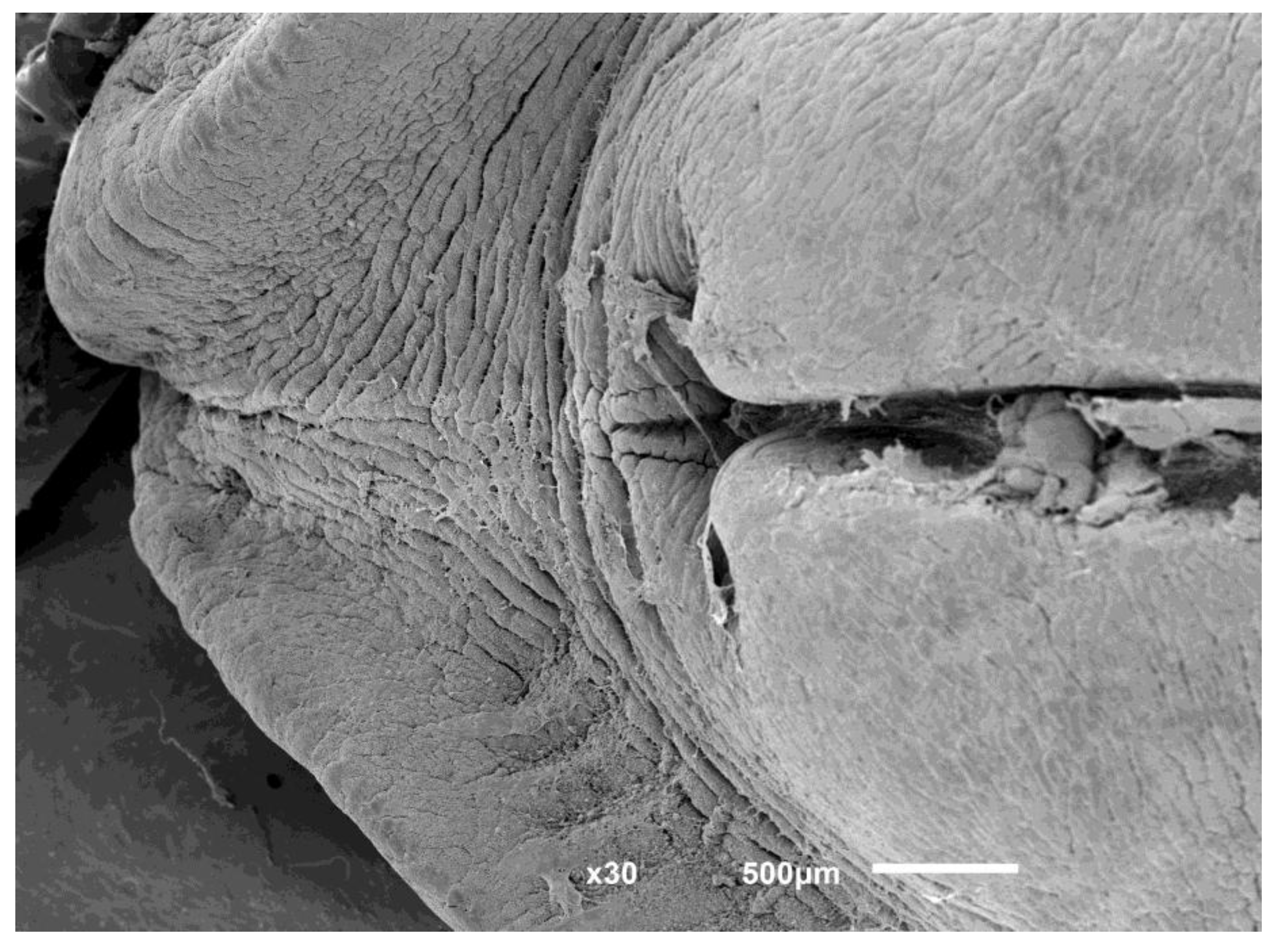
Figure 2.
Foot and byssus of Bathymodiolus azoricus. (A) The extended foot (arrow), a muscular, tongue-like organ used for locomotion and byssus deposition, protruding from between the valves. (B) Ventral view showing byssal threads (arrow) secreted by the foot’s byssal gland, anchoring the animal to the substrate. The shell displays the characteristic brown coloration of B. azoricus, an oblong outline, and concentric ring-like growth lines; irregular marks on the shell surface correspond to sites of former byssal-plaque adhesion from neighboring mussels. Scale bars, 1 cm.
Figure 2.
Foot and byssus of Bathymodiolus azoricus. (A) The extended foot (arrow), a muscular, tongue-like organ used for locomotion and byssus deposition, protruding from between the valves. (B) Ventral view showing byssal threads (arrow) secreted by the foot’s byssal gland, anchoring the animal to the substrate. The shell displays the characteristic brown coloration of B. azoricus, an oblong outline, and concentric ring-like growth lines; irregular marks on the shell surface correspond to sites of former byssal-plaque adhesion from neighboring mussels. Scale bars, 1 cm.
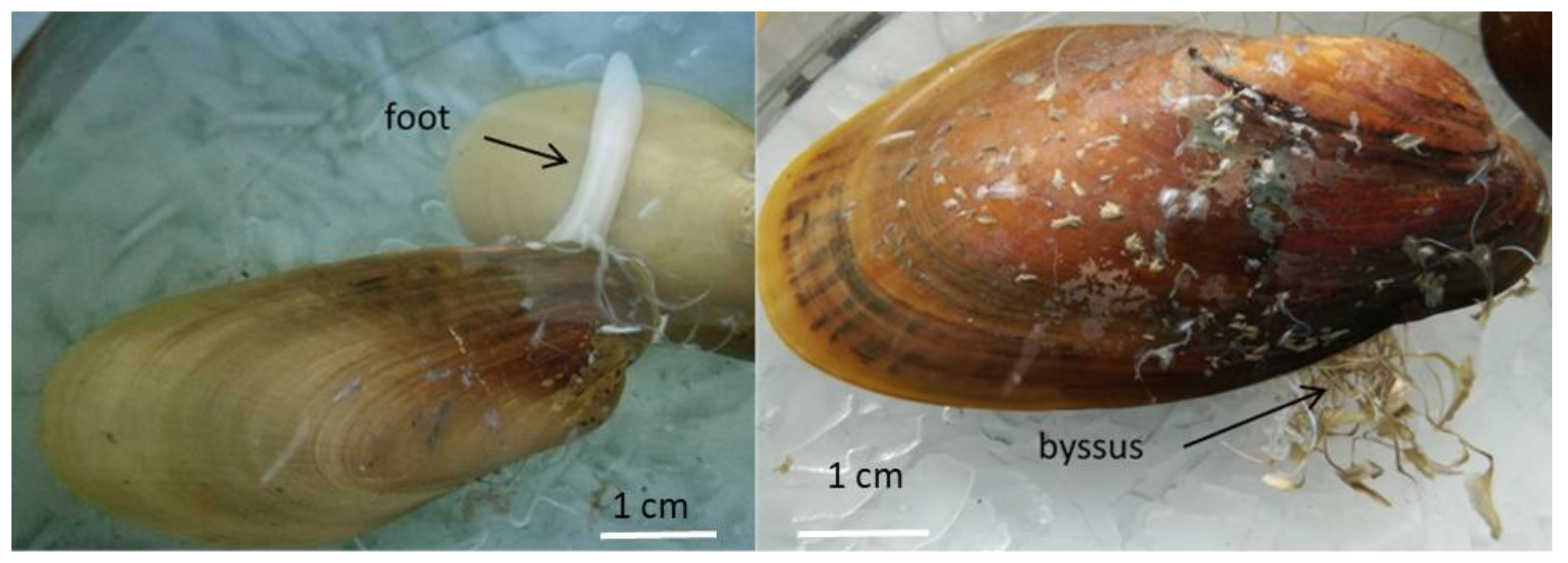
Figure 3.
Phenoloxidase-mediated wound response following mechanical shell injury in Bathymodiolus azoricus. (A) A mechanical hole drilled through the shell without disrupting the underlying mantle tissue. (B) A prophenoloxidase reaction occurring around the wound site, characterized by the formation and deposition of melanized proteins as a result of phenoloxidase activity — a response typical of injuries observed in crustaceans and insects. (C) Higher-magnification view of the darkened deposition around the wound, showing melanin and quinone accumulation. Scale bars, 5 mm (A, C), 10 mm (B).
Figure 3.
Phenoloxidase-mediated wound response following mechanical shell injury in Bathymodiolus azoricus. (A) A mechanical hole drilled through the shell without disrupting the underlying mantle tissue. (B) A prophenoloxidase reaction occurring around the wound site, characterized by the formation and deposition of melanized proteins as a result of phenoloxidase activity — a response typical of injuries observed in crustaceans and insects. (C) Higher-magnification view of the darkened deposition around the wound, showing melanin and quinone accumulation. Scale bars, 5 mm (A, C), 10 mm (B).
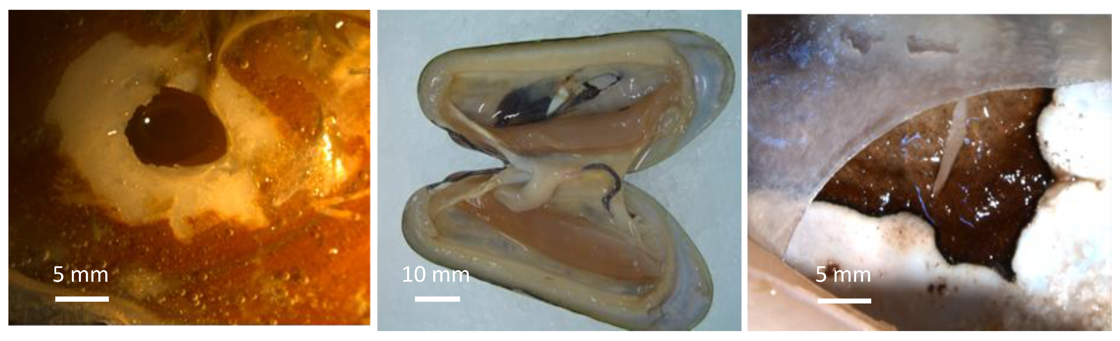
Figure 4.
Pairwise alignment of the Bathymodiolus azoricus foot-tissue transcript encoding a mussel foot protein-2 (Mfp-2)–like adhesive protein against Mytilus californianus foot protein 2 (GenBank AST36139.1). The alignment spans 231 residues (57% identity, 65% positives; E = 2×10−65) and highlights in bold red the tandem cysteine-rich “…KCKCVGGY…” repeat units diagnostic of the EGF-like domain architecture characteristic of mussel byssal plaque proteins. This alignment was originally generated during the 2017–2019 foot-tissue transcriptome analysis carried out under the 3B’s Research Group (University of Minho, supervised by Dr Tiago Silva). The same transcript was independently recovered in a 2025/2026 re-analysis of the foot transcriptome Trinity assembly, using an updated annotation pipeline with a targeted DIAMOND blastx search against a curated Mfp reference panel: TRINITY_DN360524_c2_g1_i1 was identified as a near-exact match (59.3% identity to UniProt Q25464/FP2_MYTGA) to the sequence shown here, with TRINITY_DN351523_c2_g1_i1 as a closely related secondary hit. The convergence of two independent analyses, conducted nearly a decade apart with different databases and tools, on the same Mfp-2 homolog corroborates the original 2017 identification of an EGF-repeat-containing adhesive protein in B. azoricus foot tissue.
Figure 4.
Pairwise alignment of the Bathymodiolus azoricus foot-tissue transcript encoding a mussel foot protein-2 (Mfp-2)–like adhesive protein against Mytilus californianus foot protein 2 (GenBank AST36139.1). The alignment spans 231 residues (57% identity, 65% positives; E = 2×10−65) and highlights in bold red the tandem cysteine-rich “…KCKCVGGY…” repeat units diagnostic of the EGF-like domain architecture characteristic of mussel byssal plaque proteins. This alignment was originally generated during the 2017–2019 foot-tissue transcriptome analysis carried out under the 3B’s Research Group (University of Minho, supervised by Dr Tiago Silva). The same transcript was independently recovered in a 2025/2026 re-analysis of the foot transcriptome Trinity assembly, using an updated annotation pipeline with a targeted DIAMOND blastx search against a curated Mfp reference panel: TRINITY_DN360524_c2_g1_i1 was identified as a near-exact match (59.3% identity to UniProt Q25464/FP2_MYTGA) to the sequence shown here, with TRINITY_DN351523_c2_g1_i1 as a closely related secondary hit. The convergence of two independent analyses, conducted nearly a decade apart with different databases and tools, on the same Mfp-2 homolog corroborates the original 2017 identification of an EGF-repeat-containing adhesive protein in B. azoricus foot tissue.
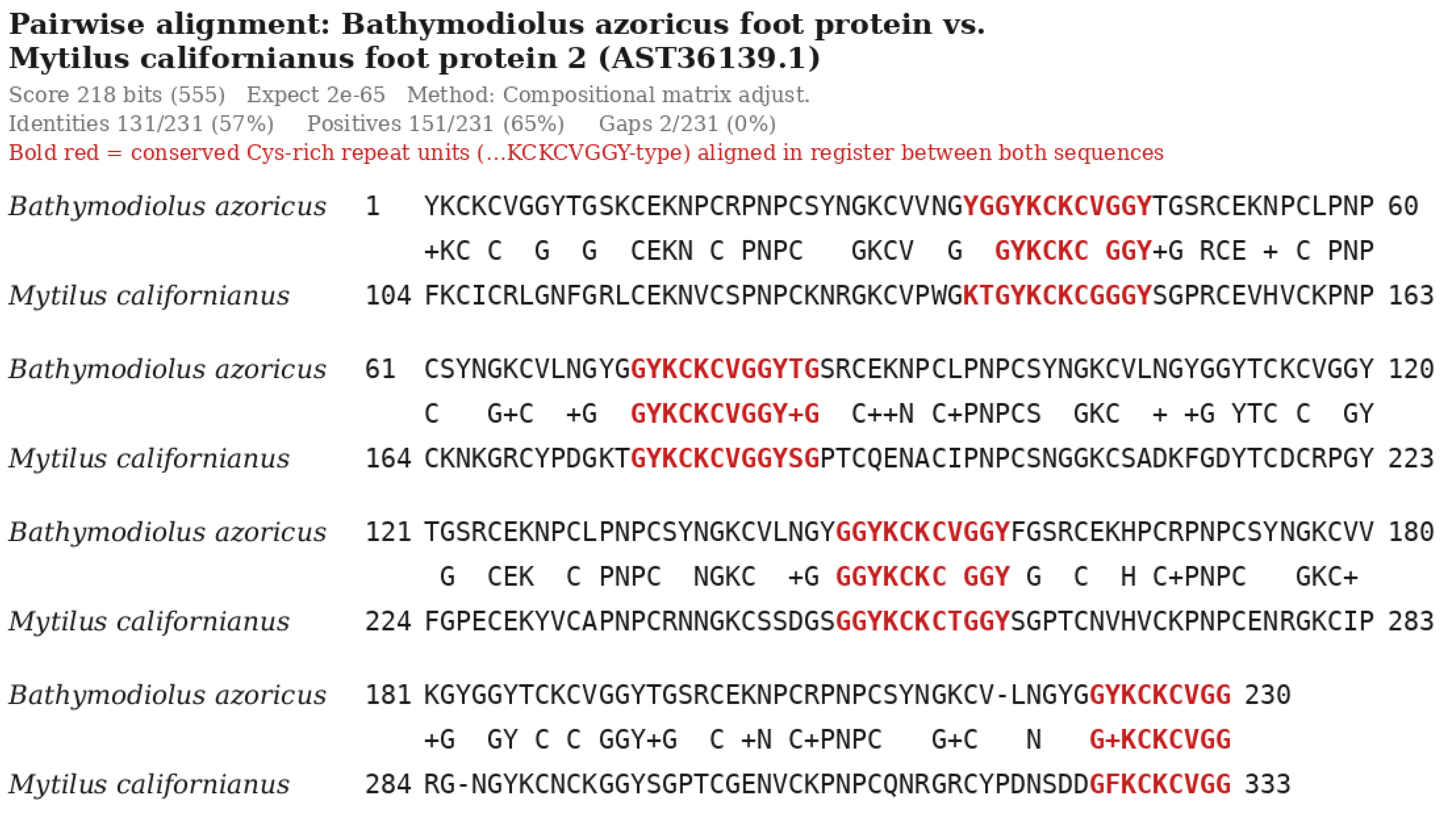

Table 1.
Candidate wound-repair- and immunity-associated genes identified by keyword and domain search of the reanalyzed B. azoricus gill transcriptome (SRR067874; see Box 1 for methodology). Contig/protein identifiers refer to the reanalysis assembly and database described here, not to the discontinued DeepSeaVent portal’s identifiers, which are no longer resolvable. Weak or negative results (VEGF, IGF, HGF, FGF ligand, proteoglycan core protein, collagen type IV specifically) are reported rather than omitted, consistent with not overstating what the data show; false-positive candidates identified during keyword screening (e.g., substring matches such as “MMPL” for “MMP”) were manually excluded. This table now also reports results from an independent reanalysis of a second, 2024 Illumina-sequenced gill transcriptome from the same species/tissue, alongside the original 2010 (454) reanalysis, for direct comparison.
Table 1.
Candidate wound-repair- and immunity-associated genes identified by keyword and domain search of the reanalyzed B. azoricus gill transcriptome (SRR067874; see Box 1 for methodology). Contig/protein identifiers refer to the reanalysis assembly and database described here, not to the discontinued DeepSeaVent portal’s identifiers, which are no longer resolvable. Weak or negative results (VEGF, IGF, HGF, FGF ligand, proteoglycan core protein, collagen type IV specifically) are reported rather than omitted, consistent with not overstating what the data show; false-positive candidates identified during keyword screening (e.g., substring matches such as “MMPL” for “MMP”) were manually excluded. This table now also reports results from an independent reanalysis of a second, 2024 Illumina-sequenced gill transcriptome from the same species/tissue, alongside the original 2010 (454) reanalysis, for direct comparison.
| Gene / family | 2010 reanalysis (SRR067874) | 2024 Illumina reanalysis | Confidence / notes |
|---|---|---|---|
| Integrin | 6 proteins, incl. c6448 (integrin alpha), c2540, c22446, c11545, c3135, c4697 | 32 transcripts | strong in both; the deeper 2024 assembly recovers substantially more paralogs |
| Tissue inhibitor of metalloproteinase (TIMP) | 7 proteins: TIMP1 (c1670), TIMP3 (c607), TIMP4 (c2320, c529), c1316, c2667, c2355 | 19 proteins (Pfam TIMP domain, e≤1e-5) | strong in both |
| EGF (epidermal growth factor) | 29 proteins bearing EGF-like / vWF-D+EGF domains (broad structural-domain sweep) | 3 proteins (tightened to true ligand-type EGF domains only: HBEGF_C / EGF_TGFA-like / EGF_Epiregulin) | NOT directly comparable: the 2010 count reflects a broad domain sweep, the 2024 count a stricter ligand-specific search; both are internally valid but methodologically distinct |
| Matrix metalloproteinase (MMP) | c1756 = MMP3/stromelysin | 11 transcripts | 2010: single confirmed hit (c1327 excluded as a false positive, a bacterial MMPL transporter); the deeper 2024 assembly recovers a substantially larger MMP family |
| Transforming growth factor beta (TGF-β) | 7 proteins, incl. c7174 (TGF-β propeptide/ligand domain) | 9 proteins (Pfam TGF_beta / TGFb_propeptide, e≤1e-5) | strong in both |
| Fibroblast growth factor (FGF) | c7365 = FGFR4 | 3 proteins (Pfam FGF ligand domain, e≤1e-5) | 2010: this is the FGF receptor, not an FGF ligand — flagged as a near-miss; 2024 resolves this with confirmed ligand-domain hits |
| Cadherin | 7 proteins: c1449, c21305, c23457, c3332, c1160, c5501, c18246 | 74 transcripts | strong in both; substantially larger recovered family in 2024 |
| Collagen (type IV) / Proteoglycan | Collagen: 68 hits across multiple types (I, VI, XIV, fibrillar), not narrowed to type IV. Proteoglycan: only 3 weak hits (HS 3-O-sulfotransferase, syndecan-binding protein, chondroitinase-related); no core proteoglycan protein identified. | Collagen type IV: 3 transcripts, alpha-1(IV) and alpha-2(IV) chains specifically confirmed. Proteoglycan: 3 transcripts, a direct hit to a basement-membrane heparan sulfate proteoglycan core protein. | 2024 resolves both open gaps flagged in the 2010 reanalysis |
| Cathepsin | 13 proteins: Cathepsin B (c79), C (c1161), L1 (c886, c9511), S (c3467), Z (c1043, c3817, c7375, c85) | 23 proteins (Pfam, e≤1e-5) | strong in both |
| Tumor necrosis factor (TNF) superfamily | 78 proteins, predominantly C1q-TNF-related proteins (C1QTNF family) and LITAF | 28 proteins (Pfam TNF / TNFSF4_N, e≤1e-5) | neither represents classical mammalian TNF-α; both are dominated by the bivalve-expanded C1q-TNF-related gene family. The lower 2024 count reflects stricter Pfam e-value filtering, not fewer true hits |
| Coagulation factor / Plasminogen / Fibrinogen | Coagulation factor: c3251 (Factor 5/8 type C domain). Plasminogen: 3 proteins, Kringle 4-domain-containing (c190, c698, c5817). Fibrinogen: 35 proteins with fibrinogen C-terminal (FReD) domains. | Coagulation factor: 15 transcripts. Plasminogen: 14 transcripts. Fibrinogen: 25 transcripts. | 2010 coagulation factor was a single confirmed hit (c2564 excluded as a false positive, “Translin-associated factor X”); 2024 substantially expands the recovered coagulation-factor family. Plasminogen and fibrinogen are broadly consistent between datasets |
| VEGF / IGF / HGF | VEGF: 3 weak hits (GO annotation “VEGFR-2 signaling pathway” only, no VEGF/VEGFR protein itself). IGF: 2 weak hits (Somatomedin-B domain within larger astacin-metalloprotease-like proteins). HGF: 0 hits under any synonym tested. | VEGF: 0. IGF: 0. HGF: 0 (one candidate BLAST hit was a bacterial false positive, excluded). | a consistent, honest negative across both independent datasets — no true ligand for any of the three confirmed in either reanalysis |
| Sialic acid-binding lectin (Siglec) | 5 proteins, incl. confirmed SIGLEC1 (c3185, c1725) | 0 (no Siglec/sialoadhesin hits; one weak Pfam hit was an unrelated enzyme, excluded) | 2010 finding not reproduced in 2024 — reported as an open discrepancy rather than omitted |
| Allograft inflammatory factor 1 (AIF1) | c3500, explicitly annotated “allograft inflammatory factor 1” with paired EF-hand domains | 71 proteins (Pfam, e≤1e-5) | strong in both; substantially larger recovered family in 2024 |
| Endothelial PAS domain protein 1 (EPAS1/HIF-2α) | weak/indirect — 1 hit (c7199) annotated “HIF-1 alpha inhibitor-related” (HIF1AN/FIH-1), not EPAS1 itself — a near-miss, not a confirmed hit | 1 direct hit: TRINITY_DN117996_c0_g1_i1, 32.7% identity, e=1.89e-08 — a genuine EPAS1 ortholog | 2024 resolves the 2010 near-miss with a direct, confirmed hit |
Table 2.
Comparison of the originally published, RT-PCR/qPCR-validated 2010 gene panel (Bettencourt et al. 2010) against the independent reanalyses of the same and a newer raw sequencing dataset (Table 1). Only three of the twenty targets surveyed in Table 1 were part of the original published, RT-PCR-validated panel; the remainder are newly identified candidates emerging from keyword/domain search of the reanalyzed data, not re-confirmations of prior validated findings.
Table 2.
Comparison of the originally published, RT-PCR/qPCR-validated 2010 gene panel (Bettencourt et al. 2010) against the independent reanalyses of the same and a newer raw sequencing dataset (Table 1). Only three of the twenty targets surveyed in Table 1 were part of the original published, RT-PCR-validated panel; the remainder are newly identified candidates emerging from keyword/domain search of the reanalyzed data, not re-confirmations of prior validated findings.
| Gene | Originally published & RT-PCR-validated (2010) | Found in 2010 reanalysis? | Found in 2024 reanalysis? | Notes |
|---|---|---|---|---|
| Integrin | Yes — confirmed elevated qPCR expression (main text, Figure 5 discussion) | Yes, 6 proteins | Yes, 32 transcripts | consistent across all three |
| EGF | Yes — contig c3243, part of the core 16-gene validated panel (Signaling category) | Yes, 29 proteins (broad domain sweep) | Yes, 3 proteins (tightened to true ligand domains) | consistent across all three; see Table 1 note on 2010 vs 2024 methodology |
| Aggrecan (proteoglycan) | Yes — contig lrc83347, RT-PCR-confirmed (Recognition category) | NOT reproduced — only 3 weak related-enzyme hits, no core proteoglycan protein | Yes, 3 transcripts — direct hit to a core heparan sulfate proteoglycan protein | apparent regression in the 2010 reanalysis, most likely explained by its much shallower assembly (26,363 contigs vs. the original paper’s 75,407 contigs + 3,071 singletons); resolved by the deeper 2024 dataset |
Table 3.
Best candidate transcripts identified for the three characterized B. azoricus foot-tissue preCol proteins (see Box 2 for search methodology). Restricted to the single strongest candidate per reference protein out of 156 total candidate transcripts passing the confidence filter; the full candidate set is not shown here. Architecture notes reflect direct visual inspection of the translated sequence for Gly-X-Y repeat content and flanking domain composition, not formal Pfam/InterPro domain annotation, which has not yet been run on these candidates.
Table 3.
Best candidate transcripts identified for the three characterized B. azoricus foot-tissue preCol proteins (see Box 2 for search methodology). Restricted to the single strongest candidate per reference protein out of 156 total candidate transcripts passing the confidence filter; the full candidate set is not shown here. Architecture notes reflect direct visual inspection of the translated sequence for Gly-X-Y repeat content and flanking domain composition, not formal Pfam/InterPro domain annotation, which has not yet been run on these candidates.
| Reference protein | Best candidate transcript | Length (aa) | Identity | E-value | Architecture note |
|---|---|---|---|---|---|
| preCol-D (O44367) | TRINITY_DN381679_c3_g3_i2 | 634 | 42.3% | 1.35×10−70 | Near-continuous Gly-X-Y repeat; short C-terminal tail |
| preCol-NG (A9QLN3) | TRINITY_DN373312_c1_g1_i5 | 652 | 39.3% | 3.05×10−70 | Near-continuous Gly-X-Y repeat; cysteine-paired C-terminal domain |
| preCol-P (O16161) | TRINITY_DN376395_c3_g3_i5 | 905 | 51.3% | 2.29×10−134 | Tripartite: Ser/Ala/Gly/His-rich flanks around central Gly-X-Y collagen core |
Disclaimer/Publisher’s Note: The statements, opinions and data contained in all publications are solely those of the individual author(s) and contributor(s) and not of MDPI and/or the editor(s). MDPI and/or the editor(s) disclaim responsibility for any injury to people or property resulting from any ideas, methods, instructions or products referred to in the content. |
© 2026 by the authors. Licensee MDPI, Basel, Switzerland. This article is an open access article distributed under the terms and conditions of the Creative Commons Attribution (CC BY) license (http://creativecommons.org/licenses/by/4.0/).
Copyright: This open access article is published under a Creative Commons CC BY 4.0 license, which permit the free download, distribution, and reuse, provided that the author and preprint are cited in any reuse.