

Submitted:
25 August 2026
Posted:
26 August 2026
You are already at the latest version
Abstract
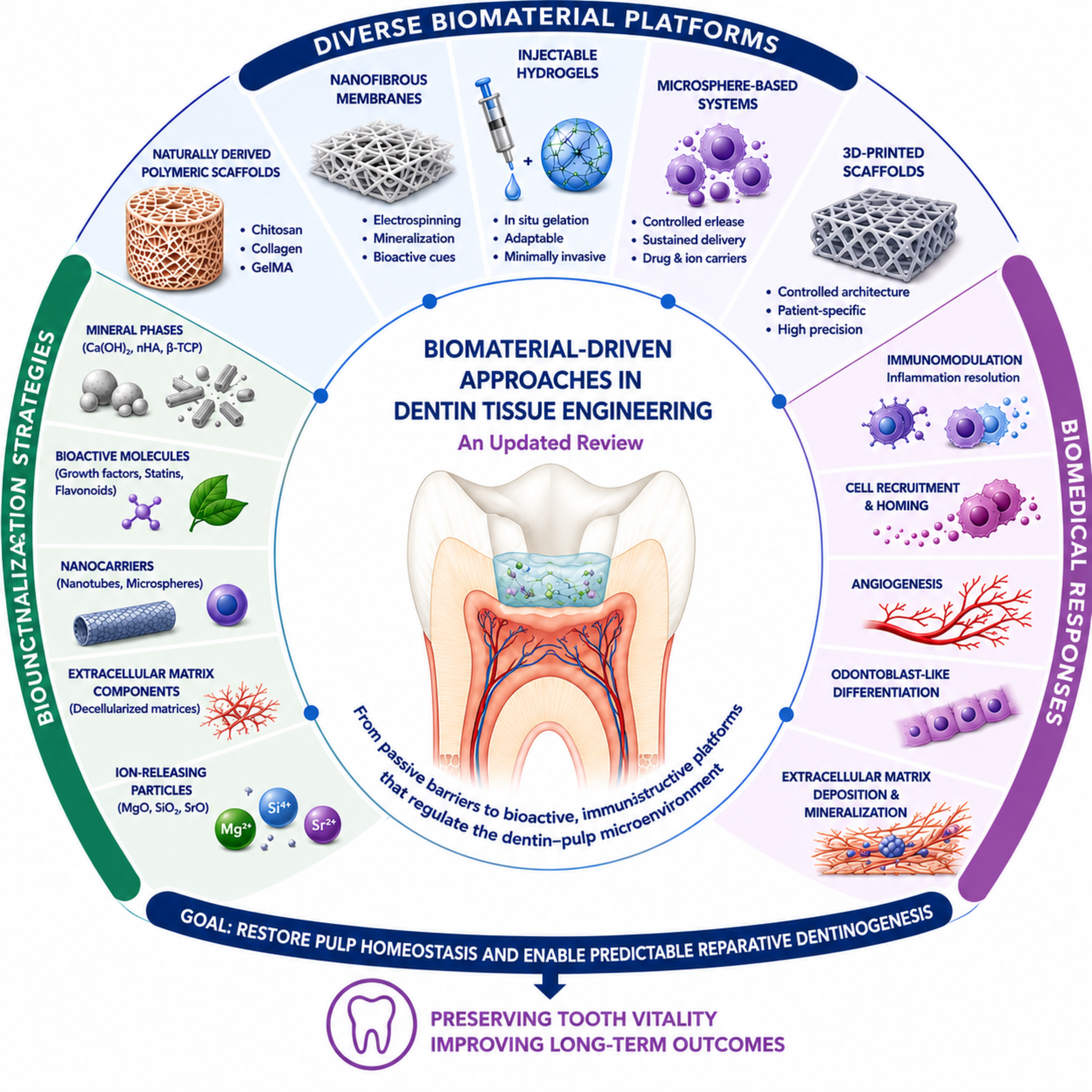
The contemporary management of dentin injuries has progressively shifted from approaches based solely on reparative mineralized barrier formation toward biologically guided strategies that preserve pulp vitality and promote endogenous regeneration. This paradigm change has driven the development of bioactive biomaterials capable of modulating the dentin–pulp microenvironment rather than serving exclusively as passive sealing materials. This review critically discusses recent advances in biomaterial-based approaches for dentin tissue engineering, emphasizing naturally derived polymeric scaffolds, nanofibrous membranes, injectable hydrogels, microsphere-based delivery systems, and three-dimensional (3D)-printed scaffolds. The biological mechanisms by which these platforms regulate inflammation, recruit endogenous progenitor cells, stimulate angiogenesis, direct odontoblast-like differentiation, and promote extracellular matrix deposition are highlighted. Particular attention is given to the sequential development of mineral-functionalized nanofibrous scaffolds, from the selection of calcium hydroxide and nano-hydroxyapatite as bioactive phases to the incorporation of fibronectin and, subsequently, the flavonoids quercetin and hesperetin to combine odontogenic signaling with immunomodulation. Emerging concepts, including immunoinstructive biomaterials, cell-homing strategies, smart hydrogels, and biofabrication technologies, are discussed in the context of their translational potential for vital pulp therapy. Rather than focusing exclusively on recreating the native tubular architecture of dentin, current regenerative strategies increasingly seek to restore pulp homeostasis and establish biological conditions that enable predictable reparative dentinogenesis. Collectively, these advances position biomaterials as active regulators of tissue regeneration and provide a framework for next-generation therapies aimed at preserving pulp vitality and improving the long-term outcomes of dentin regeneration.
Keywords:
dentin
; dental pulp
; tissue engineering
; regenerative endodontics
; dental materials
; tissue scaffolds
; hydrogels
; nanofibers
; microspheres
1. Introduction
The dentin–pulp complex is a structurally and functionally integrated biological unit that preserves tooth vitality, mechanical integrity, sensory function, and defense throughout life. Dentin is the major mineralized component of the tooth and displays a highly organized hierarchical architecture in which apatite nanocrystals, collagen fibrils, non-collagenous proteins, and dentinal tubules collectively determine its mechanical and biological behavior. Beyond acting as a passive barrier, dentin constitutes a dynamic extracellular matrix produced and maintained by odontoblasts. Its organic matrix sequesters growth factors, cytokines, and signaling molecules that can be released by demineralization or tissue injury and regulate cell recruitment, angiogenesis, odontoblastic differentiation, matrix deposition, and mineralization. Dentin therefore functions as both a load-bearing tissue and an instructive biological reservoir capable of directing endogenous repair responses (Smith et al., 2012; Cooper et al., 2014; Bertassoni, 2017; Soares et al., 2021).
The biological activity of dentin is inseparable from the maintenance of a viable pulp. Odontoblast cell bodies are positioned at the dentin–pulp interface, while their processes extend into the dentinal tubules, creating a structural and signaling continuum between both tissues. Through this interface, chemical, mechanical, thermal, and microbial stimuli reaching dentin are sensed and transmitted to the pulp, which coordinates vascular, neural, immune, and reparative responses. The pulp, in turn, provides the progenitor cells, vascular support, innervation, and molecular mediators required for dentin formation and homeostasis. Mild injury may stimulate surviving odontoblasts to deposit reactionary dentin, whereas more severe damage and odontoblast death require the recruitment and differentiation of pulp-derived progenitor cells into odontoblast-like cells responsible for reparative dentinogenesis. Nevertheless, the resulting mineralized tissue is commonly irregular and does not fully reproduce the tubular architecture, composition, or functionality of native dentin (de Souza Costa et al., 2014; Cooper et al., 2014; Soares et al., 2021).
Historically, the management of pulp exposure was guided by the assumption that inflamed pulp tissue had limited healing potential. Depending on the presumed severity of inflammation, treatment therefore progressed from direct pulp capping or pulpotomy to complete pulp removal and conventional root canal treatment. Although these interventions can preserve the tooth and restore clinical function, they predominantly induce repair or replace the pulp with an inert filling material rather than re-establishing the biological functions of the native dentin–pulp complex (Soares et al., 2021).
Advances in pulp biology have substantially changed this paradigm. The dental pulp is now recognized as an immunocompetent and regenerative tissue in which odontoblasts, fibroblasts, immune cells, endothelial cells, neural components, and mesenchymal stem/progenitor cells interact dynamically after injury. Odontoblasts act as sentinel cells that detect microbial signals and initiate innate immune responses, while pulp stromal and immune cells regulate the balance between inflammation, tissue protection, and repair. Pulp-derived mesenchymal stromal cells may also contribute to host defense through the secretion of antimicrobial and immunomodulatory factors (Le Fournis et al., 2020; Bergmann et al., 2020; Le Fournis et al., 2021; Soares et al., 2021; Ravenscroft et al., 2022; Le Fournis et al., 2026). These findings indicate that successful dentin regeneration does not depend exclusively on the presence of stem cells or on complete suppression of inflammation. Instead, it requires a controlled microenvironment in which microbial challenge is contained, destructive inflammation is resolved, and the viability and regenerative competence of resident cells are maintained.
This improved understanding of pulp immunobiology has also exposed important limitations in current clinical diagnosis. The categories of reversible and irreversible pulpitis are determined mainly from symptoms, sensibility tests, and radiographic findings, which provide only indirect information about the histological condition and spatial distribution of inflammation. Therefore, a clinical diagnosis of irreversible pulpitis does not necessarily indicate uniform and irreversible damage throughout the pulp. Inflammation may be concentrated in the coronal tissue, while deeper pulp compartments retain viable vessels, progenitor cells, fibroblasts, and immune-regulatory mechanisms capable of supporting healing and continued dentinogenesis (Vazavandi et al., 2022; Loo et al., 2025; Torabinejad et al., 2026). Emerging molecular studies have identified candidate biomarkers associated with pulp inflammation and potential therapeutic targets, but clinically applicable methods capable of accurately determining tissue viability and regenerative competence remain limited (Le Fournis et al., 2026).
The discrepancy between clinical diagnosis and actual tissue status has expanded interest in biologically based treatments that preserve the greatest possible amount of competent pulp. Conservative management and vital pulp therapies have demonstrated that mature permanent teeth with carious pulp exposure, including selected cases clinically diagnosed as irreversible pulpitis, may retain healing potential when infected tissue is appropriately removed and the remaining pulp is adequately protected. Evidence for regenerative endodontic procedures in these teeth is still limited and does not yet establish clinical predictability; however, the available findings challenge the assumption that all clinically inflamed pulps are beyond recovery. They also support a transition from treatment decisions based exclusively on diagnostic labels toward strategies informed by residual tissue viability, inflammatory status, and regenerative potential (Taha et al., 2020; Torabinejad et al., 2026).
Collectively, these biological and clinical advances have shifted the therapeutic paradigm from attempting to regenerate native dentin toward preserving or restoring a biologically competent pulp microenvironment capable of supporting organized tissue repair. Although complete recapitulation of primary dentin architecture represents the ideal developmental outcome, perfectly organized dentinal tubules are not a prerequisite for pulp vitality or long-term clinical success. The critical determinant of successful therapy is instead an immunologically balanced, biologically functional pulp capable of resolving inflammation, preserving vascularity, and depositing a continuous mineralized barrier that seals and protects the remaining tissue. Achieving this outcome requires effective control of infection and inflammation, preservation of the endogenous stem-cell niche, recruitment of progenitor cells, vascular support, odontoblast-like cell differentiation, coordinated extracellular matrix deposition, and spatially regulated mineralization. Because native healing mechanisms rarely orchestrate all these biological events in a predictable manner, particularly following extensive tissue injury, advanced biomaterials have become essential for actively directing the pulp microenvironment toward a regenerative rather than a chronic inflammatory state (Soares et al., 2021).
Accordingly, biomaterials for dentin tissue engineering are evolving from passive sealing or filling materials into bioactive and immunoinstructive platforms designed to reproduce essential functions of the native extracellular matrix. By integrating structural support with biochemical, mechanical, and immunomodulatory signals, these systems may exploit dentin-derived bioactivity, regulate inflammatory responses, promote endogenous cell recruitment, and direct the formation of more organized mineralized tissue. This review therefore critically examines the principal biomaterial platforms, bioactive signaling strategies, cell-homing approaches, and biofabrication technologies being developed for dentin tissue engineering.
2. Current Clinical Management of Pulp Exposure: Concepts and Biological Limitations
The clinical management of pulp exposure has progressively shifted toward preservation of the greatest possible amount of biologically competent pulp tissue. This approach is consistent with minimally invasive dentistry and with the contemporary understanding that pulp vitality is essential for maintaining sensory function, immune surveillance, vascular supply, and continued dentin deposition. Accordingly, vital pulp therapy (VPT) has become the primary treatment strategy for exposed pulps when infection can be controlled and a viable tissue compartment remains capable of healing (Chien et al., 2026; Schwendicke et al., 2026).
Vital pulp therapy (VPT) includes direct pulp capping, partial pulpotomy, and full pulpotomy, selected according to the origin and extent of the exposure, the inflammatory status of the pulp, aseptic control, and the ability to obtain hemostasis and an effective coronal seal. Direct pulp capping may be considered for selected mechanical, traumatic, or carious exposures when a favorable pulp condition can be maintained, whereas partial and full pulpotomy permit removal of the most inflamed coronal tissue while preserving deeper pulp compartments with greater biological competence (Taha et al., 2020; Chien et al., 2026; Schwendicke et al., 2026). The therapeutic objective is therefore to control infection, remove irreversibly damaged tissue, preserve pulp vitality, and stimulate formation of a mineralized barrier capable of protecting the remaining tissue.
Calcium hydroxide was historically regarded as the reference material for pulp capping because of its alkaline pH, antibacterial activity, and ability to induce mineralized tissue formation. However, its mechanism depends on the development of a superficial zone of chemical necrosis, followed by inflammation, recruitment of progenitor cells, and deposition of reparative dentin. Thus, tissue repair is initiated only after a new material-induced injury. Moreover, the resulting mineralized barrier may exhibit porosity, tunnel defects, heterogeneous thickness, and incomplete adaptation, which can compromise its protective function and facilitate bacterial reinfiltration (Goldberg et al., 2008; Sangwan et al., 2013). Figure 1 recapitulates this mechanism.
Hydraulic calcium silicate cements, particularly mineral trioxide aggregate and Biodentine®, have improved the clinical predictability of VPT because of their sealing ability, biocompatibility, sustained calcium-ion release, and capacity to induce mineralized barrier formation. These materials are generally associated with more consistent tissue responses and higher long-term success rates than calcium hydroxide (Peskersoy et al., 2021; Cushley et al., 2021). Their biological activity is strongly influenced by hydration, alkalinization, ion release, and interactions with the dentin–pulp interface. Although they can promote favorable repair, they were not primarily designed to provide direct and independently tunable control of immune resolution, cell recruitment, vascular support, and matrix organization.
Current VPT should therefore be understood as a clinically effective but biologically limited approach. Its success is primarily based on bacterial control, tissue preservation, sealing, and stimulation of reparative dentinogenesis. The main limitation of current pulp-capping materials is therefore not their inability to recreate the exact tubular architecture of native dentin, since complete structural replication of dentinal tubules is not necessarily required to restore protection and function. The critical issue is that conventional materials provide limited control over the inflammatory response and biological modulation of repair and regeneration within the remaining pulp. They stimulate mineralized tissue deposition but do not actively regulate the transition from antimicrobial defense and inflammation toward resolution, vascular stabilization, progenitor-cell activity, and organized matrix deposition. As a result, healing remains highly dependent on the pre-existing biological condition of the pulp and on the ability of the tissue to autonomously resolve inflammation.
Accordingly, the next generation of biomaterials for dentin engineering should move beyond conventional sealing and nonspecific calcium-driven mineralization. Bioactive and immunoinstructive platforms are needed to establish a more favorable balance between inflammation and regeneration, thereby enabling the remaining pulp tissue to coordinate a more predictable and biologically organized repair response (Figure 2).
3. Biomaterials for Dentin Tissue Engineering
Dentin tissue engineering emphasizes regeneration of a mineralized extracellular matrix capable of restoring structural integrity and protecting the dentin–pulp complex; however, this objective remains biologically inseparable from preservation of a viable, vascularized, and innervated pulp. Accordingly, biomaterials must create a temporary microenvironment that supports resident cell survival and recruitment, directs dental pulp stem/progenitor cells toward an odontoblast-like phenotype, and stimulates organized matrix deposition and mineralization at the dentin–pulp interface.
Contemporary regenerative strategies increasingly recognize biomaterials as biologically instructive platforms capable of actively regulating cellular behavior through biochemical, mechanical, architectural, and physicochemical cues. From a translational perspective, these scaffolds can be broadly categorized into three complementary clinical approaches: (i) cut-to-size bulk scaffolds, fabricated by conventional manufacturing techniques or three-dimensional (3D) printing and adapted to the defect geometry; (ii) injectable scaffolds, delivered as precursor formulations that undergo thermally, chemically, or physically induced in situ gelation, enabling minimally invasive application and intimate adaptation to irregular dentin defects; and (iii) powdered microsphere-based scaffolds, applied locally as dry particulate systems that become hydrated and stabilized by tissue fluids, forming a conformable bioactive matrix capable of adapting to complex dentin defects while serving as a reservoir for the controlled presentation or release of bioactive molecules.
3.1. Naturally Derived Polymeric Scaffolds with Mineralized Architecture
A major paradigm shift in dentin–pulp tissue engineering occurred with the transition from conventional calcium-based pulp-capping materials to biomimetic polymeric scaffolds incorporating bioactive mineral phases. Rather than relying on the intense alkaline environment generated by hydraulic cements to induce superficial tissue injury followed by reparative dentin bridge formation, these scaffolds were conceived to establish a biologically favorable pulp microenvironment capable of supporting endogenous tissue repair. Their design was inspired by the composition of the native dentin extracellular matrix (ECM), combining naturally derived polymers with bioactive mineral components to provide structural support while actively regulating cellular behavior, extracellular matrix deposition, and mineralization (Soares et al., 2020; Soares et al., 2021).
This conceptual shift emerged from the recognition that calcium- and phosphate-containing minerals are not merely structural constituents of mineralized tissues but also potent biological regulators. When incorporated into polymeric matrices, these mineral phases modify scaffold porosity, degradation kinetics, mechanical behavior, and surface chemistry while continuously releasing therapeutic ions capable of regulating cell migration, proliferation, odontoblastic differentiation, angiogenesis, extracellular matrix synthesis, and mineral deposition. Among the earliest studies supporting this concept, Okamoto et al. (2020) demonstrated that biodegradable hydroxyapatite-containing polymeric composites promoted pulp tissue repair while simultaneously providing mechanical support for tissue organization. Beyond their role as mineral carriers, these composite scaffolds enhanced cellular organization and supported the deposition of mineralized tissue, providing early evidence that scaffold composition could directly influence biological repair processes through the combined effects of structural support and bioactive mineral signaling (Okamoto et al., 2020).
A recent scoping review summarizing more than four decades of scaffold development demonstrated that contemporary regenerative platforms consistently combine naturally derived polymers, bioactive mineral phases, pharmacological functionalization, and extracellular matrix-derived materials. Despite the diversity of materials and manufacturing approaches, the biological outcomes reported across studies remain remarkably consistent, including enhanced cell adhesion, migration, angiogenesis, odontogenic differentiation, mineralized matrix deposition, and formation of vascularized pulp-like tissues. Collectively, these findings indicate that scaffold development has progressively shifted from providing passive structural support toward actively directing endogenous regenerative processes through coordinated biochemical, physicochemical, and architectural signaling (Soares et al., 2021).
The application of naturally derived polymers further expanded this concept. Chitosan rapidly became one of the most extensively investigated biomaterials because of its excellent biocompatibility, biodegradability, antimicrobial activity, and structural similarity to native glycosaminoglycans. Unlike conventional pulp-capping materials, which directly expose pulp tissue to highly alkaline conditions, incorporation of calcium hydroxide into porous chitosan scaffolds enabled gradual calcium-ion release while minimizing abrupt changes in local pH. This biomimetic scaffold exhibited an interconnected porous architecture, controlled degradation profile, and sustained calcium release, significantly increasing alkaline phosphatase activity and the expression of odontogenic markers, including COL1A1, DSPP, and DMP1, while promoting extensive extracellular matrix mineralization by human dental pulp cells (Soares et al., 2020). These findings demonstrated that naturally derived polymers can function not merely as passive structural supports but as biologically instructive matrices capable of directing endogenous reparative responses.
As the understanding of mineral-mediated signaling advanced, numerous mineral phases were incorporated into chitosan scaffolds to optimize their regenerative potential. β-glycerophosphate, calcium aluminate, calcium silicate, nanohydroxyapatite, β-tricalcium phosphate, and biomineralized calcium phosphate generated through simulated body fluid treatment were all shown to enhance odontogenic differentiation and mineralized matrix formation while improving scaffold bioactivity (Soares et al., 2017; Cassiano et al., 2020; Soares et al., 2021; Leite et al., 2022; Bordini et al., 2024). Importantly, these studies demonstrated that the biological effects of mineralized scaffolds extended beyond cells directly contacting the biomaterial. Continuous release of calcium, phosphate, silicon, and aluminum ions generated paracrine signaling capable of influencing neighboring cell populations, reinforcing the concept that successful dentin–pulp regeneration depends primarily on coordinated modulation of the pulp microenvironment rather than direct biomaterial–cell interactions alone.
Concurrently, increasing attention has been directed toward the influence of scaffold architecture itself. It is now recognized that successful dentin–pulp regeneration depends not only on scaffold composition but also on the spatial organization of the biomaterial. Hierarchically organized porous scaffolds facilitate endogenous cell migration, vascular infiltration, nutrient diffusion, waste removal, extracellular matrix deposition, and tissue remodeling, while simultaneously providing mechanical stability during the healing process. Consequently, scaffold architecture is now regarded as an instructive biological parameter capable of directly regulating regenerative outcomes rather than merely determining mechanical performance (Wang et al., 2011).
This concept was further strengthened by the development of the mineral-induced bubbling technique, in which bioactive mineral particles simultaneously function as calcium-releasing components and pore-forming agents during chitosan scaffold fabrication. The decomposition of these particles generates highly interconnected macroporous networks with homogeneous pore distribution, significantly improving nutrient transport, fluid exchange, cellular infiltration, extracellular matrix deposition, and mineralization compared with conventional freeze-dried scaffolds. Importantly, these improvements were achieved primarily through optimization of scaffold architecture rather than modification of chemical composition, demonstrating that three-dimensional organization independently contributes to regenerative performance (Soares et al., 2020; Soares et al., 2021; de Melo et al., 2022; Gallinari et al., 2023) (Figure 3).
Although mineral ions effectively stimulate odontogenic differentiation and mineral deposition, successful repair of the dentin–pulp complex also depends on regulating inflammation, endogenous cell recruitment, angiogenesis, and extracellular matrix remodeling. This realization led to the emergence of pharmacologically functionalized scaffolds, in which naturally derived polymers evolved from structural biomaterials into localized drug-delivery platforms capable of integrating physicochemical and biochemical signaling within the same regenerative construct (Bordini et al., 2020; Cassiano et al., 2020; Soares et al., 2021).
One demonstration of this concept involved the incorporation of 1α,25-dihydroxyvitamin D3 into calcium aluminate/chitosan scaffolds. Rather than replacing calcium-mediated bioactivity, vitamin D3 acted synergistically with sustained calcium release, significantly enhancing human dental pulp cell migration, alkaline phosphatase activity, mineralized matrix deposition, and the expression of odontogenic markers, including DSPP and DMP1, compared with either stimulus alone (Bordini et al., 2020). Simvastatin subsequently emerged as another relevant small molecule for dentin–pulp regeneration because of its pleiotropic effects on angiogenesis, chemotaxis, osteogenic/odontogenic differentiation, and mineralized tissue formation. Initially incorporated into chitosan and calcium aluminate/chitosan scaffolds, low-dose simvastatin enhanced alkaline phosphatase activity and mineralized matrix deposition without compromising cytocompatibility, demonstrating that localized drug delivery could further potentiate the regenerative performance of mineralized scaffolds (Soares et al., 2018; Cassiano et al., 2020). This concept was further advanced through the development of a highly porous chitosan–calcium hydroxide (CH–Ca) scaffold combining sustained calcium release, localized simvastatin delivery, and an interconnected macroporous architecture designed to facilitate endogenous cell recruitment and infiltration. Validation in biomimetic artificial pulp chamber models and in vivo critical-size calvarial defects demonstrated enhanced cell migration, odontogenic differentiation, mineralized tissue formation, and tissue integration, supporting this multifunctional scaffold as an instructive, cell-free regenerative platform capable of coordinating host-driven repair through the interplay of scaffold architecture, ionic signaling, and localized pharmacological cues (Soares et al., 2021).
The immunomodulatory potential of simvastatin in combination with scaffolds for vital pulp therapy was previously demonstrated by Soares et al. (2018b), on LPS-induced pro-inflammatory phenotype on dental pulp cells in vitro, with simvastatin playing an important role on cell recovery and pulp regeneration in vitro and in vivo. More recently, Gallinari et al. (2026) extended this concept to regenerative conditions challenged by inflammation, demonstrating that simvastatin-functionalized CH–Ca scaffolds could modulate the response of bone cells exposed to TNF-α in vitro, attenuating the upregulation of inflammatory mediators while restoring osteogenic gene expression suppressed by the inflammatory stimulus. These findings were further supported in a rat calvarial model of TNF-α-induced osteolytic injury, in which simvastatin-loaded CH–Ca scaffolds enhanced bone regeneration and accelerated defect healing. Collectively, these studies demonstrate the progressive evolution of mineralized chitosan scaffolds from passive structural matrices toward multifunctional instructive platforms, in which ionic and pharmacological signaling can enhance regenerative and immunomodulatory responses while preserving a cell-free strategy that harnesses endogenous cells for tissue repair.
The concept of pharmacological functionalization was expanded beyond small molecules through the incorporation of bioactive proteins. Divband et al. (2022) developed chitosan-based hydrogels capable of sustained release of basic fibroblast growth factor (bFGF), demonstrating significant enhancement of dental pulp stem cell survival together with increased expression of angiogenic markers including VEGFR-2, Tie-2, and Angiopoietin-1. These findings broadened the biological scope of naturally derived polymeric scaffolds by demonstrating that successful regeneration depends not only on odontoblastic differentiation but also on the establishment of an adequately vascularized pulp microenvironment capable of sustaining long-term tissue viability (Divband et al., 2022).
These studies established naturally derived polymeric scaffolds as the first generation of multifunctional biologically instructive biomaterials for dentin–pulp tissue engineering. Their major contribution was not simply the induction of mineral deposition, but the demonstration that scaffold composition, hierarchical architecture, controlled ionic release, localized delivery of pharmacological molecules and growth factors, and tissue-specific extracellular matrix signals can be rationally integrated to regulate endogenous cell recruitment, immunomodulation, angiogenesis, odontoblast-like cell differentiation, and organized mineralized barrier formation.
3.2. Nanofibrous Biomembranes and Mineralized Extracellular Matrix Mimicry
The extracellular matrix (ECM) of dentin possesses a highly hierarchical organization in which nanoscale type I collagen fibrils form the organic framework that directs apatite nucleation, crystal growth, and mineralized tissue organization. Beyond providing structural support, this fibrillar architecture establishes a complex biological microenvironment that regulates odontoblast polarization, cell adhesion, migration, mechanotransduction, extracellular matrix secretion, and tissue remodeling. Consequently, reproducing the nanoscale organization of the dentin ECM has become a major objective in dentin–pulp tissue engineering, giving rise to the development of nanofibrous biomembranes capable of simultaneously providing structural guidance and biological instruction. Evidence that nanoscale fibrillar environments modulate odontoblast responsiveness to Wnt5a while promoting three-dimensional cellular polarization further supports the concept that nanofiber architecture actively regulates cellular phenotype rather than simply serving as a passive scaffold (Gupte and Ma, 2012).
Electrospinning has emerged as one of the most versatile fabrication techniques for producing these biomimetic membranes because it allows control over fiber diameter, alignment, interfiber spacing, porosity, and surface chemistry. These structural characteristics directly influence protein adsorption, cell attachment, cytoskeletal organization, migration, nutrient diffusion, and lineage commitment. Importantly, electrospun nanofibers also provide a suitable platform for incorporating bioactive components, including mineral phases, extracellular matrix proteins, peptides, nanoparticles, and pharmacological molecules. As a result, electrospun scaffolds have evolved from simple structural matrices into multifunctional biomaterials capable of reproducing multiple structural and biological functions of the native ECM (Gupte and Ma, 2012).
Among the synthetic polymers investigated for dentin regeneration, polycaprolactone (PCL) has received particular attention because of its electrospinnability, mechanical stability, and slow degradation profile. However, pristine PCL is hydrophobic and provides limited biological instruction. This limitation motivated the incorporation of mineral phases capable of reproducing aspects of the inorganic component of dentin while providing localized ionic signals to pulp cells. A direct comparison of PCL nanofibers containing Ca(OH)2, nano-hydroxyapatite (nHA), or β-glycerophosphate established an important starting point for this platform. Although the tested mineral phases produced distinct physicochemical and biological responses, Ca(OH)2- and nHA-containing scaffolds showed the most favorable overall performance, supporting cell adhesion, proliferation, and mineralized matrix deposition. The Ca(OH)2 formulation also provided sustained calcium release and upregulated DSPP and DMP1 expression, indicating that the choice of mineral phase determines not only scaffold composition but also the odontogenic signaling generated at the cell–material interface (Anselmi et al., 2024).
The nHA-based pathway was subsequently investigated in a dedicated PCL nanofibrous platform. Increasing nHA concentration modified fiber topography and interfibrillar spacing while maintaining cytocompatibility. PCL scaffolds containing 1% or 2% nHA released greater amounts of calcium and phosphate, favored cell adhesion and spreading, increased alkaline phosphatase activity, and upregulated odontogenic genes. Mineralized matrix formation was concentration-dependent and was approximately ninefold higher with PCL + 2% nHA than with pristine PCL, identifying this formulation as a promising mineral substrate for cell-homing–based vital pulp therapy (Mendes Soares et al., 2022).
In parallel, the Ca(OH)2pathway was refined by combining 0.4% Ca(OH)2-containing PCL nanofibers with fibronectin (FN), an extracellular matrix protein associated with cell adhesion and migration. Ca(OH)2 and FN increased migration, spreading, and viability, with their combination intensifying these early cell–material interactions. However, FN did not independently enhance odontogenic markers or mineralized matrix formation. Ca(OH)2remained the principal odontogenic component, increasing DMP1 expression and mineralized matrix deposition irrespective of FN loading. These findings distinguish the complementary roles of the components: FN primarily supports cell recruitment and adhesion, whereas Ca(OH)2provides the mineral-related signal that drives odontogenic activity (Anselmi et al., 2022).
Together, these studies defined two complementary mineral-functionalized nanofibrous platforms: nHA reproduced a calcium- and phosphate-rich mineral substrate favoring odontogenic differentiation and mineralization, whereas Ca(OH)2/FN combined a calcium-releasing phase with an adhesive ECM cue supporting recruitment and adhesion. The next advance was to adapt these mineral-functionalized scaffolds to the inflammatory conditions expected after pulp exposure. In the Ca(OH)2-based platform, quercetin (QU) was incorporated into PCL/polyethylene oxide nanofibers containing Ca(OH)2. Under lipopolysaccharide stimulation and in a three-dimensional artificial pulp chamber model, the scaffold reduced inflammatory and oxidative responses while upregulating ALPL, OCN, and DSPP expression and preserving mineralized matrix formation. Thus, QU added an immunomodulatory function to the Ca(OH)2-mediated odontogenic signal, extending the platform from differentiation support toward regeneration under inflammatory challenge (Anselmi et al., 2025).
A similar progression was achieved for the nHA-based platform through alkaline surface hydrolysis and incorporation of hesperetin (HT). Surface-engineered PCL/nHA scaffolds displayed improved wettability, calcium release, and controlled HT delivery without compromising the fibrous architecture. The system supported migration, proliferation, odontogenic differentiation, and mineralized matrix formation while downregulating pro-inflammatory mediators and increasing pro-resolving signaling. In vivo, it also promoted favorable tissue integration, reduced inflammatory infiltration, and an early M2-skewed macrophage response. These results demonstrate how a mineral substrate can be further developed into an immunomodulatory and dentinogenic interface (Mendes Soares et al., 2026).
A further translational step was achieved by moving from homogeneous multifunctional membranes toward spatially organized bilayer nanofibrous scaffolds in which distinct therapeutic functions were assigned to each layer (Anselmi et al., 2026). A photocrosslinkable GelMA scaffold was engineered with a pulp-facing layer composed of finer fibers containing ibuprofen (IBP) and a superficial layer formed by thicker fibers incorporating amorphous magnesium phosphate (AMP). This architecture was designed to address two temporally and spatially distinct requirements of vital pulp therapy: early control of the inflammatory microenvironment at the pulp–material interface and subsequent support of mineralized tissue formation. The IBP-containing layer provided an initial burst followed by sustained drug release and suppressed IL-1α, TNF-α, and IL-6 secretion through inhibition of NF-κB activation without compromising cytocompatibility. Conversely, the AMP-containing layer continuously released Mg2+ and PO43− ions and enhanced alkaline phosphatase activity, mineral deposition, and the expression of mineralization-related genes, including COL1A1, RUNX2, and ALPL. Importantly, this functional compartmentalization was preserved under inflammatory conditions in an LPS-challenged artificial pulp chamber model: the bilayer scaffold rapidly downregulated IL1A, IL1B, and TNF, whereas prolonged culture increased ALPL, DSPP, and OCN expression, supporting a transition from inflammatory control toward odontogenic repair. In vivo subcutaneous implantation further demonstrated biocompatibility and complementary biological responses between the layers, with IBP reducing M1 macrophage polarization and AMP promoting sequential expression of RUNX2, ALP, and osteocalcin associated with ongoing mineralization (Figure 4).
Beyond the biological effects of its individual components, the bilayer design introduces an important translational concept for vital pulp therapy: spatial control of scaffold architecture may simultaneously regulate biological signaling and the physical interface between the exposed pulp and the definitive restoration. The finer, IBP-containing fibers can be positioned directly against the pulp to provide localized immunomodulation, whereas the thicker and denser AMP-containing superficial layer may provide a more compact coronal interface over the regenerative compartment. Such architectural asymmetry could potentially improve scaffold sealing and reduce the permeability of the regenerative construct, thereby limiting the diffusion of components released from overlying restorative materials toward the pulp. Although this protective barrier function requires direct experimental validation through permeability and trans-scaffold diffusion studies, it represents a clinically relevant design direction because successful VPT requires not only biological regeneration but also effective isolation of the healing pulp from the restorative environment. Thus, the bilayer scaffold represents a transition from multifunctional nanofibers toward a spatially programmed regenerative membrane in which immunomodulation, biomineralization, and restoration-oriented barrier properties can be integrated within a single construct.
Viewed sequentially, these studies describe a coherent evolution in nanofibrous scaffold design: mineral-phase screening first identified CH and nHA as promising bioactive substrates; dedicated formulations subsequently clarified their contributions to cell recruitment, odontogenic differentiation, and mineralization; pharmacological functionalization then introduced control over the inflammatory microenvironment; and, finally, bilayer architectures enabled the spatial compartmentalization of complementary therapeutic functions. This progression shifts the design paradigm from nanofibers that merely mimic the fibrillar organization of the dentin ECM toward spatially programmed, multifunctional interfaces capable of coordinating inflammation resolution, endogenous cell recruitment, odontogenic signaling, mineralized tissue formation, and potentially the physical protection required for definitive restorative treatment. Such integration may be particularly relevant for next-generation VPT, in which successful biomaterials must not only instruct resident pulp cells to regenerate but also establish a stable interface between the regenerating dentin–pulp complex and the restored tooth.
3.3. Injectable and Photocrosslinkable Hydrogels for Dentin Regeneration
Injectable hydrogels have emerged as particularly attractive scaffolds for dentin–pulp regeneration because they can be delivered in a minimally invasive manner, conform to defects with irregular geometry, and create hydrated three-dimensional environments resembling the native extracellular matrix. These characteristics are especially relevant for vital pulp therapy, in which the biomaterial must be precisely positioned over a small pulp exposure or within a confined dentin defect while maintaining intimate contact with the surrounding tissue. In contrast to preformed scaffolds, injectable systems can fill the available space without extensive shaping, whereas subsequent gelation stabilizes the material at the application site. Their high-water content, interconnected polymeric networks, tunable degradation, and capacity to incorporate cells, drugs, extracellular matrix components, mineral phases, and biological signals have positioned hydrogels as adaptable platforms for both dentin repair and more comprehensive regeneration of the dentin–pulp complex (Amani et al., 2026; Dal-Fabbro et al., 2025).
Injectability alone, however, is not sufficient to ensure clinical performance. A low-viscosity precursor facilitates delivery but may also flow away from the exposed pulp, undergo dilution by tissue fluids, or fail to maintain the geometry required for tissue protection. Injectable hydrogels must therefore undergo a controlled sol–gel transition after placement. This stabilization may be triggered by temperature, ionic interactions, enzymatic reactions, pH changes, or light exposure. Among these strategies, photopolymerization is especially relevant to restorative dentistry because it allows spatial and temporal control over scaffold formation. The hydrogel precursor can first be positioned and adapted to the defect and then rapidly crosslinked in situ using a light source, generating a stable network only after satisfactory placement. Photocrosslinking also enables modulation of stiffness, porosity, swelling, degradation, and drug-release behavior through changes in polymer concentration, photoinitiator content, irradiation parameters, and degree of functionalization (Omidian et al., 2026).
Natural hydrogels encompass a wide range of biomaterials, including gelatin, collagen, chitosan, carrageenan, alginate, hyaluronic acid, fibrin, and extracellular matrix-derived polymers. Their inherent biocompatibility, biodegradability, and biological recognition make them attractive for dentin–pulp regeneration, although limited mechanical strength, rapid degradation, and batch variability often require chemical modification, hybridization with synthetic polymers, or incorporation of reinforcing particles to improve their physicochemical performance while preserving biological functionality (Amani et al., 2026). More recently, these advances have led to the development of smart hydrogels, which dynamically adjust their swelling, degradation, permeability, stiffness, or cargo release in response to local stimuli such as pH, temperature, enzymatic activity, reactive oxygen species, or ionic composition (Omidian et al., 2026). Because the pulp microenvironment undergoes profound biochemical changes during inflammation and healing, stimulus-responsive hydrogels offer the opportunity to temporally coordinate drug release with tissue repair, more closely recapitulating the dynamic sequence of physiological regeneration than conventional constitutive delivery systems (Dal-Fabbro et al., 2025).
Collagen-based injectable matrices represent a traditional ECM-mimic biomaterial. Highly tunable oligomeric collagen hydrogels were developed to support dental tissue regeneration by providing a fibrillar matrix closely related to the principal organic component of dentin and pulp extracellular matrices. Their polymerization, fibril density, stiffness, and pore architecture can be adjusted to regulate cell behavior and tissue development (Pankajakshan et al., 2020). Collagen matrices offer strong biological familiarity and cell recognition, although their mechanical stability and handling may be lower than those of covalently crosslinked synthetic or methacrylated systems. This limitation has encouraged the development of composite hydrogels that combine collagen-derived bioactivity with more controllable crosslinking mechanisms. Fish collagen peptide–GelMA composite hydrogels exemplify this hybrid rationale. The addition of fish-derived collagen peptides to photopolymerizable GelMA was intended to enhance the biological activity of the hydrogel while maintaining its injectability and light-mediated stabilization. Such formulations may provide additional peptide signals for pulp cells and potentially reduce reliance on mammalian-derived collagen sources (Zhao et al., 2025). Nevertheless, source-dependent variability, degree of hydrolysis, peptide composition, gelation behavior, and immunological considerations must be carefully evaluated before clinical translation.
Gelatin methacryloyl (GelMA) has become one of the principal photocrosslinkable hydrogels investigated for dentin regeneration. GelMA is derived from gelatin and retains collagen-associated bioactive sequences, including RGD motifs that support integrin-mediated cell adhesion and matrix metalloproteinase-sensitive sites that enable cell-mediated degradation and tissue remodeling. Methacryloyl functionalization permits the formation of a covalently crosslinked network in the presence of a photoinitiator and light, while preserving many of the biological advantages of gelatin. Accordingly, GelMA combines injectability, cell responsiveness, adjustable mechanics, and in situ stabilization. The use of visible-light-responsive photoinitiators, such as lithium phenyl-2,4,6-trimethylbenzoylphosphinate (LAP), further improves its translational relevance because polymerization may be performed with LED devices already available in dental practice (Omidian et al., 2026).
Early development of injectable GelMA platforms demonstrated that the hydrogel could be used not only as a structural scaffold but also as a carrier for nanocontainers capable of controlling therapeutic release. An injectable multifunctional system was created by loading dexamethasone into halloysite nanotubes and incorporating these drug-containing nanotubes into photocrosslinkable GelMA. The nanotubes protected the drug and enabled controlled release, whereas the hydrogel supplied injectability, three-dimensional cell support, and in situ light-mediated stabilization. Incorporation of the dexamethasone-loaded nanotubes increased the mechanical strength of GelMA without adversely affecting its swelling, degradation, porous architecture, or cytocompatibility. More importantly, sustained dexamethasone delivery restored alkaline phosphatase activity and mineral deposition in lipopolysaccharide-stimulated dental stem cells, demonstrating that an injectable hydrogel could simultaneously modulate inflammation and recover the mineralizing capacity of progenitor cells under a compromised microenvironment (Bordini et al., 2021). This study introduced an important conceptual shift: injectable hydrogels should not be designed only to support cells under ideal conditions, but also to restore regeneration in biologically challenged tissues. Clinically exposed pulps are frequently affected by bacterial products and inflammatory mediators that suppress odontogenic signaling. Therefore, the therapeutic value of an injectable scaffold lies partly in its ability to modify this local environment (Figure 5).
More recent systems have reinforced this immunoregenerative approach. A baicalein-loaded methacrylated gelatin hydrogel was designed to combine injectability with the controlled presentation of a flavonoid possessing anti-inflammatory, antioxidant, and pro-mineralizing activities. The system modulated inflammatory responses while supporting events associated with mineralized tissue formation, illustrating how photopolymerizable hydrogels can be engineered to coordinate tissue protection with odontogenic stimulation rather than treating these biological objectives separately (Sahadi et al., 2026). Similar principles have been explored using dexamethasone complexed with β-cyclodextrin before incorporation into a hydrogel. Cyclodextrin inclusion complexes can improve the dispersion and apparent solubility of hydrophobic drugs while reducing burst release. In this configuration, the hydrogel provides the structural and injectable phase, whereas the inclusion complex regulates the local availability of dexamethasone. This approach demonstrates that the pharmacological performance of an injectable hydrogel can be controlled at multiple levels, including drug complexation, diffusion through the polymer network, hydrogel degradation, and cell-mediated remodeling (Tootla et al., 2026).
The incorporation of tissue-specific extracellular matrix (ECM) components represents an important strategy to enhance the biological specificity of injectable hydrogels. By incorporating dentin- or bone-derived extracellular matrices, these biomaterials reproduce native biochemical cues, including collagen, non-collagenous proteins, growth factors, and mineral-associated molecules, that promote endogenous cell recruitment, odontoblastic differentiation, and matrix deposition more effectively than conventional hydrogel systems (Han et al., 2024; Osman et al., 2025). ECM-derived hydrogels preserve the injectability and adaptability of polymeric networks while improving biological performance through enhanced mineralization, high cell viability, and, in chitosan-based formulations, intrinsic antimicrobial activity against Enterococcus faecalis (Osman et al., 2025). This concept has been further expanded through the development of thermoresponsive ECM-derived hydrogels, which remain injectable during delivery and undergo gelation at physiological temperature, enabling minimally invasive application without external light activation (Amani et al., 2026). Such systems may be particularly advantageous in deep or optically inaccessible defects, whereas photocrosslinkable hydrogels continue to offer superior spatial and temporal control over gelation.
Within this bioinspired strategy, incorporation of decellularized bovine bone matrix or nano-hydroxyapatite into GelMA generated injectable hydrogels combining the conformability of photocrosslinkable polymers with mineral and extracellular matrix-derived biological cues. While mineral particles enhanced mechanical properties, the decellularized matrix preserved native biochemical signals that promoted greater alkaline phosphatase activity and mineralized matrix deposition by human dental pulp cells, demonstrating that tissue-derived particles can significantly enhance the regenerative performance of injectable hydrogels (da Silva et al., 2024). This platform was adapted as a 3D-printable bioink, enabling precise control of scaffold architecture and porosity while maintaining its cell-homing capacity. In biomimetic artificial pulp chamber models, the printed GelMA–decellularized bone matrix scaffold promoted endogenous cell migration and enhanced odontogenic responses, illustrating the versatility of photocrosslinkable hydrogels as both injectable materials and architecturally controlled scaffolds for dentin–pulp regeneration (da Silva et al., 2026).
Metal oxide functionalization transformed GelMA into an ion-releasing and biologically instructive pulp-capping material. The incorporation of selected metal oxides into photocrosslinkable GelMA was proposed to provide bioactive inorganic signals while maintaining hydrogel injectability and light-mediated stabilization. Such systems integrate the extracellular matrix-like features of GelMA with the physicochemical and biological effects of SrO-, SiO2-, and MgO-derived species, potentially modulating pulp-cell viability, migration, differentiation, and mineralized matrix deposition (Bordini et al., 2026). The central challenge is to balance particle concentration and ion release because the same inorganic phase that enhances mineralization at an appropriate dose may compromise photopolymerization, optical penetration, mechanical behavior, or cytocompatibility when excessively concentrated.
Carrageenan-based hydrogels have introduced antimicrobial delivery into injectable pulp-regenerative systems. An injectable κ-carrageenan hydrogel carrier was shown to preserve antibiofilm activity while supporting dental pulp-derived stem-cell viability in a dentin-based model (Alajlan et al., 2026). This dual behavior is particularly important because microbial control and cell compatibility are often antagonistic goals. A clinically useful pulp-regenerative hydrogel must suppress residual microorganisms or biofilm activity without producing concentrations of antimicrobial agents that damage pulp progenitor cells. In this context, the hydrogel carrier can localize the antimicrobial compound, reduce its diffusion to surrounding tissues, and maintain a therapeutic concentration within the defect.
Self-assembling peptide nanofibers represent an alternative injectable strategy for reproducing the fibrillar architecture of the dentin extracellular matrix through molecular rather than structural engineering. Unlike electrospun synthetic polymers, these systems spontaneously assemble into highly hydrated nanofibrous networks under physiological conditions, generating supramolecular structures that closely resemble the dimensions and organization of native collagen fibrils. In addition to their structural similarity, self-assembling peptides allow precise presentation of cell-recognition motifs and bioactive sequences throughout the scaffold, enabling highly controlled regulation of cell–material interactions. Preliminary in vivo studies have demonstrated that peptide-based artificial scaffolds can support pulp tissue regeneration and the formation of vascularized connective tissue, providing important proof-of-concept that molecularly engineered nanofibrous matrices can function as biologically instructive scaffolds even in the absence of mineral functionalization (Akgun et al., 2025). Although challenges remain regarding their mechanical stability and clinical handling, peptide nanofibers considerably broaden the spectrum of nanofibrous biomaterials available for dentin–pulp engineering by introducing molecular self-assembly as an alternative to conventional electrospinning.
More recently, the distinction between electrospun membranes and hydrogel-based scaffolds has become progressively less defined through the development of hybrid biomaterials combining fibrillar and hydrogel components. These composite systems seek to reproduce multiple structural levels of the native extracellular matrix by integrating the mechanical guidance provided by nanofibrous architectures with the highly hydrated environment characteristic of natural soft tissues. One representative example is the incorporation of β-tricalcium phosphate into GelMA-based nanocomposite scaffolds, producing hybrid matrices capable of simultaneously supporting cellular infiltration, matrix deposition, and mineralized tissue formation (Han et al., 2024). Although these materials are structurally distinct from conventional electrospun membranes, they further reinforce the central concept that successful dentin–pulp biomaterials should simultaneously reproduce both the organic and inorganic components of the extracellular matrix while providing a permissive environment for cellular activity.
Despite their considerable promise, injectable hydrogels still face important translational challenges before widespread clinical application. Successful performance depends on balancing injectability, mechanical stability, degradation kinetics, and biological activity while ensuring efficient crosslinking without compromising cell viability or tissue integrity. The incorporation of mineral particles, extracellular matrix components, drug carriers, and other bioactive additives further increases formulation complexity by altering polymerization efficiency, light penetration, and degradation behavior. Equally important, degradation must be synchronized with tissue formation to provide temporary structural support while allowing progressive cellular infiltration and matrix replacement. Beyond material optimization, greater emphasis should be placed on biologically and clinically relevant experimental models. Most current studies rely on simplified in vitro systems that inadequately reproduce the anatomical, optical, inflammatory, and microbiological complexity of the dentin–pulp interface. Future investigations should therefore integrate dentin barriers, inflammatory microenvironments, bacterial challenges, clinically relevant light-delivery conditions, and advanced biomimetic models capable of simultaneously evaluating sealing ability, endogenous cell recruitment, vascularization, innervation, and functional integration with newly formed dentin. Addressing these challenges will be essential for translating injectable hydrogels from promising experimental biomaterials into predictable regenerative therapies for vital pulp treatment.
3.4. Microsphere-Based Systems: Modular Platforms for Dentin–Pulp Regeneration
Microsphere-based biomaterials have emerged as highly versatile platforms for dentin–pulp tissue engineering by combining the advantages of injectable biomaterials with precise spatial control over cells, bioactive molecules, and physicochemical signals. Unlike bulk scaffolds, microspheres function as modular regenerative units that can be delivered through minimally invasive procedures. Following application, they assemble into interconnected three-dimensional constructs while maintaining interparticle spaces that facilitate oxygen diffusion, nutrient transport, vascular ingrowth, cell migration, and extracellular matrix deposition. Their controllable size, high surface-area-to-volume ratio, structural versatility, and ease of fabrication using techniques such as emulsification, microfluidics, and electrostatic droplet generation have established microspheres as attractive building blocks for regenerative endodontics (Kuang et al., 2015; Li et al., 2016).
The earliest applications of microspheres in pulp engineering primarily focused on cell delivery. Nanofibrous poly(L-lactic acid) (PLLA) microspheres were designed to reproduce the nanoscale fibrillar architecture of collagen, providing physical cues that enhanced dental pulp stem cell adhesion, proliferation, and odontogenic differentiation (Kuang et al., 2015). This concept was further advanced through the development of hollow nanofibrous PLLA microspheres capable of encapsulating stem cells within an internal cavity while preserving the extracellular matrix-like nanofibrous surface. Dynamic cell seeding generated larger and denser tissue constructs, and subsequent implantation into endodontically treated mouse molars demonstrated the formation of pulp-like tissue intimately integrated with native dentin as the microspheres gradually degraded, establishing injectable microspheres as effective temporary templates for tissue regeneration (Kuang et al., 2015).
As the field evolved, the role of microspheres expanded beyond cell carriers toward multifunctional delivery systems capable of controlling the spatial and temporal presentation of regenerative signals. One of the first examples was the dual-microsphere strategy developed by Li et al. (2016), in which nanofibrous PLLA microspheres supported dental pulp stem cells while heparin-conjugated gelatin microspheres provided sustained vascular endothelial growth factor (VEGF) release. When injected into disinfected human premolar root canals, this modular construct promoted vascularized pulp-like tissue extending throughout the canal space, with abundant blood vessels and odontoblast-like cells aligned along the existing dentinal walls. These findings demonstrated that independently engineering cellular and biochemical components within complementary microsphere populations could substantially improve regenerative outcomes compared with cell delivery alone.
The versatility of microsphere-based systems has stimulated the incorporation of an increasingly diverse repertoire of therapeutic agents. Hollow hydroxyapatite microspheres loaded with dexamethasone were initially shown to enhance odontogenic differentiation of human dental pulp cells by combining sustained corticosteroid release with the bioactivity of a calcium phosphate carrier (Zhang et al., 2020). These in vitro findings were validated in vivo, where dexamethasone-loaded hollow hydroxyapatite microspheres promoted favorable pulp healing and reparative dentin formation following direct pulp capping in rat molars, illustrating the therapeutic potential of localized drug delivery while minimizing systemic exposure (Liu et al., 2023). Similar strategies have incorporated other pharmacological agents with regenerative potential. Simvastatin-loaded chitosan microspheres stimulated odontogenic differentiation while exploiting the pleiotropic osteogenic and angiogenic properties of statins, supporting their application as bioactive carriers for dentin tissue engineering (Bronze-Uhle et al., 2025a). Likewise, metformin-encapsulated PLGA microspheres incorporated into calcium phosphate pulp-capping cement created a multifunctional biomaterial capable of combining sustained drug delivery, stem-cell support, and bioactive mineralization, representing a promising strategy for next-generation vital pulp therapy (Alenizy et al., 2026).
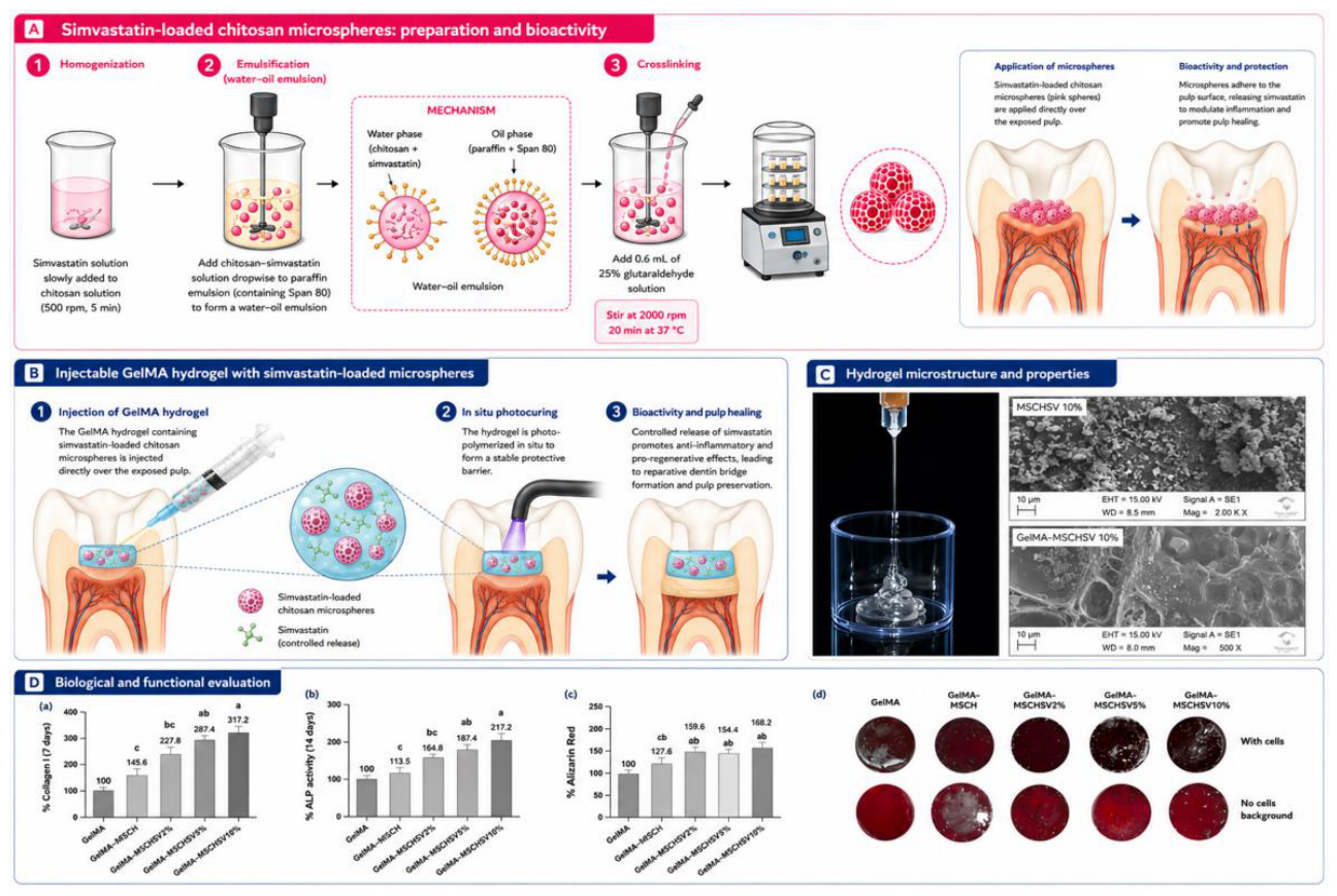
Despite these promising results, microspheres alone are not ideal clinical biomaterials for dentin regeneration. Their particulate nature may compromise adaptation to irregular pulp exposures, reduce retention at the implantation site, and permit particle displacement before complete tissue integration. Consequently, recent strategies have focused on incorporating drug-loaded microspheres into injectable hydrogels, creating hybrid systems that combine the sustained release kinetics of microspheres with the structural stability, injectability, and defect adaptation provided by the surrounding hydrogel. Using this approach, simvastatin-loaded chitosan microspheres were incorporated into a photocrosslinkable GelMA hydrogel to generate an injectable platform for direct pulp capping. The hybrid construct maintained the controlled release profile of simvastatin while preserving hydrogel degradability and injectability, and significantly enhanced odontogenic differentiation and mineralized matrix deposition by human dental pulp cells. Importantly, the hydrogel acted not merely as a passive carrier but as a structural framework that retained the microspheres at the implantation site, facilitated clinical handling, and created a three-dimensional microenvironment capable of supporting tissue neoformation, illustrating how injectable matrices can overcome important translational limitations associated with microsphere-based delivery systems (Bronze-Uhle et al., 2025b).
Figure 6.
Simvastatin-loaded chitosan microspheres incorporated into an injectable GelMA hydrogel for dental pulp bioactivity and regenerative applications. (A) Schematic representation of the preparation of simvastatin-loaded chitosan microspheres (MSCHSV) by water–oil emulsification and chemical crosslinking, followed by their proposed direct application to exposed dental pulp and controlled simvastatin release. (B) Incorporation of MSCHSV into an injectable GelMA hydrogel and proposed application as a pulp-capping platform. The microsphere-containing hydrogel is injected into the exposure site and photocrosslinked in situ, forming a stable barrier capable of providing sustained simvastatin delivery and supporting pulp preservation and reparative dentin formation. (C) Representative macroscopic image demonstrating the injectability of the GelMA–MSCHSV formulation and scanning electron microscopy (SEM) images showing the morphology of MSCHSV10% and the microstructure of GelMA containing MSCHSV10%. (D)Biological and functional evaluation of GelMA formulations containing increasing concentrations of simvastatin-loaded chitosan microspheres (GelMA, GelMA–MSCH, GelMA–MSCHSV2%, GelMA–MSCHSV5%, and GelMA–MSCHSV10%), including (a) collagen deposition, (b) alkaline phosphatase (ALP) activity, (c) Alizarin Red staining/quantification, and (d) representative images of Alizarin Red staining in the presence of cells and corresponding cell-free backgrounds. Different lowercase letters indicate statistically significant differences among groups (p < 0.05). Panels C and D were adapted from Bronze-Uhle et al., 2025b, published by Wiley under Creative Commons CC BY 3.0.
Figure 6.
Simvastatin-loaded chitosan microspheres incorporated into an injectable GelMA hydrogel for dental pulp bioactivity and regenerative applications. (A) Schematic representation of the preparation of simvastatin-loaded chitosan microspheres (MSCHSV) by water–oil emulsification and chemical crosslinking, followed by their proposed direct application to exposed dental pulp and controlled simvastatin release. (B) Incorporation of MSCHSV into an injectable GelMA hydrogel and proposed application as a pulp-capping platform. The microsphere-containing hydrogel is injected into the exposure site and photocrosslinked in situ, forming a stable barrier capable of providing sustained simvastatin delivery and supporting pulp preservation and reparative dentin formation. (C) Representative macroscopic image demonstrating the injectability of the GelMA–MSCHSV formulation and scanning electron microscopy (SEM) images showing the morphology of MSCHSV10% and the microstructure of GelMA containing MSCHSV10%. (D)Biological and functional evaluation of GelMA formulations containing increasing concentrations of simvastatin-loaded chitosan microspheres (GelMA, GelMA–MSCH, GelMA–MSCHSV2%, GelMA–MSCHSV5%, and GelMA–MSCHSV10%), including (a) collagen deposition, (b) alkaline phosphatase (ALP) activity, (c) Alizarin Red staining/quantification, and (d) representative images of Alizarin Red staining in the presence of cells and corresponding cell-free backgrounds. Different lowercase letters indicate statistically significant differences among groups (p < 0.05). Panels C and D were adapted from Bronze-Uhle et al., 2025b, published by Wiley under Creative Commons CC BY 3.0.
Recent developments have further expanded microsphere functionality by integrating ion-mediated signaling and biomimetic biological instruction. Strontium-doped hydroxyapatite microspheres simultaneously provided osteo-odontogenic ionic stimulation and proangiogenic signaling, promoting coordinated regeneration of the dentin–pulp complex through enhanced vascularization and mineralized tissue formation (Chen et al., 2026). Rather than relying on isolated recombinant growth factors, more sophisticated systems have incorporated complex biological secretomes. GelMA microspheres loaded with human platelet lysate provided sustained release of multiple endogenous growth factors for several weeks, enhancing dental pulp stem cell migration, angiogenesis, extracellular matrix deposition, and odontoblast-like differentiation following implantation into human tooth fragments (Zhang et al., 2021).
More recently, microsphere design has begun to incorporate concepts derived from the emerging understanding of pulp immunobiology. Spatial transcriptomic analyses of human pulpitis have demonstrated that interactions between fibroblasts and immune cells are central determinants of tissue repair and may distinguish reversible from irreversible inflammatory responses (Zaky et al., 2026). This biological insight has stimulated the development of immunoinstructive microspheres capable of responding to pathological microenvironments rather than simply releasing therapeutic molecules at constant rates. For example, pH-responsive peptide microsphere/carboxymethyl chitosan complexes exploit the acidic conditions associated with inflamed pulp tissues to trigger localized therapeutic activity, improving protection of the dentin–pulp complex while adapting biomaterial behavior to the inflammatory status of the tissue (Wang et al., 2025). Such responsive systems represent an important conceptual evolution toward intelligent biomaterials capable of dynamically interacting with their biological environment.
Therefore, microsphere-based biomaterials have evolved from injectable cell carriers into sophisticated modular regenerative platforms capable of integrating structural support, controlled drug delivery, extracellular matrix mimicry, angiogenic stimulation, immunomodulation, and localized biological signaling. Beyond serving as scaffolds or drug reservoirs, next-generation microspheres are increasingly designed as dynamic building blocks that orchestrate multiple biological events simultaneously, reproducing the spatial and temporal complexity of the dentin–pulp microenvironment. Future advances are expected to integrate responsive biomaterials, extracellular vesicles, secretome-based therapies, immune modulation, and programmable release systems, further strengthening the translational potential of microsphere-based strategies for regenerative endodontics.
3.5. Three-Dimensional Printed Scaffolds for Cell-Homing–Based Dentin Regeneration
Additive manufacturing has introduced a new level of architectural control into biomaterial design for dentin regeneration. Conventional scaffold-processing methods, including phase separation, particle leaching, gas foaming, molding, and lyophilization, can generate porous matrices but provide limited control over pore size, geometry, interconnectivity, and spatial distribution. Three-dimensional (3D) printing overcomes some of these limitations by fabricating constructs layer by layer from computer-aided designs, enabling reproducible control of external shape, internal architecture, and material distribution. These capabilities are particularly relevant to dentin regeneration because scaffold geometry can regulate cell infiltration, organization, differentiation, and mineralized matrix deposition rather than merely providing structural support (Zhao et al., 2024; Daghrery et al., 2026).
Although the terms are frequently used interchangeably, 3D printing and 3D bioprinting represent distinct biofabrication strategies. Conventional 3D printing generates acellular constructs that may subsequently be seeded with cells or implanted as cell-free scaffolds intended to recruit endogenous cell populations. In contrast, bioprinting involves the controlled deposition of living cells together with biomaterials and, in some cases, bioactive molecules within a cell-laden bioink. Accordingly, bioprinting requires simultaneous optimization of printability, structural fidelity, crosslinking, cell viability, and biological functionality, whereas acellular printing primarily focuses on the relationship among material processability, architecture, mechanical stability, degradation, and postimplantation cell interactions. For cell-homing–based dentin regeneration, acellular printed scaffolds may offer a more readily translatable approach because they avoid exogenous cell isolation, expansion, storage, and transplantation while using scaffold-derived structural and biochemical cues to recruit resident progenitor cells and direct odontoblastic differentiation (Zhao et al., 2024; Daghrery et al., 2026).
The principal advantage of 3D printing in this context is not simply the ability to reproduce a defect shape but to transform scaffold architecture into a biological regulatory cue. Pore dimensions, strand spacing, surface topography, and interconnectivity determine the available area for protein adsorption and cell attachment while controlling cell penetration, nutrient transport, degradation, and matrix organization (Zhao et al., 2024; Huang et al., 2023). Hierarchical structures combining microporosity with interconnected macropores are particularly attractive: smaller pores increase surface area and cell–material interactions, whereas larger pores provide pathways for endogenous cell infiltration and newly deposited tissue. However, increasing porosity can reduce mechanical integrity and compromise shape fidelity, requiring a balance between biological accessibility and structural stability (Daghrery et al., 2026).
The regulatory effect of architecture has been demonstrated in ordered macroporous hydrogel systems. Indirectly printed GelMA scaffolds with grid or honeycomb architectures showed that geometry alone can differentially regulate mineralizing cell behavior. Although larger honeycomb pores enhanced alkaline phosphatase activity, the grid architecture more effectively promoted odontoblastic differentiation and mineralized matrix deposition (de Carvalho et al., 2026). The importance of balancing pore accessibility with structural stability was further demonstrated using extrusion-printed GelMA scaffolds with controlled infill densities. Scaffolds printed with 60% infill maintained greater structural stability and promoted higher cell proliferation, alkaline phosphatase activity, and mineralized matrix deposition than non-printed GelMA. Incorporation of decellularized bovine bone matrix microparticles further enhanced these effects, demonstrating that printed architecture and bioactive composition act synergistically to regulate odontogenic behavior (da Silva et al., 2026).
Synthetic thermoplastic polymers, particularly poly(ε-caprolactone) (PCL), have been extensively used to generate mechanically stable printed scaffolds with reproducible pore networks. Their slow degradation and structural stability facilitate space maintenance, but hydrophobicity and the absence of intrinsic cell-recognition sites frequently limit endogenous cell attachment and biological integration. Consequently, PCL has increasingly been combined with hydroxyapatite, bioactive glass, calcium silicate, or hydrophilic polymers to generate constructs that couple architectural stability with mineral-inducing activity. These modifications improve dental pulp stem-cell adhesion, proliferation, alkaline phosphatase activity, and mineral deposition, demonstrating that cell-homing scaffolds require not only open pathways for infiltration but also surfaces permissive to cell anchorage and differentiation (Mousavi Nejad et al., 2021; Zhao et al., 2024).
Mineral and pharmacological functionalization further transforms printed constructs from structurally defined supports into instructive platforms for dentin formation. Three-dimensionally printed mesoporous calcium silicate/calcium sulfate/PCL scaffolds containing quercetin rapidly developed apatite on their surfaces and enhanced cell viability, mineralization, and the expression of odontogenic proteins, including DSPP and DMP-1 (Yeh et al., 2022). Similarly, printable self-assembling peptide matrices containing amorphous magnesium phosphate combined a nanofibrous extracellular matrix-like environment with a controlled macroporous grid. The porous architecture supported cellular infiltration, while the mineral phase provided signals associated with mineralized matrix formation (Dubey et al., 2020). These findings illustrate a central principle of cell-homing scaffold design: architecture determines whether endogenous cells can enter and occupy the construct, whereas biochemical composition determines how those recruited cells behave after infiltration.
Dentin-derived matrices provide an even more tissue-specific approach by reintroducing biological molecules naturally involved in dentinogenesis. Partial demineralization exposes collagen fibrils and dentinal tubules while releasing or preserving dentin-associated proteins and growth factors, including type I collagen, DSPP, DMP-1, TGF-β1, decorin, and biglycan. Incorporation of soluble and insoluble dentin matrix fractions into printable alginate hydrogels improved the viability and odontogenic differentiation of encapsulated stem cells from the apical papilla, demonstrating that dentin-derived components retain instructive biological activity after printing (Athirasala et al., 2018). Although this study used a cell-laden bioink, its findings are also relevant to acellular scaffold design because the same matrix-associated cues could potentially recruit endogenous progenitor cells and induce odontoblastic differentiation after implantation.
Demineralized dentin matrix particles have subsequently been incorporated into fibrinogen–gelatin bioinks to improve printability and provide tissue-specific biological instruction. Increasing particle concentration enhanced viscosity, shear-thinning behavior, printing resolution, and construct stacking while maintaining high cell viability and increasing DSPP and DMP1 gene expression and mineral deposition (Han et al., 2021). Likewise, printed biocomposites containing decellularized extracellular matrix and calcium phosphate ceramics promoted stronger osteo/odontogenic differentiation than collagen-based controls, with increased calcification and expression of dentin-associated markers (Kim et al., 2022). Printed treated dentin matrix/PCL scaffolds also released collagenous and non-collagenous proteins associated with dentinogenesis, including DSPP and DMP-1, while supporting cell adhesion, differentiation, biomimetic mineralization, and formation of an odontogenic microenvironment (Huang et al., 2023). Together, these studies indicate that tissue-derived matrices can bridge the gap between architectural precision and biological specificity, enabling printed scaffolds to reproduce aspects of the native dentin extracellular matrix rather than relying exclusively on isolated recombinant factors.
Within a cell-homing strategy, however, the biological value of a printed scaffold should not be inferred solely from the response of cells pre-encapsulated during fabrication. A true cell-free platform must demonstrate that endogenous cells can migrate from the surrounding tissue into the construct, remain viable within its interconnected architecture, acquire an odontoblast-like phenotype, and deposit an organized mineralized matrix. This concept was directly investigated using a biomimetic artificial pulp chamber model designed to reproduce key features of the dentin–pulp interface. The system incorporated a perforated human dentin disc positioned above a three-dimensional collagen matrix containing human dental pulp cells, while a hydrostatic pressure equivalent to physiological intrapulpal pressure was maintained throughout cultivation. Acellular injected and 3D-printed GelMA and GelMA enriched with decellularized bovine bone matrix microparticles (BMdc) were placed within the dentin perforation, allowing cell migration from the underlying three-dimensional culture toward the scaffolds to be evaluated under a dentin barrier and continuous fluid pressure. Cells remained viable within the resident 3D culture and progressively migrated through the defect to colonize the hydrogel surface. Both the printed architecture and incorporation of the decellularized matrix enhanced cell recruitment, with the GelMA–BMdc scaffold printed at 60% infill showing the greatest number of migrated cells. This formulation also supported cell adhesion and spreading and enhanced odontogenic differentiation and mineralized matrix deposition, demonstrating that the combination of controlled macroporosity and matrix-derived biological signals can function as an instructive cell-homing platform in a more clinically relevant experimental environment (da Silva et al., 2026) (Figure 7).
Ultimately, the significance of additive manufacturing for dentin regeneration extends beyond geometric customization: printing becomes a means of engineering an instructive microenvironment in which endogenous cells are recruited and guided toward organized reparative dentinogenesis without requiring exogenous cell transplantation. In this framework, printing becomes not merely a manufacturing technique but a means of engineering a cell-directing microenvironment in which endogenous cells are recruited and guided toward organized reparative dentinogenesis without requiring exogenous cell transplantation.
4. Conclusions
Biomaterials for dentin tissue engineering have progressed from passive supports and nonspecific mineralizing agents toward bioinstructive systems capable of coordinating cell recruitment, differentiation, matrix deposition, and inflammatory control. Naturally derived scaffolds, nanofibrous membranes, injectable hydrogels, microsphere-based systems, and three-dimensional printed constructs provide complementary strategies for controlling scaffold composition, architecture, delivery, and biological signaling. The sequential development of the PCL-based nanofibrous platform illustrates this progression particularly clearly, from mineral-phase selection to lineage-specific signaling and, finally, immunomodulation. Nevertheless, most available evidence remains preclinical and is derived from heterogeneous in vitro or ectopic models. Translation will require standardized testing under dentin barriers, inflammatory and microbial challenges, clinically relevant handling and light-curing conditions, and orthotopic models capable of evaluating sealing, pulp vitality, vascularization, tissue organization, and long-term mineralized barrier quality. Within these limitations, the most promising strategy is not a single material class but the rational integration of structural support, mineral signaling, and temporally controlled immunomodulation.
References
- Smith, A.J.; Scheven, B.A.; Takahashi, Y.; Ferracane, J.L.; Shelton, R.M.; Cooper, P.R. Dentine as a bioactive extracellular matrix. Archives of Oral Biology 2012, 57, 109–121. [CrossRef]
- Cooper, P.R.; Holder, M.J.; Smith, A.J. Inflammation and regeneration in the dentin–pulp complex: a double-edged sword. Journal of Endodontics 2014, 40, S46–S51. [CrossRef]
- Bertassoni, L.E. Dentin on the nanoscale: hierarchical organization, mechanical behavior and bioinspired engineering. Dental Materials 2017, 33, 637–649. [CrossRef]
- Soares, D.G.; Bordini, E.A.F.; Swanson, W.B.; de Souza Costa, C.A.; Bottino, M.C. Platform technologies for regenerative endodontics from multifunctional biomaterials to tooth-on-a-chip strategies. Clinical Oral Investigations 2021, 25, 4749–4779. [CrossRef]
- de Souza Costa, C.A.; Hebling, J.; Scheffel, D.L.; Soares, D.G.; Basso, F.G.; Ribeiro, A.P. Methods to evaluate and strategies to improve the biocompatibility of dental materials and operative techniques. Dental Materials 2014, 30, 769–784. [CrossRef]
- Le Fournis, C.; et al. Pulp fibroblast contribution to the local control of pulp inflammation via complement activation. Journal of Endodontics 2020, 46, S26–S32. [CrossRef]
- Bergmann, P.; et al. Complement activation links inflammation to dental tissue regeneration. Clinical Oral Investigations 2020, 24, 4185–4196. [CrossRef]
- Le Fournis, C.; et al. Fibroblasts control macrophage differentiation during pulp inflammation. Journal of Endodontics 2021, 47, 1427–1434. [CrossRef]
- Ravenscroft, N.; et al. Novel antibacterial properties of the human dental pulp multipotent mesenchymal stromal cell secretome. American Journal of Pathology 2022, 192, 956–969. [CrossRef]
- Le Fournis, C.; et al. Macrophage–pulp fibroblast interactions modulate initial dental pulp regeneration in vitro. International Endodontic Journal 2026, 59, 852–862. [CrossRef]
- Vazavandi, A.; Raoof, M.; et al. Clinical, radiological, and histological correlation in diagnosis of pulpitis. Dental Research Journal 2022, 19, 25. [CrossRef]
- Loo, A.; et al. Symptom correlation and spatial distribution of inflammatory mediators in pulpitis—a preliminary study. International Endodontic Journal 2025. [CrossRef]
- Torabinejad, M.; et al. Regenerative endodontics in teeth with irreversible pulpitis: a scoping review. International Endodontic Journal 2026. [CrossRef]
- Taha, N.A.; et al. Conservative management of mature permanent teeth with carious pulp exposure. Journal of Endodontics 2020, 46, S33–S41. [CrossRef]
- Chien, Y.-C.; et al. What outcomes matter in vital pulp therapy? Current concepts and challenges. Australian Dental Journal 2026. [CrossRef]
- Schwendicke, F.; et al. Deep caries management: EFCD–ESE–ORCA S3-level clinical practice guideline. International Endodontic Journal 2026, 59, 1298–1315. [CrossRef]
- Goldberg, M.; Farges, J.-C.; Lacerda-Pinheiro, S.; et al. Inflammatory and immunological aspects of dental pulp repair. Pharmacological Research 2008, 58, 137–147. [CrossRef]
- Sangwan, P.; et al. Tertiary dentinogenesis with calcium hydroxide: a review of proposed mechanisms. International Endodontic Journal 2013, 46, 3–19. [CrossRef]
- Peskersoy, C.; et al. Efficacy of different calcium silicate materials as pulp-capping agents: a randomized clinical trial. Journal of Dental Sciences 2021, 16, 723–731. [CrossRef]
- Cushley, S.; et al. Efficacy of direct pulp capping for management of cariously exposed pulps in permanent teeth: a systematic review and meta-analysis. International Endodontic Journal 2021, 54, 556–571. [CrossRef]
- Soares, D.G.; et al. Characterization of novel calcium hydroxide-mediated highly porous chitosan–calcium scaffolds for potential application in dentin tissue engineering. Journal of Biomedical Materials Research Part B 2020, 108, 2546–2559. [CrossRef]
- Okamoto, M.; et al. Performance of a biodegradable composite with hydroxyapatite as a scaffold in pulp tissue repair. Polymers 2020, 12, 937. [CrossRef]
- Soares, D.G.; et al. Odontogenic differentiation potential of human dental pulp cells cultured on a calcium-aluminate-enriched chitosan–collagen scaffold. Clinical Oral Investigations 2017, 21, 2827–2839. [CrossRef]
- Cassiano, F.B.; et al. Simvastatin-enriched macro-porous chitosan–calcium-aluminate scaffold for mineralized tissue regeneration. Brazilian Dental Journal 2020, 31, 385–391. [CrossRef]
- Leite, M.L.; et al. Calcium silicate-coated porous chitosan scaffold as a cell-free tissue engineering system for direct pulp capping. Dental Materials 2022, 38, 1763–1776. [CrossRef]
- Bordini, E.A.F.; et al. Chitosan–calcium aluminate as a cell-homing scaffold: its bioactivity testing in a microphysiological dental pulp platform. Alternatives to Laboratory Animals 2024, 52, 107–116. [CrossRef]
- de Melo, M.A.; et al. Mineral-induced bubbling effect and biomineralization as strategies to create highly porous and bioactive scaffolds for dentin tissue engineering. Journal of Biomedical Materials Research Part B: Applied Biomaterials 2022, 110, 1757–1770. [CrossRef]
- Bordini, E.A.F.; et al. Synergistic potential of 1α,25-dihydroxyvitamin D3 and calcium-aluminate–chitosan scaffolds with dental pulp cells. Clinical Oral Investigations 2020, 24, 663–674. [CrossRef]
- Soares, D.G.; et al. Biological analysis of simvastatin-releasing chitosan scaffold as a cell-free system for pulp-dentin regeneration. Journal of Endodontics 2018a, 44, 971–976.e1. [CrossRef]
- Soares, D.G.; et al. Simvastatin and nanofibrous poly(L-lactic acid) scaffolds promote the odontogenic potential of dental pulp cells in an inflammatory environment. Acta Biomaterialia 2018b, 68, 190–203. [CrossRef]
- Divband, B.; et al. Towards induction of angiogenesis in dental pulp stem cells using chitosan-based hydrogels releasing basic fibroblast growth factor. BioMed Research International 2022, 2022, 5401461. [CrossRef]
- Wang, J.; Ma, H.; Jin, X.; Hu, J.; Liu, X.; Ni, L.; Ma, P.X. The effect of scaffold architecture on odontogenic differentiation of human dental pulp stem cells. Biomaterials 2011, 32, 7822–7830. [CrossRef]
- Gupte, M.J.; Ma, P.X. Nanofibrous scaffolds for dental and craniofacial applications. Journal of Dental Research 2012, 91, 227–234. [CrossRef]
- Anselmi, C.; Mendes Soares, I.P.; Mota, R.L.M.; Leite, M.L.; Ribeiro, R.A.O.; Fernandes, L.O.; Bottino, M.C.; de Souza Costa, C.A.; Hebling, J. Functionalization of PCL-based fiber scaffolds with different sources of calcium and phosphate and odontogenic potential on human dental pulp cells. Journal of Functional Biomaterials 2024, 15, 97. [CrossRef]
- Mendes Soares, I.P.; Anselmi, C.; Kitagawa, F.A.; Ribeiro, R.A.O.; Leite, M.L.; de Souza Costa, C.A.; Hebling, J. Nano-hydroxyapatite-incorporated polycaprolactone nanofibrous scaffold as a dentin tissue engineering-based strategy for vital pulp therapy. Dental Materials 2022, 38, 960–977. [CrossRef]
- Anselmi, C.; Mendes Soares, I.P.; Leite, M.L.; Kitagawa, F.A.; de Souza Costa, C.A.; Hebling, J. Cytocompatibility and bioactivity of calcium hydroxide-containing nanofiber scaffolds loaded with fibronectin for dentin tissue engineering. Clinical Oral Investigations 2022, 26, 4031–4047. [CrossRef]
- Anselmi, C.; Mendes Soares, I.P.; Chang, S.; Cardoso, L.M.; de Carvalho, A.B.G.; Dal-Fabbro, R.; de Souza Costa, C.A.; Bottino, M.C.; Hebling, J. Quercetin–calcium hydroxide scaffolds modulate dental pulp stem cell response in vitro under a simulated inflammatory environment. International Endodontic Journal 2025, 58, 1073–1090. [CrossRef]
- Mendes Soares, I.P.; Anselmi, C.; Dal-Fabbro, R.; Fernandes, L.O.; Pinto, G.C.; Piazza, R.D.; de Souza Costa, C.A.; Hebling, J.; Bottino, M.C. Immunomodulatory and dentinogenic potential of surface-engineered hesperetin-functionalized composite scaffolds for pulp–dentin regeneration. Materials Today Bio 2026, 37, 102930. [CrossRef]
- Anselmi, C.; et al. Bilayer gelatin-methacryloyl scaffold for pulp inflammation suppression and dentin-like tissue regeneration. Journal of Controlled Release 2026, 389, 114469. [CrossRef]
- Amani, H.; et al. Hydrogel-based strategies for dentin–pulp complex regeneration: a comprehensive review. Cell and Tissue Banking 2026, 27. [CrossRef]
- Dal-Fabbro, R.; et al. Recent advances in injectable hydrogel biotherapeutics for regenerative dental medicine. Macromolecular Bioscience 2025, 25, e00096. [CrossRef]
- Omidian, H.; et al. Smart hydrogels for craniofacial regeneration. Cells 2026, 15, 1054. [CrossRef]
- Pankajakshan, D.; et al. Injectable highly tunable oligomeric collagen matrices for dental tissue regeneration. ACS Applied Bio Materials 2020, 3, 859–868. [CrossRef]
- Zhao, Y.; et al. Photopolymerizable fish collagen peptide–GelMA composite hydrogels for dental pulp regeneration. International Journal of Biological Macromolecules 2025, 332, 148628. [CrossRef]
- Bordini, E.A.F.; et al. Injectable multifunctional drug delivery system for hard tissue regeneration under inflammatory microenvironments. ACS Applied Bio Materials 2021, 4, 6993–7006. [CrossRef]
- Sahadi, B.O.; et al. Immunomodulatory and pro-mineralizing effects of an injectable baicalein-loaded methacrylated gelatin hydrogel for vital pulp therapy. Biomaterials Advances 2026, 183, 214768. [CrossRef]
- Tootla, S.; et al. Dexamethasone/β-cyclodextrin inclusion complex hydrogel for vital pulp therapy. Odontology 2026, 114, 702–710. [CrossRef]
- Han, Y.; et al. GelMA/TCP nanocomposite scaffold for vital pulp therapy. Acta Biomaterialia 2024, 173, 495–508. [CrossRef]
- Osman, M.; Sharmin, Z.; Suchy, S.; et al. Bioinspired smart dentin extracellular matrix–chitosan hydrogels for dentin–pulp complex regeneration. Journal of Dentistry 2025, 159, 105811. [CrossRef]
- da Silva, T.M.; et al. Photo-crosslinkable hydrogel incorporated with bone matrix particles for advancements in dentin tissue engineering. Journal of Biomedical Materials Research Part A 2024, 112, 2273–2288. [CrossRef]
- da Silva, T.M.; et al. Three-dimensional printed GelMA–bone matrix scaffold promotes cell homing and odontogenic responses in an artificial pulp chamber model. Clinical Oral Investigations 2026. [CrossRef]
- Bordini, E.A.F.; et al. Functionalization of photocrosslinkable GelMA hydrogel with metal oxides for direct pulp capping. BioMed Research International 2026, 2026. [CrossRef]
- Alajlan, M.; et al. An injectable κ-carrageenan hydrogel carrier maintains antibiofilm activity while supporting dental pulp-derived stem cell viability in a dentin-based model. Journal of Dentistry 2026, 171, 106713. [CrossRef]
- Akgun, O.M.; Tekinay, A.B.; Tansık, G.; Yıldırım, C.; Guven Polat, G. Pulp regeneration using a peptide nanofiber artificial scaffold on animal models: A preliminary study. Advances in Clinical and Experimental Medicine 2025, 34, 407–419. [CrossRef]
- Kuang, R.; et al. Nanofibrous spongy microspheres enhance odontogenic differentiation of human dental pulp stem cells. Advanced Healthcare Materials 2015, 4, 1993–2000. [CrossRef]
- Li, X.; et al. Pulp regeneration in a full-length human tooth root using a hierarchical nanofibrous microsphere system. Acta Biomaterialia 2016, 35. [CrossRef]
- Zhang, W.; et al. Dexamethasone-loaded hollow hydroxyapatite microspheres promote odontogenic differentiation of human dental pulp cells in vitro. Odontology 2020, 108, 222–230. [CrossRef]
- Liu, Y.; et al. Experimental study of dexamethasone-loaded hollow hydroxyapatite microspheres applied to direct pulp capping of rat molars. Frontiers in Endocrinology 2023, 14, 1192420. [CrossRef]
- Bronze-Uhle, E.S.; et al. Simvastatin-loaded chitosan microspheres as a biomaterial for dentin tissue engineering. Journal of Biomedical Materials Research Part B 2025a, 113, e35536. [CrossRef]
- Alenizy, H.A.; et al. Novel metformin-encapsulating poly(lactic-co-glycolic acid) microspheres in calcium phosphate pulp-capping cement with dental pulp stem cells for regenerative applications. Materials 2026, 19, 487. [CrossRef]
- Bronze-Uhle, E.S.; et al. Simvastatin-Release Photocurable Hydrogel with Microsphere Delivery System for Improved Dental Applications. J. Appl. Polym. Sci. 2025b, 142, e57284. [CrossRef]
- Chen, X.; et al. Strontium-doped hydroxyapatite microspheres loaded with iloprost promote dentin–pulp complex regeneration. Frontiers in Bioengineering and Biotechnology 2026, 13, 1726285. [CrossRef]
- Zhang, W.; et al. Platelet lysate-functionalized gelatin methacrylate microspheres for improving angiogenesis in endodontic regeneration. Acta Biomaterialia 2021, 136, 441–455. [CrossRef]
- Zaky, S.H.; et al. Pilot spatial transcriptomics of dental pulpitis suggests immune–fibroblast profiling linked to reversibility. Journal of Translational Medicine 2026. [CrossRef]
- Wang, X.; et al. Two-stepped pH-responsive peptide microsphere/carboxymethyl chitosan complex: enhanced protection of an inflamed dentin–pulp complex. Journal of Materials Chemistry B 2025, 13, 4879–4892. [CrossRef]
- Zhao, F.; Zhang, Z.; Guo, W. The 3-dimensional printing for dental tissue regeneration: the state of the art and future challenges. Frontiers in Bioengineering and Biotechnology 2024, 12, 1356580. [CrossRef]
- Daghrery, A.; Mendes Soares, I.P.; dos Reis-Prado, A.H.; de Souza Araújo, I.J.; Dal-Fabbro, R.; Bottino, M.C. Advances in 3D printed scaffolds for periodontal regeneration. Current Oral Health Reports 2026, 13, 1. [CrossRef]
- Huang, Y.; Zhang, Z.; Bi, F.; Tang, H.; Chen, J.; Huo, F.; et al. Personalized 3D-printed scaffolds with multiple bioactivities for bioroot regeneration. Advanced Healthcare Materials 2023, 12, e2300625. [CrossRef]
- de Carvalho, L.A.M.; Stuani, V.T.; da Silva, I.S.P.; Berteli, T.S.; Gonçalves, N.G.; Soares, D.G.; Bordini, E.A.F. Hydrogel microarchitecture as a regulatory cue for in vitro odontogenic differentiation. Journal of Applied Oral Science 2026, 34, e20250607. [CrossRef]
- Mousavi Nejad, Z.; Zamanian, A.; Saeidifar, M.; Vanaei, H.R.; Salar Amoli, M. 3D bioprinting of polycaprolactone-based scaffolds for pulp–dentin regeneration: investigation of physicochemical and biological behavior. Polymers 2021, 13, 4442. [CrossRef]
- Yeh, C.-L.; Bhorade, R.; Hsu, T.-T.; Chen, C.-Y.; Lin, C.-P. Mechanical assessment and odontogenic behavior of a 3D-printed mesoporous calcium silicate/calcium sulfate/poly-ε-caprolactone composite scaffold. Journal of the Formosan Medical Association 2022, 121, 510–518. [CrossRef]
- Dubey, N.; Ferreira, J.A.; Malda, J.; Bhaduri, S.B.; Bottino, M.C. Extracellular matrix/amorphous magnesium phosphate bioink for 3D bioprinting of craniomaxillofacial bone tissue. ACS Applied Materials & Interfaces 2020, 12, 23752–23763. [CrossRef]
- Athirasala, A.; Tahayeri, A.; Thrivikraman, G.; França, C.M.; Monteiro, N.; Tran, V.; Ferracane, J.L.; Bertassoni, L.E. A dentin-derived hydrogel bioink for 3D bioprinting of cell-laden scaffolds for regenerative dentistry. Biofabrication 2018, 10, 024101. [CrossRef]
- Han, J.; Jeong, W.; Kim, M.K.; Nam, S.H.; Park, E.K.; Kang, H.W. Demineralized dentin matrix particle-based bio-ink for patient-specific shaped 3D dental tissue regeneration. Polymers 2021, 13, 1294. [CrossRef]
- Kim, D.S.; et al. Fabrication of bone-derived decellularized extracellular matrix/ceramic-based biocomposites and their osteo/odontogenic differentiation ability for dentin regeneration. Bioengineering & Translational Medicine 2022, 7, e10317. [CrossRef]
Figure 1.
Mechanism of action and limitations of calcium hydroxide in vital pulp therapy. The upper left panel depicts the clinical application of calcium hydroxide as a direct pulp capping material. The upper right panel summarizes its main biological properties, including alkaline pH, antibacterial activity, and the ability to induce mineralized tissue formation. The central sequence illustrates the mechanism of action of calcium hydroxide. (1) Following placement, the material induces a superficial zone of chemical necrosis at the pulp interface. (2) The necrotic layer triggers an inflammatory response in the underlying vital pulp tissue. (3) Inflammatory signaling promotes the recruitment and activation of progenitor cells. (4) These cells differentiate and deposit a reparative dentin bridge beneath the capping material. This sequence highlights that tissue repair is initiated only after a new material-induced injury. The lower panels illustrate the main structural limitations of the reparative dentin bridge formed after calcium hydroxide treatment, including porosity, tunnel defects, heterogeneous thickness, and incomplete adaptation to the dentinal walls. These defects may compromise the integrity of the mineralized barrier, reduce its sealing capacity, and facilitate bacterial reinfiltration, ultimately affecting the long-term success of vital pulp therapy.
Figure 1.
Mechanism of action and limitations of calcium hydroxide in vital pulp therapy. The upper left panel depicts the clinical application of calcium hydroxide as a direct pulp capping material. The upper right panel summarizes its main biological properties, including alkaline pH, antibacterial activity, and the ability to induce mineralized tissue formation. The central sequence illustrates the mechanism of action of calcium hydroxide. (1) Following placement, the material induces a superficial zone of chemical necrosis at the pulp interface. (2) The necrotic layer triggers an inflammatory response in the underlying vital pulp tissue. (3) Inflammatory signaling promotes the recruitment and activation of progenitor cells. (4) These cells differentiate and deposit a reparative dentin bridge beneath the capping material. This sequence highlights that tissue repair is initiated only after a new material-induced injury. The lower panels illustrate the main structural limitations of the reparative dentin bridge formed after calcium hydroxide treatment, including porosity, tunnel defects, heterogeneous thickness, and incomplete adaptation to the dentinal walls. These defects may compromise the integrity of the mineralized barrier, reduce its sealing capacity, and facilitate bacterial reinfiltration, ultimately affecting the long-term success of vital pulp therapy.
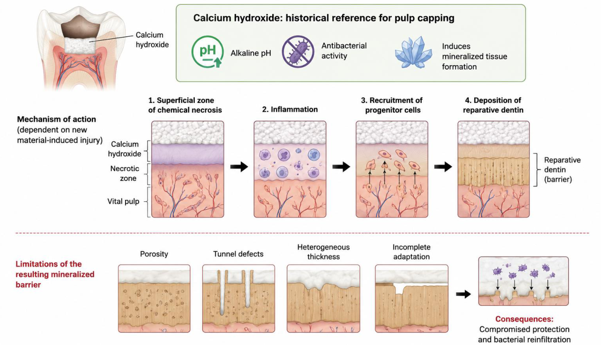
Figure 2.
From passive pulp capping to biologically guided vital pulp therapy. This schematic illustrates the paradigm shift from conventional pulp-capping materials toward next-generation bioactive biomaterials capable of actively modulating the healing microenvironment. The proposed biomaterial should integrate structural support with immunomodulatory and regenerative functions, including regulation of the inflammatory response, promotion of inflammation resolution, vascular stabilization, recruitment and activation of endogenous progenitor cells, and guidance of organized extracellular matrix deposition. These coordinated biological events are expected to promote the formation of a continuous and functional reparative dentin bridge, resulting in more predictable healing, reduced dependence on the initial biological condition of the pulp, and long-term preservation of pulp vitality.
Figure 2.
From passive pulp capping to biologically guided vital pulp therapy. This schematic illustrates the paradigm shift from conventional pulp-capping materials toward next-generation bioactive biomaterials capable of actively modulating the healing microenvironment. The proposed biomaterial should integrate structural support with immunomodulatory and regenerative functions, including regulation of the inflammatory response, promotion of inflammation resolution, vascular stabilization, recruitment and activation of endogenous progenitor cells, and guidance of organized extracellular matrix deposition. These coordinated biological events are expected to promote the formation of a continuous and functional reparative dentin bridge, resulting in more predictable healing, reduced dependence on the initial biological condition of the pulp, and long-term preservation of pulp vitality.
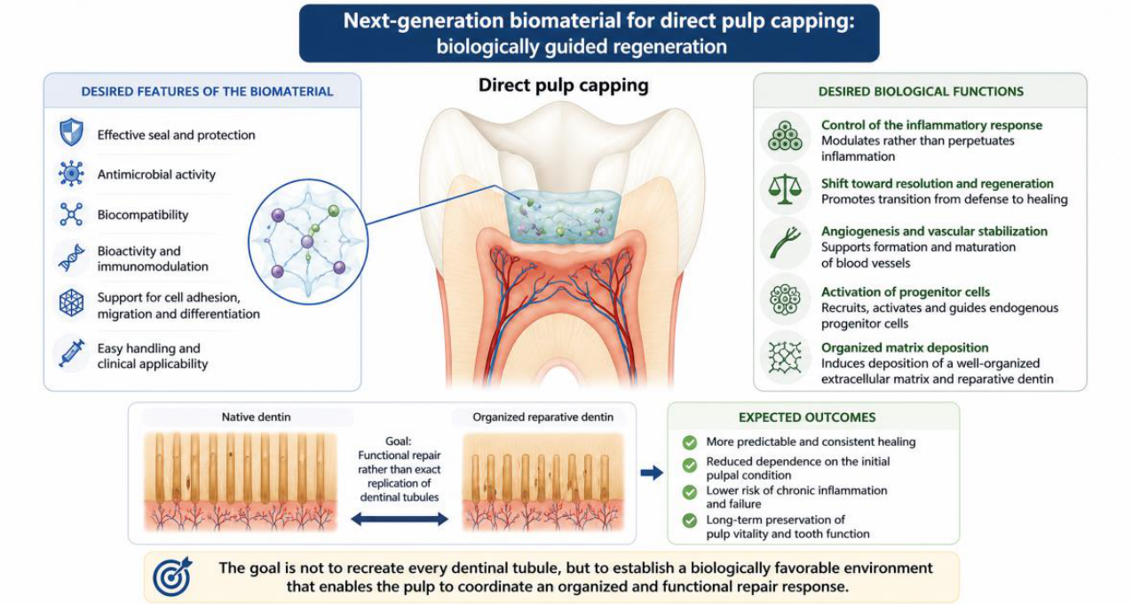
Figure 3.
Chitosan-based mineralized macroporous scaffolds and their proposed application for pulp tissue regeneration. Incorporation of a Ca(OH)2 suspension into an acidic chitosan solution, followed by a controlled gradual-freezing protocol, generated the chitosan–calcium (CH–Ca) scaffold (A), a cut-to-size biomaterial characterized by a highly porous architecture, interconnected pore network, and calcium associated with the chitosan matrix (B). The incorporation of Ca(OH)2 into the acidic chitosan solution induces a CO2-mediated bubbling effect through its reaction with acetic acid. CO2 nucleation and bubble expansion, followed by phase separation and freezing, contribute to the formation and preservation of the interconnected macroporous architecture, while released Ca2+ may interact with functional groups of the chitosan chains (C). The proposed translational application is illustrated in panels 1–3: the scaffold is adapted to the pulp exposure site (1); its macroporous network and calcium release support recruitment, adhesion, and infiltration of resident cells, followed by mineralized matrix deposition (2); and progressive scaffold remodeling and replacement by reparative dentin ultimately result in the formation of a mineralized barrier and restoration of pulp integrity (3). Panels A and B were adapted from Gallinari et al. (2023), © 2023 the authors, published by SciELO under the CC BY-NC-ND 4.0 license, and reused in accordance with the authors’ retained rights.
Figure 3.
Chitosan-based mineralized macroporous scaffolds and their proposed application for pulp tissue regeneration. Incorporation of a Ca(OH)2 suspension into an acidic chitosan solution, followed by a controlled gradual-freezing protocol, generated the chitosan–calcium (CH–Ca) scaffold (A), a cut-to-size biomaterial characterized by a highly porous architecture, interconnected pore network, and calcium associated with the chitosan matrix (B). The incorporation of Ca(OH)2 into the acidic chitosan solution induces a CO2-mediated bubbling effect through its reaction with acetic acid. CO2 nucleation and bubble expansion, followed by phase separation and freezing, contribute to the formation and preservation of the interconnected macroporous architecture, while released Ca2+ may interact with functional groups of the chitosan chains (C). The proposed translational application is illustrated in panels 1–3: the scaffold is adapted to the pulp exposure site (1); its macroporous network and calcium release support recruitment, adhesion, and infiltration of resident cells, followed by mineralized matrix deposition (2); and progressive scaffold remodeling and replacement by reparative dentin ultimately result in the formation of a mineralized barrier and restoration of pulp integrity (3). Panels A and B were adapted from Gallinari et al. (2023), © 2023 the authors, published by SciELO under the CC BY-NC-ND 4.0 license, and reused in accordance with the authors’ retained rights.
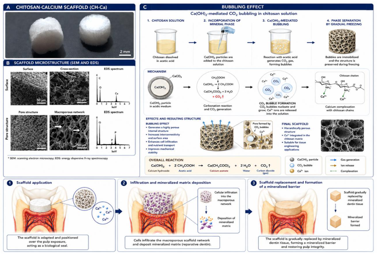
Figure 4.
Bilayer electrospun GelMA nanofiber scaffold with spatially compartmentalized immunomodulatory and mineralizing functions for vital pulp therapy. The bilayer scaffold was designed with a pulp-facing layer composed of finer GelMA nanofibers containing ibuprofen (IBP), intended to provide localized anti-inflammatory activity, and a superficial layer composed of thicker GelMA nanofibers containing amorphous magnesium phosphate (AMP), providing a mineralizing and potentially more compact coronal interface. Representative SEM images and fiber-diameter distributions illustrate the morphological differences between the GelMA 10% + 10% IBP and GelMA 20% + 5% AMP layers before and after photocrosslinking. (A) The biological performance of the bilayer scaffold was evaluated in an artificial pulp chamber (APC) model simulating pulp exposure, in which a perforated dentin disc separated the scaffold from a three-dimensional DPSC culture positioned on the pulpal side. The scaffold was placed over the simulated exposure with the IBP-containing layer facing the DPSC culture. (B) Under LPS-induced inflammatory conditions, gene-expression analysis after 3 h showed modulation of the inflammatory markers IL1A, IL1B, and TNF, indicating an early immunomodulatory effect of the bilayer scaffold. (C) After 14 days, expression of the mineralization- and odontogenic-related markers ALPL, RUNX2, OCN, and DSPP was evaluated, demonstrating a shift toward a regenerative and mineralizing response. Experimental groups included Control, GelMA double-layer scaffold (GelMA DL), and the 10% IBP/5% AMP bilayer scaffold (BL). Different letters indicate statistically significant differences among groups and experimental conditions. The images and data presented were adapted from Anselmi et al. (2026) under the CC BY 4.0 license.
Figure 4.
Bilayer electrospun GelMA nanofiber scaffold with spatially compartmentalized immunomodulatory and mineralizing functions for vital pulp therapy. The bilayer scaffold was designed with a pulp-facing layer composed of finer GelMA nanofibers containing ibuprofen (IBP), intended to provide localized anti-inflammatory activity, and a superficial layer composed of thicker GelMA nanofibers containing amorphous magnesium phosphate (AMP), providing a mineralizing and potentially more compact coronal interface. Representative SEM images and fiber-diameter distributions illustrate the morphological differences between the GelMA 10% + 10% IBP and GelMA 20% + 5% AMP layers before and after photocrosslinking. (A) The biological performance of the bilayer scaffold was evaluated in an artificial pulp chamber (APC) model simulating pulp exposure, in which a perforated dentin disc separated the scaffold from a three-dimensional DPSC culture positioned on the pulpal side. The scaffold was placed over the simulated exposure with the IBP-containing layer facing the DPSC culture. (B) Under LPS-induced inflammatory conditions, gene-expression analysis after 3 h showed modulation of the inflammatory markers IL1A, IL1B, and TNF, indicating an early immunomodulatory effect of the bilayer scaffold. (C) After 14 days, expression of the mineralization- and odontogenic-related markers ALPL, RUNX2, OCN, and DSPP was evaluated, demonstrating a shift toward a regenerative and mineralizing response. Experimental groups included Control, GelMA double-layer scaffold (GelMA DL), and the 10% IBP/5% AMP bilayer scaffold (BL). Different letters indicate statistically significant differences among groups and experimental conditions. The images and data presented were adapted from Anselmi et al. (2026) under the CC BY 4.0 license.
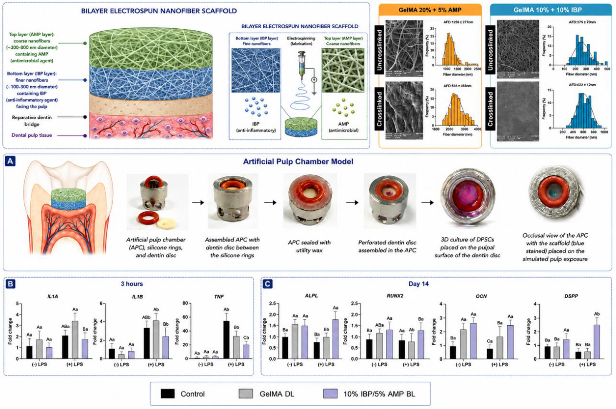
Figure 5.
GelMA-based dexamethasone delivery strategy for inflammation modulation and dentin–pulp regeneration. Schematic representation of a photocrosslinkable GelMA-based drug-delivery system designed for vital pulp therapy under inflammatory conditions. (1) Pulp exposure and bacterial stimuli induce a pro-inflammatory microenvironment characterized by increased cytokine production and activation of the NF-κB pathway. (2) GelMA hydrogel containing dexamethasone (DEX) is applied to the exposed pulp and photocrosslinked in situ using visible light and LAP as photoinitiator, providing local drug delivery while maintaining GelMA-mediated cell adhesion and matrix remodeling. (2b) Representative TEM image of halloysite nanotubes (HNTs) used as DEX carriers and SEM image illustrating the porous morphology of the HNT-modified GelMA hydrogel. (3) Transwell-based pulp-capping model used to evaluate GelMA, GelMA + 5% HNT, and GelMA + 5% HNT-DEX10% hydrogels in the presence or absence of LPS-induced inflammatory challenge. Mineralized matrix deposition was evaluated by Alizarin Red staining after 21 days. Adapted from Bordini et al., 2021, published by ACS under Creative Commons CC BY-NC-ND 4.0.
Figure 5.
GelMA-based dexamethasone delivery strategy for inflammation modulation and dentin–pulp regeneration. Schematic representation of a photocrosslinkable GelMA-based drug-delivery system designed for vital pulp therapy under inflammatory conditions. (1) Pulp exposure and bacterial stimuli induce a pro-inflammatory microenvironment characterized by increased cytokine production and activation of the NF-κB pathway. (2) GelMA hydrogel containing dexamethasone (DEX) is applied to the exposed pulp and photocrosslinked in situ using visible light and LAP as photoinitiator, providing local drug delivery while maintaining GelMA-mediated cell adhesion and matrix remodeling. (2b) Representative TEM image of halloysite nanotubes (HNTs) used as DEX carriers and SEM image illustrating the porous morphology of the HNT-modified GelMA hydrogel. (3) Transwell-based pulp-capping model used to evaluate GelMA, GelMA + 5% HNT, and GelMA + 5% HNT-DEX10% hydrogels in the presence or absence of LPS-induced inflammatory challenge. Mineralized matrix deposition was evaluated by Alizarin Red staining after 21 days. Adapted from Bordini et al., 2021, published by ACS under Creative Commons CC BY-NC-ND 4.0.
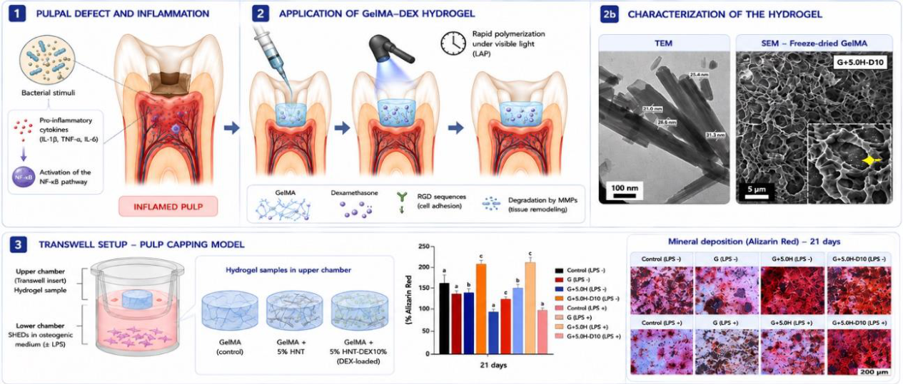
Figure 7.
Development and evaluation of 3D-printed GelMA scaffolds functionalized with bone matrix-derived components (BMdc) for dentin–pulp regeneration. (1) Hydrogel preparation. GelMA solution (15% w/v) was prepared in PBS and combined with LAP photoinitiator (0.075% w/v) and BMdc particles (1% w/v; 75 µm), followed by vortex mixing to obtain a homogeneous GelMA–BMdc bioink. (2) Three-dimensional printing of scaffolds and characterization.GelMA and GelMA–BMdc formulations were extrusion-printed into grid-like scaffolds with different infill densities (40%, 50%, and 60%) to modulate scaffold architecture and pore size. Representative top-view and cross-sectional images illustrate the resulting scaffold microarchitecture. Pore size and cell viability were evaluated to determine the influence of BMdc incorporation and infill density on scaffold properties and biological performance. (3) Evaluation in an artificial pulp chamber (APC) under simulated pulp pressure. The APC system was connected to a liquid-column perfusion system generating a hydrostatic pressure of 20 cm H2O (14.7 mmHg), reproducing physiological pulp pressure. Cell migration toward GelMA and GelMA–BMdc scaffolds, including the 60% infill formulations, was evaluated after 14 days, with representative fluorescence images and quantitative cell counts demonstrating cell recruitment through the dentin toward the hydrogel. (4) Proposed regenerative mechanism and formation of a mineralized barrier. Following scaffold placement over exposed pulp tissue, the 3D-printed GelMA–BMdc scaffold is proposed to promote cell recruitment and infiltration, followed by extracellular matrix deposition and mineralization. Progressive scaffold degradation and bioactive signaling support reparative dentin formation, ultimately resulting in a continuous mineralized dentin bridge that restores pulp–dentin integrity and provides a biological seal. Different lowercase letters indicate statistically significant differences among groups (p < 0.05). Adapted from da Silva et al., 2026, published by Springer under Creative Commons CC BY 4.0.
Figure 7.
Development and evaluation of 3D-printed GelMA scaffolds functionalized with bone matrix-derived components (BMdc) for dentin–pulp regeneration. (1) Hydrogel preparation. GelMA solution (15% w/v) was prepared in PBS and combined with LAP photoinitiator (0.075% w/v) and BMdc particles (1% w/v; 75 µm), followed by vortex mixing to obtain a homogeneous GelMA–BMdc bioink. (2) Three-dimensional printing of scaffolds and characterization.GelMA and GelMA–BMdc formulations were extrusion-printed into grid-like scaffolds with different infill densities (40%, 50%, and 60%) to modulate scaffold architecture and pore size. Representative top-view and cross-sectional images illustrate the resulting scaffold microarchitecture. Pore size and cell viability were evaluated to determine the influence of BMdc incorporation and infill density on scaffold properties and biological performance. (3) Evaluation in an artificial pulp chamber (APC) under simulated pulp pressure. The APC system was connected to a liquid-column perfusion system generating a hydrostatic pressure of 20 cm H2O (14.7 mmHg), reproducing physiological pulp pressure. Cell migration toward GelMA and GelMA–BMdc scaffolds, including the 60% infill formulations, was evaluated after 14 days, with representative fluorescence images and quantitative cell counts demonstrating cell recruitment through the dentin toward the hydrogel. (4) Proposed regenerative mechanism and formation of a mineralized barrier. Following scaffold placement over exposed pulp tissue, the 3D-printed GelMA–BMdc scaffold is proposed to promote cell recruitment and infiltration, followed by extracellular matrix deposition and mineralization. Progressive scaffold degradation and bioactive signaling support reparative dentin formation, ultimately resulting in a continuous mineralized dentin bridge that restores pulp–dentin integrity and provides a biological seal. Different lowercase letters indicate statistically significant differences among groups (p < 0.05). Adapted from da Silva et al., 2026, published by Springer under Creative Commons CC BY 4.0.
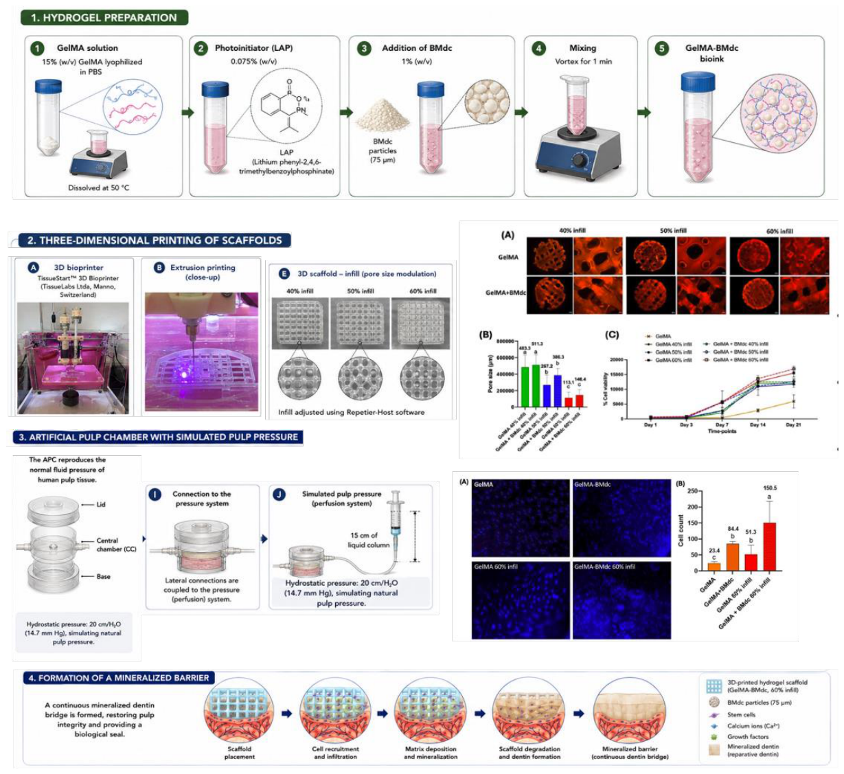
Disclaimer/Publisher’s Note: The statements, opinions and data contained in all publications are solely those of the individual author(s) and contributor(s) and not of MDPI and/or the editor(s). MDPI and/or the editor(s) disclaim responsibility for any injury to people or property resulting from any ideas, methods, instructions or products referred to in the content. |
© 2026 by the authors. Licensee MDPI, Basel, Switzerland. This article is an open access article distributed under the terms and conditions of the Creative Commons Attribution (CC BY) license (http://creativecommons.org/licenses/by/4.0/).
Copyright: This open access article is published under a Creative Commons CC BY 4.0 license, which permit the free download, distribution, and reuse, provided that the author and preprint are cited in any reuse.