

Submitted:
24 August 2026
Posted:
25 August 2026
You are already at the latest version
Abstract
The primary motor cortex (M1) controls voluntary movements through the coordinated interactions between excitatory pyramidal neurons (Pyr) and inhibitory parvalbumin-expressing interneurons (PV-INs). Although PV-INs are critical for regulating motor output and motor coordination, their role in age-related motor decline remains unclear. Here, we investigated how aging alters PV-INs modulation of corticospinal tract (CST) in mice. Aged mice (>14 Months) exhibited significant motor impairments, including deficits in balance and coordination in the balance beam and rope-pulling tasks, while general locomotor activity remained largely preserved. To determine whether these deficits were associated with altered motor cortical circuitry, we combined optogenetics, ex-vivo electrophysiology, and in-vivo electromyographic (eEMG) recordings. Optogenetic activation of PV-INs in aged M1 slices elicited glutamate receptor-dependent depolarizing postsynaptic potentials in layer V pyramidal neurons, indicating recruitment of a glutamate-dependent excitatory circuit. Notably, inhibitory transmission from PV-INs to pyramidal neurons remained intact, suggesting functional remodeling rather than loss of inhibitory connectivity. In vivo, optogenetic activation of PV-INs significantly enhanced CST-mediated hindlimb eEMG responses in aged mice but had little effect in young animals, demonstrating an age-dependent change in PV-IN regulation of motor output. Together, these findings reveal a previously unrecognized form of age-related circuit plasticity in the motor cortex. We propose that PV-INs undergo functional remodeling during aging, preserving inhibitory signaling while enhancing the recruitment of excitatory network mechanisms that facilitate CST output. This adaptive reorganization may represent a compensatory response to age-related motor circuit dysfunction and identifies inhibitory microcircuits as potential therapeutic targets for improving motor function in the aging brain.
Keywords:
parvalbumin interneuron
; primary motor cortex
; corticospinal tract
; postsynaptic potential
; aging
1. Introduction
The primary motor cortex (M1) is a central hub for motor planning, execution, and adaptation [1]. Its function relies on the coordinated activity of excitatory pyramidal neurons and diverse populations of inhibitory interneurons regulate neuronal excitability, temporal precision, and network synchronization [2]. Among cortical interneurons, parvalbumin-expressing interneurons (PV-INs) represent the largest subtype (~40% of inhibitory neurons) in the cortex [3,4]. PV-INs form dense synaptic connections onto the soma and proximal dendrites of pyramidal neurons, positioning them as powerful regulators of cortical output [5] and interact with other types of interneurons. Through their fast-spiking properties and extensive connectivity [6], PV-INs synchronize pyramidal neuron activity and contribute to the precise timing required for motor coordination and skilled movement.
Within M1, layer V pyramidal neurons give rise to the corticospinal tract (CST), the principal descending pathway controlling voluntary movement [7]. These corticospinal neurons project directly to spinal motor circuits and are essential for limb control, balance, and locomotion. PV-INs exert strong inhibitory control over layer V pyramidal neurons and are therefore strategically positioned to regulate CST output [5,8] Beyond simply suppressing excitation, accumulating evidence suggests that PV-INs shape cortical information processing by regulating network oscillations, synchronizing neuronal ensembles, and improving the fidelity of cortical output signals. Disruption of PV-IN function has been implicated in a variety of neurological and neurodevelopmental disorders characterized by motor dysfunction, including autism spectrum disorders, epilepsy, and stroke [9], however, their role in age-related motor decline remains poorly understood.
Aging is associated with progressive impairments in motor performance, including deficits in balance, coordination, gait stability, and movement precision [10]. These functional declines are associated with alterations in motor cortical activity and CST function [11]. Increasing evidence suggests that aging is also accompanied by substantial reorganization of inhibitory circuits, including changes in interneuron connectivity, synaptic transmission, and excitatory-inhibitory (E/I) balance [12]. Such alterations may profoundly influence how motor commands are processed and transmitted through cortical networks. Notably, studies of motor recovery after stroke have demonstrated that targeted manipulation of inhibitory interneuron populations can restore motor function by re-establishing appropriate E/I balance and network dynamics [13,14]. These findings raise the possibility that adaptive changes within inhibitory circuits may similarly influence motor function during normal aging. Despite the recognized importance of PV-INs in motor cortical processing, little is known about how aging affects PV-IN mediated regulation of CST output. In particular, it remains unclear whether age-related motor deficits are associated with alterations in PV-IN connectivity, synaptic signaling, or network function within M1. Addressing these questions is essential for understanding how cortical microcircuits adapt to age-related challenges and whether inhibitory interneurons contribute to compensatory mechanisms that preserve motor function.
In the present study, we combined behavioral analysis, optogenetics, ex-vivo electrophysiology, retrograde tracing, and in-vivo electromyographic recordings to investigate how aging alters PV-IN regulation of CST output. We demonstrate that aging is associated with motor coordination deficits and profound changes in PV-IN mediated cortical signaling. While canonical inhibitory transmission from PV-INs remains intact, activation of PV-INs in the aged motor cortex recruits a glutamate-dependent excitatory circuit and enhances CST motor output. These findings reveal a previously unrecognized form of age-related circuit remodeling and suggest that inhibitory microcircuits may undergo adaptive reorganization to preserve motor cortex function during aging.
2. Materials and Methods
2.1. Animals
This study used a hybrid mouse line, PV-cre/Ai27 (Jackson Strain # 017320/# 012567) which expresses channelrhodopsin-2/tdTomato fusion protein on PV-INs, or PV-cre/Ai39 (Jackson Strain # 017320/# 014539), which expresses Halorhodopsin/EYFP fusion protein on PV-INs. Experimental mice were divided into 2 groups: aged (14-24 months-old) and young adult (3-6 months-old) groups. Animals were housed with ad libitum access to food and water under the care of the UCLA Division of Laboratory Animal Medicine (DLAM). Mice were maintained on a reversed light/dark cycle of 12 hours (light started at 9 PM). All procedures were performed in accordance with protocol approved by the Institutional Animal Care and Use Committee (IACUC) and guidelines of the National Institutes of Health.
2.2. Behavioral Tests
All behavioral tests were performed during the animal’s dark cycle (9 AM-9 PM). For all behavioral tests, mice were in habitat in the testing room in their home cages for at least 1 h. The test animal had another break for at least another 1 h between different tests if more than one test was performed on the same day. The surfaces for all the behavioral tests were carefully sanitized using 75% ethanol between animals to remove any odor and odorant cues. Grid walking. A custom-designed setup was built to obtain 3D information using only one camera [15]. Two side mirrors were used along the shorter and longer side of a transparent cage with wire mesh at the bottom. A top mounted camera captured the movement of the animal on the wire mesh located in the central part of the image, while the two mirrored side-view images were projected on the two sides of the video image. The central image provided information about the animal’s speed, location and orientation; the mirrored images were used to detect the movement of a specific body part through the grid, i.e., depth, duration and 1-dimensional side projected location. A custom-developed program in Igor Pro 6.32 (Wavemetrics, Lake Oswego, OR) was used to analyze the video recordings. The combination of the three image segments yielded the 3D data necessary to determine the exact body part (snout/head, tail or limbs) protruding through the grid, hereafter referred to as a ‘fault’. Multiple parameters could thus be derived based on the 3D information: total travel distance, average speed, time-instantaneous speed, fault number, fault-speed relation, animal location trajectory, center-peripheral location preferences, fault locations on the grid, normalized relative fault location to the body part, fault duration-depth matrices for each limb, distribution of the fault depth and fault durations for each limb, head and tail, grouping the faults based on their depth and duration, animal rotations, etc. Balance beam. The apparatus for the beam-walking test consists of a round horizontal bar 100 cm long and 20 mm in diameter and was elevated 50 cm in a second container, a cushion was placed under the beam to protect the mice in case of falling. Two different types of beams with different surface materials were used: smooth surface and rough surface. A stop was fixed to each end of the beam (see picture in Figure 1B). A top mounted camera captured the movement of the animals on the beam, and a custom-developed program in Igor Pro 6.32 (Wavemetrics, Lake Oswego, OR) was used to do the post-analysis of the video recordings. For the test trial, each animal was placed at the center of the beam and allowed to freely explore for 1 min. Animals that successfully remained on the beam after the exploration period were recorded for up to 2 min. Trials were terminated immediately if the animal fell from the beam during the recording period. Behavioral parameters, including normalized total travel distance on the beam, average locomotor speed, and the center-to-periphery occupancy ratio, were quantified for analysis. Rope pulling. The test method was modified from Khademullah’s study [16]. The testing apparatus was a transparent polyethylene cylinder (15 cm in diameter) with a thin stick crossing at the top (35 cm in height). One end of the rope (3 mm in diameter) was attached and turned over the stick and loosely fixed at the center. To eliminate potential distraction, 3 walls (rear and sides, leaving the top uncovered) were assembled out of white foam board around the apparatus (Figure 1C). Mice were allowed a 5 min habituation period in the cylinder with the string placed inside prior to recording. String movement was towards the floor of the apparatus if the animal performed the pulling behavior. Before recording, the rope pulled during the habituation period was reset to the starting point and the recordings were made with a camera placed on one side of the cylinder (15 cm away from recording table) for 2 mins. After testing, mice were returned to their home cages. To assess the fore limb function, the length of the string pulled during the 2 min recording was documented in cm. Time was recorded for the period during which the mouse was gripping onto the string with either one or both paws and engaged in a pulling behavior. To assess hindlimb function, time was recorded for the period during which the mouse was gripping the string with either one or both paws while in standing position on its hind paws. Time spent pulling the string while in a seated or crouched position was recorded as time spent pulling, but not time spent standing on hindlimbs.
2.3. Motor Output Recordings and Stimulation
In vivo recordings were performed on anesthetized animals. Mice were initially anesthetized with isoflurane, followed by intraperitoneal administration of urethane (1.5 g/kg; U2500, Sigma) for sustained anesthesia [17]. The body temperature was maintained at 37° ± 0.5 using a water-circular heating pad throughout the procedure.
Electrode placement and stimulation protocols: A cranial window (2 x 2 mm) was opened over the left primary motor cortex (center: AP: 0 mm, ML: 1.5 mm) to accommodate recording electrodes and optical fibers. Opto-electrode design and stimulation. A custom-built opto-electrode was used for combining electrical and optical stimulation. The electrode consisted of two bare-tipped tungsten wires (0.005 inch in diameter) paired with 10 mm-long optical fibers (100 μm core diameter, Thor lab, cat # CFMLC21L10). The opto-electrode was positioned at a depth of 400-500 μm to target layer V pyramidal neurons in the hindlimb representation area of M1 (AP: 0.55 ± 1.04 mm, ML: 1.47 ± 0.32 mm) [18]. Electrical stimulation: A 200 Hz, 500-ms train of biphasic pulses (200 μs duration) was delivered to evoke CST-mediated muscle activity. Stimulation intensity (0.2-0.9 mA) was adjusted to elicit observable right hindlimb withdrawal or twitch. If no movement was detected, the electrode was repositioned iteratively until a response was observed. Optical stimulation: PV-INs were selectively modulated using either 475 nm light (20 Hz, 5 ms pulses) or 590 nm light (continuously) delivered via the optical fiber. Electromyography (EMG) recording. EMG signals were recorded from the biceps femoris muscle using two Teflon-coated wires (0.125 mm diameter, bared 2 mm at the tips) inserted via a 26-gauge needle guide. Wires were bent at the tips to secure placement. The stimulation protocol consisted of three sequential phases designed to assess both baseline and PV-IN modulated motor responses. First, a control period was established by delivering four electrical stimulations to the motor cortex at 30-second intervals to record baseline CST-mediated muscle activity. Next, during the light stimulation period, another set of four electrical stimulations was administered while simultaneously activating PV-INs through optical stimulation, allowing observation of how inhibitory interneuron modulation influences motor output. Finally, to isolate the effects of PV-IN activation, a single light-only trial was conducted without concurrent electrical stimulation. This sequential approach enabled systematic comparison of motor responses under control, combining modulation, and PV-IN-specific conditions. The recording was made using the AD instrument octal-bio-amplifier and digitized at 10 kHz with Powerlab digital data acquisition device and LabChart software (ADInstruments Ltd., Colarado Springs, CO, USA). The EMG activity was analyzed offline in Igor Pro 8 (Wavemetrics, Lake Oswego, OR).
2.4. Ex Vivo Slices and Patch Clamp Recordings
Mice were anesthetized with isoflurane and decapitated following UCLA Chancellor’s Animal Research Committee protocol. Horizontal 350 μm thick slices were cut on a Leica VT1000S vibratome in ice-cold N-Methyl-D-Glutamine (NMDG)-based HEPES-buffered solution, containing (in mM): 135 NMDG, 10 D-glucose, 4 MgCl2, 0.5 CaCl2, 1 KCl, 1.2 KH2PO4, 20 HEPES, 27 sucrose (bubbled with 100% O2, pH 7.4, 290-300 mOsm/L). Then, slices were incubated at 32 °C in a reduced sodium artificial CSF (ACSF), containing (in mM): NaCl 85, D-glucose 25, sucrose 55, KCl 2.5, NaH2PO4 1.25, CaCl2 0.5, MgCl2 4, NaHCO3 26, pH 7.3-7.4 when bubbled with 95% O2, 5% CO2. After 30 min, the low sodium ACSF was substituted for normal ACSF at room temperature, containing (in mM): NaCl 126, D-glucose 10, MgCl2 2, CaCl2 2, KCl 2.5, NaH2PO4 1.25, Na Pyruvate 1.5, L-Glutamine 1, NaHCO3 26, pH 7.3-7.4 when bubbled with 95% O2, 5% CO2. For recording, brain slices were transferred to a submerged recording chamber at 34 °C and perfused at 5 ml/min with ACSF. All salts were purchased from Sigma-Aldrich. Slices were visualized under IR-DIC upright microscope (Olympus BX-51WI, 20x XLUMPlan FL N objective). Either the post-synaptic potential or action potential firing was evoked by a single pulse (5 ms) blue light (475 nm) through the objective lens. Cell attached recordings were obtained from M1 cortical layer V pyramidal neurons or nearby PV-INs with borosilicate patch pipettes (4-6 MΩ, outer and inner diameter of 0.8 and 1.5 mm with 0.15 filament, King Precision Glass, Claremont, CA, USA) containing ACSF (the same as above) as the internal pipette solution. Recordings were obtained using an Axon-patch 200B amplifier (Molecular Devices, San Jose, CA, USA), low-pass filtered at 5 kHz (Bessel, 8-pole) and digitized at 10 kHz with a National Instruments data acquisition board (BNC 2110, National Instruments, Austin, TX, USA). All data were acquired and analyzed with EVAN (custom-designed LabView-based software).
2.5. Statistical Analyses
Animals were randomly assigned to experimental procedures. Sample sizes were not predetermined by statistical power analysis; however, efforts were made to minimize animal use while ensuring adequate data collection.
Data are presented as mean ± SEM unless otherwise indicated. Comparisons between two independent groups were performed using the two-tailed Mann-Whitney test when data did not meet assumptions of normality. Two-way ANOVA was used for analyses involving two independent variables, followed by appropriate post hoc multiple-comparison tests when applicable. Statistical values and exact p values are reported in the text, figure legends, and tables. Differences were considered statistically significant at p < 0.05. All statistical analyses were performed using GraphPad Prism 9.0 (GraphPad Software, San Diego, CA, USA).
3. Results
3.1. Aged Mice Exhibit Impaired Motor Coordination but Preserved General Locomotion
In the grid walking test, we employed a sensitive detection and analysis method, as described in method. Parameters including limb fault number (instances of limbs out of the grid), total travel distance, average speed, and preference were quantified (Figure 1A-E). No statistically significant differences were observed between aged (n = 18) and young (n = 12) mice across all these measures, indicating that general locomotion activity was largely preserved with aging.
In contrast, the rope pulling test revealed deficits in fine motor performance and hindlimb function in aged mice (Figure 1F-H). Although the total rope-pulling distance was lower in aged mice (35.00 ± 2.89 cm) than in young mice (88.00 ± 32.62 cm), this difference did not reach statistical significance. However, the duration of hindlimb-supported standing was significantly reduced in aged mice (3.75 ± 0.25 s, n = 4) compared with young mice (10.8 ± 2.35 s, n = 5; p = 0.0159, Mann-Whitney test). These findings suggest impaired hindlimb strength and postural control in aged animals.
The balance beam test further demonstrated age-related deficits in coordination (Figure 1I-L). On the rough-surfaced beam, both age groups were able to traverse the beam, and no significant differences were detected in travel distance or speed. However, on the smooth beam, all aged mice fell within the first minute and were unable to complete the following 2-min testing period, whereas young mice successfully traversed the beam, covering 4539.23 ± 1219.79 mm at an average speed of 81.37 ± 20.45 mm/s. Travel distance and speed were significantly reduced in aged mice (p = 0.0023 and p = 0.001, respectively, Mann-Whitney test).
Together, these behavioral results indicate that aging produces marked impairments in motor coordination and balance while leaving general locomotor activity relatively intact.
3.2. Light-Evoked Depolarizing PSPs Are Only Observed in Pyramidal Neurons from Aged M1 Cortex
To examine age-related changes in communication between PV-INs and pyramidal neurons, we performed loose cell-attached recordings in acute M1 slices from young and aged PV-Cre/Ai27 mice. PV-INs were identified by tdTomato expression and their robust light-evoked firing, whereas pyramidal neurons were identified by their morphology, laminar location, and absence of direct light-evoked spiking.
In slices from aged mice, single-pulse blue-light stimulation (5 ms) reliably evoked depolarizing postsynaptic potentials (PSPs) in layer V pyramidal neurons (Figure 2A). In contrast, no light-evoked PSPs were detected in pyramidal neurons from young mice. The evoked PSPs in aged mice had an average amplitude of 0.32 ± 0.01 mV, a 10-90% rise time of 1.20 ± 0.08 ms, a rise rate of 0.23 ± 0.02 V/s, and a decay time constant of 2.45 ± 0.25 ms (n = 24 neurons from 7 aged mice; Figure 2C-F). No obvious morphological differences were observed between recorded pyramidal neurons from young and aged animals under DIC microscopy (Figure 2B). To determine the synaptic mechanisms underlying these PSPs, we first applied the GABAA receptor antagonist gabazine (GBZ, 40 μM). Gabazine significantly increased both the amplitude and duration of the evoked PSPs, producing a depolarizing response followed by a delayed hyperpolarizing component. Subsequent application of GBZ together with the glutamate receptor antagonists DNQX (10 μM) and APV (50 μM) completely abolished the light-evoked response (Figure 3A). These results indicate that activation of PV-INs in the aged M1 cortex recruits a glutamate-dependent excitatory circuit that ultimately generates depolarizing PSPs in pyramidal neurons.
Recordings from tdTomato-labeled PV-INs showed that light stimulation reliably triggered action potentials, and firing probability increased further in the presence of GBZ and after combined GBZ, DNQX, and APV treatment (Figure 3B). Analysis of spike probability within a 2-ms window revealed that the temporal pattern of PV-IN firing closely correlated with the size and shape of the PSPs recorded in pyramidal neurons (Figure 3C), suggesting a strong functional relationship between PV-IN activity and pyramidal neuron responses in the aged motor cortex.
3.3. PV-INs Maintain Inhibitory Coupling with Pyramidal Neurons During Prolonged Stimulation in the Aged Motor Cortex
To investigate the functional connectivity between PV-INs and pyramidal neurons in the aged M1, we examined inhibitory synaptic transmission during prolonged optogenetic manipulation of PV-IN activity. Previous experiments using single-pulse photo-stimulation demonstrated that activation of PV-INs elicited glutamate-dependent postsynaptic responses in pyramidal neurons, raising the possibility of direct and/or polysynaptic communication between these neuronal populations in the aged cortical network.
To further characterize this interaction, whole-cell patch-clamp recordings were obtained from pyramidal neurons to monitor postsynaptic currents, while cell-attached recordings were performed from PV-INs to verify their responsiveness to sustained photo-stimulation. Brain slices were stimulated with either single-pulse or paired-pulse blue-light trains (2 ms inter-pulse interval) delivered at 20 Hz.
Simultaneous recordings revealed a strong temporal coupling between PV-IN firing and inhibitory synaptic responses in pyramidal neurons. Single-pulse photo-stimulation reliably evoked action potentials in PV-INs and generated corresponding IPSCs in pyramidal neurons, demonstrating robust inhibitory transmission from PV-INs to pyramidal cells (Figure 4A). During paired-pulse stimulation, PV-INs faithfully followed both light pulses throughout the stimulation train, and pyramidal neurons exhibited IPSCs containing distinct primary and secondary peaks that mirrored the paired-pulse activation pattern (Figure 4B). These findings indicate that PV-INs retain the capacity to reliably drive inhibitory synaptic transmission during prolonged repetitive activation in the aged motor cortex.
We next examined whether sustained manipulation of PV-IN activity produces lasting alterations in inhibitory network function. Spontaneous IPSCs were analyzed before and after a prolonged 10 s period of optogenetic activation. Continuous activation of PV-INs did not significantly alter either phasic or tonic IPSC activity, suggesting that prolonged excitation of PV-INs does not induce persistent changes in inhibitory drive within the local cortical network (Figure 4C). In contrast, prolonged optogenetic inhibition of PV-INs using eNpHR3.0 produced a pronounced rebound increase in phasic IPSC activity following termination of the light stimulus, whereas tonic IPSC activity remained unchanged (Figure 4D). These results suggest that transient suppression of PV-IN activity engages compensatory network mechanisms that selectively enhance phasic inhibitory transmission upon release from inhibition, but not the transient activation of PV-INs. Together, these findings demonstrate that PV-INs maintain effective inhibitory control over pyramidal neurons in the aged motor cortex and reveal distinct network responses to prolonged activation versus inhibition of PV-IN activity.
3.4. Activation of M1 PV-INs Enhances CST-Mediated Motor Output in Aged Mice
The primary motor cortex (M1) transmits motor commands to the spinal cord primarily through layer V CST pyramidal neurons. Because aging is associated with deficits in motor performance, we next investigated whether local PV-IN activity modulates CST mediated output in the aged mice.
To determine whether aging selectively affects CST pyramidal neurons, we first identified CST pyramidal neurons using retrograde viral tracing from the lumbar spinal cord (L4-L6). After one month of expression, a large population of layer V pyramidal neurons in M1 was labeled (data not shown). Whole-cell recordings were then obtained from both retrogradely labeled CST pyramidal neurons and neighboring unlabeled pyramidal neurons in aged mice. Comparison of intrinsic membrane properties, including resting membrane potential, membrane input resistance, membrane capacitance, membrane time constant, and rheobase, revealed no significant differences between the two populations (Table 1). These findings suggest that aging-related changes in M1 circuitry are not restricted to CST pyramidal neurons but likely involve broader network reorganization.
We next examined the functional consequences of PV-IN activation on CST-mediated motor output in vivo. Electrical stimulation was delivered to the hindlimb representation area of M1 while electromyographic responses (eEMGs) were recorded from the contralateral tibialis anterior (TA) muscle. Activation of layer V pyramidal neurons generated CST-dependent motor responses, allowing assessment of cortical output under baseline conditions and during optogenetic activation of local PV-INs.
Representative recordings are shown in Figure 5A, B. In aged mice, optogenetic activation of PV-INs significantly increased the amplitude of M1-evoked EMG responses (0.08 ± 0.01 mV vs. 0.14 ± 0.02 mV; event n = 19, p < 0.0001, two-way ANOVA). In contrast, PV-IN activation did not affect EMG amplitude in young mice (0.063 ± 0.007 mV vs. 0.066 ± 0.007 mV; event n = 34, p = 0.7949) (Figure 5C). These results indicate that PV-IN recruitment selectively enhances corticospinal motor output in the aged cortex.
Analysis of EMG event duration revealed a modest but significant increase in young mice following PV-IN activation (0.33 ± 0.02 s vs. 0.35 ± 0.02 s; p = 0.0357), whereas no effect was observed in aged mice (0.407 ± 0.033 s vs. 0.408 ± 0.030 s; p = 0.9937) (Figure 5D). Event frequency was largely unaffected in young mice but was modestly reduced in aged animals following PV-IN activation (412.05 ± 2.70 Hz vs. 407.07 ± 0.97 Hz; p = 0.0483) (Figure 5E). No significant changes were detected in inter-event intervals (Figure 5F).
To further evaluate the temporal organization of motor output, we quantified burstiness and memory of the detected EMG events. Burstiness measures the tendency of events to cluster in time, whereas memory reflects correlations between consecutive inter-event intervals. Although the distribution of burstiness values in aged mice appeared shifted toward lower values compared with young animals, neither burstiness nor memory differed significantly between age groups or between control and PV-IN activation conditions (Figure 5G, H). Thus, while PV-IN activation enhances the magnitude of corticospinal motor output in aged mice, it has minimal effects on the temporal structure of EMG activity.
Together, these findings demonstrate that local PV-IN activation selectively facilitates CST-mediated motor output in aged mice, supporting the idea that inhibitory microcircuits may undergo compensatory functional remodeling to maintain motor cortex output during aging.
4. Discussion
The main finding of the present study was the appearance of light-evoked depolarizing postsynaptic potentials (PSPs) in layer V pyramidal neurons following PV-IN stimulation in aged, but not young, M1 cortex. These PSPs were abolished by glutamate receptor antagonists, indicating that glutamatergic transmission is required for their generation. While PV-INs are traditionally considered inhibitory interneurons, our findings suggest that activation of PV-INs in the aged motor cortex recruits a glutamate-dependent excitatory circuit that ultimately depolarizes pyramidal neurons. Importantly, the prolonged stimulation experiments demonstrated that canonical inhibitory transmission from PV-INs to pyramidal neurons remains intact in aged mice, indicating that aging does not simply convert inhibitory PV-IN synapses into excitatory ones. Rather, aging appears to alter the balance between direct inhibitory and indirect excitatory network mechanisms engaged by PV-IN activity.
Several mechanisms may contribute to this phenomenon. First, aging has been associated with extensive remodeling of cortical microcircuits, including changes in interneuron connectivity, synaptic strength, and network synchronization [19,20]. PV-INs play a critical role in coordinating cortical oscillations and shaping the temporal precision of pyramidal neuron firing [21,22,23]. Age-related alterations in these functions may enhance the recruitment of recurrent excitatory pathways following PV-IN activation, resulting in the emergence of glutamate-dependent depolarizing responses. Second, aging may alter the intrinsic properties and molecular identity of PV-INs themselves [24]. Previous studies have reported age-dependent changes in calcium buffering proteins [25,26], GABAergic signaling components [27,28], and interneuron excitability [19], all of which could influence how PV-IN activity propagates through local cortical circuits.
Another potential mechanism involves alterations in neuronal chloride homeostasis. The inhibitory efficacy of GABAergic transmission critically depends on intracellular chloride concentration, which is primarily regulated by the potassium-chloride cotransporter KCC2 and the sodium-potassium-chloride cotransporter NKCC1 [29,30]. Several studies have reported reduced KCC2 expression and impaired chloride extrusion in aging and neurodegenerative conditions, leading to a depolarizing shift in the GABA reversal potential [31,32,33]. Under such conditions, GABAergic inputs that are normally inhibitory may become less inhibitory or even partially depolarizing. Although our pharmacological experiments indicate that glutamatergic transmission is required for the observed PSPs, altered chloride regulation could increase the excitability of local circuits and facilitate the recruitment of polysynaptic excitatory pathways following PV-IN activation. Future studies examining KCC2 and NKCC1 expression, intracellular chloride dynamics, and GABA reversal potentials in aged M1 neurons will be important for testing this possibility.
Our in vivo recordings further revealed that optogenetic activation of M1 PV-INs significantly enhanced CST-mediated EMG responses in aged mice, despite the canonical inhibitory nature of PV interneurons. At first glance, this finding appears paradoxical; however, it is consistent with an emerging view that PV-INs improve cortical computation by increasing the signal-to-noise ratio of network activity rather than simply suppressing excitation [33,34]. PV-INs preferentially inhibit weakly active or temporally inappropriate neuronal ensembles, thereby sharpening the recruitment of behaviorally relevant pyramidal neurons [35]. In the aged cortex, where inhibitory-excitatory balance and network fidelity are frequently disrupted, activation of PV-INs may restore more precise temporal coordination of cortical activity and enhance the effectiveness of corticospinal output. The selective increase in EMG amplitude observed in aged mice, but not young mice, suggests that PV-IN recruitment may function as a compensatory mechanism that partially offsets age-related circuit dysfunction, consistent with broader evidence that the aging brain can recruit adaptive or compensatory circuit-level changes to help preserve function [36,37]
An additional observation supporting a circuit-level mechanism is that CST-projecting pyramidal neurons exhibited intrinsic membrane properties similar to those of neighboring unlabeled pyramidal neurons. This finding suggests that aging-related changes are unlikely to arise from selective alterations in corticospinal neurons themselves. Instead, the data support a model in which aging primarily affects local microcircuit organization and neuronal interactions, thereby modifying how motor commands are processed and transmitted through otherwise preserved corticospinal output neurons.
Together, our findings support a model in which aging induces functional remodeling of PV-INs mediated cortical circuits. This remodeling appears to preserve inhibitory transmission while simultaneously enhancing the recruitment of glutamate-dependent excitatory pathways and altering the influence of PV-INs on motor cortex output. Such adaptive circuit changes may represent an endogenous compensatory mechanism that helps maintain motor function despite age-related deterioration of cortical networks.
Author Contributions
As defined by the CRediT taxonomy (https://casrai.org/credit/), author contributions were as follows. Conceptualization: X.W., I.M., and D.C.L.; Data curation: X.W.; Formal analysis: X.W.; Funding acquisition: X.W., D.C.L.; Investigation: X.W., I.M., Y.W., A.G., G.S., V.N., A.S., R.H., S.H., D.C.L.; Software: X.W., I.M.; Visualization: X.W.; Writing - original draft: X.W., D.C.L.; Project administration: X.W., D.C.L.
Funding
This work was supported by NIH/NIDA Grant R01DA047637.
Acknowledgments
This study was made possible by the generous support of the Chen Family, the content is solely the responsibility of the authors and does not necessarily reflect the official views of the Chen Family. We would like to thank Dr. László Molnár and Dr. Albert M. Barth for designing the original Igor procedures for behavioral video analysis and electromyograms (EMG) analysis. We also thank Dr. Carlos Cepeda for insightful comments and editing.
Conflicts of Interest
The authors declare no competing financial interests.
References
- Lemon, R.N. Descending pathways in motor control. Annu Rev. Neurosci. 2008, 31, 195–218. [Google Scholar] [CrossRef] [PubMed]
- Tsodyks, M.V.; Skaggs, W.E.; Sejnowski, T.J.; McNaughton, B.L. Paradoxical effects of external modulation of inhibitory interneurons. J. Neurosci. 1997, 17, 4382–4388. [Google Scholar] [CrossRef] [PubMed]
- Rudy, B.; Fishell, G.; Lee, S.; Hjerling-Leffler, J. Three groups of interneurons account for nearly 100% of neocortical GABAergic neurons. Dev. Neurobiol. 2011, 71, 45–61. [Google Scholar] [CrossRef] [PubMed]
- Markram, H.; Toledo-Rodriguez, M.; Wang, Y.; Gupta, A.; Silberberg, G.; Wu, C. Interneurons of the neocortical inhibitory system. Nat. Rev. Neurosci. 2004, 5, 793–807. [Google Scholar] [CrossRef] [PubMed]
- Kuramoto, E.; Tanaka, Y.R.; Hioki, H.; Goto, T.; Kaneko, T. Local Connections of Pyramidal Neurons to Parvalbumin-Producing Interneurons in Motor-Associated Cortical Areas of Mice. eNeuro 2022, 9. [Google Scholar] [CrossRef] [PubMed]
- Kawaguchi, Y.; Kubota, Y. GABAergic cell subtypes and their synaptic connections in rat frontal cortex. Cereb. Cortex 1997, 7, 476–486. [Google Scholar] [CrossRef] [PubMed]
- Ueno, M.; Nakamura, Y.; Li, J.; Gu, Z.; Niehaus, J.; Maezawa, M.; Crone, S.A.; Goulding, M.; Baccei, M.L.; Yoshida, Y. Corticospinal Circuits from the Sensory and Motor Cortices Differentially Regulate Skilled Movements through Distinct Spinal Interneurons. Cell Rep. 2018, 23, 1286–1300 e1287. [Google Scholar] [CrossRef] [PubMed]
- Womelsdorf, T.; Schoffelen, J.M.; Oostenveld, R.; Singer, W.; Desimone, R.; Engel, A.K.; Fries, P. Modulation of neuronal interactions through neuronal synchronization. Science 2007, 316, 1609–1612. [Google Scholar] [CrossRef] [PubMed]
- Okabe, N.; Wei, X.; Abumeri, F.; Batac, J.; Hovanesyan, M.; Dai, W.; Azarapetian, S.; Campagna, J.; Pilati, N.; Marasco, A.; et al. Parvalbumin interneurons regulate rehabilitation-induced functional recovery after stroke and identify a rehabilitation drug. Nat. Commun. 2025, 16, 2556. [Google Scholar] [CrossRef] [PubMed]
- Harris, K.D.; Shepherd, G.M. The neocortical circuit: themes and variations. Nat. Neurosci. 2015, 18, 170–181. [Google Scholar] [CrossRef] [PubMed]
- Chamberland, S.; Nebet, E.R.; Valero, M.; Hanani, M.; Egger, R.; Larsen, S.B.; Eyring, K.W.; Buzsaki, G.; Tsien, R.W. Brief synaptic inhibition persistently interrupts firing of fast-spiking interneurons. Neuron 2023, 111, 1264–1281 e1265. [Google Scholar] [CrossRef] [PubMed]
- Foss-Feig, J.H.; Adkinson, B.D.; Ji, J.L.; Yang, G.; Srihari, V.H.; McPartland, J.C.; Krystal, J.H.; Murray, J.D.; Anticevic, A. Searching for Cross-Diagnostic Convergence: Neural Mechanisms Governing Excitation and Inhibition Balance in Schizophrenia and Autism Spectrum Disorders. Biol. Psychiatry 2017, 81, 848–861. [Google Scholar] [CrossRef] [PubMed]
- Linortner, P.; Jehna, M.; Johansen-Berg, H.; Matthews, P.; Schmidt, R.; Fazekas, F.; Enzinger, C. Aging associated changes in the motor control of ankle movements in the brain. Neurobiol. Aging 2014, 35, 2222–2229. [Google Scholar] [CrossRef] [PubMed]
- Jankowsky, J.L.; Melnikova, T.; Fadale, D.J.; Xu, G.M.; Slunt, H.H.; Gonzales, V.; Younkin, L.H.; Younkin, S.G.; Borchelt, D.R.; Savonenko, A.V. Environmental enrichment mitigates cognitive deficits in a mouse model of Alzheimer’s disease. J. Neurosci. 2005, 25, 5217–5224. [Google Scholar] [CrossRef] [PubMed]
- Barth, A.M.; Mody, I. Novel test of motor and other dysfunctions in mouse neurological disease models. J. Neurosci. Methods 2014, 221, 151–158. [Google Scholar] [CrossRef] [PubMed]
- Khademullah, C.S.; De Koninck, Y. A novel assessment of fine-motor function reveals early hindlimb and detectable forelimb deficits in an experimental model of ALS. Sci. Rep. 2022, 12, 17010. [Google Scholar] [CrossRef] [PubMed]
- Barth, A.M.; Mody, I. Changes in hippocampal neuronal activity during and after unilateral selective hippocampal ischemia in vivo. J. Neurosci. 2011, 31, 851–860. [Google Scholar] [CrossRef] [PubMed]
- Ayling, O.G.; Harrison, T.C.; Boyd, J.D.; Goroshkov, A.; Murphy, T.H. Automated light-based mapping of motor cortex by photoactivation of channelrhodopsin-2 transgenic mice. Nat. Methods 2009, 6, 219–224. [Google Scholar] [CrossRef] [PubMed]
- Rozycka, A.; Liguz-Lecznar, M. The space where aging acts: focus on the GABAergic synapse. Aging Cell 2017, 16, 634–643. [Google Scholar] [CrossRef] [PubMed]
- Guet-McCreight, A.; Tripathy, S.; Sibille, E.; Hay, E. Linking Age Changes in Human Cortical Microcircuits to Impaired Brain Function and EEG Biomarkers. Aging Cell 2026, 25, e70329. [Google Scholar] [CrossRef] [PubMed]
- Isaacson, J.S.; Scanziani, M. How inhibition shapes cortical activity. Neuron 2011, 72, 231–243. [Google Scholar] [CrossRef] [PubMed]
- Cardin, J.A. Inhibitory Interneurons Regulate Temporal Precision and Correlations in Cortical Circuits. Trends Neurosci. 2018, 41, 689–700. [Google Scholar] [CrossRef] [PubMed]
- Hu, H.; Gan, J.; Jonas, P. Interneurons. Fast-spiking, parvalbumin(+) GABAergic interneurons: from cellular design to microcircuit function. Science 2014, 345, 1255263. [Google Scholar] [CrossRef] [PubMed]
- Ueno, H.; Takao, K.; Suemitsu, S.; Murakami, S.; Kitamura, N.; Wani, K.; Okamoto, M.; Aoki, S.; Ishihara, T. Age-dependent and region-specific alteration of parvalbumin neurons and perineuronal nets in the mouse cerebral cortex. Neurochem Int. 2018, 112, 59–70. [Google Scholar] [CrossRef] [PubMed]
- Moyer, J.R., Jr.; Furtak, S.C.; McGann, J.P.; Brown, T.H. Aging-related changes in calcium-binding proteins in rat perirhinal cortex. Neurobiol. Aging 2011, 32, 1693–1706. [Google Scholar] [CrossRef] [PubMed]
- Nikoletopoulou, V.; Tavernarakis, N. Calcium homeostasis in aging neurons. Front Genet 2012, 3, 200. [Google Scholar] [CrossRef] [PubMed]
- Pandya, M.; Palpagama, T.H.; Turner, C.; Waldvogel, H.J.; Faull, R.L.; Kwakowsky, A. Sex- and age-related changes in GABA signaling components in the human cortex. Biol. Sex. Differ. 2019, 10, 5. [Google Scholar] [CrossRef] [PubMed]
- Banuelos, C.; Beas, B.S.; McQuail, J.A.; Gilbert, R.J.; Frazier, C.J.; Setlow, B.; Bizon, J.L. Prefrontal cortical GABAergic dysfunction contributes to age-related working memory impairment. J. Neurosci. 2014, 34, 3457–3466. [Google Scholar] [CrossRef] [PubMed]
- Kaila, K.; Price, T.J.; Payne, J.A.; Puskarjov, M.; Voipio, J. Cation-chloride cotransporters in neuronal development, plasticity and disease. Nat. Rev. Neurosci. 2014, 15, 637–654. [Google Scholar] [CrossRef] [PubMed]
- Blaesse, P.; Airaksinen, M.S.; Rivera, C.; Kaila, K. Cation-chloride cotransporters and neuronal function. Neuron 2009, 61, 820–838. [Google Scholar] [CrossRef] [PubMed]
- Keramidis, I.; McAllister, B.B.; Bourbonnais, J.; Wang, F.; Isabel, D.; Rezaei, E.; Sansonetti, R.; Degagne, P.; Hamel, J.P.; Nazari, M.; et al. Restoring neuronal chloride extrusion reverses cognitive decline linked to Alzheimer’s disease mutations. Brain 2023, 146, 4903–4915. [Google Scholar] [CrossRef] [PubMed]
- Di Cristo, G.; Awad, P.N.; Hamidi, S.; Avoli, M. KCC2, epileptiform synchronization, and epileptic disorders. Prog. Neurobiol. 2018, 162, 1–16. [Google Scholar] [CrossRef] [PubMed]
- Yeganeh, F.; Knauer, B.; Guimaraes Backhaus, R.; Yang, J.W.; Stroh, A.; Luhmann, H.J.; Stuttgen, M.C. Effects of optogenetic inhibition of a small fraction of parvalbumin-positive interneurons on the representation of sensory stimuli in mouse barrel cortex. Sci. Rep. 2022, 12, 19419. [Google Scholar] [CrossRef] [PubMed]
- Atallah, B.V.; Bruns, W.; Carandini, M.; Scanziani, M. Parvalbumin-expressing interneurons linearly transform cortical responses to visual stimuli. Neuron 2012, 73, 159–170. [Google Scholar] [CrossRef] [PubMed]
- Agetsuma, M.; Hamm, J.P.; Tao, K.; Fujisawa, S.; Yuste, R. Parvalbumin-Positive Interneurons Regulate Neuronal Ensembles in Visual Cortex. Cereb. Cortex 2018, 28, 1831–1845. [Google Scholar] [CrossRef] [PubMed]
- Park, D.C.; Reuter-Lorenz, P. The adaptive brain: aging and neurocognitive scaffolding. Annu Rev. Psychol. 2009, 60, 173–196. [Google Scholar] [CrossRef] [PubMed]
- Cabeza, R.; Anderson, N.D.; Locantore, J.K.; McIntosh, A.R. Aging gracefully: compensatory brain activity in high-performing older adults. Neuroimage 2002, 17, 1394–1402. [Google Scholar] [CrossRef] [PubMed]
Figure 1.
Behavioral assessment of locomotor and motor coordination in young and aged mice. (A-E) Grid walking test. (A) Representative overview of the grid walking assay and analysis. From left to right: representative locomotor trajectory (gray) recorded during the 10-min test; color-coded markers indicating limb faults and nose-reaching events beyond the grid; heat map showing the preferred occupancy of the arena, with the center region outlined in red and the surrounding area defined as the periphery; and schematic illustrations of limb fault scoring from head-up and rear-view perspectives. A top-view image of the grid walking apparatus is also shown, consisting of a mesh-bottom walking box (20 × 30 cm base, 20 cm height, transparent acrylic walls, 5 mm thickness) with two mirrors positioned at approximately 45° beneath the apparatus to facilitate visualization and scoring of limb placement errors. (B-E) Quantification of grid walking performance in young and aged mice, including (B) total limb fault number, (C) total travel distance (m), (D) average locomotor speed (mm/s), and (E) center-to-periphery ratio during the 10-min testing period. No significant differences were observed between young and aged mice in any of these locomotor parameters. (F-H) Rope-pulling test. (F) Representative illustration of the rope-pulling apparatus. Quantification of (G) the total rope length pulled during the 2-min testing period (cm) and (H) cumulative hindlimb standing time (s) while pulling the rope. Young mice spent significantly more time standing on their hind limbs during rope-pulling behavior than aged mice. (J-L) Balance beam test. (J) Representative images of the balance beam apparatus consisting of two beams with different surface textures (upper, rough surface; lower, smooth surface). Quantification of (K) total travel distance and (L) average travel speed (mm/s) on the beam during the 2-min test period. Statistical significance is indicated as labeled in the figures. P ≤ 0.05.
Figure 1.
Behavioral assessment of locomotor and motor coordination in young and aged mice. (A-E) Grid walking test. (A) Representative overview of the grid walking assay and analysis. From left to right: representative locomotor trajectory (gray) recorded during the 10-min test; color-coded markers indicating limb faults and nose-reaching events beyond the grid; heat map showing the preferred occupancy of the arena, with the center region outlined in red and the surrounding area defined as the periphery; and schematic illustrations of limb fault scoring from head-up and rear-view perspectives. A top-view image of the grid walking apparatus is also shown, consisting of a mesh-bottom walking box (20 × 30 cm base, 20 cm height, transparent acrylic walls, 5 mm thickness) with two mirrors positioned at approximately 45° beneath the apparatus to facilitate visualization and scoring of limb placement errors. (B-E) Quantification of grid walking performance in young and aged mice, including (B) total limb fault number, (C) total travel distance (m), (D) average locomotor speed (mm/s), and (E) center-to-periphery ratio during the 10-min testing period. No significant differences were observed between young and aged mice in any of these locomotor parameters. (F-H) Rope-pulling test. (F) Representative illustration of the rope-pulling apparatus. Quantification of (G) the total rope length pulled during the 2-min testing period (cm) and (H) cumulative hindlimb standing time (s) while pulling the rope. Young mice spent significantly more time standing on their hind limbs during rope-pulling behavior than aged mice. (J-L) Balance beam test. (J) Representative images of the balance beam apparatus consisting of two beams with different surface textures (upper, rough surface; lower, smooth surface). Quantification of (K) total travel distance and (L) average travel speed (mm/s) on the beam during the 2-min test period. Statistical significance is indicated as labeled in the figures. P ≤ 0.05.
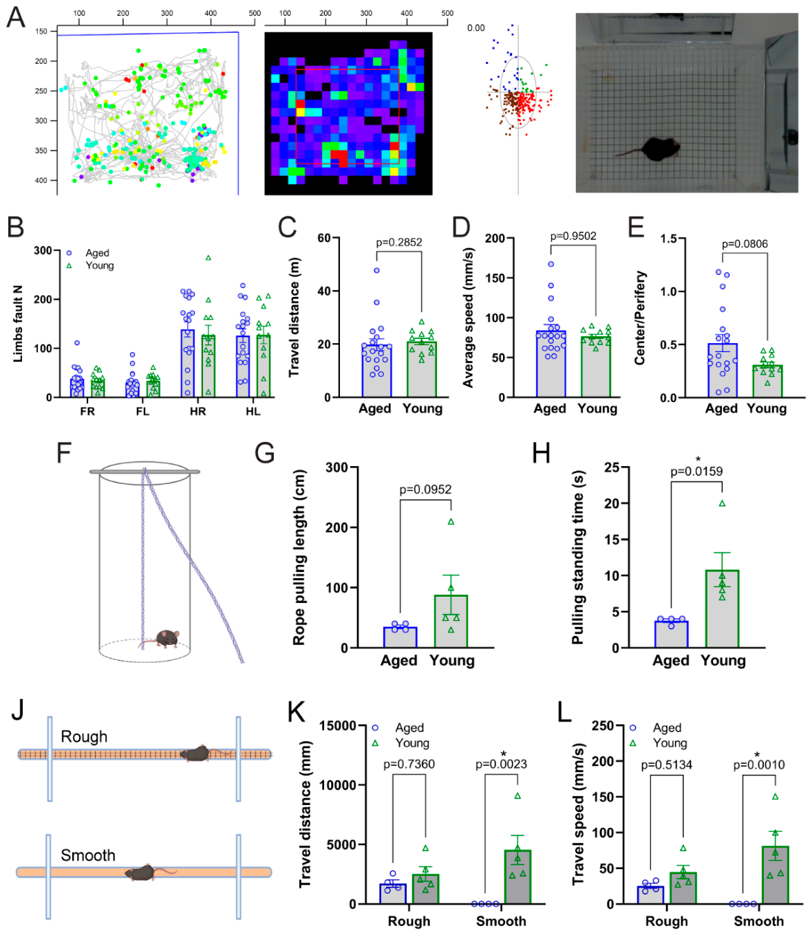
Figure 2.
Optogenetic activation of M1 layer V PV interneurons evokes postsynaptic potentials (PSPs) in layer V pyramidal neurons exclusively in aged mice (>15 months). (A) Representative light-evoked PSP traces recorded from M1 layer V pyramidal neurons using loose-attached single-unit recordings. Representative traces from aged mice (top) show robust light-evoked depolarizing PSPs, whereas no detectable responses were observed in slices from young mice (bottom). The blue bar above each trace indicates the timing and duration (5 ms) of the blue-light stimulation. (B) Representative infrared differential interference contrast (IR-DIC) image of a layer V pyramidal neuron during loose-attached single-unit recording, with the recording pipette visible. (C-F) Quantitative analysis of the light-evoked PSPs recorded from M1 layer V pyramidal neurons, including (C) PSP amplitude, (D) 10-90% rise time, (E) 10-90% rise rate, and (F) decay time (tau). Light-evoked PSPs were consistently observed in slices from aged mice but were absent in slices prepared from young mice.
Figure 2.
Optogenetic activation of M1 layer V PV interneurons evokes postsynaptic potentials (PSPs) in layer V pyramidal neurons exclusively in aged mice (>15 months). (A) Representative light-evoked PSP traces recorded from M1 layer V pyramidal neurons using loose-attached single-unit recordings. Representative traces from aged mice (top) show robust light-evoked depolarizing PSPs, whereas no detectable responses were observed in slices from young mice (bottom). The blue bar above each trace indicates the timing and duration (5 ms) of the blue-light stimulation. (B) Representative infrared differential interference contrast (IR-DIC) image of a layer V pyramidal neuron during loose-attached single-unit recording, with the recording pipette visible. (C-F) Quantitative analysis of the light-evoked PSPs recorded from M1 layer V pyramidal neurons, including (C) PSP amplitude, (D) 10-90% rise time, (E) 10-90% rise rate, and (F) decay time (tau). Light-evoked PSPs were consistently observed in slices from aged mice but were absent in slices prepared from young mice.
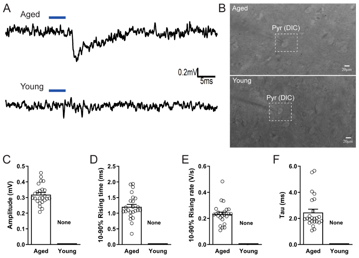
Figure 3.
Optogenetically evoked PSPs in aged M1 slices are mediated by glutamatergic synaptic transmission and are associated with PV interneuron firing patterns. (A) Representative light-evoked PSPs recorded from M1 layer V pyramidal neurons in brain slices from aged mice under three perfusion conditions: ACSF (artificial cerebrospinal fluid), ACSF containing 40 μM gabazine (GBZ) to block GABAA receptor-mediated transmission, and ACSF containing 40 μM GBZ together with 10 μM DNQX and 50 μM APV to block glutamatergic synaptic transmission. Light-evoked PSPs persisted in the presence of GBZ but were abolished following the addition of DNQX and APV. (B) Representative light-evoked action potential firing recorded from optogenetically labeled PV interneurons using the cell-attached patch-clamp configuration under the same three pharmacological conditions described in (A). PV interneurons exhibited reliable light-evoked firing regardless of pharmacological treatment. (C) Action potential probability as a function of time following blue-light stimulation. Spike occurrence was analyzed with a temporal resolution of approximately 2 ms from recordings obtained under ACSF, ACSF + 40 μM GBZ, and ACSF + 40 μM GBZ + 10 μM DNQX + 50 μM APV. The red curves represent the average action potential probability calculated from n = 4 slices (ACSF), n = 5 slices (GBZ), and n = 4 slices (GBZ + DNQX + APV), with each slice obtained from a different animal.
Figure 3.
Optogenetically evoked PSPs in aged M1 slices are mediated by glutamatergic synaptic transmission and are associated with PV interneuron firing patterns. (A) Representative light-evoked PSPs recorded from M1 layer V pyramidal neurons in brain slices from aged mice under three perfusion conditions: ACSF (artificial cerebrospinal fluid), ACSF containing 40 μM gabazine (GBZ) to block GABAA receptor-mediated transmission, and ACSF containing 40 μM GBZ together with 10 μM DNQX and 50 μM APV to block glutamatergic synaptic transmission. Light-evoked PSPs persisted in the presence of GBZ but were abolished following the addition of DNQX and APV. (B) Representative light-evoked action potential firing recorded from optogenetically labeled PV interneurons using the cell-attached patch-clamp configuration under the same three pharmacological conditions described in (A). PV interneurons exhibited reliable light-evoked firing regardless of pharmacological treatment. (C) Action potential probability as a function of time following blue-light stimulation. Spike occurrence was analyzed with a temporal resolution of approximately 2 ms from recordings obtained under ACSF, ACSF + 40 μM GBZ, and ACSF + 40 μM GBZ + 10 μM DNQX + 50 μM APV. The red curves represent the average action potential probability calculated from n = 4 slices (ACSF), n = 5 slices (GBZ), and n = 4 slices (GBZ + DNQX + APV), with each slice obtained from a different animal.
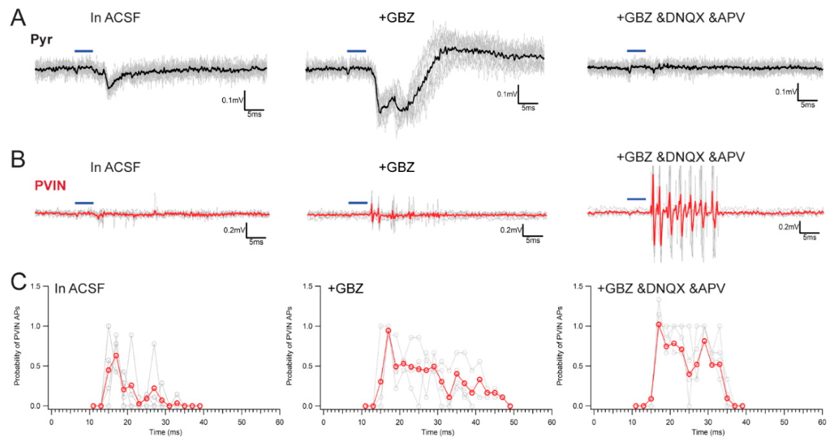
Figure 4.
Functional coupling between PV interneurons and pyramidal neurons in the aged motor cortex during prolonged optogenetic stimulation. (A) A 20 Hz train of single blue-light pulses (5 ms pulse duration, 1 s) reliably evoked action potentials in PV interneurons (red) and corresponding inhibitory postsynaptic currents (IPSCs) in layer V pyramidal neurons (black), demonstrating effective inhibitory synaptic transmission. The temporal relationship among photostimulation, PV interneuron firing, and IPSCs is summarized on the right. (B) A 20 Hz paired-pulse stimulation protocol (5 ms pulse duration, 2 ms inter-pulse interval, 1 s) reliably elicited action potential firing in PV interneurons and generated IPSCs with distinct primary and secondary peaks corresponding to the first and second light pulses, respectively. The timing relationships between photostimulation, PV interneuron firing, and IPSCs are illustrated on the right. (C) Effects of prolonged PV interneuron activation on inhibitory synaptic transmission. Spontaneous IPSCs were analyzed before and after 10 s of continuous photostimulation. Neither phasic nor tonic inhibitory currents were significantly altered following prolonged PV interneuron activation. (D) Effects of prolonged PV interneuron inhibition on inhibitory synaptic transmission. Continuous photoinhibition of PV interneurons for 10 s using eNpHR3.0 induced a rebound increase in phasic IPSC activity following light offset, whereas tonic inhibitory currents remained unchanged. For both (C) and (D), representative traces are shown on the left. The corresponding phasic and tonic current values from the example recordings are indicated in the distribution plots (middle). Comparisons of Hedges’g effect sizes and linear regression slopes are summarized in the right panels.
Figure 4.
Functional coupling between PV interneurons and pyramidal neurons in the aged motor cortex during prolonged optogenetic stimulation. (A) A 20 Hz train of single blue-light pulses (5 ms pulse duration, 1 s) reliably evoked action potentials in PV interneurons (red) and corresponding inhibitory postsynaptic currents (IPSCs) in layer V pyramidal neurons (black), demonstrating effective inhibitory synaptic transmission. The temporal relationship among photostimulation, PV interneuron firing, and IPSCs is summarized on the right. (B) A 20 Hz paired-pulse stimulation protocol (5 ms pulse duration, 2 ms inter-pulse interval, 1 s) reliably elicited action potential firing in PV interneurons and generated IPSCs with distinct primary and secondary peaks corresponding to the first and second light pulses, respectively. The timing relationships between photostimulation, PV interneuron firing, and IPSCs are illustrated on the right. (C) Effects of prolonged PV interneuron activation on inhibitory synaptic transmission. Spontaneous IPSCs were analyzed before and after 10 s of continuous photostimulation. Neither phasic nor tonic inhibitory currents were significantly altered following prolonged PV interneuron activation. (D) Effects of prolonged PV interneuron inhibition on inhibitory synaptic transmission. Continuous photoinhibition of PV interneurons for 10 s using eNpHR3.0 induced a rebound increase in phasic IPSC activity following light offset, whereas tonic inhibitory currents remained unchanged. For both (C) and (D), representative traces are shown on the left. The corresponding phasic and tonic current values from the example recordings are indicated in the distribution plots (middle). Comparisons of Hedges’g effect sizes and linear regression slopes are summarized in the right panels.
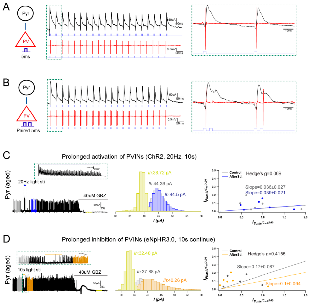
Figure 5.
Activation of M1 PV interneurons facilitates corticospinal tract (CST)-mediated evoked EMG activity in aged mice. (A, B) Representative raw and filtered electromyography (EMG) recordings. Gray traces show the raw EMG signals, whereas black (no light stimulation) and blue (light stimulation) traces represent EMG signals band-pass filtered between 200 and 2,000 Hz. Enlarged views of representative filtered EMG event clusters are shown on the right. The upper panels illustrate recordings obtained without optogenetic stimulation of M1 PV interneurons, whereas the lower panels show recordings during blue-light activation of PV interneurons. Yellow binary traces indicate the event detection windows identified by the automated detection algorithm. (C-F) Quantitative analysis of detected EMG events under control conditions (black) and during optogenetic activation of M1 PV interneurons (blue), including (C) average event amplitude, (D) event duration, (E) inter-event interval (IEI), and (F) event frequency. (G, H) Burstiness-memory analysis of EMG activity. Scatter plots illustrate the distributions of burstiness and memory of detected EMG events under control conditions and during optogenetic activation of M1 PV interneurons, allowing comparison of the temporal organization of CST-mediated motor output between the two conditions.
Figure 5.
Activation of M1 PV interneurons facilitates corticospinal tract (CST)-mediated evoked EMG activity in aged mice. (A, B) Representative raw and filtered electromyography (EMG) recordings. Gray traces show the raw EMG signals, whereas black (no light stimulation) and blue (light stimulation) traces represent EMG signals band-pass filtered between 200 and 2,000 Hz. Enlarged views of representative filtered EMG event clusters are shown on the right. The upper panels illustrate recordings obtained without optogenetic stimulation of M1 PV interneurons, whereas the lower panels show recordings during blue-light activation of PV interneurons. Yellow binary traces indicate the event detection windows identified by the automated detection algorithm. (C-F) Quantitative analysis of detected EMG events under control conditions (black) and during optogenetic activation of M1 PV interneurons (blue), including (C) average event amplitude, (D) event duration, (E) inter-event interval (IEI), and (F) event frequency. (G, H) Burstiness-memory analysis of EMG activity. Scatter plots illustrate the distributions of burstiness and memory of detected EMG events under control conditions and during optogenetic activation of M1 PV interneurons, allowing comparison of the temporal organization of CST-mediated motor output between the two conditions.
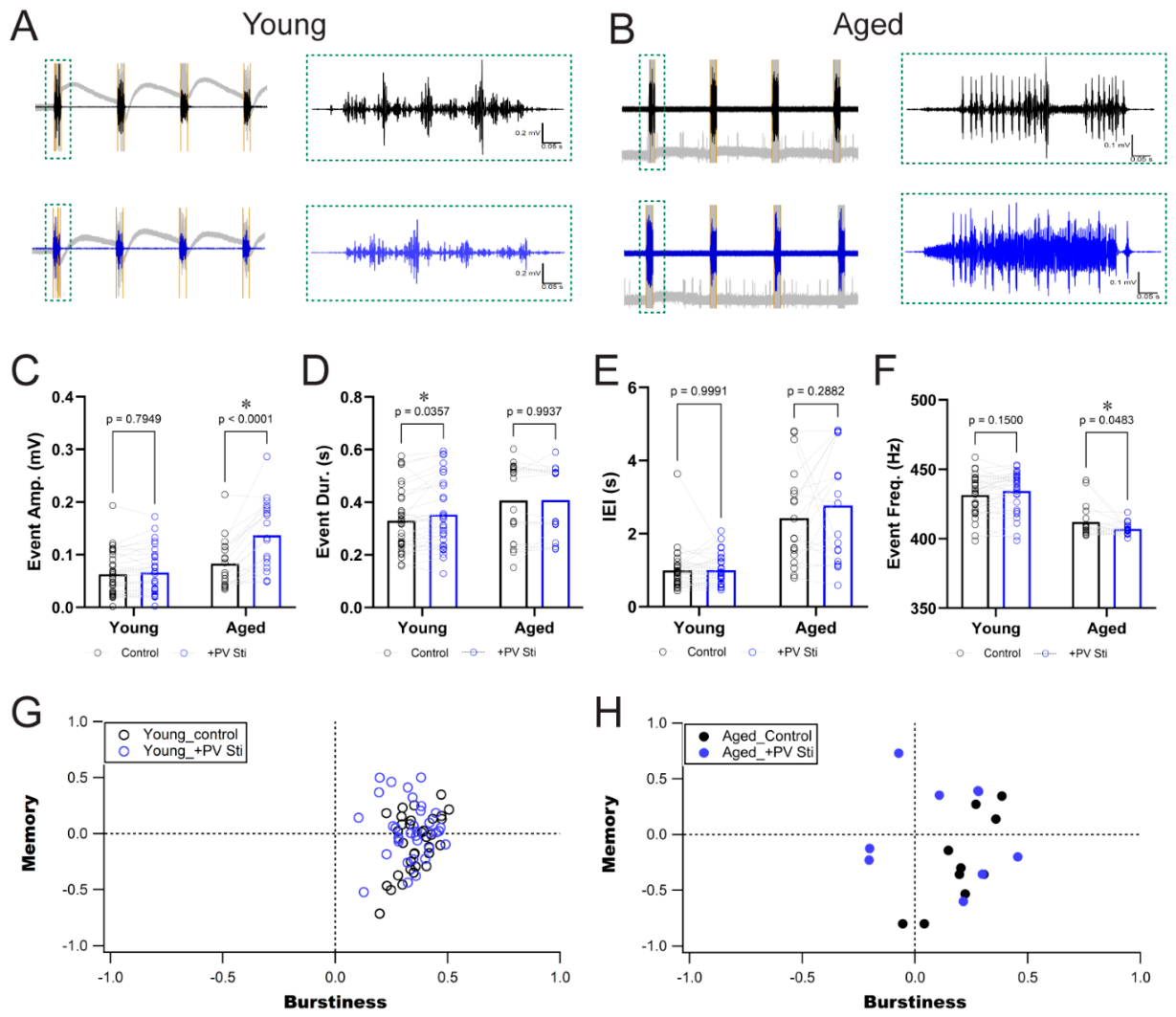

Table 1.
Intrinsic membrane properties of CST-projecting and unlabeled pyramidal neurons in the aged M1 cortex.
Table 1.
Intrinsic membrane properties of CST-projecting and unlabeled pyramidal neurons in the aged M1 cortex.
| n | Cm (pF) | Rm (MΩ) | Tau (ms) | RMP (mV) | Rheobase (pA) | |
| PYR (unlabeled) | 5 | 106.9±23.1 | 255.6±33.4 | 2.18±0.5 | -71.64±0.8 | 326±42.6 |
| PYR (retro) | 5 | 159.8±16.2 | 195.6±25.2 | 3.44±0.4 | -68.12±1.9 | 327±55.9 |
Values are presented as mean ± SEM. No significant differences were detected between groups.
Disclaimer/Publisher’s Note: The statements, opinions and data contained in all publications are solely those of the individual author(s) and contributor(s) and not of MDPI and/or the editor(s). MDPI and/or the editor(s) disclaim responsibility for any injury to people or property resulting from any ideas, methods, instructions or products referred to in the content. |
© 2026 by the authors. Licensee MDPI, Basel, Switzerland. This article is an open access article distributed under the terms and conditions of the Creative Commons Attribution (CC BY) license.
Copyright: This open access article is published under a Creative Commons CC BY 4.0 license, which permit the free download, distribution, and reuse, provided that the author and preprint are cited in any reuse.