

Submitted:
20 August 2026
Posted:
21 August 2026
You are already at the latest version
Abstract
Nematode growth medium (NGM) has been the standard culture medium for Caenorhabditis elegans for over five decades and contains peptone to support proliferation of the bacterial food source, Escherichia coli OP50. However, the increasing use of paraformaldehyde (PFA)-killed OP50 in studies of metabolism, aging, and host-microbe interactions raise the question of whether peptone remains necessary once bacterial proliferation has been eliminated. Here, we systematically compared conventional and peptone-free NGM using PFA-killed OP50 as the bacterial food source. Peptone removal completely prevented residual bacterial proliferation and markedly reduced fungal contamination, thereby improving culture stability. Although worms cultured on peptone-free NGM exhibited reduced food preference and pharyngeal pumping, they maintained normal growth, reproduction, embryonic viability, lifespan, and chemotactic behavior. Peptone-free NGM also reduced basal intracellular reactive oxygen species (ROS) and lipid accumulation, accompanied by downregulation of genes involved in oxidative stress responses and lipogenesis. In addition, worms displayed enhanced locomotor activity and significantly greater resistance to oxidative, ultraviolet, and thermal stress. Collectively, these findings demonstrate that peptone is dispensable when PFA-killed OP50 is used as the bacterial food source. Peptone-free NGM therefore represents a simple, inexpensive, and practical refinement of the conventional culture system for C. elegans studies employing PFA-killed bacterial diets.
Keywords:
Caenorhabditis elegans
; peptone-free NGM
; paraformaldehyde-killed OP50
; culture stability
; physiological resilience
1. Introduction
Caenorhabditis elegans (C. elegans) is one of the most extensively used model organisms in biological and biomedical research owing to its short life cycle, transparent body, invariant cell lineage, fully sequenced genome, and remarkable genetic tractability [1,2,3,4,5]. Since its introduction by Brenner in 1974, C. elegans has become a powerful experimental system for investigating diverse biological processes, including development, aging, metabolism, neurobiology, host-microbe interactions, stress responses, and numerous human diseases [3,4,6,7,8,9]. The ability to culture large, age-synchronized populations together with compatibility with genetic manipulation, fluorescence imaging, high-throughput phenotypic screening, and genome-editing technologies have further established C. elegans as a premier experimental model. These advantages have facilitated their widespread use in both fundamental biological research and translational biomedical applications [4,6,10,11].
Routine laboratory maintenance of C. elegans relies on cultivation on nematode growth medium (NGM) plates seeded with Escherichia coli (E. coli) OP50 as the bacterial food source. Since the original formulation described by Brenner (1974), NGM has remained largely unchanged and consists of agar, sodium chloride, peptone, cholesterol, calcium chloride, magnesium sulfate, and potassium phosphate buffer [3,6]. Among these components, peptone serves as a rich source of peptides, amino acids, and nitrogen that supports the growth and proliferation of E. coli OP50, enabling the formation of a dense bacterial lawn that serves as the primary food source for C. elegans [3,6]. However, the same nutrient-rich environment also favors the growth of unintended bacterial and fungal contaminants introduced during routine laboratory handling [6,12,13,14]. Consequently, bacterial and fungal contamination represents a common challenge during routine C. elegans culture, often necessitating frequent transfer of worms to fresh plates, increased maintenance efforts, and, in severe cases, loss of experimental cultures or valuable strains [6,12].
To minimize microbial contamination, laboratories commonly employ improved aseptic techniques, routine transfer of worms onto fresh plates, antibiotics, or antifungal agents such as Nystatin [6]. In parallel, several studies have explored modifications of conventional NGM by reducing or eliminating peptone for specific applications. Studies have demonstrated that altering bactopeptone concentration influenced bacterial availability and lifespan in C. elegans, while more recent studies have described peptone-free monoxenic media as economical alternatives for worm cultivation and maintenance [15,16,17,18]. Likewise, peptone-free media have occasionally been incorporated into specialized protocols to minimize bacterial overgrowth or facilitate defined nutritional conditions [19,20,21,22,23,24]. Nevertheless, these studies primarily focused on culture methodology or nutritional manipulation rather than evaluating the physiological consequences of peptone removal or its impact on microbial contamination.
More recently, paraformaldehyde (PFA)-killed E. coli OP50 has gained increasing attention as a standardized bacterial food source for investigations of nutrition, dietary restriction, metabolism, aging, host-microbe interactions, and drug responses because bacterial proliferation is eliminated while bacterial biomass and nutritional content are largely preserved [25,26,27,28]. The adoption of PFA-killed bacteria fundamentally changes the requirement for peptone within the culture medium. Since bacterial replication is intentionally prevented, the original purpose of peptone to support bacterial growth is largely eliminated. Under these conditions, residual peptone may instead serve primarily as a nutrient source for contaminating microorganisms inadvertently introduced during routine culture handling. Therefore, when killed bacteria are used as the food source, the continued inclusion of peptone in NGM becomes questionable.
Beyond its effects on culture maintenance, altering the nutritional composition of the culture environment can profoundly influence C. elegans physiology. Dietary composition and bacterial metabolism regulate numerous biological processes, including feeding behaviour, energy homeostasis, lipid metabolism, oxidative stress, locomotion, reproduction, lifespan, and resistance to environmental stressors [25,26,27,29,30,31,32,33,34]. Consequently, eliminating peptone from NGM could potentially modify the nutritional microenvironment surrounding the worms even when bacterial biomass is standardized using PFA-killed OP50. Despite the widespread use of conventional NGM for over five decades, no study has systematically investigated whether peptone remains necessary under these culture conditions or whether its removal can improve culture stability without compromising normal worm physiology.
In the present study, we hypothesized that removal of peptone from NGM would suppress microbial contamination while maintaining normal growth and development of C. elegans when PFA-killed OP50 is used as the bacterial food source. To test this hypothesis, we comprehensively compared conventional and peptone-free NGM with respect to bacterial and fungal contamination, feeding behavior, growth, reproduction, lifespan, chemotaxis, locomotion, lipid accumulation, basal oxidative status, and resistance to oxidative, ultraviolet, and thermal stress. Furthermore, we examined the expression of genes associated with oxidative stress responses, lipid metabolism, locomotion, and stress adaptation to identify molecular changes associated with peptone-free culture conditions. Our findings demonstrate that peptone-free NGM substantially improves culture stability while preserving normal development and reproduction, reducing basal oxidative stress and lipid accumulation, enhancing locomotor activity, and increasing physiological resilience against multiple environmental stressors. These findings establish peptone-free NGM as a simple, inexpensive, and effective alternative to conventional NGM for routine maintenance and physiological studies employing PFA-killed bacterial diets.
2. Materials and Methods
2.1. Reagents
2.1.1. Bleaching Solution (2X)
The bleaching solution was prepared as previously described [10]. A total of 1 mL of 2X bleaching solution was freshly prepared by mixing 0.3 mL of 4% NaOCl, 0.625 mL of 1 M NaOH, and 0.125 mL of double-distilled water.
2.1.2. Nematode Growth Medium (NGM) and Peptone-Free NGM
Conventional NGM (NGM(Peptone+)) was prepared as previously described [6]. Briefly, for 1 liter of NGM, 3 g of NaCl, 2.5 g of peptone, and 17 g of agar were dissolved in 975 mL of double-distilled water. Peptone-free NGM (NGM(Peptone-)) was prepared using the same formulation except that peptone was omitted. The mixture was autoclaved at 121 °C for 20 minutes. After cooling to approximately 55 °C, the following sterile-filtered supplements were added: 1 mL of 1 M CaCl₂, 1 mL of 1 M MgSO₄, 25 mL of 1 M potassium phosphate buffer (pH 6.0), 1 mL of 5 mg/mL cholesterol in ethanol, and 1.25 mL of 10 mg/mL Nystatin solution in 70% ethanol. The medium was then mixed thoroughly and poured into 35- and 60-mm petri plates under sterile conditions. Plates were allowed to solidify at room temperature and stored at 4 °C until use.
2.1.3. M9 Buffer
M9 buffer was prepared as previously described [10]. A total of 100 mL of M9 buffer was prepared by dissolving 0.6 g of Na2HPO4, 0.3 g of KH2PO4, and 0.5 g of NaCl in double-distilled water. Then, 0.1 mL of 1M MgSO4 was added, and the volume was adjusted to 100 mL with double-distilled water. The solution was autoclaved and allowed to cool to room temperature before use.
2.1.4. S-Basal Buffer
S-basal buffer was prepared as previously described [6]. A total of 100 mL of S-basal buffer was prepared by dissolving 0.59 g of NaCl in double-distilled water. Then, 5 mL of potassium phosphate buffer (pH 6.0) was added, and the final volume was adjusted to 100 mL with double-distilled water. The solution was autoclaved and allowed to cool to room temperature before adding 0.1 mL of 5 mg/mL cholesterol in ethanol.
2.1.5. S-Complete Buffer
S-complete buffer was prepared as previously described [6]. A total of 100 mL of S-complete buffer was prepared by mixing 1 mL of 1 M potassium citrate buffer (pH 6.0), 0.1 mL of 10X trace metal solution, 0.3 mL of 1 M CaCl2, and 0.3 mL of 1 M MgSO4 with 98.3 mL of S-basal buffer.
2.2. Methods
2.2.1. C. elegans Strains, PFA-Killed OP50, and Culture Conditions
The C. elegans strain N2 Bristol (wild-type) and GRU101 were maintained on 60 mm NGM plates seeded with 30 µL of PFA-killed E. coli OP50. To prepare PFA-killed OP50, a 500 mL culture of E. coli OP50 was incubated overnight (~16 hrs) at 37°C in an orbital shaker at 200 rpm. The culture was then treated with 1% paraformaldehyde (PFA) for 2 hours, followed by four washes with autoclaved double-distilled water to remove residual PFA. The OP50 cell pellet was resuspended in sterile S-complete buffer at a final concentration of 250 mg/mL (~5x10¹⁰ OP50/mL). Worm cultures were grown and maintained at 20°C. Some strains were provided by the CGC, which is funded by NIH Office of Research Infrastructure Programs (P40 OD010440).
2.2.2. Synchronization of C. elegans Population
Confluent 60 mm NGM plates with adult worms were washed with 1 mL of M9 buffer to collect gravid adults and laid eggs. The M9 buffer containing worms and eggs was transferred to a 15 mL Falcon tube, followed by the addition of 13 mL of M9 buffer. The suspension was centrifuged at 1500 rpm for 2 minutes at room temperature. The supernatant was carefully discarded without disturbing the worm pellet, and the washing step was repeated with 14 mL of M9 buffer until the buffer appeared clear of bacteria. To the pellet, 1 mL of M9 buffer and 1 mL of bleaching solution were added, and the mixture was vortexed at 2500 rpm for 6 minutes continuously. The reaction was stopped by adding 12 mL of M9 buffer, followed by centrifugation at 2000 rpm for 1 minute at room temperature. The pellet was washed three additional times with 14 mL of M9 buffer to remove debris. The egg pellet was resuspended in 1 mL of M9 buffer and incubated at 20°C for 16 hours at 30 rpm.
2.2.3. Bacterial and Fungal Growth Measurement
To evaluate bacterial growth, live E. coli OP50 and OP50 treated with 0.25%, 0.5%, or 1% PFA (approximately 5 × 10¹⁰ cells/mL) were diluted 10-35 in sterile phosphate-buffered saline. A 100 µL aliquot of each dilution was spread onto conventional NGM(Peptone+) and peptone-free NGM(Peptone-) plates and incubated at 37 °C for 16 h. Bacterial growth was quantified by enumerating colony-forming units (CFU) using an automatic colony counter (Scan 4000, Interscience) equipped with Scan software (version 8), and the results were expressed as CFU/mL. For fungal growth analysis, a sterile filter paper disc containing actively growing Penicillium chrysogenum mycelium was placed at the center of NGM(Peptone+) and NGM(Peptone-) plates supplemented with or without Nystatin (12.5 µg/mL). Plates were incubated at 26 °C for 5 days, and fungal colony diameter was measured daily using a digital caliper. Fungal growth rate was expressed as the increase in colony diameter per day (mm/day).
2.2.4. Food Preference Assay
Food preference was evaluated using a two-choice assay. Briefly, 60-mm Petri dishes were prepared with two equal halves consisting of NGM(Peptone+) and NGM(Peptone-). After the media had solidified, 10 μL of 1% PFA-killed E. coli OP50 suspension was spotted at the center of each half of the plate and allowed to air-dry. The assay plates were maintained at 20 °C until use. Approximately 25 age-synchronized L4 worms were placed at the center of each assay plate, equidistant from the two bacterial lawns, and incubated at 20 °C. The number of worms present on each bacterial lawn was recorded after 1 and 8 h of incubation. Worms remaining outside both bacterial lawns were excluded from the analysis. Food preference was expressed as the percentage of worms present on each bacterial lawn and calculated using the following equation: . Three independent biological experiments were performed with three assay plates per experiment (n = 9 biological replicates).
2.2.5. Pharyngeal Pumping Assay
Pharyngeal pumping was quantified using the PharaGlow pipeline as previously described [35]. Briefly, age-synchronized L1 larvae of the transgenic strain GRU101, which expresses yellow fluorescent protein (YFP) specifically in the pharyngeal muscles while exhibiting a wild-type phenotype, were cultured on NGM(Peptone+) or NGM(Peptone-) plates seeded with 1% PFA-killed E. coli OP50 and maintained at 20 °C until they reached the L4 stage. Videos of freely moving L4 worms were acquired using a Nikon SMZ25 stereo fluorescence microscope equipped with a GFP filter cube, a D-LEDI fluorescence illumination system, and an OPTO-EDU A59.2225-20MMA monochrome camera. Fluorescence videos were recorded at a 1× zoom setting for 30 s at 30 frames per second (fps) under identical imaging conditions for all experimental groups. Pharyngeal pumping events were automatically detected and quantified using the PharaGlow software with the default analysis parameters. The mean pumping rate for each worm was calculated and expressed as pumping frequency (Hz). A total of 80 worms per condition from three independent biological replicates were analyzed.
2.2.6. Body Size and Volume Measurement
Body length and body volume of C. elegans were quantified using the WorMachine image analysis platform [36]. Briefly, age-synchronized L1 worms were cultured on NGM(Peptone+) or NGM(Peptone-) plates seeded with 1% PFA-killed E. coli OP50 and maintained at 20 °C. Worms were imaged at 0, 24, 48, 72, 96, and 120 h after L1 plating. Under the culture conditions used, these imaging time points corresponded approximately to the predominant L1, L2, L3, L4, young adult, and gravid adult stages, respectively. Bright-field images were acquired using a Nikon SMZ25 stereomicroscope equipped with a high-resolution monochrome camera (OPTO-EDU A59.2225-20MMA). All images were captured at a 1× zoom setting under identical illumination conditions to ensure consistent image quality across experimental groups. Captured images were analyzed using WorMachine implemented in MATLAB (R2024a) following the standard protocol described by Hakim et al. (2018). The software automatically identifies individual worms, segments them from the background, and extracts morphological features using skeletonization and image-processing algorithms. Body length was determined from the worm skeleton, while additional morphological parameters, including midwidth and thickness, were extracted from the software output. Worm body volume was subsequently calculated in Microsoft Excel using an ellipsoidal approximation according to the following equation: . A minimum of 200 worms per condition were analyzed. All images were processed using identical analysis parameters. Worms that were overlapping, touching, or incorrectly segmented were excluded from the analysis to ensure accurate morphometric measurements.
2.2.7. Brood Size and Embryonic Viability Analysis
Brood size and embryonic viability were assessed as previously described [37]. Briefly, age-synchronized L1 worms were cultured on NGM(Peptone+) or NGM(Peptone-) plates seeded with 1% PFA-killed E. coli OP50 and maintained at 20 °C until they reached the L4 stage. Individual L4-stage hermaphrodites were then transferred onto fresh 35-mm NGM(Peptone+) or NGM(Peptone-) plates seeded with 1% PFA-killed E. coli OP50 and maintained at 20 °C. Each worm was allowed to lay eggs for 24 h, after which the adult was transferred daily to a fresh plate for a total of three consecutive days. Plates containing the laid embryos were subsequently incubated at 20 °C for an additional 24 h to allow viable embryos to hatch. Following incubation, the number of live larvae and unhatched embryos on each plate was counted using a stereomicroscope (Nikon SMZ745T). Hatched larvae were considered viable progeny, whereas unhatched embryos after the incubation period were considered non-viable, consistent with established methods [37]. Brood size was calculated as the total number of progeny (live larvae plus unhatched embryos) produced per hermaphrodite over the three-day reproductive period. Embryonic viability (%) was calculated using the following equation: . Three individual worms were analyzed per condition in each biological replicate (n = 9 worms per condition). All assays were performed under identical experimental conditions to ensure reproducibility.
2.2.8. Lifespan Assay
Lifespan analysis was performed as previously described with minor modifications [38]. Briefly, age-synchronized L1 worms were cultured on NGM(Peptone+) or NGM(Peptone-) plates supplemented with 120 μM 5-fluoro-2′-deoxyuridine (FUdR) and seeded with 1% PFA-killed E. coli OP50. Worms were maintained at 20 °C throughout the experiment. Worm survival was scored at 2-day intervals using a stereomicroscope (Nikon SMZ745T) until all worms had died. Worms were considered dead when they failed to respond to gentle touch with a platinum wire pick. Worms that died due to desiccation after crawling off the agar surface, or other non-age-related causes were censored from the analysis. Survival curves were generated using the Kaplan-Meier method, and statistical significance between groups was determined using the log-rank (Mantel-Cox) test.
2.2.9. Chemotaxis Assay
Chemotactic responses toward an attractant and a repellent were evaluated using a four-quadrant chemotaxis assay as previously described with minor modifications [39]. Briefly, age-synchronized L1 worms were cultured on NGM(Peptone+) or NGM(Peptone-) plates seeded with 1% PFA-killed Escherichia coli OP50 and maintained at 20 °C until they reached the L4 stage. Chemotaxis assays were performed on 60-mm chemotaxis agar plates divided into four equal quadrants consisting of two opposite test quadrants and two opposite control quadrants. A circle (0.5 cm radius) was marked at the center of each plate, and worms remaining within this region at the end of the assay were excluded from analysis. Test and control spots were positioned equidistant from the center in their respective quadrants. Separate assay plates were prepared for attractant and repellent assays. For the attractant assay, 2 μL of 1% (v/v) diacetyl was applied to each test spot, whereas 2 μL of 100% glycerol was applied to each test spot for the repellent assay. In both assays, 2 μL of double-distilled water was applied to each control spot. Approximately 25 age-synchronized L4 worms were washed three times with M9 buffer to remove residual bacteria and transferred to the center of each assay plate. After the liquid was absorbed into the agar, plates were incubated at 20 °C for 1 h. The number of worms present in each quadrant that had crossed the central circle was then recorded. The chemotactic index was calculated using the following equation: . A chemotaxis index of +1 indicates complete attraction toward the test compound, whereas a value of -1 indicates complete avoidance. Three independent biological experiments were performed with three assay plates per condition (n = 9 biological replicates).
2.2.10. Locomotion Analysis
Locomotion of C. elegans was analyzed at the L4 stage using video-based tracking as previously described [40,41]. Briefly, age-synchronized L1 worms were cultured on NGM(Peptone+) or NGM(Peptone-) plates seeded with 1% PFA-killed E. coli OP50 and maintained at 20 °C until they reached the L4 stage. Videos of freely moving L4 worms were acquired using a Nikon SMZ25 stereomicroscope equipped with a high-resolution monochrome camera (OPTO-EDU A59.2225-20MMA). Recordings were captured at a 1× zoom setting, with a frame rate of 28 fps and a duration of 60 s under identical illumination conditions for all experimental groups. Two locomotion parameters were quantified: body length traveled per second (BLPS) and body bends per second (BBPS). For BLPS measurements, videos of worms crawling on NGM agar plates were recorded. For BBPS measurements, 200 μL of M9 buffer was added to the surface of the NGM plate immediately before video acquisition to induce swimming behavior. Videos were analyzed using ImageJ (version 1.54f) with the wrMTrck plugin (version 1.04). The plugin automatically tracks individual worms and quantifies locomotion parameters, including BLPS and BBPS, based on frame-by-frame movement and body posture analysis. A minimum of 300 worms per condition were analyzed across three independent biological replicates. All recordings and analyses were performed under identical imaging and analysis parameters to ensure consistency across experimental groups.
2.2.11. Lipid Quantification by Nile Red Staining
Intracellular lipid accumulation was quantified by Nile Red staining as previously described with minor modifications [42]. Briefly, age-synchronized L1 worms were cultured on NGM(Peptone+) or NGM(Peptone-) plates seeded with 1% PFA-killed Escherichia coli OP50 and maintained at 20 °C until they reached the L4 stage. All worms from each plate were collected into 1.5-mL microcentrifuge tubes, washed three times with phosphate-buffered saline containing 0.01% Triton X-100 (PBST) to remove residual bacteria, and fixed in 40% (v/v) isopropanol for 3 min at room temperature. Following fixation, worms were incubated with freshly prepared Nile Red working solution for 2 h at room temperature in the dark. Excess stain was removed by washing the worms with PBST for 30 min, after which the worms were resuspended in the remaining buffer and mounted onto glass microscope slides. Fluorescence images were acquired using an inverted fluorescence microscope (OPTO-EDU A16.2615-L-4) equipped with a GFP filter set at 20× magnification. All images were captured using identical exposure time, illumination intensity, and camera settings to ensure consistency between experimental groups. Fluorescence intensity was quantified using ImageJ (version 1.54f). Individual worms were manually outlined, and the mean fluorescence intensity was measured following background subtraction. Thirty randomly selected worms per condition from three independent biological replicates were analyzed. Lipid accumulation was expressed as normalized Nile Red fluorescence intensity.
2.2.12. Intracellular ROS Measurement
Intracellular reactive oxygen species (ROS) levels were quantified using 2′,7′-dichlorodihydrofluorescein diacetate (H₂DCFDA) as previously described with minor modifications [43]. Briefly, age-synchronized L1 worms were cultured on NGM(Peptone+) or NGM(Peptone-) plates seeded with 1% PFA-killed E. coli OP50 and maintained at 20 °C until they reached the L4 stage. L4 worms were washed three times with M9 buffer to remove residual bacteria and resuspended in M9 buffer. Approximately 50 worms were transferred into each well of a black 96-well microplate containing 40 μL of M9 buffer. Subsequently, 50 μL of H₂DCFDA working solution was added to each well to obtain a final dye concentration of 25 μM. Blank wells containing M9 buffer and H₂DCFDA without worms were included to correct for background fluorescence. Fluorescence was measured immediately using a multimode microplate reader (BioTek Synergy H1) at an excitation wavelength of 490 nm and an emission wavelength of 530 nm. Fluorescence intensity was recorded at 1-h intervals for 8 h. Blank-subtracted fluorescence values were used to determine intracellular ROS levels. Three independent biological experiments were performed with three technical replicates per condition.
2.2.13. Oxidative Stress-Resistance Assay
Oxidative stress resistance was assessed using a paraquat-induced survival assay as previously described with minor modifications [44]. Briefly, age-synchronized L1 worms were cultured on NGM(Peptone+) or NGM(Peptone-) plates seeded with 1% PFA-killed E. coli OP50 and maintained at 20 °C until they reached the L4 stage. Freshly prepared 500 mM paraquat (methyl viologen) solution in M9 buffer was dispensed into the wells of a 96-well microplate. Approximately 15 synchronized L4 worms were transferred into each paraquat-containing well, and the plates were maintained at 20 °C throughout the assay. Worm survival was assessed at 1-h intervals using a stereomicroscope (Nikon SMZ745T). Before scoring, the plate was gently agitated to stimulate worm movement. Worms that failed to respond to gentle agitation and showed no visible body movement were scored as dead. Survival was monitored until all worms had died. A minimum of 240 worms per condition were analyzed across three independent biological replicates. Survival curves were generated using the Kaplan-Meier method, and statistical significance between groups was determined using the log-rank (Mantel-Cox) test.
2.2.14. Ultraviolet-Stress Resistance Assay
Ultraviolet (UV) stress resistance was assessed by determining the survival of worms following UV-C irradiation. Briefly, age-synchronized L1 worms were cultured on NGM(Peptone+) or NGM(Peptone-) plates seeded with 1% PFA-killed E. coli OP50 and maintained at 20 °C until they reached the L4 stage. Age-synchronized L4 worms were transferred onto fresh NGM(Peptone+) or NGM(Peptone-) plates supplemented with 120 μM FUdR and seeded with 1% PFA-killed Escherichia coli OP50. The worms were then directly exposed to 254 nm UV-C radiation (3 mJ/cm²) using a UV crosslinker (Analytik Jena UVP CL-3000). Following irradiation, worms were maintained at 20 °C on their respective culture plates, and survival was monitored daily using a stereomicroscope (Nikon SMZ745T) until all worms had died. Worms were considered dead when they failed to respond to gentle touch with a platinum wire pick. Worms that died due to desiccation after crawling off the agar surface, or other non-age-related causes were censored from the analysis. A minimum of 180 worms per condition were analyzed across three independent biological replicates. Survival curves were generated using the Kaplan-Meier method, and statistical significance between groups was determined using the log-rank (Mantel-Cox) test.
2.2.15. Thermotolerance Assay
Thermotolerance was assessed by determining the survival of worms under acute heat stress. Briefly, age-synchronized L1 worms were cultured on NGM(Peptone+) or NGM(Peptone-) plates seeded with 1% PFA-killed E. coli OP50 and maintained at 20 °C until they reached the L4 stage. Age-synchronized L4 worms maintained on NGM(Peptone+) or NGM(Peptone-) plates were directly exposed to acute heat stress by transferring the plates to a 35 °C incubator. Worms were maintained at 35 °C throughout the assay, and survival was monitored at 12-h intervals using a stereomicroscope (Nikon SMZ745T) until all worms had died. Worms were considered dead when they failed to respond to gentle touch with a platinum wire pick. Worms that died due to desiccation after crawling off the agar surface, or other non-age-related causes were censored from the analysis. A minimum of 300 worms per condition were analyzed across three independent biological replicates. Survival curves were generated using the Kaplan-Meier method, and statistical significance between groups was determined using the log-rank (Mantel-Cox) test.
2.2.16. Quantitative PCR and Relative Gene Expression Analysis
Relative gene expression was determined by quantitative real-time PCR (qPCR). Briefly, age-synchronized L1 worms were cultured on NGM(Peptone+) or NGM(Peptone-) plates seeded with 1% PFA-killed Escherichia coli OP50 and maintained at 20 °C until they reached the L4 stage. L4 worms were collected, washed three times with M9 buffer to remove residual bacteria, pelleted by centrifugation, and stored at -80 °C until RNA extraction. Total RNA was extracted using TRIzol™ Reagent (Invitrogen) following the standard TRIzol-chloroform extraction method according to the manufacturer's instructions. RNA concentration and purity were determined using a BioTek Synergy H1 multimode microplate reader (Agilent Technologies), and RNA integrity was verified by agarose gel electrophoresis. First-strand cDNA was synthesized from 1 μg of total RNA using M-MLV Reverse Transcriptase, random hexamer primers, dNTPs, RNase inhibitor, and the appropriate reaction buffer (Aura Biotechnologies) according to the manufacturer's instructions. Quantitative real-time PCR was performed in a 20 μL reaction volume using GoTaq® qPCR Master Mix (Promega) on a CFX Opus 96 Real-Time PCR System (Bio-Rad Laboratories). Each reaction was performed in triplicate using gene-specific primers (Table S1). The thermal cycling conditions consisted of an initial denaturation at 95 °C for 2 min, followed by 40 cycles of 95 °C for 15 s and 60 °C for 30 s. A melt curve analysis was performed following amplification to verify the specificity of each PCR product. The expression of target genes was normalized to the reference gene tba-1, and relative gene expression was calculated using the 2-ΔΔCt method [45]. Relative expression levels are presented as log10 fold change relative to worms cultured on NGM(Peptone+). Three independent biological replicates were analyzed for each experimental group.
2.2.17. Statistical Analysis
All experiments were performed using at least three independent biological replicates unless otherwise indicated. Data are presented as mean ± standard error of the mean (SEM) unless otherwise stated. Statistical analyses were performed using GraphPad Prism v.10. Comparisons between two experimental groups were performed using an unpaired two-tailed Student's t-test for bacterial and fungal growth, food preference, pharyngeal pumping rate, brood size, embryonic viability, chemotaxis index, Nile Red fluorescence intensity, locomotion parameters (BLPS and BBPS), and relative gene expression. Body length, body volume, and intracellular ROS measurements were analyzed using two-way analysis of variance (ANOVA). Survival data from lifespan, oxidative stress resistance, UV stress resistance, and thermotolerance assays were analyzed using the Kaplan-Meier method, and statistical significance between survival curves was determined using the log-rank (Mantel-Cox) test. A P value of < 0.05 was considered statistically significant. Statistical significance is indicated as follows: nsP > 0.05; *P < 0.05; **P < 0.01; ***P < 0.001; and ****P < 0.0001.
3. Results
3.1. Peptone-Free NGM Suppresses Bacterial and Fungal Contamination
To determine whether removal of peptone from NGM affects microbial contamination, conventional NGM (NGM(Peptone+)) and peptone-free NGM (NGM(Peptone-)) plates were seeded with either live Escherichia coli OP50 or OP50 treated with different concentrations (0.25%, 0.5%, and 1%) of paraformaldehyde (PFA). Bacterial growth was quantified by colony-forming units (CFU/mL). In parallel, the growth of Penicillium chrysogenum (P. chrysogenum) was evaluated on NGM(Peptone+) and NGM(Peptone-) plates in the presence or absence of Nystatin. We observed that bacterial proliferation occurred on conventional NGM irrespective of whether live or PFA-treated OP50 was used, whereas no detectable bacterial growth was observed on peptone-free NGM under any condition (Figure 1A). Furthermore, P. chrysogenum exhibited significantly reduced growth on peptone-free NGM compared with conventional NGM, and fungal growth was completely inhibited in the presence of Nystatin (Figure 1B). These results suggest that removal of peptone effectively suppresses bacterial proliferation and markedly reduces fungal contamination, thereby improving the microbial stability of NGM culture plates.
3.2. Peptone-Free NGM Alters Feeding Behavior of C. elegans
To investigate whether the absence of peptone influences feeding behavior, age-synchronized L4 worms were subjected to a food preference assay between bacterial lawns grown on NGM(Peptone+) and NGM(Peptone-). In addition, pharyngeal pumping rate was quantified as an indicator of food intake. We observed that worms showed a significantly greater preference for bacterial lawns grown on conventional NGM than those grown on peptone-free NGM at both 1 h and 8 h after the start of the assay (Figure 2A). Consistent with this observation, GRU101 worms maintained on peptone-free NGM exhibited a significantly lower pharyngeal pumping rate than worms cultured on conventional NGM (Figure 2B). Together, these results indicate that peptone-free NGM alters feeding behavior in C. elegans.
3.3. Peptone-Free NGM Does Not Adversely Affect Growth, Reproduction, Lifespan, or Chemotactic Behavior in C. elegans
To determine whether removal of peptone affects normal physiological development, age-synchronized L1 worms were cultured on NGM(Peptone+) and NGM(Peptone-) plates seeded with 1% PFA-killed E. coli OP50 at 20 °C. Body length, body volume, brood size, embryonic viability, lifespan, and chemotaxis were subsequently evaluated. We observed that worms cultured on peptone-free NGM exhibited no significant differences in body length, body volume, brood size, embryonic viability, lifespan, or chemotactic responses toward diacetyl, glycerol, and water when compared with worms cultured on conventional NGM (Figure 3A–F). Together, these results indicate that peptone-free NGM does not adversely affect growth, reproductive capacity, lifespan, or chemotactic behavior of C. elegans.
3.4. Peptone-Free NGM Reduces Basal Oxidative Stress and Lipid Accumulation in C. elegans
To investigate whether removal of peptone influences basal oxidative status and lipid metabolism, age-synchronized L4 worms were cultured on NGM(Peptone+) and NGM(Peptone-), followed by quantification of intracellular reactive oxygen species (ROS), Nile Red fluorescence, and expression of oxidative stress- and lipid metabolism-associated genes. We observed that worms maintained on peptone-free NGM exhibited significantly lower intracellular ROS levels throughout the measurement period compared with worms maintained on conventional NGM (Figure 4A). Consistent with the reduced basal ROS levels, quantitative PCR analysis revealed significant downregulation of pmk-1, skn-1, and gcs-1, whereas gst-4 expression remained unchanged (Figure 4B). Furthermore, Nile Red staining demonstrated a significant reduction in lipid accumulation in worms cultured on peptone-free NGM (Figure 4C). Correspondingly, the lipogenic genes sbp-1, fasn-1, fat-6, fat-7, and dgat-2 were significantly downregulated (Figure 4D). To further examine lipid metabolic pathways, we analyzed the expression of additional lipid metabolism-associated genes. nhr-49 and lips-17 were significantly downregulated, whereas acs-2 expression remained unchanged in worms cultured on peptone-free NGM (Figure S1A). These findings further support the suppression of lipogenic gene expression under peptone-free culture conditions. Together, these results suggest that peptone-free NGM lowers basal oxidative stress and suppresses lipid accumulation in C. elegans.
3.5. Peptone-Free NGM Enhances Locomotor Activity in C. elegans
To determine whether removal of peptone influences locomotor behavior, age-synchronized L4 worms cultured on NGM(Peptone+) and NGM(Peptone-) were subjected to locomotion analysis. Body length traveled per second (BLPS), body bends per second (BBPS), and expression of locomotion-associated genes were subsequently evaluated. We observed that worms cultured on peptone-free NGM exhibited significantly higher BLPS and BBPS than worms cultured on conventional NGM (Figure 5A,B). Consistent with the improved locomotor activity, unc-17 expression was significantly increased, whereas egl-4 expression was significantly decreased in worms maintained on peptone-free NGM (Figure 5C). To further investigate the molecular basis of altered locomotion, we examined the expression of additional locomotion-associated genes. The expression of unc-13, myo-3, and unc-54 was not significantly different between worms cultured on NGM(Peptone+) and NGM(Peptone-) (Figure S1B), suggesting that the enhanced locomotor activity is associated primarily with altered neuronal signaling rather than changes in the expression of muscle structural genes. Together, these results suggest that peptone-free NGM enhances locomotor activity in C. elegans.
3.6. Peptone-Free NGM Enhances Physiological Resilience Against Oxidative, Ultraviolet, and Thermal Stress in C. elegans
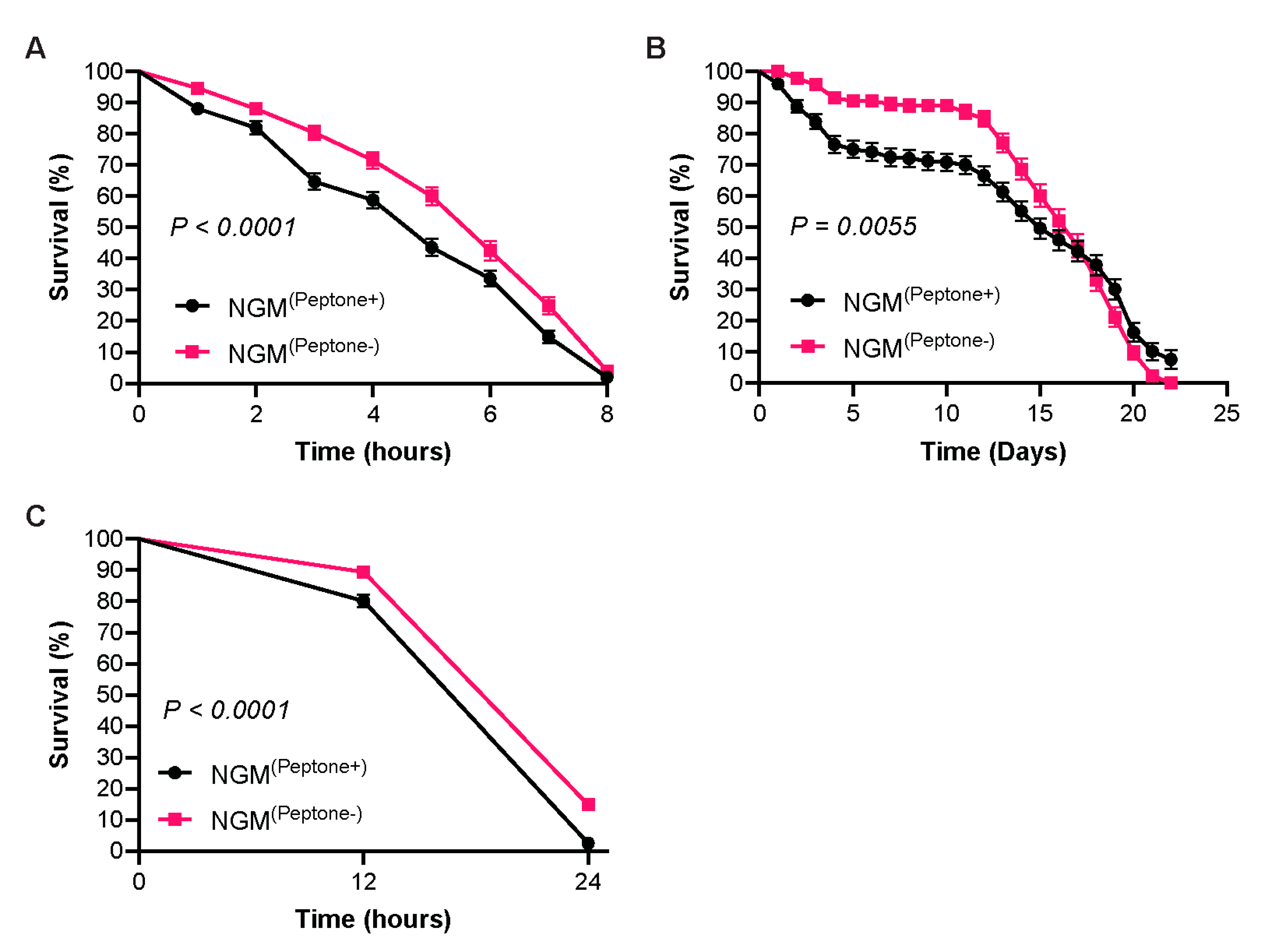
To determine whether culturing worms on peptone-free NGM improves stress resistance, age-synchronized L4 worms were exposed to oxidative stress induced by paraquat, ultraviolet (UV) irradiation, or elevated temperature (35 °C), and survival was monitored over time. We observed that worms cultured on peptone-free NGM exhibited significantly improved survival under oxidative stress, UV stress, and thermal stress compared with worms cultured on conventional NGM (Figure 6A–C). Basal expression analysis of heat shock-associated genes further revealed significant downregulation of hsf-1, hsp-70, and hsp-90, whereas hsp-16.2 expression remained unchanged (Figure S1C). These results indicate that peptone-free NGM enhances physiological resilience against multiple environmental stressors despite reduced basal expression of several heat shock response genes.
3. Discussion
The present study systematically evaluated whether peptone remains necessary in NGM when paraformaldehyde (PFA)-killed Escherichia coli OP50 is used as the bacterial food source. Although peptone has been an integral component of NGM since its original formulation by Brenner to support bacterial proliferation [3], its requirement under conditions where bacterial replication is intentionally eliminated has not previously been investigated. Here, we demonstrate that removal of peptone markedly suppresses bacterial and fungal contamination while preserving normal growth, reproduction, lifespan, and chemotactic behavior of C. elegans. In addition, peptone-free NGM reduced basal oxidative stress and lipid accumulation, enhanced locomotor activity, and improved resistance to oxidative, ultraviolet, and thermal stress. Collectively, these findings indicate that peptone-free NGM represents a practical refinement of the conventional culture system for experiments employing PFA-killed bacterial diets.
One of the most immediate advantages of peptone-free NGM was the marked suppression of both bacterial and fungal contamination. Previous studies reported that treatment of OP50 with 0.25% or 0.5% PFA effectively prevents bacterial proliferation on conventional NGM [27,28]. However, under our experimental conditions, limited bacterial growth was still detected on conventional NGM even after treatment with 1% PFA, whereas no detectable bacterial proliferation occurred on peptone-free NGM (Figure 1A). These findings suggest that a small number of bacteria may occasionally survive or recover following PFA treatment and subsequently proliferate when nutrients are available. Thus, removal of peptone eliminates the nutrient source required for bacterial expansion and provides an additional safeguard against contamination. Likewise, peptone-free NGM markedly reduced the growth of Penicillium chrysogenum, and complete inhibition was achieved when combined with Nystatin (Figure 1B). Since fungal germination depends on the availability of amino acids and other nutrients [46,47], these findings indicate that peptone deprivation creates an unfavorable environment for fungal proliferation. To our knowledge, this is the first study demonstrating that removal of peptone simultaneously suppresses both bacterial and fungal contamination without adversely affecting C. elegans physiology.
Previous studies demonstrated that C. elegans preferentially accumulates on live or mock-treated OP50 rather than PFA-killed bacteria [26,27,28]. Consistent with our bacterial growth assay, worms in the present study preferred bacterial lawns on conventional NGM over those on peptone-free NGM, suggesting that limited bacterial proliferation on conventional NGM may influence food preference (Figure 2A). Worms cultured on peptone-free NGM also exhibited a lower pharyngeal pumping rate (Figure 2B), which may reflect differences in bacterial lawn characteristics rather than reduced nutritional availability. Importantly, body growth, reproduction, and lifespan remained unchanged (Figure 3), indicating that the nutritional requirements of the worms were fully met despite altered feeding behavior.
Previous studies reported that worms fed PFA-killed OP50 exhibit a modest developmental delay, reduced body size, and lower brood size compared with worms fed live OP50 or mock-treated bacteria [25,27,28]. In contrast, worms cultured on peptone-free NGM showed no significant differences in developmental body length, body volume, brood size, or embryonic viability compared with those maintained on conventional NGM (Figure 3A–D). These findings are consistent with a recent study demonstrating that PFA-killed OP50 supports normal reproductive capacity comparable to live bacteria [26], indicating that removal of peptone does not adversely affect post-embryonic growth or reproductive fitness when bacterial nutrition is provided through PFA-killed OP50. Chemotactic responses toward both attractive and aversive cues were also unaffected, indicating that sensory function remained intact (Figure 3F). Together, these findings demonstrate that elimination of peptone preserves fundamental aspects of C. elegans physiology and further support the conclusion that, once bacterial proliferation is prevented, the primary role of peptone is to sustain bacterial growth rather than directly contribute to worm nutrition.
Previous studies have shown that bacterial diet is an important determinant of redox homeostasis and lipid metabolism in C. elegans [29,30,32,33,34]. In agreement with these observations, worms cultured on peptone-free NGM exhibited significantly lower intracellular ROS together with reduced expression of oxidative stress-associated genes, including pmk-1, skn-1, and gcs-1 (Figure 4A,B). Rather than indicating impaired antioxidant defenses, these transcriptional changes likely reflect a lower basal oxidative burden under peptone-free culture conditions. Similarly, worms maintained on peptone-free NGM accumulated significantly less neutral lipid and displayed coordinated downregulation of the lipogenic regulators sbp-1, fasn-1, fat-6, fat-7, and dgat-2 (Figure 4C,D). Although nhr-49 and lips-17 were also reduced, acs-2 expression remained unchanged (Figure S1A), suggesting that reduced lipid accumulation results primarily from decreased lipogenesis rather than enhanced β-oxidation. Together, these findings indicate that eliminating peptone improves metabolic homeostasis without inducing a classical dietary restriction or starvation response.
Bacterial diet also profoundly influences locomotion and neuromuscular function in C. elegans [25,31,34]. Consistent with previous reports, worms cultured on peptone-free NGM displayed significantly greater locomotor activity despite reduced pharyngeal pumping and no detectable changes in growth, reproduction, lifespan, or chemotaxis (Figure 2B, 3 and 5A,B). The improved locomotion was accompanied by increased expression of unc-17, which encodes the vesicular acetylcholine transporter, and reduced expression of the locomotion suppressor egl-4, whereas unc-13, myo-3, and unc-54 remained unchanged (Figure 5C and S1B) [48,49,50,51,52,53,54]. These findings suggest that enhanced locomotion results from subtle modulation of cholinergic signaling rather than broad alterations in synaptic transmission or muscle structure and is consistent with the improved metabolic and redox homeostasis observed under peptone-free culture conditions.
Finally, worms maintained on peptone-free NGM exhibited significantly greater resistance to oxidative, ultraviolet, and thermal stress (Figure 6). Interestingly, these improvements occurred despite lower basal expression of pmk-1, skn-1, gcs-1, hsf-1, hsp-70, and hsp-90, whereas hsp-16.2 remained unchanged (Figure 4B and S1C). Rather than reflecting impaired stress responses, reduced basal expression of these genes likely indicates diminished constitutive stress signaling, allowing worms to respond more efficiently to acute environmental challenges [55,56,57]. Importantly, enhanced stress tolerance occurred without extending lifespan, supporting the concept that physiological resilience and longevity are related but distinct aspects of organismal health [55,56,57,58].
Collectively, our findings support a model in which removal of peptone suppresses residual bacterial proliferation and microbial contamination without altering the nutritional adequacy of PFA-killed OP50. Consequently, worms experience a lower basal physiological burden, characterized by reduced oxidative stress and lipid accumulation, which is accompanied by improved locomotor performance and greater resilience to environmental stress while maintaining normal growth, reproduction, lifespan, and sensory function. These observations indicate that, under conditions where bacterial proliferation is intentionally prevented, the continued inclusion of peptone primarily benefits contaminating microorganisms rather than C. elegans. Therefore, peptone-free NGM represents a simple, inexpensive, and experimentally advantageous refinement of the conventional culture system for studies employing PFA-killed bacterial diets.
The present study has several limitations that warrant consideration. All experiments were performed using wild-type C. elegans cultured with PFA-killed OP50 under standard laboratory conditions. Whether similar benefits extend to live bacterial diets, alternative bacterial species, diverse genetic backgrounds, or different environmental conditions remains to be determined. Furthermore, comprehensive transcriptomic, metabolomic, and proteomic analyses will be valuable for elucidating the molecular mechanisms underlying the physiological improvements observed under peptone-free culture conditions.
4. Conclusion
In conclusion, our findings demonstrate that peptone is not required in NGM when bacterial proliferation is intentionally prevented through the use of PFA-killed OP50 as the bacterial food source. Under these conditions, removal of peptone markedly reduces bacterial and fungal contamination while preserving normal growth, reproduction, lifespan, and sensory behavior in C. elegans. More importantly, peptone-free NGM reduces basal oxidative stress and lipid accumulation, enhances locomotor activity, and improves resistance to oxidative, UV, and thermal stress, collectively indicating enhanced physiological resilience. These findings establish peptone-free NGM as a simple, inexpensive, and practical refinement of the conventional culture system that minimizes contamination-related variability without compromising normal C. elegans physiology. Given the increasing use of PFA-killed bacterial diets in studies of metabolism, aging, neurobiology, and host-microbe interactions, peptone-free NGM represents a rational and experimentally advantageous culture medium for future C. elegans research.
Data Availability
Data will be made available on request.
Declaration of Interests
The authors declare no competing interests.
Author Contributions
P.M., S.B. and A.K. performed experiments and analyzed the results. K.R.B. designed and supervised the research work; wrote, edited, and revised the manuscript.
Funding
This research did not receive any specific grant from funding agencies in the public, commercial, or not-for-profit sectors.
Acknowledgments
The authors acknowledge UPES, Dehradun, India, for providing institutional support and infrastructure for this research. The authors also thank Yash Khanduri for his insightful discussions, which contributed to the conceptual foundation of this study.
References
- Fernandez-Abascal, J.; Bianchi, L. A protocol for imaging calcium and chloride in C. elegans glia upon touch stimulation. STAR Protoc. 2022, 3, 101282. [Google Scholar] [CrossRef] [PubMed]
- Roozen, M.C.; Kas, M.J.H. Assessing genetic conservation of human sociability-linked genes in C. elegans. Behav. Genet. 2025, 55, 141–152. [Google Scholar] [CrossRef] [PubMed]
- Brenner, S. The genetics of Caenorhabditis elegans. Genetics 1974, 77(1), 71–94. [Google Scholar] [CrossRef] [PubMed]
- Corsi, A.K.; Wightman, B.; Chalfie, M. A Transparent Window into Biology: A Primer on Caenorhabditis elegans. Genetics 2015, 200, 387–407. [Google Scholar] [CrossRef] [PubMed]
- Jeayeng, S.; Thongsroy, J.; Chuaijit, S. Caenorhabditis elegans as a Model to Study Aging and Photoaging. Biomolecules 2024, 14, 1235. [Google Scholar] [CrossRef] [PubMed]
- Stiernagle, T. Maintenance of C. elegans. WormBook 2006. [Google Scholar] [CrossRef] [PubMed]
- Kaletta, T.; Hengartner, M.O. Finding function in novel targets: C. elegans as a model organism. Nat. Rev. Drug Discov. 2006, 5, 387–399. [Google Scholar] [CrossRef] [PubMed]
- Baugh, L.R. To Grow or Not to Grow: Nutritional Control of Development DuringCaenorhabditis elegansL1 Arrest. Genetics 2013, 194, 539–555. [Google Scholar] [CrossRef] [PubMed]
- Markaki, M.; Tavernarakis, N. Caenorhabditis elegans as a model system for human diseases. Curr. Opin. Biotechnol. 2020, 63, 118–125. [Google Scholar] [CrossRef] [PubMed]
- Porta-de-la-Riva, M.; et al. Basic Caenorhabditis elegans Methods: Synchronization and Observation. J. Vis. Exp. 2012, 64. [Google Scholar] [CrossRef] [PubMed]
- Dickinson, D.J.; Goldstein, B. CRISPR-Based Methods for Caenorhabditis elegans Genome Engineering. Genetics 2016, 202, 885–901. [Google Scholar] [CrossRef] [PubMed]
- Hodgkin, J.; Kuwabara, P.E.; Corneliussen, B. A novel bacterial pathogen, Microbacterium nematophilum, induces morphological change in the nematode C. elegans. Curr. Biol. 2000, 10, 1615–1618. [Google Scholar] [CrossRef] [PubMed]
- Chalissery, J., et al., <italic>Rearing C. elegans on Parafilm-wrapped NGM Plates Impacts Habituation Behavior</italic>. MicroPubl Biol, 2023. <bold>2023</bold>.
- Shinn-Thomas, J.H.; Scanga, S.E.; Spica, P.S.; Nariya, H.K.; Klempic, E.; Brockett, M.R. Wrapping culture plates with Parafilm M® increases Caenorhabditis elegans growth. BMC Res. Notes 2019, 12, 1–5. [Google Scholar] [CrossRef] [PubMed]
- Hosono, R.; Nishimoto, S.; Kuno, S. Alterations of life span in the nematode Caenorhabditis elegans under monoxenic culture conditions. Exp. Gerontol. 1989, 24, 251–264. [Google Scholar] [CrossRef] [PubMed]
- Le, T.S.; Nguyen, T.T.H.; Huong, B.T.M.; Nguyen, H.G.; Ha, B.H.; Nguyen, V.S.; Nguyen, M.H.; Nguyen, H.-H.; Wang, J. Cultivation of Caenorhabditis elegans on new cheap monoxenic media without peptone. J. Nematol. 2021, 53, 1–23. [Google Scholar] [CrossRef] [PubMed]
- Greer, E.L.; Brunet, A. Different dietary restriction regimens extend lifespan by both independent and overlapping genetic pathways in C. elegans. Aging Cell 2009, 8, 113–127. [Google Scholar] [CrossRef] [PubMed]
- Stastna, J.J.; Snoek, L.B.; Kammenga, J.E.; Harvey, S.C. Genotype-dependent lifespan effects in peptone deprived Caenorhabditis elegans. Sci. Rep. 2015, 5, 16259–16259. [Google Scholar] [CrossRef] [PubMed]
- Higurashi, S.; Tsukada, S.; Aleogho, B.M.; Park, J.H.; Al-Hebri, Y.; Tanaka, M.; Nakano, S.; Mori, I.; Noma, K. Bacterial diet affects the age-dependent decline of associative learning in Caenorhabditis elegans. eLife 2023, 12. [Google Scholar] [CrossRef] [PubMed]
- Akduman, N.; Lightfoot, J.W.; Röseler, W.; Witte, H.; Lo, W.-S.; Rödelsperger, C.; Sommer, R.J. Bacterial vitamin B12 production enhances nematode predatory behavior. ISME J. 2020, 14, 1494–1507. [Google Scholar] [CrossRef] [PubMed]
- MacNeil, L.T.; Watson, E.; Arda, H.E.; Zhu, L.J.; Walhout, A.J. Diet-Induced Developmental Acceleration Independent of TOR and Insulin in C. elegans. Cell 2013, 153, 240–252. [Google Scholar] [CrossRef] [PubMed]
- Quinones Tavarez, Z., et al., <italic>Microbiota-Influenced Toxicological Responses in Caenorhabditis elegans Exposed to Strawberry and Menthol E-Liquids.</italic> MicroPubl Biol, 2025. <bold>2025</bold>.
- Kissoyan, K.A.; Drechsler, M.; Stange, E.-L.; Zimmermann, J.; Kaleta, C.; Bode, H.B.; Dierking, K. Natural C. elegans Microbiota Protects against Infection via Production of a Cyclic Lipopeptide of the Viscosin Group. Curr. Biol. 2019, 29, 1030–1037.e5. [Google Scholar] [CrossRef] [PubMed]
- Dirksen, P.; Marsh, S.A.; Braker, I.; Heitland, N.; Wagner, S.; Nakad, R.; Mader, S.; Petersen, C.; Kowallik, V.; Rosenstiel, P.; et al. The native microbiome of the nematode Caenorhabditis elegans: gateway to a new host-microbiome model. BMC Biol. 2016, 14, 1–16. [Google Scholar] [CrossRef] [PubMed]
- Stuhr, N.L.; Curran, S.P. Different methods of killing bacteria diets differentially influence Caenorhabditis elegans physiology. 2023, 2023. [Google Scholar] [CrossRef] [PubMed]
- Thériault, V.; De la Rosa, C.M.A.; Miard, S.; Taubert, S.; Picard, F. Impact of bacterial inactivation methods on Caenorhabditis elegans feeding and healthspan. Sci. Rep. 2025, 15. [Google Scholar] [CrossRef] [PubMed]
- Beydoun, S.; et al. Methodology to Metabolically Inactivate Bacteria for Caenorhabditis elegans Research. J. Vis. Exp. 2023, 197. [Google Scholar] [CrossRef] [PubMed]
- Beydoun, S.; Choi, H.S.; Dela-Cruz, G.; Kruempel, J.; Huang, S.; Bazopoulou, D.; Miller, H.A.; Schaller, M.L.; Evans, C.R.; Leiser, S.F. An alternative food source for metabolism and longevity studies in Caenorhabditis elegans. Commun. Biol. 2021, 4, 1–9. [Google Scholar] [CrossRef] [PubMed]
- Stuhr, N.L.; Curran, S.P. Bacterial diets differentially alter lifespan and healthspan trajectories in C. elegans. Commun. Biol. 2020, 3, 1–18. [Google Scholar] [CrossRef] [PubMed]
- Melov, S.; et al. The Influence of Bacterial Diet on Fat Storage in C. elegans. PLoS ONE 2009, 4(10). [Google Scholar] [CrossRef] [PubMed]
- Ribeiro, C.; et al. Bacterially produced metabolites protect C. elegans neurons from degeneration. PLoS Biol. 2020, 18(3). [Google Scholar] [CrossRef] [PubMed]
- A A Neve, I.; Sowa, J.N.; Lin, C.-C.J.; Sivaramakrishnan, P.; Herman, C.; Ye, Y.; Han, L.; Wang, M.C. Escherichia coli Metabolite Profiling Leads to the Development of an RNA Interference Strain for Caenorhabditis elegans. G3 Genes|Genomes|Genetics 2020, 10, 189–198. [Google Scholar] [CrossRef] [PubMed]
- Bhat, A.; Cox, R.L.; Hendrickson, B.G.; Das, N.K.; Schaller, M.L.; Tuckowski, A.M.; Wang, E.; Shah, Y.M.; Leiser, S.F. A diet of oxidative stress–adapted bacteria improves stress resistance and lifespan in C. elegans via p38-MAPK. Sci. Adv. 2024, 10, eadk8823. [Google Scholar] [CrossRef] [PubMed]
- Huang, Q.; Li, R.; Yi, T.; Cong, F.; Wang, D.; Deng, Z.; Zhao, Y.-L. Phosphorothioate-DNA bacterial diet reduces the ROS levels in C. elegans while improving locomotion and longevity. Commun. Biol. 2021, 4, 1–13. [Google Scholar] [CrossRef] [PubMed]
- Bonnard, E.; Liu, J.; Zjacic, N.; Alvarez, L.; Scholz, M. Automatically tracking feeding behavior in populations of foraging C. elegans. eLife 2022, 11. [Google Scholar] [CrossRef] [PubMed]
- Hakim, A.; Mor, Y.; Toker, I.A.; Levine, A.; Neuhof, M.; Markovitz, Y.; Rechavi, O. WorMachine: machine learning-based phenotypic analysis tool for worms. BMC Biol. 2018, 16, 8. [Google Scholar] [CrossRef] [PubMed]
- Jaramillo-Lambert, A.; Kwah, J.K. Measuring Embryonic Viability and Brood Size in Caenorhabditis elegans. J. Vis. Exp. 2023, 192. [Google Scholar] [CrossRef] [PubMed]
- Sutphin, G.L.; Kaeberlein, M. Measuring Caenorhabditis elegans Life Span on Solid Media. J. Vis. Exp. 2009. [Google Scholar] [CrossRef] [PubMed]
- Chin-Sang, I.; Palmer, C.; O. Margie, C. elegans Chemotaxis Assay. J. Vis. Exp. 2013, 74. [Google Scholar] [CrossRef]
- Husson, S.J.; Costa, W.S.; Schmitt, C.; Gottschalk, A. Keeping track of worm trackers. WormBook 2012, 1–17. [Google Scholar] [CrossRef] [PubMed]
- Morimoto, R.I.; et al. Investigating the Spreading and Toxicity of Prion-like Proteins Using the Metazoan Model Organism C. elegans. J. Vis. Exp. 2015, 95. [Google Scholar] [CrossRef]
- Curran, S.P.; et al. Quantification of Lipid Abundance and Evaluation of Lipid Distribution in Caenorhabditis elegans by Nile Red and Oil Red O Staining. J. Vis. Exp. 2018, 133. [Google Scholar] [CrossRef] [PubMed]
- Yoon, D.S.; Lee, M.-H.; Cha, D.S. Measurement of Intracellular ROS in Caenorhabditis elegans Using 2’,7’-Dichlorodihydrofluorescein Diacetate. Bio-Protocol 2018, 8, e2774–e2774. [Google Scholar] [CrossRef] [PubMed]
- Pause, A.; Possik, E. Measuring Oxidative Stress Resistance of Caenorhabditis elegans in 96-well Microtiter Plates. J. Vis. Exp. 2015, 99. [Google Scholar] [CrossRef] [PubMed]
- Livak, K.J.; Schmittgen, T.D. Analysis of relative gene expression data using real-time quantitative PCR and the 2(-Delta Delta C(T)) Method. Methods 2001, 25(4), 402–8. [Google Scholar] [PubMed]
- Osherov, N.; May, G. Conidial Germination in Aspergillus nidulans Requires RAS Signaling and Protein Synthesis. Genetics 2000, 155, 647–656. [Google Scholar] [CrossRef] [PubMed]
- Osherov, N. The molecular mechanisms of conidial germination. FEMS Microbiol. Lett. 2001, 199(2), 153–160. [Google Scholar] [CrossRef]
- Alfonso, A.; Grundahl, K.; Duerr, J.S.; Han, H.-P.; Rand, J.B. The Caenorhabditis elegans unc-17 Gene: a Putative Vesicular Acetylcholine Transporter. Science 1993, 261, 617–619. [Google Scholar] [CrossRef] [PubMed]
- Rand, J. Acetylcholine. WormBook 2007. [Google Scholar] [CrossRef] [PubMed]
- Fujiwara, M.; Sengupta, P.; McIntire, S.L. Regulation of Body Size and Behavioral State of C. elegans by Sensory Perception and the EGL-4 cGMP-Dependent Protein Kinase. Neuron 2002, 36, 1091–1102. [Google Scholar] [CrossRef] [PubMed]
- Raizen, D.M.; Cullison, K.M.; I Pack, A.; Sundaram, M.V. A Novel Gain-of-Function Mutant of the Cyclic GMP-Dependent Protein Kinase egl-4 Affects Multiple Physiological Processes in Caenorhabditis elegans. Genetics 2006, 173, 177–187. [Google Scholar] [CrossRef] [PubMed]
- Richmond, J.E.; Davis, W.S.; Jorgensen, E.M. UNC-13 is required for synaptic vesicle fusion in C. elegans. Nat. Neurosci. 1999, 2, 959–964. [Google Scholar] [CrossRef] [PubMed]
- Honda, S.; Epstein, H.F. Modulation of muscle gene expression in Caenorhabditis elegans: differential levels of transcripts, mRNAs, and polypeptides for thick filament proteins during nematode development. Proc. Natl. Acad. Sci. 1990, 87, 876–880. [Google Scholar] [CrossRef] [PubMed]
- Francis, G.R.; Waterston, R.H. Muscle organization in Caenorhabditis elegans: localization of proteins implicated in thin filament attachment and I-band organization. J. Cell Biol. 1985, 101, 1532–1549. [Google Scholar] [CrossRef] [PubMed]
- Houthoofd, K.; Vanfleteren, J.R. The longevity effect of dietary restriction in Caenorhabditis elegans. Exp. Gerontol. 2006, 41, 1026–1031. [Google Scholar] [CrossRef] [PubMed]
- Back, P.; Braeckman, B.P.; Matthijssens, F. ROS in AgingCaenorhabditis elegans: Damage or Signaling? Oxidative Med. Cell. Longev. 2012, 2012, 1–14. [Google Scholar] [CrossRef] [PubMed]
- López-Otín, C.; Blasco, M.A.; Partridge, L.; Serrano, M.; Kroemer, G. Hallmarks of aging: An expanding universe. Cell 2023, 186, 243–278. [Google Scholar] [CrossRef] [PubMed]
- Kenyon, C.J. The genetics of ageing. Nature 2010, 464(7288), 504–512. [Google Scholar] [CrossRef] [PubMed]
Figure 1.
Peptone-free NGM suppresses bacterial and fungal contamination. (A) Representative images showing bacterial growth on conventional NGM (NGM(Peptone+)) and peptone-free NGM (NGM(Peptone-)) plates seeded with live Escherichia coli OP50 or OP50 treated with 0.25%, 0.5%, or 1% paraformaldehyde (PFA). Right panel shows quantification of bacterial growth as colony-forming units (CFU/mL) (n = 9 biological replicates per condition). (B) Representative images of Penicillium chrysogenum growth on NGM(Peptone+) and NGM(Peptone-) plates in the presence or absence of Nystatin. Right panel shows quantification of fungal growth rate (mm/day) (n = 9 biological replicates per condition). Data are presented as mean ± SEM from three independent biological experiments. The two-tailed Student’s t test was applied to calculate statistical significance. **P < 0.01, ***P < 0.001, ****P < 0.0001. Scale bar = 10 mm.
Figure 1.
Peptone-free NGM suppresses bacterial and fungal contamination. (A) Representative images showing bacterial growth on conventional NGM (NGM(Peptone+)) and peptone-free NGM (NGM(Peptone-)) plates seeded with live Escherichia coli OP50 or OP50 treated with 0.25%, 0.5%, or 1% paraformaldehyde (PFA). Right panel shows quantification of bacterial growth as colony-forming units (CFU/mL) (n = 9 biological replicates per condition). (B) Representative images of Penicillium chrysogenum growth on NGM(Peptone+) and NGM(Peptone-) plates in the presence or absence of Nystatin. Right panel shows quantification of fungal growth rate (mm/day) (n = 9 biological replicates per condition). Data are presented as mean ± SEM from three independent biological experiments. The two-tailed Student’s t test was applied to calculate statistical significance. **P < 0.01, ***P < 0.001, ****P < 0.0001. Scale bar = 10 mm.
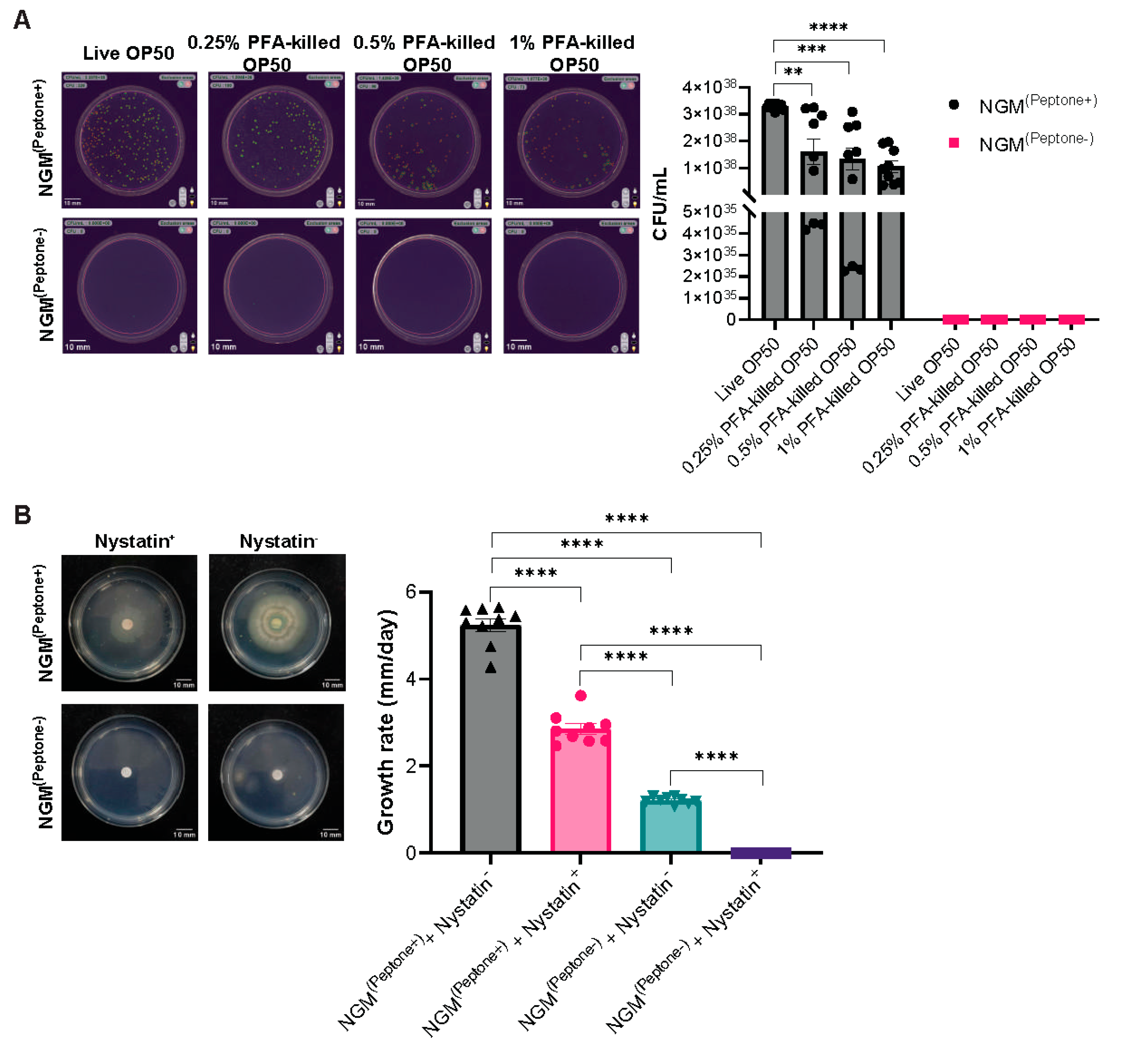
Figure 2.
Peptone-free NGM alters feeding behavior of C. elegans. (A) Food preference assay showing the percentage of age-synchronized L4 worms present on bacterial lawns grown on NGM(Peptone+) or NGM(Peptone-) after 1 and 8 h of incubation (n = 9 biological replicates per condition). (B) Pharyngeal pumping rate of age-synchronized L4 worms maintained on NGM(Peptone+) or NGM(Peptone-) (n = 80 worms per condition). Data are presented as mean ± SEM from three independent biological experiments. The two-tailed Student’s t test was applied to calculate statistical significance. ****P < 0.0001.
Figure 2.
Peptone-free NGM alters feeding behavior of C. elegans. (A) Food preference assay showing the percentage of age-synchronized L4 worms present on bacterial lawns grown on NGM(Peptone+) or NGM(Peptone-) after 1 and 8 h of incubation (n = 9 biological replicates per condition). (B) Pharyngeal pumping rate of age-synchronized L4 worms maintained on NGM(Peptone+) or NGM(Peptone-) (n = 80 worms per condition). Data are presented as mean ± SEM from three independent biological experiments. The two-tailed Student’s t test was applied to calculate statistical significance. ****P < 0.0001.
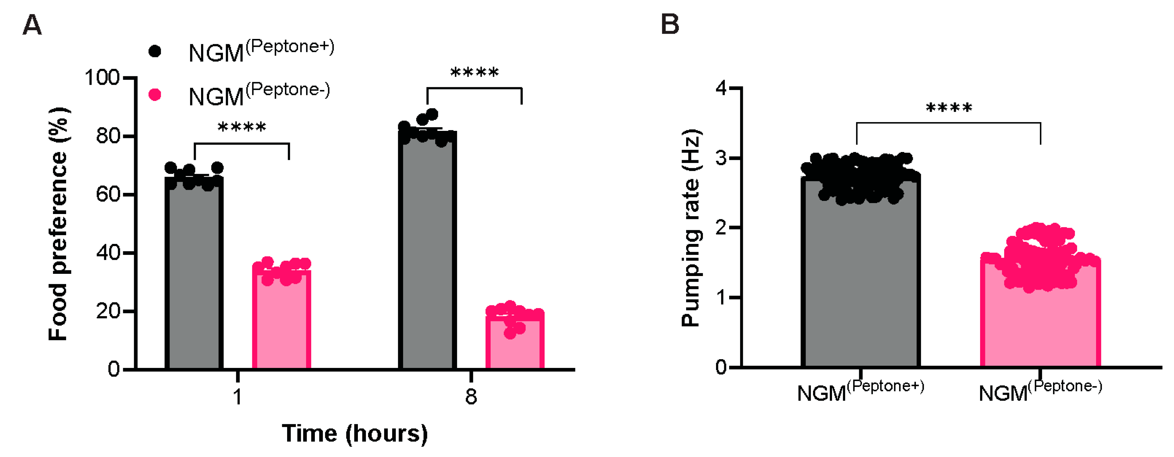
Figure 3.
Peptone-free NGM does not adversely affect growth, reproduction, lifespan, or chemotactic behavior of C. elegans. (A) Developmental body length and (B) body volume of age-synchronized worms cultured on NGM(Peptone+) and NGM(Peptone-), measured at 0, 24, 48, 72, 96, and 120 h after L1 plating (n ≥ 200 worms per condition). (C) Brood size and (D) embryonic viability of hermaphrodites maintained on NGM(Peptone+) and NGM(Peptone-) (n = 9 worms per condition). (E) Kaplan-Meier survival curves showing lifespan of worms maintained on NGM(Peptone+) and NGM(Peptone-) (n ≥ 130 worms per condition). (F) Chemotaxis indices toward diacetyl (attractant), glycerol (repellent), and double distilled water (control) (n = 9 biological replicates per condition). Body length and body volume were analyzed using two-way ANOVA. Brood size, embryonic viability, and chemotaxis were analyzed using an unpaired two-tailed Student's t-test. Lifespan was analyzed using the log-rank (Mantel-Cox) test. Data are presented as mean ± SEM from three independent experiments. nsP > 0.05.
Figure 3.
Peptone-free NGM does not adversely affect growth, reproduction, lifespan, or chemotactic behavior of C. elegans. (A) Developmental body length and (B) body volume of age-synchronized worms cultured on NGM(Peptone+) and NGM(Peptone-), measured at 0, 24, 48, 72, 96, and 120 h after L1 plating (n ≥ 200 worms per condition). (C) Brood size and (D) embryonic viability of hermaphrodites maintained on NGM(Peptone+) and NGM(Peptone-) (n = 9 worms per condition). (E) Kaplan-Meier survival curves showing lifespan of worms maintained on NGM(Peptone+) and NGM(Peptone-) (n ≥ 130 worms per condition). (F) Chemotaxis indices toward diacetyl (attractant), glycerol (repellent), and double distilled water (control) (n = 9 biological replicates per condition). Body length and body volume were analyzed using two-way ANOVA. Brood size, embryonic viability, and chemotaxis were analyzed using an unpaired two-tailed Student's t-test. Lifespan was analyzed using the log-rank (Mantel-Cox) test. Data are presented as mean ± SEM from three independent experiments. nsP > 0.05.
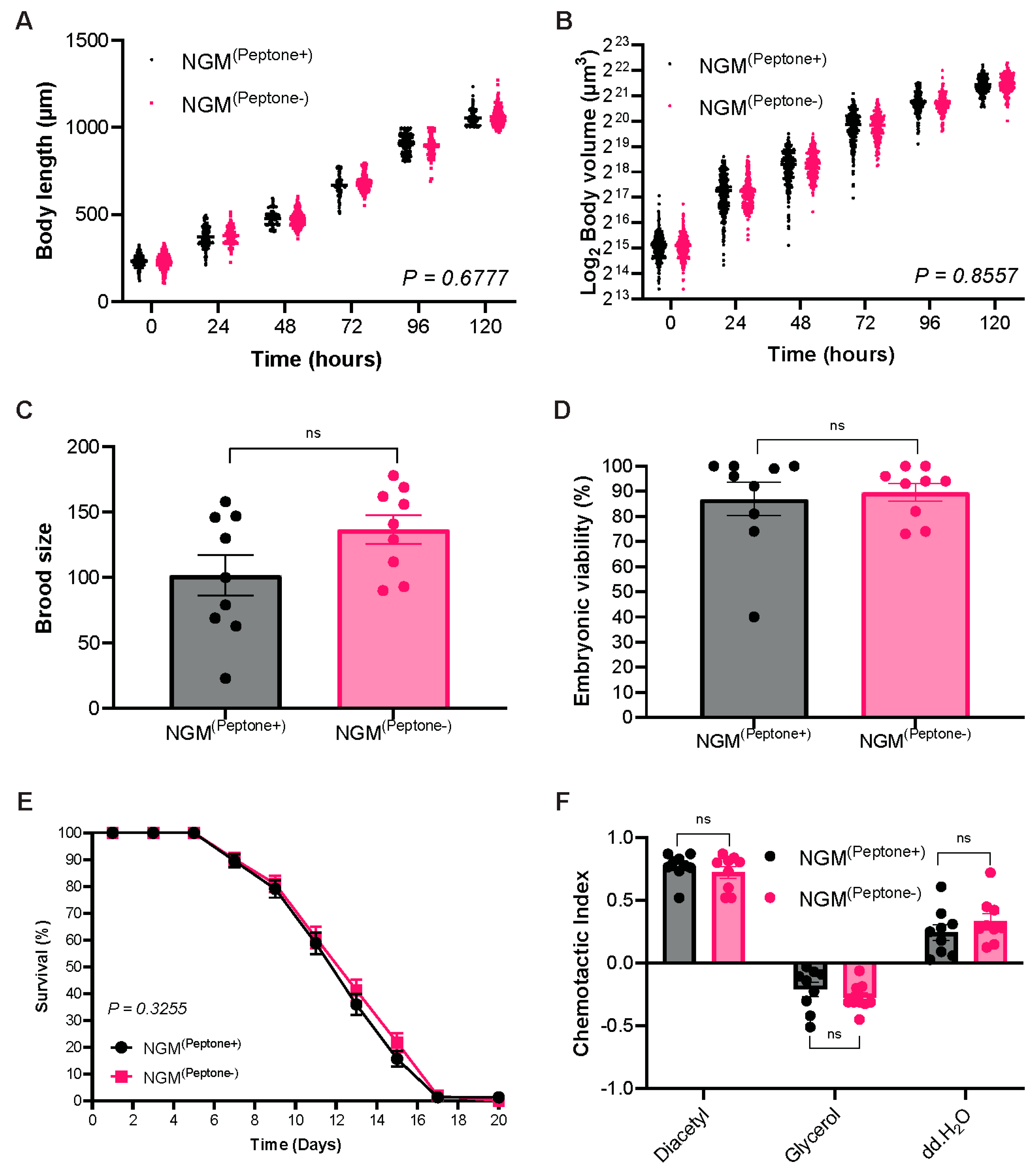
Figure 4.
Peptone-free NGM reduces basal oxidative stress and lipid accumulation in C. elegans. (A) Intracellular reactive oxygen species (ROS) levels in age-synchronized L4 worms maintained on NGM(Peptone+) and NGM(Peptone-), measured using H2DCFDA fluorescence over an 8-h period. (B) Relative mRNA expression of oxidative stress response-associated genes (pmk-1, skn-1, gcs-1, and gst-4) determined by quantitative PCR. Expression levels were normalized to tba-1 and are presented as log10 fold change relative to worms cultured on NGM(Peptone+). (C) Representative bright-field, fluorescence, and merged images of Nile Red-stained L4 worms cultured on NGM(Peptone+) and NGM(Peptone-). Right panel shows quantification of Nile Red fluorescence intensity (n = 30 worms per condition). Scale bar = 100 µm. (D) Relative mRNA expression of lipogenesis-related genes (sbp-1, fasn-1, fat-6, fat-7, and dgat-2) measured by quantitative PCR. Gene expression was normalized to tba-1 and expressed as log10 fold change relative to NGM(Peptone+). Data are presented as mean ± SEM from three independent experiments. Intracellular ROS was analyzed using two-way ANOVA. Nile Red Fluorescence intensity and qPCR data were analyzed using an unpaired two-tailed Student's t-test. nsP > 0.05, **P < 0.01, ****P < 0.0001.
Figure 4.
Peptone-free NGM reduces basal oxidative stress and lipid accumulation in C. elegans. (A) Intracellular reactive oxygen species (ROS) levels in age-synchronized L4 worms maintained on NGM(Peptone+) and NGM(Peptone-), measured using H2DCFDA fluorescence over an 8-h period. (B) Relative mRNA expression of oxidative stress response-associated genes (pmk-1, skn-1, gcs-1, and gst-4) determined by quantitative PCR. Expression levels were normalized to tba-1 and are presented as log10 fold change relative to worms cultured on NGM(Peptone+). (C) Representative bright-field, fluorescence, and merged images of Nile Red-stained L4 worms cultured on NGM(Peptone+) and NGM(Peptone-). Right panel shows quantification of Nile Red fluorescence intensity (n = 30 worms per condition). Scale bar = 100 µm. (D) Relative mRNA expression of lipogenesis-related genes (sbp-1, fasn-1, fat-6, fat-7, and dgat-2) measured by quantitative PCR. Gene expression was normalized to tba-1 and expressed as log10 fold change relative to NGM(Peptone+). Data are presented as mean ± SEM from three independent experiments. Intracellular ROS was analyzed using two-way ANOVA. Nile Red Fluorescence intensity and qPCR data were analyzed using an unpaired two-tailed Student's t-test. nsP > 0.05, **P < 0.01, ****P < 0.0001.
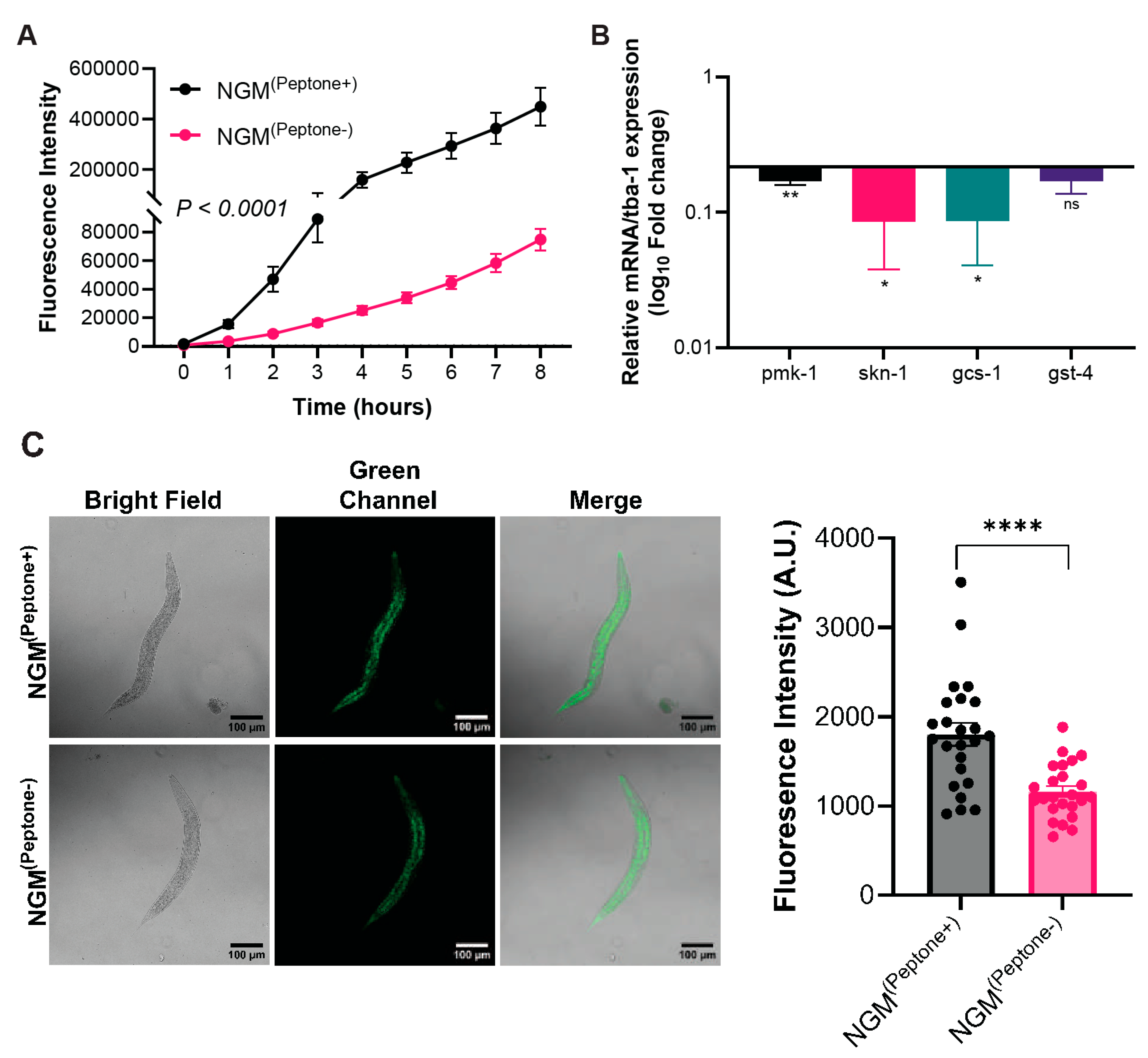
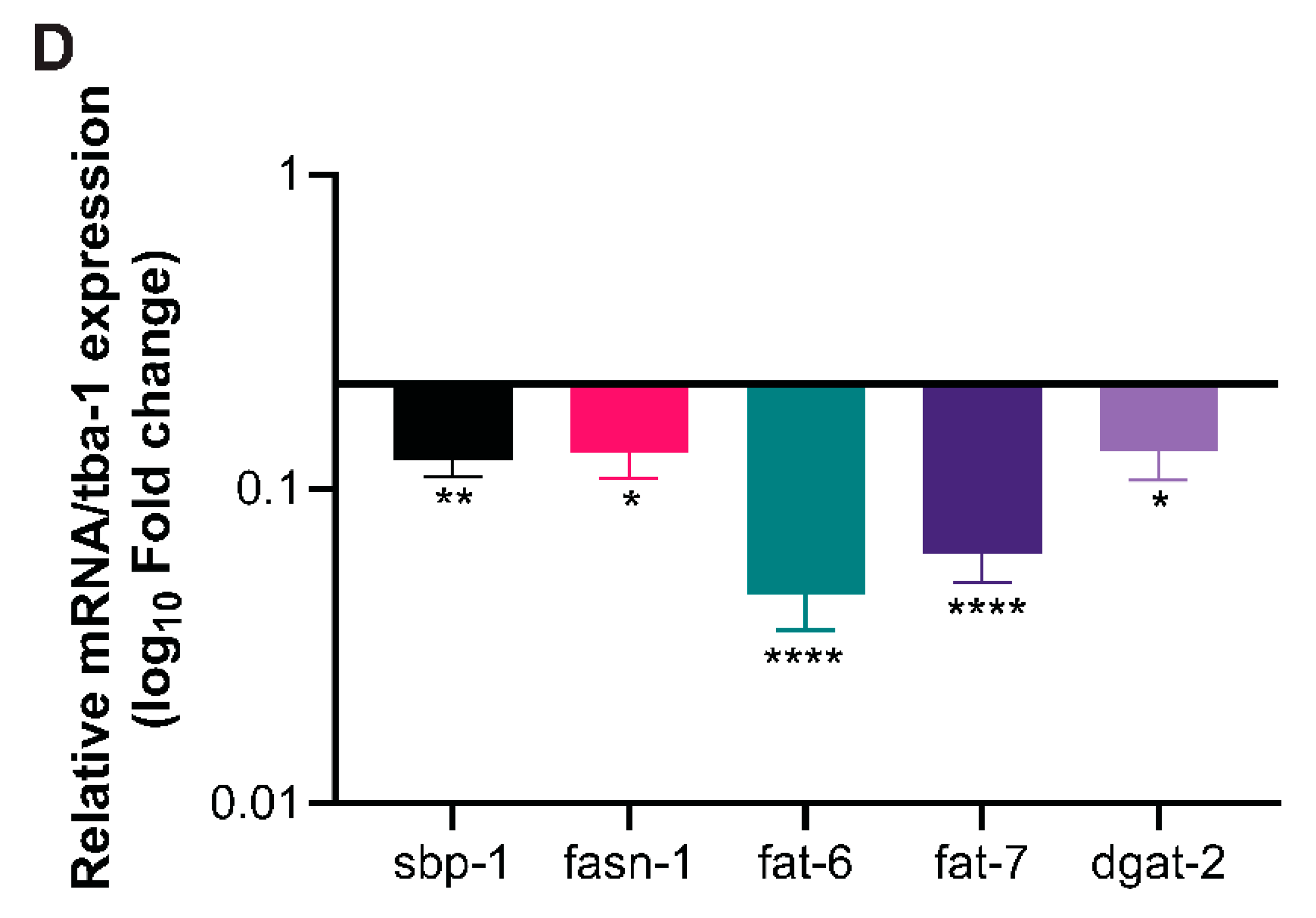
Figure 5.
Peptone-free NGM enhances locomotor activity in C. elegans. (A) Body length traveled per second (BLPS) and (B) Body bends per second (BBPS) of age-synchronized L4 worms maintained on NGM(Peptone+) and NGM(Peptone-) (n ≥ 300 worms per condition). (C) Relative mRNA expression of locomotion-associated genes (unc-17 and egl-4) measured by quantitative PCR. Gene expression was normalized to tba-1 and presented as log10 fold change relative to worms cultured on NGM(Peptone+). Data are presented as mean ± SEM from three independent experiments. Statistical significance was determined using an unpaired two-tailed Student's t-test. *P < 0.05, ****P < 0.0001.
Figure 5.
Peptone-free NGM enhances locomotor activity in C. elegans. (A) Body length traveled per second (BLPS) and (B) Body bends per second (BBPS) of age-synchronized L4 worms maintained on NGM(Peptone+) and NGM(Peptone-) (n ≥ 300 worms per condition). (C) Relative mRNA expression of locomotion-associated genes (unc-17 and egl-4) measured by quantitative PCR. Gene expression was normalized to tba-1 and presented as log10 fold change relative to worms cultured on NGM(Peptone+). Data are presented as mean ± SEM from three independent experiments. Statistical significance was determined using an unpaired two-tailed Student's t-test. *P < 0.05, ****P < 0.0001.
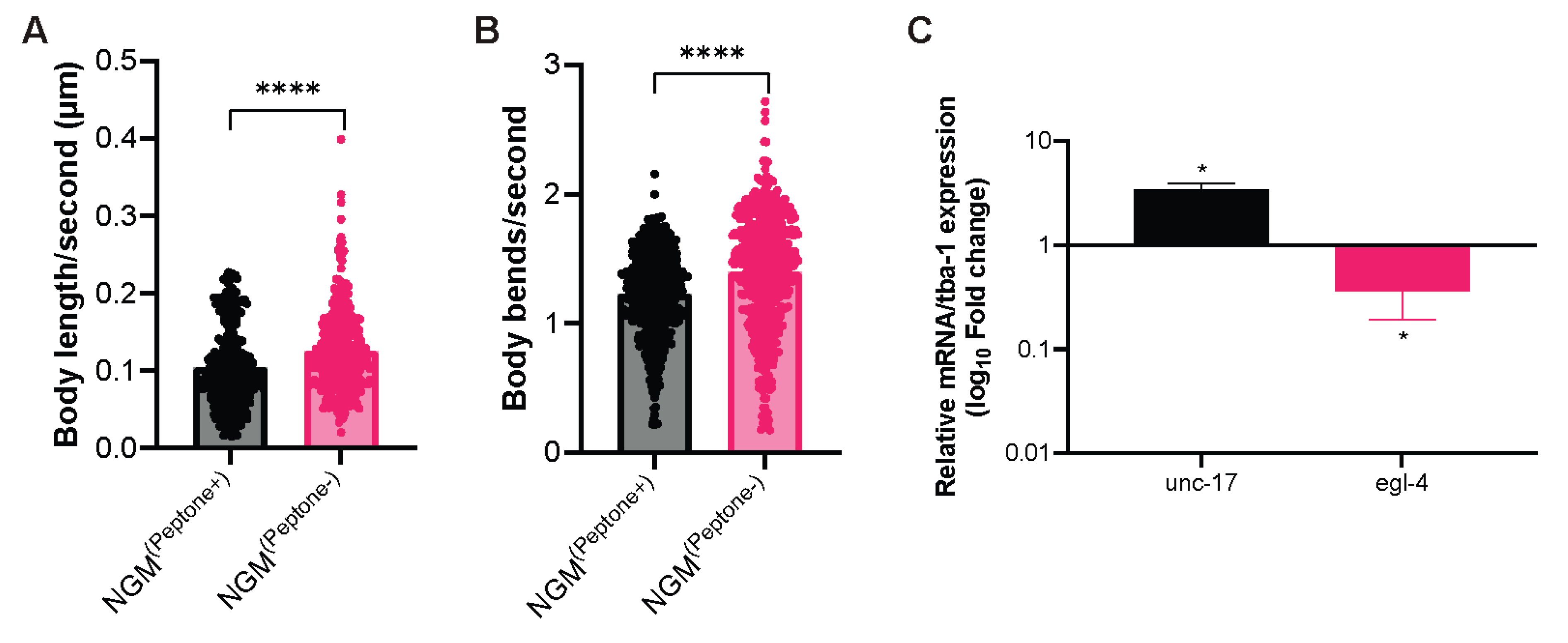
Figure 6.
Peptone-free NGM enhances resistance to oxidative, ultraviolet, and thermal stress in C. elegans. (A) Oxidative stress resistance of age-synchronized L4 worms cultured on NGM(Peptone+) and NGM(Peptone-) following exposure to paraquat (n ≥ 240 worms per condition). Survival was monitored at the indicated time points. (B) Ultraviolet (UV) stress resistance assay showing worm survival following UV exposure (n = 180 worms per condition). (C) Thermotolerance assay showing survival of worms maintained at 35 °C (n ≥ 300 worms per condition). Survival curves were analyzed using the Kaplan-Meier method, and statistical significance between groups was determined using the log-rank (Mantel-Cox) test. Data are presented as mean ± SEM from three independent experiments. Statistical significance is indicated as follows: oxidative stress, P < 0.0001; UV stress, P = 0.0055; and thermotolerance, P < 0.0001.
Figure 6.
Peptone-free NGM enhances resistance to oxidative, ultraviolet, and thermal stress in C. elegans. (A) Oxidative stress resistance of age-synchronized L4 worms cultured on NGM(Peptone+) and NGM(Peptone-) following exposure to paraquat (n ≥ 240 worms per condition). Survival was monitored at the indicated time points. (B) Ultraviolet (UV) stress resistance assay showing worm survival following UV exposure (n = 180 worms per condition). (C) Thermotolerance assay showing survival of worms maintained at 35 °C (n ≥ 300 worms per condition). Survival curves were analyzed using the Kaplan-Meier method, and statistical significance between groups was determined using the log-rank (Mantel-Cox) test. Data are presented as mean ± SEM from three independent experiments. Statistical significance is indicated as follows: oxidative stress, P < 0.0001; UV stress, P = 0.0055; and thermotolerance, P < 0.0001.
Disclaimer/Publisher’s Note: The statements, opinions and data contained in all publications are solely those of the individual author(s) and contributor(s) and not of MDPI and/or the editor(s). MDPI and/or the editor(s) disclaim responsibility for any injury to people or property resulting from any ideas, methods, instructions or products referred to in the content. |
© 2026 by the authors. Licensee MDPI, Basel, Switzerland. This article is an open access article distributed under the terms and conditions of the Creative Commons Attribution (CC BY) license (http://creativecommons.org/licenses/by/4.0/).
Copyright: This open access article is published under a Creative Commons CC BY 4.0 license, which permit the free download, distribution, and reuse, provided that the author and preprint are cited in any reuse.