
Submitted:
14 August 2026
Posted:
17 August 2026
You are already at the latest version
Abstract
Measuring behavior in free-moving, undisturbed rodents is essential for characterizing pain pathways and evaluating analgesic therapies. Current approaches increasingly use machine learning to analyze video recordings of mice and rats. Many existing methods rely on pain behaviors defined by human observers; however, these approaches are susceptible to observer bias. Alternatively, software can be trained to autonomously label anatomical regions of interest, enabling automatic identification of movement locations and trajectories without requiring subjective classification of pain behaviors. More recent work has incorporated three-dimensional (3D) imaging to capture pain-related behaviors. Algorithms have been applied to automatically identify facial features for quantifying orofacial pain, yet 3D imaging has not previously been used for this application. In the present study, 3D imaging was employed to assess behavioral changes associated with orofacial pain in freely moving male and female rats. Both inflammatory and neuropathic pain models were evaluated. Inflammatory pain was induced by placement of a ligature around the masseter tendon, whereas neuropathic pain was induced by injection of varicella zoster virus (VZV) into the whisker pad. Behavioral testing was performed before injury and following treatment with ketoprofen or gabapentin, respectively. Animals were recorded seven days after the start of treatment, with and without drug administration. The software tracked the locations of 12 discrete body parts within the cage and quantified inter-body-part distances, movement speed, and changes in speed over time. Face wiping, rearing, paw to face and tail tip behaviors were automatically detected to determine bout frequency, activity duration as a fraction of total observation time, and average bout duration. Following ligature placement or VZV treatment, tail tip speed decreased but rearing and/or facial wiping behaviors increased, and these responses were attenuated by drug treatment. These findings demonstrate that specific behavioral patterns can be used to identify pain-related spontaneous behavior(s) in rodent models of inflammatory and neuropathic orofacial pain. In conclusion, this study is the first to use 3D imaging to quantify spontaneous orofacial pain responses in rats.
Keywords:
machine learning
; pain
; orofacial
; preclinical
; animal model
1. Introduction
It is necessary to develop more clinically relevant measures of pain as many preclinical results have not translated to effective pain medications [1,2,3]. A combination of tests has been suggested to close the gap between clinical and preclinical results [4,5]. Measures of spontaneous behavior may be key in bridging this gap [4,5]. Spontaneous behavior is one test that can be recorded and quantitated to complement evoked and reward/punishment measures for pain. This study addresses an improved measure of spontaneous behavior using 3D behavioral tracking. Recently, machine learning software has identified both evoked and spontaneous behaviors related to pain [6,7,8,9,10,11]. Recent studies have applied algorithms to autonomously identify behavior [12,13]. Training a computer requires manually labeling areas of interest and then developing a system to automatically analyze limb location and trajectories [14]. These include measuring paw speed [6,15], licking [16], licking and biting [17] and vocalization [18]. Orofacial pain-related spontaneous behaviors include changes in feeding behavior and changes in facial features [19,20,21]. Moreover, machine learning of rodent facial features can be quantitated by computer and used as a correlate for pain [11,22,23].
In prior studies spontaneous orofacial facial pain was measured using two-dimensional modeling techniques [9,10,11] but recent studies are increasing the amount of information gathered from recording spontaneous behavior by using three-dimensional (3D) modeling [8,17,24,25,26,27]. For example, autistic behaviors were automatically identified from 3D imaging and machine learning software [27]. A recent 2023 study used 3D images and machine learning software to identify pain-related spontaneous behaviors after carrageenan injection of the paw and after inducing arthritis [8]. Clifford Woolf’s lab developed a specialized chamber to measure spontaneous behavior for detecting pain in multiple models [28]. To date no study has used 3D modeling to measure orofacial pain and this report will be the first to use 3D modeling to measure spontaneous orofacial pain behaviors. These tests will be completed in male and female rats and using both an inflammatory and a neuropathic pain model.
In this study persistent inflammatory and persistent neuropathic orofacial pain behaviors were measured. Orofacial inflammatory pain was induced by placing a ligature around the masseter tendon [29]. Neuropathic pain was produced by injecting the whisker pad with varicella zoster virus (VZV) to cause zoster-associated “shingles” pain [30]. Comparing the non-pain to the pain group identified behaviors altered by pain. Treating with ketoprofen or gabapentin, respectively, was expected to reverse the pain response. Further supporting that the behavior was pain specific. The animals were recorded 7 days after beginning treatment with and without drug. The computer was trained to identify tail tip, middle and base of the tail, eyes, lower jaw, nose, tip of ears, front legs and paws, base of the neck, mid back, the rear legs and paws. The distance between body parts, speed of motion and rate of change in the speed were calculated. Face wiping, rearing, paw to face and tail tip behaviors were detected using this software to obtain bout frequency, activity duration as fraction of total observation time and average bout duration.
2. Materials and Methods
2.1. Animal Husbandry and Usage
The Texas A&M University Institutional Animal Care and Use Committee approved the experimental protocol (24-0298). Male and female Long Evans rats 250-280 grams were purchased from Charles River (Wilmington, MA) and kept on a 12 hour light/dark cycle with lights on at 07:00 am. They were acclimated for one week before experimentation. The rats were given food and water ad libitum.
2.2. Treatment and Groups
Twenty adult rats of each sex were treated with vehicle or drug (Fig. 1A and 1B, DAY 0). Next, a ligature was placed around the masseter tendon or the rats received sham surgery. Ten male rats and ten female rats had two chromic gut (4.0) ligatures placed around the masseter tendon bilaterally. The ligatures were about two millimeters apart. Ten male rats and ten female rats were sham operated rats and received the same surgery but the tendon was not ligated. After seven days half of the ligature rats and half of the sham rats were injected with vehicle 100 µl PBS (Fig. 1C) or 5 mg/kg ketoprofen subcutaneously (Fig. 1D). Recording of behavior began 30 minutes after the injection. Dosage of ketoprofen was based on the dosage recommended in the Formulary for Laboratory Animals [31]. Ketoprofen was obtained from Covetrus and manufactured by Zoetis as a 100 mg/ml solution.
Figure 1.
Timeline for treatment of the rats. Panels A and B show the ligature experiment and panels C and D show the VZV experiment. Panels A and B start off with sham or ligature on DAY 0. On DAY 7 rats were injected with vehicle (no ketoprofen) or ketoprofen and recorded after 30 minutes. Panels C and D start off (DAY 0) with injection of VZV or MeWo control cells (sham) followed by daily injections of vehicle (no gabapentin) or gabapentin. On the seventh day the animals were recorded 30 minutes after injecting vehicle or gabapentin. Arrows indicate recording of spontaneous behavior for 1 hour.
Figure 1.
Timeline for treatment of the rats. Panels A and B show the ligature experiment and panels C and D show the VZV experiment. Panels A and B start off with sham or ligature on DAY 0. On DAY 7 rats were injected with vehicle (no ketoprofen) or ketoprofen and recorded after 30 minutes. Panels C and D start off (DAY 0) with injection of VZV or MeWo control cells (sham) followed by daily injections of vehicle (no gabapentin) or gabapentin. On the seventh day the animals were recorded 30 minutes after injecting vehicle or gabapentin. Arrows indicate recording of spontaneous behavior for 1 hour.
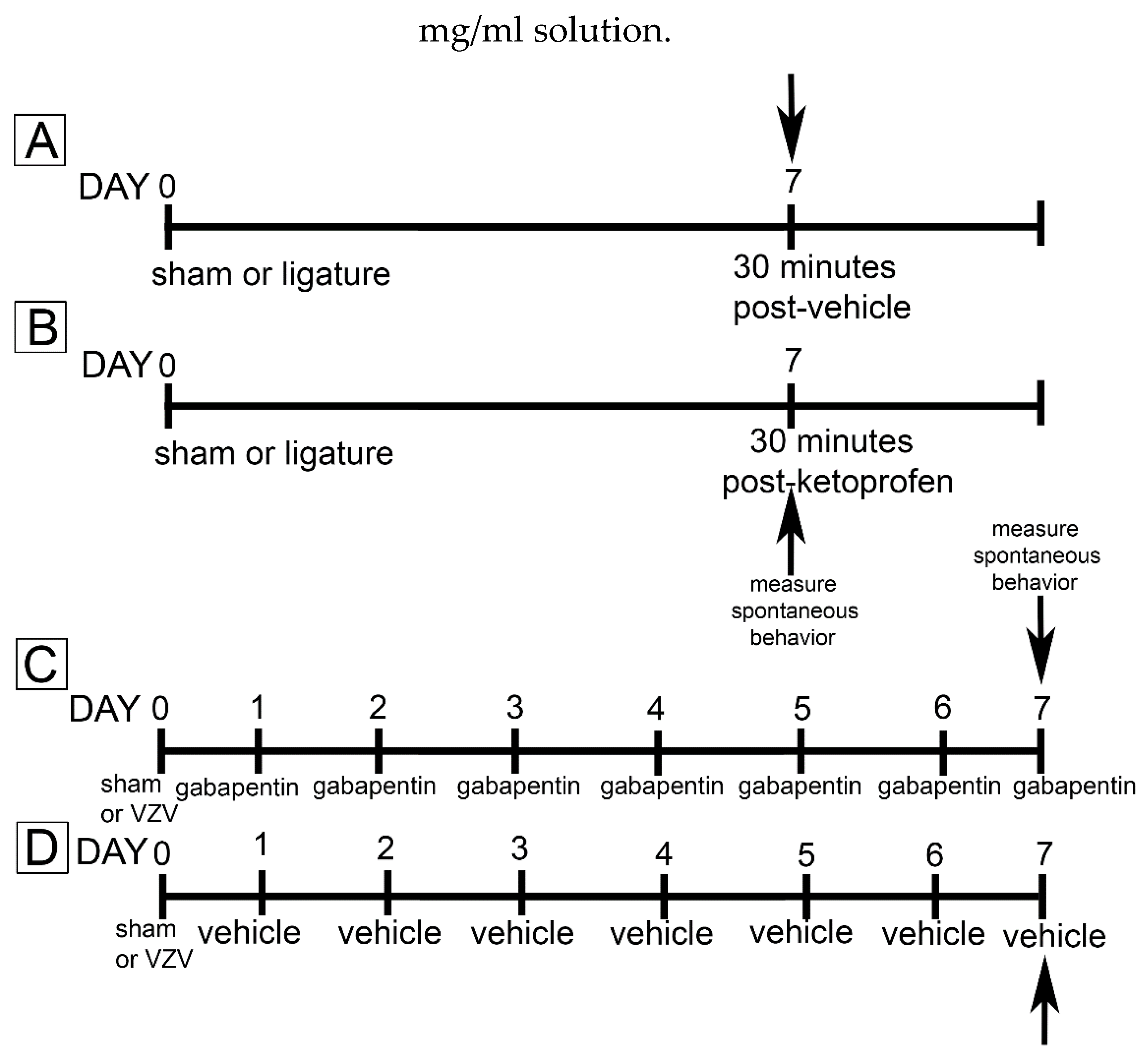
Ten male and ten female rats received VZV injection (50,000 pfu in 100 µl PBS) and ten male and ten female rats received sham (MeWo cells in 100 µl PBS) injection into the whisker pad bilaterally (Fig. 1C and 1D, DAY 0). Half of the VZV rats and half of the sham rats were injected daily (intraperitoneal) with gabapentin (60 mg/kg in 100 µl, cat# PHR1049, Millipore Sigma). The gabapentin dose was based on a previous neuropathic pain study [32]. The remaining half of the VZV rats and the sham rats were injected daily (intraperitoneal) with vehicle (100 µl PBS). On the seventh day a one-hour recording of the rats was completed after injecting gabapentin or vehicle to determine spontaneous behavior (Fig. 1C and 1D, DAY 7).
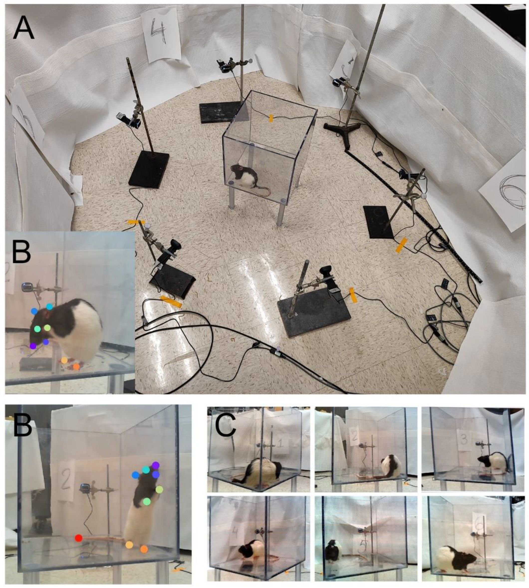
All rats were video recorded for one hour (light phase) in a transparent 30 cm x 30 cm x 30 cm plexiglass cage to measure undisturbed spontaneous behavior after treatment. For the recording, six cameras (Microsoft LifeCam HD-3000) were placed around the cage at 60 degrees relative to each other to cover the entire 360 degree view of the animal and about 45 cm from the center of the cage (Fig. 2A). The videos from the six cameras were captured using VLC media player 3.0.21 software and synchronized at the beginning of each recording to allow subsequent matching of the frames from different camera views (Fig. 2C). Cameras were calibrated with a Charuco board using Anipose software [33]. Rats were acclimated to the plexiglass cage for 30 minutes before recording. Recording was completed by individuals blinded to the treatment given to each group.
Figure 2.
Setup for recording using six cameras. Panel A shows an overhead view of the six cameras and the clear acrylic chamber in the center with a 300 gram Long Evans rat. Panels labeled B show how the software was trained to identify tail tip, middle and base of the tail, eyes, lower jaw, nose, tip of ears, front legs and paws, base of neck, mid back, the rear legs and paws. On image each body part is identified with a different colored dot. Note: image does not have all body parts indicated in Table 1 becaurse the angle of the animal prevents imaging all body parts simultaneously. The top panel B is a rat face wiping and the bottom panel B is a rat rearing. Panel C shows active recording from the six cameras from six different angles. The images were captured on a single computer using VLC media player 3.0.21 software.
Figure 2.
Setup for recording using six cameras. Panel A shows an overhead view of the six cameras and the clear acrylic chamber in the center with a 300 gram Long Evans rat. Panels labeled B show how the software was trained to identify tail tip, middle and base of the tail, eyes, lower jaw, nose, tip of ears, front legs and paws, base of neck, mid back, the rear legs and paws. On image each body part is identified with a different colored dot. Note: image does not have all body parts indicated in Table 1 becaurse the angle of the animal prevents imaging all body parts simultaneously. The top panel B is a rat face wiping and the bottom panel B is a rat rearing. Panel C shows active recording from the six cameras from six different angles. The images were captured on a single computer using VLC media player 3.0.21 software.
2.3. 3D Tracking
To track animal body parts in 3D, we, first, trained a neural network model to identify body parts of interest on video frames. For that, we labeled about 8000 images on the frames from preliminary videos. The following body parts were tracked: nose, lower jaw, eyes, ears, front and hind paws, base of tail, and tip of tail (Fig. 2B). We used DeepLabCut [34] to train a ResNet101 model and to subsequently analyze the video recordings to produce 2D coordinates for each body part on each frame. Second, we used Anipose to triangulate the 2D positions identified with DeepLabCut on the six camera recordings into a 3D time series for each body part. The raw mean average precision for identification of a body part was 84%.
2.4. Behavior Identification
Knowing 3D positions of body parts at sub-second resolution allowed us to define specific body part locations within the cage and measure the distance between different parts. For example, position of nose, lower jaw, eyes, or ears 12 cm above the cage floor would indicate rearing. A distance between a front paw and any of the head features less then 2 cm would most likely indicate face wiping. We compared distributions of each parameter (12 for body part position and 58 for pairwise inter-body part distances, Table 1) between sham and painful state. Any behavior where there was a difference between sham and painful state was included in this study.

Table 1.
Parameters, collected and used in analysis.
| 12 body part positions | 58 pairwise inter-body part distances | |
|---|---|---|
| Body part location, x, y, z coordinates, in mm | Distance between each body part below, in mm | |
| Nose left eye right eye lower jaw left ear right ear left front paw right front paw left hind paw right hind paw base of tail tip of tail |
nose to left ear nose to right ear nose to left front paw nose to right front paw nose to left hind paw nose to right hind paw nose to base of tail nose to tip of tail left eye to left ear left eye to right ear left eye to left front paw left eye to right front paw left eye to left hind paw left eye to right hind paw left eye to base of tail left eye to tip of tail right eye to left ear right eye to right ear right eye to left front paw right eye to right front paw right eye to left hind paw right eye to right hind paw right eye to base of tail right eye to tip of tail lower jaw to right front paw lower jaw to left front paw lower jaw to right hind paw lower jaw to left front paw lower jaw to base of tail lower jaw to tip of tail left ear to right ear left ear to left front paw left ear to right front paw left ear to left hind paw left ear to right hind paw left ear to base of tail left ear to tip of tail |
right ear to left front paw right ear to right front paw right ear to left hind paw right ear to right hind paw right ear to base of tail right ear to tip of tail left front paw to right front paw left front paw to left hind paw left front paw to right hind paw left front paw to base of tail left front paw to tip of tail right front paw to left hind paw right front paw to right hind paw right front paw to base of tail right front paw to tip of tail left hind paw to right hind paw left hind paw to base of tail left hind paw to tip of tail right hind paw to base of tail right hind paw to tip of tail base of tail to tip of tail |
Thus, a body part position or inter-body part distance time series for an animal was dichotomized such that each frame (and the corresponding time period) was assigned a “yes” or “no” behavior. The “yes” frames were accumulated over the frames of the session producing a cumulative time course of a particular behavior over the session time. To determine the effect of a treatment (pain induction, pain relief, or both) the cumulative behavior from a baseline recording was subtracted from the post-treatment cumulative behavior. Baseline was obtained from a 1 hour recording using the naïve rat before any treatment was started. Thus, the intervention effect could be represented by a single line and a single number representing the difference in the cumulative behavior time between baseline and treatment at the end of 1-hour observation period. Measurements completed in this study include speed of the base of the tail, moving front paw to nose, face wiping, and rearing.
2.5. Statistics and Analysis
Statistical analysis of the treatment effects was completed using two-way ANOVA with repeated measure comparing before ligature and VZV to after ligature and VZV for each animal. The independent variables were drug and treatment and the dependent variables were cumulative time difference of spontaneous behavior or number of behavioral events. Comparison of groups was performed using Sidak’s post-hoc tests. Male and female data was compared using a Mann-Whitney test. The data provided >80% power to detect the specified effect size at a level of α = 0.05. Values were reported as the mean ± SEM.
3. Results
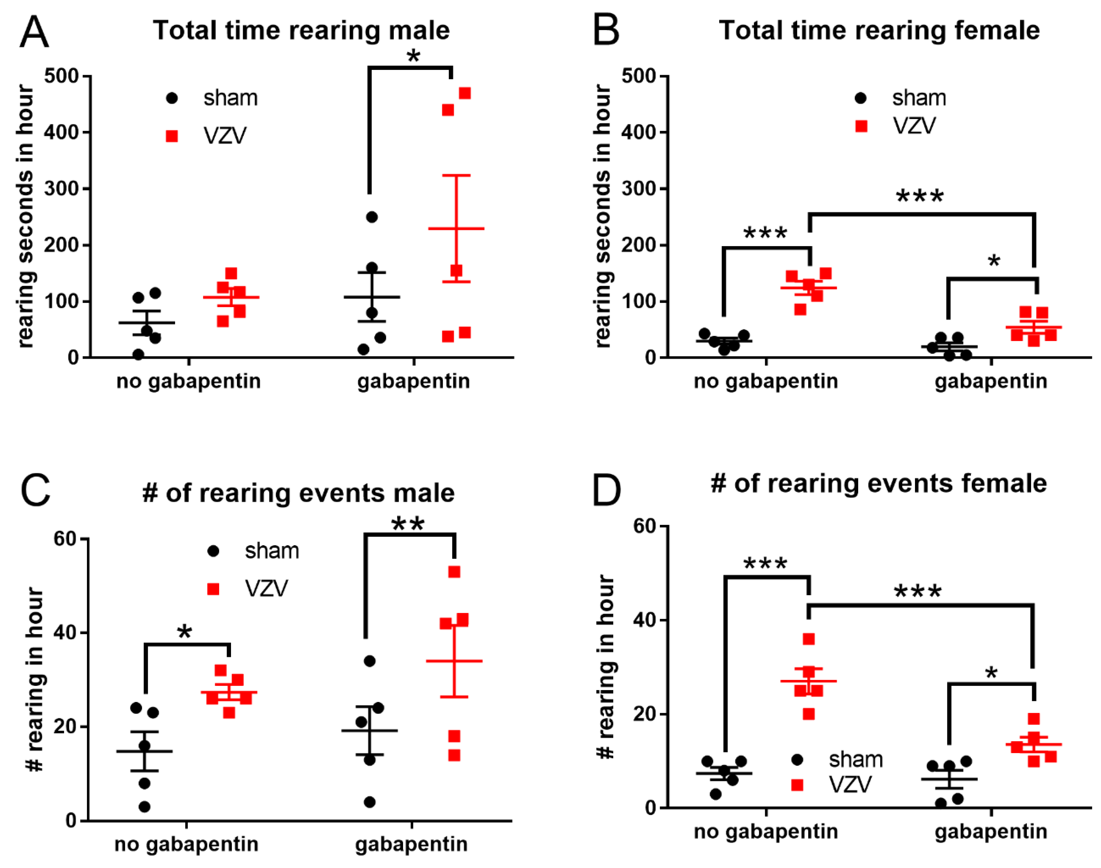
After recording the rats for one hour the total rearing time increased in males (F1,8 = 8.5, p=0.019) and females (F1,8 = 74.5, p<0.0001) after VZV injection (Fig. 3A and B). Gabapentin treatment significantly (F1,8 = 13.8, p=0.006) decreased the rearing time in females (Fig. 3B). The number of rearing events also increased in both male (F1,8 = 27.5, p=0.0008) and females (F1,8 = 44.1, p=0.0002) (Fig. 3C and D). Again, gabapentin treatment significantly decreased (F1,8 = 15.4, p=0.004) the number of rearing events in females (Fig. 3D). After treating with gabapentin females had a significantly fewer rearing events than males.
Comparing the male sham gabapentin group (Fig. 3, black circles, panel C) with the female sham gabapentin group (Fig. 3, black circles, panel D) there was significantly fewer rearing events (p<0.05). Comparing the male VZV gabapentin group (Fig. 3, red squares, panel C) with the female VZV gabapentin group (Fig. 3, red squares, panel D) there was significantly fewer rearing events (p<0.05).
Figure 3.
The number of rearing events and the amount of time rearing was altered in a neuropathic pain model. To induce neuropathic pain the whisker pad was subcutaneously injected with VZV or control MeWo cells (sham) bilaterally. After whisker pad injection the animals were given daily intraperitoneal injections of gabapentin (60 mg/kg in 100 µl) or vehicle (100 µl PBS); labeled as no gabapentin. The amount of time rearing was measured from the recordings seven days after injection of the whisker pad for both males (panel A) and females (panel B) rats. The number of rearing events was counted within the one-hour recording for males (panel C) and for female (panel D) rats. *= p<0.05, **=p<0.01, ***<p<0.001. Each point on the graph is from an individual rat.
Figure 3.
The number of rearing events and the amount of time rearing was altered in a neuropathic pain model. To induce neuropathic pain the whisker pad was subcutaneously injected with VZV or control MeWo cells (sham) bilaterally. After whisker pad injection the animals were given daily intraperitoneal injections of gabapentin (60 mg/kg in 100 µl) or vehicle (100 µl PBS); labeled as no gabapentin. The amount of time rearing was measured from the recordings seven days after injection of the whisker pad for both males (panel A) and females (panel B) rats. The number of rearing events was counted within the one-hour recording for males (panel C) and for female (panel D) rats. *= p<0.05, **=p<0.01, ***<p<0.001. Each point on the graph is from an individual rat.
The amount of time face wiping (Fig. 4A) and the number of face wipes (Fig. 4C) did not significantly change after injecting the VZV into the whisker pad of males. In females the amount of face wiping (Fig. 4B) significantly increased (F1,8 = 9.4, p=0.015) and the number of face wipes (Fig. 4D) significantly increased (F1,8 = 11.0, p=0.01). Gabapentin significantly decreased the amount of face wiping time (F1,8 = 20.7, p=0.0019) (Fig. 4B) and the number of face wipes (F1,8 = 42.5, p=0.0002) (Fig. 4D) in females.
Figure 4.
The total time face wiping and the number of face wiping events was altered in a neuropathic pain model. To induce neuropathic pain the whisker pad was injected with VZV or vehicle (sham) bilaterally followed by daily i.p. injections of gabapentin or vehicle (no gabapentin). The amount of time face wiping 7 days after treatment is show for both males (panel A) and females (panel B). Seven days after treatment began the number of face wiping events in one hour of recording is shown for males (panel C) and for females (panel D). **=p<0.01, ***<p<0.001. Each point on the graph is from an individual rat.
Figure 4.
The total time face wiping and the number of face wiping events was altered in a neuropathic pain model. To induce neuropathic pain the whisker pad was injected with VZV or vehicle (sham) bilaterally followed by daily i.p. injections of gabapentin or vehicle (no gabapentin). The amount of time face wiping 7 days after treatment is show for both males (panel A) and females (panel B). Seven days after treatment began the number of face wiping events in one hour of recording is shown for males (panel C) and for females (panel D). **=p<0.01, ***<p<0.001. Each point on the graph is from an individual rat.
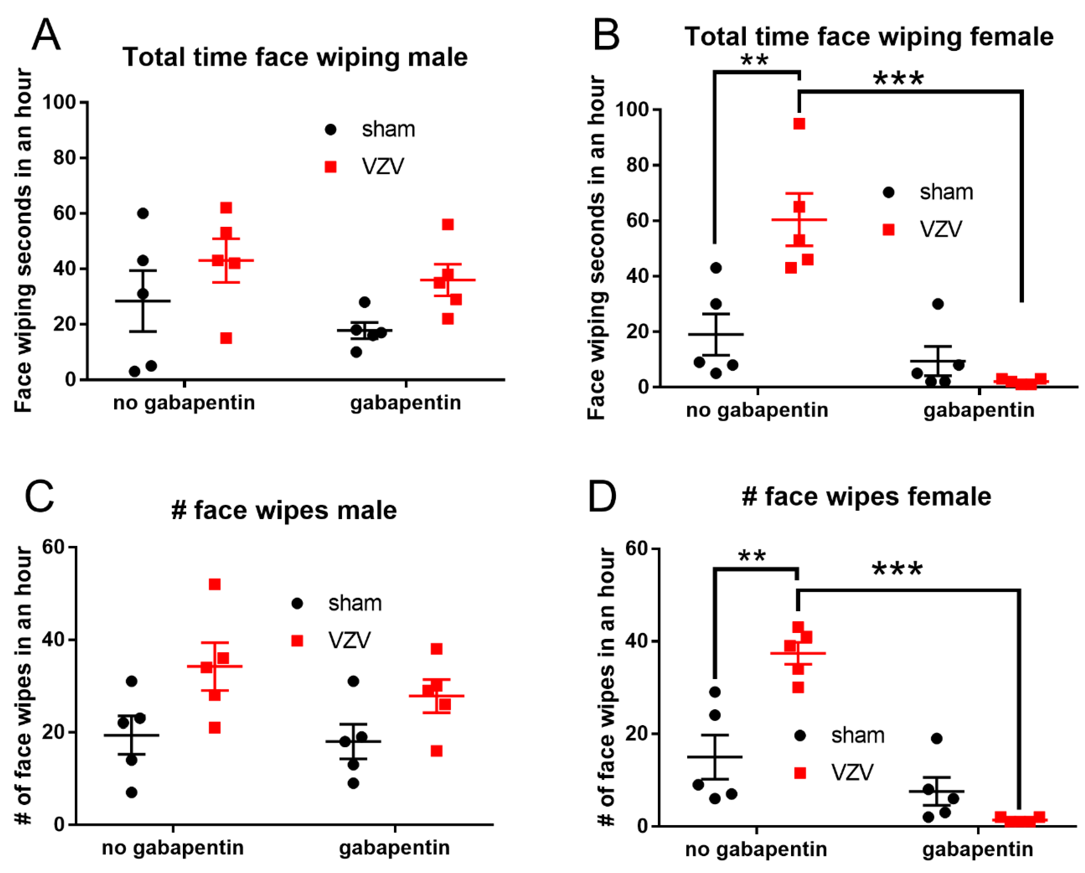
When comparing the males to the females the females had significantly (p<0.01) less face wiping time (compare red squares in Fig. 4A to Fig. 4B) and face wiping events (compare red squares, Fig. 4C to Fig. 4D) in the VZV gabapentin treatment group.
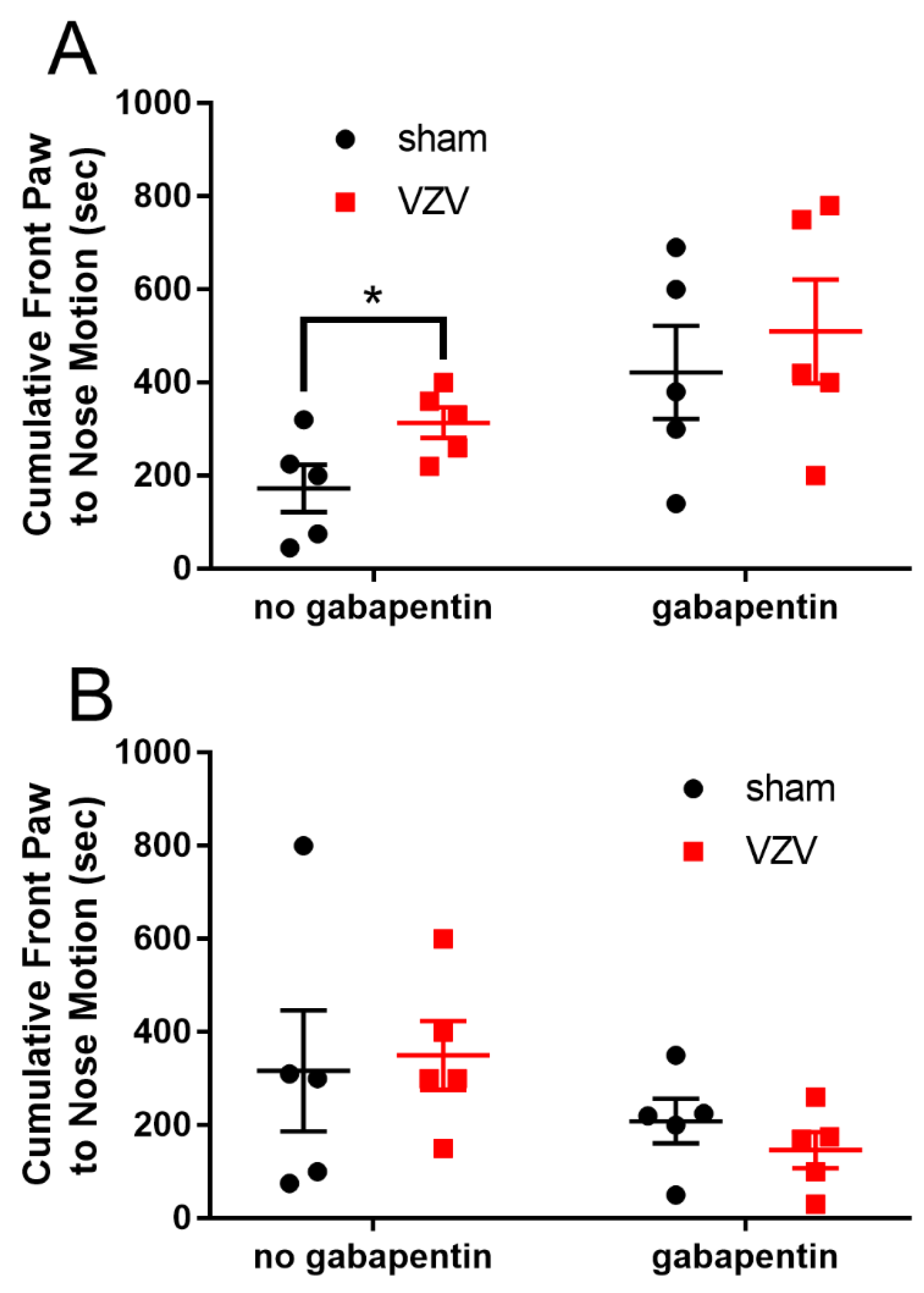
The time the rat was moving the front paw to the nose significantly increased in male rats (F1,8 = 16.3, p=0.004) (Fig. 5A) but not female rats (Fig. 5B) after injection of VZV.
Figure 5.
The total time the front paw moved toward the face was recorded for one hour. The recording was completed after injecting VZV or vehicle (sham) into the whisker pad bilaterally and after daily i.p. injections of gabapentin or vehicle (no gabapentin). Seven days after treatment spontaneous behavior was recorded in both male (panel A) and female (panel B) rats. * =p<0.05, **=p<0.01. Each point on the graph is from an individual rat.
Figure 5.
The total time the front paw moved toward the face was recorded for one hour. The recording was completed after injecting VZV or vehicle (sham) into the whisker pad bilaterally and after daily i.p. injections of gabapentin or vehicle (no gabapentin). Seven days after treatment spontaneous behavior was recorded in both male (panel A) and female (panel B) rats. * =p<0.05, **=p<0.01. Each point on the graph is from an individual rat.
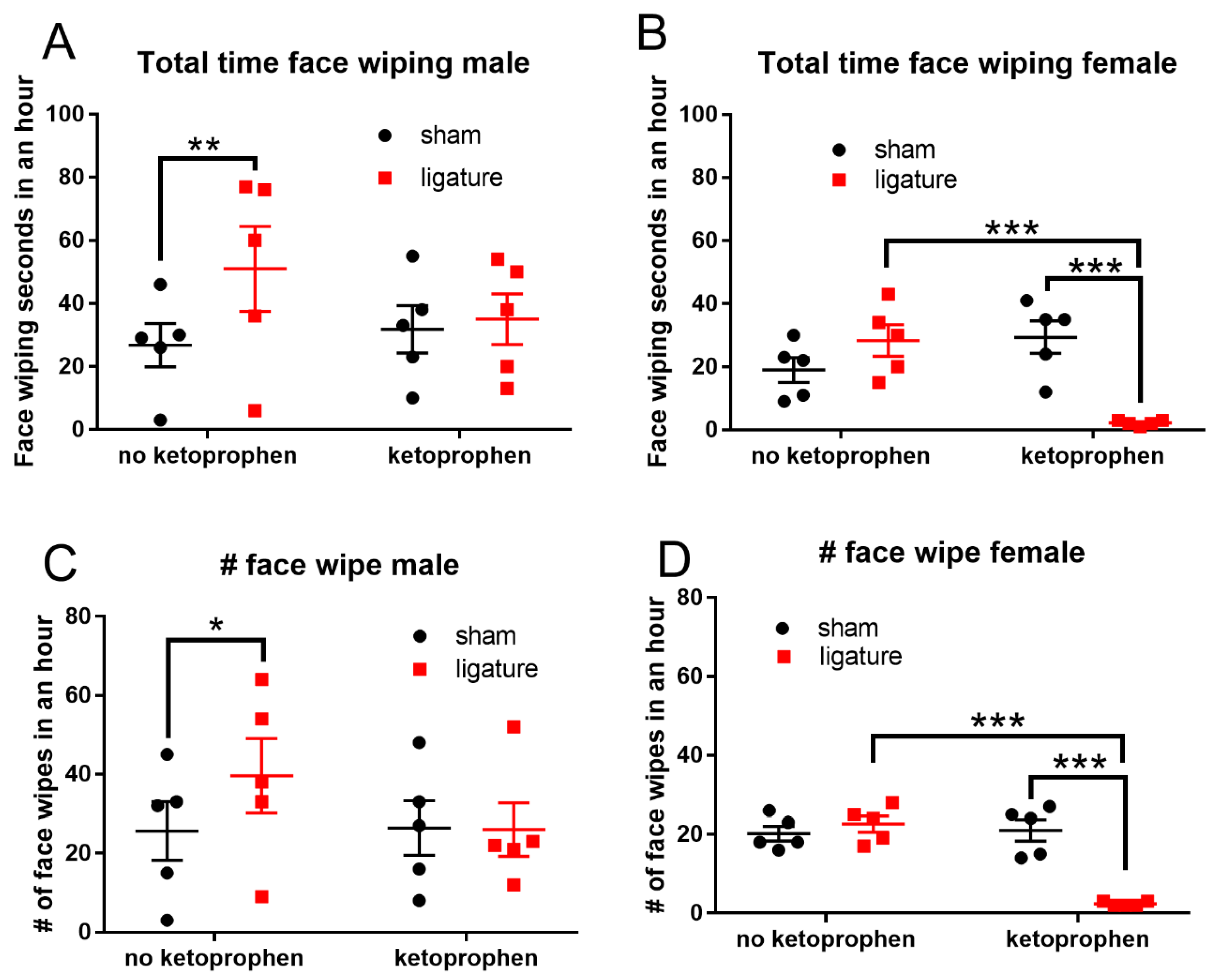
In an inflammatory model where the masseter tendon was ligated the amount of time face wiping significantly increased in male rats (F1,8 = 11.1, p=0.01) (Fig. 6A). Also, the number of times that the male rats wiped their face within the one-hour was significantly increased (F1,8 = 4.2, p=0.05) (Fig. 6C). Ketoprofen had no significant effect on male behavior (Fig. 6A, C). In contrast, ketoprofen significantly (F1,8 = 16.8, p=0.003) decreased the amount of face wiping time (Fig. 6B) and the number of face wipes (Fig. 6D) in females injected with VZV. Females had significantly (p<0.01) less face wiping time (compare red squares in Fig. 6A to Fig. 6B) and face wiping events (compare red squares in Fig. 6C to Fig. 6D) as compared to males after ligature and ketoprofen treatment.
Figure 6.
The total time face wiping and the number of face wiping events was altered in an inflammatory model. To induce inflammation of the orofacial region the masseter tendon was ligatured bilaterally. Sham operated rats and received the same surgery but the tendon was not ligated. Seven days after ligature the rats were divided so that half were injected with ketoprofen and half were injected with vehicle (no ketoprofen). Thirty minutes after injection the rats behavior was recorded for one hour. The amount of time face wiping within the one hour recording is show for both males (panel A) and females (panel B). The number of face wiping events in that one hour of recording are shown for males (panel C) and for females (panel D). **=p<0.01, ***<p<0.001. Each point on the graph is from an individual rat.
Figure 6.
The total time face wiping and the number of face wiping events was altered in an inflammatory model. To induce inflammation of the orofacial region the masseter tendon was ligatured bilaterally. Sham operated rats and received the same surgery but the tendon was not ligated. Seven days after ligature the rats were divided so that half were injected with ketoprofen and half were injected with vehicle (no ketoprofen). Thirty minutes after injection the rats behavior was recorded for one hour. The amount of time face wiping within the one hour recording is show for both males (panel A) and females (panel B). The number of face wiping events in that one hour of recording are shown for males (panel C) and for females (panel D). **=p<0.01, ***<p<0.001. Each point on the graph is from an individual rat.
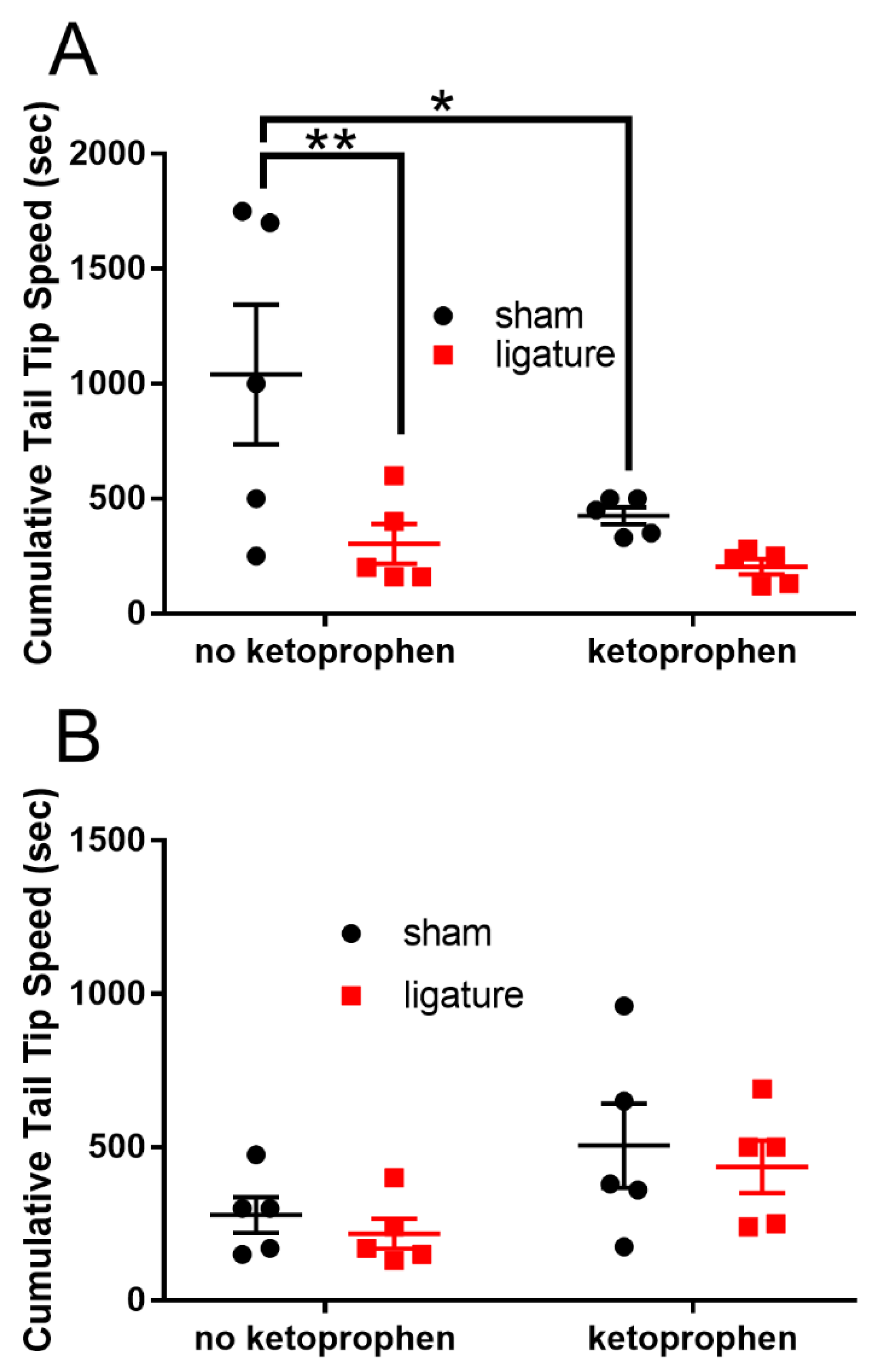
Ligature did significantly (F1,8 = 4, p=0.05) decreased the speed of the tail tip in male rats (Fig. 7A) and ketoprofen also decreased this speed (Fig. 7A). Ligature and ketoprofen did not significantly alter the tail tip speed in female rats (Fig. 7B).
Figure 7.
Ligature of the master tendon reduced the tail tip speed. Seven days after ligature of the masseter tendon the tail tip speed was recorded in both male (panel A) and female (panel B) rats after injecting vehicle (no ketoprofen) or ketoprofen. *= p<0.05. Each point on the graph is from an individual rat.
Figure 7.
Ligature of the master tendon reduced the tail tip speed. Seven days after ligature of the masseter tendon the tail tip speed was recorded in both male (panel A) and female (panel B) rats after injecting vehicle (no ketoprofen) or ketoprofen. *= p<0.05. Each point on the graph is from an individual rat.
4. Discussion
Recording of rats over one hour determined spontaneous changes in behavior that correlated to pain. Our lab demonstrated that ligature of the masseter tendon induces pain in a rat [35] and that administering NSAIDS reduces the pain response [36]. Injecting varicella zoster virus into the whisker pad of rats induced a shingles like pain response that can be attenuated using gabapentin [30]. By treating with pain relieving drugs we expect that reversal of a behavior by drug treatment indicated a pain response. Thus, a drug response measured with a 3D imaging method would indicate a behavior that correlates to pain.
Most existing methods rely on action categories defined by human experts to establish correlation with pain behaviors [7], making it difficult to directly identify pain behaviors. These methods usually rely on the intervention of human experts and are therefore prone to observer bias thus, limiting the objectivity and reproducibility [37]. To address this potential bias, we developed a method requiring computer selection of body parts along with analysis of the location and movement that can identify various animal behaviors unique to the pain group.
An additional strength of these studies is the attempt to quantify spontaneous pain-related behaviors in freely moving animals and validate these behaviors pharmacologically with standard analgesic drugs. Many preclinical studies do not translate into clinical results and there is a challenge to improve clinical outcomes [1,2,3]. Spontaneous or ongoing pain-related behaviors may better capture clinically relevant aspects of pain versus conventional evoked reflex-based assays [4]. In support of this idea, a recent meta-analysis study demonstrated that measuring pain-related spontaneous behaviors in preclinical models mirrored clinical performance and could enhance clinical translation [38]. Thus, developing an improved method for measuring pain-related spontaneous behavior would result in more experimental drugs being successful in the clinic.
Rearing and face wiping increased after injection of VZV. Gabapentin reversed this response. Gabapentin is a drug used to treat shingles pain after VZV infection [39] thus, we hypothesize that the ameliorating effect of gabapentin on rearing and face wiping indicates these behaviors are pain specific. Other labs have shown that during the neuropathic phase post-formalin injection mice show an increased face wiping [40,41]. Face wiping also increases after ligature of the infraorbital nerve and gabapentin reduces the wiping response [42,43,44]. This reduction could be due to attenuation of pain and/or locomotion. Comparison of the sham animals having no gabapentin to the sham animals treated with gabapentin suggests there is no significant effect in rearing or face wiping. If gabapentin altered locomotion we would expect the sham animals treated with gabapentin to have reduced rearing and face wiping behavior in comparison to the sham animals with no gabapentin. This was not observed. In contrast, once pain was induced by injecting VZV there was an effect of gabapentin as shown when comparing the VZV no gabapentin group to the VZV gabapentin group. We cannot eliminate the possibility that gabapentin altered locomotion after VZV treatment.
In addition, the reduced tail tip speed after ketoprofen treatment could be due to drug effects on locomotion thus, not measuring locomotor activity is a weakness of this study.
Rearing decreases in rodents with infraorbital nerve ligature [45] but rearing increased in our VZV model. Thus far, rearing has never been measured after VZV injection and may result in a different response than infraorbital nerve ligature. A few reports do describe increased rearing due to novel environments [46,47] but rats in these studies were conditioned to chambers for 30 minutes before recording and novelty would be expected to be extinguished before recording. One potential explanation for this increased rearing response is that after VZV the stress response was elevated and this mild stressor increased rearing, consistent with reports of mild stress increasing rearing [48,49,50].
Ligature of the masseter tendon increased face wiping, consistent with increased face wiping during the inflammatory phase following formalin injection [40,41]. Inflammation in the orofacial region increases the face wiping response [51,52]. Reducing inflammation or treating with opiates reduced face wiping behavior suggesting that wiping behavior correlates to pain and is an indicator of orofacial pain [51,52]. Females had less face wiping time and fewer face wiping events as compared to males after ligature and ketoprofen treatment. This may be the result of ketoprofen having a greater effect in females verses males.
Much of the behavioral changes were specific to females. Females typically show a greater VZV induced neuropathic pain response than males [30] and the female rats had fewer rearing, face wiping events and less face wiping time than males after gabapentin treatment in this study. Inflammatory pain induced by ligature of the masseter tendon also shows a sex difference with females showing a greater pain response than males [53]. One explanation for the sex difference in behavioral may be that anxiety and stress are greater in females and influence the response [54]. Another possibility is that sex hormones alter neuron or glial function at a molecular level to change the pain response [55].
After performing our 3D recordings tail speed was reduced by ligature and ketoprofen treatment. A recent article using 3D imaging demonstrated a reduced speed of motion after inducing pain [13]. Speed of motion was also altered by the drug alone suggesting the drug slows the speed of the animal and could be considered a side effect of the drug unrelated to the pain response.
Future work will include longer recordings with multiple animals. Behaviors related to pain may not be observed within a one-hour recording or with a singly housed animal. We shall also incorporate measurement during the dark phase as this an active period for the rodent and could reveal pain specific behaviors. Higher resolution cameras can identify each whisker and each digit on the paw, this resolution could be used to identify more behaviors such as whisker contraction or paw flexion, respectively.
In conclusion, initial 3D imaging studies reveal behavioral changes in both a neuropathic and inflammatory pain model. These changes in behavior are consistent with previous studies but further refinement of this method is needed to determine the natural behaviors modified by pain.
Acknowledgments
This study was funded through NIH/NIDCR DE022129 to PRK. The research presented in this manuscript is free of conflict of interest.
References
- Rice, A.S.C.; Eisenach, J.C. Improving Preclinical Development of Novel Interventions to Treat Pain: Insanity Is Doing the Same Thing Over and Over and Expecting Different Results. Anesth. Analg. 2022, 135, 1128–1136. [Google Scholar] [CrossRef] [PubMed]
- Tappe-Theodor, A.; Kuner, R. Studying ongoing and spontaneous pain in rodents – challenges and opportunities. Eur. J. Neurosci. 2014, 39, 1881–1890. [Google Scholar] [CrossRef] [PubMed]
- Yezierski, R.P.; Hansson, P. Inflammatory and Neuropathic Pain From Bench to Bedside: What Went Wrong? J. Pain 2018, 19, 571–588. [Google Scholar] [CrossRef] [PubMed]
- Draxler, P.; Moen, A.; Galek, K.; Boghos, A.; Ramazanova, D.; Sandkühler, J. Spontaneous, Voluntary, and Affective Behaviours in Rat Models of Pathological Pain. Front Pain Res. 2021, 2, 672711. [Google Scholar] [CrossRef] [PubMed]
- Huerta, M.Á.; Cisneros, E.; Alique, M.; Roza, C. Strategies for measuring non-evoked pain in preclinical models of neuropathic pain: Systematic review. Neurosci. Biobehav. Rev. 2024, 163, 105761. [Google Scholar] [CrossRef] [PubMed]
- Jones, J.M.; Foster, W.; Twomey, C.R.; Burdge, J.; Ahmed, O.M.; Pereira, T.D.; Wojick, J.A.; Corder, G.; Plotkin, J.B.; Abdus-Saboor, I. A machine-vision approach for automated pain measurement at millisecond timescales. eLife 2020, 9. [Google Scholar] [CrossRef] [PubMed]
- Chen, Y.-H.; Chen, W.-H.; Wang, C.-Y.; Liao, H.-Y.M.; Liao, J.C.; Chen, C.-C. Deep Learning-based Animal Behavior Analysis: Insights from Mouse Chronic Pain Models. arXiv 2025, arXiv:2508.05138. [Google Scholar]
- Bohic, M.; Pattison, L.A.; Jhumka, Z.A.; Rossi, H.; Thackray, J.K.; Ricci, M.; Mossazghi, N.; Foster, W.; Ogundare, S.; Twomey, C.R.; et al. Mapping the neuroethological signatures of pain, analgesia, and recovery in mice. Neuron 2023, 111, 2811–2830.e2818. [Google Scholar] [CrossRef] [PubMed]
- Tuttle, A.H.; Molinaro, M.J.; Jethwa, J.F.; Sotocinal, S.G.; Prieto, J.C.; Styner, M.A.; Mogil, J.S.; Zylka, M.J. A deep neural network to assess spontaneous pain from mouse facial expressions. Mol. Pain 2018, 14, 1744806918763658. [Google Scholar] [CrossRef] [PubMed]
- Chiang, C.Y.; Chen, Y.P.; Tzeng, H.R.; Chang, M.H.; Chiou, L.C.; Pei, Y.C. Deep Learning-Based Grimace Scoring Is Comparable to Human Scoring in a Mouse Migraine Model. J. Pers. Med. 2022, 12. [Google Scholar] [CrossRef] [PubMed]
- McCoy, E.S.; Park, S.K.; Patel, R.P.; Ryan, D.F.; Mullen, Z.J.; Nesbitt, J.J.; Lopez, J.E.; Taylor-Blake, B.; Vanden, K.A.; Krantz, J.L.; et al. Development of PainFace software to simplify, standardize, and scale up mouse grimace analyses. Pain 2024, 165, 1793–1805. [Google Scholar] [CrossRef] [PubMed]
- Tillmann, J.F.; Hsu, A.I.; Schwarz, M.K.; Yttri, E.A. A-SOiD, an active-learning platform for expert-guided, data-efficient discovery of behavior. Nat. Methods 2024, 21, 703–711. [Google Scholar] [CrossRef] [PubMed]
- Ashiquzzaman, A.; Lee, E.; Znaub, B.F.; Sakib, A.N.; Chung, G.; Kim, S.S.; Kim, Y.R.; Kwon, H.S.; Chung, E. MoSeq based 3D behavioral profiling uncovers neuropathic behavior changes in diabetic mouse model. Sci. Rep. 2025, 15, 15114. [Google Scholar] [CrossRef] [PubMed]
- Gu, A.; Goel, K.; Ré, C. Efficiently modeling long sequences with structured state spaces. arXiv 2021, arXiv:2111.00396. [Google Scholar]
- Abdus-Saboor, I.; Fried, N.T.; Lay, M.; Burdge, J.; Swanson, K.; Fischer, R.; Jones, J.; Dong, P.; Cai, W.; Guo, X.; et al. Development of a Mouse Pain Scale Using Sub-second Behavioral Mapping and Statistical Modeling. Cell Rep. 2019, 28, 1623–1634.e1624. [Google Scholar] [CrossRef] [PubMed]
- Wotton, J.M.; Peterson, E.; Anderson, L.; Murray, S.A.; Braun, R.E.; Chesler, E.J.; White, J.K.; Kumar, V. Machine learning-based automated phenotyping of inflammatory nocifensive behavior in mice. Mol. Pain 2020, 16, 1744806920958596. [Google Scholar] [CrossRef] [PubMed]
- Barkai, O.; Zhang, B.; Turnes, B.L.; Arab, M.; Yarmolinsky, D.A.; Zhang, Z.; Barrett, L.B.; Woolf, C.J. ARBEL: A Machine Learning Tool with Light-Based Image Analysis for Automatic Classification of 3D Pain Behaviors. bioRxiv 2024. [Google Scholar] [CrossRef] [PubMed]
- Cordeiro, A.F.d.S.; Nääs, I.d.A.; Baracho, M.d.S.; Jacob, F.G.; Moura, D.J.d. THE USE OF VOCALIZATION SIGNALS TO ESTIMATE THE LEVEL OF PAIN IN PIGLETS. Eng. Agrícola 2018, 38. [Google Scholar]
- Harper, R.P.; Kerins, C.A.; Talwar, R.; Spears, R.; Hutchins, B.; Carlson, D.S.; McIntosh, J.E.; Bellinger, L.L. Meal pattern analysis in response to temporomandibular joint inflammation in the rat. J.Dent.Res. 2000, 79, 1704–1711. [Google Scholar] [CrossRef] [PubMed]
- Andresen, N.; Wöllhaf, M.; Hohlbaum, K.; Lewejohann, L.; Hellwich, O.; Thöne-Reineke, C.; Belik, V. Towards a fully automated surveillance of well-being status in laboratory mice using deep learning: Starting with facial expression analysis. PLoS ONE 2020, 15, e0228059. [Google Scholar] [CrossRef] [PubMed]
- Langford, D.J.; Bailey, A.L.; Chanda, M.L.; Clarke, S.E.; Drummond, T.E.; Echols, S.; Glick, S.; Ingrao, J.; Klassen-Ross, T.; Lacroix-Fralish, M.L.; et al. Coding of facial expressions of pain in the laboratory mouse. Nat. Methods 2010, 7, 447–449. [Google Scholar] [CrossRef] [PubMed]
- Dalla Costa, E.; Pascuzzo, R.; Leach, M.C.; Dai, F.; Lebelt, D.; Vantini, S.; Minero, M. Can grimace scales estimate the pain status in horses and mice? A statistical approach to identify a classifier. PLoS ONE 2018, 13, e0200339. [Google Scholar] [CrossRef] [PubMed]
- Vidal, A.; Jha, S.; Hassler, S.; Price, T.; Busso, C. Face detection and grimace scale prediction of white furred mice. Mach. Learn. With Appl. 2022, 8, 100312. [Google Scholar] [CrossRef]
- Dunn, T.W.; Marshall, J.D.; Severson, K.S.; Aldarondo, D.E.; Hildebrand, D.G.C.; Chettih, S.N.; Wang, W.L.; Gellis, A.J.; Carlson, D.E.; Aronov, D.; et al. Geometric deep learning enables 3D kinematic profiling across species and environments. Nat. Methods 2021, 18, 564–573. [Google Scholar] [CrossRef] [PubMed]
- Lauer, J.; Zhou, M.; Ye, S.; Menegas, W.; Schneider, S.; Nath, T.; Rahman, M.M.; Di Santo, V.; Soberanes, D.; Feng, G.; et al. Multi-animal pose estimation, identification and tracking with DeepLabCut. Nat. Methods 2022, 19, 496–504. [Google Scholar] [CrossRef] [PubMed]
- Tsuruda, Y.; Akita, S.; Yamanaka, K.; Matsumoto, Y.; Yamamoto, M.; Sano, Y.; Furuichi, T.; Takemura, H. 3D Body Parts Tracking of Mouse Based on RGB-D Video from Under an Open Field. Annu Int. Conf. IEEE Eng. Med. Biol. Soc. 2021, 2021, 7252–7255. [Google Scholar] [CrossRef] [PubMed]
- Huang, K.; Han, Y.; Chen, K.; Pan, H.; Zhao, G.; Yi, W.; Li, X.; Liu, S.; Wei, P.; Wang, L. A hierarchical 3D-motion learning framework for animal spontaneous behavior mapping. Nat. Commun. 2021, 12, 2784. [Google Scholar] [CrossRef] [PubMed]
- Barkai, O.; Zhang, B.; Turnes, B.; Arab, M.; Yarmolinsky, D.; Zhang, Z.; Barrett, L.; Woolf, C. A machine learning tool with light-based image analysis for automatic classification of 3D pain behaviors. Cell Rep. Methods 2025, 5, 101145. [Google Scholar] [CrossRef] [PubMed]
- Guo, W.; Wang, H.; Zou, S.; Wei, F.; Dubner, R.; Ren, K. Long lasting pain hypersensitivity following ligation of the tendon of the masseter muscle in rats: a model of myogenic orofacial pain. Mol. Pain 2010, 6, 40. [Google Scholar] [CrossRef] [PubMed]
- Stinson, C.; Deng, M.; Yee, M.B.; Bellinger, L.L.; Kinchington, P.R.; Kramer, P.R. Sex differences underlying orofacial varicella zoster associated pain in rats. BMC Neurol. 2017, 17, 95. [Google Scholar] [CrossRef] [PubMed]
- Hawk, C.T.; Hawk, C.T.; Leary, S.L.; Morris, T.H.; American College of Laboratory Animal, M. European College of Laboratory Animal, M. Formulary for laboratory animals / compiled by C. Terrance Hawk, Steven L. Leary, Timothy H. Morris ; in association with the American College of Laboratory Animal Medicine and the European College of Laboratory Animal Medicine, Third edition. ed.; Blackwell Publishing: 2005.
- Surcheva, S.; Todorova, L.; Maslarov, D.; Vlaskovska, M. Preclinic and clinic effectiveness of gabapentin and pregabalin for treatment of neuropathic pain in rats and diabetic patients. Biotechnol. Biotechnol. Equip. 2017, 31, 568–573. [Google Scholar] [CrossRef]
- Karashchuk, P.; Rupp, K.L.; Dickinson, E.S.; Walling-Bell, S.; Sanders, E.; Azim, E.; Brunton, B.W.; Tuthill, J.C. Anipose: A toolkit for robust markerless 3D pose estimation. Cell Rep. 2021, 36, 109730. [Google Scholar] [CrossRef] [PubMed]
- Mathis, A.; Mamidanna, P.; Cury, K.M.; Abe, T.; Murthy, V.N.; Mathis, M.W.; Bethge, M. DeepLabCut: markerless pose estimation of user-defined body parts with deep learning. Nat. Neurosci. 2018, 21, 1281–1289. [Google Scholar] [CrossRef] [PubMed]
- Kramer, P.R.; Bellinger, L.L. Reduced GABA receptor alpha6 expression in the trigeminal ganglion enhanced myofascial nociceptive response. Neuroscience 2013, 245C, 1–11. [Google Scholar] [CrossRef] [PubMed]
- Shu, H.; Liu, S.; Tang, Y.; Schmidt, B.L.; Dolan, J.C.; Bellinger, L.L.; Kramer, P.R.; Bender, S.D.; Tao, F. A Pre-Existing Myogenic Temporomandibular Disorder Increases Trigeminal Calcitonin Gene-Related Peptide and Enhances Nitroglycerin-Induced Hypersensitivity in Mice. Int. J. Mol. Sci. 2020, 21. [Google Scholar] [CrossRef] [PubMed]
- Tuyttens, F.A.M.S.; Heerkens, J.L.T.; Jacobs, L.; Nalon, E.; Ott, S.; Stadig, L.E.; Ampe, B. Observer bias in animal behaviour research: can we believe what we score, if we score what we believe? Anim. Behav. 2014, 90, 273–280. [Google Scholar] [CrossRef]
- Alique, M.; Cisneros, E.; Roza, P.; Roza, C.; Huerta, M.Á. Enhancing clinical translation of analgesics for neuropathic pain: a systematic review and meta-analysis on the role of non-evoked pain assessment in preclinical trials. Pain 2025, 166, 2473–2486. [Google Scholar] [CrossRef] [PubMed]
- Berry, J.D.; Petersen, K.L. A single dose of gabapentin reduces acute pain and allodynia in patients with herpes zoster. Neurology 2005, 65, 444–447. [Google Scholar] [CrossRef] [PubMed]
- Hunskaar, S.; Hole, K. The formalin test in mice: dissociation between inflammatory and non-inflammatory pain. Pain 1987, 30, 103–114. [Google Scholar] [CrossRef] [PubMed]
- Clavelou, P.; Pajot, J.; Dallel, R.; Raboisson, P. Application of the formalin test to the study of orofacial pain in the rat. Neurosci. Lett. 1989, 103, 349–353. [Google Scholar] [CrossRef] [PubMed]
- Vos, B.P.; Strassman, A.M.; Maciewicz, R.J. Behavioral evidence of trigeminal neuropathic pain following chronic constriction injury to the rat's infraorbital nerve. J. Neurosci. 1994, 14, 2708–2723. [Google Scholar] [CrossRef] [PubMed]
- Ding, W.; You, Z.; Shen, S.; Yang, J.; Lim, G.; Doheny, J.T.; Chen, L.; Zhu, S.; Mao, J. An Improved Rodent Model of Trigeminal Neuropathic Pain by Unilateral Chronic Constriction Injury of Distal Infraorbital Nerve. J. Pain 2017, 18, 899–907. [Google Scholar] [CrossRef] [PubMed]
- Eriksson, J.; Jablonski, A.; Persson, A.K.; Hao, J.X.; Kouya, P.F.; Wiesenfeld-Hallin, Z.; Xu, X.J.; Fried, K. Behavioral changes and trigeminal ganglion sodium channel regulation in an orofacial neuropathic pain model. Pain 2005, 119, 82–94. [Google Scholar] [CrossRef] [PubMed]
- Islam, J.; Kc, E.; Kim, S.; Kim, H.K.; Park, Y.S. Stimulating GABAergic Neurons in the Nucleus Accumbens Core Alters the Trigeminal Neuropathic Pain Responses in a Rat Model of Infraorbital Nerve Injury. Int. J. Mol. Sci. 2021, 22. [Google Scholar] [CrossRef] [PubMed]
- Shan, X.; Sawangjit, A.; Born, J.; Inostroza, M. Rearing Behavior as Indicator of Spatial Novelty and Memory in Developing Rats. Eur. J. Neurosci. 2025, 61, e70162. [Google Scholar] [CrossRef] [PubMed]
- Lever, C.; Burton, S.; O'Keefe, J. Rearing on hind legs, environmental novelty, and the hippocampal formation. Rev. Neurosci. 2006, 17, 111–133. [Google Scholar] [CrossRef] [PubMed]
- McKinney, M.M.; Dupont, W.D.; Corson, K.J.; Wallace, J.M.; Jones, C.P. Physiologic and Behavioral Effects in Mice Anesthetized with Isoflurane in a Red-tinted or a Traditional Translucent Chamber. J. Am. Assoc. Lab Anim. Sci. 2022, 61, 322–332. [Google Scholar] [CrossRef] [PubMed]
- Pokk, P.; Väli, M. The effects of flumazenil, Ro 154513 and beta-CCM on the behaviour of control and stressed mice in the staircase test. J. Psychopharmacol. 2001, 15, 155–159. [Google Scholar] [CrossRef] [PubMed]
- de Sousa, C.N.S.; da Silva Medeiros, I.; Vasconcelos, G.S.; de Aquino, G.; Filho, F.; de Almeida, J.C.; Alves, A.; Macêdo, D.S.; Leal, L.; Vasconcelos, S.M.M. Anxiolytic Effect of Carvedilol in Chronic Unpredictable Stress Model. Oxid. Med. Cell Longev. 2022, 2022, 6906722. [Google Scholar] [CrossRef] [PubMed]
- Romero-Reyes, M.; Pardi, V.; Akerman, S. A potent and selective calcitonin gene-related peptide (CGRP) receptor antagonist, MK-8825, inhibits responses to nociceptive trigeminal activation: Role of CGRP in orofacial pain. Exp. Neurol. 2015, 271, 95–103. [Google Scholar] [CrossRef] [PubMed]
- Romero-Reyes, M.; Akerman, S.; Nguyen, E.; Vijjeswarapu, A.; Hom, B.; Dong, H.W.; Charles, A.C. Spontaneous behavioral responses in the orofacial region: a model of trigeminal pain in mouse. Headache 2013, 53, 137–151. [Google Scholar] [CrossRef] [PubMed]
- Kramer, P.R.; Bellinger, L.L. Infusion of Gabralpha6 siRNA into the trigeminal ganglia increased the myogenic orofacial nociceptive response of ovariectomized rats treated with 17beta-estradiol. Neuroscience 2014, 278, 144–153. [Google Scholar] [CrossRef] [PubMed]
- Sturman, O.; Germain, P.L.; Bohacek, J. Exploratory rearing: a context- and stress-sensitive behavior recorded in the open-field test. Stress 2018, 21, 443–452. [Google Scholar] [CrossRef] [PubMed]
- Ray, P.R.; Shiers, S.; Caruso, J.P.; Tavares-Ferreira, D.; Sankaranarayanan, I.; Uhelski, M.L.; Li, Y.; North, R.Y.; Tatsui, C.; Dussor, G.; et al. RNA profiling of human dorsal root ganglia reveals sex differences in mechanisms promoting neuropathic pain. Brain 2023, 146, 749–766. [Google Scholar] [CrossRef] [PubMed]
Disclaimer/Publisher’s Note: The statements, opinions and data contained in all publications are solely those of the individual author(s) and contributor(s) and not of MDPI and/or the editor(s). MDPI and/or the editor(s) disclaim responsibility for any injury to people or property resulting from any ideas, methods, instructions or products referred to in the content. |
© 2026 by the authors. Licensee MDPI, Basel, Switzerland. This article is an open access article distributed under the terms and conditions of the Creative Commons Attribution (CC BY) license (http://creativecommons.org/licenses/by/4.0/).
Copyright: This open access article is published under a Creative Commons CC BY 4.0 license, which permit the free download, distribution, and reuse, provided that the author and preprint are cited in any reuse.