
Submitted:
14 August 2026
Posted:
14 August 2026
You are already at the latest version
Abstract
Purpose: To summarize current evidence on the incidence, magnitude, and possible predictors of rebound after cessation of myopia control treatments and to identify strategies that may help minimize rebound in practice.
Methods: In this systematic review, we aimed to evaluate potential rebound effects following the cessation of optical, pharmacological, and low-level light therapy interventions used for myopia control. In accordance with the Preferred Reporting Items for Systematic Reviews and Meta-Analyses (PRISMA) guidelines, a systematic literature search was conducted, and 1027 articles published up to November 2025 were identified through the following databases: PubMed, Web of Science, Scopus, and ClinicalTrials.gov. Three investigators independently performed the selection process according to these inclusion and exclusion criteria.
Results: In most studies, axial length (AL) and spherical equivalent refraction (SER) values were higher in cessation groups than in groups that continued myopia control treatment. HAL/DIMS spectacles for myopia control interventions showed weak to moderate rebound effects after treatment cessation, with a mean rebound of 0.09 ± 0.17 mm (0.06 to 0.11) in AL and −0.16 ± 0.43 D (-0.18 to -0.13) in SER. Multifocal contact lenses generally showed weak or no rebound after treatment cessation, whereas Ortho-K lenses were more frequently associated with moderate to strong rebound effects, suggesting a mean rebound of 0.05 ± 0.61 mm (-0.43 to 0.62) in AL and 0.05 ± 0.60 D (-0.22 to 0.41) in SER. Similarly, the mean rebound effect in the atropine intervention was concentration-dependent and was 0.14 ± 0.19 mm (-0.02 to 0.35) in AL and -0.23 ± 0.47 D (-0.87 to 0.43) in SER. LLLT intervention showed moderate to strong rebound effects with the mean rebound effect of 0.23 ± 0.26 mm (0.15 to 0.30) in AL and -0.48 ± 0.37 D (-0.71 to -0.25) in SER. Overall, the treatment continuation subgroup showed a mean axial length (AL) change of 0.15 ± 0.16 mm (range: −0.09 to 0.46) and a mean spherical equivalent refraction (SER) change of −0.25 ± 0.41 D (range: −0.67 to 0.38). In contrast, the treatment cessation subgroup demonstrated a mean AL change of 0.25 ± 0.26 mm (0.00 to 0.58) and a mean SER change of −0.55 ± 0.44 D (-1.15 to -0.01).
Conclusion: Various myopia control interventions produce rebound effects of differing magnitude in different parameters, including AL, SE, and choroidal thickness. Spectacle lenses and contact lenses showed the lowest rebound, whereas high-dose atropine and LLLT were associated with the greatest rebound, highlighting the dose-dependent nature of this phenomenon.
Keywords:
myopia
; systematic reviews
; myopia treatment
; rebound effects
1. Introduction
The global rise in myopia [1,2], particularly in East and Southeast Asia [2], has become a major public health concern, with projections indicating that nearly half of the world’s population will be myopic by 2050 [2]. Since it often begins and progresses quickly in childhood, timely early interventions are essential. Myopia may result from disrupted emmetropization driven by the eye’s attempt to compensate for peripheral hyperopic blur [3,4], with Bruch’s membrane, comprising five structural layers, potentially exerting biomechanical influences on ocular shape and thereby contributing to both emmetropization and myopization [5]. In addition, myopia develops through complex interactions between genetic and environmental factors, with more than 450 associated genetic loci identified and key modifiable risks such as near work and reduced outdoor time playing significant roles [6].
Spectacle lenses have been used for myopia control in children and adolescents, with evidence from randomized controlled trials showing they can slow progression by affecting spherical refraction, axial length, and peripheral myopic defocus causing increase in choroidal thickness, making them a safe, non-invasive option to reduce high-myopia risk and guide lens selection [7,8,9,10]. In addition to spectacle lenses, other interventions such as gas-permeable contact lenses, orthokeratology lenses, and soft dual focus contact lenses have been extensively used for myopia control, offering alternative strategies to slow progression [11,12,13,14]. Atropine eye drops, particularly at low concentrations, have emerged as an effective, dose-dependent therapy for myopia control, with widespread use, especially in East Asia, helping to slow progression in high-risk children [15,16]. Randomized trials have demonstrated a clear dose-dependent efficacy of atropine for myopia control, with higher concentrations (0.5–1%) producing greater short-term reductions in spherical equivalent progression and axial elongation but at the cost of increased rebound after cessation [17] compared with low-dose atropine (0.01–0.05%) [18,19,20]. A novel molecule, 7-methylxanthine (7-MX), has emerged as a promising oral therapy for controlling myopia progression and excessive axial elongation [21]. Long-term clinical studies from Denmark demonstrate dose-dependent and sustained efficacy in children, with no significant safety concerns reported [21]. Repeated low-level red-light (RLRL) therapy, which enhances cellular energy supply and metabolism [22] to promote tissue repair [23], has recently attracted global attention as a novel scientific approach for myopia treatment [24,25,26].
Although there is convincing evidence for the efficacy of these different interventions for myopia control [9,16,27,28,29,30,31] uncertainties remain, particularly concerning the risk of myopic rebound following treatment cessation. The rebound effect, characterized by accelerated myopia progression in axial length or spherical equivalent following discontinuation of treatment, has been suggested as a major factor contributing to the relatively high discontinuation rates observed with various myopia control interventions [32]. Evidence suggests that optical interventions generally exhibit a lower magnitude of rebound compared with pharmacological or light-based therapies, although the latter typically demonstrate greater efficacy in slowing myopia progression during active treatment [33]. Understanding the potential for rebound is therefore essential for clinicians and patients when planning long-term management strategies, ensuring continuity of care, and selecting the most appropriate intervention for sustained myopia control.
Although several studies have evaluated that the rebound effect after stopping myopia control treatments varies widely and depends on the type and concentration of the intervention [16,34,35,36,37,38,39], to date, no systematic review has comprehensively analysed changes in axial length (AL) and spherical equivalent refraction (SER) after cessation of myopia control treatments across all study designs, including randomized and non-randomized studies. Also, the differences in study designs, treatment durations and washout periods have led to inconsistent findings, making the overall pattern of rebound unclear. Therefore, this systematic review aimed to summarize current evidence on the incidence, magnitude and possible predictors of rebound after cessation of myopia control treatments and to identify strategies that may help minimize rebound in practice.
2. Materials and Methods
2.1. Study Design, Data Source, and Search Strategy
In this systematic review, we aimed to evaluate the potential rebound effects that occur following the cessation of optical, pharmacological, and low-level light therapy interventions used for myopia control. This systematic review was conducted in accordance with the Preferred Reporting Items for Systematic Reviews and Meta-Analyses (PRISMA) guidelines [40]. A systematic literature search was conducted, and 1027 articles published up to November 2025 were identified through the following databases: PubMed, Web of Science, Scopus, and ClinicalTrials.gov. The search combined three key concepts: myopia, treatment cessation or rebound, and relevant interventions, using the following Boolean strategy: (myopia OR nearsightedness) AND (discontinue OR cease OR cessation OR stop OR stopped OR rebound) AND (contact lens OR dual-focus contact lens OR MiSight contact lens OR extended depth of focus (EDOF) contact lens, OR orthokeratology OR spectacles OR Defocus incorporated multiple segments (DIMS) spectacles OR highly aspherical lenslets (HAL) OR atropine OR low-level light therapy OR LLLT). The reference lists of all retrieved articles were also reviewed to identify additional studies that met the inclusion criteria.
2.2. Study Selection
A total of 1027 studies were initially identified. After removing duplicates, the remaining articles underwent successive screening stages, including title, abstract, and full-text review. Three investigators independently performed the selection process according to these inclusion and exclusion criteria. Studies were included if they met the following criteria: (1) randomized controlled trials (RCTs) or other interventional studies, including post-hoc analyses reporting new findings on rebound effects; (2) studies reporting axial length (AL) and/or spherical equivalent (SE) measurements; and (3) studies enrolling children with myopia who underwent treatment for at least 6 months and subsequently discontinued the intervention for more than 2 weeks. There were no restrictions on country, follow-up duration, sample size, or study results.
Studies were excluded if they were unrelated to myopia control, did not report rebound effects after therapy cessation, were non-English publications, or appeared in unindexed journals. Titles and abstracts were first screened to remove clearly irrelevant studies. Full-text articles and eligible abstract-only publications were then assessed for eligibility based on reported post-cessation changes in axial length (AL) and/or spherical equivalent refraction (SER).
2.3. Data Extraction and Risk of Bias Assessment
Data from each included study were independently extracted and summarized in tables by three researchers. The following information was collected from each article: (1) author and year of publication, (2) study design, (3) follow-up duration (in months), (4) number of participants, (5) mean age of participants (in years), (6) participant sex distribution (male/female), (7) inclusion criteria, (8) intervention in the study group, (9) intervention in the control group, (10) baseline axial length (AL) and spherical equivalent refraction (SER), and (11) washout period of myopia control therapy in months.
The following data were extracted regarding outcomes of the studies: (12) continuation period rate of AL (mm)/SE (Ds), (13) cessation period rate of AL (mm)/SE (Ds), and (14) Rebound Effect. Rebound effects of myopia control therapies were defined as ‘(Progression during cessation period) − (Progression during continuation period)’. Rebound effect is classified into four levels based on changes in the AL or SE [41] as follows: “no rebound effect” (an AL ≤ 0 mm/year or SE ≥ 0 D/year); “weak rebound effect” (an AL from 0 to 0.09 mm/year or SE from 0 to −0.25 D/year); “moderate rebound effect” (an AL from 0.09 to 0.18 mm/year or SE from −0.25 to −0.50 D/year); and “strong rebound effect” (an AL ≥ 0.18 mm/year or SE ≤ −0.50 D/year).
Mean ± standard deviation (SD) values were calculated for each variable to summarize clinical outcomes and rebound effects. Specifically, the mean AL and SER rebound effect was determined based on the data reported in each study. The changes in AL and SER before and after cessation of treatment were manually extracted to calculate the differences in myopia progression during the treatment and discontinuation periods.
The studies that remained after abstract and full-text screenings were evaluated for methodological quality. To minimize the risk of bias, three authors independently assessed the included RCTs using the Cochrane Risk of Bias tool (without meta-analysis) [42], which considers the following domains: randomization process, adherence to intervention, missing outcome data, outcome measurement, selective reporting, and overall risk of bias. A synopsis of each study’s quality was generated based on these criteria to guide interpretation of the findings. For non-randomized studies, methodological quality was assessed using the Risk of Bias in Non-Randomized Studies of Interventions (ROBINS-I) tool [43], which evaluates potential bias across seven domains: confounding, participant selection, classification of interventions, deviations from intended interventions, missing data, outcome measurement, and selective reporting of results. A fourth author subsequently reviewed and compared these assessments to resolve discrepancies and ensure consistency.
The Robvis tool [44] was used to visually summarize the risk of bias across included studies. Robvis generates color-coded plots for each domain assessed by the Cochrane tool, providing a clear and thorough overview of study quality both individually and collectively included in this review.
3. Results
3.1. Literature Search
The study selection of this systemic review is outlined in Figure 1. Initially, 1027 studies were recorded. After eliminating the duplicates, and reviewing the abstract and full text articles, 21 studies, published between 2011 and 2025, were included in this systemic review. A total of 1927 participants including 907 females (47.07%) and 1020 males (52.93%) with average age of 10.8 ± 1.7 years were included in this review. After a structured and reproducible literature search, a total of 21 studies were identified which include HAL/DIMS spectacles [34,45], Soft multifocal contact lenses [36,46,48,51], Orthokeratology (Ortho-K) [35,37,47,49,50,52], atropine [53,54,55,56,57,58,59], and RLRL [38,39] studies. The mean washout period of the included studies was 10.6 ± 9.8 months. Table 1 presents the detailed characteristics of the studies included in this review.
3.2. Outcomes
The rebound effect outcomes are presented and summarized in Table 2 and Table 3 respectively. In most studies, axial length (AL) and spherical equivalent refraction (SER) values were higher in cessation groups than in groups that continued myopia control treatment. HAL/DIMS spectacles for myopia control interventions showed weak to moderate rebound effects after treatment cessation with a mean rebound of 0.09 ± 0.17 mm (0.06 to 0.11) in AL and −0.16 ± 0.43 D (-0.18 to -0.13) in SER. Multifocal contact lenses generally showed weak or no rebound after treatment cessation, whereas Ortho-K lenses were more frequently associated with moderate to strong rebound effects, suggesting a mean rebound of 0.05 ± 0.61 mm (-0.43 to 0.62) in AL and 0.05 ± 0.60 D (-0.22 to 0.41) in SER. Similarly, the mean rebound effect in atropine intervention was concentration-dependent and found to be 0.14 ± 0.19 mm (-0.02 to 0.35) in AL and -0.23 ± 0.47 D (-0.87 to 0.43) in SER. LLLT intervention showed moderate to strong rebound effects with the mean rebound effect of 0.23 ± 0.26 mm (0.15 to 0.30) in AL and -0.48 ± 0.37 D (-0.71 to -0.25) in SER.
In a comprehensive assessment of all myopia control interventions, the treatment continuation subgroup showed a mean axial length (AL) change of 0.15 ± 0.16 mm (range: −0.09 to 0.46) and a mean spherical equivalent refraction (SER) change of −0.25 ± 0.41 D (range: −0.67 to 0.38). In contrast, the treatment cessation subgroup demonstrated a mean AL change of 0.25 ± 0.26 mm (0.00 to 0.58) and a mean SER change of −0.55 ± 0.44 D (-1.15 to -0.01). These findings indicate a mean rebound effect of 0.11 ± 0.43 (-0.43 to 0.62) mm for AL and −0.20 ± 0.53 D for SER (-0.87 to 0.43), suggesting a intervention-dependent post-cessation increase in axial elongation and myopic progression.
3.3. Risk of Bias Assessment
Figure 2 presents the potential risk of bias in the included studies, assessed using the RoB 2 and ROBINS-I tools. Among the 16 RCTs, 13 studies [34,37,38,39,45,46,51,53,54,55,56,57,58] were assessed as having a low risk of bias, while 3 studies [36,47,48] had a moderate risk. Of the 5 non-RCTs, 2 studies [35,49] were rated as high risk and 3 studies [50,52,59] as moderate risk of bias. No RCTs were excluded, as more than 75% of them were at low risk of bias. For non-RCTs, although some studies had moderate or high risk of bias, studies with moderate risk comprised more than 50%, so all were retained in the review. The overall risk of bias summary of the domains used in each study is presented in Figure 3.
4. Discussion
Indeed, there are large volume of literature published over the past decade on myopia interventions [60], this systematic review aimed to assess changes in axial length (AL) and spherical equivalent (SE) after cessation of different myopia-control interventions, focusing on highly relevant original studies, including both randomized and non-randomized designs, identified through a targeted keyword-based search strategy. This review limited the summaries of rebound for optical (spectacles and contact lenses), atropine, and repeated low level laser therapies to randomized and non-randomized clinical trials for at least 1 year in duration, published before October 2025. Nonetheless, several included clinical trials lacked complete reporting of all checklist items (Table 1 and Table 2), which limited direct comparability across studies. While the trials were generally well conducted, detailed assessment of methodological quality and potential risk of bias identified deficiencies in reporting of certain required items.
Although myopia primarily develops in childhood, refractive changes can continue into adulthood [61], potentially predisposing individuals to rebound following intervention cessation. while the accelerated progression observed after treatment discontinuation. The effectiveness of current myopia-control strategies is demonstrated by their ability to significantly slow myopia progression during active treatment, and the rebound effects following treatment cessation [38,53] are clinically undesirable, however, these effects after treatment cessation serve as evidence of the effectiveness of myopia-control interventions, highlighting their role in suppressing ocular growth help prevent progression to high myopia. Because different interventions show varying rebound effects, it is important to study and compare these interventions to guide the selection of optimal strategies for future use.
4.1. Spectacles
Myopia-control spectacle lenses act by modifying peripheral retinal image signals, either by inducing peripheral myopic defocus [62] or by reducing retinal contrast [63]. These designs are based on the hypothesis that peripheral image quality influences ocular growth and myopia progression, and they typically combine a central distance-correction zone with surrounding treatment zones or contrast-modulating elements [62]. Therefore, the new-generation spectacle lens designs (HAL or DIMS) have been shown to effectively slow myopia progression in children [9,26,27].
However, a moderate rebound effect after cessation of HAL spectacles was found in a study by Sankaridurg et al. which [34]. In this study, children wearing highly aspherical lenslets (HAL) showed slower myopia progression than those with single-vision (SV) lenses. Switching from HAL to SV lenses led to increased progression, while switching from SV to HAL slowed progression. In contrast, Lam et al. [45] reported DIMS spectacle lenses provided a sustained effect of slowing myopia progression and axial elongation in myopic children who wore DIMS lenses for up to 6 years with weak rebound effect (approximately 0.22 D/year and 0.12 mm/year), after cessation of DIMS spectacles wear, with a longer duration of study and washout period of 72 and 30 months, respectively.
Although DIMS and HAL lenses were essentially equivalent in slowing myopia progression [64], the presence of a central distance-correction zone, extra peripheral myopic defocus, appears to play an important role in controlling myopia, particularly in influencing axial elongation [65]. The recently FDA-approved Essilor Stellest spectacle lens, based on the optical design of HAL lenses, has been clinically shown to slow myopia progression in children, with a 71% reduction in refractive progression and a 53% reduction in axial elongation over 24 months, and approximately 0.7 mm less axial elongation and 1.7 D less myopia progression over five years compared with conventional single-vision spectacles [66,67]. Its aspherical Lenslet design may potentially reduce rebound and provide enhanced long-term control, although post-cessation effects have yet to be assessed.
4.2. Contact Lens
Dual-focus contact lenses generate simultaneous clear and myopically defocused retinal images, with their myopia-control effect mediated primarily through altered central retinal activity [13]. Both inner and outer retinal responses increase predominantly within the central 10° under dual-focus conditions, with peak responses occurring at myopic defocus levels close to +2.00 D, indicating a centrally weighted retinal mechanism rather than peripheral signaling [13]. Optically, these lenses employ a central distance-correction zone surrounded by concentric peripheral zones alternating between distance correction and myopic defocus, enabling full refractive correction while maintaining consistent retinal exposure to myopic defocus across gaze directions, preserving visual acuity and normal accommodation [14]. Concerning post-treatment myopia progression, weak to moderate rebound effects were reported by Weng et al. [51], Ruiz-Pomeda et al. [36], and Anstice et al. [46] over follow-up periods ranging from 10 to 36 months, whereas no rebound effect was observed by Cheng et al. [48] after 42 months of follow-up.
Growing evidence [50] indicates that visual stimuli influence choroidal thickness changes involved in ocular growth regulation. Orthokeratology lenses have been shown to effectively control myopia in children, possibly through the induction of relative peripheral myopic defocus following lens wear; however, the precise underlying mechanism remains unclear. In orthokeratology treatment, refractive changes are difficult to assess because of corneal molding by the rigid gas-permeable lenses, so myopia progression is typically monitored using axial length measurements [68]. Rebound effects following cessation of orthokeratology lenses have been reported with varying magnitude across studies. Swarbrick et al. [47] reported a strong rebound after 12 months, while the other studies reported weak to moderate rebound, including Santodomingo-Rubido et al. [49] (24 months), Cho et al. [35] (24 months), Li et al. [50] (12 months), Wang et al. [52] (20 months), and Zhu et al. [37] (13 months). These findings suggest that the rebound effect after Ortho-K discontinuation is variable and may depend on follow-up duration, washout period, and study design.
Consistent with previous clinical evidence [69], both orthokeratology and dual-focus soft contact lenses demonstrate comparable efficacy in slowing myopia progression, allowing either modality to be selected in clinical practice based on patient preference, practitioner expertise, and individual suitability. Data on rebound effects following cessation of dual-focus contact lens wear remain limited, therefore, further longitudinal studies are required to compare the magnitude of rebound between dual-focus contact lenses and orthokeratology.
4.3. Atropine
Although the exact mechanism of atropine in myopia control is not fully understood, accumulating evidence suggests that atropine inhibits myopia progression through a multimodal mechanism involving retinal neuromodulation and downstream structural effects. Beyond muscarinic receptor blockade, atropine modulates retinal inhibitory and excitatory neurotransmitter pathways, enhancing dopaminergic signaling, a key inhibitor of axial elongation [70]. While muscarinic receptors contribute to ocular growth regulation, atropine’s antimyopic effect is not solely mediated by cholinergic pathways, indicating the involvement of additional, non-cholinergic mechanisms [71,72,73]. Downstream, atropine induces adaptive choroidal and scleral responses that collectively act to limit axial elongation [74,75].
Randomized trials and meta-analyses show that topical atropine slows childhood myopia in a concentration-dependent way, with higher concentrations (0.5–1%) giving the strongest short-term reductions but causing photophobia, accommodation loss, and rebound [76,77], while low doses (≤0.05%) provide a clinically meaningful effect with minimal side effects and excellent tolerability across Asian and Western populations [78,79]. Across atropine cessation studies, the magnitude of myopic rebound demonstrates a clear concentration-dependent pattern, occurring markedly after stopping ≥0.5% atropine but minimally with 0.01–0.05%, and while phased high-to-low-dose regimens reduce it but do not eliminate this effect [80,81]. In the ATOM2 follow-up, Chia et al.[53] reported substantially greater post-cessation myopia progression with higher atropine concentrations (0.5% and 0.1%) compared with minimal rebound after 0.01% treatment. This dose–response relationship was further supported by the LAMP washout analysis by Yam et al. [54], which showed progressively greater rebound in axial length with increasing prior atropine concentration, despite continued differences in on-treatment efficacy. Consistent with these findings, Medghalchi et al. [55] observed smaller rebound effects following cessation of 0.01% atropine compared with higher concentrations, while multiple recent studies evaluating 0.01% alone [56,57,58,59] reported weak or negligible rebound. Collectively, these results indicate that lower concentrations atropine particularly 0.01% provides the least rebound effect, however, further longitudinal studies are needed to compare these interventions.
Atropine efficacy for myopia control is both dose- and ethnicity-dependent, with higher concentrations producing greater reductions in spherical equivalent and axial elongation, and East Asian children demonstrating the largest treatment effect compared with South Asian and white European children [82]. In previous studies, although higher atropine concentrations have been associated with greater rebound, multiple evidence remains limited, particularly across ethnic groups since rebound effects may also vary by ethnicity, underscoring the need for further population-specific studies.
4.4. LLLT
Although the mechanism of low-level red-light therapy (LLLT) in myopia control is not fully understood, current evidence suggests that red-light exposure enhances retinal dopamine release, which regulates axial eye growth and choroidal thickness [83,84]. Through activation of retinal photoreceptors and downstream signaling pathways, LLLT increases choroidal blood perfusion, supports scleral remodeling, and ultimately slows axial elongation, thereby delaying myopia progression [37].
Since the use of low-level red-light therapy (LLLT) in myopia control is a relatively novel intervention [85], the available evidence remains limited. Nevertheless, findings from Xiong et al. [38] and Chen et al. [39] suggest a moderate to strong rebound effect following treatment cessation, warranting cautious interpretation and the need for further long-term, well-designed studies. Although no statistically significant differences were found among the three LRL powers, a trend suggests higher powers may be more effective [86]. Rebound effects may also vary with power, which requires further investigation with larger samples and longer follow-up.
Overall, in this systematic review, spectacle lenses and contact lens wear were associated with less rebound compared to atropine and LLLT interventions. Overall, Chia et al. [53] reported that 0.5% atropine caused the greatest rebound, followed by LLLT as reported by Xiong et al. [38]. Rebound effects after discontinuation of contact lenses appear variable, with some studies reporting weak or no rebound [46,48] and others observing stronger effects [47], likely influenced by follow-up duration, washout period, and study design. Emerging spectacle lens designs offer a safe and practical alternative for myopia control, avoiding the pharmacological side effects of atropine and the allergy, infection, and discomfort risks associated with contact lenses, while enabling greater autonomy for children [87]. However, dual-focus soft contact lenses [88], orthokeratology [89], and low-dose atropine [15,16] have all been shown to safely and effectively slow myopia progression in children, with DFCL reducing eye growth by 71% over three years, OK lenses supporting long-term control, and low-dose atropine demonstrating minimal rebound effects. Short-term studies have suggested the LLLT to be safe [25]; however, long-term studies are needed to fully assess its efficacy and potential rebound effects.
The dropout rate of due to spectacle non-tolerance is 2.1% ranging from 1.6% to 3.0%. The non-tolerance is reported majorly due to refraction error, (47.4%), followed by errors in communication (16.3%), dispensing (13.5%), non-adaptation (9.7%), data entry (8.7%), binocular vision (7.4%) and ocular pathology (6.4%) [90]. Reports indicate annual retention rate for soft contact lenses ranges from 17% to 26% [91,92], with the reasons of poor distance vision (26%), poor near vision (16%), discomfort (14%) and handling problems (15%), while that in orthokeratology lenses ranges from 3% to 9% [93,94] with the reasons of short sleep time, economic difficulties, poor effect after wearing the lenses , and corneal infiltrates.[95]. Repka MX et al.[96] reported a retention rate of 5% during low-dose atropine treatment of myopia. The dropout rate associated with red-light low-level (RLRL) therapy appears to be low [97]; however, it was not explicitly reported in the studies included in the present analysis and should therefore be systematically documented and evaluated in future evidence. High dropout rates can bias rebound estimates if participants with faster or slower post-cessation progression is selectively lost to follow-up, leading to under- or overestimation of true rebound effects making it difficult to reliably compare rebound effects across different myopia control interventions.
Experimental and clinical evidence suggests that ocular growth is regulated through a coordinated signaling cascade originating in the retina and transmitted via the retinal pigment epithelium and choroid to the sclera, the primary determinant of eye size and shape. During myopia development, scleral remodeling, characterized by altered collagen composition, extracellular matrix reorganization, and biomechanical weakening, facilitates axial elongation. Following cessation of myopia-control interventions, disruption of these growth-inhibitory signals may reactivate scleral remodeling processes, allow accelerated eye growth and contribute to rebound effects. This biological susceptibility may be particularly relevant in younger children and in eyes with established scleral thinning, where compensatory elongation may occur more rapidly [98,99]. Age may influence the magnitude of rebound following myopia control interventions, as younger children generally exhibit faster axial elongation. Supporting this, a comparative study of LLLT and orthokeratology found that younger children showed greater axial elongation under LLLT, whereas older children exhibited more pronounced choroidal thickening with OK lenses [85]. Although rebound was not assessed in this study, these age-dependent treatment responses suggest that the potential for rebound may vary across age groups. These areas warrant further investigation.
4.5. Strengths and Limitations
A key strength of this systematic review is that it includes both RCTs and non-RCTs, to the best of our knowledge, allowing for a comprehensive understanding of myopia control interventions with an overall low risk of bias.
The review is limited by heterogeneity among interventions, which may make direct comparisons difficult, and differences in follow-up durations and washout periods, potentially affecting the assessment of rebound and treatment effects. Inclusion of studies conducted several years apart may introduce bias due to changes in study design and methodology over time. Including both abstract-only and full-text articles could limit the depth and quality of extracted data, while studies from multiple countries may introduce variability in demographics and clinical characteristics, influencing the generalizability of results.
Future research should focus on long-term, well-designed studies to better evaluate the sustained efficacy and rebound effects of myopia control interventions, including LLLT, low-dose atropine, and optical therapies. Standardized study designs with uniform follow-up durations and washout periods are needed to reduce heterogeneity and allow for more reliable comparisons. Future studies should include large, multicenter cohorts with diverse ethnic population to better define dose-dependent rebound effects and optimize population-specific atropine treatment and discontinuation strategies, while mechanistic studies could further elucidate the pathways underlying treatment effects.
5. Conclusion
Each 1-diopter increase in myopia is associated with a 67% increase in myopic maculopathy prevalence, whereas slowing myopia progression by 1 diopter can reduce the risk by about 40%, irrespective of baseline myopia level [100]. In myopia control, progression at any level is important, because every diopter counts [100]. Beyond assessing rebound effects, this systematic review highlights the efficacy and significance of various interventions in reducing myopia progression and their potential long-term benefits for ocular health. In conclusion, this systematic review demonstrates that various myopia control interventions produce rebound effects of differing magnitude in different parameters including AL, SE, choroidal thickness and so on. Spectacle lenses and contact lenses showed the lowest rebound, whereas high-dose atropine and LLLT were associated with the greatest rebound, highlighting the dose-dependent nature of this phenomenon. Multiple studies suggest that the magnitude of rebound may also depend on follow-up duration, washout period, and study design, raising the possibility that greater myopia control during treatment could lead to a larger post-cessation rebound. Comprehensive future research is needed to clarify the precise mechanisms underlying these effects and to confirm these findings across diverse populations and study settings.
Author Contributions
Conceptualization, R.G.; Methodology, R.G., P.D, B.K.and S.R.; Literature Search, R.G., P.D., B.K. and S.R.; Writing—Original Draft Preparation R.G.; Formal Analysis; R.G; Writing—Review and Editing, R.G.and P.D.; Visualization, R.G., P.D; Supervision; P.D.; Project Administration, R.G.and P.D; Funding Acquisition, P.D. All authors have read and agreed to the published version of the manuscript.
Funding
This research received no external funding.
Institutional Review Board Statement
Not applicable.
Informed Consent Statement
Not applicable.
Data Availability Statement
The datasets generated and/or analyzed during the current study are available from the corresponding author upon reasonable request. The data are not publicly available because they contain information from human participants and are subject to institutional ethical and privacy restrictions.
Conflicts of Interest
The authors declare no conflict of interest.
References
- Dolgin E. The myopia boom. Nature. 2015 Mar 19;519(7543):276. [CrossRef]
- Holden, B.A.; Fricke, T.R.; Wilson, D.A.; Jong, M.; Naidoo, K.S.; Sankaridurg, P.; Wong, T.Y.; Naduvilath, T.; Resnikoff, S. Global Prevalence of Myopia and High Myopia and Temporal Trends from 2000 through 2050. Ophthalmology 2016, 123, 1036–1042. [CrossRef]
- Smith EL, Hung LF, Huang J, Blasdel TL, Humbird TL, Bockhorst KH. Effects of optical defocus on refractive development in monkeys: evidence for local, regionally selective mechanisms. Investigative ophthalmology & visual science. 2010 Aug 1;51(8):3864-73. [CrossRef]
- Smith III EL. Prentice Award Lecture 2010: a case for peripheral optical treatment strategies for myopia. Optometry and Vision Science. 2011 Sep 1;88(9):1029-44.
- Jonas JB, Ohno-Matsui K, Panda-Jonas S. Myopia: anatomic changes and consequences for its etiology. Asia-Pacific Journal of Ophthalmology. 2019 Sep 1;8(5):355-9. [CrossRef]
- He X, Li SM. Gene–environment interaction in myopia. Ophthalmic and Physiological Optics. 2023 Nov;43(6):1438-48.
- D’Andrea L, Rinaldi M, Piscopo R, Iorio F, La Padula S, Maqsood S, Elalfy M, Melenzane A, Confalonieri F, Ozkan O, Utine CA. Efficacy of spectacle lenses for myopia control: a meta-analysis of randomised controlled trials. British Journal of Ophthalmology. 2025 Sep 5. [CrossRef]
- Bao J, Yang A, Huang Y, Li X, Pan Y, Ding C, Lim EW, Zheng J, Spiegel DP, Drobe B, Lu F. One-year myopia control efficacy of spectacle lenses with aspherical lenslets. British Journal of Ophthalmology. 2022 Aug 1;106(8):1171-6. [CrossRef]
- Bao J, Huang Y, Li X, Yang A, Zhou F, Wu J, Wang C, Li Y, Lim EW, Spiegel DP, Drobe B. Spectacle lenses with aspherical lenslets for myopia control vs single-vision spectacle lenses: a randomized clinical trial. JAMA ophthalmology. 2022 May 1;140(5):472-8.
- Tarutta EP, Kondratova SE, Milash SV. Early changes in choroidal thickness and axial length in the use glasses of monofocal lenses and highly aspherical lenslets in children with newly diagnosed myopia. Vestnik oftalmologii. 2024;140(5):18-24. [CrossRef]
- Garcia-del Valle AM, Blázquez V, Gros-Otero J, Infante M, Culebras A, Verdejo A, Sebastián J, García M, Bueno S, Piñero DP. Efficacy and safety of a soft contact lens to control myopia progression. Clinical and Experimental Optometry. 2021 Jan 2;104(1):14-21. [CrossRef]
- Yang B, Liu L, Cho P. Effectiveness of orthokeratology and myopia control spectacles in a real-world setting in China. Contact Lens and Anterior Eye. 2024 Jun 1;47(3):102167.
- Turnbull PR, Goodman LK, Phillips JR. Dual-focus contact lenses for myopia control modify central retinal electrophysiology in humans. Ophthalmic and Physiological Optics. 2023 Sep;43(5):1100-9. [CrossRef]
- Chamberlain P, Peixoto-de-Matos SC, Logan NS, Ngo C, Jones D, Young G. A 3-year randomized clinical trial of MiSight lenses for myopia control. Optometry and Vision Science. 2019 Aug 1;96(8):556-67. [CrossRef]
- Li FF, Yam JC. Low-concentration atropine eye drops for myopia progression. The Asia-Pacific Journal of Ophthalmology. 2019 Sep 1;8(5):360-5. [CrossRef]
- Ha A, Kim SJ, Shim SR, Kim YK, Jung JH. Efficacy and safety of 8 atropine concentrations for myopia control in children: a network meta-analysis. Ophthalmology. 2022 Mar 1;129(3):322-33.
- Chua W, Balakrishnan V, Tan D, Chan Y, ATOM Study Group. Efficacy results from the atropine in the treatment of myopia (ATOM) study. Investigative Ophthalmology & Visual Science. 2003 May 1;44(13):3119-.
- Chia A, Lu QS, Tan D. Five-year clinical trial on atropine for the treatment of myopia 2: myopia control with atropine 0.01% eyedrops. Ophthalmology. 2016 Feb 1;123(2):391-9.
- Zhang XJ, Zhang Y, Yip BH, Kam KW, Tang F, Ling X, Ng MP, Young AL, Wu PC, Tham CC, Chen LJ. Five-year clinical trial of the low-concentration atropine for myopia progression (LAMP) study: phase 4 report. Ophthalmology. 2024 Sep 1;131(9):1011-20. [CrossRef]
- Wei S, Li SM, An W, Du J, Liang X, Sun Y, Zhang D, Tian J, Wang N. Safety and efficacy of low-dose atropine eyedrops for the treatment of myopia progression in Chinese children: a randomized clinical trial. JAMA ophthalmology. 2020 Nov 1;138(11):1178-84.
- Trier K. 7-methylxanthine for myopia control in Denmark–Clinical results 2003–2023. Acta Ophthalmologica. 2024 Jan;102. [CrossRef]
- Liu Y, Zhu M, Yan X, Li M, Xiang Y. The effect of repeated low-level red-light therapy on myopia control and choroid. Translational vision science & technology. 2024 Oct 1;13(10):29-. [CrossRef]
- Zhu Q, Cao X, Zhang Y, Zhou Y, Zhang J, Zhang X, Zhu Y, Xue L. Repeated low-level red-light therapy for controlling onset and progression of myopia-a review. International journal of medical sciences. 2023 Sep 4;20(10):1363. [CrossRef]
- Liu G, Li B, Rong H, Du B, Wang B, Hu J, Zhang B, Wei R. Axial length shortening and choroid thickening in myopic adults treated with repeated low-level red light. Journal of Clinical Medicine. 2022 Dec 17;11(24):7498. [CrossRef]
- Ren J, Xu JN, Liu YZ, Gu XL, Wang Y. Short-term effectiveness and safety of photobiomodulation on low-to-moderate myopia. Lasers in Medical Science. 2025 Feb 15;40(1):95. [CrossRef]
- Liu L, Wang Y, Liu F, Yu X, Xie L, Tan S, Liu J, Liu Y, Huang J, Zhang S, Jiang Y. Effects of repeated low-level red-light therapy on macular retinal thickness and microvascular system in children with myopia. Photodiagnosis and Photodynamic Therapy. 2024 Feb 1;45:103938. [CrossRef]
- Li X, Huang Y, Yin Z, Liu C, Zhang S, Yang A, Drobe B, Chen H, Bao J. Myopia control efficacy of spectacle lenses with aspherical lenslets: results of a 3-year follow-up study. American journal of ophthalmology. 2023 Sep 1;253:160-8. [CrossRef]
- Liu J, Lu Y, Huang D, Yang J, Fan C, Chen C, et al. The efficacy of defocus incorporated multiple segments lenses in slowing myopia progression: results from diverse clinical circumstances. Ophthalmology. 2023; 130: 542–550.
- Lipson MJ, Brooks MM, Koffler BH. The role of orthokeratology in myopia control: a review. Eye & contact lens. 2018 Jul 1;44(4):224-30. [CrossRef]
- Sankaridurg P, Bakaraju RC, Naduvilath T, Chen X, Weng R, Tilia D, Xu P, Li W, Conrad F, Smith III EL, Ehrmann K. Myopia control with novel central and peripheral plus contact lenses and extended depth of focus contact lenses: 2-year results from a randomised clinical trial. Ophthalmic and Physiological Optics. 2019 Jul;39(4):294-307.
- Tang J, Liao Y, Yan N, Dereje SB, Wang J, Luo Y, Wang Y, Zhou W, Wang X, Wang W. Efficacy of repeated low-level red-light therapy for slowing the progression of childhood myopia: a systematic review and meta-analysis. American journal of ophthalmology. 2023 Aug 1;252:153-63. [CrossRef]
- Chiu YC, Tsai PC, Lee SH, Wang JH, Chiu CJ. Systematic review of myopia progression after cessation of optical interventions for myopia control. Journal of Clinical Medicine. 2023 Dec 21;13(1):53. [CrossRef]
- Sánchez-Tena MÁ, Ballesteros-Sánchez A, Martinez-Perez C, Alvarez-Peregrina C, De-Hita-Cantalejo C, Sánchez-González MC, Sánchez-González JM. Assessing the rebound phenomenon in different myopia control treatments: A systematic review. Ophthalmic and Physiological Optics. 2024 Mar;44(2):270-9. [CrossRef]
- Sankaridurg P, Weng R, Tran H, Spiegel DP, Drobe B, Ha T, et al. Spectacle lenses with highly aspherical lenslets for slowing myopia: a randomized, double-blind, cross-over clinical trial. Am J Ophthalmol. 2023; 247: 18–24. [CrossRef]
- Cho, P.; Cheung, S.W. Discontinuation of orthokeratology on eyeball elongation (DOEE). Cont. Lens Anterior Eye 2017, 40, 82–87. [CrossRef]
- Ruiz-Pomeda, A.; Prieto-Garrido, F.L.; Hernández Verdejo, J.L.; Villa-Collar, C. Rebound Effect in the Misight Assessment Study Spain (Mass). Curr. Eye Res. 2021, 46, 1223–1226. [CrossRef]
- Zhu Q, Yin J, Li X, Hu M, Xue L, Zhang J, et al. Effects of long-term wear and discontinuation of orthokeratology lenses on the eyeball parameters in children with myopia. Int J Med Sci. 2023; 20: 50–56. [CrossRef]
- Xiong R, Zhu Z, Jiang Y, Kong X, Zhang J, Wang W, et al. Sustained and rebound effect of repeated low-level red-light therapy on myopia control: a 2-year post-trial follow-up study. Clin Exp Ophthalmol. 2022; 50: 1013–1024. [CrossRef]
- Chen H, Wang W, Liao Y, Zhou W, Li Q, Wang J, et al. Low-intensity red-light therapy in slowing myopic progression and the rebound effect after its cessation in Chinese children: a randomized controlled trial. Graefes Arch Clin Exp Ophthalmol. 2023; 261: 575–584. [CrossRef]
- Page MJ, Moher D, Bossuyt PM, Boutron I, Hoffmann TC, Mulrow CD, Shamseer L, Tetzlaff JM, Akl EA, Brennan SE, Chou R. PRISMA 2020 explanation and elaboration: updated guidance and exemplars for reporting systematic reviews. bmj. 2021 Mar 29;372.
- Brennan NA, Toubouti YM, Cheng X, Bullimore MA. Efficacy in myopia control. Progress in retinal and eye research. 2021 Jul 1;83:100923. [CrossRef]
- Campbell M, McKenzie JE, Sowden A, Katikireddi SV, Brennan SE, Ellis S, Hartmann-Boyce J, Ryan R, Shepperd S, Thomas J, Welch V. Synthesis without meta-analysis (SWiM) in systematic reviews: reporting guideline. bmj. 2020 Jan 16;368. [CrossRef]
- Sterne JA, Hernán MA, Reeves BC, Savović J, Berkman ND, Viswanathan M, Henry D, Altman DG, Ansari MT, Boutron I, Carpenter JR. ROBINS-I: a tool for assessing risk of bias in non-randomised studies of interventions. bmj. 2016 Oct 12;355. [CrossRef]
- McGuinness LA, Higgins JPT. Risk-of-bias VISualization (robvis): an R package and Shiny web app for visualizing risk-of-bias assessments. Res Synth Methods. 2021; 12: 55–61. [CrossRef]
- Lam CSY, Tang WC, Zhang HY, Lee PH, Tse DYY, Qi H, et al. Long-term myopia control effect and safety in children wearing DIMS spectacle lenses for 6 years. Sci Rep. 2023; 13: 5475. [CrossRef]
- Anstice, N.S.; Phillips, J.R. Effect of dual-focus soft contact lens wear on axial myopia progression in children. Ophthalmology 2011, 118, 1152–1161. [CrossRef]
- Swarbrick, H.A.; Alharbi, A.; Watt, K.; Lum, E.; Kang, P. Myopia control during orthokeratology lens wear in children using a novel study design. Ophthalmology 2015, 122, 620–630. [CrossRef]
- Cheng, X.; Xu, J.; Chehab, K.; Exford, J.; Brennan, N. Soft Contact Lenses with Positive Spherical Aberration for Myopia Control. Optom. Vis. Sci. 2016, 93, 353–366. [CrossRef]
- Santodomingo-Rubido, J.; Villa-Collar, C.; Gilmartin, B.; Gutiérrez-Ortega, R.; Sugimoto, K. Long-term Efficacy of Orthokeratology Contact Lens Wear in Controlling the Progression of Childhood Myopia. Curr. Eye Res. 2017, 42, 713–720. [CrossRef]
- Li Z, Hu Y, Cui D, Long W, He M, Yang X. Change in subfoveal choroidal thickness secondary to orthokeratology and its cessation: a predictor for the change in axial length. Acta ophthalmologica. 2019 May;97(3):e454-9. [CrossRef]
- Weng R, Lan W, Bakaraju R, Conrad F, Naduvilath T, Yang Z, et al. Efficacy of contact lenses for myopia control: insights from a randomised, contralateral study design. Ophthalmic Physiol Opt. 2022; 42: 1253–1263. [CrossRef]
- Wang A, Yang C, Shen L, Wang J, Zhang Z, Yang W. Axial length shortening after orthokeratology and its relationship with myopic control. BMC Ophthalmol. 2022; 22:243. [CrossRef]
- Chia A, Chua WH, Wen L, Fong A, Goon YY, Tan D. Atropine for the treatment of childhood myopia: changes after stopping atropine 0.01%, 0.1% and 0.5%. American journal of ophthalmology. 2014 Feb 1;157(2):451-7. [CrossRef]
- Yam JC, Zhang XJ, Zhang Y, Wang YM, Tang SM, Li FF, et al. Three-year clinical trial of low-concentration atropine for myopia progression (LAMP) study: continued versus washout: phase 3 report. Ophthalmology. 2022; 129: 308–321. [CrossRef]
- Medghalchi A, Behboudi H, Akbari M, Moghadam RS, Kazemnejad E, Sabnan S. The preventive role of atropine eye drops on myopia progression: a double-blind randomized clinical trial. Int J Prev Med. 2023; 14:45. [CrossRef]
- Wei S, Li SM, An W, Du J, Liang X, Sun Y, Gan J, Bai W, Tian J, Cai Z, Yin L. Myopia progression after cessation of low-dose atropine eyedrops treatment: a two-year randomized, double-masked, placebo-controlled, cross-over trial. Acta ophthalmologica. 2023 Mar;101(2):e177-84. [CrossRef]
- Hieda O, Hiraoka T, Fujikado T, Ishiko S, Hasebe S, Torii H, Takahashi H, Tanaka S, Kinoshita S, ATOM-J Study Group Nakamura Yo Sotozono Chie Oshika Tetsuro Morimoto Takeshi Nishida Kohji Nishikawa Noriko Song Young-Seok Tokutake Tomoki Nishi Yasuyo Shigeno Yuta Kurihara Toshihide Negishi Kazuno Tsubota Kazuo Ono Masafumi Nakai Tomoko Tan Donald. Assessment of myopic rebound effect after discontinuation of treatment with 0.01% atropine eye drops in Japanese school-age children. Japanese Journal of Ophthalmology. 2023 Sep;67(5):602-11. [CrossRef]
- Lee SS, Nilagiri VK, Lingham G, Blaszkowska M, Sanfilippo PG, Franchina M, Clark A, Mackey DA. Myopia progression following 0.01% atropine cessation in Australian children: Findings from the Western Australia–Atropine for the Treatment of Myopia (WA-ATOM) study. Clinical & Experimental Ophthalmology. 2024 Jul;52(5):507-15. [CrossRef]
- Erdinest N, Shemer A, Morad S, Atar-Vardi M, London N, Landau D, Dubinsky-Pertzov B, Pras E, Naroo SA, Morad Y. Changes in Axial Length and Refraction After Gradual Versus Prompt Cessation of Atropine 0.01% Treatment for Myopia Management. Current Eye Research. 2025 Oct 8:1-8. [CrossRef]
- Lawrenson JG, Shah R, Huntjens B, Downie LE, Virgili G, Dhakal R, Verkicharla PK, Li D, Mavi S, Kernohan A, Li T. Interventions for myopia control in children: a living systematic review and network meta-analysis. Cochrane Database of Systematic Reviews. 2023(2). [CrossRef]
- Bullimore MA, Lee SS, Schmid KL, Rozema JJ, Leveziel N, Mallen EA, Jacobsen N, Iribarren R, Verkicharla PK, Polling JR, Chamberlain P. IMI—onset and progression of myopia in young adults. Investigative ophthalmology & visual science. 2023 May 1;64(6):2-. [CrossRef]
- Lam CS, Tang WC, Tse DY, Lee RP, Chun RK, Hasegawa K, Qi H, Hatanaka T, To CH. Defocus Incorporated Multiple Segments (DIMS) spectacle lenses slow myopia progression: a 2-year randomised clinical trial. British Journal of Ophthalmology. 2020 Mar 1;104(3):363-8. [CrossRef]
- Arias A, Ohlendorf A, Artal P, Wahl S. In-depth optical characterization of spectacle lenses for myopia progression management. Optica. 2023 May 11;10(5):594-603. [CrossRef]
- Lembo A, Schiavetti I, Serafino M, Caputo R, Nucci P. Comparison of the performance of myopia control in European children and adolescents with defocus incorporated multiple segments (DIMS) and highly aspherical lenslets (HAL) spectacles. BMJ Paediatrics Open. 2024 Dec 31;8(1):e003187. [CrossRef]
- Carlà MM, Boselli F, Giannuzzi F, Gambini G, Caporossi T, De Vico U, Savastano A, Baldascino A, Rizzo C, Kilian R, Rizzo S. Overview on defocus incorporated multiple segments lenses: a novel perspective in myopia progression management. Vision. 2022 Apr 2;6(2):20. [CrossRef]
- Kekevian B. Myopia Correction Spectacles Receive Marketing Authorization From US FDA.
- Li X, Huang Y, Liu C, Chang X, Cui Z, Yang Q, Drobe B, Bullimore MA, Chen H, Bao J. Essilor Stellest lenses: 5-year results.
- Cheung SW, Cho P. Validity of axial length measurements for monitoring myopic progression in orthokeratology. Investigative ophthalmology & visual science. 2013 Mar 1;54(3):1613-5. [CrossRef]
- Turnbull PR, Munro OJ, Phillips JR. Contact lens methods for clinical myopia control. Optometry and Vision Science. 2016 Sep 1;93(9):1120-6. [CrossRef]
- Barathi VA, Chaurasia SS, Poidinger M, Koh SK, Tian D, Ho C, Iuvone PM, Beuerman RW, Zhou L. Involvement of GABA transporters in atropine-treated myopic retina as revealed by iTRAQ quantitative proteomics. Journal of proteome research. 2014 Nov 7;13(11):4647-58. [CrossRef]
- Lind, G.J.; Chew, S.J.; Marzani, D.; Wallman, J. Muscarinic acetylcholine receptor antagonists inhibit chick scleral chondrocytes. Investig. Ophthalmol. Vis. Sci. 1998, 39, 2217–2231.
- McBrien, N.A.; Arumugam, B.; Gentle, A.; Chow, A.; Sahebjada, S. The M4 muscarinic antagonist MT-3 inhibits myopia in chick: Evidence for site of action. Ophthalmic Physiol. Opt. 2011, 31, 529–539. [CrossRef]
- Fischer, A.J.; Miethke, P.; Morgan, I.G.; Stell, W.K. Cholinergic amacrine cells are not required for the progression and atropine-mediated suppression of form-deprivation myopia. Brain Res. 1998, 794, 48–60. [CrossRef]
- .Zhang Z, Zhou Y, Xie Z, Chen T, Gu Y, Lu S, Wu Z. The effect of topical atropine on the choroidal thickness of healthy children. Scientific reports. 2016 Oct 7;6(1):34936. [CrossRef]
- Yu Q, Zhou JB. Scleral remodeling in myopia development. International Journal of Ophthalmology. 2022 Mar 18;15(3):510. [CrossRef]
- Hou P, Wu D, Nie Y, Wei H, Liu L, Yang G. Comparison of the efficacy and safety of different doses of atropine for myopic control in children: a meta-analysis. Frontiers in Pharmacology. 2023 Sep 11;14:1227787. [CrossRef]
- Yam JC, Zhang XJ, Zhang Y, Wang YM, Tang SM, Li FF, Kam KW, Ko ST, Yip BH, Young AL, Tham CC. Three-year clinical trial of low-concentration atropine for myopia progression (LAMP) study: continued versus washout: phase 3 report. Ophthalmology. 2022 Mar 1;129(3):308-21. [CrossRef]
- Chen C, Yao J. Efficacy and adverse effects of atropine for myopia control in children: a meta-analysis of randomised controlled trials. Journal of ophthalmology. 2021;2021(1):4274572. [CrossRef]
- Yam JC, Zhang XJ, Zhang Y, Yip BH, Tang F, Wong ES, Bui CH, Kam KW, Ng MP, Ko ST, Yip WW. Effect of low-concentration atropine eyedrops vs placebo on myopia incidence in children: the LAMP2 randomized clinical trial. Jama. 2023 Feb 14;329(6):472-81. [CrossRef]
- Wu PC, Chuang MN, Choi J, Chen H, Wu G, Ohno-Matsui K, Jonas JB, Cheung CM. Update in myopia and treatment strategy of atropine use in myopia control. Eye. 2019 Jan;33(1):3-13. [CrossRef]
- Ye L, Xu H, Shi Y, Yin Y, Yu T, Peng Y, Li S, He J, Zhu J, Xu X. Efficacy and safety of consecutive use of 1% and 0.01% atropine for myopia control in Chinese children: the atropine for children and adolescent myopia progression study. Ophthalmology and therapy. 2022 Dec;11(6):2197-210. [CrossRef]
- Biswas S, Lanca C, Kynman-Sprinks D, Grzybowski A. Ethnic and dose-dependent differences in atropine efficacy for myopia control: a systematic review and meta-analysis. British Journal of Ophthalmology. 2025 Oct 30. [CrossRef]
- Zhou, X.; Pardue, M.T.; Iuvone, P.M.; Qu, J. Dopamine signaling and myopia development: What are the key challenges. Prog. Retin. Eye Res. 2017, 61, 60–71. [CrossRef]
- Liu G, Li B, Rong H, Du B, Wang B, Hu J, Zhang B, Wei R. Axial length shortening and choroid thickening in myopic adults treated with repeated low-level red light. Journal of Clinical Medicine. 2022 Dec 17;11(24):7498. [CrossRef]
- Xiong F, Mao T, Liao H, Hu X, Shang L, Yu L, Lin N, Huang L, Yi Y, Zhou R, Zhou X. Orthokeratology and low-intensity laser therapy for slowing the progression of myopia in children. BioMed research international. 2021;2021(1):8915867. [CrossRef]
- Zhou W, Liao Y, Wang W, Sun Y, Li Q, Liu S, Tang J, Li L, Wang X. Efficacy of different powers of low-level red light in children for myopia control. Ophthalmology. 2024 Jan 1;131(1):48-57. [CrossRef]
- Lupon M, Nolla C, Cardona G. New designs of spectacle lenses for the control of myopia progression: a scoping review. Journal of clinical medicine. 2024 Feb 19;13(4):1157. [CrossRef]
- Chamberlain P, Bradley A, Arumugam B, Hammond D, McNally J, Logan NS, Jones D, Ngo C, Peixoto-de-Matos SC, Hunt C, Young G. Long-term effect of dual-focus contact lenses on myopia progression in children: a 6-year multicenter clinical trial. Optometry and Vision Science. 2022 Mar 1;99(3):204-12. [CrossRef]
- Hiraoka T, Sekine Y, Okamoto F, Mihashi T, Oshika T. Safety and efficacy following 10-years of overnight orthokeratology for myopia control. Ophthalmic and Physiological Optics. 2018 May;38(3):281-9. [CrossRef]
- Bist J, Kaphle D, Marasini S, Kandel H. Spectacle non-tolerance in clinical practice–a systematic review with meta-analysis. Ophthalmic and Physiological Optics. 2021 May;41(3):610-22. [CrossRef]
- Morgan PB, Sulley AL. Challenges to the new soft contact lens wearer and strategies for clinical management. Contact Lens and Anterior Eye. 2023 Jun 1;46(3):101827. [CrossRef]
- Sulley A, Young G, Hunt C. Factors in the success of new contact lens wearers. Contact Lens and Anterior Eye. 2017 Feb 1;40(1):15-24. [CrossRef]
- Santodomingo-Rubido J, Villa-Collar C, Gilmartin B, Gutiérrez-Ortega R. Orthokeratology vs. spectacles: adverse events and discontinuations. Optometry and Vision Science. 2012 Aug 1;89(8):1133-9.
- Kakita T, Hiraoka T, Oshika T. Influence of overnight orthokeratology on axial elongation in childhood myopia. Investigative ophthalmology & visual science. 2011 Apr 1;52(5):2170-4. [CrossRef]
- Ma L, Xu M, Wang J, Niu X. Analysis of the reasons for the discontinuation of orthokeratology lens use: a 4-year retrospective study. Eye & Contact Lens. 2022 Aug 1;48(8):335-9. [CrossRef]
- Repka MX, Weise KK, Chandler DL, Wu R, Melia BM, Manny RE, Kehler LA, Jordan CO, Raghuram A, Summers AI, Lee KA. Low-dose 0.01% atropine eye drops vs placebo for myopia control: a randomized clinical trial. JAMA ophthalmology. 2023 Aug 1;141(8):756-65.
- Dong J, Zhu Z, Xu H, He M. Myopia control effect of repeated low-level red-light therapy in Chinese children: a randomized, double-blind, controlled clinical trial. Ophthalmology. 2023 Feb 1;130(2):198-204. [CrossRef]
- Metlapally R, Wildsoet CF. Scleral mechanisms underlying ocular growth and myopia. Progress in molecular biology and translational science. 2015 Jan 1;134:241-8.
- Zhang Y, Wildsoet CF. RPE and choroid mechanisms underlying ocular growth and myopia. Progress in molecular biology and translational science. 2015 Jan 1;134:221-40.
- Bullimore MA, Brennan NA. Myopia control: why each diopter matters. Optometry and Vision Science. 2019 Jun 1;96(6):463-5. [CrossRef]
Figure 1.
PRISMA flow chart of study selection for the systemic review.
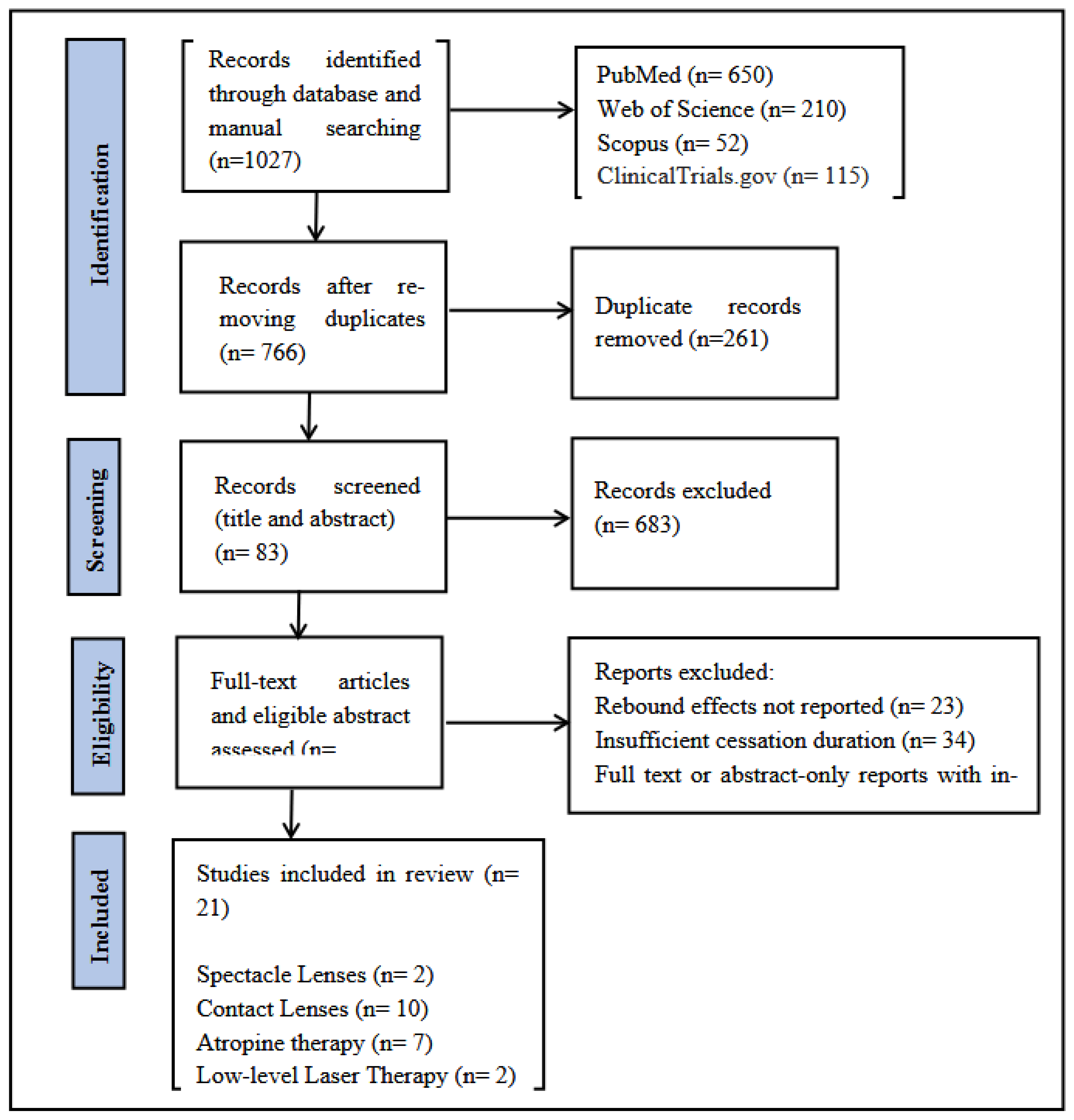
Figure 2.
Risk of bias assessment of the included studies. (a) Risk of bias assessment of the randomized studies. (b) Risk of bias assessment of the non- randomized studies.
Figure 2.
Risk of bias assessment of the included studies. (a) Risk of bias assessment of the randomized studies. (b) Risk of bias assessment of the non- randomized studies.
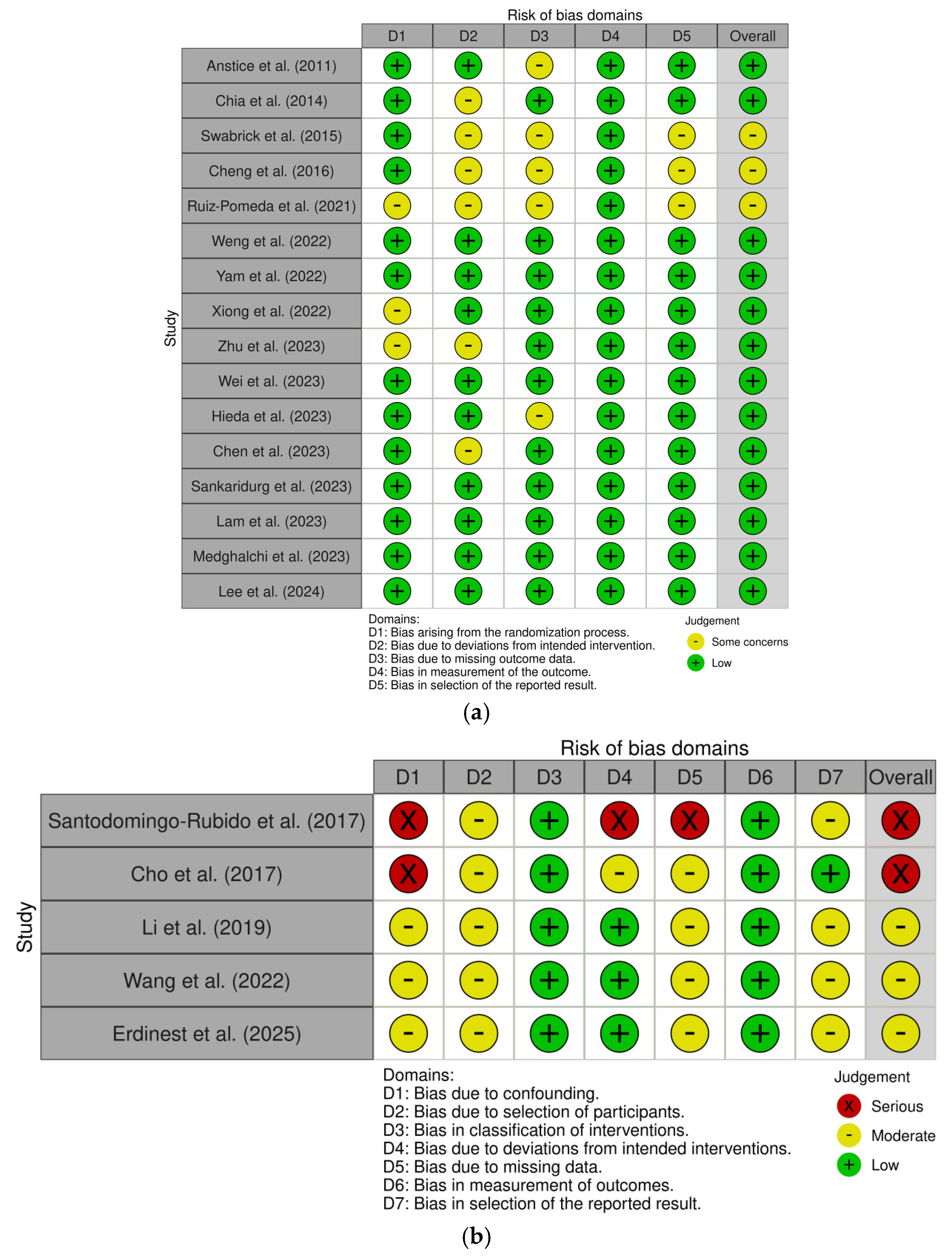
Figure 3.
Summary of risk of bias assessment of the included studies. (a) Summary of risk of bias assessment of the randomized studies. (b) Summary of risk of bias assessment of the non- randomized studies.
Figure 3.
Summary of risk of bias assessment of the included studies. (a) Summary of risk of bias assessment of the randomized studies. (b) Summary of risk of bias assessment of the non- randomized studies.
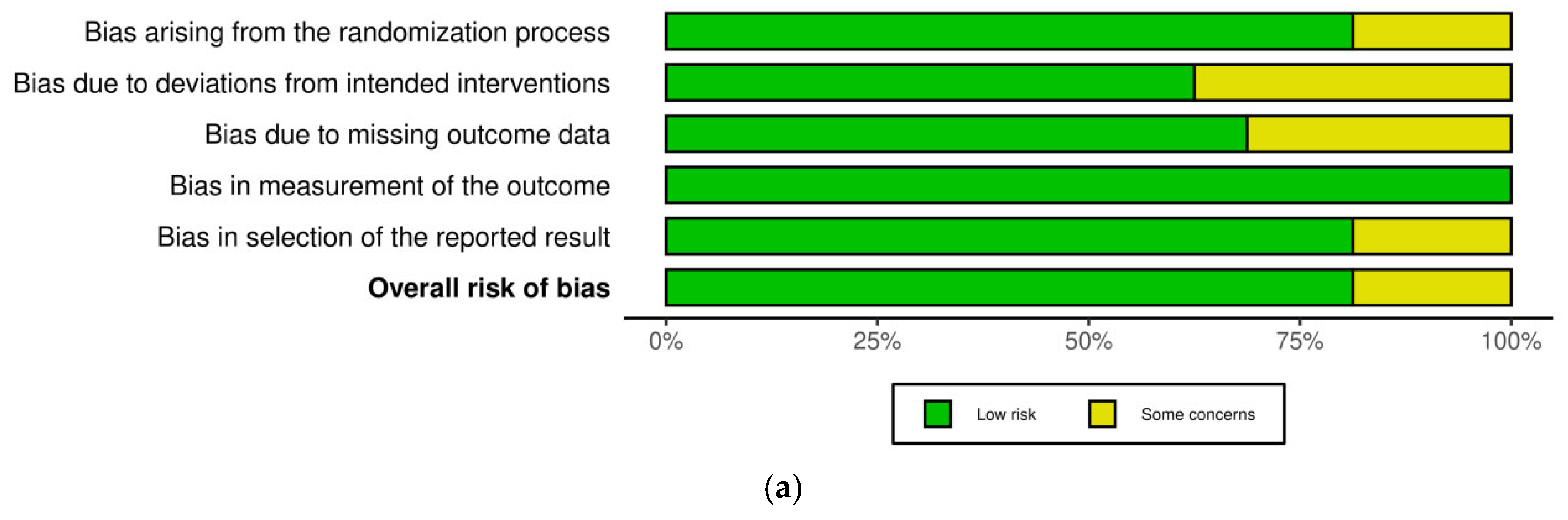
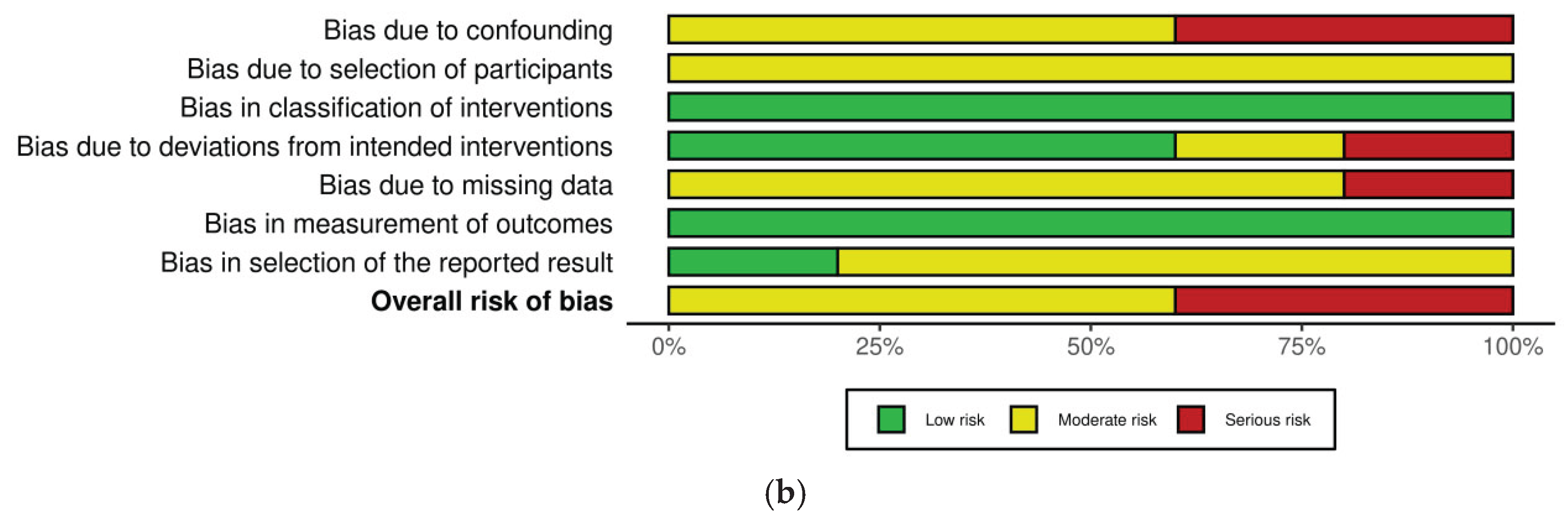

Table 1.
Overview of included studies.
| Intervention | Author/Year | Design | F/U (months) | Participants (IG/CG) | Age (Years) (IG/TG) ( mean ± SD) | Sex (M/F) | Inclusion Criteria | Intervention | Control | Baseline SE (IG/TG) ( mean ± SD) | Baseline AL (IG/TG) ( mean ± SD) | Washout period (months) |
|---|---|---|---|---|---|---|---|---|---|---|---|---|
| Spectacles | Sankaridurg et al. (2023) [34] | RCT | 19 | 54/65 | 11.2±1.6/10.9±1.7 | 65/54 | -0.75 to -4.75 | HAL spectacles | SVS | -3.47±1.16/-3.37±1.22 | 25.1±0.8/24.9±0.8 | 6 |
| Lam et al. (2023) [45] | RCT | 72 | 50/40 | 9.96±1.04/10.43±1.09 | 43/47 | -1.00 to -5.00 | DIMS spectacles | SVS | -3.02±0.70/-2.67±0.71 | 24.83±0.55/24.53±0.58 | 30 | |
| Contact Lens |
Anstice et al. (2011) [46] | RCT | 10 | 40 | 13.4±0.9 | NR | NR | Dual-Focus CL | SVS | -2.71±1.10 | NR | 10 |
| Swarbrick et al. (2015) [47] | RCT | 12 | 26 | 13.4±1.9 | 14/12 | NR | OK-Lens | GP contralateral | NR | NR | 6 | |
| Cheng et al. (2016) [48] | RCT | 42 | 127 (82) | 9.7±1.1 | NR | -0.75 to -4.00 | SCL | Spherical SCL | NR | NR | 18 | |
| Santodomingo-Rubido et al. (2017) [49] | Non-RCT | 24 | 14/16 | 10.4±0.5 | NR | NR | OK-Lens | SVS | NR | NR | 6 | |
| Cho et al. (2017) [35] | Non-RCT | 24 | 44/13 | 8–14 | NR | -0.75 to -4.00 | OK-Lens | SVS | NR | NR | 14 | |
| Li et al. (2019) [50] | Non-RCT | 12 | 29/21 | 11.92±1.70 | 22/28 | -1.00 to -4.00 | OK-Lens | SVS | -3.16±0.85/-2.98±1.34 | 25.18±0.77/24.82±0.71 | 1 | |
| Ruiz-Pomeda et al. (2021) [36] | RCT | 36 | 31/24 | 12.31±1.71/11.52±1.69 | NR | -0.75 to -3.50 | SMCL | SVS | NR | NR | 12 | |
| Weng et al. (2022) [51] | RCT | 12 | 65/30 | 13/11.9 | 47/48 | <-5.00 | SMCL | SVCL | -1.91±0.72/-2.08±0.64 | 24.5±0.8/24.5±0.6 | 6 | |
| Wang et al. (2022) [52] | Non-RCT | 20 | 54/85 | 10.8±1.5 | 32/22 | <-5.00 | OK-Lens | SVS | -2.98±1.25/-1.98±1.58 | 24.68±0.90/23.89±0.86 | 1 | |
| Zhu et al. (2023) [37] | RCT | 13 | 142/137 | 9.2±1.5 | 138/141 | -1.00 to -5.00 | OK-Lens | SVS | -2.74±0.39/-2.73±0.41 | 23.53±0.19/23.40±0.16 | 1 | |
| Atropine |
Chia et al. (2014) [53] | RCT | 24 | 347/NR | 11.7±1.5 | 177/170 | ≥-2.00 | 0.5/0.1/0.01% | Placebo | NR | NR | 12 |
| Yam et al. (2022) [54] | RCT | 36 | 254/72 | 10.7±1.7/11.1±1.9 | 181/145 | ≥-1.00 | 0.05/0.025/0.01% | Placebo | NR | NR | 12 | |
| Medghalchi et al. (2023) [55] | RCT | 12 | 40/20 | 10.6±3.5/12±3.6 | 35/32 | -2.00 to -6.00 | 0.1/0.01% | Placebo | -1.99±0.61/-1.92±0.93 | 23.92±0.45/24.00±0.54 | 6 | |
| Wei et al. (2023) [56] | RCT | 24 | 65/68 | 9.9±1.6/9.2±1.6 | 68/65 | -1.00 to -6.00 | 0.01% | Placebo | -2.67±1.41 | 24.65±0.89 | 12 | |
| Hieda et al. (2023) [57] | RCT | 24 | 200/NR | NR | NR | NR | 0.01% | Placebo | NR | NR | 1,12 | |
| Lee et al. (2024) [58] | RCT | 36 | 104/49 | 11.2±2.7/12.2±2.5 | 60/29 | ≤-1.50 | 0.01% | Placebo | -3.13/-3.56 | 24.6/24.7 | 12 | |
| Erdinest et al. (2025) [59] | Non-RCT | 24 | 50 | 10.33±1.04 | NR | NR | 0.01% | NR | -4.42±1.24 | NR | 12 | |
| LLLT |
Xiong et al. (2022) [38] | RCT | 24 | 63/51 | 10.4±1.5/10.8±3 | 53/61 | -1.00 to -5.00 | LLLT | SVS | -1.77±0.57/-2.76±1.15 | 24.89±0.94/24.58±0.94 | 12 |
| Chen et al. (2023) [39] | RCT | 15 | 46/40 | 9±1.9/8.9±1.2 | 52/34 | -0.75 to -6.00 | LLLT | SVS | NR | NR | 3 |
ALS: axial length shortening group; NALS: no axial length shortening group; RCT: randomized control trial; HAL: highly aspheric lenslets; SVS: single-vision spectacles; IG: intervention group; TG: treatment group; DIMS: defocus incorporated multiple segments; RS: Retrospective; SMCL: Soft multifocal contact lens; SVCL: Single vision contact lens.
Table 2.
Myopia progression after cessation.
| Intervention | Author/Year | Treatment continuation
AL (mm) |
Treatment continuation SE (D) | Treatment cessation AL (mm) | Treatment cessation SE (D) | Rebound AL (mm) | Rebound SE (D) | Category (AL/SE) |
|---|---|---|---|---|---|---|---|---|
| Spectacles | Sankaridurg et al. (2023) [34] | 0.06 ± 0.15 | −0.20 ± 0.31 | 0.17 ± 0.13 | −0.33 ± 0.27 | 0.11 ± 0.20 | −0.13 ± 0.41 | Moderate |
| Lam et al. (2023) [45] | 0.25 ± 0.24 | −0.30 ± 0.65 | 0.31 ± 0.21 | −0.48 ± 0.37 | 0.06 ± 0.32 | −0.18 ± 0.75 | Weak | |
| Contact Lens | Anstice et al. (2011) [46] | 0.13 ± 0.10 | −0.53 ± 0.40 | 0.17 ± 0.11 | −0.46 ± 0.46 | 0.04 ± 0.15 | 0.07 ± 0.61 | Weak/None |
| Swarbrick et al. (2015) [47] | −0.04 ± 0.18 | NR | 0.58 ± 1.54 | NR | 0.62 ± 1.55 | NR | Strong | |
| Cheng et al. (2016) [48] | 0.24 | −0.67 | 0.19 | −0.26 | −0.43 | 0.41 | None | |
| Santodomingo-Rubido et al. (2017) [49] | 0.14 ± 0.11 | NR | 0.16 ± 0.11 | NR | 0.02 ± 0.16 | NR | Weak | |
| Cho et al. (2017) [35] | 0.17 ± 0.46 | NR | 0.26 ± 0.14 | NR | 0.09 ± 0.48 | NR | Moderate | |
| Li et al. (2019) [50] | 0.17 ± 0.16 | NR | 0.23 ± 0.17 | NR | 0.06 ± 0.23 | NR | Weak | |
| Ruiz-Pomeda et al. (2021) [36] | 0.15 ± 0.04 | −0.24 ± 0.09 | 0.22 ± 0.11 | −0.46 ± 0.39 | 0.07 ± 0.12 | −0.22 ± 0.40 | Weak | |
| Weng et al. (2022) [51] | 0.16 ± 0.16 | −0.46 ± 0.66 | 0.28 ± 0.16 | −0.52 ± 0.36 | 0.12 ± 0.23 | −0.06 ± 0.75 | Moderate/Weak | |
| Wang et al. (2022) [52] | ALS: 0.18 ± 0.20NALS: 0.46 ± 0.18 | NR | 0.28 ± 0.190.52 ± 0.17 | NR | 0.10 ± 0.050.06 ± 0.05 | NR | Moderate | |
| Zhu et al. (2023) [37] | 0.22 ± 0.11 | NR | 0.00 ± 0.97 | NR | −0.22 ± 0.98 | NR | None | |
| Atropine | Chia et al. (2014) [53] | 0.5%, 0.1%, 0.01%: NR | NR | NR | −1.15 ± 0.81−1.04 ± 0.83−0.72 ± 0.72 | 0.35 ± 0.200.33 ± 0.180.19 ± 0.13 | −0.87 ± 0.52−0.68 ± 0.45−0.28 ± 0.33 | Strong |
| Yam et al. (2022) [54] | 0.17 ± 0.140.20 ± 0.150.24 ± 0.18 | −0.28 ± 0.42−0.35 ± 0.37−0.38 ± 0.49 | 0.33 ± 0.170.29 ± 0.140.29 ± 0.15 | −0.68 ± 0.49−0.57 ± 0.38−0.56 ± 0.40 | 0.16 ± 0.220.09 ± 0.210.05 ± 0.23 | −0.40 ± 0.65−0.22 ± 0.53−0.18 ± 0.63 | Moderate | |
| Medghalchi et al. (2023) [55] | −0.08 ± 0.07−0.09 ± 0.11 | 0.38 ± 0.320.24 ± 0.21 | 0.08 ± 0.070.04 ± 0.09 | −0.03 ± 0.20−0.01 ± 0.16 | 0.16 ± 0.130.13 ± 0.14 | −0.41 ± 0.38−0.25 ± 0.26 | Moderate | |
| Wei et al. (2023) [56] | 0.30 ± 0.20 | −0.48 ± 0.42 | 0.39 ± 0.17 | −0.78 ± 0.43 | 0.09 ± 0.26 | −0.30 ± 0.60 | Moderate | |
| Hieda et al. (2023) [57] | 1 mo: NR12 mo: NR | NR | NR | NR | 0.02−0.02 | −0.06−0.13 | Weak | |
| Lee et al. (2024) [58] | 0.16 | −0.31 | 0.19 | −0.40 | 0.03 | −0.09 | Moderate/Weak | |
| Erdinest et al. (2025) [59] | GC: NRPC: NR | NR | NR | NR | 0.15 ± 0.100.25 ± 0.18 | 0.21 ± 0.240.43 ± 0.26 | Strong/Weak | |
| LLLT | Xiong et al. (2022) [38] | 0.12 ± 0.16 | −0.20 ± 0.56 | 0.42 ± 0.20 | −0.91 ± 0.48 | 0.30 ± 0.26 | −0.71 ± 0.74 | Strong |
| Chen et al. (2023) [39] | 0.01 | 0.05 | 0.16 | −0.20 | 0.15 | −0.25 | Moderate |
GC: Gradual cessation; Mo: Months; NR: Not reported; PC; Prompt cessation.
Table 3.
Rebound effect of different myopia control interventions.
| Intervention | Washout Period (months)
(mean ± SD) |
Rebound Effect | ||
|---|---|---|---|---|
| AL (mm) (mean± SD) |
SE (Ds) (mean± SD) |
Category | ||
| Spectacles | 18.00 ± 16.97 | 0.09 ± 0.17 | -0.16 ± 0.43 | Weak |
| Contact Lens | 6.91 ± 5.92 | 0.05 ± 0.61 | 0.05 ± 0.60 | Weak/ No rebound |
| Atropine | 9.87 ± 4.16 | 0.14 ± 0.19 | -0.23 ± 0.47 | Moderate/Weak |
| LLLT | 7.50 ± 6.36 | 0.23 ± 0.26 | -0.48 ± 0.37 | Strong |
Disclaimer/Publisher’s Note: The statements, opinions and data contained in all publications are solely those of the individual author(s) and contributor(s) and not of MDPI and/or the editor(s). MDPI and/or the editor(s) disclaim responsibility for any injury to people or property resulting from any ideas, methods, instructions or products referred to in the content. |
© 2026 by the authors. Licensee MDPI, Basel, Switzerland. This article is an open access article distributed under the terms and conditions of the Creative Commons Attribution (CC BY) license.
Copyright: This open access article is published under a Creative Commons CC BY 4.0 license, which permit the free download, distribution, and reuse, provided that the author and preprint are cited in any reuse.


