

Submitted:
14 August 2026
Posted:
14 August 2026
You are already at the latest version
Abstract
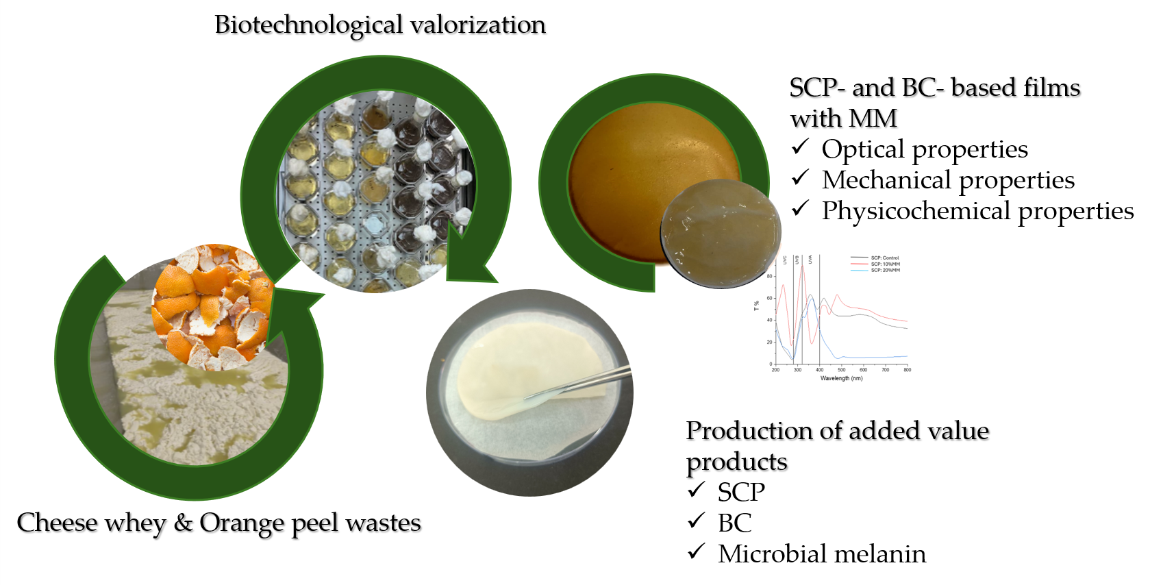
This study proposes a sustainable biorefinery strategy to develop biodegradable packaging films by integrating three microbial biopolymers produced from agro-industrial residues. Cheese whey and citrus waste were used as renewable substrates for producing single-cell protein (SCP), bacterial cellulose (BC), and microbial melanin (MM). SCP consists of dried microbial biomass rich in protein, while BC is a renewable biopolymer with outstanding physicochemical properties. MM, a natural pigment produced by black yeasts, is a multifunctional biopolymer with considerable technological potential. In this work, SCP and BC were employed as two distinct film-forming matrices, whereas MM was incorporated into both types of films as a functional additive to enhance their performance. To the best of the authors’ knowledge, this is the first study reporting the incorporation of microbial melanin into both SCP- and BC-based biodegradable films. SCP and BC were produced by Kluyveromyces marxianus EXF-5288 and Komagataeibacter rhaeticus UNIWA AAK2, respectively, while microbial melanin was produced by Exophiala phaeomuriformis EXF-6108 cultivated on citrus waste. Melanin incorporation significantly improved the UV-shielding performance of both film types, particularly in the UVC and UVB regions, while also affecting color, water solubility, and mechanical properties. SCP-based films containing 20% melanin exhibited complete UVC/UVB blocking and increased opacity. Overall, the findings demonstrate that waste-derived microbial biopolymers enable sustainable biodegradable packaging and promote a circular bioeconomy.
Keywords:
microbial melanin
; packaging
; biopolymers
; agricultural wastes
1. Introduction
The increasing accumulation of plastic waste and the generation of large quantities of agro-industrial residues have intensified the need for sustainable resource management and circular bioeconomy strategies. Food packaging represents one of the largest applications of conventional plastics, accounting for approximately 40% of global plastic production [1], thereby contributing substantially to environmental pollution and waste generation. Consequently, the replacement of petroleum-based plastics with biodegradable and renewable alternatives has become a major priority for both academia and industry.
Among the emerging bio-based materials, microbial polymers such as single-cell protein (SCP) and bacterial cellulose (BC) have attracted considerable attention as sustainable alternatives for food-packaging applications because they can be produced through microbial fermentation without competing for agricultural land while exhibiting desirable film-forming properties [2,3,4]. Recent studies have demonstrated the potential of cheese whey-derived SCP as a novel edible packaging material, owing to its favorable film-forming ability, high nutritional value, satisfactory oxygen barrier properties, and promising sensory characteristics [5,6,7]. Similarly, BC has emerged as an attractive packaging biopolymer due to its unique three-dimensional nanofibrillar network, which provides high tensile strength, a high degree of polymerization and crystallinity, excellent water-holding capacity, mechanical stability and excellent biodegradability [8,9]. In addition, the abundant hydroxyl groups on the BC surface enable its functionalization through various modification strategies, allowing the enhancement of its physicochemical, barrier, and antimicrobial properties [10]. Collectively, SCP and BC represent two complementary microbial platforms with considerable potential for the development of next-generation sustainable food-packaging materials.
Meanwhile, cheese whey is among the most abundant agro-industrial residues, with approximately 9 L generated per kilogram of cheese produced. Due to its high organic load, it also represents one of the most environmentally problematic dairy waste streams, necessitating effective management strategies [11]. Plethora of studies have demonstrated that the biotechnological valorization of cheese whey as a low-cost substrate for the production of both SCP [12,13] and BC [14,15] is highly promising. Therefore, converting cheese whey into microbial bioproducts such as BC and SCP simultaneously address waste management challenges while promoting sustainable material and protein production.
Melanin is a naturally occurring biopolymer found in animals, plants, fungi, yeasts, and bacteria, where it performs various protective biological functions. Owing to its unique physicochemical properties, including strong ultraviolet (UV) absorption, antioxidant activity, free radical scavenging ability, metal-chelating capacity, and protection against environmental stresses, melanin has attracted increasing attention as a sustainable functional biomaterial with applications in biomedicine, cosmetics, agriculture, textiles, and food-related technologies [16]. Microbial production has emerged as an attractive alternative to conventional pigment sources because it is independent of seasonal variations, allows controlled production, and can be integrated with the valorization of agro-industrial residues, supporting circular bioeconomy strategies [17].
These multifunctional properties also make microbial melanin a promising additive for biodegradable food-packaging materials. Its UV-shielding and antioxidant properties can reduce light-induced deterioration and oxidative degradation of food products, while its incorporation into bio-based polymeric matrices may enhance the functional performance of packaging films without compromising their sustainability [18]. However, the application of waste-derived microbial melanin in microbial biopolymer-based packaging materials remains largely unexplored, highlighting the need for the development of novel fully bio-based active packaging systems.
Therefore, the aim of this study was to develop and evaluate two complementary microbial-based packaging platforms, namely bacterial cellulose and single-cell protein films, produced through the valorization of cheese whey and functionalized with microbial melanin biosynthesized from orange processing waste. The physicochemical, optical, barrier, and mechanical properties of the developed films were investigated to assess their potential as sustainable active food-packaging materials. To the best of our knowledge, this is the first study integrating these waste-derived microbial products into a unified waste-to-packaging strategy.
2. Materials and Methods
2.1. Raw Materials & Pre-Treatment
Citrus wastes (orange peels) were obtained from a local shop in Lemnos (Greece), dried at 80 °C and been grounded with a laboratory grinder to obtain a constant particle size of 1cm. To obtain free sugars, peels were immersed in deionized water (80oC, 60 min, 280 rpm) followed by centrifugation (9000 rpm, 4 °C, 10 min) and stored at -20 °C until further use. Concentration of free sugars of orange peel waste extract (OPWE) were determined by DNS method [19] and reached ~33.0 g/L.
Cheese whey was obtained from a regional cheese manufacturer (Lemnos, Greece) and preserved at −20 ± 2 °C for subsequent use. DCW was generated through sterilization at 121 °C for 20 minutes, followed by centrifugation at 9000 rpm for 15 minutes at 4 °C using a Universal 320R centrifuge (Hettich, Tuttlingen, Germany), and filtration. DCW contained approximately ~37.0 g/L lactose and a pH value of ~ 6.7.
2.2. Microbial Strains and Growth Media
Yeast strains Exophiala phaeomuriformis EXF- 6108 and Kluyveromyces marxianus EXF-5288 were kindly provided by Infrastructural Centre Mycosmo, MRIC UL, Slovenia. Bacterial strain Komagataeibacter rhaeticus UNIWA AAK2 was kindly provided by Department of Wine, School of Food Science, University of West Attica.
E. phaeomuriformis EXF- 6108 and K marxianus EXF-5288 were maintained on YPDA slants, while K. rhaeticus was maintained on HS medium slants. All strains were stored at 4 °C and subcultured periodically.
Batch fermentations were carried out in 250 mL Erlenmeyer flasks with a 50 mL working volume. A 48h preculture (T=30oC, 180 rpm) was used as the inoculum in all experiments, at 2% (v/v) for E. phaeomuriformis EXF-6108 and K. marxianus EXF-5288, and 10% (v/v) for K. rhaeticus UNIWA AAK2. The pH was controlled using 5 M NaOH for E. phaeomuriformis (pH 6) and K. rhaeticus (pH 6), and 5 M H₂SO₄ for K. marxianus (pH 3.5).
E. phaeomuriformis EXF-6108 was cultivated at 30 °C and 180 rpm using OPWE-derived sugars (20 g/L), supplemented with 5 g/L peptone, 5 g/L yeast extract, 1 g/L KH₂PO₄, 2 g/L MgSO₄, and 1 g/L NaCl (C/N ≈ 6.7). K. rhaeticus UNIWA AAK2 was cultivated statically at 30 °C using deproteinized cheese whey (DCW), or glucose (≈20 g/L total sugars), supplemented with 5 g/L yeast extract, 5 g/L peptone, 2.7 g/L Na₂HPO₄, and 1.15 g/L citric acid. K. marxianus EXF-5288 was cultivated at 20 °C and 180 rpm using DCW as the sole carbon source (70 g/L lactose), with mineral salts (g/L): KH₂PO₄, 7.0; Na₂HPO₄, 2.5; MgSO₄·7H₂O, 1.5; FeCl₃·6H₂O, 0.15; CaCl₂·2H₂O, 0.15; ZnSO₄·7H₂O, 0.02; and MnSO₄·H₂O, 0.06, while urea (0.22%, w/w) served as the nitrogen source as previously reported [12].
2.3. Biomass and BC Production
Yeast biomass was recovered through three successive centrifugation cycles performed at 9,000 rpm for 10 min at 4 °C using a Universal 320R-Hettich centrifuge (Tuttlingen, Germany). The resulting biomass was quantified gravimetrically by drying the collected material at approximately 85 °C until a constant mass was reached.
BC pellicles formed at the air–liquid interface were harvested, washed thoroughly with deionized water, and purified by treatment with 1 M NaOH at 80 °C for approximately 80 min to remove bacterial cells. The membranes were subsequently rinsed with deionized water until pH neutralization and then dried at 40 °C until constant weight.
2.4. Extraction and Recovery of Microbial Melanin
Microbial melanin was extracted according to Selvakumar et al. [20] with some modifications. Briefly, after centrifugation of the fermentation medium the supernatant’s pH was adjusted to 12 with 1 M NaOH and incubated overnight at 70 °C and 300 rpm to promote melanin solubilization. Subsequently, the pH was lowered to 2 with 5 M H₂SO₄ to precipitate the pigment. The precipitated melanin was collected by centrifugation (9,000 rpm, 4 °C, 10 min) and washed repeatedly with deionized water until neutral pH was achieved. To further purify the pigment, the alkaline dissolution–acid precipitation procedure was repeated twice. After each cycle, the precipitated melanin was collected by centrifugation and washed repeatedly with deionized water until neutral pH was achieved. After that, the purified microbial melanin was concentrated at 10 mg/mL and the resulting melanin stock solution was stored for further use.
2.5. Fourier-Transform Infrared Resonance (FT-IR) Analysis of Microbial Melanin
Fourier-transform infrared (FT-IR) spectroscopy was performed using a PerkinElmer Spectrum 100 IR spectrometer to characterize purified microbial melanin. Synthetic melanin (Sigma-Aldrich) was used as a reference standard. Samples were analyzed under a nitrogen atmosphere over the spectral range of 380–4000 cm⁻¹ with a resolution of 4 cm⁻¹. Two spectra were acquired for each sample at different loadings to ensure reproducibility. The purified microbial melanin was identified by comparison with the FT-IR spectrum of the synthetic melanin standard.
2.6. Development of SCP- and BC- Based Films Enriched with MM
2.6.1. Development of SCP-Based Films Enriched with MM
SCP- based films were prepared according to Koukoumaki et al. [5] with minor modifications. Briefly, dry biomass (7%, w/w) was dispersed in deionized water, and the pH was adjusted to 8.0 with 0.5 M NaOH. Melanin stock solution was incorporated at final concentrations of 10 or 20% (v/v), after which the film-forming mixture was heated at 80 °C for 30 min to induce protein denaturation. The mixture was then rapidly cooled, and 50% (w/w, based on dry biomass) glycerol was added as a plasticizer. After homogenization (5,500 rpm, 15 min; HG-15D, Witeg, Germany), the solution was degassed in an ultrasonic bath (P70H, Elma Ultrasonic, Weinfelden, Switzerland) for 20 min.
The degassed film-forming solution was cast onto 9 cm Petri dishes and dried in an environmental chamber at 25 ± 1 °C and 55 ± 2% relative humidity for 24–48 h. The dried films were carefully peeled from the casting surface and conditioned at 25 ± 1 °C before further characterization. A control batch was prepared without melanin addition into solution.
2.6.2. Development of BC-Based Films Enriched with MM
BC pellicles were submerged in melanin stock solution for 48 h at room temperature to allow absorption. Subsequently, BC was rinsed with deionized water to remove any excess solution and neutralize the pH and were dried at 40 °C until constant weight. BC pellicles without melanin addition served as control.
2.7. Characterization of SCP- and BC-Based Films Enriched with MM
2.7.1. Film Thickness
Film thickness was determined using a digital thickness gauge (F.F. Group Tools, Frankfurt, Germany) with an accuracy of 0.002 mm. Measurements were taken at three different locations on each film prior to characterization, and the mean value was used for subsequent analyses.
2.7.2. Light Transmission and Film Opacity
The optical properties of the films were determined using a UV–Vis spectrophotometer (UV-1900i, Shimadzu, Japan). UV–Vis absorbance spectra were measured in the wavelength range of 250–800 nm, while film opacity was calculated from the absorbance at 600 nm using the following equation:
where A600 is the absorbance at 600 nm and x is the film thickness (mm).
Opacity = A600/x
2.7.3. Analysis of Color Parameters
Film color characteristics were assessed with a Lovibond LC100 Spectrocolorimeter. The measurements included the CIELAB parameters L*, a*, and b*, along with hue angle (h*) and chroma (C*). The L* scale ranged from 0 (black) to 100 (white), whereas positive and negative a* values represented red and green tones, respectively. Similarly, positive b* values indicated yellow coloration, while negative values corresponded to blue tones.
2.7.4. Moisture Content, Solubility and Swelling Index Analysis
Moisture content (MC%) of film stripes (2 x 2 cm) was determinate at 105oC according to Koukoumaki et al. [5] as follows:
where M0 refers to initial film weight and M1 refers to film weight after drying.
MC (%)= [(M0-M1 )/M0] * 100
Solubility (S%) and Swelling Index (S.I %) were calculated according to Papadaki et al. [21] according to the following equations:
where S0= the initial film weight (g) and S1= weight of dried film (g).
where S.I0= the initial film weight (g) and S.I1= weight of dried film (g).
S (%) = [(S0-S1 )/S0] * 100
S.I (%) = [(S.I1-S.I0 )/S.I0] * 100
2.7.5. Water Vapor Permeability (WVP)
Water vapor permeability (WVP) was determined using the gravimetric cup method based on ASTM, Procedure B (1995) [22], following the methodology reported by Ramos et al. [23] with minor modifications. Deionized water was placed inside permeability cups, which were subsequently sealed with the film samples. The assembled cups were maintained in an environmental chamber at 25 °C and a relative humidity of 50 ± 2% for 7 days.
The water vapor transmission rate (WVTR) was calculated from the slope of the linear regression of the weight gained over time in days (d) (R2 ≥ 0.99). WVP (g ⋅ mm/ m2 ⋅ d ⋅ kPa) was then calculated using the following equation:
where WVTR (g/ m2 d) is the slope (g/d) divided by the transfer area of the film (m2), x (mm) is the film thickness, and Δp (kPa) is the partial water vapor pressure across the film
WVP= WVTR*x/ Δp
2.7.6. Mechanical Properties
Mechanical properties, including tensile strength (TS) and elongation at break (E), were determined following the procedures described in ASTM D882 [24] and Piccirilli et al. [25]. Measurements were performed using a Texture Analyzer (TA.XT.plus C, Stable Micro Systems, Surrey, UK) equipped with A/MTG Mini Tensile Grips, and the data were analyzed using Texture Exponent Software (Version 6.1.18.0, Stable Micro Systems).
Film specimens were cut into strips measuring 10 × 60 mm, with an initial grip separation of 30 mm. The tests were conducted at a crosshead speed of 0.05 mm/s to obtain stress–strain curves. Tensile strength (TS, MPa) and elongation at break (E, %) were calculated using the following equations:
where F is the maximum force (N), a is the film thickness (m), x is the film width (m), d is the elongation at the point of rupture (mm), and l is the initial gauge length of the film specimen (mm).
TS (MPa)= F / (a* x )* 10-6
E (%)= (d /l)* 100
2.8. Statistical Analysis
All experiments in films were performed using three independent batches (N = 3). For each batch, every measurement was carried out in triplicate, and the mean value was used for subsequent statistical analysis. The resulting data were organized in Microsoft Excel and statistically evaluated using XLSTAT software (Version 2018.1, Addinsoft). Differences among treatments were assessed by one-way analysis of variance (ANOVA), followed by Tukey’s honestly significant difference (HSD) post hoc test for multiple comparisons. Statistical significance was established at P ≤ 0.05. Two independent fermentation batches were prepared for each experimental condition (N = 2). The results presented are expressed as the mean of the measurements obtained from these independent biological replicates.
3. Results
3.1. Fourier-Transform Infrared Resonance (FT-IR) Analysis of Microbial Melanin
FT-IR spectra of synthetic melanin and melanin extracted from E. phaeomuriformis displayed the characteristic absorption bands associated with melanin (Figure 1). A broad band at 3660–3014 cm⁻¹ was assigned to the stretching vibrations of O–H groups from carboxylic acids and phenolic hydroxyls, as well as N–H stretching of aromatic amino groups within the indole and pyrrole structures of melanin. The absorption band at 2924 cm⁻¹ corresponded to C–H stretching vibrations of aliphatic methylene groups, while the peak at 1710 cm⁻¹ was attributed to C=O stretching of carbonyl groups. The band at 1612 cm⁻¹ was assigned to vibrations of carboxylic acid (COOH) groups. The absorption bands in the 1481–1373 cm⁻¹ region were attributed to vibrations associated with the indolic structure of melanin, including C–N/C=N and conjugated aromatic vibrations, which are characteristic of the melanin polymer. The band at 759 cm⁻¹ was assigned to the out-of-plane bending vibration of aromatic C–H bonds, a characteristic feature of melanin pigments [26].
Notably, both spectra exhibited the characteristic melanin peaks at 1511, 1385, and 759 cm⁻¹, confirming the presence of the melanin polymer in the pigment extracted from E. phaeomuriformis. The close agreement between the FT-IR spectra of the extracted and synthetic melanin indicates that the fungal pigment shares the principal structural features of the reference melanin.
3.2. Light Transmittance
The UV–Vis transmittance spectra of the BC- and SCP based films are presented in Figure 2 and Figure 3. Melanin incorporation affected the optical properties of the films in a polymer-dependent manner. In BC: Gly+MM films, the addition of melanin resulted in lower transmittance throughout most of the UV region (200–400 nm), indicating improved UV-shielding properties compared with the BC:Gly films. In contrast, only minor differences in UV transmittance were observed between BC:DCW films with and without melanin, suggesting a limited effect of melanin on the UV barrier properties of this film platform. Furthermore, increasing the microbial melanin concentration from 10% to 20% in SCP-based films markedly reduced UV transmittance, particularly in the UV-B (280–320 nm) and UV-A (320–400 nm) regions (Figure 3), demonstrating a concentration-dependent enhancement of the UV-blocking capacity. In the visible region (400–800 nm), melanin-containing SCP films exhibited lower light transmittance than the SCP: control films, indicating reduced transparency with increasing melanin content.
3.3. Color Analysis & Film Opacity of BC- and SCP-Based Films
As shown in Table 1, regarding BC-based films, both the change in the cultivation substrate and the incorporation of melanin resulted in significant differences in the color parameters (p < 0.05). Microbial melanin incorporation significantly decreased the L* values, indicating a darker film appearance, while simultaneously increasing film opacity. Specifically, L* values of BC films produced by glycose consumption decreased from 77.2± 0.5 to 37.4± 1.8 and from 84.1± 0.7 to 51.6± 3.0 for BC films cultivated in DCW.
3.4. Water-Related Properties of BC- and SCP-Based Films
The cultivation substrate significantly affected the moisture content (MC) of the BC films. Films produced from glucose-grown bacterial cellulose exhibited a significantly higher moisture content (20.0 ± 1.6%) than those produced from DCW-grown bacterial cellulose (14.4 ± 1.9%). Melanin incorporation influenced the moisture content differently depending on the cultivation substrate. Specifically, the MC of BC films decreased significantly to 9.6 ± 0.7% following melanin incorporation, whereas a slight increase was observed in films produced from DCW-grown bacterial cellulose.
In contrast, melanin incorporation exerted a similar effect on the other water-related properties regardless of the cultivation substrate. Specifically, film solubility, swelling index, and water vapor permeability increased significantly in all melanin-containing films as shown in Table 3.
In SCP-based films, MM incorporation resulted in a slight decrease in moisture content, irrespective of the concentration of the additive. Film solubility also decreased significantly, from 48.3 ± 0.3% in the control films to 38.8 ± 0.9% and 36.8 ± 0.6% following the incorporation of 10% and 20% MM, respectively. In contrast, both the swelling index (SI) and water vapor permeability (WVP) increased progressively with increasing MM concentration as presented in Table 4.
3.5. Mechanical Properties of BC- and SCP-Based Films
Melanin incorporation significantly affected the mechanical properties of the BC-based films. Specifically, the addition of melanin significantly increased both the tensile strength (TS) and elongation at break (E%) of the films, regardless of the cultivation substrate (glucose or DCW) (Figure 5 a,b). The TS increased from 4.40± 1.13 to 6.04± 1.20 MPa for BC films and from 3.70± 1.21 to 8.54± 1.20 MPa for BC films. Similarly, the E% increased from 0.98± 0.21 to 1.33± 0.10% for BC films and from 1.71± 0.40 to 2.39± 0.10% for BC films.
In contrast, melanin incorporation adversely affected the mechanical properties of the SCP-based films. Tensile strength decreased progressively with increasing melanin concentration, from 1.24 ± 0.01 MPa in the control films to 0.41± 0.17 MPa in films containing 20% melanin (Figure 6 a,b). Elongation at break showed a slight increase following the incorporation of 10% melanin, reaching 3.86± 0.0%; however, a further increase in melanin concentration to 20% resulted in a decrease in E%.
4. Discussion
Melanin incorporation significantly influenced the color characteristics of the developed films. Specifically, the L* value decreased in both SCP- and BC-based films, indicating reduced lightness and the formation of darker films. This behavior is consistent with previous studies on melanin-containing protein-based films [27] and cellulose-based films [28], which similarly reported a significant decrease in L following melanin addition, reflecting the intrinsic dark pigmentation of melanin.
Melanin incorporation generally improved the UV shielding properties of the films, as demonstrated by the reduction in UV transmittance, particularly in the UV-B and UV-A regions. This behavior is aligned with previous studies examined incorporation of melanin in polymeric films [29,30] and is attributed to melanin’s aromatic structure which enables efficient absorption of UV radiation and rapid conversion of photon energy from UV light into heat to prevent destructive photodegradation [31].
The moisture content (MC) of SCP-based films without melanin was 20.7 ± 0.3%, which is in agreement with previous findings for SCP-based films (25.9 ± 0.3%) reported by Koukoumaki et al. [5]. In the present study, melanin incorporation reduced the MC of both SCP- and BC-based films. Similar observations have been reported for alginate/PVA films containing melanin nanoparticles [32]. However, several studies have shown that melanin incorporation generally increases the MC of films prepared from different biopolymers, including chitosan, alginate, and carrageenan [33,34,35]. These discrepancies are most likely related to the different physicochemical properties of the polymer matrices and the resulting interactions between melanin and the functional groups of each biopolymer. Such interactions may either decrease or increase the availability of free hydroxyl groups capable of binding water molecules, ultimately affecting the moisture content of the films.
A similar explanation can be proposed for the differences observed in water solubility. In the present study, melanin addition significantly decreased the solubility of SCP-based films, whereas it increased the solubility of BC-based films. The reduced solubility observed in SCP-based films may be attributed to the formation of intermolecular interactions between melanin and the polymer chains, resulting in a more compact polymer network and consequently limiting film dissolution in water, as previously suggested by [35]. Conversely, the increased solubility of BC-based films may indicate weaker interactions between melanin and the cellulose matrix or structural modifications that facilitated water penetration and polymer dissolution.
In contrast to the MC results, both film platforms exhibited increased water vapor permeability (WVP) and swelling index (SI) following melanin incorporation. A similar increase in swelling index after melanin addition has been reported for protein-based films [29]. Regarding WVP, most previous studies have demonstrated that melanin improves the water vapor barrier properties of biopolymer films, resulting in lower WVP values in cellulose-based films [30] as well as in gelatin-based films [27]. Nevertheless, increases in WVP have also been reported in cellulose-based films [36] and agar-based films [37], indicating that the effect of melanin is highly dependent on the polymer matrix and the quality of filler dispersion. An increase in WVP may result from non-uniform melanin distribution within the polymer network, leading to the formation of micro voids or discontinuities that facilitate water vapor diffusion. Therefore, the increase in WVP observed in the present study is likely associated with the specific interactions between melanin and the SCP- and BC-based matrices, as well as possible structural heterogeneities introduced during film formation.
Regarding the effect of microbial melanin on the mechanical properties of BC-based films, the present study demonstrated a clear reinforcing effect regardless of the culture medium used for bacterial cellulose production. Specifically, the tensile strength increased from 4.4 ± 0.9 to 6.0 ± 1.3 MPa for BC films and from 3.7 ± 1.7 to 8.5 ± 2.4 MPa for BC films following melanin incorporation. Similar improvements have been reported for cellulose-based films containing melanin [30,38]. In contrast, melanin incorporation into SCP-based films resulted in a reduction in tensile strength, particularly at higher melanin concentrations. A similar decrease in mechanical strength has also been reported for other protein-based films containing melanin [39], suggesting that the reinforcing effect of melanin is not universal but strongly depends on the characteristics of the polymer matrix.
The opposite mechanical responses observed for BC- and SCP-based films are likely attributed to differences in the interactions between melanin and each polymer matrix. In cellulose-based films, the abundance of hydroxyl groups promotes strong hydrogen bonding with the functional groups of melanin, leading to improved stress transfer, increased tensile strength, and reduced chain mobility, which is reflected by lower elongation at break. Similar reinforcement mechanisms have been reported for melanin-containing cellulose and PVA nanocomposites, where homogeneous melanin dispersion and strong interfacial interactions enhanced the mechanical performance [40].
On the contrary, in protein-based matrices, melanin may not act as an efficient reinforcing agent. Lower polymer–filler compatibility and disruption of existing protein–protein interactions may reduce the continuity of the polymer network and create stress-concentration sites, ultimately decreasing tensile strength. Therefore, the effect of melanin on the mechanical properties of biopolymer films depends primarily on polymer–melanin compatibility and the quality of melanin dispersion within the polymer matrix.
5. Conclusions
The present study demonstrates that the holistic valorization of agro-industrial by-products, namely cheese whey and orange processing residues, for the development of alternative packaging materials is feasible following the biorefinery concept. The results indicate that microbially produced melanin can enhance the performance of packaging materials by improving their UV-shielding capacity as well as their mechanical properties. However, its effect on the physicochemical characteristics of the resulting materials is strongly dependent on the type of polymer matrix in which it is incorporated. These findings highlight the potential of microbial melanin as a multifunctional bio-based additive for sustainable packaging applications while emphasizing the importance of optimizing polymer–melanin interactions. Further research should focus on evaluating the performance of these material platforms in real food packaging systems, including shelf-life studies, migration and safety assessments, scalability of the production process, and end-of-life biodegradation or recyclability, in order to facilitate their industrial implementation within a circular bioeconomy framework.
Author Contributions
Conceptualization: D.S and D.I.K; methodology: D.IK and D.S; validation: D.S. ; formal analysis: F.G and E.T; investigation: D.I.K, F.G and E.T; resources: D.S; writing—original draft preparation: D.I.K; writing—review and editing: D.S; supervision: D.S; project administration: D.S; funding acquisition: D.S. All authors have read and agreed to the published version of the manuscript.
Funding
This research received no external funding.
Institutional Review Board Statement
Not applicable.
Informed Consent Statement
Not applicable.
Data Availability Statement
The original contributions presented in this study are included in the article. Further inquiries can be directed to the corresponding author.
Conflicts of Interest
The authors declare no conflicts of interest.
Abbreviations
The following abbreviations are used in this manuscript:
| SCP | Single Cell Protein |
| BC | Bacterial Cellulose |
| MM | Microbial melanin |
| DCW | Deproteinized cheese whey |
References
- European Economic and Social Committee. Recycling Food Packaging & Food Waste in Plastics Revolution; Publications Office of the European Union: Luxembourg, 2020. [Google Scholar] [CrossRef] [PubMed]
- Yan, Y.; Liu, L.; Wang, F.; Zhang, H. Bacterial cellulose as a promising biodegradable bioplastic for sustainability. Nat. Commun. 2026, 17. [Google Scholar] [CrossRef] [PubMed]
- Çalhan, A.; Hasanoğlu, A. Bacterial cellulose as a next-generation membrane material for selective transport: properties, fabrication, and applications. Cellulose 2026, 33, 1167–1211. [Google Scholar] [CrossRef]
- Baddigam, K.R.; Chee, B.S.; Guilloud, E.; Venkatesh, C.; Koninckx, H.; Windey, K.; Fournet, M.B.; Hedenqvist, M.; Svagan, A.J. High oxygen barrier packaging materials from protein-rich single-celled organisms. Commun. Chem. 2025, 8, 1–9. [Google Scholar] [CrossRef] [PubMed]
- Koukoumaki, D.I.; Papanikolaou, S.; Ioannou, Z.; Gkatzionis, K.; Sarris, D. The Development of Novel Edible Films from Single-Cell Protein Produced by the Biotechnological Valorization of Cheese Whey. Appl. Microbiol. 2024, 4, 1030–1041. [Google Scholar] [CrossRef]
- Koukoumaki, D.I.; Gkatzionis, K.; Panagiotou, M.; Sarris, D. Consumer Perception of Novel Edible Films Produced by Single Cell Protein Derived From Cheese Whey Bio-Valorization. J. Sens. Stud. 2024, 39. [Google Scholar] [CrossRef]
- Singha, S.; Mahmutovic, M.; Zamalloa, C.; Stragier, L.; Verstraete, W.; Svagan, A.J.; Das, O.; Hedenqvist, M.S. Novel Bioplastic from Single Cell Protein as a Potential Packaging Material. ACS Sustain. Chem. Eng. 2021, 9, 6337–6346. [Google Scholar] [CrossRef]
- dos Santos, M.R.; Durval, I.J.B.; de Medeiros, A.D.M.; Júnior, C.J.G.d.S.; Converti, A.; Costa, A.F.d.S.; Sarubbo, L.A. Biotechnology in Food Packaging Using Bacterial Cellulose. Foods 2024, 13, 3327. [Google Scholar] [CrossRef] [PubMed]
- Zhou, R.; Guo, C.; Li, Q.; Li, Z.; Fan, W.; Chen, X.; Dai, J.; Zhang, Q. Development of Bacterial Cellulose-Based Films Incorporated with Epigallocatechin-3-Gallate for Active Food Packaging. Foods 2026, 15, 785. [Google Scholar] [CrossRef] [PubMed]
- Ntovolou, I.; Ioannou, D.; Koukoumaki, D.I.; Sarris, D.; Gogolides, E.; Ellinas, K. On demand control of bacterial cellulose membranes wetting properties via micro/nano-topography and surface chemistry modification. Appl. Surf. Sci. 2026, 747. [Google Scholar] [CrossRef]
- Dinkçi, N.; Akdeniz, V.; Akalın, A.S. Recent Trends and Developments to Valorize Sheep and Goat Cheese Whey for Small and Medium-Size Enterprises. Foods 2026, 15, 1217. [Google Scholar] [CrossRef] [PubMed]
- Koukoumaki, D.I.; Papanikolaou, S.; Ioannou, Z.; Mourtzinos, I.; Sarris, D. Single-Cell Protein and Ethanol Production of a Newly Isolated Kluyveromyces marxianus Strain through Cheese Whey Valorization. Foods 2024, 13, 1892. [Google Scholar] [CrossRef] [PubMed]
- Delmoitié, B.; Regueira, A.; De Wever, H.; Ganigué, R.; Sakarika, M. Assessing the economic viability, environmental sustainability, and scalability of microbial protein from cheese whey permeate in Europe. Chem. Eng. J. 2026, 529. [Google Scholar] [CrossRef]
- Bekatorou, A.; Plioni, I.; Sparou, K.; Maroutsiou, R.; Tsafrakidou, P.; Petsi, T.; Kordouli, E. Bacterial Cellulose Production Using the Corinthian Currant Finishing Side-Stream and Cheese Whey: Process Optimization and Textural Characterization. Foods 2019, 8, 193. [Google Scholar] [CrossRef] [PubMed]
- Lappa, I.K.; Kachrimanidou, V.; Alexandri, M.; Papadaki, A.; Kopsahelis, N. Novel Probiotic/Bacterial Cellulose Biocatalyst for the Development of Functional Dairy Beverage. Foods 2022, 11, 2586. [Google Scholar] [CrossRef] [PubMed]
- Tsouko, E.; Tolia, E.; Sarris, D. Microbial Melanin: Renewable Feedstock and Emerging Applications in Food-Related Systems. Sustainability 2023, 15, 7516. [Google Scholar] [CrossRef]
- Campanhol, B.S.; Ribeiro, B.D.; Casellato, F.; Medina, K.J.D.; Sponchiado, S.R.P. Improvement of DOPA-Melanin Production by Aspergillus nidulans Using Eco-Friendly and Inexpensive Substrates. J. Fungi 2023, 9, 714. [Google Scholar] [CrossRef] [PubMed]
- Ferraz, A.R.; Goulão, M.; Santo, C.E.; Anjos, O.; Serralheiro, M.L.; Pintado, C.M.B.S. Novel, Edible Melanin-Protein-Based Bioactive Films for Cheeses: Antimicrobial, Mechanical and Chemical Characteristics. Foods 2023, 12, 1806. [Google Scholar] [CrossRef] [PubMed]
- Miller, G.L. Use of Dinitrosalicylic Acid Reagent for Determination of Reducing Sugar. Anal. Chem. 1959, 31, 426–428. [Google Scholar] [CrossRef]
- Selvakumar, P.; Rajasekar, S.; Periasamy, K.; Raaman, N. Isolation and characterization of melanin pigment from Pleurotus cystidiosus (telomorph of Antromycopsis macrocarpa). World J. Microbiol. Biotechnol. 2008, 24, 2125–2131. [Google Scholar] [CrossRef]
- Papadaki, A.; Kachrimanidou, V.; Lappa, I.K.; Andriotis, H.; Eriotou, E.; Mandala, I.; Kopsahelis, N. Tuning the physical and functional properties of whey protein edible films: Effect of pH and inclusion of antioxidants from spent coffee grounds. Sustain. Chem. Pharm. 2022, 27. [Google Scholar] [CrossRef]
- opusiewicz, Ł.; Drozłowska, E.; Trocer, P.; Kostek, M.; ’Sliwi ’ nski, M.; Henriques, M.H.; Bartkowiak, A.; Sobolewski, P. Whey protein concentrate/isolate biofunctional films modified with melanin from watermelon (Citrullus lanatus) seeds. Materials 2020, 13, 3876. [Google Scholar] [CrossRef] [PubMed]
- Ramos, Ó.L.; Reinas, I.; Silva, S.I.; Fernandes, J.C.; Cerqueira, M.A.; Pereira, R.N.; Vicente, A.A.; Poças, M.F.; Pintado, M.E.; Malcata, F.X. Effect of whey protein purity and glycerol content upon physical properties of edible films manufactured therefrom. Food Hydrocoll. 2013, 30, 110–122. [Google Scholar] [CrossRef]
- ASTM. Standard test method for tensile properties of thin plastic sheeting. ASTM D 882-02; Annual Book of ASTM Standards. ASTM: Philadelphia, PA, USA, 2002; pp. 1–9. [Google Scholar]
- Piccirilli, G.; Soazo, M.; Pérez, L.; Delorenzi, N.; Verdini, R. Effect of storage conditions on the physicochemical characteristics of edible films based on whey protein concentrate and liquid smoke. Food Hydrocoll. 2019, 87, 221–228. [Google Scholar] [CrossRef]
- El-Batal, A.I.; El-Sayyad, G.S.; El-Ghamery, A.; Gobara, M. Response Surface Methodology Optimization of Melanin Production by Streptomyces cyaneus and Synthesis of Copper Oxide Nanoparticles Using Gamma Radiation. J. Clust. Sci. 2016, 28, 1083–1112. [Google Scholar] [CrossRef]
- Shankar, S.; Wang, L.-F.; Rhim, J.-W. Effect of melanin nanoparticles on the mechanical, water vapor barrier, and antioxidant properties of gelatin-based films for food packaging application. Food Packag. Shelf Life 2019, 21. [Google Scholar] [CrossRef]
- Macieja, S.; Środa, B.; Zielińska, B.; Roy, S.; Bartkowiak, A.; Łopusiewicz, Ł. Bioactive Carboxymethyl Cellulose (CMC)-Based Films Modified with Melanin and Silver Nanoparticles (AgNPs)—The Effect of the Degree of CMC Substitution on the In Situ Synthesis of AgNPs and Films’ Functional Properties. Int. J. Mol. Sci. 2022, 23, 15560. [Google Scholar] [CrossRef] [PubMed]
- Łopusiewicz, Ł.; Drozłowska, E.; Trocer, P.; Kostek, M.; Śliwiński, M.; Henriques, M.H.F.; Bartkowiak, A.; Sobolewski, P. Whey Protein Concentrate/Isolate Biofunctional Films Modified with Melanin from Watermelon (Citrullus lanatus) Seeds. Materials 2020, 13, 3876. [Google Scholar] [CrossRef] [PubMed]
- Łopusiewicz, Ł.; Kwiatkowski, P.; Drozłowska, E.; Trocer, P.; Kostek, M.; Śliwiński, M.; Polak-Śliwińska, M.; Kowalczyk, E.; Sienkiewicz, M. Preparation and Characterization of Carboxymethyl Cellulose-Based Bioactive Composite Films Modified with Fungal Melanin and Carvacrol. Polymers 2021, 13, 499. [Google Scholar] [CrossRef] [PubMed]
- Roy, S.; Rhim, J.-W. New insight into melanin for food packaging and biotechnology applications. Crit. Rev. Food Sci. Nutr. 2021, 62, 4629–4655. [Google Scholar] [CrossRef] [PubMed]
- Yang, M.; Li, L.; Yu, S.; Liu, J.; Shi, J. High performance of alginate/polyvinyl alcohol composite film based on natural original melanin nanoparticles used as food thermal insulating and UV–vis block. Carbohydr. Polym. 2020, 233, 115884. [Google Scholar] [CrossRef] [PubMed]
- Roy, S.; Rhim, J.-W. Carrageenan-based antimicrobial bionanocomposite films incorporated with ZnO nanoparticles stabilized by melanin. Food Hydrocoll. 2019, 90, 500–507. [Google Scholar] [CrossRef]
- Roy, S.; Van Hai, L.; Kim, H.C.; Zhai, L.; Kim, J. Preparation and characterization of synthetic melanin-like nanoparticles reinforced chitosan nanocomposite films. Carbohydr. Polym. 2020, 231, 115729. [Google Scholar] [CrossRef] [PubMed]
- Łopusiewicz, Ł.; Macieja, S.; Śliwiński, M.; Bartkowiak, A.; Roy, S.; Sobolewski, P. Alginate Biofunctional Films Modified with Melanin from Watermelon Seeds and Zinc Oxide/Silver Nanoparticles. Materials 2022, 15, 2381. [Google Scholar] [CrossRef] [PubMed]
- Roy, S.; Kim, H.C.; Kim, J.W.; Zhai, L.; Zhu, Q.Y.; Kim, J. Incorporation of melanin nanoparticles improves UV-shielding, mechanical and antioxidant properties of cellulose nanofiber based nanocomposite films. Mater. Today Commun. 2020, 24. [Google Scholar] [CrossRef]
- Roy, S.; Rhim, J.-W. Agar-based antioxidant composite films incorporated with melanin nanoparticles. Food Hydrocoll. 2019, 94, 391–398. [Google Scholar] [CrossRef]
- Atakhanov, A.; Ashurov, N.; Abdurazakov, M.; Shakhobutdinov, S.; Yugay, S.; Mamadiyorov, B.; Abdulmyanova, L.; Ruzieva, D.; Nasmetova, S.; Gulyamova, T.; et al. Preparation and properties of nanocomposite films based on carboxymethylcellulose and melanin from Cladosporium endophytic fungus. Polym. Bull. 2025, 82, 12031–12049. [Google Scholar] [CrossRef]
- Elmi, F.; Roshan, M.E.; Elmi, M.M.; Taleshi, M.S. Physicochemical characterization of bionanocomposite films based on tuna skin gelatin (Thunnus tonggol), squid ink melanin, and earthworm-derived glycolipoprotein G-90. Int. J. Biol. Macromol. 2025, 335, 149327. [Google Scholar] [CrossRef] [PubMed]
- Wang, Y.; Wang, Z.; Ma, P.; Bai, H.; Dong, W.; Xie, Y.; Chen, M. Strong nanocomposite reinforcement effects in poly(vinyl alcohol) with melanin nanoparticles. RSC Adv. 2015, 5, 72691–72698. [Google Scholar] [CrossRef]
Figure 1.
Fourier-transform infrared (FT-IR) spectra of synthetic melanin and melanin extracted from Exophiala phaeomuriformis strain EXF-6108 cultivated in OPWE.
Figure 1.
Fourier-transform infrared (FT-IR) spectra of synthetic melanin and melanin extracted from Exophiala phaeomuriformis strain EXF-6108 cultivated in OPWE.
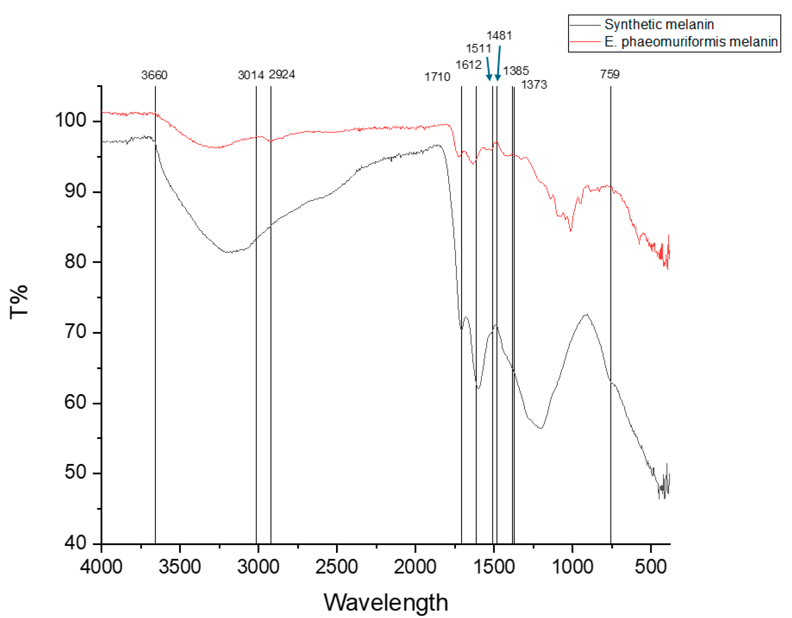
Figure 2.
UV–Vis transmittance spectra of the BC- based films cultivated in glycose (a) and in deproteinized cheese whey (b) with and without microbial melanin addition.
Figure 2.
UV–Vis transmittance spectra of the BC- based films cultivated in glycose (a) and in deproteinized cheese whey (b) with and without microbial melanin addition.
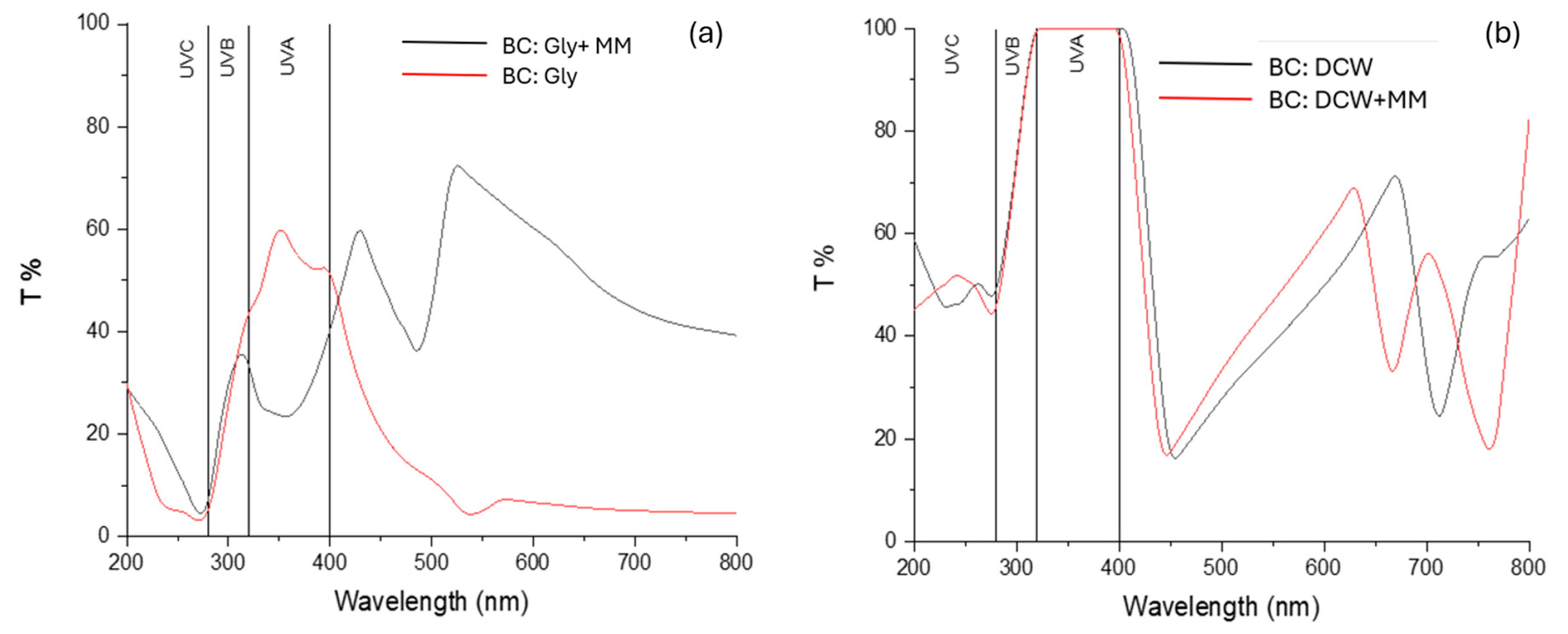
Figure 3.
UV–Vis transmittance spectra of the SCP- based films with and without microbial melanin addition.
Figure 3.
UV–Vis transmittance spectra of the SCP- based films with and without microbial melanin addition.
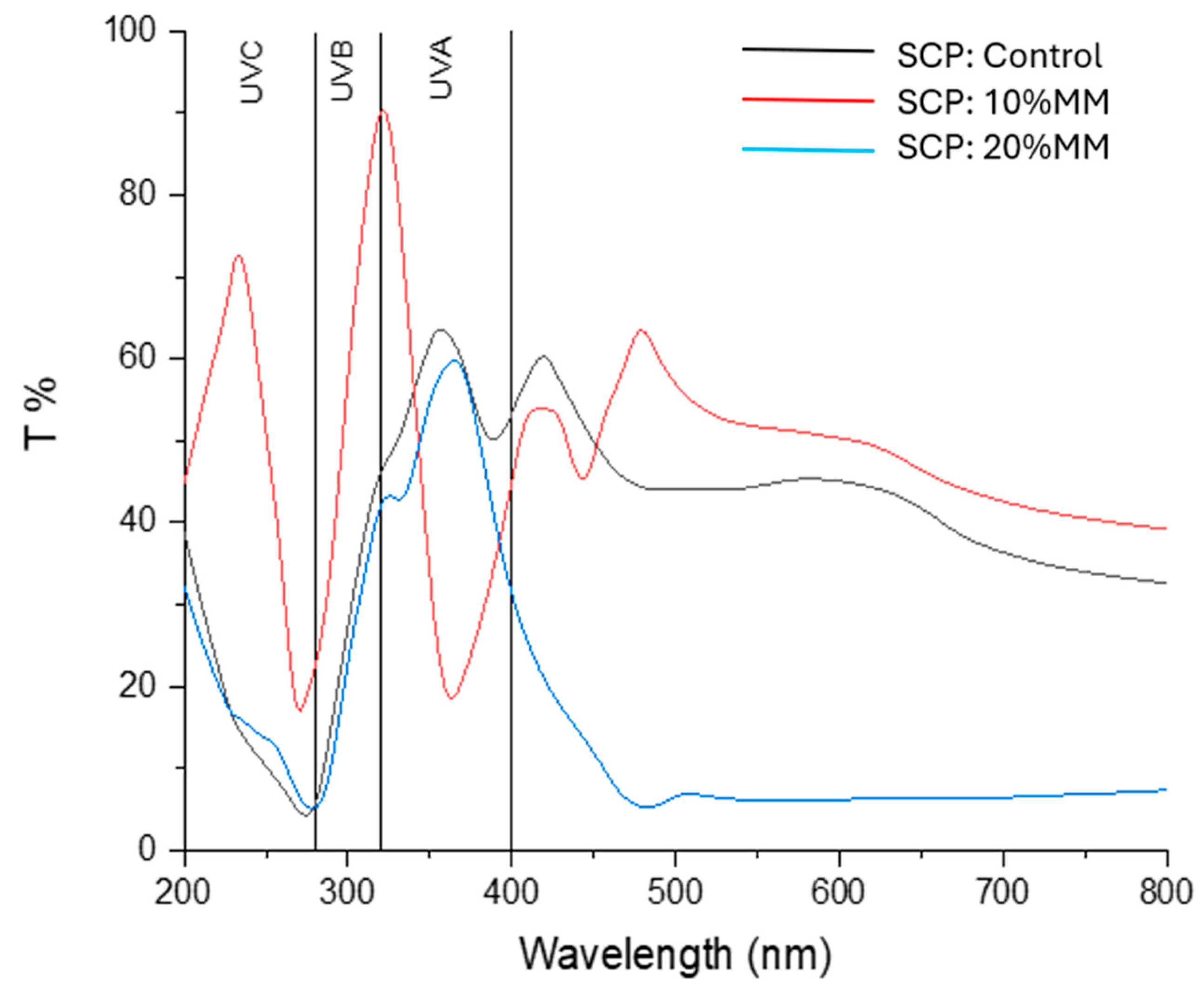
Figure 4.
Developed SCP-based films with microbial melanin addition at 10 and 20% v/v.
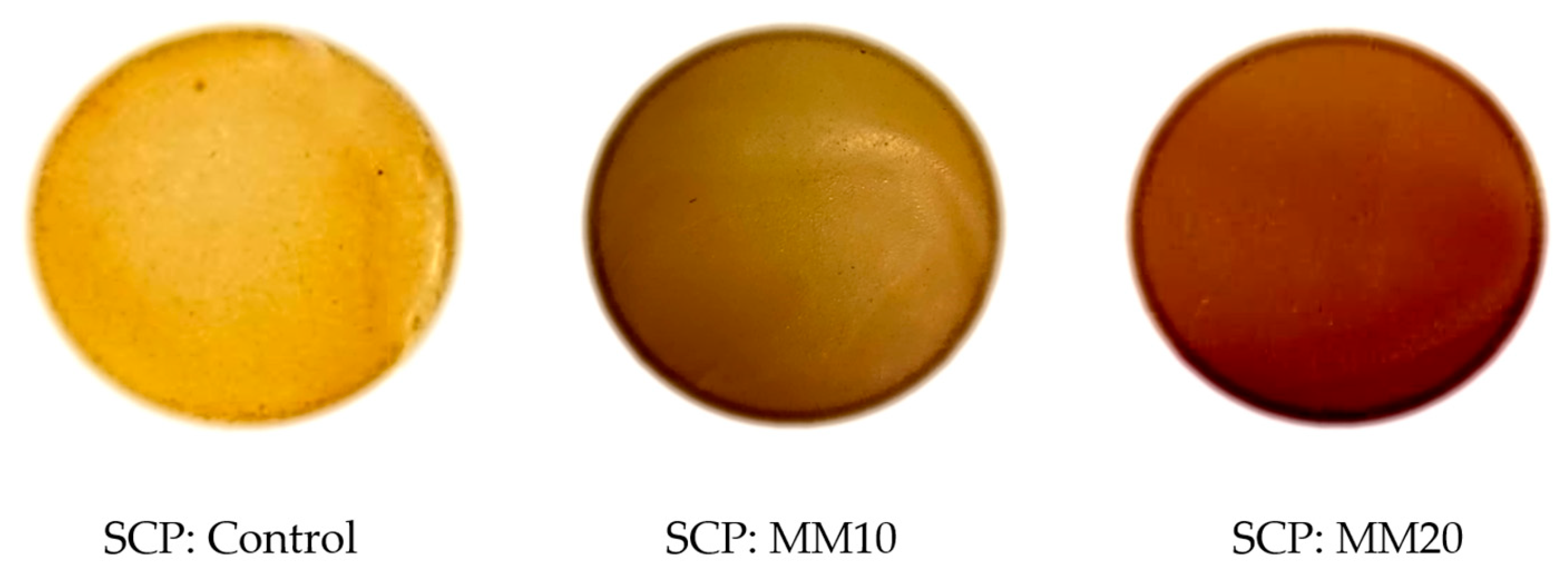
Figure 5.
Mechanical properties in terms of tensile strnght (MPa) (a) and elongation at break (%) (b) of BC- based films. Results are expressed as mean values accompanied by their corresponding standard deviations. Within each parameter, values marked with different lowercase superscript letters differ significantly (p < 0.05). All analyses were carried out using three independent experimental replicates (N=3).
Figure 5.
Mechanical properties in terms of tensile strnght (MPa) (a) and elongation at break (%) (b) of BC- based films. Results are expressed as mean values accompanied by their corresponding standard deviations. Within each parameter, values marked with different lowercase superscript letters differ significantly (p < 0.05). All analyses were carried out using three independent experimental replicates (N=3).
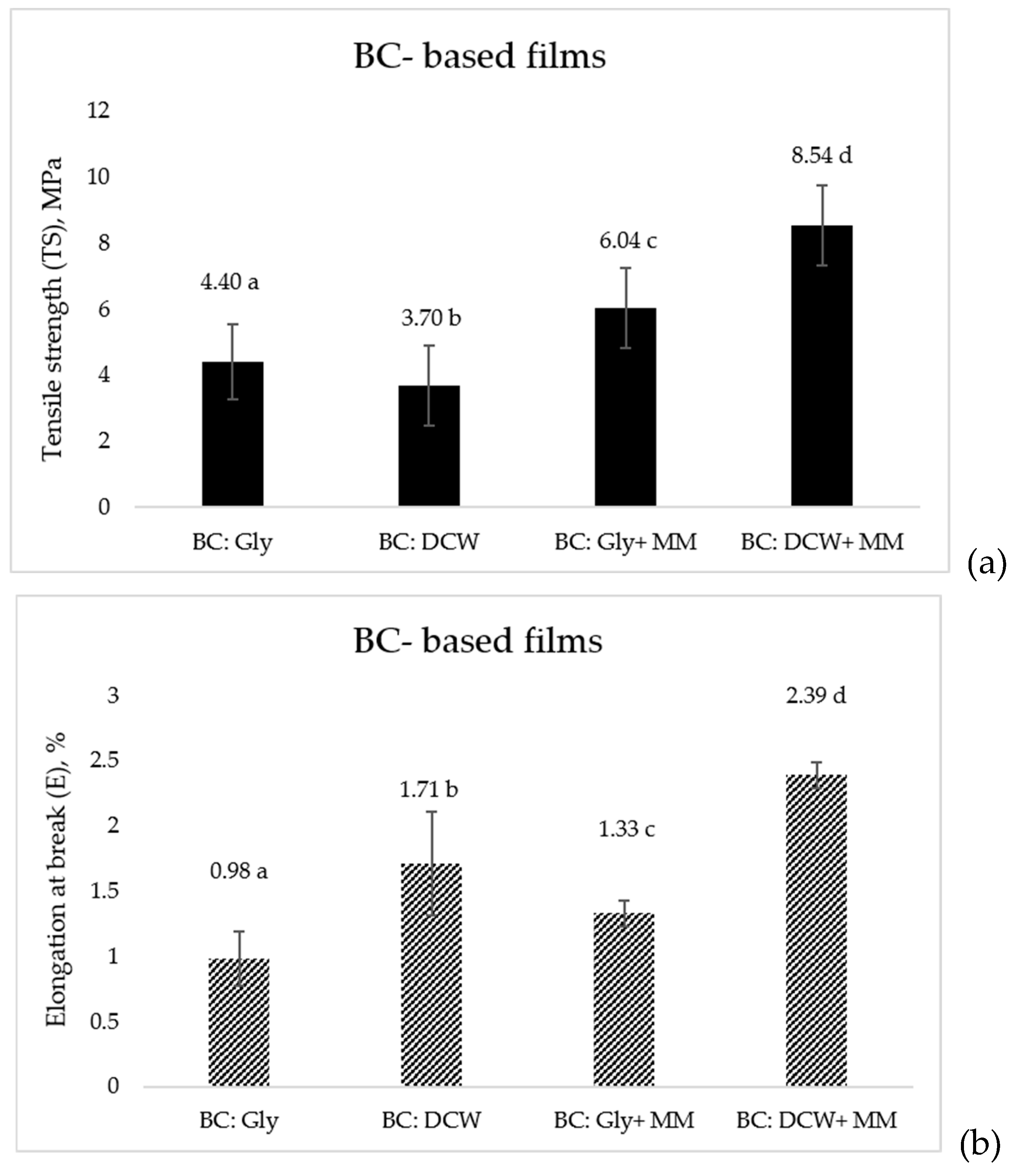
Figure 6.
Mechanical properties in terms of tensile strnght (MPa) (a) and elongation at break (%) (b) of SCP- based films. Results are expressed as mean values accompanied by their corresponding standard deviations. Within each parameter, values marked with different lowercase superscript letters differ significantly (p < 0.05). All analyses were carried out using three independent experimental replicates (N=3).
Figure 6.
Mechanical properties in terms of tensile strnght (MPa) (a) and elongation at break (%) (b) of SCP- based films. Results are expressed as mean values accompanied by their corresponding standard deviations. Within each parameter, values marked with different lowercase superscript letters differ significantly (p < 0.05). All analyses were carried out using three independent experimental replicates (N=3).
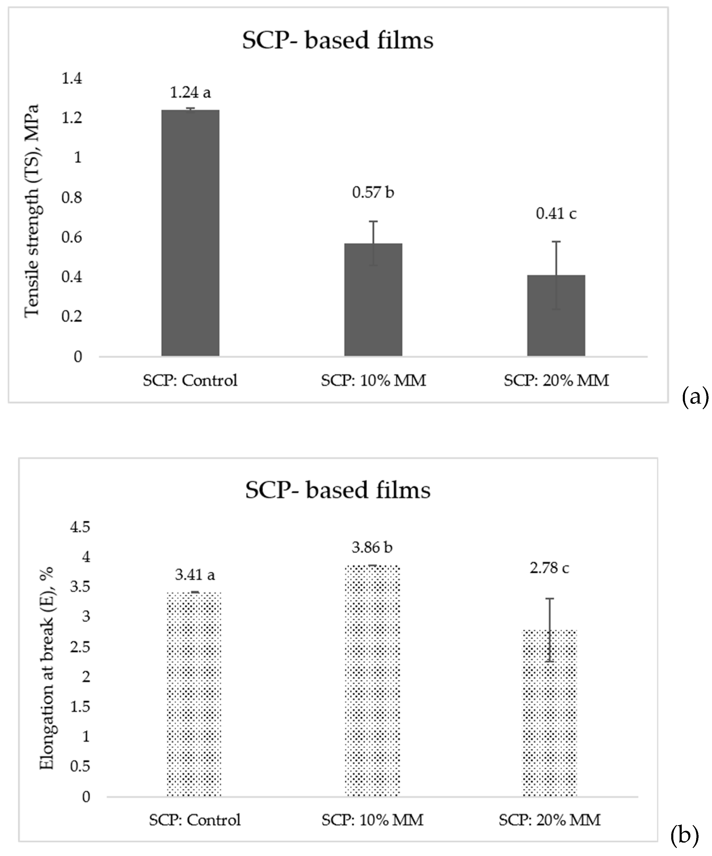

Table 1.
Optical properties of BC-based films with and without microbial melanin incorporation.
| BC- based films | L* | a* | b* | C* | h* | Film opacity |
|---|---|---|---|---|---|---|
| BC: Gly | 77.2± 0.5 a | 4.4± 0.4 a | 16.4± 2.1 a | 17.0± 2.1 a | 74.9± 0.7 a | 0.8± 0.0 a |
| BC: DCW | 84.1± 0.7 b | 3.4± 0.1 b | 1.2± 0.7 b | 3.7± 0.3 b | 341.6± 7.9 b | 0.3± 0.0 b |
| BC: Gly+ MΜ | 37.4± 1.8 c | 10.4± 0.4 c | 15.0± 1.8 c | 18.3± 1.7 c | 55.0± 2.3 c | 1.2± 0.0 c |
| BC: DCW+ MΜ | 51.6± 3.0 d | 10.3± 0.8 d | 16.5± 1.7 d | 19.5± 1.8 d | 57.8± 0.9 d | 0.4± 0.0 d |
Results are expressed as mean values accompanied by their corresponding standard deviations. Within each parameter, values marked with different lowercase superscript letters differ significantly (p < 0.05). BC: bacterial cellulose; Gly: glycose; DCW: deproteinized cheese whey; MΜ: microbial melanin. All analyses were carried out using three independent experimental replicates (N=3).
Table 2.
Optical properties of SCP-based films with increased concentrations of microbial melanin.
| SCP- based films | L* | a* | b* | C* | h* | Film opacity |
|---|---|---|---|---|---|---|
| SCP: Control | 61.8± 2.0a | 11.3± 1.3a | 42.7± 2.0a | 44.2± 2.3a | 75.2± 0.9a | 0.1± 0.0a |
| SCP: MΜ10 | 43.0± 1.9b | 21.4± 1.5b | 41.5± 1.4b | 46.8± 0.6b | 62.7± 2.4b | 0.2± 0.0b |
| SCP: MΜ20 | 44.5± 2.4c | 21.1± 1.1b | 41.0± 3.3 b | 46.2± 2.4b | 62.6± 2.4b | 1.1± 0.0c |
Results are expressed as mean values accompanied by their corresponding standard deviations. Within each parameter, values marked with different lowercase superscript letters differ significantly (p < 0.05). SCP: single cell protein; MΜ10: 10% addition of microbial melanin; MΜ20: 20% addition of microbial melanin. All analyses were carried out using three independent experimental replicates (N=3).
Table 3.
The effect of microbial melanin of BC-based films in water- related properties.
| BC- based films | MC (%) | S (%) | S.I (%) | WVP (g⋅mm/(m2⋅d⋅kPa) |
|---|---|---|---|---|
| BC: Gly | 20± 1.6a | 43.1 ± 4.1a | 607.2 ± 6.7a | 3.4 ± 1.0a |
| BC: DCW | 14.4± 1.9b | 12.5 ± 2.4b | 262.2 ± 8.0b | 0.9 ± 0.1b |
| BC: Gly+ MM | 9.6 ± 0.7c | 53.3 ± 2.9c | 680.0 ± 11.6c | 8.0± 3.0c |
| BC: DCW+ MM | 18.2± 1.2d | 56.0 ± 4.5d | 491.5 ± 4.7d | 1.8± 0.7b |
Results are expressed as mean values accompanied by their corresponding standard deviations. Within each parameter, values marked with different lowercase superscript letters differ significantly (p < 0.05). BC: bacterial cellulose; Gly: glycose; DCW: deproteinized cheese whey; MΜ: microbial melanin. All analyses were carried out using three independent experimental replicates (N=3).
Table 4.
The effect of different concentrations of microbial melanin in SCP-based films in water- related properties.
Table 4.
The effect of different concentrations of microbial melanin in SCP-based films in water- related properties.
| SCP- based films | MC (%) | S (%) | S.I (%) | WVP (g⋅mm/(m2⋅d⋅kPa) |
|---|---|---|---|---|
| SCP: Control | 20.7± 0.3a | 48.3± 0.3a | 5.2± 0.3a | 8.1± 3.2a |
| SCP: MM10 | 18.7± 0.3b | 38.8± 0.9b | 7.1± 0.3b | 9.2± 3.5b |
| SCP: MM20 | 18.4± 0.4b | 36.8± 0.6c | 9.0± 0.2c | 9.5± 3.7c |
Results are expressed as mean values accompanied by their corresponding standard deviations. Within each parameter, values marked with different lowercase superscript letters differ significantly (p < 0.05). SCP: single cell protein; MΜ10: 10% addition of microbial melanin; MΜ20: 20% addition of microbial melanin. All analyses were carried out using three independent experimental replicates (N=3).
Disclaimer/Publisher’s Note: The statements, opinions and data contained in all publications are solely those of the individual author(s) and contributor(s) and not of MDPI and/or the editor(s). MDPI and/or the editor(s) disclaim responsibility for any injury to people or property resulting from any ideas, methods, instructions or products referred to in the content. |
© 2026 by the authors. Licensee MDPI, Basel, Switzerland. This article is an open access article distributed under the terms and conditions of the Creative Commons Attribution (CC BY) license (http://creativecommons.org/licenses/by/4.0/).
Copyright: This open access article is published under a Creative Commons CC BY 4.0 license, which permit the free download, distribution, and reuse, provided that the author and preprint are cited in any reuse.