

Submitted:
12 August 2026
Posted:
13 August 2026
You are already at the latest version
Abstract
Protein tyrosyl phosphorylation is a dynamic reversible regulatory process that is es-sential for virtually all aspects of cellular function. Dysregulation of protein tyrosyl phosphorylation is one of the main causes of human diseases such as cancer, diabetes, autoimmune disorders, and neurological diseases. Protein tyrosine kinases (PTKs) have paved the way towards defining the significance of protein tyrosyl phosphorylation in human health and disease. However, homeostatic control of protein tyrosyl phosphor-ylation is also regulated by protein tyrosine phosphatases (PTPs). The actions of PTPs are diverse functioning to both positively and negatively regulate signaling pathways through specific substrate dephosphorylation. The PTKs are now matured targets with numerous drugs that have been developed for the treatment of human diseases such as cancer and immunological disorders. In contrast, PTPs are relatively in their infancy as compared with their PTK counterparts with regards to drug development for the treatment of human diseases. However, mounting evidence supports the view that PTPs are not only valid targets but are indeed targetable. This review will highlight recent developments in the PTP field which provide support for the emergence of PTPs as therapeutic targets for diseases such as cancer, obesity, type II diabetes and rare diseases.
Keywords:
protein phosphatases
; phosphorylation
; cancer
; tumor suppressor
; signal transduction
1. Introduction
Signal transduction is an intricate process that transmits extracellular signals through a series of events involving protein tyrosine phosphorylation and dephosphorylation to produce a broad spectrum of signal responses. Protein tyrosine kinases (PTKs) transfer phosphate groups from ATP to target tyrosine residues in proteins, thereby activating signal transduction pathways linked to cell growth, proliferation, differentiation, metabolism, and cell death. Protein tyrosine phosphatases (PTPs) are a group of enzymes that mediate the dephosphorylation of proteins on tyrosine residues. PTPs play an important role in the regulation of cell growth, proliferation, and differentiation. A total of 95 PTKs are counteracted by 107 PTPs to maintain the net balance of cellular tyrosyl phosphorylation. The PTP superfamily is characterized by a consensus signature motif HC(X)5R, which designates the active site of these enzymes. PTPs can be grouped into two classes. The first class is characterized by cysteine-based phosphotyrosine selectivity, and the second class constitutes the dual-specificity phosphatases (DUSPs). The traditional PTP genes can be further classified into non-transmembrane PTPs and transmembrane receptor PTPs (RPTPs) [1,2,3,4,5].
Mutations in both the PTKs and PTPs are documented to be linked with various human diseases [6,7]. The significance of PTKs in the context of cancer has been thoroughly investigated, but studies on PTPs have lagged behind PTKs since these proteins were assumed to function as housekeeping proteins [8,9]. However, it is now abundantly clear that PTPs are critical for the maintenance of protein tyrosyl phosphorylation levels and work in concert with the PTKs [4]. The success of PTKs as drug targets for the treatment of human diseases such as cancer and immunological disorders has provided a conceptual platform from which to investigate whether PTPs can be similarly targeted therapeutically. Although the PTP family is extensive much of the focus regarding, the development of therapeutics for PTPs against diseases such as cancer, immunological disorders and metabolic disease has largely focused on a handful of PTPs. These PTPs include SHP-2, PTP-1B, TC-PTP, PRLs, STEP and more recently the DUSPs, in particular MKP-5. As such, these PTPs represent the forefront of PTP therapeutic development and serve as excellent representations of the advancements and trajectory of PTP research.
1.1. Protein Phosphatases
Protein phosphatases are a group of enzymes that remove phosphate groups from phosphorylated amino acid residues, counterbalancing the action of kinases and playing essential roles in regulating cellular signaling [10]. Based on substrate specificity and structural characteristics, protein phosphatases are broadly classified into three major families: serine/threonine protein phosphatases (PSPs), protein tyrosine phosphatases (PTPs), and dual-specificity phosphatases (DUSPs).
1.2. Serine/Threonine Protein Phosphatases (PSPs)
Serine/threonine protein phosphatases (PSPs) primarily dephosphorylate phosphoserine and phosphothreonine residues [11]. This family includes the phosphoprotein phosphatase (PPP) family (e.g., PP1, PP2A, PP2B/calcineurin, PP4, PP5, PP6) and the protein phosphatase, Mg²⁺/Mn²⁺-dependent (PPM) family (e.g., PP2C, PPM1D) [11]. In cancer, PSPs can act as either tumor suppressors or promoters, depending on context. For example, PP2A is well known for its tumor-suppressive role by negatively regulating key oncogenic pathways, such as the AKT and ERK pathways. However, paradoxically, inhibiting PP2A has also shown potential as a therapeutic strategy in certain cancers [12,13]. Besides, dysregulation of some PSPs, such as overexpression of PPM1D, has been implicated in tumorigenesis and therapy resistance [14,15].
1.3. Protein Tyrosine Phosphatases (PTPs)
Protein tyrosine phosphatases (PTPs) selectively dephosphorylate phosphotyrosine residues. This group encompasses classical receptor-type (e.g., PTPRA, PTPRF, PTPRB) and non-receptor-type PTPs (e.g., PTP1B, SHP1/PTPN6, SHP2/PTPN11) [10]. PTPs have diverse roles in cancer: while some, such as PTEN, function as well-established tumor suppressors through PI3K/AKT pathway inhibition, others like SHP2 (encoded by PTPN11) can promote tumor growth and are emerging therapeutic targets in RAS-driven cancers [16,17].
1.4. Dual-specificity Phosphatases (DUSPs)
Dual-specificity phosphatases (DUSPs) can dephosphorylate both phosphoserine/threonine and phosphotyrosine residues. This family includes DUSP1–DUSP28, among others, many of which are MAPK phosphatases that regulate ERK, JNK, and p38 pathways [18,19]. Dysregulated expression of DUSPs contributes to cancer progression by affecting cell proliferation, survival, and drug resistance [19]. For example, DUSP6 specifically inactivates ERK1/2 and can act as a tumor suppressor in certain contexts. However, in some cases, high levels of DUSP6 are associated with increased cancer cell growth [20,21].
Together, these Phosphatase families tightly regulate signaling cascades involved in proliferation, apoptosis and DNA damage responses. In cancer, their roles are complex and often context-dependent, making them attractive as targets for therapeutic intervention.
Figure 1.
The Superfamily of Protein Phosphatases.
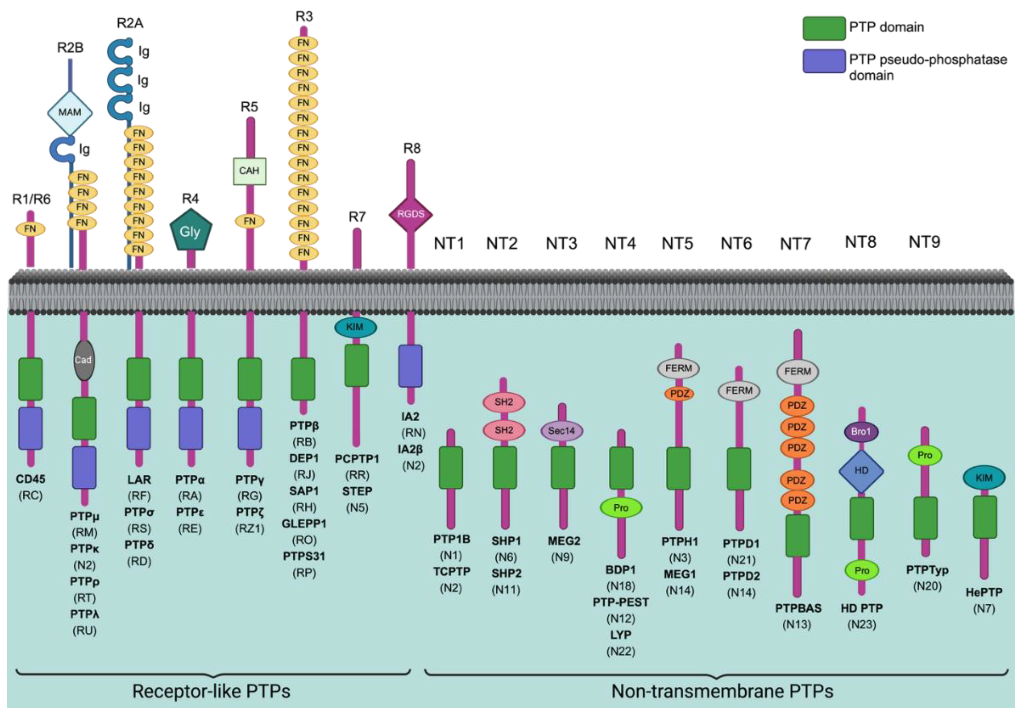
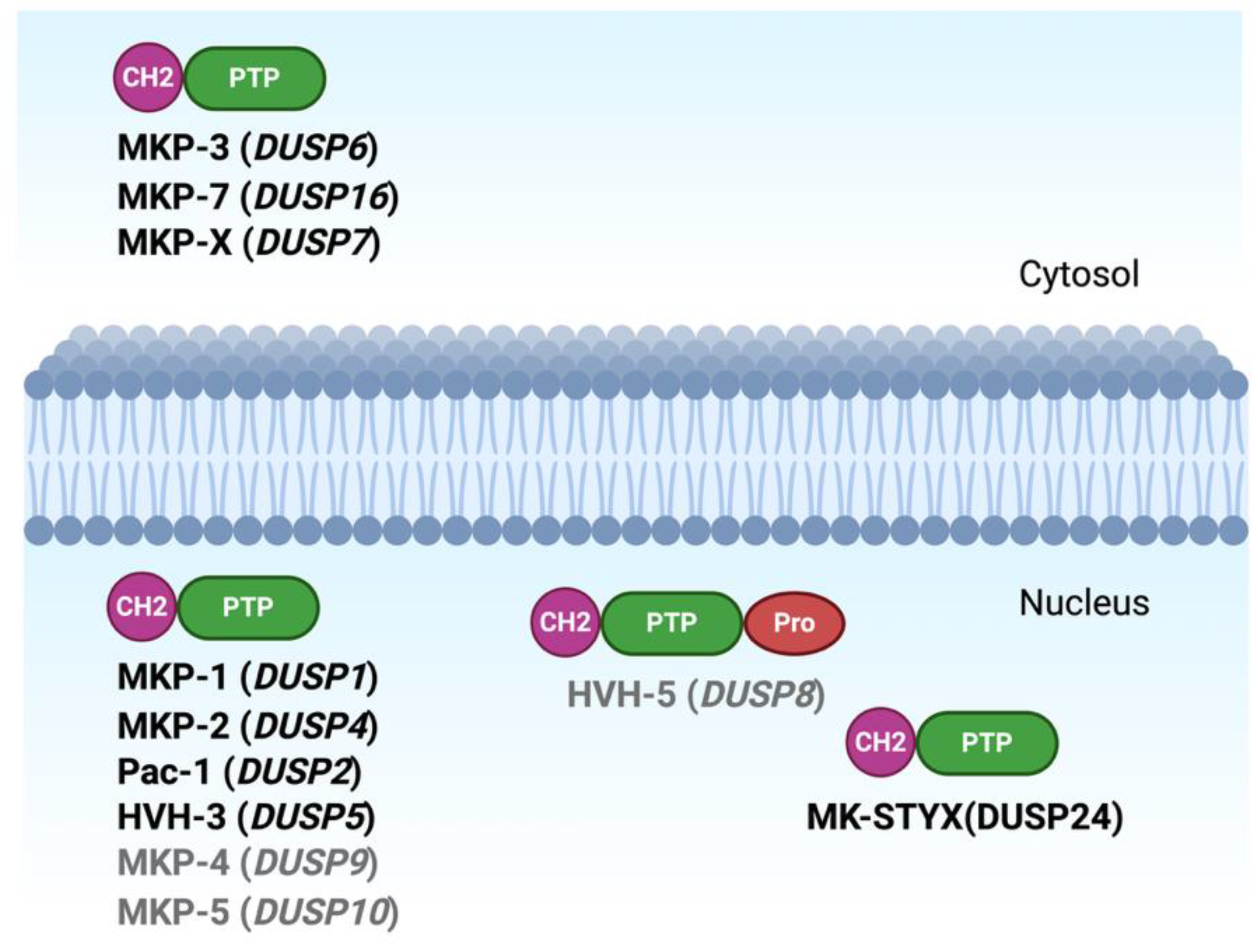
Figure 2.
The Superfamily of Protein Phosphatases and Dual-Specificity Phosphatases.
2. Protein Tyrosine Phosphatases in Cancer:
SHP2: Src homology region 2 (SH2)-containing protein tyrosine phosphatase 2 (SHP2) is a ubiquitously expressed non-receptor PTP that is encoded by the PTPN11 gene [1]. SHP2 transduces signals from several growth factor, cytokine, and extracellular matrix receptors that are mitogenic, pro-survival, pro-cell-fate, and/or promigratory [1,22]. SHP2 has two adjacent SH2 domains (N-SH2 and C-SH2) at its N-terminus, and a PTP catalytic domain, followed by two tyrosine phosphorylation sites (Y542 and Y580) and a proline-rich region at its C-terminus [23]. In the normal state SHP2 is in a closed conformation in which the catalytic domain is blocked by the N-SH2 domain [23]. Whenever growth factors or cytokines stimulate the cell, phosphorylated tyrosine residues on proteins like receptor tyrosine kinases (RTKs), cytokine receptors, and scaffolding adaptors bind to the N-SH2 domain [24]. This binding disrupts autoinhibition and activates the enzyme [24].
Mutations in the PTPN11 gene lead to Noonan Syndrome (NS) and Noonan Syndrome with Multiple Lentigines (NSML), and since SHP2 is involved in many signaling pathways, it is implicated in a variety of cancer types [25,26,27]. In acute myeloid leukemia (AML), SHP2 functions as an oncogenic mediator by binding to PD-1 expressed on leukemia stem/progenitor cells, initiating downstream signaling that promotes leukemic proliferation and survival, bypassing immune control and promoting proliferation [28]. In clear cell renal cell carcinoma (ccRCC), SHP2 acts downstream of RGC32 to activate NF-κB/EGFR signaling, enhancing proliferation, invasion, and epithelial-mesenchymal transition (EMT) [29]. In cervical cancer, SHP2 in tumor-associated macrophages (TAMs) participates in the PD-1/IRE1α/SHP2/HIF1α axis to promote angiogenesis and cell migration [30]. Inhibition of this pathway with PD-1 monoclonal antibodies increases SHP2 levels yet suppresses HIF1α-driven neovascularization, demonstrating SHP2’s involvement in tumor vascular biology and metastasis regulation [30]. In BRAF(V600E) mutant colorectal cancer, SHP2 activation is necessary for the oncogenic MUC1-C protein to drive proliferation and BRAF inhibitor resistance [31] while in other colorectal cancers, high SHP2 expression correlates with reduced metastasis and improved survival, illustrating SHP2’s context-dependent role in cancer progression [32,33]. Moreover, SHP2 also sustains ERK/MAPK signaling as an oncogenic amplifier in esophageal squamous cell carcinoma, but in myeloid cells, SHP2 downregulates STAT3/STAT6 signaling, and deficiency in SHP2 has been shown to promote esophageal carcinoma [34,35]. Additional oncogenic roles include folic-acid-driven proliferation via the caveolin-1-SHP2-p-cSrcY416 pathway and drug resistance in breast cancer, sustained FGR and KRAS signaling in non-small cell lung cancer (NSCLC) to drive drug resistance and developing anoikis resistance and EMT through TGF-β1/SHP2/JAK2/STAT3 and SHP2/Grb2 signaling pathways [36,37,38,39,40]. Additionally, cancer cell-derived exosomal circUSP7 upregulates the expression of SHP2, which causes immunosuppression by promoting CD8+ T cell dysfunction [39]. SHP2 also drives hypoxia-induced aggressiveness in oral cancers and suppresses IFN-α anti-proliferative signaling in hepatocellular carcinoma and renal cancer [41,42,43]. In thyroid cancer, PD-1 blockade suppresses an intrinsic SHP2/Ras/MAPK program, delaying tumor growth, while in anaplastic thyroid carcinoma, SHP2 inhibition enhances immunogenicity and dendritic cell-mediated phagocytosis [44,45].
More recently, SHP2 inhibition has also gained attention as a rational combination strategy in RAS- and RTK-driven malignancies. Because SHP2 functions downstream of multiple RTKs and facilitates RAS/MAPK pathway activation, SHP2 inhibitors are being evaluated with RAS-pathway inhibitors and RTK-directed therapies to overcome adaptive pathway reactivation and therapeutic resistance. In KRAS-mutant lung cancer, clinical studies have examined SHP2 inhibitors in combination with KRAS G12C inhibitors, including sotorasib plus RMC-4630 in KRAS G12C-mutant NSCLC and other advanced solid tumors, as well as broader studies combining SHP2 inhibitors with ERK, EGFR, HER2, VEGF, PI3K, or PD-1 pathway inhibitors [46]. Similarly, the SHP2 inhibitor TNO155 has been evaluated alone and in combination with the EGFR inhibitor nazartinib in advanced solid tumors, supporting the concept that SHP2 blockade may suppress RTK-mediated signaling dependencies [46]. In breast cancer, SHP2 inhibition has also been proposed as a way to overcome RTK-mediated resistance, including resistance to PI3K inhibition in luminal and triple-negative breast cancer models, further supporting SHP2 as a therapeutic node in both lung and breast cancer signaling networks [47].
PTP1B: Protein tyrosine phosphatase 1B (PTP1B), which is encoded by the PTPN1 gene, is a highly studied phosphatase. It has been observed to function as either a tumor suppressor or promoter, depending on the specific cellular environment. Structurally and mechanistically related to TC-PTP, PTP1B plays pivotal roles in diverse oncogenic signaling pathways, including PI3K/AKT, Src, and Hippo-YAP [48].
In hepatocellular carcinoma (HCC), PTP1B overexpression enhances tumor growth, proliferation, migration, invasion, and angiogenesis by sustaining PI3K/AKT activation and suppressing AMPK activity [49]. In breast cancer, the PTP1B/PI3K/AKT pathway induces proliferation and suppresses apoptosis, and these effects can be reversed using derivatives of oleanolic acid glycosides, such as OA-Br-1 [50]. Consistently, PTP1B expression correlates with immune infiltration and checkpoint gene expression, further linking PTP1B activity to tumor immune microenvironment modulation [48]. PTP1B also promotes tumor progression in glioblastoma by activating Cdk3 through dephosphorylation, leading to Rb phosphorylation, E2F-dependent transcription, and G1/S transition, thereby accelerating proliferation [51]. Another study supported this by showing that miR-34c-mediated inhibition of PTP1B suppresses glioma cell proliferation [52]. In melanoma, PTP1B promotes tumor growth and metastasis by dephosphorylating Src at Tyr527, maintaining its active conformation and driving downstream pro-metastatic signaling [53]. Similarly, PTP1B has been shown to have a role in tumor immune evasion in colorectal cancer [54]. On the other hand, PTP1B can also exert tumor-suppressive effects in some cancers. In cholangiocarcinoma, PTP1B interacts with the Hippo pathway effector Yes-associated protein (YAP), which is a driver for oncogenesis; PTP1B inhibits YAP activity, decreasing cholangiocarcinoma cellular proliferation [55].
TC-PTP/PTPN2: The PTPN2 gene codes for human T-cell protein tyrosine phosphatase (TC-PTP) belonging to the family of classical non-receptors PTPs [56]. TC-PTP contains a conserved catalytic domain and a variable length C-terminus. PTPN2 is closely related to PTP1B, possessing similar enzymatic activity and around 72% sequence identity in their catalytic domains [57,58]. Because of alternative splicing at its carboxyl terminus, PTPN2 encodes two distinct isoforms in humans and rodents: a 48.5 kDa TC-PTP (TC48) and 45 kDa TC-PTP (TC45). Key variations that distinguish the two isoforms are found at their C-terminus. In TC45, the inclusion of a nuclear localization signal (NLS) allows it to act within the nucleus and cytoplasm, gaining access to a wide variety of substrates. TC48 also has an NLS but uniquely has a hydrophobic C-terminus element, which leads to it being localized to the endoplasmic reticulum (ER) [57,58].
The role of TC-PTP in cancer is quite complex, with demonstrated potential to act not only as a tumor suppressor but also as a driver of cancer progression [59,60]. TC-PTP has been shown to act as a suppressor of tumor development in that a loss of TC-PTP was found to promote the development of breast, liver, and skin cancers, often through influencing STAT-3 signaling [61]. In fact, several components of the JAK-STAT pathway are TC-PTP substrates and have gained increased attention as prospective targets in cancer immunotherapeutic approaches [62]. In other studies, TC-PTP has shown itself to be a promoter of cancer progression, such as within KRAS-dependent cancer cell lines where TC-PTP promoted membrane localization of KRAS, and a loss of TC-PTP corresponded with marked attenuation of cancer cell proliferation and enhanced apoptosis [63]. Despite the very clear and mounting evidence to suggest that TC-PTP plays a critical role in cancer suppression and progression, there remains no commercially available agents targeting TC-PTP, though work is being done to fill this gap. Of note, researchers are exploring the use of proteolysis-targeting chimera (PROTAC) technology to overcome intrinsic challenges associated with phosphatase-targeting drug design and have taken the first steps towards the development of immunotherapeutic agents targeting the phosphatase [56].
Figure 3.
Schematic overview of signaling pathways regulated by PTPs in cancer. PTPs effects on proteins are indicated by lines with dots and downstream signaling effects are indicated by lines with arrows.
Figure 3.
Schematic overview of signaling pathways regulated by PTPs in cancer. PTPs effects on proteins are indicated by lines with dots and downstream signaling effects are indicated by lines with arrows.
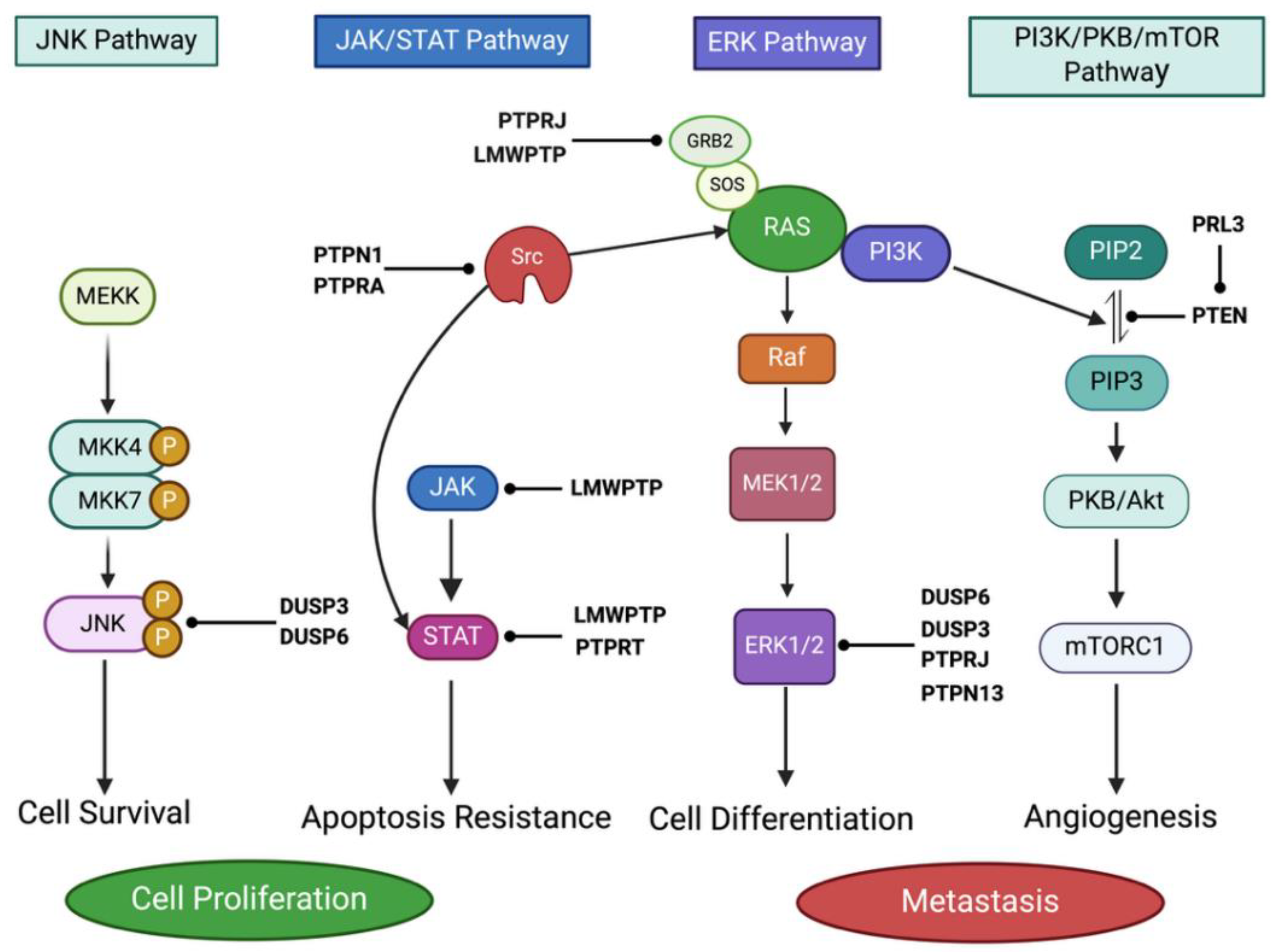
Several tumor suppressor PTPs have been identified whose loss has been shown to be associated with abnormal growth of tumors, metastasis in in vitro and in vivo models (Table 1). Recent studies have shown that PTPs can also act as oncogenes to induce tumor growth. A few human cancers have abnormally high expression of these oncogenic PTPs, which promotes tumor development, growth, and metastasis and, ultimately, lowers survival. In case of breast cancer, leukemia and gliomas, SHP2 acts as an oncogene and dysregulates the EGFR/Ras/MAPK pathway [64,65,66,67,68,69,70]. PTP1B acts as oncogene in ovarian, gastric, prostate & breast cancer and dysregulates the Src/Ras&PI3/Akt pathway [71,72,73,74,75,76,77,78]. PTP4A3 acts as oncogene in Breast, gastric & colon cancers and dysregulates the PDGF, integrin & EphA signaling pathway [79,80,81,82].
The development of PTP inhibitors is still difficult, even though oncogenic PTPs are appealing molecules for the creation of tailored treatments. Because of the great degree of conservation in the active site of both tumor suppressor and oncogenic PTP subtypes, selectivity presents a significant obstacle in the development of PTP inhibitors. Thus, the inhibitors that target the active sites of oncogenic PTPs also inhibit tumor suppressor PTPs in healthy patients and potentially result in unfavorable side effects. However, these negative side effects could be avoided by generating selective PTP inhibitors that bind to regions outside of the PTP active site [98].
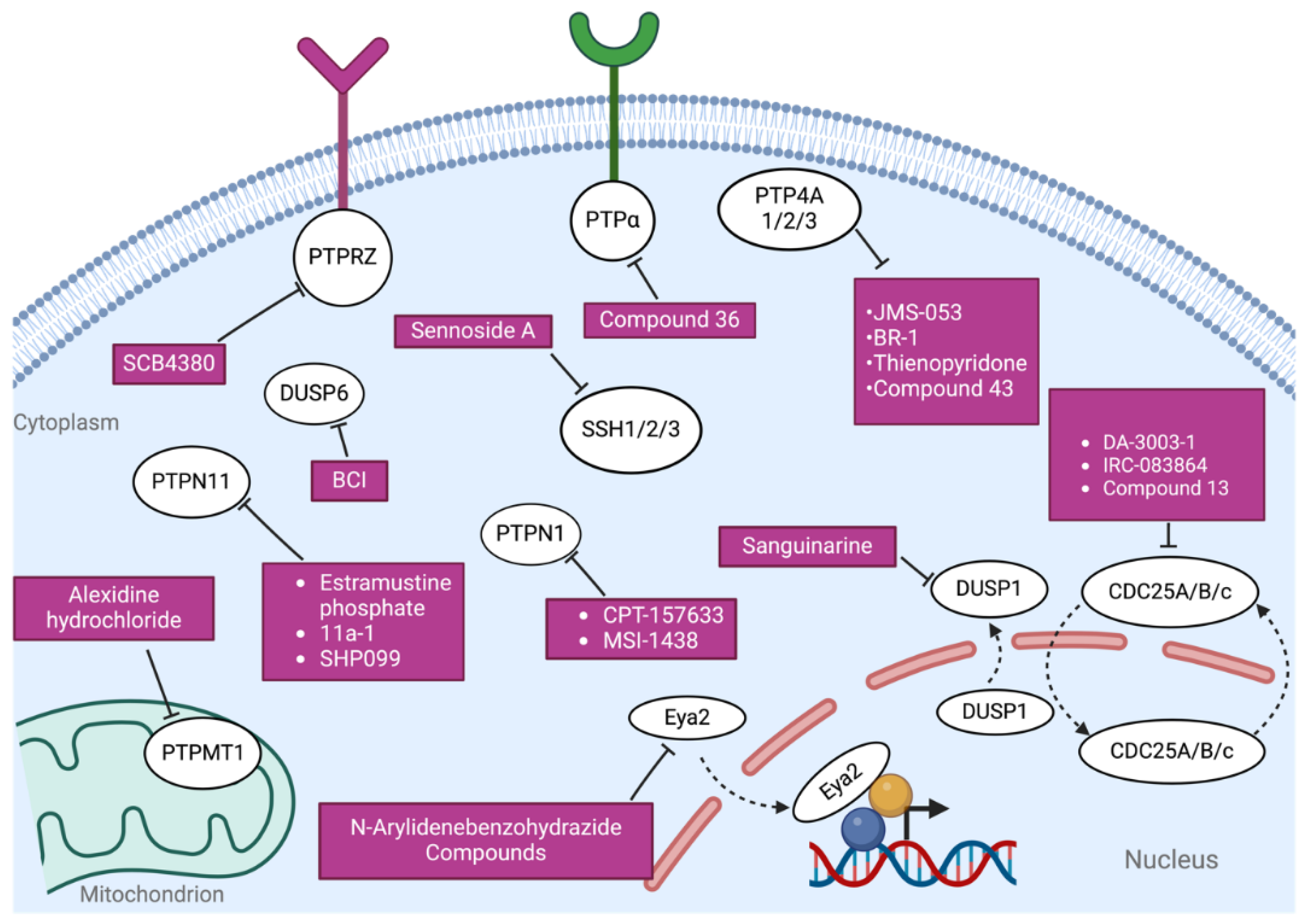
Figure 4.
Subcellular localization of PTP inhibitors. A schematic representation of the subcellular localization of PTPs and corresponding SMIs.
Figure 4.
Subcellular localization of PTP inhibitors. A schematic representation of the subcellular localization of PTPs and corresponding SMIs.
3. Future of PTP Inhibitors for Cancer Therapy:
Recent studies have demonstrated that oncogenic PTPs are appealing candidates for the development of targeted therapeutics for many cancer types. There aren't enough safe and effective PTP inhibitors remain scarce for clinical use, as the development of selective PTP inhibitors has proven difficult. Phase I clinical trial results with the SHP2 inhibitor SSG demonstrated that patients with stage IV melanoma and advanced solid tumors could tolerate doses of 1,200 and 900 mg/m2, respectively [98,99,100]. Comparably, positive results from the PTP1B inhibitor MSI-1436C phase I clinical trial, which is now underway and is establishing the drug's safety and tolerability in the treatment of metastatic breast cancer, will be instrumental to the translation of this drug to the clinical setting. To successfully generate highly specific PTP inhibitors for cancer therapy, more research is needed to unravel specific essential locations situated outside of the active sites, even though various other phosphatases have also been demonstrated to be interesting targets.
Targeting PTPN2 can improve the efficacy of human CAR T cells against solid tumors. CRISPR-Cas9-mediated deletion of PTPN2, or pharmacologic inhibition using the PTPN2/PTP1B inhibitor ABBV-CLS-484, enhanced CAR and cytokine signaling in Lewis Y-specific CAR T cells [101]. PTPN2 targeting increased antigen-induced CAR T cell expansion, activation, cytotoxicity, interferon-associated signaling, and tumor repression in human tumor xenograft and patient-derived xenograft models [101]. Importantly, PTPN2 deletion or inhibition also promoted the persistence of CD8+ CAR T cells with long-lived stem cell memory-like features, suggesting that PTPN2 inhibition may help address major limitations of CAR T therapy in solid tumors, including poor persistence and tumor microenvironment-mediated exhaustion. PTPN22 has also emerged as another immune-restricted phosphatase target for cancer immunotherapy. Genetic loss or small-molecule inhibition of PTPN22 with L-1 augmented antitumor immunity by increasing CD8+ T cell activation and shifting tumor-associated macrophages toward MHC-II-expressing, M1-like phenotypes [102]. PTPN22 inhibition also enhanced the activity of anti-PD-1 therapy, and patients carrying the PTPN22 rs2476601 variant showed improved responses to checkpoint inhibition, supporting PTPN22 as a druggable systemic immunotherapy target [102].
Recent advances in the understanding of phosphatases in cancer have shown that these critical growth-regulatory molecules are just as "druggable" as kinases. However, targeting phosphatases is far more challenging than targeting kinases because many of them are constitutively active and are frequently controlled by binding partners or subunits. Previous studies have demonstrated that inhibition of oncogenic phosphatases can suppress the growth of human cancers. The central challenge is to develop safe and effective strategies to target these cancer-promoting phosphatases. Once this obstacle is cleared, a completely a new class of cancer therapeutics will become available to address numerous serious cancers.
3.1. Dual-specificity Phosphatases (DUSP) in Cancer:
DUSP1: Dual-specificity Phosphatase 1 (DUSP1), also known as MKP-1, is a stress-inducible phosphatase that dephosphorylates JNK and p38 (with weaker activity toward ERK) to regulate MAPK signaling cascades [103]. In cancer, DUSP1 can act either as a tumor promoter or suppressor. In pancreatic ductal adenocarcinoma, DUSP1 promotes tumor growth by suppressing ferroptosis through inhibition of lipid peroxidation [104,105]. In high-grade serous ovarian carcinoma, DUSP1 overexpression drives paclitaxel resistance by promoting drug efflux via p38 MAPK signaling, and its inhibition blocks cell cycle progression by disrupting the AMPK/mTORC pathways [104,105]. In non-small cell lung cancer, DUSP1 is upregulated in osimertinib-resistant cells and mediates therapeutic resistance by inhibiting MAPK-induced apoptosis [106]. In cervical cancer, it suppresses JAK2/STAT3 signaling by downregulating oncogenic miR-21, limiting migration and invasion [107]. In hepatocellular carcinoma, it promotes autophagy via the AMPK–ULK1–Beclin-1 axis, leading to tumor suppression [108]. Additionally, in endometrial and prostate cancers, DUSP1 impairs EMT and promotes apoptosis by targeting EPHA2, ERK/JNK, and Snail, correlating with reduced metastatic potential [109,110].
DUSP2: Dual-specificity Phosphatase 2 (DUSP2) is a nuclear MAPK phosphatase that inactivates ERK1/2 (and to a lesser extent p38 and JNK) to modulate inflammatory and proliferative signaling in immune cells [111]. DUSP2 is predominantly regarded as a tumor suppressor across multiple cancers. In gastric cancer, DUSP2 suppresses ERK-driven EMT, invasion, and metastasis by restoring epithelial markers and repressing mesenchymal genes, with its expression negatively regulated by SKA3 [111]. In pancreatic ductal adenocarcinoma, DUSP2 inhibits AKT signaling and promotes apoptosis under hypoxia; conversely, AKT activation produces TRIM2, which degrades DUSP2, promoting survival and resistance [112]. Epigenetic mechanisms such as DNTTIP1–HDAC1-mediated transcriptional repression and circEIF3I–miR-361-3p post-transcriptional silencing suppress DUSP2 expression in nasopharyngeal and hepatocellular carcinoma, enhancing metastasis and tumor growth [113,114]. In prostate and bladder cancers, DUSP2 loss correlates with EMT, oxidative stress, immune evasion, and poor clinical outcomes, while its restoration inhibits tumor progression and sensitizes cells to therapy through modulation of MAPK, mTOR, and immune pathways [115,116,117].
DUSP6: Dual-specificity Phosphatase 6 (DUSP6), also known as MKP-3, is a cytosolic phosphatase that selectively dephosphorylates ERK1/2, acting as a negative feedback regulator of the RAS/ERK pathway [21]. In cancer, it can exhibit both tumor-suppressive and oncogenic functions. As a tumor suppressor, DUSP6 is frequently downregulated in RAS-driven pancreatic, lung, and liver cancers, where its loss results in sustained ERK signaling and enhanced proliferation and metastasis [118,119]. On the other hand, as a tumor promoter, DUSP6 overexpression in therapy-resistant cancers such as HER2-positive breast and cisplatin-resistant ovarian cancer dampens toxic ERK signaling, supports drug tolerance, and correlates with poor outcomes [120,121]. DUSP6 also contributes to metastasis and metabolic reprogramming by promoting Notch1 signaling in colorectal cancer, suppressing EMT in renal cell carcinoma, and modulating glycolysis via ERK-PKM2 in non-small cell lung cancer [21,122,123,124]. Furthermore, it facilitates DNA damage repair and therapy resistance by enhancing DNA-PKcs recruitment at double-strand breaks in glioblastoma and prolonging G2/M cell cycle arrest in esophageal squamous cell carcinoma [125,126].
DUSP16: Dual-specificity Phosphatase 16 (DUSP16), also known as MKP-7, dephosphorylates JNK and p38 MAPKs to regulate stress-induced apoptotic and inflammatory signaling cascades [127,128]. It contains an allosteric site centered around Tyr271 that is essential for MAPK binding and catalytic activity [129]. In nasopharyngeal, colorectal, gastric, breast, and head and neck tumors, DUSP16 is overexpressed and promotes chemoresistance by suppressing JNK/p38-mediated apoptosis and mitochondrial death signaling [130]. In hematologic cancers, DUSP16 maintains leukemic stemness and proliferation of CD34⁺ hematopoietic progenitors with expression epigenetically regulated by HDAC1/3 [131]. Alternatively, in osteosarcoma and endometrial cancer, DUSP16 acts as a tumor suppressor by inhibiting EMT and glycolytic reprogramming through regulation of the JNK-c-JUN axis [132,133]. Hypoxia-induced circular forms of DUSP16 (circDUSP16) also enhance glycolysis, drug resistance, and tumor growth by sponging tumor-suppressive miRNAs in gastric, colorectal, and breast cancers [134,135,136].
4. Conclusions and Future Perspectives:
4.1. Conclusions:
Collectively, this review establishes PTPs as validated, high-value drug targets rather than the passive “housekeeping” enzymes they were once assumed to be. Their diverse regulatory functions across cell types make them attractive targets not only in oncology but also in inflammatory and metabolic disease. Despite this promise, progress in the PTP field lags considerably behind that of kinases: although 107 PTPs counterbalance the activity of 95 PTKs to maintain cellular phosphotyrosine homeostasis, understanding of PTP structure, function, and inhibitor development remains far less advanced than for kinases.
A central theme of this review is that PTP function is highly context and tissue-dependent rather than fixed: the same phosphatase can act as an oncogene in one cancer type and a tumor suppressor in another. SHP2, for example, drives AML, clear cell renal cell carcinoma, and BRAF-mutant colorectal cancer, yet is associated with reduced metastasis and improved survival in other colorectal cancer subsets. PTP1B similarly contributes to hepatocellular carcinoma and melanoma but suppresses cholangiocarcinoma growth through inhibition of YAP. This dual, context-dependent role is arguably the most important conceptual insight of this review: PTP inhibition cannot be applied as a universal cancer strategy, and rational drug development must instead account for tumor type and genetic driver context.
Among the PTP family, SHP2, PTP1B, and PP2A currently have the most mature mechanistic and translational data, positioning them as the most clinically advanced near-term candidates for drug development. Even within this well-studied group, roles can shift unexpectedly: a recent single-cell transcriptomic study found that depleting SHP2 in Myc-driven liver tumors despite SHP2 being conventionally viewed as pro-tumorigenic instead generated an immunosuppressive, tumor-permissive microenvironment, underscoring that even extensively characterized PTPs can reverse their apparent role depending on genetic driver context.
The principal technical barrier to translating this biology into safe therapeutics is selectivity, Because the catalytic active sites of oncogenic and tumor-suppressor PTPs are highly conserved, inhibitors directed at the active site risk simultaneously blocking protective, tumor-suppressive PTPs in healthy tissue, raising the potential for adverse effects.
4.2. Future Perspectives:
Given SHP2’s clearly context-dependent dual role, future drug discovery should invest as heavily in SHP2 activators as in inhibitors, allowing the pharmacological direction of treatment to be selected based on tumor genetics rather than applying a one-size-fits-all inhibition strategy. Second, moving inhibitor design beyond the catalytic site toward allosteric or other non-active-site regions offers the most promising route to isoform-selective agents that spare tumor-suppressive PTPs in normal tissue. Third, PTP-targeted agents should be further integrated into combination and immunotherapy regimens, building on encouraging early precedent. In RAS- and RTK-driven cancers, pairing SHP2 inhibitors with KRAS G12C inhibitors (sotorasib plus RMC-4630) or with EGFR inhibitors (TNO155 plus nazartinib) has yielded encouraging results. Beyond these, targeting PTPN2 has been shown to boost CAR T-cell persistence and effector function against solid tumors, while inhibiting PTPN22 can potentiate the response to anti-PD-1 checkpoint blockade. Future research should continue to expand this combinatorial framework across RTK, RAS-pathway, and checkpoint-inhibitor-based regimens.
Ultimately, achieving safe, selective PTP-targeting agents would unlock an entirely new class of cancer therapeutics one capable of addressing numerous malignancies that remain underserved by kinase-focused drug development.
Author Contributions
Conceptualization, S.P.; methodology, S.P.; writing—original draft preparation, S.P and J.P.; writing—review and editing, S.P., J.P., M.L., S.K., C.G.A., ; supervision, V.A.S., ; funding support for article, S.P., A.K.; All authors have read and agreed to the published version of the manuscript.
Funding
This article is supported by S.P., A.K.; and Jacob Churg Award (to V.S.S).
Institutional Review Board Statement
Not applicable.
Informed Consent Statement
Not applicable.
Data Availability Statement
No new data were created or analyzed in this study. Data sharing is not applicable to this article.
Acknowledgments
Thanks to my mentor Anton M Bennett (Dept. of Pharmacology, Yale School of Medicine) for his invaluable support and guidance.
Conflicts of Interest
“The authors declare no conflicts of interest.”.
Abbreviations
The following abbreviations are used in this manuscript:
| SH2 | Src homology region 2 |
| SHP2 | SH2-containing protein tyrosine phosphatase 2 |
| PTP1B | Protein tyrosine phosphatase 1B |
| TC-PTP | T-cell protein tyrosine phosphatase |
| DUSP | Dual-specificity phosphatases |
| DUSP1 | Dual-specificity phosphatase 1 |
| DUSP2 | Dual-specificity phosphatase 2 |
| DUSP6 | Dual-specificity phosphatase 6 |
| DUSP16 | Dual-specificity phosphatase 16 |
| PROTAC | Proteolysis-targeting chimera |
| NS | Noonan syndrome |
| NSML | Noonan syndrome with multiple lentigines |
| AML | Acute myeloid leukemia |
| NSCLC | Non-small cell lung cancer |
| HCC | Hepatocellular carcinoma |
| EMT | Epithelial–mesenchymal transition |
| YAP | Yes-associated protein |
| NLS | Nuclear localization signal |
| ER | Endoplasmic reticulum |
| PPP | Phosphoprotein phosphatase family |
| PPM | Mg²⁺/Mn²⁺-dependent protein phosphatase family |
| SSG | Sodium stibogluconate |
| IFN | Interferon |
References
- Soulsby, M.; Bennett, A.M. Physiological Signaling Specificity by Protein Tyrosine Phosphatases. Physiology 2009, 24, 281–289. [Google Scholar] [CrossRef] [PubMed]
- Alonso, A.; Sasin, J.; Bottini, N.; Friedberg, I.; Osterman, A.; Godzik, A.; Hunter, T.; Dixon, J.; Mustelin, T. Protein Tyrosine Phosphatases in the Human Genome. Cell 2004, 117, 699–711. [Google Scholar] [CrossRef] [PubMed]
- Andersen, J.N.; Mortensen, O.H.; Peters, G.H.; Drake, P.G.; Iversen, L.F.; Olsen, O.H.; Jansen, P.G.; Andersen, H.S.; Tonks, N.K.; Møller, N.P. Structural and Evolutionary Relationships among Protein Tyrosine Phosphatase Domains. Mol. Cell. Biol. 2001, 21, 7117–7136. [Google Scholar] [CrossRef] [PubMed]
- Tonks, N.K. Protein Tyrosine Phosphatases: From Genes, to Function, to Disease. Nat. Rev. Mol. Cell Biol. 2006, 7, 833–846. [Google Scholar] [CrossRef] [PubMed]
- Perla, S.; Qiu, B.; Dorry, S.; Yi, J.S.; Bennett, A.M. Identification of Protein Tyrosine Phosphatase (PTP) Substrates. Methods Mol. Biol. 2024, 2743, 123–133. [Google Scholar] [CrossRef] [PubMed]
- Cicenas, J.; Zalyte, E.; Bairoch, A.; Gaudet, P. Kinases and Cancer. Cancers 2018, 10, 63. [Google Scholar] [CrossRef] [PubMed]
- Motiwala, T.; Jacob, S.T. Role of Protein Tyrosine Phosphatases in Cancer. Prog. Nucleic Acid. Res. Mol. Biol. 2006, 81, 297–329. [Google Scholar] [CrossRef] [PubMed]
- Sangwan, V.; Park, M. Receptor Tyrosine Kinases: Role in Cancer Progression. Curr. Oncol. 2006, 13, 191–193. [Google Scholar] [CrossRef] [PubMed]
- Tonks, N.K. PTP1B: from the sidelines to the front lines! FEBS Lett. 2003, 546, 140–148. [Google Scholar] [CrossRef] [PubMed]
- Stanford, S.M.; Bottini, N. Targeting protein phosphatases in cancer immunotherapy and autoimmune disorders. Nat. Rev. Drug Discov. 2023, 22, 273–294. [Google Scholar] [CrossRef] [PubMed]
- Nguyen, H.; Kettenbach, A.N. Substrate and phosphorylation site selection by phosphoprotein phosphatases. Trends Biochem. Sci. 2023, 48, 713–725. [Google Scholar] [CrossRef] [PubMed]
- Brautigan, D.L.; Farrington, C.; Narla, G. Targeting protein phosphatase PP2A for cancer therapy: development of allosteric pharmaceutical agents. Clin. Sci. (Lond.) 2021, 135, 1545–1556. [Google Scholar] [CrossRef] [PubMed]
- Ronk, H.; Rosenblum, J.S.; Kung, T.; Zhuang, Z. Targeting PP2A for cancer therapeutic modulation. Cancer Biol. Med. 2022, 19, 1428–1439. [Google Scholar] [CrossRef] [PubMed]
- Deng, W.; Li, J.; Dorrah, K.; Jimenez-Tapia, D.; Arriaga, B.; Hao, Q.; et al. The role of PPM1D in cancer and advances in studies of its inhibitors. Biomed. Pharmacother. 2020, 125, 109956. [Google Scholar] [CrossRef] [PubMed]
- Kucheryavenko, A.S.; Muzyko, E.A.; Perfilova, V.N.; Kaplanov, K.D.; Frolov, M.Y. The role of the PPM1D gene in tumor pathogenesis. Biomed. Khim. 2025, 71, 19–28. [Google Scholar] [CrossRef] [PubMed]
- Carnero, A.; Blanco-Aparicio, C.; Renner, O.; Link, W.; Leal, J.F. The PTEN/PI3K/AKT signalling pathway in cancer, therapeutic implications. Curr. Cancer Drug Targets 2008, 8, 187–198. [Google Scholar] [CrossRef] [PubMed]
- Punekar, S.R.; Velcheti, V.; Neel, B.G.; Wong, K.K. The current state of the art and future trends in RAS-targeted cancer therapies. Nat. Rev. Clin. Oncol. 2022, 19, 637–655. [Google Scholar] [CrossRef] [PubMed]
- Singh, M.K.; Altameemi, S.; Lares, M.; Newton, M.A.; Setaluri, V. Role of dual specificity phosphatases (DUSPs) in melanoma cellular plasticity and drug resistance. Sci. Rep. 2022, 12, 14395. [Google Scholar] [CrossRef] [PubMed]
- Zandi, Z.; Kashani, B.; Alishahi, Z.; Pourbagheri-Sigaroodi, A.; Esmaeili, F.; Ghaffari, S.H.; et al. Dual-specificity phosphatases: therapeutic targets in cancer therapy resistance. J. Cancer Res. Clin. Oncol. 2022, 148, 57–70. [Google Scholar] [CrossRef] [PubMed]
- Moncho-Amor, V.; Pintado-Berninches, L.; Ibanez de Caceres, I.; Martin-Villar, E.; Quintanilla, M.; Chakravarty, P.; et al. Role of Dusp6 Phosphatase as a Tumor Suppressor in Non-Small Cell Lung Cancer. Int. J. Mol. Sci. 2019, 20. [Google Scholar] [CrossRef] [PubMed]
- Png, C.W.; Weerasooriya, M.; Li, H.; Hou, X.; Teo, F.Y.; Huang, S.; et al. DUSP6 regulates Notch1 signalling in colorectal cancer. Nat. Commun. 2024, 15, 10087. [Google Scholar] [CrossRef] [PubMed]
- Solman, M.; Woutersen, D.T.J.; den Hertog, J. Modeling (not so) rare developmental disorders associated with mutations in the protein-tyrosine phosphatase SHP2. Front. Cell Dev. Biol. 2022, 10, 1046415. [Google Scholar] [CrossRef] [PubMed]
- Song, Z.; Wang, M.; Ge, Y.; Chen, X.P.; Xu, Z.; Sun, Y.; et al. Tyrosine phosphatase SHP2 inhibitors in tumor-targeted therapies. Acta Pharm. Sin. B 2021, 11, 13–29. [Google Scholar] [CrossRef] [PubMed]
- Mohi, M.G.; Neel, B.G. The role of Shp2 (PTPN11) in cancer. Curr. Opin. Genet. Dev. 2007, 17, 23–30. [Google Scholar] [CrossRef] [PubMed]
- Yi, J.S.; Huang, Y.; Kwaczala, A.T.; Kuo, I.Y.; Ehrlich, B.E.; Campbell, S.G.; et al. Low-dose dasatinib rescues cardiac function in Noonan syndrome. JCI Insight 2016, 1, e90220. [Google Scholar] [CrossRef] [PubMed]
- Yi, J.S.; Perla, S.; Enyenihi, L.; Bennett, A.M. Tyrosyl phosphorylation of PZR promotes hypertrophic cardiomyopathy in PTPN11-associated Noonan syndrome with multiple lentigines. JCI Insight 2020, 5. [Google Scholar] [CrossRef] [PubMed]
- Yi, J.S.; Perla, S.; Bennett, A.M. An Assessment of the Therapeutic Landscape for the Treatment of Heart Disease in the RASopathies. Cardiovasc Drugs Ther. 2023, 37, 1193–1204. [Google Scholar] [CrossRef] [PubMed]
- Suo, S.; Li, F.; Zhao, D.; Qiao, J.; Liang, C.; Hoang, D.H.; et al. Acute Myeloid Leukemia Cell-Intrinsic PD-1 Functions Promoting Leukemia Development By Recruiting SHP2. Blood 2021, 138, 1173. [Google Scholar] [CrossRef]
- Zhang, J.; Sun, Y.; Tang, K.; Xu, H.; Xiao, J.; Li, Y. RGC32 promotes the progression of ccRCC by activating the NF-kappaB/SHP2/EGFR signaling pathway. Aging 2024, 16. [Google Scholar] [CrossRef] [PubMed]
- Hao, X.; Zhao, W.; Zhang, X.; Lu, X.; Wang, C.; Zhang, Z. Treatment of tumor-associated macrophages with PD-1 monoclonal antibodies affects vascular generation in cervical cancer via the PD-1/IRE1alpha/SHP2/HIF1alpha signaling pathway. Aging 2024, 16, 12335–12345. [Google Scholar] [CrossRef] [PubMed]
- Morimoto, Y.; Yamashita, N.; Hirose, H.; Fushimi, A.; Haratake, N.; Daimon, T.; et al. MUC1-C is necessary for SHP2 activation and BRAF inhibitor resistance in BRAF(V600E) mutant colorectal cancer. Cancer Lett. 2023, 559, 216116. [Google Scholar] [CrossRef] [PubMed]
- Liu, X.; Li, M.; Chen, L.; Wen, F.; Zheng, S.; Ge, W. High expression of SHP2 predicts a promising prognosis in colorectal cancer. Indian J. Pathol. Microbiol. 2024, 67, 29–35. [Google Scholar] [CrossRef] [PubMed]
- Yu, M.; Xu, C.; Zhang, H.; Lun, J.; Wang, L.; Zhang, G.; et al. The tyrosine phosphatase SHP2 promotes proliferation and oxaliplatin resistance of colon cancer cells through AKT and ERK. Biochem. Biophys. Res. Commun. 2021, 563, 1–7. [Google Scholar] [CrossRef] [PubMed]
- Li, S.; Qu, J.; Wang, X.; Zou, Q.; Li, C. SHP2 is involved in the occurrence, development and prognosis of cancer. Oncol. Lett. 2023, 26, 393. [Google Scholar] [CrossRef] [PubMed]
- Li, Q.; Min, X.; Tang, K.; Bai, J.; Bo, X.; Guo, P. SHP-2 deficiency in myeloid cells promotes metastasis of esophageal carcinoma via increasing cathepsins and inflammatory factor secretion. Ann. Oncol. 2023, 34. [Google Scholar] [CrossRef]
- Wang, H.-C. Folic acid via progesterone receptor induces differential effects on proliferation of breast and colon cancer cells through caveolin/cSrc-mediated pathway. ESMO Open 2025, 10. [Google Scholar] [CrossRef]
- Chen, H.; Libring, S.; Ruddraraju, K.V.; Miao, J.; Solorio, L.; Zhang, Z.Y.; et al. SHP2 is a multifunctional therapeutic target in drug resistant metastatic breast cancer. Oncogene 2020, 39, 7166–7180. [Google Scholar] [CrossRef] [PubMed]
- Karunaraj, P.; Scheele, R.; Wells, M.L.; Rathod, R.; Abrahamson, S.; Taylor, L.C.; Gokulu, I.S.; Chowdhury, L.; Kazmi, A.; Song, W.; et al. A Hotspot Phosphorylation Site on SHP2 Drives Oncoprotein Activation and Drug Resistance. Nat. Commun. 2026. [Google Scholar] [CrossRef] [PubMed]
- Chen, S.W.; Zhu, S.Q.; Pei, X.; Qiu, B.Q.; Xiong, D.; Long, X.; et al. Cancer cell-derived exosomal circUSP7 induces CD8(+) T cell dysfunction and anti-PD1 resistance by regulating the miR-934/SHP2 axis in NSCLC. Mol. Cancer 2021, 20, 144. [Google Scholar] [CrossRef] [PubMed]
- Wang, L.N.; Zhang, Z.T.; Wang, L.; Wei, H.X.; Zhang, T.; Zhang, L.M.; et al. TGF-beta1/SH2B3 axis regulates anoikis resistance and EMT of lung cancer cells by modulating JAK2/STAT3 and SHP2/Grb2 signaling pathways. Cell Death Dis. 2022, 13, 472. [Google Scholar] [CrossRef] [PubMed]
- Pandruvada, S.; Draveck, J.; Varano, I.; Jackson, A. Tyrosine phosphatase SHP2 is a determinant of cancer cell aggressiveness in hypoxic oral cancers. J. Biol. Chem. 2025, 301. [Google Scholar] [CrossRef]
- Sheng, Y.; Lin, Y.; Qiang, Z.; Shen, X.; He, Y.; Li, L.; et al. Protein kinase a suppresses antiproliferative effect of interferon-alpha in hepatocellular carcinoma by activation of protein tyrosine phosphatase SHP2. J. Biol. Chem. 2025, 301, 108195. [Google Scholar] [CrossRef] [PubMed]
- Xi, R.; Cao, Y.; Fu, N.; Sheng, Y.; Yu, J.; Li, L.; et al. Allosteric inhibition of the tyrosine phosphatase SHP2 enhances the anti-tumor immunity of interferon alpha through induction of caspase-1-mediated pyroptosis in renal cancer. Int. Immunopharmacol. 2024, 143, 113498. [Google Scholar] [CrossRef] [PubMed]
- Liotti, F.; Kumar, N.; Prevete, N.; Marotta, M.; Sorriento, D.; Ierano, C.; et al. PD-1 blockade delays tumor growth by inhibiting an intrinsic SHP2/Ras/MAPK signalling in thyroid cancer cells. J. Exp. Clin. Cancer Res. 2021, 40, 22. [Google Scholar] [CrossRef] [PubMed]
- Marotta, M.; Zirpoli, S.; Prevete, N.; De Simone, C.; Di Cristofano, A.; Liotti, F.; et al. SHP2 is a multifunctional target in anaplastic thyroid carcinoma: Cell intrinsic and immune-dependent anti-tumor effects. Biomed. Pharmacother. 2025, 190, 118384. [Google Scholar] [CrossRef] [PubMed]
- Amgen ClinicalTrials.gov ID NCT04185883. Sotorasib Activity in Subjects With Advanced Solid Tumors With KRAS p.G12C Mutation (CodeBreak 101) 2025 [ 2026, May 23]. Available from: https://clinicaltrials.gov/study/NCT04185883. gov ID NCT04185883.
- Novartis Pharmaceuticals Corporation, N.; ClinicalTrials.gov ID NCT03114319. Dose Finding Study of TNO155 in Adult Patients With Advanced Solid Tumors 2023 [Available from: https://www.cancer.gov/research/participate/clinical-trials-search/v?id=NCI-2017-01726.
- Zhao, R.; Chen, S.; Cui, W.; Xie, C.; Zhang, A.; Yang, L.; et al. PTPN1 is a prognostic biomarker related to cancer immunity and drug sensitivity: from pan-cancer analysis to validation in breast cancer. Front. Immunol. 2023, 14, 1232047. [Google Scholar] [CrossRef] [PubMed]
- Yuan, F.; Gao, Q.; Tang, H.; Shi, J.; Zhou, Y. Ophiopogonin-B targets PTP1B to inhibit the malignant progression of hepatocellular carcinoma by regulating the PI3K/AKT and AMPK signaling pathways. Mol. Med. Rep. 2022, 25. [Google Scholar] [CrossRef] [PubMed]
- Song, X.; Wang, L.; Liu, Y.; Xu, K.; Cai, P.; Liu, J.; et al. Discovery of novel oleanolic acid glycoside derivatives targeting PTP1B/PI3K/AKT signaling pathway for the treatment of breast cancer. Bioorg. Chem. 2025, 157, 108296. [Google Scholar] [CrossRef] [PubMed]
- Villamar-Cruz, O.; Loza-Mejia, M.A.; Vivar-Sierra, A.; Saldivar-Ceron, H.I.; Patino-Lopez, G.; Olguin, J.E.; et al. A PTP1B-Cdk3 Signaling Axis Promotes Cell Cycle Progression of Human Glioblastoma Cells through an Rb-E2F Dependent Pathway. Mol. Cell. Biol. 2023, 43, 631–649. [Google Scholar] [CrossRef] [PubMed]
- Shu, Y.; Yao, S.; Cai, S.; Li, J.; He, L.; Zou, J.; et al. miR-34c inhibits proliferation of glioma by targeting PTP1B. Acta Biochim. Biophys. Sin. 2021, 53, 325–332. [Google Scholar] [CrossRef] [PubMed]
- Wang, Q.; Pan, Y.; Zhao, L.; Qi, F.; Liu, J. Protein tyrosine phosphatase 1B(PTP1B) promotes melanoma cells progression through Src activation. Bioengineered 2021, 12, 8396–8406. [Google Scholar] [CrossRef] [PubMed]
- Flores-Romero, G.; Perez-Perez, A.K.; Galdamez-Velazquez, M.; Suarez-Rodriguez, S.; Torres-Solano, A.; Gonzalez-Perez, M. Study of the anticancerogenic properties of 4-(α-l-ramnosiloxi) benzyl isothiocyanate with a focus on colorectal cancer using quantum chemistry. World J. Pharm. Pharm. Sci. 2023, 12, 74–84. [Google Scholar]
- Watkins, R.; Buckarma, E.; Tomlinson, J.; Gores, G.; Smoot, R. Protein tyrosine phosphatase 1b modulates yes associated protein and cholangiocarcinoma cell growth. HPB 2023, 25. [Google Scholar] [CrossRef]
- Miao, J.; Dong, J.; Miao, Y.; Bai, Y.; Qu, Z.; Jassim, B.A.; et al. Discovery of a selective TC-PTP degrader for cancer immunotherapy. Chem. Sci. 2023, 14, 12606–12614. [Google Scholar] [CrossRef] [PubMed]
- Singh, J.P.; Li, Y.; Chen, Y.Y.; Hsu, S.D.; Page, R.; Peti, W.; et al. The catalytic activity of TCPTP is auto-regulated by its intrinsically disordered tail and activated by Integrin alpha-1. Nat. Commun. 2022, 13, 94. [Google Scholar] [CrossRef] [PubMed]
- Tang, X.E.; Cheng, Y.Q.; Tang, C.K. Protein tyrosine phosphatase non-receptor type 2 as the therapeutic target of atherosclerotic diseases: past, present and future. Front. Pharmacol. 2023, 14, 1219690. [Google Scholar] [CrossRef] [PubMed]
- Asare, O.; Shim, L.; Lee, C.J.; Delgado, J.; Quailes, N.; Zavala, K.; et al. Loss of TC-PTP in keratinocytes leads to increased UVB-induced autophagy. Cell Death Discov. 2025, 11, 80. [Google Scholar] [CrossRef] [PubMed]
- Akamatsu, Y.; Onishi, M.; Nagano, T.; Oka, M.; Kamada, S.; Iwasaki, T. The tumor promoter 12-O-tetradecanoylphorbol-13-acetate suppresses cell proliferation in metastatic melanoma through TC-PTP/PTPN2 and SH-PTP2/PTPN11. J. Biochem. 2025. [Google Scholar] [CrossRef] [PubMed]
- Goh, P.K.; Wiede, F.; Zeissig, M.N.; Britt, K.L.; Liang, S.; Molloy, T.; et al. PTPN2 elicits cell autonomous and non-cell autonomous effects on antitumor immunity in triple-negative breast cancer. Sci. Adv. 2022, 8, eabk3338. [Google Scholar] [CrossRef] [PubMed]
- Perez-Quintero, L.A.; Abidin, B.M.; Tremblay, M.L. Immunotherapeutic implications of negative regulation by protein tyrosine phosphatases in T cells: the emerging cases of PTP1B and TCPTP. Front. Med. 2024, 11, 1364778. [Google Scholar] [CrossRef] [PubMed]
- Huang, Z.; Liu, M.; Li, D.; Tan, Y.; Zhang, R.; Xia, Z.; et al. PTPN2 regulates the activation of KRAS and plays a critical role in proliferation and survival of KRAS-driven cancer cells. J. Biol. Chem. 2020, 295, 18343–18354. [Google Scholar] [CrossRef] [PubMed]
- Bunda, S.; Burrell, K.; Heir, P.; Zeng, L.; Alamsahebpour, A.; Kano, Y.; et al. Inhibition of SHP2-mediated dephosphorylation of Ras suppresses oncogenesis. Nat. Commun. 2015, 6, 8859. [Google Scholar] [CrossRef] [PubMed]
- Dance, M.; Montagner, A.; Salles, J.P.; Yart, A.; Raynal, P. The molecular functions of Shp2 in the Ras/Mitogen-activated protein kinase (ERK1/2) pathway. Cell Signal. 2008, 20, 453–459. [Google Scholar] [CrossRef] [PubMed]
- Diaz, M.E.; Gonzalez, L.; Miquet, J.G.; Martinez, C.S.; Sotelo, A.I.; Bartke, A.; et al. Growth hormone modulation of EGF-induced PI3K-Akt pathway in mice liver. Cell Signal. 2012, 24, 514–523. [Google Scholar] [CrossRef] [PubMed]
- Tartaglia, M.; Cotter, P.D.; Zampino, G.; Gelb, B.D.; Rauen, K.A. Exclusion of PTPN11 mutations in Costello syndrome: further evidence for distinct genetic etiologies for Noonan, cardio-facio-cutaneous and Costello syndromes. Clin. Genet. 2003, 63, 423–426. [Google Scholar] [CrossRef] [PubMed]
- Xu, R.; Yu, Y.; Zheng, S.; Zhao, X.; Dong, Q.; He, Z.; et al. Overexpression of Shp2 tyrosine phosphatase is implicated in leukemogenesis in adult human leukemia. Blood 2005, 106, 3142–3149. [Google Scholar] [CrossRef] [PubMed]
- Zhou, X.; Coad, J.; Ducatman, B.; Agazie, Y.M. SHP2 is up-regulated in breast cancer cells and in infiltrating ductal carcinoma of the breast, implying its involvement in breast oncogenesis. Histopathology 2008, 53, 389–402. [Google Scholar] [CrossRef] [PubMed]
- Zhou, X.D.; Agazie, Y.M. Inhibition of SHP2 leads to mesenchymal to epithelial transition in breast cancer cells. Cell Death Differ. 2008, 15, 988–996. [Google Scholar] [CrossRef] [PubMed]
- Bentires-Alj, M.; Neel, B.G. Protein-tyrosine phosphatase 1B is required for HER2/Neu-induced breast cancer. Cancer Res. 2007, 67, 2420–2424. [Google Scholar] [CrossRef] [PubMed]
- Dube, N.; Bourdeau, A.; Heinonen, K.M.; Cheng, A.; Loy, A.L.; Tremblay, M.L. Genetic ablation of protein tyrosine phosphatase 1B accelerates lymphomagenesis of p53-null mice through the regulation of B-cell development. Cancer Res. 2005, 65, 10088–10095. [Google Scholar] [CrossRef] [PubMed]
- Hoekstra, E.; Das, A.M.; Swets, M.; Cao, W.; van der Woude, C.J.; Bruno, M.J.; et al. Increased PTP1B expression and phosphatase activity in colorectal cancer results in a more invasive phenotype and worse patient outcome. Oncotarget 2016, 7, 21922–21938. [Google Scholar] [CrossRef] [PubMed]
- Julien, S.G.; Dube, N.; Read, M.; Penney, J.; Paquet, M.; Han, Y.; et al. Protein tyrosine phosphatase 1B deficiency or inhibition delays ErbB2-induced mammary tumorigenesis and protects from lung metastasis. Nat. Genet. 2007, 39, 338–346. [Google Scholar] [CrossRef] [PubMed]
- Liu, H.; Wu, Y.; Zhu, S.; Liang, W.; Wang, Z.; Wang, Y.; et al. PTP1B promotes cell proliferation and metastasis through activating src and ERK1/2 in non-small cell lung cancer. Cancer Lett. 2015, 359, 218–225. [Google Scholar] [CrossRef] [PubMed]
- Wang, J.; Liu, B.; Chen, X.; Su, L.; Wu, P.; Wu, J.; et al. PTP1B expression contributes to gastric cancer progression. Med. Oncol. 2012, 29, 948–956. [Google Scholar] [CrossRef] [PubMed]
- Wang, N.; She, J.; Liu, W.; Shi, J.; Yang, Q.; Shi, B.; et al. Frequent amplification of PTP1B is associated with poor survival of gastric cancer patients. Cell Cycle 2015, 14, 732–743. [Google Scholar] [CrossRef] [PubMed]
- Wiener, J.R.; Hurteau, J.A.; Kerns, B.J.; Whitaker, R.S.; Conaway, M.R.; Berchuck, A.; et al. Overexpression of the tyrosine phosphatase PTP1B is associated with human ovarian carcinomas. Am. J. Obstet. Gynecol. 1994, 170, 1177–1183. [Google Scholar] [CrossRef] [PubMed]
- Cramer, J.M.; Zimmerman, M.W.; Thompson, T.; Homanics, G.E.; Lazo, J.S.; Lagasse, E. Deletion of Ptp4a3 reduces clonogenicity and tumor-initiation ability of colitis-associated cancer cells in mice. Stem Cell Res. 2014, 13, 164–171. [Google Scholar] [CrossRef] [PubMed]
- den Hollander, P.; Rawls, K.; Tsimelzon, A.; Shepherd, J.; Mazumdar, A.; Hill, J.; et al. Phosphatase PTP4A3 Promotes Triple-Negative Breast Cancer Growth and Predicts Poor Patient Survival. Cancer Res. 2016, 76, 1942–1953. [Google Scholar] [CrossRef] [PubMed]
- Fiordalisi, J.J.; Dewar, B.J.; Graves, L.M.; Madigan, J.P.; Cox, A.D. Src-mediated phosphorylation of the tyrosine phosphatase PRL-3 is required for PRL-3 promotion of Rho activation, motility and invasion. PLoS ONE 2013, 8, e64309. [Google Scholar] [CrossRef] [PubMed]
- Walls, C.D.; Iliuk, A.; Bai, Y.; Wang, M.; Tao, W.A.; Zhang, Z.Y. Phosphatase of regenerating liver 3 (PRL3) provokes a tyrosine phosphoproteome to drive prometastatic signal transduction. Mol. Cell. Proteom. 2013, 12, 3759–3777. [Google Scholar] [CrossRef] [PubMed]
- Dong, H.; Xie, C.; Yao, Z.; Zhao, R.; Lin, Y.; Luo, Y.; et al. PTPRO-related CD8(+) T-cell signatures predict prognosis and immunotherapy response in patients with breast cancer. Front. Immunol. 2022, 13, 947841. [Google Scholar] [CrossRef] [PubMed]
- Xiong, Y.; Zhang, X.; Xie, W.; Yin, Y.; Qian, Y.; Ying, X.; et al. DUSP4 inhibited tumor cell proliferation by downregulating glycolysis via p-ERK/p-PGK1 signaling in ovarian cancer. Cancer Cell Int. 2025, 25, 87. [Google Scholar] [CrossRef] [PubMed]
- Kong, T.; Laranjeira, A.B.A.; Yang, K.; Fisher, D.A.C.; Yu, L.; Poittevin De La Fregonniere, L.; et al. DUSP6 mediates resistance to JAK2 inhibition and drives leukemic progression. Nat. Cancer 2023, 4, 108–127. [Google Scholar] [CrossRef] [PubMed]
- Sarmasti Emami, S.; Ge, A.; Zhang, D.; Hao, Y.; Ling, M.; Rubino, R.; et al. Identification of PTPN12 Phosphatase as a Novel Negative Regulator of Hippo Pathway Effectors YAP/TAZ in Breast Cancer. Int. J. Mol. Sci. 2024, 25. [Google Scholar] [CrossRef] [PubMed]
- Zheng, C.; Liu, T.; Wang, A.Q.; Chen, X.A.; Zhang, R.Z.; Wang, X.C.; et al. Protein tyrosine phosphatase receptor type kappa (PTPRK) revisited: evolving insights into structure, function, and pathology. J. Transl. Med. 2025, 23, 534. [Google Scholar] [CrossRef] [PubMed]
- Young, K.A.; Wojdyla, K.; Lai, T.; Mulholland, K.E.; Aldaz Casanova, S.; Antrobus, R.; et al. The receptor protein tyrosine phosphatase PTPRK promotes intestinal repair and catalysis-independent tumour suppression. J. Cell Sci. 2024, 137. [Google Scholar] [CrossRef] [PubMed]
- Liu, X.; Jiang, W.G.; Ye, L. Abstract P2-02-11: Unveiling the subtype-specific role of PTPRK in triple-negative breast cancer: Implications for targeted therapies. Clin. Cancer Res. 2025, 31, P2–02–11. [Google Scholar] [CrossRef]
- Swierczewska, M.; Sterzynska, K.; Wojtowicz, K.; Kazmierczak, D.; Izycki, D.; Nowicki, M.; et al. PTPRK Expression Is Downregulated in Drug Resistant Ovarian Cancer Cell Lines, and Especially in ALDH1A1 Positive CSCs-Like Populations. Int. J. Mol. Sci. 2019, 20. [Google Scholar] [CrossRef] [PubMed]
- Lv, Z.; Wang, T.; Cao, X.; Sun, M.; Qu, Y. The role of receptor-type protein tyrosine phosphatases in cancer. Precis. Med. Sci. 2023, 12, 48–57. [Google Scholar] [CrossRef]
- Boni, C.; Sorio, C. The Role of the Tumor Suppressor Gene Protein Tyrosine Phosphatase Gamma in Cancer. Front. Cell Dev. Biol. 2021, 9, 768969. [Google Scholar] [CrossRef] [PubMed]
- Ismail, M.A.; Sorio, C.; Al-Dewik, N. Protein Tyrosine Phosphatase Receptor Gamma as Potential Therapeutic Target for Chronic Myeloid Leukemia Patients. Cancer Control 2022, 29, 10732748221140201. [Google Scholar] [CrossRef] [PubMed]
- Derkyi-Kwarteng, L.; Ghartey, F.N.; Aidoo, E.; Addae, E.; Imbeah, E.G.; Brown, A.A.; et al. A retrospective analysis suggests PTEN expression is associated with favorable clinicopathological features of breast cancer. Sci. Rep. 2024, 14, 21645. [Google Scholar] [CrossRef] [PubMed]
- Gupta, S.; To, T.M.; Graf, R.; Kadel, E.E., 3rd; Reilly, N.; Albarmawi, H. Real-World Overall Survival and Treatment Patterns by PTEN Status in Metastatic Castration-Resistant Prostate Cancer. JCO Precis. Oncol. 2024, 8, e2300562. [Google Scholar] [CrossRef] [PubMed]
- Xu, X.; Yu, Y.; Zhang, W.; Ma, W.; He, C.; Qiu, G.; et al. SHP-1 inhibition targets leukaemia stem cells to restore immunosurveillance and enhance chemosensitivity by metabolic reprogramming. Nat. Cell Biol. 2024, 26, 464–477. [Google Scholar] [CrossRef] [PubMed]
- Chen, J.L.; Chu, P.Y.; Huang, C.T.; Huang, T.T.; Wang, W.L.; Lee, Y.H.; et al. Interfering B cell receptor signaling via SHP-1/p-Lyn axis shows therapeutic potential in diffuse large B-cell lymphoma. Mol. Med. 2022, 28, 93. [Google Scholar] [CrossRef] [PubMed]
- Bollu, L.R.; Mazumdar, A.; Savage, M.I.; Brown, P.H. Molecular Pathways: Targeting Protein Tyrosine Phosphatases in Cancer. Clin. Cancer Res. 2017, 23, 2136–2142. [Google Scholar] [CrossRef] [PubMed]
- Naing, A.; Reuben, J.M.; Camacho, L.H.; Gao, H.; Lee, B.N.; Cohen, E.N.; et al. Phase I Dose Escalation Study of Sodium Stibogluconate (SSG), a Protein Tyrosine Phosphatase Inhibitor, Combined with Interferon Alpha for Patients with Solid Tumors. J. Cancer 2011, 2, 81–89. [Google Scholar] [CrossRef] [PubMed]
- Yi, T.; Elson, P.; Mitsuhashi, M.; Jacobs, B.; Hollovary, E.; Budd, G.T.; et al. Phosphatase inhibitor, sodium stibogluconate, in combination with interferon (IFN) alpha 2b: phase I trials to identify pharmacodynamic and clinical effects. Oncotarget 2011, 2, 1155–1164. [Google Scholar] [CrossRef] [PubMed]
- Du, X.; Goh, P.K.; Ma, C.; Coughlan, E.; Greatorex, S.; Porter, L.H.; et al. Targeting PTPN2 enhances human CAR T cell efficacy and the development of long-term memory in mouse xenograft models. Sci. Transl. Med. 2025, 17, eadk0627. [Google Scholar] [CrossRef] [PubMed]
- Ho, W.J.; Croessmann, S.; Lin, J.; Phyo, Z.H.; Charmsaz, S.; Danilova, L.; et al. Systemic inhibition of PTPN22 augments anticancer immunity. J. Clin. Investig. 2021, 131. [Google Scholar] [CrossRef] [PubMed]
- Shen, J.; Zhang, Y.; Yu, H.; Shen, B.; Liang, Y.; Jin, R.; et al. Role of DUSP1/MKP1 in tumorigenesis, tumor progression and therapy. Cancer Med. 2016, 5, 2061–2068. [Google Scholar] [CrossRef] [PubMed]
- Xie, Y.; Kuang, F.; Liu, J.; Tang, D.; Kang, R. DUSP1 Blocks autophagy-dependent ferroptosis in pancreatic cancer. J. Pancreatol. 2020, 3, 154–160. [Google Scholar] [CrossRef]
- Sanders, B.E.; Yamamoto, T.M.; McMellen, A.; Woodruff, E.R.; Berning, A.; Post, M.D.; et al. Targeting DUSP Activity as a Treatment for High-Grade Serous Ovarian Carcinoma. Mol. Cancer Ther. 2022, 21, 1285–1295. [Google Scholar] [CrossRef] [PubMed]
- He, W.; Liu, P.; Lei, Q.; Xu, J.; Liu, L. DUSP1 Promotes Osimertinib Drug-Tolerant Persistence by Inhibiting MAPK/ERK Signaling in Non-small Cell Lung Cancer. Mol. Biotechnol. 2025, 67, 1256–1268. [Google Scholar] [CrossRef] [PubMed]
- Chen, H.; Ren, S.; Wan, H.; Wei, W.; Luo, Y.; Cai, M. DUSP1 regulates the JAK2/STAT3 signaling pathway through targeting miR-21 in cervical cancer cells. Cell Mol. Biol. (Noisy-le-grand) 2023, 69, 40–44. [Google Scholar] [CrossRef] [PubMed]
- Wen, Z.; Qi, J.; Ruan, Q.; Wen, C.; Huang, G.; Yang, Z.; et al. Formosanin C induces autophagy-mediated cell death in hepatocellular carcinoma through activating DUSP1/AMPK/ULK1/Beclin1 signaling pathway. Phytomedicine 2025, 138, 156404. [Google Scholar] [CrossRef] [PubMed]
- Lei, S.; He, X.; Yang, X.; Gu, X.; He, Y.; Wang, J. A mechanism study of DUSP1 in inhibiting malignant progression of endometrial carcinoma by regulating ERK/AP-1 axis and dephosphorylation of EPHA2. J. Cancer 2023, 14, 634–645. [Google Scholar] [CrossRef] [PubMed]
- Martinez-Martinez, D.; Toledo Lobo, M.V.; Baquero, P.; Ropero, S.; Angulo, J.C.; Chiloeches, A.; et al. Downregulation of Snail by DUSP1 Impairs Cell Migration and Invasion through the Inactivation of JNK and ERK and Is Useful as a Predictive Factor in the Prognosis of Prostate Cancer. Cancers 2021, 13. [Google Scholar] [CrossRef] [PubMed]
- Zhang, C.; Zhao, S.; Tan, Y.; Pan, S.; An, W.; Chen, Q.; et al. The SKA3-DUSP2 Axis Promotes Gastric Cancer Tumorigenesis and Epithelial-Mesenchymal Transition by Activating the MAPK/ERK Pathway. Front. Pharmacol. 2022, 13, 777612. [Google Scholar] [CrossRef] [PubMed]
- Zhang, Y.; Kong, R.; Yang, W.; Hu, K.; Zhao, Z.; Li, L.; et al. DUSP2 recruits CSNK2A1 to suppress AKT1-mediated apoptosis resistance under hypoxic microenvironment in pancreatic cancer. Cancer Lett. 2023, 568, 216288. [Google Scholar] [CrossRef] [PubMed]
- Ding, S.; Gao, Y.; Lv, D.; Tao, Y.; Liu, S.; Chen, C.; et al. DNTTIP1 promotes nasopharyngeal carcinoma metastasis via recruiting HDAC1 to DUSP2 promoter and activating ERK signaling pathway. EBioMedicine 2022, 81, 104100. [Google Scholar] [CrossRef] [PubMed]
- Ni, L.; Gao, Q.; Zhao, Q.; Dai, K.; Jin, M.; Fu, C.; et al. Circ-EIF3I Promotes Hepatocellular Carcinoma Progression Through Modulating miR-361-3p/DUSP2 Axis. DNA Cell Biol. 2024, 43, 258–266. [Google Scholar] [CrossRef] [PubMed]
- Zou, F.; Rao, T.; Chen, W.; Song, T.; Li, T.; Hu, W.; et al. DUSP2 affects bladder cancer prognosis by down-regulating MEK/ERK and P38 MAPK signaling pathways through PTPN7. Cell Signal. 2023, 112, 110893. [Google Scholar] [CrossRef] [PubMed]
- Ren, D.; Li, W.; Zeng, R.; Liu, X.; Liang, H.; Xiong, W.; et al. Retinoblastoma-associated protein is important for TRIM24-mediated activation of the mTOR signaling pathway through DUSP2 action in prostate cancer. Cell Death Differ. 2024, 31, 592–604. [Google Scholar] [CrossRef] [PubMed]
- Yang, G.; Chen, X.; Quan, Z.; Liu, M.; Guo, Y.; Tang, Y.; et al. Comprehensive analysis of the FOXA1-related ceRNA network and identification of the MAGI2-AS3/DUSP2 axis as a prognostic biomarker in prostate cancer. Front Oncol. 2023, 13, 1048521. [Google Scholar] [CrossRef] [PubMed]
- Kidger, A.M.; Saville, M.K.; Rushworth, L.K.; Davidson, J.; Stellzig, J.; Ono, M.; et al. Suppression of mutant Kirsten-RAS (KRAS(G12D))-driven pancreatic carcinogenesis by dual-specificity MAP kinase phosphatases 5 and 6. Oncogene 2022, 41, 2811–2823. [Google Scholar] [CrossRef] [PubMed]
- Zhao, H.; Chen, W.; Zhu, Y.; Lou, J. Hypoxia promotes pancreatic cancer cell migration, invasion, and epithelial-mesenchymal transition via modulating the FOXO3a/DUSP6/ERK axis. J. Gastrointest. Oncol. 2021, 12, 1691–1703. [Google Scholar] [CrossRef] [PubMed]
- Momeny, M.; Tienhaara, M.; Sharma, M.; Chakroborty, D.; Varjus, R.; Takala, I.; et al. DUSP6 inhibition overcomes neuregulin/HER3-driven therapy tolerance in HER2+ breast cancer. EMBO Mol. Med. 2024, 16, 1603–1629. [Google Scholar] [CrossRef] [PubMed]
- Gao, Y.; Li, H.; Han, Q.; Li, Y.; Wang, T.; Huang, C.; et al. Overexpression of DUSP6 enhances chemotherapy-resistance of ovarian epithelial cancer by regulating the ERK signaling pathway. J. Cancer 2020, 11, 3151–3164. [Google Scholar] [CrossRef] [PubMed]
- Pu, Y.; Han, J.; Zhang, M.; Liu, M.; Abdusamat, G.; Liu, H. SKA1 promotes tumor metastasis via SAFB-mediated transcription repression of DUSP6 in clear cell renal cell carcinoma. Aging 2022, 14, 9679–9698. [Google Scholar] [CrossRef] [PubMed]
- Tai, C.H.; Wang, C.C.; Shih, Y.C.; Chen, Y.J.; Chen, B.K.; Chen, L.C. Functional Role of DUSP6 in the EGFR-mediated Progression of Head and Neck Squamous Cell Carcinoma. Anticancer Res. 2024, 44, 2545–2554. [Google Scholar] [CrossRef] [PubMed]
- Wang, X.L.; Lu, S.C.; Sun, C.; Jin, W.G.; Fan, Y.W.; Shu, Y.S.; et al. Tripartite motif protein 11 (TRIM11), an oncogene for human lung cancer via the DUSP6-mediated ERK1/2 signaling pathway. Cancer Biol. Ther. 2021, 22, 324–332. [Google Scholar] [CrossRef] [PubMed]
- Nair, J.; Basha Syed, S.; Mahaddalkar, T.; Ketkar, M.; Thorat, R.; Sastri Goda, J.; et al. DUSP6 regulates radiosensitivity in glioblastoma by modulating the recruitment of phosphorylated DNAPKcs at DNA double-strand breaks. J. Cell Sci. 2021, 134. [Google Scholar] [CrossRef] [PubMed]
- Li, L.; Lv, L.; Xu, J.C.; He, Q.; Chang, N.; Cui, Y.Y.; et al. RIG-I Promotes Tumorigenesis and Confers Radioresistance of Esophageal Squamous Cell Carcinoma by Regulating DUSP6. Int. J. Mol. Sci. 2023, 24. [Google Scholar] [CrossRef] [PubMed]
- Enomoto, A.; Fukasawa, T.; Terunuma, H.; Nakagawa, K.; Yoshizaki, A.; Sato, S.; et al. Deregulated JNK signaling enhances apoptosis during hyperthermia. Int. J. Hyperth. 2024, 41, 2335199. [Google Scholar] [CrossRef] [PubMed]
- Enomoto, A.; Fukasawa, T. JNK signaling dominance in hyperthermia. Cell Stress Chaperones 2025, 30, 100080. [Google Scholar] [CrossRef] [PubMed]
- Shillingford, S.; Zhang, L.; Surovtseva, Y.; Dorry, S.; Lolis, E.; Bennett, A.M. A novel site on dual-specificity phosphatase MKP7/DUSP16 is required for catalysis and MAPK binding. J. Biol. Chem. 2022, 298, 102617. [Google Scholar] [CrossRef] [PubMed]
- Low, H.B.; Wong, Z.L.; Wu, B.; Kong, L.R.; Png, C.W.; Cho, Y.L.; et al. DUSP16 promotes cancer chemoresistance through regulation of mitochondria-mediated cell death. Nat. Commun. 2021, 12, 2284. [Google Scholar] [CrossRef] [PubMed]
- Wang, X.; Broxmeyer, H.E. DUSP16 is a regulator of human hematopoietic stem and progenitor cells and promotes their expansion ex vivo. Leukemia 2021, 35, 1516–1520. [Google Scholar] [CrossRef] [PubMed]
- Liu, W.; Wang, B.; Duan, A.; Shen, K.; Zhang, Q.; Tang, X.; et al. Exosomal transfer of miR-769-5p promotes osteosarcoma proliferation and metastasis by targeting DUSP16. Cancer Cell Int. 2021, 21, 541. [Google Scholar] [CrossRef] [PubMed]
- Pi, J.; Wang, Y.; Zhao, Y.; Yang, J. FBXL18 promotes endometrial carcinoma progression via destabilizing DUSP16 and thus activating JNK signaling pathway. Cancer Cell Int. 2025, 25, 180. [Google Scholar] [CrossRef] [PubMed]
- Wei, D.; Zhang, F.; Li, M.; Fan, Z.; Ma, J.; Ji, J.; et al. CircDUSP16 mediates the effect of triple-negative breast cancer in pirarubicin via the miR-1224-3p/TFDP2 axis. Biochem. Pharmacol. 2025, 232, 116719. [Google Scholar] [CrossRef] [PubMed]
- Ma, L.; Li, H.; Lin, Y.; Wang, G.; Xu, Q.; Chen, Y.; et al. CircDUSP16 Contributes to Cell Development in Esophageal Squamous Cell Carcinoma by Regulating miR-497-5p/TKTL1 Axis. J. Surg. Res. 2021, 260, 64–75. [Google Scholar] [CrossRef] [PubMed]
- Wang, G.; Yang, H. CircRNA DUSP16 Knockdown Suppresses Colorectal Cancer Progression by Regulating the miR-432-5p/E2F6 Axis. Cancer Manag. Res. 2021, 13, 6599–6609. [Google Scholar] [CrossRef] [PubMed]

Table 1.
List of Tumor suppressor PTPs and Cancer.
| Cancer | PTP | Signaling Pathway | Ref |
|---|---|---|---|
| Breast | PTPRO | P53/FOXM1 | [83] |
| Ovarian | DUSP4 | MAPK | [84] |
| Leukemia | DUSP6 | MAPK | [85] |
| Breast | PTPN12 | EGFR, HER2, mTOR | [86] |
| NSCLC, Colorectal, Prostate, Lymphoma, Breast, Liver, Ovarian | PTPRK | EGFR, AKT | [87,88,89,90] |
| Colorectal Cancer | PTPRF | Wnt | [91] |
| Ovarian, Breast, Leukemia | PTPRG | Pi3K/AKT, FGFR, AXL, BCR:ABL1 | [91,92,93] |
| Prostate, Breast | PTEN | PI3K/AKT | [94,95] |
| Leukemia, Lymphoma | SHP1 | AKT-β-catenin, BCR signaling pathway | [96,97] |
Disclaimer/Publisher’s Note: The statements, opinions and data contained in all publications are solely those of the individual author(s) and contributor(s) and not of MDPI and/or the editor(s). MDPI and/or the editor(s) disclaim responsibility for any injury to people or property resulting from any ideas, methods, instructions or products referred to in the content. |
© 2026 by the authors. Licensee MDPI, Basel, Switzerland. This article is an open access article distributed under the terms and conditions of the Creative Commons Attribution (CC BY) license (http://creativecommons.org/licenses/by/4.0/).
Copyright: This open access article is published under a Creative Commons CC BY 4.0 license, which permit the free download, distribution, and reuse, provided that the author and preprint are cited in any reuse.