

Submitted:
12 August 2026
Posted:
13 August 2026
You are already at the latest version
Abstract
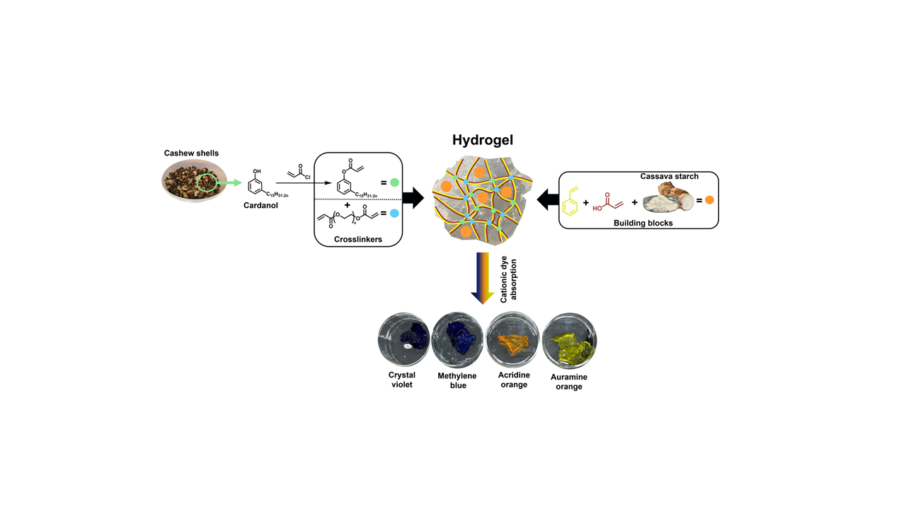
The utilization of naturally derived sustainable components is an increasing trend in polymer and materials science and technology. This paper reports on the synthesis of superabsorbent hydrogels (SAHs) containing acrylic acid (AA), styrene (St, 10 wt.% of AA) and poly(ethylene glycol diacrylate), PEGDA using one-pot radical suspension copolymerization. Cassava starch (CS, 1 wt.% of AA) was added to the copolymerization mixture as natural hydrophilic gel modifier. Cardanol acrylate (CA) was used as natural alternative to PEGDA and its effect on SAHs properties (gel content, swelling rate, swelling capacity, morphology, thermal stability, rheology and dye absorption) was investigated. The swelling capacity reached its maximum at 63,000% for the gel containing 50% of CA. CA also significantly increased SAH rigidity as revealed by rheology tests that confirmed the gel-like behavior of all networks, as well. The amphiphilic gels showed remarkable binding capacity for selected dyes: 47.95 mg/g for Crystal Violet, 46.37 mg/g for Acridine Orange, 42.50 mg/g for Methylene Blue and 23.48 mg/g for Auramine Orange, indicating a promising application potential for environmental cleanup of aqueous waste.
Keywords:
superabsorbent hydrogels
; cardanol acrylate
; cassava starch
; crosslinking copolymerization
; dye absorption
1. Introduction
Hydrogels are natural or synthetic networks composed of chemically or physically crosslinked polymers capable of swelling and retaining a large amount of water in their structure [1]. They represent a very important class of materials due to their usefulness in numerous applications including contact lenses [2], drug delivery [3,4,5,6,7], wound dressing [8], tissue engineering [9], supercapacitors [1], soil hydration [10], hygiene products [11], heavy metal removal [12], and dye absorption [13,14,15,16]. Superabsorbent hydrogels (SAH) were first introduced in 1978 in Japan from the crosslinking of starch and poly (acrylic acid) and were used as personal hygiene products for women [17]. They were further improved to be used as baby diapers in Germany and France in the 80s [17]. Most commercially available SAH are made from petroleum-based vinyl monomers, which are challenging to degrade and pose a significant environmental threat. Recently, the market has been transferring to more bio-based alternatives to chemicals derived from fossil sources [18].
Starch is an inexpensive and renewable material often used in biobased SAHs [19]. It could be derived from Cassava (Manihot esculenta), a shrub considered as a reliable source since it can grow in harsh natural conditions [20]. The plant contains very high amounts of starch which contribute to about 70% of its dry matter [20]. Cassava starch is an excellent candidate for hydrogel production owing to its higher solubility in water compared to other tuber starches and in distinction to cereal starches the gel has better stability [20].
Cardanol [21] is recently considered as a bio-based sustainable alternative to some typical crosslinkers for AA-based hydrogels such as N,N-methylene-bis-acrylamide (MBA) and poly (ethylene glycol) diacrylate (PEGDA). It is the by-product of the cashew industry, obtained as the major component of the cashew nut shell liquid (CNSL), a natural resin contained in the honeycomb-like shell of cashew nuts. Cardanol consists of a phenol substituted in the meta position with unsaturated C15 linear alkyl chains averaging two unsaturations per molecule [21]. Its use as a building block of naturally derived polymers is increasingly investigated [22,23,24]. It is also used in several applications including plasticizers [25,26,27], coating crosslinkers [28,29,30], and surfactants [31,32]. Cardanol acrylate (CA) is obtained by attaching acrylate moieties to the cardanol molecule. This modification allows cardanol to self-polymerize and form oligomers [33] or be utilized with other vinyl monomers. Hu et al. [34] used CA as reactive diluent in the synthesis of UV-curable polyurethane acrylate resins. They found that the addition of CA increased C=C conversion, and improved thermal stability, viscosity, shrinkage, and hardness among other properties [34].
An environmental concern that SAHs help tackle efficiently is dye removal from aqueous waste. Every year, about 800,000 tons of dyes are released in the environment [35]. They come essentially from effluents of many industries such as textile, pulp and paper, food and beverages, and the plastic industry [36]. These dyes cause not only aesthetic problems but are also serious threats to human and aquatic health [37]. They increase chemical demand in oxygen and decrease light penetration, which hinders the photosynthesis process under water [38]. These dyes also show carcinogenic effects. For example, Auramine Orange (AO), widely used in the textile and paper industry, is among dyes identified as carcinogenic by the International Agency for Cancer Research [39]. Crystal violet (CV) is also highly utilized in industry and biomedical studies [40,41] and is shown to have cytogenic, genetic, mutagenic and carcinogenic effects [42]. Methylene Blue (MB) is highly used in the textile, cosmetic [43], and paper industry [44]. It persists in water and doesn’t decompose under light.
Super absorbent hydrogels are an interesting alternative to many previously used materials for dye removal from effluent waters [45]. They are also the focus of this study, where SAHs were prepared using AA, styrene, and cassava starch, with CA used as a partial replacement for PEGDA. Acrylic acid imparts hydrophilicity and improves water absorption and swelling, while styrene introduces hydrophobic domains that reinforce the mechanical stability of the hydrogel structure and hydrophobic binding [46]. Cassava starch enhances the hydrogel’s environmental sustainability. Besides sustainability, CA brings unique binding attributes to the amphiphilic networks due to its aromatic rings and long alkyl chain. Emulsion polymerization was chosen as the synthesis method to ensure uniform incorporation of the hydrophilic and hydrophobic components. The aim of this study is to show how components such as Cassava starch and styrene as well as partial replacement of PEGDA by CA affect gel formation, morphology, thermal, swelling, and rheological properties of the hydrogels. Their dye binding capabilities are also explored.
2. Materials and Methods
2.1. Materials
Starch was extracted from Cassava tubers sourced from Côte d’Ivoire. Cardanol (Cardolite, Harrisburg, PA, United States) was used as received. Styrene (St, 99%), 2,2’-azobisisobutyronitrile (AIBN, 98%), and Acryloyl Chloride (99%) were supplied by Millipore Sigma (St. Louis, MO, USA) and were used without further purification. Triethylamine (TCI America, United States), chloroform (J. T. Baker, Phillipsburg, NJ, USA), methanol (Burdick and Jackson, Muskegon, MI, USA), acetone (J. T. Baker, Phillipsburg, NJ, USA), diethyl ether (J. T. Baker, Phillipsburg, NJ, USA), sodium dodecyl sulfate (SDS, Sigma Aldrich, Milwaukee, WI, USA), potassium persulfate (KPS, Beantown Chemical, Hudson, NH, USA), acetic acid (Fisher Chemical, Pittsburg, PA, USA), sodium acetate anhydrous (Fischer Scientific, Pittsburg, PA, USA), NaCl (Macron, Philipsburg, NJ, USA), NaOH (Macron, Philipsburg, NJ, USA ), Na2CO3 (Fisher Science Education, Nazareth, PA, USA), NaHCO3 (Fischer Science Education, Nazareth, PA, USA), PEGDA (Mn=250 Da, Aldrich, Milwaukee, WI, USA), HCl (EMD Millipore Corporation, Billerica, MA) were used as received. The following dyes (all from Aldrich, Milwaukee, WI, USA) were used in the binding experiments without further purification: Acridine Orange (AcrO, dye content ~90%), Auramine Orange (AO, dye content ~85%), Bromophenol Blue (BPB, dye content ~95%), Crystal Violet (CV, dye content ~98%), Methylene Blue (MB, dye content ~98%), and Methyl Orange (MO, dye content ~93%).
2.2. Methods
2.2.1. Synthesis of Cardanol Acrylate
Cardanol acrylate was synthesized according to the procedure shown in Scheme 1.
In a two-neck round bottom flask equipped with a reflux condenser, 30 g of cardanol (0.1 mol) were dissolved in 100 mL of acetone and 14.08 mL of triethylamine (10.12 g, 0.1 mol) were added as an acid neutralizer. The mixture was cooled in ice bath to 0 °C, 8.41 mL of acryloyl chloride (9.05 g, 0.1 mol) was cautiously added dropwise through the neck to the reaction mixture. The reaction was carried out for 16 h at 55 °C under reflux. The reaction mixture was added dropwise to 100 mL diethyl ether to precipitate the triethylamine hydrochloride, a salt formed by the reaction between triethylamine and the hydrochloric acid produced during the reaction. The mixture was then rinsed with sodium carbonate and DI water until neutral, vacuum filtered, dehydrated with sodium sulfate, and concentrated using a rotary evaporator. The product was a yellow-orange liquid. Yield (87.43%).
ATR-FTIR (cm-1): 3450 (w, residual OH stretch), 1746 (s, C=O stretch), 1635 (w, C=C stretch), 1401 (s, H-C=C bend), 1230 (s, C(=O)-O stretch), 1025 (m, O-C-C stretch), 983 (m, =C-H out-of-plane bend).
1H NMR (600 MHz, CDCl3): δ 7.29 (t, 1H, Ar-H), 7.14 (d, 1H, Ar-H) 7.07 (s, 1H, Ar-H); 6.61 (dd, J= 17.38 Hz, 1H, =CH2); 6.33 (dd, J=27.77 Hz, 1H, =CH-), 6.00 (dd, J= 10.46 Hz, =CH2), (0.8-5.8 ppm, Cardanol signals).
13C NMR (151 MHz, CDCl3): δ 164.76 (C=O), 150.69 (Ar-C), 144.78 (Ar-C), 132.46 (=CH-), 129.23 (=CH2), 128.19 (Ar-C), 121.47 (Ar-C), 118.77 (Ar-C).
2.2.2. Synthesis of Hydrogels
The emulsion polymerization was carried out using a one pot technique. The organic phase, which contained 8 mg AIBN (0.049 mmol), 0 or 1.1 mL styrene (co)monomer (1 g, 9.6 mmol), 3 mL toluene, and 0 - 8 mg of CA/PEGDA mixtures were first loaded in a pre-dried 50 mL test tube, capped with a rubber septum and purged with nitrogen. For the aqueous phase, 100 mg of starch was gelatinized in 5 mL DI water at 90 °C. 9.52 mL of acrylic acid (10 g, 138.8 mmol) was partially neutralized (70% neutralized) with NaOH (6 M, 16.2 mL). The starch solution and acrylic acid were mixed and sonicated at room temperature for 5 minutes. Then, SDS (80 mg, 0.406 mmol) were added, followed by addition of potassium persulfate (72 mg, 0.266 mmol), sonicated for 3 min and thorough mixing until full dissolution. This aqueous phase was then added with a syringe to the test tube containing the organic phase, immediately forming an emulsion. The mixtures in the sealed test tubes were vortexed for 1 minute each and heated in an oil bath at 60 °C. Polymerization occurs within 20 min. The products were washed with 100 mL methanol, followed by immersion in 3 chloroform portions (100 mL each) over three days to remove all uncrosslinked products – after 24 h the initial 100 mL chloroform portion was removed, and fresh chloroform was added. The chloroform fractions were concentrated using a rotavapor and used to quantify the amount of extractables and the gel content.
2.2.3. Fourier Transform Infrared Spectroscopy (FTIR)
FTIR analysis was performed in Attenuated Total Reflectance (ATR) mode using a Perkin Elmer Frontier instrument (PerkinElmer, Shelton, CT, USA). All samples were air dried and analyzed in the medium infrared region (4000 – 550 cm-1) at 32 scans. The transmittance spectra were converted to absorbance spectra with baseline correction, and normalization through the instrument software.
2.2.4. Nuclear Magnetic Resonance (NMR)
1H- and 13C NMR analyses were performed on a Bruker 600 MHz instrument (Bruker Corporation, Billerica, MA, USA) at 16 scans for 1H, and 256 scans for 13C in deuterated chloroform (CDCl3) and DMSO-d6 at room temperature. Chemical shifts were referenced to the residual peaks of CHCl3 or DMSO at 7.26 ppm and 2.5 ppm for 1H NMR and 77 ppm for 13C NMR using CDCl3.
2.2.5. Gel Content
The chloroform fractions at the end of the reaction were concentrated using a rotary evaporator and used to quantify the soluble polymer fraction and the gel fraction.
The soluble polymer fraction is obtained using the following formula:
where is the mass of the dry hydrogel after synthesis, is the mass of the hydrogel after the extraction of the soluble polymer fraction and subsequent drying.
The gel fraction, which is the complementary percentage, is given by the following equation:
2.2.6. Scanning Electron Microscopy
SEM imaging was performed using a JEOL IT 100 instrument (JEOL USA, Inc.; Peabody, MA, USA). Samples were sputter-coated with 22.5 nm Au/Pd to improve conductivity before imaging. Secondary electrons were detected in high vacuum at an acceleration voltage of 10 kV, an aperture size of 2, and a probe current of 50.
2.2.7. Swelling Rate
The swelling rate is the profile of the swelling capacity of a hydrogel against time [47,48]. To measure the swelling rate, approximately 100 mg of hydrogel samples were weighed and immersed in 100 mL of DI water. The samples were then removed from the water, gently wiped with a lint-free paper and the weight was recorded after 5, 10, 15, 30, 45, 60, 90, 120, 180, 300, 600 and 1440 min (24 h). The measurements were done in triplicates and averaged. The swelling capacity is given by equation (3).
Where is the swelling capacity, is the weight of the dried hydrogel, and is the weight of the swelled hydrogel. The swelling rate values are then fitted into a Voigt model [49] given by the equation (4).
Where is the swelling time, is the swelling capacity at time , is the equilibrium swelling capacity, and the rate parameter (time required to reach 0.63 of the equilibrium swelling) [50].
2.2.7. Maximum Swelling Capacity and Absorbance Under Load
Hydrogels samples of approximately 100 mg were immersed in 100 mL deionized water and swelled for 24 h. After swelling, hydrogels were wiped gently with a lint-free paper and weighed to determine the maximum swelling capacity. For the quantification of the absorbance under load (AUL), the swelled samples were loaded in a system consisting of a custom 3D-printed PLA apparatus composed of an outer and inner cylinder supported by a stainless-steel spacer to let the excess water drip, Figure 1. After hydrogel was loaded, a brass weight of 500 g was placed on top of the inner cylinder to allow the liquid to drip, the weight of the sample was recorded and AUL was calculated. Both swelling capacity, and AUL were all calculated using previously mentioned equation (3).
2.2.8. Salt and pH sensitivity
Salt sensitivity was measured in the same conditions as the maximum swelling capacity (100 mg hydrogel, 24 h) in different solutions. Hydrogels were swelled in a 0.9% NaCl solution for the determination of the salt sensitivity. For the pH sensitivity, buffer solutions of sodium acetate/acetic acid (pH=5.05) and sodium carbonate/bicarbonate (pH 9.17) were used. All measurements were done in triplicates and averaged.
2.2.9. UV-Vis Spectroscopy
UV-Vis analyses were performed on a Shimadzu UV-1900i instrument (Shimadzu Scientific Instruments, Columbia, MD, USA) in 1 cm quartz cuvettes. The measurements were conducted with aqueous dye solutions at room temperature.
2.2.10. Dye Binding
Dry pieces of the PEGDA50% / CA50% hydrogel weighing about 10 mg each were immersed in 50 mL dye solutions with concentration 10 mg/L. The UV-Vis spectra of the solutions were recorded between 800-350 nm before and after immersion of the hydrogel for 24 h. The concentrations of the solutions were calculated using the Bouguer-Lambert-Beer equation (Beer’s law) using the molar extinction coefficients of the dyes used in the binding experiments. They were established by UV-Vis spectroscopy of aqueous solutions with different concentrations (R> 0.99, Figure S1). Using the absorbance values from the UV-Vis measurements, Figure S2, the equilibrium dye uptake was calculated by the equation (5).
where and are the molar concentrations of the dye solutions before and after absorption by the hydrogel respectively. is the molar mass of the dye, is the mass of the dry hydrogel used, and is the volume of the dye solution.
2.2.11. Thermogravimetric Analysis
TGA measurements were carried out using a high resolution TGA instrument Discovery 550 (TA Instruments, New Castle, DE, USA). Approximately 10 mg of sample was loaded into a platinum pan and heated from room temperature to 600 °C. Onset and endset temperatures as well as maximum decomposition temperatures were obtained from the TGA and the derivative thermogravimetry (DTG) curves.
2.2.12. Differential Scanning Calorimetry (DSC)
DSC analysis was carried out using a calorimeter TA Instruments Discovery 250 (TA Instruments, New Castle, DE, USA). The sample and the reference pan were heated from 20 °C to 150 °C at a rate of 10 °C/ min, kept at an isotherm for 5 min, then cooled down to 0 °C at a rate of 10 °C/min, kept at an isotherm for 5 min, and finally heated again to 150 °C at a rate of 10 °C/min.
2.2.13. Rheology
Rheological properties were measured using a Discovery HR-3 rheometer (TA instruments, New Castle, DE, USA) with temperature-controlled lower Peltier plate geometry 20 mm. Hydrogels of diameter 20 mm and thickness 5000 ± 300 μm were cut after chloroform extraction and used for analysis. Prior to oscillatory frequency sweep experiments, oscillatory strain sweep measurements were first conducted to determine the linear viscoelastic region of the hydrogel networks. The frequency sweep experiments were performed on the hydrogels at 25 °C in the range 0.01–250 rad/s. Experiments were carried out in triplicates, and the average value was reported.
3. Results and Discussions
3.1. Synthesis of Cardanol Acrylate
Cardanol acrylate (CA) was successfully synthesized via esterification of the cardanol phenolic hydroxyl group with acryloyl chloride in acetone under anhydrous conditions, using triethylamine as a stoichiometric organic base, producing a yellow-orange liquid in 87.4% yield. In this reaction, triethylamine deprotonates the phenol to generate a more reactive phenoxide nucleophile while simultaneously scavenging the HCl formed during the acyl substitution as triethylammonium chloride, preventing acid-catalyzed side reactions and driving the equilibrium toward full conversion [34]. The use of an ice-bath during acryloyl chloride addition, followed by reflux at 55 °C for 16 h, ensured controlled exotherm management during the exothermic acylation step and complete consumption of the phenolic hydroxyl group, as confirmed by the near-total disappearance of the phenolic O–H stretch at ~3350 cm−1 in the FTIR spectrum (Section 3.1.1) and by the disappearance of the phenolic proton in the 1H NMR spectrum (Section 3.1.2). The purification protocol involving precipitation of triethylammonium chloride in diethyl ether, followed by alkaline washing with Na2CO3 and drying over Na2SO4 effectively removed ionic byproducts and residual acidity, as evidenced by the absence of any ammonium salt signals in the 1H NMR spectrum of the purified product. Importantly, no radical inhibitor was required during the reaction, as the moderate reflux temperature (55 °C) under inert atmosphere was insufficient to trigger premature self-polymerization of the acrylate moiety. The dual reactive character of CA, bearing both an acrylate group available for free-radical copolymerization and the residual unsaturations of the C15 alkyl side chain capable of secondary crosslinking, makes it a particularly versatile bio-based crosslinker candidate, as discussed extensively by Caillol et al. [21,22,24]. These results are further confirmed by the following detailed FTIR and NMR characterizations.
3.1.1. FTIR of Cardanol Acrylate
Figure 2 presents the transmittance spectra of cardanol (black) and cardanol acrylate (orange). The broad peak at 3350 cm−1, corresponding to intermolecular hydrogen bonds of the phenolic proton in cardanol, nearly disappears after the reaction indicating successful substitution by the acrylate group. The strong peak at 1746 cm−1 in the cardanol acrylate spectrum corresponds to the acrylate carbonyl bond. The small bump at 1635 cm−1 is caused by the stretching vibration of the C=C bond in the acrylate group. The strong peak at 1401 cm−1 is due to the bending vibration of the H-C=C bond in the acrylate group. The peak at 1230 cm−1 suggests the presence of C(=O)-O bonds.
The ester O-C-C bond exhibits a strong signal at 1025 cm−1. The peak at 983 cm−1 indicates the presence of an out-of-plane bending vibration in the =C-H bond. These results are consistent with those reported by Hu et al.[34].
3.1.2. 1H NMR of Cardanol Acrylate
Figure 3 shows the proton NMR spectra of cardanol and cardanol acrylate (CA). Differences are observed in the regions corresponding to the acrylate double bond and the aromatic ring. The phenolic proton 1 disappears due to substitution by the acrylate group.
The singlet and doublet at 6.65 ppm representing protons 2, the doublet at 6.75 ppm corresponding to proton 4, and the triplet at 7.14 ppm representing proton 3 in the cardanol structure are all shifted downfield, appearing at 6.96 ppm (protons 2’), 7.07 ppm (4’), and 7.29 ppm (3’). These shifts are attributed to the inductive effect of the additional oxygen atom from the acrylate group. Acrylate double bond protons are identified based on coupling constant differences, proton 5 (trans to 7, and cis to 6) exhibits the highest coupling constant (J = 27.77 Hz) and appears as a doublet of doublets centered at 6.33 ppm. Proton 6 (geminal to 7 and cis to 5) has the smallest coupling constant (J = 10.46 Hz) and appears at 6.00 ppm, while proton 7 (geminal to 6 and trans to 5) has a moderate coupling constant (J = 17.38 Hz) and appears at 6.61 ppm. These results are in accordance with those reported by Hu et al. [34].
3.1.3. 13C NMR of Cardanol Acrylate
Figure 4 shows the carbon NMR spectrum of cardanol (blue) and cardanol acrylate (red). In the cardanol spectrum, saturated carbon signals appear between 14.24 ppm and 35.95 ppm and remain unaffected by acrylate substitution.
Unsaturated aliphatic and aromatic ring carbons appear between 112.61 and 155.55 ppm. Upon substitution of the phenolic proton with the acrylate group, new peaks emerge between 112.58 and 164.76 ppm in the cardanol acrylate spectrum. Notable changes include three new peaks from acrylate carbons (g, h, i) at 164.76 ppm, 132.46 ppm, and 129.23 ppm, respectively. The aromatic ring carbons shift: a from 155.55 ppm to 150.69 ppm (a’), b from 115.43 ppm to 118.77 ppm (b’), c from 112.61 ppm to 121.47 ppm (c’), d from 145.08 ppm to 144.78 ppm (d’), e from 129.52 ppm to 128.19 ppm (e’), and f from 121.13 ppm to 126.09 ppm (f’). These results also align with those previously reported by Hu et al. [34].
3.2. Synthesis of Hydrogels
Superabsorbent hydrogels based on partially neutralized acrylic acid (70%), styrene, and cassava starch were successfully prepared via oil-in-water suspension polymerization using a dual initiator system (AIBN in the organic phase, KPS in the aqueous phase) and SDS as surfactant.
All formulations yielded stable, coherent gel materials in yields ranging from 88.5% to 96.8% (Table 1), confirming the efficiency of emulsion polymerization as a method for incorporating both hydrophilic and hydrophobic components into a single crosslinked architecture.
The use of an oil-in-water emulsion approach allowed the simultaneous polymerization of the water-soluble acrylic acid phase and the toluene-dissolved styrene/crosslinker phase, producing a phase-segregated but covalently interconnected amphiphilic gel, consistent with the semi-interpenetrating network (semi-IPN) concept described in recent literature [51,52]. The system is referred to as a semi-IPN since starch is not independently crosslinked. However, limited grafting of acrylic acid onto starch backbone chains via KPS-mediated hydrogen abstraction cannot be excluded under these conditions, consistent with observations reported by Ismail et al. [52]. The partial neutralization of acrylic acid to 70% with NaOH prior to polymerization is a well-established strategy in SAH synthesis that balances electrostatic repulsion within the growing network and promotes increased swelling while preventing excessive chain mobility that would otherwise reduce gel content [52,53]. Gelatinization of cassava starch at 90 °C before its incorporation facilitated the complete disruption of crystalline amylopectin domains and ensured homogeneous dispersion in the aqueous phase, which is essential to avoid phase separation during polymerization and to enable physical entrapment within the crosslinked network [54]. The gel content of all formulations exceeded 98% (Table 1), demonstrating a quantitative incorporation of both comonomers and crosslinkers into the insoluble/swellable network, irrespective of the PEGDA/CA ratio used. Furthermore, the hydrogels with higher content of CA (75% and 100%) showed nearly total crosslinking, which demonstrates the efficiency of CA as a standalone crosslinker. The 1H NMR analysis of methanol and dichloromethane (DCM) extracts, Figure 5 and Figure 6, confirmed that the extractable fractions consisted only of acrylic acid oligomers, residual SDS, and linear polystyrene, with no detectable crosslinker signal, indicating complete incorporation of both PEGDA and CA into the network. The formation of a stable emulsion within seconds of combining the organic and aqueous phases, followed by onset of gelation within 20 minutes at 60 °C, is consistent with the previously reported rapid kinetics of persulfate-initiated radical polymerization of acrylic acid at moderately elevated temperatures. [52] and with the accelerated polymerization behavior in emulsion systems described by Kovačič and Silverstein [51]. These results collectively confirm the successful formation of well-defined, highly crosslinked semi-IPN systems. The NMR analyses of the extracts gave further evidence of the incorporation of each component.
3.2.1. 1H NMR of Methanol Extracts
Figure 5 shows the proton NMR spectra of methanol extracts, which were obtained after the first 24-h treatment by soaking the hydrogels formed.
The spectra show signals of acrylic acid (+) and SDS (*) as confirmed by signal overlaps in Supporting Information 1H NMR spectra (Figures S3 and S4). This suggests that only unreacted acrylic acid and SDS are released in the methanolic fractions. A further investigation is carried out after three days of DCM extraction.
3.2.2. 1H NMR of DCM Extracts
Figure 6 shows the DCM extracts which were obtained after three additional days of immersion of the hydrogels in fresh DCM portions. These extracts are composed of, acrylic acid (+) (Figure S3), SDS (*) (Figure S4) and linear polystyrene (▫) at 6.3 – 7.2 ppm and 1 – 2.5 ppm (Figure S5).
The samples without starch did not release any non-crosslinked poly(styrene), which indicates that the absence of starch may enhance styrene incorporation into the network. In contrast, the absence of styrene shows prominent unreacted acrylic acid peaks, which might be interpreted as an enhanced styrene/acrylic acid copolymerization tendency. No crosslinker signal (PEGDA or CA) was identified in the spectra, confirming the full incorporation of the crosslinkers in the hydrogel networks.
3.3. Gel Content
Table 1 shows the gel yield, the soluble polymer fraction (), and the gel content. All formulations showed a high gel content between 98 and 100%. The gel content remains very high 99.41% - 99.76% for the first three hydrogels crosslinked with PEGDA only (w/o starch, w/o styrene, PEGDA 100%). It then decreases slightly to ~98.5% and ~98.4% for the hydrogels crosslinked with mixtures of CA and PEGDA (CA 25% and CA 50%) with further increases to nearly 100% when the CA content increased (CA 75% and CA 100%). The decreasing gel content at 25% and 50% of CA in the mixture could be attributed to the steric effects and differences in reactivities between the two crosslinkers. The CA structure is composed of a phenyl substituted acrylate group with alkenes in its long C15 side chain (Scheme 1). The phenyl group has a resonance effect that can enhance the reactivity of CA compared to PEGDA. And this mismatch in reactivity together with the steric differences could cause a minor loss in crosslinking efficiency at certain ratios.
3.4. Scanning Electron Microscopy
SEM images of the hydrogels are shown in Figure 7, revealing their morphology. All hydrogels exhibit a homogeneous surface and highly porous inner structure. Images at higher magnification (500×) are provided in the Supporting Information, Figure S6. It was seen that macropores with diameters of several hundred microns were randomly distributed in the hydrogel. Conversely, micropores of ≤10 microns were uniformly distributed in the networks, Figure 7. The pore formation mechanism can be attributed to two parallel processes. The formation of macropores as seen in Figure 7 and Figures 7 and S6, can be caused by gas evolution during the AIBN decomposition at the initiation stage [55]. Pore sizes of this origin can vary between 1 and 3000 μm [56]. The micropores formation, however, could be attributed to emulsion templating. In this process droplets of the dispersed phase act as templates during copolymerization [53]. The (co)monomers (co)polymerize around these droplets, forming a crosslinked network. After that, the droplets are removed by solvent wash and drying, leaving behind voids (pores) corresponding to the droplet size with diameters ranging from 1 to 30 μm [53]. Previous studies [52] reported similar porous structures in hydrogels synthesized via emulsion polymerization of acrylic acid, poly(styrene), and starch reinforced with bentonite.
3.5. Swelling Rate
One of the most important properties of superabsorbent polymers is their swelling rate, which shows how quickly the hydrogel can absorb and retain water in its structure [56]. To compare swelling rates the values are fitted to a Voigt model [49,50], and the rate parameter, which is the value at which the swelling ratio reaches 63% of the hydrogel’s maximum swelling capacity, is used as a baseline for the comparison of the swelling rates. Figure 8 (a) shows the swelling profile of all the hydrogels. Figure 8 (b-h) show the Voigt model describes well the hydrogels swelling behavior All hydrogels show a strong correlation with R2> 0.93, meaning that 93% of the values can be fitted in the model.
It was observed that the equilibrium swelling capacity is smaller for the hydrogels without starch and without styrene. The other networks produced with a mixture of crosslinkers (CA 25%, CA 50%, and CA 75%) have higher equilibrium swelling capacity values than the hydrogels crosslinked with pure PEGDA and CA (Table S1). The rate parameter () values (Table S1) are lined up in the following increasing order: w/o styrene < w/o starch < PEGDA 100% < CA 50% < CA 100% < CA 75% < CA 25%. The rate parameters show that the absence of styrene and starch causes hydrogel to reach swell equilibrium faster. This could be due to the relative hydrophobicity of both materials compared to acrylic acid. The other hydrogels show no significant differences.
3.6. Maximum Swelling Capacity and Absorbance Under Load (AUL)
Figure 9 shows the maximum swelling capacity (blue bars) and the absorbance under load (orange bars). Swelling capacity was measured as the amount of fluid retained in the hydrogels structure after 24 h. The swelling capacity increases from 53,000% for PEGDA 100% to 63,000% for CA 50%. It then decreases back to 55,000% for CA 100%. It is well known that a higher crosslinking density is negatively correlated to the swelling capacity [57]. This means that differences in reactivity between CA and PEGDA would promote a looser network for mixtures of crosslinkers, thus enhancing their swelling capacity. The hydrogels without starch and styrene show notably lower swelling capacities (i.e., higher crosslinking density) compared to the other hydrogels. Most probably, starch being inactive in radical polymerization acts as an inert spacer between the radically formed crosslinks. In addition, styrene could be involved in several reaction pathways, none of them producing crosslinks: homopolymerization, copolymerization with acrylic acid or copolymerization with CA forming linear polymers or copolymers that would increase the intrinsic hydrophobicity of the semi-IPNs with concomitant reduction of their swelling capacity. Absorbance under Load (AUL) measures the hydrogel’s ability to retain absorbed fluid under applied pressure. AUL is considered a measure of the mechanical strength of a hydrogel, since logically stronger hydrogels can withstand more pressure [58]. In this study, when 500 g of load (339.5 N/m2) is applied to the hydrogels, load-free swelling capacity decreases only slightly across the entire line of networks with the loss in swelling between 5 and 11% of the original swelling capacity of the hydrogels. This shows that all hydrogels are mechanically strong regardless of their degree of crosslinking.
3.7. Salt and pH Sensitivity of Hydrogels
All hydrogels showed similar responses to changes in the medium – saline, basic or acidic. In all cases a loss in swelling capacity was observed: ~87.7% in saline environment, ~83.9% at pH 5 and ~79.9% at pH 9, Figure 9 and Figure 10. These changes can be attributed to the balance of electrostatic interactions, osmotic pressure, and ion-exchange dynamics within the amphiphilic gels [59,60]. When the polymer network absorbs pure water, a fraction of its acidic functional groups most probably undergoes partial dissociation, releasing hydrogen ions (H+) into the interior solvent while generating conjugate anions (COO−) that remain covalently bound to the network [61]. The partial neutralization of the hydrogel (70%) also introduces Na+ counterions that further enhance swelling. When the surrounding solution consists of water molecules and different ions the networks behave differently showing that the swelling behavior of the gel is highly dependent on the ionic composition of the external solution.
Saline solution contains Na+ and Cl− ions, the acidic medium (pH 5) has an excess of H+ and negative counterions, while the basic solution (pH 9) has an excess of HO− and positive counterions. Diffusion of these charged species in the interior of the networks leads to changes in the ionic equilibrium between the fixed negative charges in the hydrogel and the positive charges in the solution, known as Donnan equilibrium [49]. The resulting partial neutralization of charges leads to reduced swelling, which is clearly seen comparing the data in Figure 9 and Figure 10.
3.8. Thermogravimetric Analysis
TGA experiments revealed a slight weight loss between 100 and 150 °C probably due to the removal of molecularly bound residual water [63] and two major degradation onsets for all hydrogels, Figure 11.
They occur in the range of 301-319 °C (5-11% weight loss) and 400-405 °C (31-36% weight loss)., Figure S7. During the first degradation stage the decomposition of uncrosslinked starch most probably occurred (the starch used in this study thermally breaks down at ~318 °C, Figure S8). However, it represents only 1% relative to the mass of acrylic acid contained in the semi-IPN and could not cause the entire percentage loss observed in this temperature interval. Acrylic acid and its derivatives (homopolymers, copolymers and crosslinking segments) are the major components of all hydrogels under this investigation, and they are probably the major causes for the two major decompositions, Figure 11. This assumption is supported by previously published data [64]. The lack of styrene in one of the hydrogels doesn’t affect markedly the decomposition profile compared with the PEGDA 100% hydrogel, Figure 11. Poly(styrene) has a maximum temperature of decomposition positioned between 390 and 420 °C [65] coinciding with the temperature of the second decomposition event in Figure 11. Due to the small styrene:acrylic acid ratio (1:10) it may not be sufficiently high to significantly affect the thermogram.
3.9. Differential Scanning Calorimetry
Figure 12shows DSC thermograms of the PEGDA/CA-crosslinked hydrogels with no observable thermal transitions between 0 and 150 °C, an absence that could be attributed to the highly crosslinked nature of the networks.
Tg of poly(styrene), poly(acrylic acid) and their copolymers are at or below 126 °C [67]. Crosslinking of the copolymers generally increases Tg, but it generally remains below 150 °C [68]. The absence of a detectable glass transition suggests a highly restricted segmental motion in the interpenetrating (co)polymer chains and crosslinked segments.
3.10. Rheology
The effect of CA addition on the viscoelastic properties (storage modulus and loss modulus) of the PEGDA/CA hydrogels was investigated through rheological measurements, Figure 13. Hydrogels do not respond instantaneously to an applied force; instead, their response depends on time [69,70].
This means that when a force is applied, part of the energy is stored reversibly, this is the elastic component known as storage modulus [71]. Some of this energy is dissipated as heat, this is the viscous component known as loss modulus [71]. A preliminary strain sweep is necessary to identify the linear viscoelastic region at which frequency sweep measurements should be conducted [72]. This region was identified at 0.5% strain (Figure S9). For all hydrogels, the storage modulus is parallel and higher to the loss modulus. This is the sign of a predominantly elastic gel behavior [72]. The storage modulus of most hydrogels that contain CA is significantly higher than that of the pure PEGDA hydrogel, revealing that the addition of cardanol increases the rigidity of the hydrogel. This increase in rigidity could be attributed to several factors – the benzene ring in CA and the multiple double bonds in its side chain that might increase crosslinking density. The loss modulus quantifies the viscous response, measuring the energy dissipated as heat during deformation [73]. The results show a similar trend, with loss modulus values increasing with CA content compared to PEGDA 100% gel. It would be reasonable to assume that the increased presence of long CA chains positively affects the fluid-like characteristics of the networks containing this compound.
3.11. Dye Binding
One advantage of hydrogels over traditional adsorbents, such as activated carbon, is their swelling capacity, which increases surface contact and enhances dye adsorption efficiency. The CA 50% hydrogel was used for all dye absorption measurements because of its highest swelling capacity and AUL. Figure 14 illustrates dye absorption results, expressed as both percentages and mg of dye per mg of gel.
The differently charged dyes can interact with the hydrogel through electrostatic attraction/repulsion with the acrylate groups/anions and π-π interactions with the aromatic rings from styrene and CA. The difference in adsorption behavior can be explained by the differences in dyes structures and their charges (Figure 15). Cationic dyes (Acridine Orange, Auramine Orange, Crystal Violet, and Methylene Blue) exhibited substantial absorption ranging from 23.48 mg/g (AO) to 42.50 mg/g (MB), 46.37 mg/g (AcrO) and 47.95 mg/g (CV). The CV binding capacity of the CA 50% SAH places this material in the middle range of other hydrogels containing naturally derived sustainable components – it is superior to some (28 mg/g [74] and 35 mg/g [75]) and inferior to others (~67 mg/g [13]). The significantly lower absorption of Auramine Orange is due to its relatively good solubility in water (10 mg/mL, [76]) thus shifting the distribution equilibrium towards the liquid phase. Nonetheless the observed binding is still significantly higher than the values achieved in previously published studies (9.225 mg/g [77]). The enhanced absorption of cationic dyes could be attributed to favorable interactions between carboxylate groups in the hydrogels and imine groups from cationic dyes in combination with π-π interactions with the aromatic rings in styrene and CA (co)polymers, Figure 15.
Anionic dyes (Methyl Orange, Bromophenol Blue), in contrast showed no adsorption. In fact, they even showed artificially negative values (Table S2). However, visual inspection of the hydrogels confirms that some amount of these dyes was absorbed by the hydrogels (MO and BpB, Figure 15). This seemingly puzzling behavior could be explained by the specifics of the binding procedure used. The gels were introduced in the aqueous dye solution in dry form. Upon contact with the water the gels began to swell selectively absorbing water molecules. Some of the dye molecules are also absorbed, but most of them stay outside the crosslinked structures. Consequently, their concentration in the water outside the networks increases, causing the intensity of the absorption maxima to increase thus falsely reporting the presence of more dye than initially dissolved.
4. Conclusions
In this study, SAHs based on acrylic acid, styrene, PEGDA and cassava starch were efficiently synthesized with high yields. Infrared and NMR spectroscopy confirmed the successful synthesis of CA, which was used as additional semi-natural modifier and potential crosslinker. Crosslinking was achieved with different ratios of CA to PEGDA producing networks with different degrees of crosslinking density. The synthesized materials were extensively characterized by different analytical techniques. NMR spectra of methanol and DCM extracts showed no crosslinker leakage, suggesting a good incorporation of all components into the network. SEM micrographs showed a highly porous structure for all hydrogels with micropores ranging from 1- 30 μm created by emulsion templating, and large macropores which formed by nitrogen gas evolution during AIBN decomposition. The swelling capacity of the networks was within the typical SAHs range reaching a maximum of 63,000%. AUL measurement demonstrated a high resistance to stress of all gels. The hydrogels displayed high sensitivity to the salinity and the pH of the aqueous media. TGA tests showed that the SAHs decomposition pattern is mostly affected by the thermal stability of the acrylic components. DSC analyses could not detect an observable glass transition within the 0-150 °C temperature interval, a sign for the rigid nature of the hydrogels. Rheology analysis showed a typical gel behavior with CA-containing hydrogels showing higher rigidity. Finally, dye binding experiments demonstrated that SAHs have good binding capacity towards cationic dyes ranging from 48 to 23 mg/g in the following order CV > AcrO > MB > AO.
Overall, this study demonstrated that acrylic acid, styrene, and cassava starch could be successfully incorporated in stable SAHs by one-pot emulsion polymerization and either PEGDA or CA, a naturally-based crosslinker. The process leads to the formation of more sustainable SAHs with enhanced structural stability and swelling capacity, that can be conveniently applied to the environmental cleanup of industrial waste effluents.
Supplementary Materials
The following supporting information can be downloaded at the website of this paper posted on Preprints.org. Figure S1. Concentration calibration curves of tested dyes derived from UV-Vis spectra. Figure S2. Dye binding experiments, UV-Vis Spectra. Figure S3. 1H NMR of acrylic acid and poly(acrylic acid) vs. MeOH extract of CA75% hydrogel. Figure S4. 1H NMR of sodium dodecyl sulfate vs. MeOH extracts of CA75% hydrogel. Figure S5. 1H NMR of linear poly(styrene) vs. DCM extracts of CA 100% and PEGDA 100%. Figure S6. SEM micrographs of hydrogels at 500x magnification with (a) w/o starch, (b) w/o styrene, (c) PEGDA 100%, (d) CA 25%, (e) CA 50%, (f) CA 75% and (g) CA 100%. Table S1. Swelling rate parameters (r) and equilibrium swelling capacity (Se) computed by Voigt’s model. Figure S7. TGA of hydrogels. First and second onset temperatures and residue at 600 °C. Figure S8. TGA and DTG thermograms of cassava starch and standard amylopectin. Figure S9. Strain sweep of PEGDA 100% hydrogel. Maximum deformation was obtained for 0.5% oscillation strain, representing the linear viscoelastic region. Table S2. Calculated absorption values for dye binding experiment. Values in red are artificially negative due to dry hydrogel swelling and selective water absorption.
Author Contributions
Conceptualization, F.A. and M.A.; methodology, M.A. and I.G.; validation D.B.; formal analysis, M.A.; investigation, M.A., D.F., B.O. and D.T.; resources, I.G.; writing—original draft preparation, M.A.; writing—review and editing, D.B. and I.G.; visualization, S.P.; supervision, I.G.; project administration, I.G.; funding acquisition, D.B, F.A. and I.G. All authors have read and agreed to the published version of the manuscript.
Funding
This research was funded in part by the Programme d′Appui Stratégique à la Recherche Scientifique (PASRES), a fund from the Centre Suisse de Recherche Scientifique (CSRS) and the Ivorian Minister of Higher Education, Abidjan (Ivory Coast), grant number 191. Partial support through funds allocated by BioInspired Institute at Syracuse University (Grant 85709-1168699-1) is acknowledged with thanks.
Data Availability Statement
All results obtained in this study are included in the paper and in the supporting information. Any questions or requests can be referred to the corresponding author.
Conflicts of Interest
The authors declare no conflicts of interest.
Abbreviations
The following abbreviations are used in this manuscript:
| AA | Acrylic acid |
| AcrO | Acridine orange |
| AIBN | 2,2’-azobisisobutyronitrile |
| AO | Auramine orange |
| BpB | Bromophenol blue |
| CA | Cardanol acrylate |
| CS | Cassava starch |
| CV | Crystal violet |
| MB | Methylene blue |
| MO | Methyl orange |
| PEGDA | Polyethylene glycol diacrylate |
| SAH | Superabsorbent hydrogel |
References
- Varaprasad, K.; Raghavendra, G.M.; Jayaramudu, T.; Yallapu, M.M.; Sadiku, R. A mini review on hydrogels classification and recent developments in miscellaneous applications. Mater. Sci. Eng. C 2017, 79, 958–971. [Google Scholar] [CrossRef] [PubMed]
- Wichterle, O.; Lim, D. Hydrophilic Gels for Biological Use. Nature 1960, 185, 117–118. [Google Scholar] [CrossRef]
- Hoare, T.R.; Kohane, D.S. Hydrogels in drug delivery: Progress and challenges. Polymer 2008, 49, 1993–2007. [Google Scholar] [CrossRef]
- Gupta, A.; Lee, J.; Ghosh, T.; Nguyen, V.Q.; Dey, A.; Yoon, B.; Um, W.; Park, J.H. Polymeric Hydrogels for Controlled Drug Delivery to Treat Arthritis. Pharmaceutics 2022, 14, 540. [Google Scholar] [CrossRef] [PubMed]
- Caló, E.; Khutoryanskiy, V.V. Biomedical applications of hydrogels: A review of patents and commercial products. Eur. Polym. J. 2015, 65, 252–267. [Google Scholar] [CrossRef]
- Gupta, P.; Vermani, K.; Garg, S. Hydrogels: From controlled release to pH-responsive drug delivery. Drug Discov. Today 2002, 7, 569–579. [Google Scholar] [CrossRef] [PubMed]
- Abdel Ghaffar, A.M.; Radwan, R.R.; Ali, H.E. Radiation synthesis of poly(starch/acrylic acid) pH sensitive hydrogel for rutin controlled release. Int. J. Biol. Macromol. 2016, 92, 957–964. [Google Scholar] [CrossRef] [PubMed]
- Liang, Y.; He, J.; Guo, B. Functional Hydrogels as Wound Dressing to Enhance Wound Healing. ACS Nano 2021, 15, 12687–12722. [Google Scholar] [CrossRef] [PubMed]
- Chyzy, A.; Plonska-Brzezinska, M.E. Hydrogel Properties and Their Impact on Regenerative Medicine and Tissue Engineering. Molecules 2020, 25, 5795. [Google Scholar] [CrossRef] [PubMed]
- Abedi-Koupai, J.; Sohrab, F.; Swarbrick, G. Evaluation of hydrogel application on soil water retention characteristics. J. Plant Nutr. 2008, 31, 317–331. [Google Scholar] [CrossRef]
- Mistry, P.A.; Konar, M.N.; Latha, S.; Chadha, U.; Bhardwaj, P.; Eticha, T.K. Chitosan superabsorbent biopolymers in sanitary and hygiene applications. Int. J. Polym. Sci. 2023, 1, 4717905. [Google Scholar] [CrossRef]
- Abdel-Halim, E.S.; Al-Deyab, S.S. Preparation of poly (acrylic acid)/starch hydrogel and its application for cadmium ion removal from aqueous solutions. React. Funct. Polym. 2014, 75, 1–8. [Google Scholar] [CrossRef]
- Getya, D.; Lucas, A.; Gitsov, I. Composite Hydrogels Based on Poly(Ethylene Glycol) and Cellulose Macromonomers as Fortified Materials for Environmental Cleanup and Clean Water Safeguarding. Int. J. Mol. Sci. 2023, 24, 7558. [Google Scholar] [CrossRef] [PubMed]
- Salunkhe, B.; Schuman, T.P. Super-Adsorbent Hydrogels for Removal of Methylene Blue from Aqueous Solution: Dye Adsorption Isotherms, Kinetics, and Thermodynamic Properties. Macromol 2021, 1, 256–275. [Google Scholar] [CrossRef]
- Granados-Carrera, C.M.; Perez-Puyana, V.M.; Jiménez-Rosado, M.; Romero, A. Hydrogel Development, Processing and Applications in Agriculture: A Review. Gels 2026, 12, 259. [Google Scholar] [CrossRef] [PubMed]
- Paulino, A.T.; Guilherme, M.R.; Reis, A.V.; Campese, G.M.; Muniz, E.C.; Nozaki, J. Removal of methylene blue dye from an aqueous media using superabsorbent hydrogel supported on modified polysaccharide. J. Coll. Interf. Sci. 2006, 301, 55–62. [Google Scholar] [CrossRef] [PubMed]
- Masuda, F. Trends in the development of superabsorbent polymers for diapers. In Superabsorbent Polymers. Science and Technology; Buchholz, F.E., Peppas, N.A., Eds.; American Chemical Society: Washington, DC, USA, 1994; Vol. 573, pp. 88–98. [Google Scholar] [CrossRef]
- Omidian, H.; Akhzarmehr, A.; Chowdhury, S.D. Advancements in Cellulose-Based Superabsorbent Hydrogels: Sustainable Solutions across Industries. Gels 2024, 10, 174. [Google Scholar] [CrossRef] [PubMed]
- Supare, K.; Mahanwar, P.A. Starch-derived superabsorbent polymers in agriculture applications: an overview. Polym. Bull. 2022, 79, 5795–5824. [Google Scholar] [CrossRef]
- Chisenga, S.M.; Workneh, T.S.; Bultosa, G.; Alimi, B.A. Progress in research and applications of cassava flour and starch: a review. J. Food. Sci. Technol. 2019, 56, 2799–2813. [Google Scholar] [CrossRef] [PubMed]
- Caillol, S. Cardanol: A promising building block for biobased polymers and additives. Curr. Op. Green Sust. Chem. 2018, 14, 26–32. [Google Scholar] [CrossRef]
- Jaillet, F.; Darroman, E.; Ratsimihety, A.; Boutevin, B.; Caillol, S. Synthesis of cardanol oil building blocks for polymer synthesis. Green Mater. 2015, 3, 59–70. [Google Scholar] [CrossRef]
- Jia, P.; Song, F.; Li, Q.; Xia, H.; Li, M.; Shu, X.; Zhou, Y. Recent Development of Cardanol Based Polymer Materials-A Review. J. Renew. Mater. 2019, 7, 601–619. [Google Scholar] [CrossRef]
- Voirin, C.; Caillol, S.; Sadavarte, N.V.; Tawade, B.V.; Boutevin, B.; Wadgaonkar, P.P. Functionalization of cardanol: towards biobased polymers and additives. Polym. Chem. 2014, 5, 3142–3162. [Google Scholar] [CrossRef]
- Adjoumane, M.M.; Edja, A.F.; Doe-Mensah, J.; Boa, D.; Driscoll, M. Effect of cardanol-based plasticizers on thermoplastic cassava (Manihot esculenta) starch properties. Ind. Crops Prod. 2023, 203, 117145. [Google Scholar] [CrossRef]
- Greco, A.; Brunetti, D.; Renna, G.; Mele, G.; Maffezzoli, A. Plasticizer for poly (vinyl chloride) from cardanol as a renewable resource material. Polym. Degr. Stab. 2010, 95, 2169–2174. [Google Scholar] [CrossRef]
- Greco, A.; Ferrari, F.; Maffezzoli, A. Effect of the epoxidation yield of a cardanol derivative on the plasticization and durability of soft PVC. Polym. Degr. Stab. 2016, 134, 220–226. [Google Scholar] [CrossRef]
- John, G.; Pillai, C. K. S. Synthesis and characterization of a self-crosslinkable polymer from cardanol: Autooxidation of poly (cardanyl acrylate) to crosslinked film. J. Polym. Sci. Part A Polym. Chem. 1993, 31, 1069–1073. [Google Scholar] [CrossRef]
- Lee, D.H.; Ahn, H.J.; Lee, J.; Woo, H.C. Biodegradable PVA–Alginate Bio-Based Polymers Incorporating Cardanol-Based Polyols for Antibacterial Applications. Polymers 2025, 17, 2792. [Google Scholar] [CrossRef] [PubMed]
- Kinaci, E.; La Scala, J.J.; Can, E.; Palmese, G.R. Preparation and characterization of cardanol based vinyl ester resins as cross-linker units. J. Appl. Polym. Sci. 2024, 141, e56129. [Google Scholar] [CrossRef]
- Faye, I.; Besse, V.; David, G.; Caillol, S. Sustainable cardanol-based ionic surfactants. Green Mater. 2017, 5, 144–>152. [Google Scholar] [CrossRef]
- Roy, A.; Fajardie, P.; Lepoittevin, B.; Baudoux, J.; Lapinte, V.; Caillol, S.; Briou, B. CNSL, a Promising Building Blocks for Sustainable Molecular Design of Surfactants: A Critical Review. Molecules 2022, 27, 1443. [Google Scholar] [CrossRef] [PubMed]
- Liu, R.; Zhang, X.; Zhu, J.; Liu, X.; Wang, Z.; Yan, J. UV-curable coatings from multiarmed cardanol-based acrylate oligomers. ACS Sust. Chem. Eng. 2015, 3, 1313–1320. [Google Scholar] [CrossRef]
- Hu, Y.; Shang, Q.; Tang, J.; Wang, C.; Zhang, F.; Jia, P.; Feng, G.; Wu, Q.; Liu, C.; Hu, L.; Lei, W.; Zhou, Y. Use of cardanol-based acrylate as reactive diluent in UV-curable castor oil-based polyurethane acrylate resins. Ind. Crops Prod. 2018, 117, 295–302. [Google Scholar] [CrossRef]
- Malana, M.A.; Ijaz, S.; Ashiq, M.N. Removal of various dyes from aqueous media onto polymeric gels by adsorption process: their kinetics and thermodynamics. Desalination 2010, 263, 249–257. [Google Scholar] [CrossRef]
- Kumar, M.; Mishra, A.; Patel, S.K.; Kushwaha, J.; Singh, S.; Mishra, V.; Singh, D.; Singh, V.; Giri, B.S.; Singhania, R.R.; et al. Environmental Impacts and Strategies for Bioremediation of Dye-Containing Wastewater. Bioengineering 2025, 12, 1043. [Google Scholar] [CrossRef] [PubMed]
- Kolya, H.; Kang, C.-W. Toxicity of Metal Oxides, Dyes, and Dissolved Organic Matter in Water: Implications for the Environment and Human Health. Toxics 2024, 12, 111. [Google Scholar] [CrossRef] [PubMed]
- Hassaan, M.A.; El Nemr, A.; Hassaan, A. Health and environmental impacts of dyes: mini review. Am. J. Environ. Sci. Eng. 2017, 1, 64–67. [Google Scholar] [CrossRef]
- International Agency for Research on Cancer (IARC) - Summaries & Evaluations. Available online: https://inchem.org/documents/iarc/suppl7/auramine.html.
- Kahr, B.; Gurney, R.W. Dyeing crystals. Chem. Rev. 2001, 101, 893–952. [Google Scholar] [CrossRef] [PubMed]
- Wainwright, M. The use of dyes in modern biomedicine. Biotech. Histochem. 2003, 78, 147–155. [Google Scholar] [CrossRef] [PubMed]
- Ho, H.K. Investigating the Acute Physiological Effects of Effluent Crystal Violet on the Freshwater Bioindicator Daphnia pulex. Honors Thesis;2024 Fall Honors Capstone Projects - Archive, University of Texas at Arlington, 2024; p. 21. Available online: https://mavmatrix.uta.edu/honors_fall2024/21.
- Khan, I.; Saeed, K.; Zekker, I.; Zhang, B.; Hendi, A.H.; Ahmad, A.; Ahmad, S.; Zada, N.; Ahmad, H.; Shah, L.A.; et al. Review on Methylene Blue: Its Properties, Uses, Toxicity and Photodegradation. Water 2022, 14, 242. [Google Scholar] [CrossRef]
- Netto, A. M.; de Oliveira Brahim Cortez, M.; Rodrigues Ferreira, J.P.; da Silva, R.C.; Rodrigues, L.N.; de Moura Guimarães, L.; Moreira, R.P.L. Sustainable Conversion of Pulp Industry Sludge into Activated Biochar for High-Performance Methylene Blue Removal. ACS Omega 2026, 11, 9497–9509. [Google Scholar] [CrossRef] [PubMed]
- Sharma, S.; Sharma, G.; Kumar, A.; AlGarni, T.S.; Naushad, M.; ALOthman, Z.A.; Stadler, F.J. Adsorption of cationic dyes onto carrageenan and itaconic acid-based superabsorbent hydrogel: Synthesis, characterization and isotherm analysis. J. Haz. Mater. 2022, 421, 126729. [Google Scholar] [CrossRef] [PubMed]
- Getya, D.; Gitsov, I. Stronger Together. Poly(Styrene) Gels Reinforced by Soft Gellan Gum. Gels 2022, 8, 607. [Google Scholar] [CrossRef] [PubMed]
- Sievers, J.; Sperlich, K.; Stahnke, T.; Kreiner, C.; Eickner, T.; Martin, H.; Guthoff, R.F.; Schünemann, M.; Bohn, S.; Stachs, O. Determination of hydrogel swelling factors by two established and a novel non-contact continuous method. J. Appl. Polym. Sci. 2021, 138, 50326. [Google Scholar] [CrossRef]
- Lin, C.; Gitsov, I. Synthesis and physical properties of reactive amphiphilic hydrogels based on poly (p-chloromethylstyrene) and poly (ethylene glycol): Effects of composition and molecular architecture. Macromolecules 2010, 43, 3256–3267. [Google Scholar] [CrossRef]
- Omidian, H.; Hashemi, S.A.; Sammes, P.G.; Meldrum, I. A model for the swelling of superabsorbent polymers. Polymer 1998, 39, 6697–6704. [Google Scholar] [CrossRef]
- Zhang, K.; Feng, W.; Jin, C. Protocol efficiently measuring the swelling rate of hydrogels. MethodsX 2020, 7, 100779. [Google Scholar] [CrossRef] [PubMed]
- Kovačič, S.; Silverstein, M.S. Hydrogels through emulsion templating: sequential polymerization and double networks. Polym. Chem. 2017, 8, 6319–6328. [Google Scholar] [CrossRef]
- Ismail, H.; Irani, M.; Ahmad, Z. Utilization of waste polystyrene and starch for superabsorbent composite preparation. J. Appl. Polym. Sci. 2013, 127, 4195–4202. [Google Scholar] [CrossRef]
- Kabiri, K.; Omidian, H.; Hashemi, S.A.; Zohuriaan-Mehr, M.J. Synthesis of fast-swelling superabsorbent hydrogels: effect of crosslinker type and concentration on porosity and absorption rate. Eur. Polym. J. 2003, 39, 1341–1348. [Google Scholar] [CrossRef]
- Wang, S.; Chao, C.; Huang, S.; Yu, J. Phase Transitions of Starch and Molecular Mechanisms. In Starch Structure, Functionality and Application in Foods; Wang, S., Ed.; Springer: Singapore, 2020; pp. 77–120. [Google Scholar] [CrossRef]
- Krstina, J.; Moad, G.; Willing, R.I.; Danek, S.K.; Kelly, D.P.; Jones, S.L.; Solomon, D.;H. Further studies on the thermal decomposition of AIBN—implications concerning the mechanism of termination in methacrylonitrile polymerization. Eur. Polym. J. 1993, 29, 379–388. [CrossRef]
- Foudazi, R.; Zowada, R.; Manas-Zloczower, I.; Feke, D.L. Porous hydrogels: present challenges and future opportunities. Langmuir 2023, 39, 2092–2111. [Google Scholar] [CrossRef] [PubMed]
- Seiffert, S. Origin of nanostructural inhomogeneity in polymer-network gels. Polym. Chem. 2017, 8, 4472–4487. [Google Scholar] [CrossRef]
- Abd Alla, S.G.; Sen, M.; El-Naggar, A.W.M. Swelling and mechanical properties of superabsorbent hydrogels based on Tara gum/acrylic acid synthesized by gamma radiation. Carbohydr. Polym. 2012, 89, 478–485. [Google Scholar] [CrossRef] [PubMed]
- Castel, D.; Ricard, A.; Audebert, R. Swelling of anionic and cationic starch-based superabsorbents in water and saline solution. J. Appl. Polym. Sci. 1990, 39, 11–29. [Google Scholar] [CrossRef]
- Li, H.; Ng, T.Y.; Yew, Y.K.; Lam, K.Y. Modeling and simulation of the swelling behavior of pH-stimulus-responsive hydrogels. Biomacromolecules 2005, 6, 109–120. [Google Scholar] [CrossRef] [PubMed]
- Buchanan, K.J.; Hird, B.; Letcher, T.M. Crosslinked poly (sodium acrylate) hydrogels. Polym. Bull. 1986, 15, 325–332. [Google Scholar] [CrossRef]
- Stell, G.; Joslin, C.G. The Donnan equilibrium: a theoretical study of the effects of interionic forces. Biophys. J. 1986, 50, 855–859. [Google Scholar] [CrossRef] [PubMed]
- Lin, C.; Gitsov, I. Synthesis and physical properties of reactive amphiphilic hydrogels based on poly (p-chloromethylstyrene) and poly (ethylene glycol): Effects of composition and molecular architecture. Macromolecules 2010, 43, 3256–3267. [Google Scholar] [CrossRef]
- McNeill, I.C.; Sadeghi, S.M.T. Thermal stability and degradation mechanisms of poly (acrylic acid) and its salts: Part 1—Poly (acrylic acid). Polym. Degr. Stab. 1990, 29, 233–246. [Google Scholar] [CrossRef]
- Kokta, B.V.; Valade, J.L.; Martin, W.N. Dynamic thermogravimetric analysis of polystyrene: Effect of molecular weight on thermal decomposition. J. Appl. Polym. Sci. 1973, 17, 1–19. [Google Scholar] [CrossRef]
- Li, X.; Silge, S.; Saal, A.; Kircher, G.; Koynov, K.; Berger, R.; Butt, H.J. Adaptation of a styrene–acrylic acid copolymer surface to water. Langmuir 2021, 37 1571. [Google Scholar] [CrossRef] [PubMed]
- Brannock, G.R.; Barlow, J.W.; Paul, D.R. Blends of styrene/acrylic acid copolymers and acrylic polymers. J. Polym. Sci. Part B Polym. Phys. 1990, 28, 871–891. [Google Scholar] [CrossRef]
- Sulaeman, A.S.; Putro, P.A.; Nikmatin, S. Thermal studies of hydrogels based on poly (acrylic acid) and its copolymers by differential scanning calorimetry: A systematic literature review. Polym. Polym. Comp. 2022, 30. [Google Scholar] [CrossRef]
- Chang, D.P.; Dolbow, J.E.; Zauscher, S. Switchable friction of stimulus-responsive hydrogels. Langmuir 2007, 23, 250–257. [Google Scholar] [CrossRef] [PubMed]
- Landrum, B.J.; Russel, W.; Zia, R.N. Delayed yield in colloidal gels: Creep, flow, and re-entrant solid regimes. J. Rheol. 2016, 60, 783–807. [Google Scholar] [CrossRef]
- Bashir, S.; Hina, M.; Iqbal, J.; Rajpar, A.H.; Mujtaba, M.A.; Alghamdi, N.A.; Wageh, S.; Ramesh, K.; Ramesh, S. Fundamental Concepts of Hydrogels: Synthesis, Properties, and Their Applications. Polymers 2020, 12, 2702. [Google Scholar] [CrossRef] [PubMed]
- Malkin, A.Y.; Derkach, S.R.; Kulichikhin, V.G. Rheology of Gels and Yielding Liquids. Gels 2023, 9, 715. [Google Scholar] [CrossRef] [PubMed]
- Hao, J.; Weiss, R.A. Viscoelastic and mechanical behavior of hydrophobically modified hydrogels. Macromolecules 2011, 44, 9390–9398. [Google Scholar] [CrossRef]
- Mahdavinia, R.G.; Hasanpour, J.; Rahmani, Z.; Karami, S.; Etemadi, H. Nanocomposite hydrogel from grafting of acrylamide onto HPMC using sodium montmorillonite nanoclay and removal of crystal violet dye. Cellulose 2013, 20, 2591–2604. [Google Scholar] [CrossRef]
- Hosseinzadeh, H.; Javadi, A. Fabrication and characterization of CMC-based magnetic superabsorbent hydrogel nanocomposites for crystal violet removal. Polym. Adv. Technol. 2016, 27, 1609–1616. [Google Scholar] [CrossRef]
- Green, F.J. The Sigma-Aldrich Handbook of Stains, Dyes and Indicators . In Aldrich Che; mical Co., Inc.: Milwaukee, WI, USA, 1990; pp. 103–104. [Google Scholar]
- Vaid, V.; Jindal, R. Biodegradable tamarind kernel powder/kappa-carrageenan hydrogel for efficient removal of cationic dyes from effluents. Water Environ. Res. 2023, 95, e10897. [Google Scholar] [CrossRef] [PubMed]
Scheme 1.
Synthesis of Cardanol acrylate.
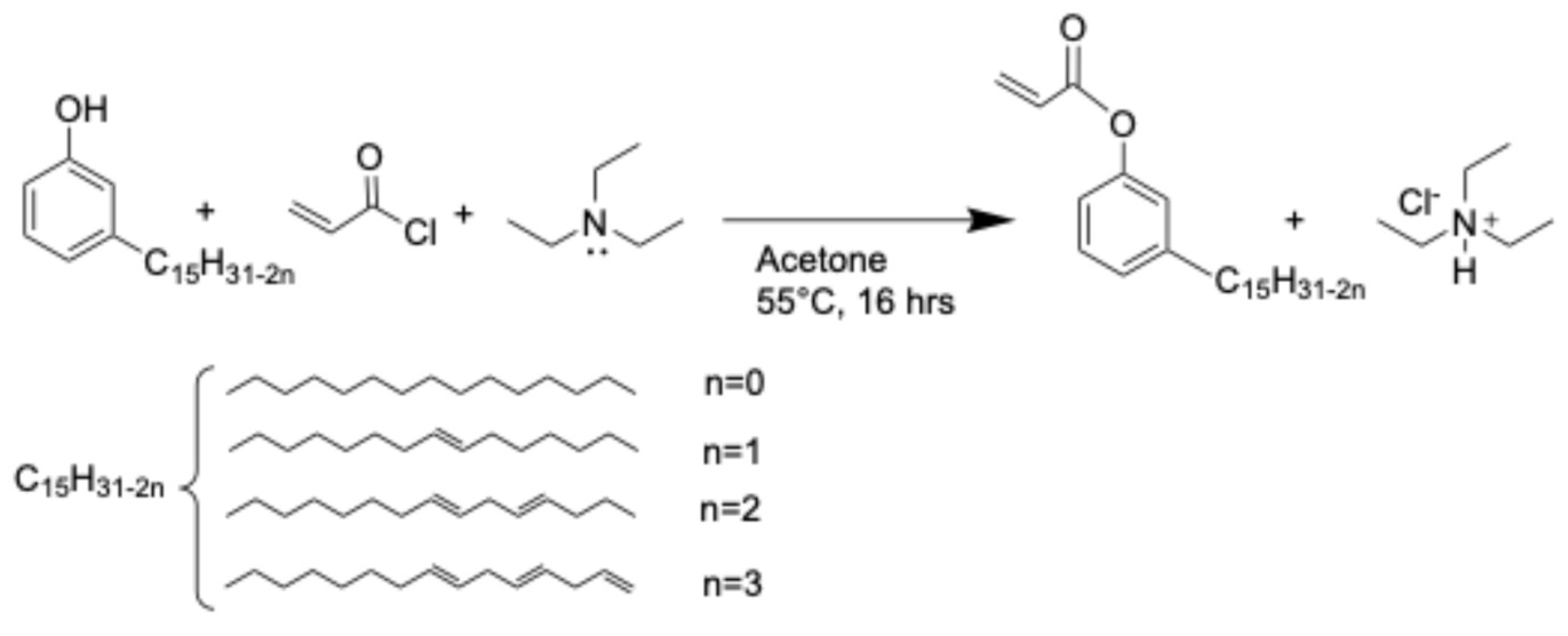
Figure 1.
Picture of PLA apparatus (a) before and (b) after loading.
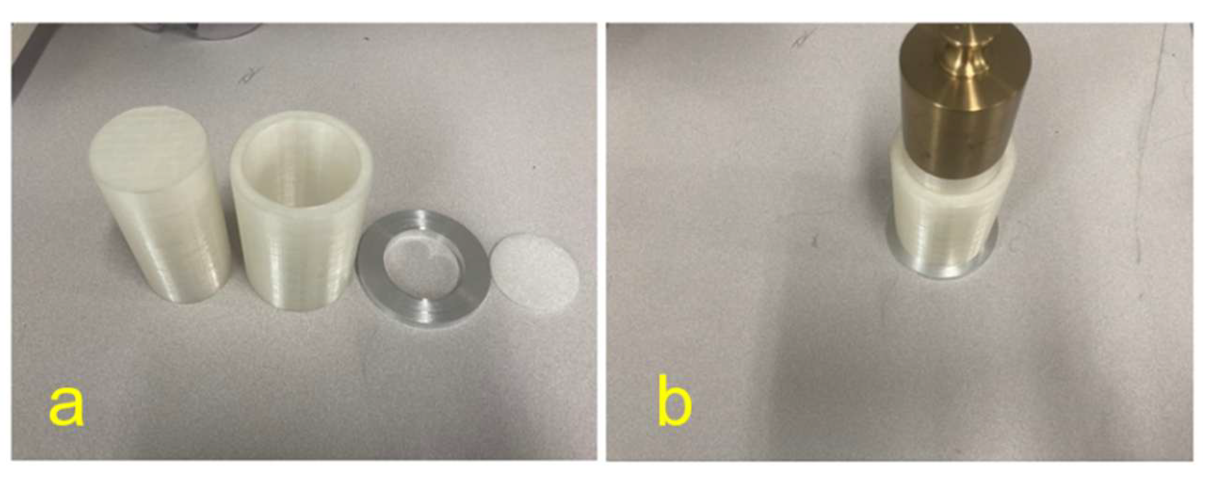
Figure 2.
FT IR spectra of Cardanol and Cardanol acrylate showing (a) the full spectrum 550 – 4000 cm-1 and (b) the expanded spectrum 900 – 1700 cm-1. See Experimental for details.
Figure 2.
FT IR spectra of Cardanol and Cardanol acrylate showing (a) the full spectrum 550 – 4000 cm-1 and (b) the expanded spectrum 900 – 1700 cm-1. See Experimental for details.
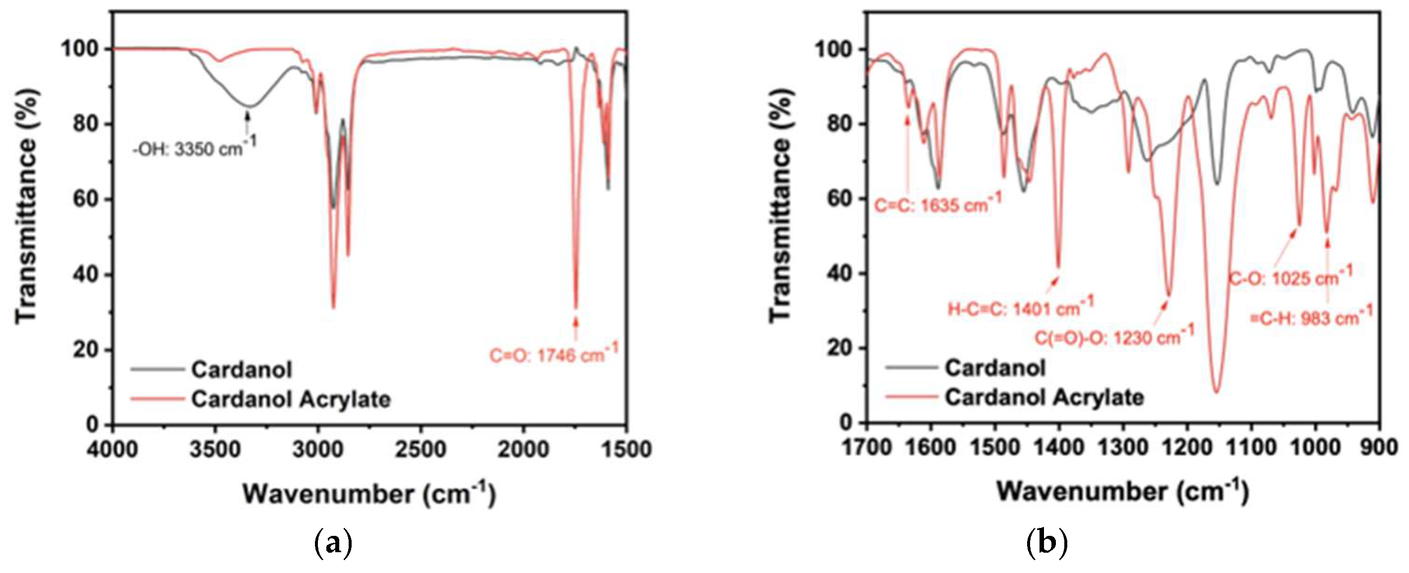
Figure 3.
1H NMR spectra of cardanol (blue) and Cardanol acrylate (red). See Experimental for details.
Figure 3.
1H NMR spectra of cardanol (blue) and Cardanol acrylate (red). See Experimental for details.
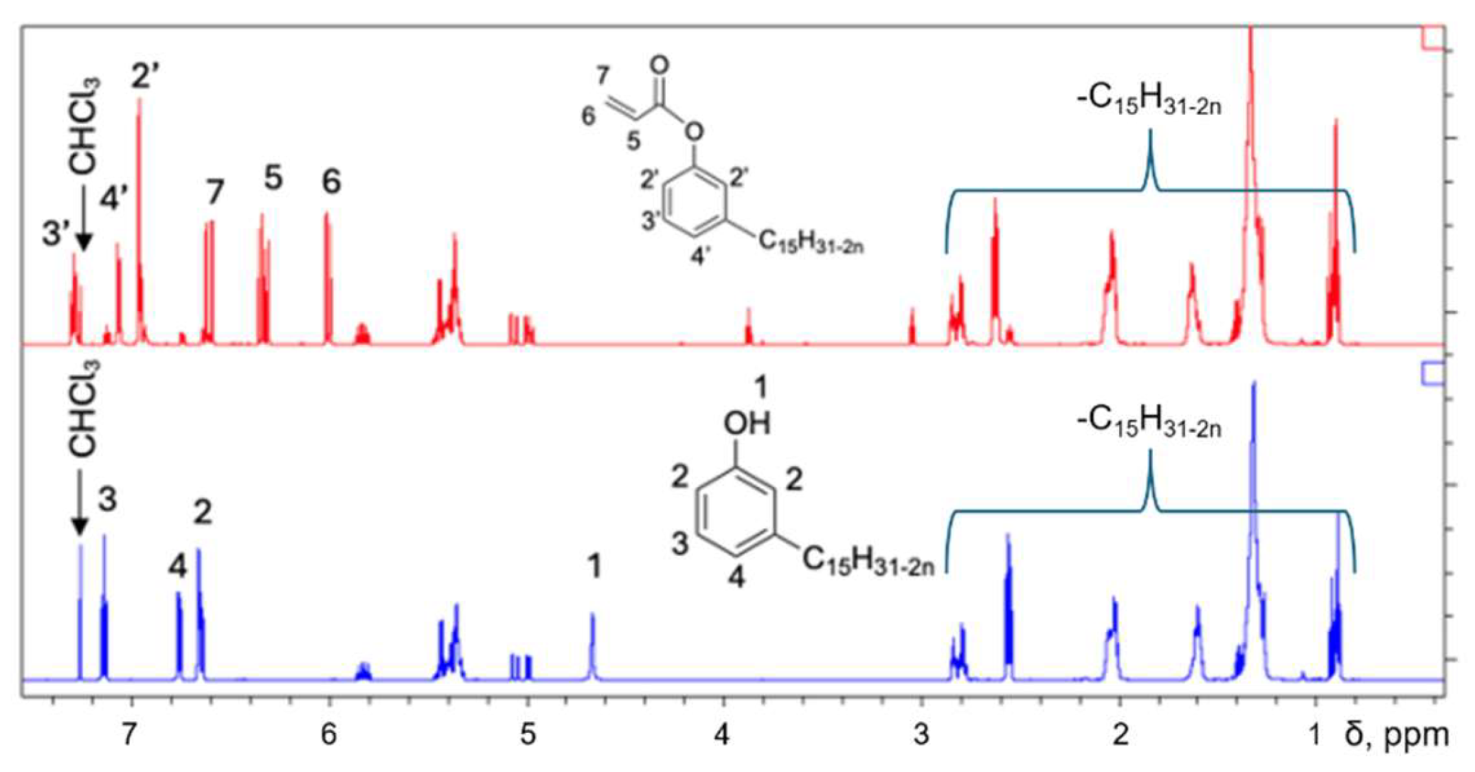
Figure 4.
13C NMR spectra of Cardanol (blue) and Cardanol acrylate (red). (x) residual CHCl3 signal. See Experimental for details.
Figure 4.
13C NMR spectra of Cardanol (blue) and Cardanol acrylate (red). (x) residual CHCl3 signal. See Experimental for details.
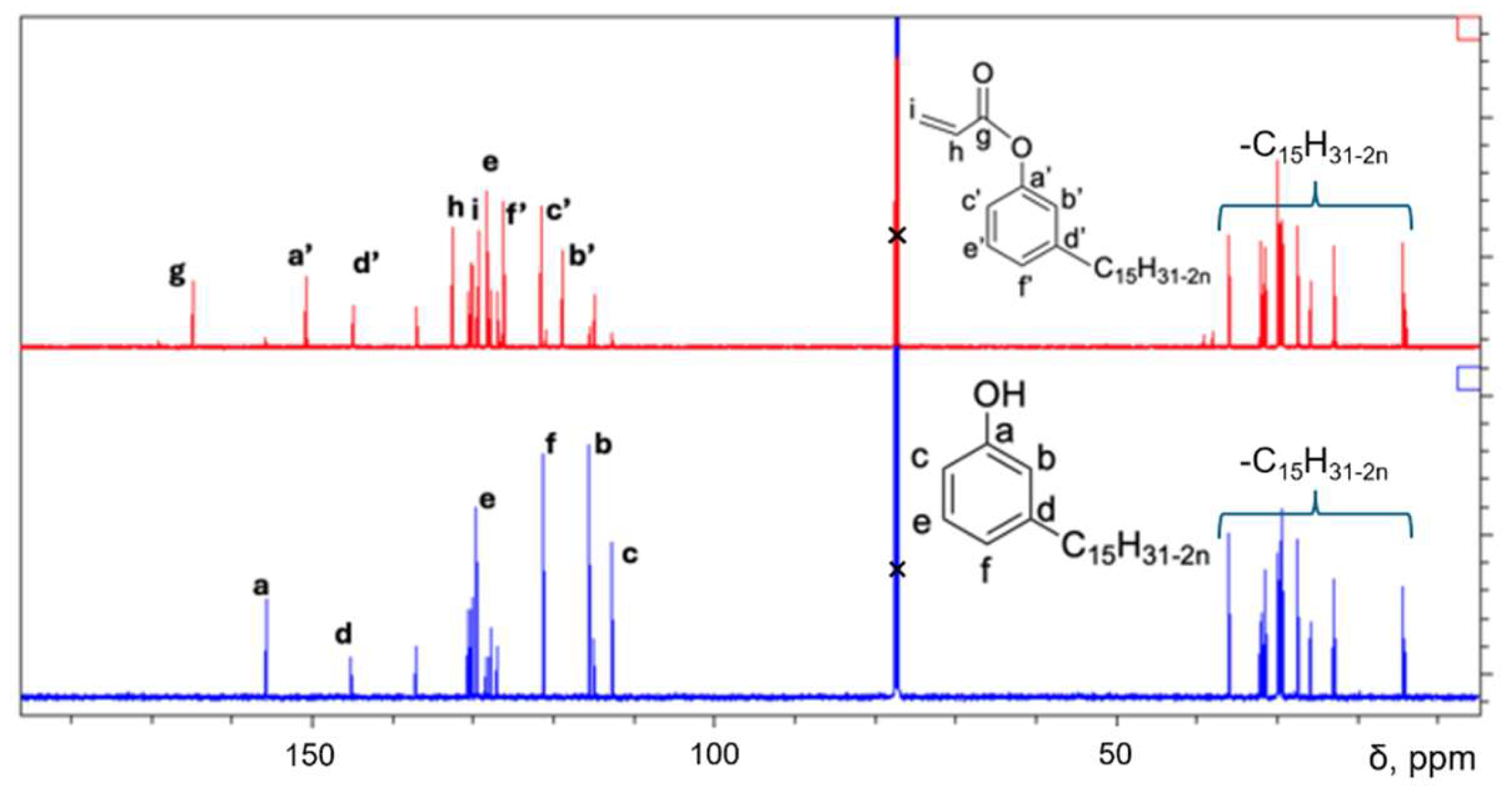
Figure 5.
1H NMR spectra of methanol extracts, (x) residual DMSO signal, (●) methanol signal. See Experimental for details.
Figure 5.
1H NMR spectra of methanol extracts, (x) residual DMSO signal, (●) methanol signal. See Experimental for details.
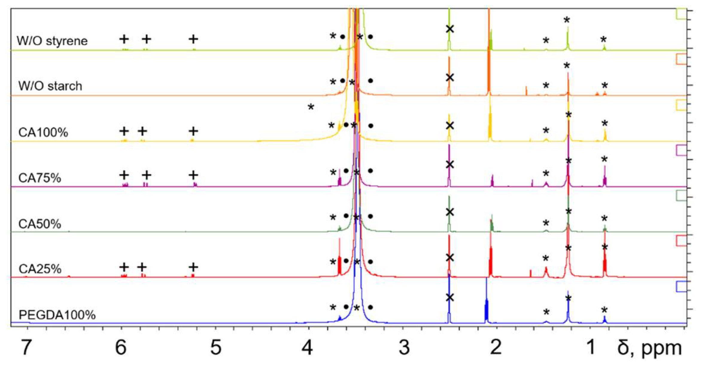
Figure 6.
1H NMR spectra of DCM extracts, (x) residual CHCl3 signal. See Experimental for details.
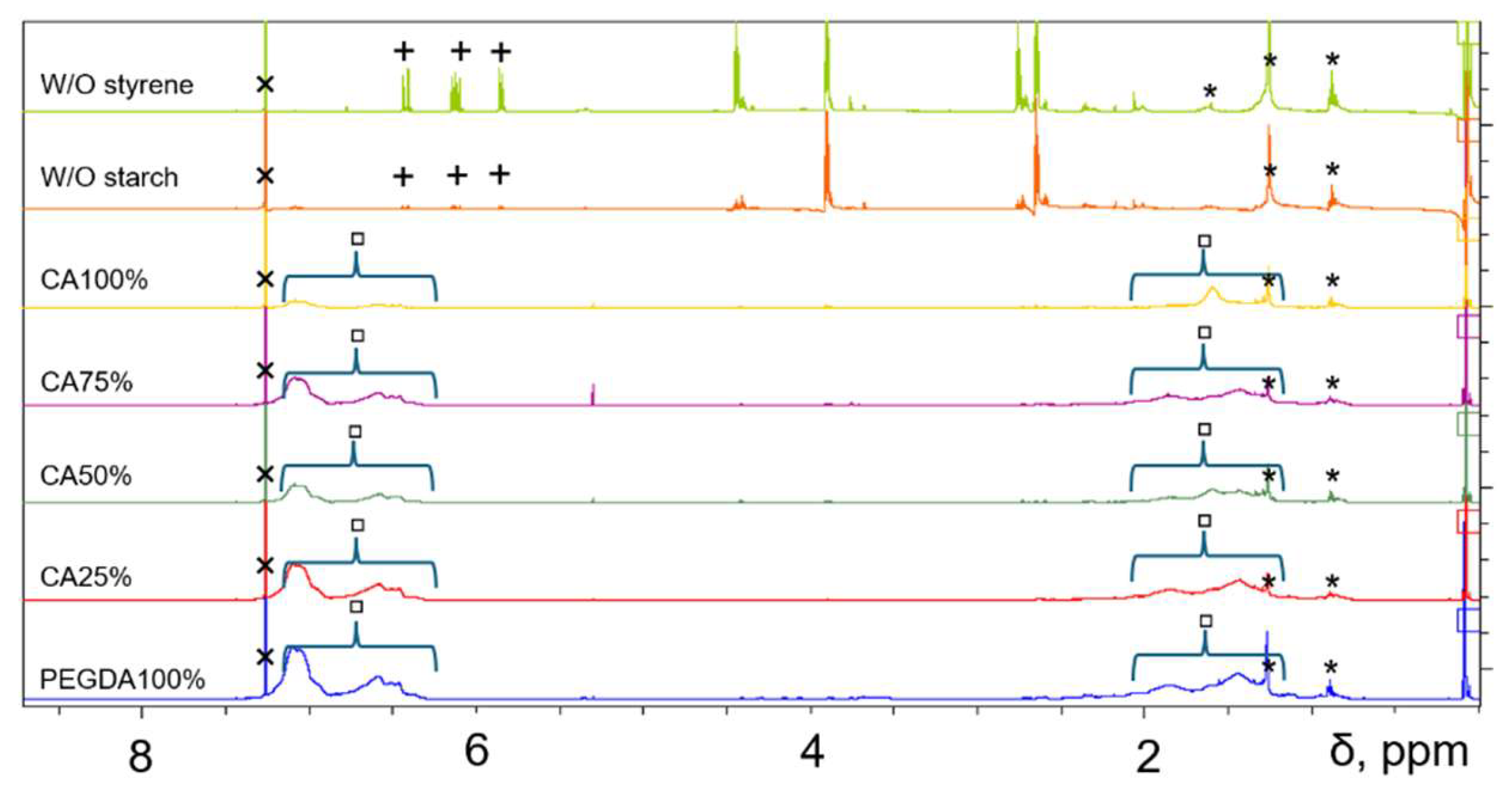
Figure 7.
SEM micrographs of hydrogels at magnification 100×. a) w/o starch, b) w/o styrene, c) PEGDA 100%, d) CA 25%, e) CA 50%, f) CA 75% and g) CA 100%. See Experimental for details.
Figure 7.
SEM micrographs of hydrogels at magnification 100×. a) w/o starch, b) w/o styrene, c) PEGDA 100%, d) CA 25%, e) CA 50%, f) CA 75% and g) CA 100%. See Experimental for details.
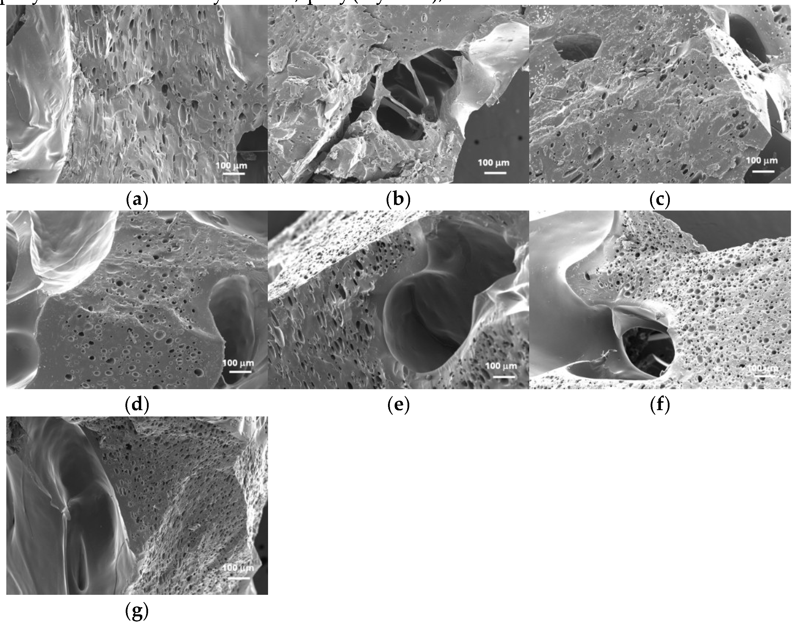
Figure 8.
(a) Swelling rate profile of all hydrogels. Voigt model fitting of (b) PEGDA 100%, (c) CA 25%, (d) CA 50%, (e) CA 75%, (f) CA 100%, (g) w/o starch, (h) w/o styrene. See Experimental for details.
Figure 8.
(a) Swelling rate profile of all hydrogels. Voigt model fitting of (b) PEGDA 100%, (c) CA 25%, (d) CA 50%, (e) CA 75%, (f) CA 100%, (g) w/o starch, (h) w/o styrene. See Experimental for details.
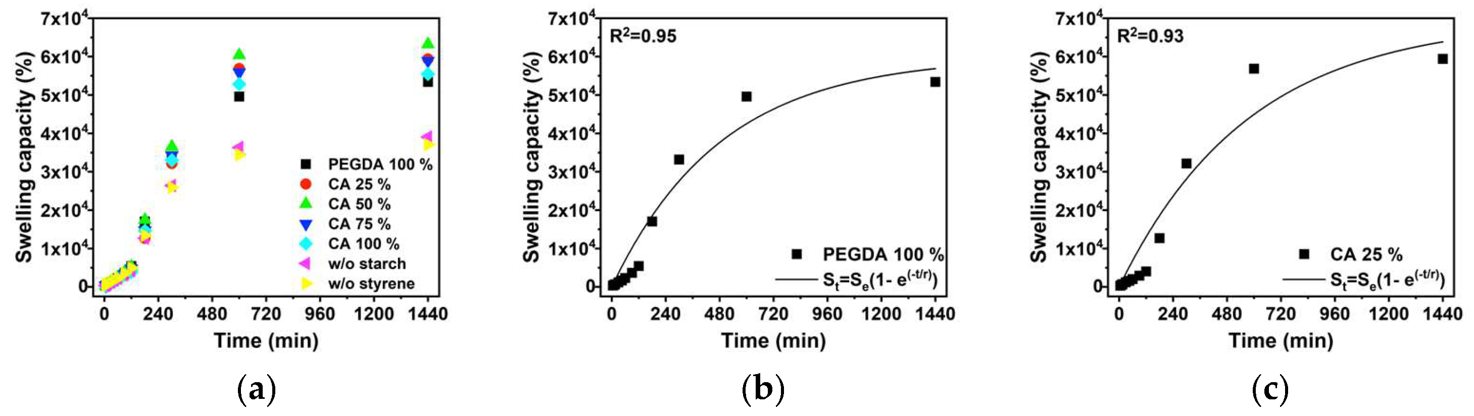
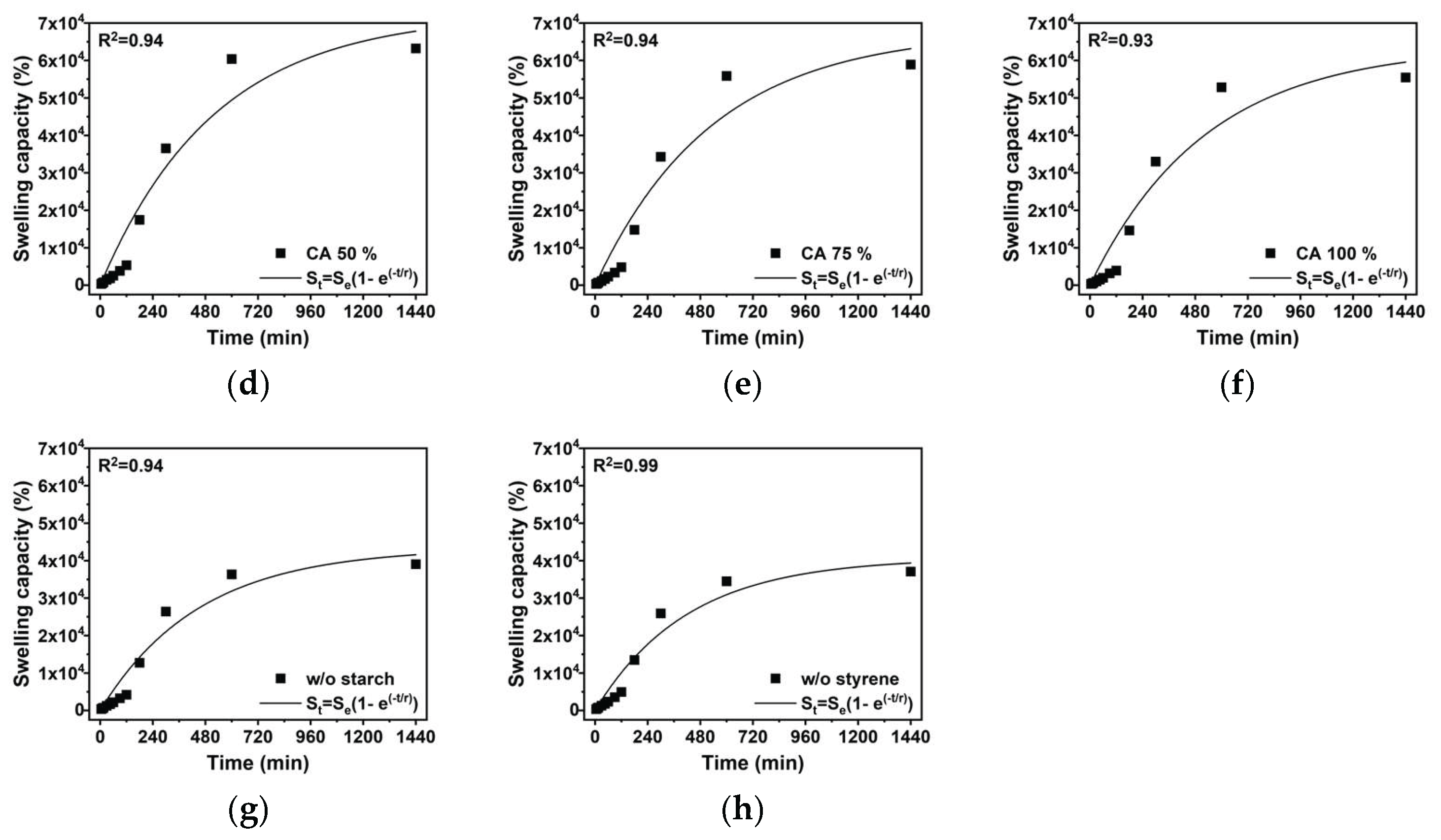
Figure 9.
Maximum swelling capacity (blue) and absorbance under load (orange) of hydrogels for an applied load of 339.5 N/m2. See Experimental and Figure 1 for details.
Figure 9.
Maximum swelling capacity (blue) and absorbance under load (orange) of hydrogels for an applied load of 339.5 N/m2. See Experimental and Figure 1 for details.
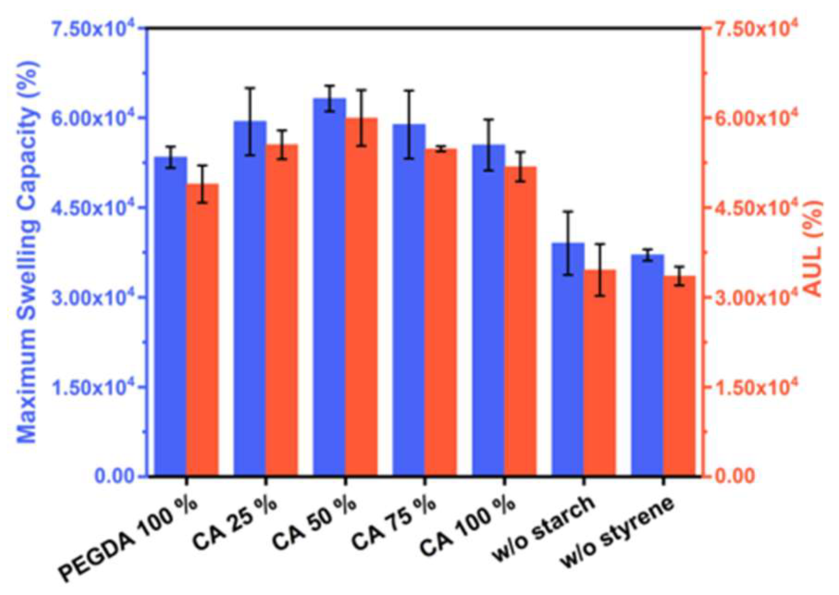
Figure 10.
Swelling of hydrogels in saline solution (yellow), acidic (orange) and basic solutions (green). See Experimental for details.
Figure 10.
Swelling of hydrogels in saline solution (yellow), acidic (orange) and basic solutions (green). See Experimental for details.
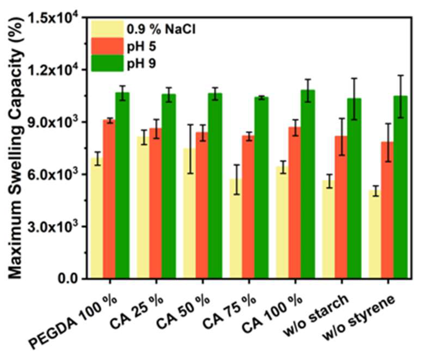
Figure 11.
TGA analysis of hydrogels. See Experimental for details.
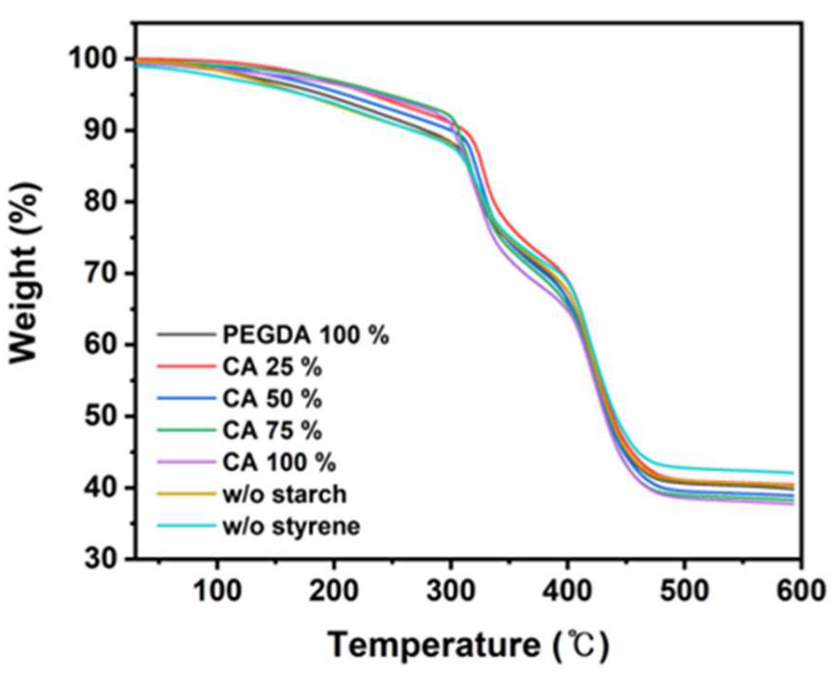
Figure 12.
DSC thermograms (2nd heating) of hydrogels at heating rate of 20 °C/min in nitrogen atmosphere. See Experimental for details.
Figure 12.
DSC thermograms (2nd heating) of hydrogels at heating rate of 20 °C/min in nitrogen atmosphere. See Experimental for details.
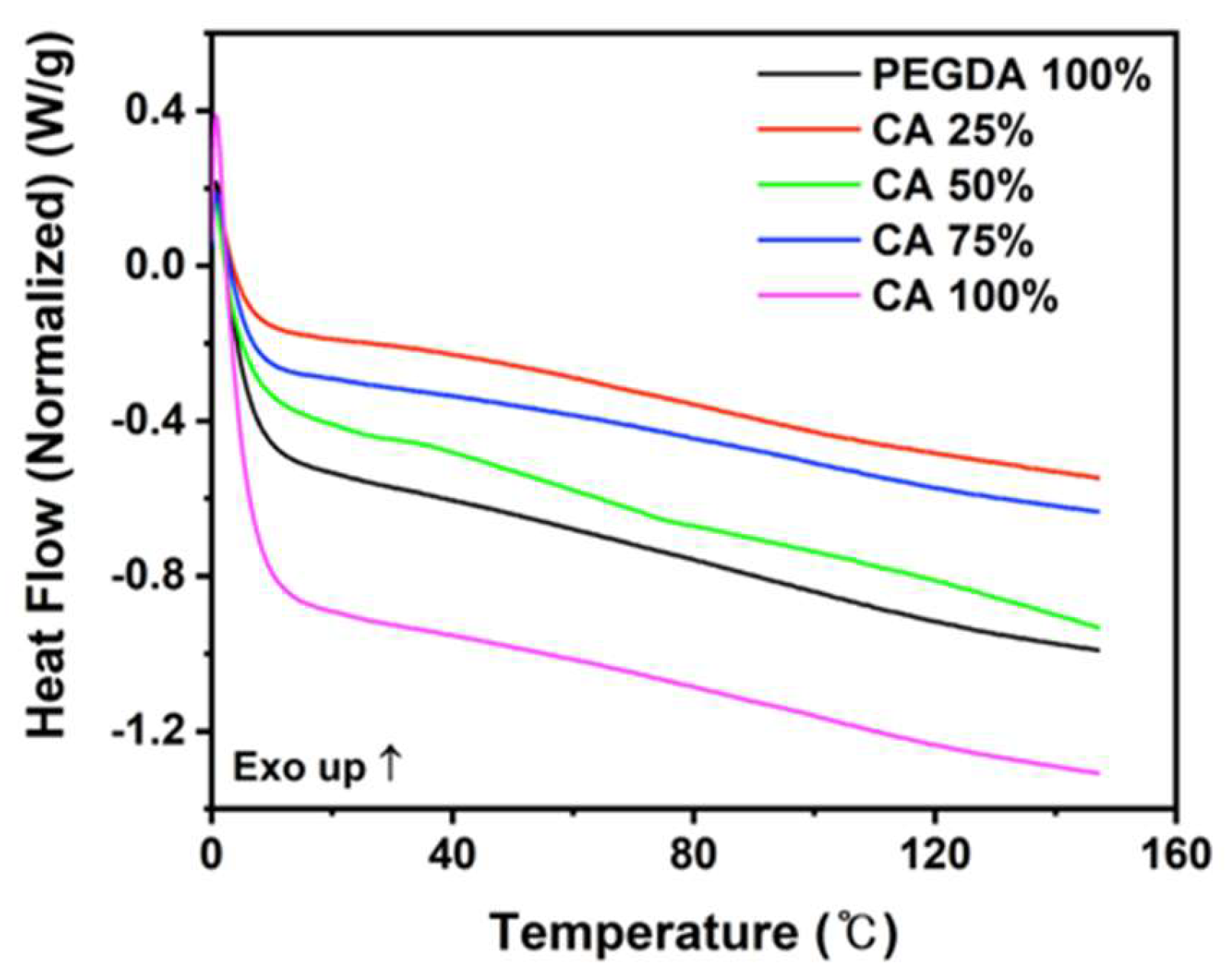
Figure 13.
Rheological properties of hydrogels. Storage moduli (solid lines), loss moduli (doted lines). See Experimental for details.
Figure 13.
Rheological properties of hydrogels. Storage moduli (solid lines), loss moduli (doted lines). See Experimental for details.
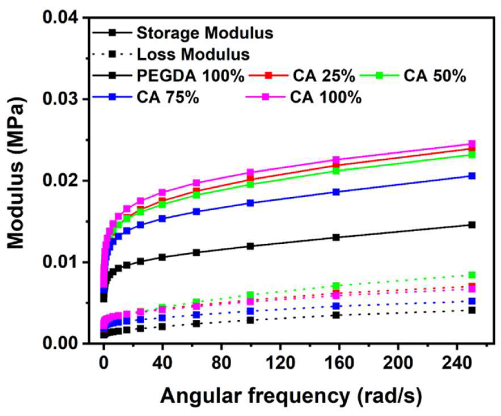
Figure 14.
Dye binding efficiency of CA 50% hydrogels. (AcrO) Acridine Orange, (AO) Auramine Orange, (CV) Crystal Violet, (MB) Methylene Blue, (MO) Methyl Orange, (BpB) Bromophenol Blue. See Experimental for details.
Figure 14.
Dye binding efficiency of CA 50% hydrogels. (AcrO) Acridine Orange, (AO) Auramine Orange, (CV) Crystal Violet, (MB) Methylene Blue, (MO) Methyl Orange, (BpB) Bromophenol Blue. See Experimental for details.
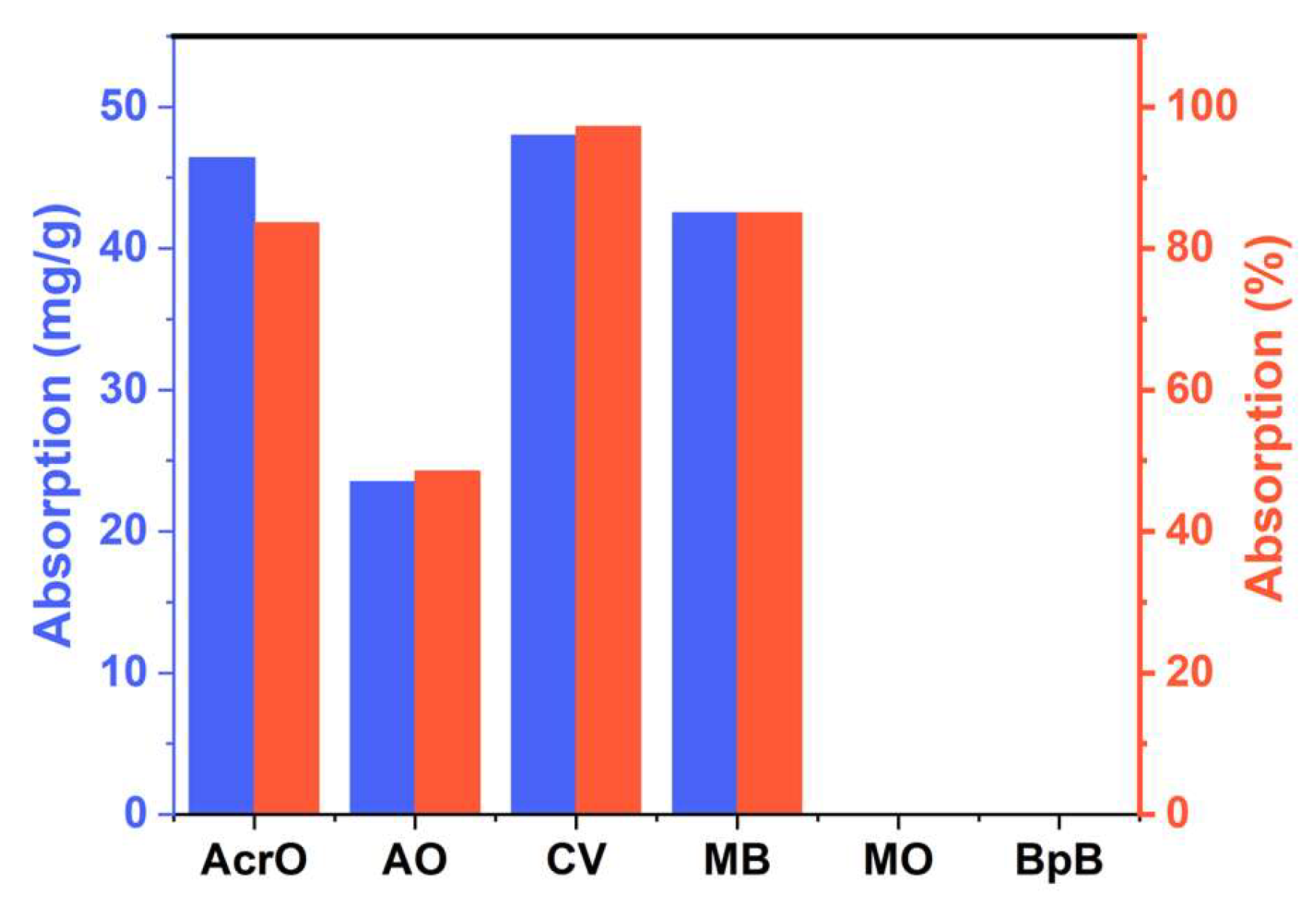
Figure 15.
Structure of investigated dyes and their absorption in CA50% hydrogels. See Experimental for details.
Figure 15.
Structure of investigated dyes and their absorption in CA50% hydrogels. See Experimental for details.
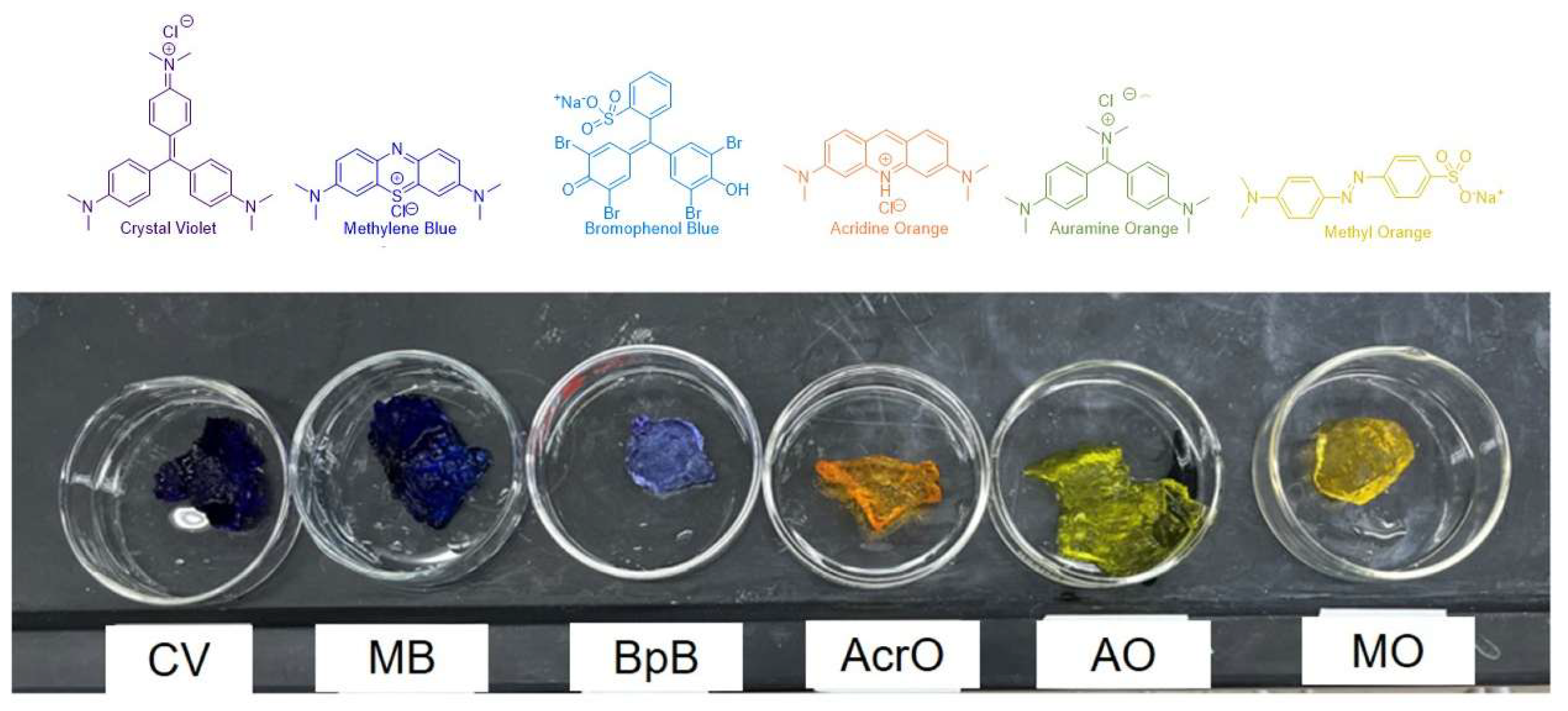

Table 1.
Hydrogels yield, polymer soluble fraction (Psf) and related gel content depending on crosslinking conditions.
Table 1.
Hydrogels yield, polymer soluble fraction (Psf) and related gel content depending on crosslinking conditions.
| Hydrogel | PEGDA (mg) | CA (mg) | Yield (%) | (%) | Gel (%) |
| w/o starch | 8 | 0 | 95.60 | 0.41 | 99.59 |
| w/o styrene | 8 | 0 | 96.74 | 0.59 | 99.41 |
| PEGDA 100% | 8 | 0 | 88.54 | 0.24 | 99.76 |
| CA 25% | 6 | 2 | 93.18 | 1.48 | 98.52 |
| CA 50% | 4 | 4 | 95.91 | 1.63 | 98.37 |
| CA 75% | 2 | 6 | 96.76 | 0.10 | 99.90 |
| CA 100% | 0 | 8 | 93.51 | 0.01 | 99.99 |
Disclaimer/Publisher’s Note: The statements, opinions and data contained in all publications are solely those of the individual author(s) and contributor(s) and not of MDPI and/or the editor(s). MDPI and/or the editor(s) disclaim responsibility for any injury to people or property resulting from any ideas, methods, instructions or products referred to in the content. |
© 2026 by the authors. Licensee MDPI, Basel, Switzerland. This article is an open access article distributed under the terms and conditions of the Creative Commons Attribution (CC BY) license (http://creativecommons.org/licenses/by/4.0/).
Copyright: This open access article is published under a Creative Commons CC BY 4.0 license, which permit the free download, distribution, and reuse, provided that the author and preprint are cited in any reuse.