
Submitted:
10 August 2026
Posted:
12 August 2026
You are already at the latest version
Abstract
Supernumerary teeth are well recognised in humans and domestic animals but are rarely reported in elephants. This case series describes four African savanna elephants (Loxodonta africana) with multiple tusk structures occupying a single alveolus. A 7-year-old bull presented with a deformed left tusk and behavioural changes. Clinical examination revealed two coronal openings, followed by eruption of three accessory tusks. Conservative intracavitary treatment and selective surgical removal of the accessory tusks preserved the primary tusk, which maintained normal eruption and function at 36-month follow-up. Additional cases comprised a bull that developed three tusks after traumatic fracture and removal of the fractured tusk remnant, an adult female with two tusks in one alveolus, and a dry specimen composed of five tusks, each with a distinct pulp canal. The continued eruption, organised morphology, separate pulp systems, and surgical separability of these structures support their interpretation as distinct supernumerary tusks rather than segmentation of the primary tusk or odontoma-like lesions. The developmental mechanism remains uncertain, but lifelong odontogenic activity in elephant tusks may permit formation of independent dentine-producing structures, and trauma may contribute in some cases. Selective removal of accessory tusks while preserving the primary tusk can provide a favourable long-term outcome.
Keywords:
elephant
; tusk
; supernumerary teeth
; hyperdontia
; hypselodont dentition
1. Introduction
Elephant dental formulae are commonly expressed as I1/0 C0/0 PM3/3 M3/3 [1]. However, elephant cheek teeth are frequently described as six sequential molars (M1-M6), reflecting their unusual pattern of horizontal tooth replacement [2]. The tusks represent the maxillary second incisor teeth [1]. They are continuously growing teeth that do not undergo normal root closure and retain lifelong odontogenic activity. Such teeth have traditionally been classified as hypselodont in the comparative anatomical literature [3], although Kertesz [4] subsequently proposed the term elodont to emphasise continuous growth rather than crown morphology. During odontogenesis, reciprocal epithelial-mesenchymal interactions initiate differentiation of ameloblasts and odontoblasts [5]. Following eruption, the enamel cap of the elephant tusk is rapidly lost, while dentine production by odontoblasts lining the pulp cavity continues throughout life [1].
Developmental abnormalities of elephant dentition are uncommon. Hypodontia and tusklessness, particularly in African savanna elephants (Loxodonta africana), are well documented and appear to have a heritable basis, possibly linked to X-chromosomal inheritance [6,7]. Reports of multiple tusk structures in elephants are rare and are largely confined to historical observations, museum specimens, or anecdotal field reports. Historical accounts have described elephants possessing three, four, five, seven, or even nine tusks, although these observations generally lacked clinical documentation or pathological evaluation [1,8]. Colyer described several examples of multiple tusks and proposed that at least some cases may have been associated with trauma to persistently growing tusks [9]. Despite these historical reports, the clinical presentation, biological behaviour, and developmental origin of supernumerary tusks remain poorly understood.
Hyperdontia refers to the presence of more than the normal number of teeth, and the additional teeth are termed supernumerary teeth [10]. Several theories have been proposed regarding the development of supernumerary teeth in humans, including phylogenetic reversion (atavism) [11], splitting of a developing tooth germ (dichotomy theory) [12], localised hyperactivity of the dental lamina [13], hereditary influences [14], and trauma [15]. However, the applicability of these mechanisms to continuously growing hypselodont (elodont) teeth such as elephant tusks remains uncertain.
The term “tusklet”, apparently introduced by Sikes [16], has been used to describe smaller tusk-like structures associated with a primary tusk within the same alveolus [16,17]. To date, these structures have not been described in a detailed clinical context, and their developmental origin remains poorly understood.
The aim of this study was to describe four African savanna elephants with multiple tusk structures within a single alveolus, including one case managed surgically with long-term follow-up, and to propose a possible developmental mechanism for supernumerary tusk formation in hypselodont (elodont) dentition.
2. Materials and Methods
Cases were identified opportunistically during clinical evaluation of African savanna elephants (Loxodonta africana). Clinical information was obtained from direct examination, veterinary records, serial photographic documentation, and follow-up reports from attending veterinarians and animal care staff. A dry tusk specimen housed at the School of Dentistry, University of Pretoria, was also available for evaluation.
In Case 1, surgical intervention was performed under general anaesthesia using standard immobilisation protocols as part of routine veterinary management [18]. Diagnostic imaging was attempted using conventional radiography; however, image quality was limited by the density and dimensions of the tusk and surrounding alveolar bone.
This study comprised retrospective clinical observations and case documentation. All procedures were performed as part of routine veterinary management and welfare-related intervention; no procedure was undertaken for research purposes.
Generative artificial intelligence was used during manuscript preparation to assist with language refinement, structural organisation, and consideration of alternative wording and interpretations. It was not used to generate or analyse clinical data. All output was critically reviewed and revised by the authors.
3. Case Descriptions
3.1. Case 1
A 7-year-old African elephant bull presented with an abnormal left tusk and recent behavioural changes, including reduced responsiveness to commands. Under general anaesthesia using etorphine and azaperone [18], clinical examination revealed a deformed tusk with a flattened coronal surface and two distinct openings, approximately 12 mm and 8 mm in diameter. No haemorrhage, purulent material, or visible pulp tissue was observed.
Radiographic examination was attempted but was non-diagnostic because of the density of the tusk and surrounding alveolar bone. The cavities were irrigated with sterile saline and dressed with calcium hydroxide powder to reduce bacterial contamination and promote tertiary dentine formation. The openings were temporarily sealed with epoxy resin to limit contamination until reassessment (Figure 1).
Over the following months, continued eruption revealed three additional tusk structures within the alveolus: a small ventral structure and two larger laterodorsal structures (Figure 2). Nine months after the initial treatment, all three structures were surgically removed under general anaesthesia. The smaller structure was thin and mobile, while the two larger structures were partially fused coronally to the primary tusk and required sectioning before extraction (Figure 3).
3.2. Case 2
A wild-caught young African elephant bull was brought into captivity and was estimated to be 3 years old at that time. Both tusks appeared normal. Approximately 2 years later, the bull fell while playing with other elephants and fractured the left tusk. Records indicated that the fractured tusk remnants were removed under general anaesthesia. Approximately 2 years later, the left tusk began to erupt with a split appearance. It was decided to trim the tusk and maintain it at a short length (Figure 6). No pulp was exposed during trimming, which the elephant permitted while conscious.
Regular trimming consistently revealed three structures: one larger and two smaller tusks (Figure 7). Trimming continued at regular intervals until the elephant died at 31 years of age from a disease unrelated to the tusk abnormality. Because of concern regarding the infectious organism associated with his death, only a limited necropsy was performed and all tissues, including the abnormal tusk, were incinerated.
3.3. Case 3
A 29-year-old African elephant cow in a sanctuary setting was observed to have two tusks within the left alveolus (Figure 8). The primary tusk was larger, with a smaller, distally positioned secondary tusk. Both were much shorter than the single right tusk and grew straight ventrally without normal curvature. No historical or clinical information regarding the onset or progression of the condition was available. No intervention was performed.
3.4. Case 4
A 30-cm abnormal dry tusk specimen from an African elephant was available for evaluation at the School of Dentistry, University of Pretoria. No clinical history or other metadata relating to the animal were available. The specimen was composed of five differently sized tusks, all attached to one another coronally. Each individual tusk had a distinct pulp canal (Figure 9).
4. Results
The four cases are summarised in Table 1. Supernumerary tusks occurred in both sexes and in animals ranging from juveniles to mature adults. In all clinically documented cases, the left tusk was affected. The affected alveoli contained between two and five tusk structures. Clinical presentation ranged from an incidental finding to behavioural abnormalities associated with tusk deformity. In the surgically managed case, selective removal of the accessory tusks while preserving the primary tusk resulted in a favourable long-term functional outcome.
5. Discussion
5.1. Aetiology
Several theories have been proposed to explain the development of supernumerary teeth, including phylogenetic reversion (atavism), splitting of a tooth germ, localised hyperactivity of the dental lamina, hereditary influences, and trauma. These mechanisms are largely derived from observations in brachydont teeth, and their applicability to continuously growing elephant tusks requires careful consideration.
Atavism is unlikely to explain the findings in the present series. This theory proposes that supernumerary teeth represent re-expression of ancestral dental patterns [11]. However, additional tusks within a single maxillary incisor alveolus do not correspond to a known ancestral dental formula in elephants and do not represent restoration of a lost tooth position. The abnormalities described here were not additional teeth in a normal anatomical series, but multiple tusk-like structures occupying the same alveolus as the primary tusk.
Dichotomy of the tooth germ, or splitting of a developing tooth bud (gemination), is another possibility [10]. This mechanism could theoretically produce a duplicated or bifid tusk. Geminated teeth, however, more commonly have two crowns that share a single root and pulp system. In Case 1, each supernumerary tusk had its own pulp tissue, the structures were surgically separable, and the primary tusk continued to erupt and function normally after their removal. These findings are difficult to reconcile with gemination.
Localised hyperactivity of the dental lamina resulting in accessory tooth bud formation is a plausible mechanism for many supernumerary teeth in humans and domestic animals [10,13]. Its applicability to the present cases is uncertain. Elephant tusks are hypselodont (elodont) teeth with lifelong growth, and the supernumerary tusks in this series appeared within the same alveolus as an established primary tusk. The available clinical evidence cannot demonstrate whether a separate, fully organised accessory tooth germ arose from the dental lamina.
A hereditary basis cannot be excluded. Supernumerary teeth have been associated with several syndromes in humans, although their patterns of inheritance and development remain incompletely understood. They also occur more frequently in affected human families than in the general population [14]. Similarly, supernumerary maxillary fourth premolar teeth have been described as a heritable condition in related Burmese cats [19]. The present case series, however, provides no evidence of familial occurrence, population clustering, or recurrence in related individuals. A hereditary predisposition therefore remains speculative.
Trauma may be relevant, particularly in Case 2, in which multiple tusk structures developed after a severe tusk fracture early in life. At the time of the initial injury, no supernumerary tusk was recognised clinically and the tusk appeared macroscopically normal. Multiple independent tusks became apparent only during subsequent regrowth. Trauma may therefore have contributed to displacement, separation, or altered organisation of odontogenic tissues during healing. It cannot, however, explain cases without a known history of tusk injury. Trauma has previously been proposed as a possible cause of multiple tusks [8,9].
Taken together, conventional theories of supernumerary tooth formation do not fully explain the findings in this series. The continuously growing nature of elephant tusks may permit developmental or reparative processes not encountered in conventional rooted teeth. Following eruption and loss of the enamel cap, dentine production by odontoblasts lining the pulp cavity persists throughout life. It is therefore conceivable that displaced, sequestered, or ectopic odontogenic mesenchymal tissues associated with the tusk apex or periapical environment may retain the capacity to form independent dentine-producing structures. These structures may then erupt alongside the primary tusk as dentinogenesis continues.
The independently erupting, morphologically organised, and surgically separable structures in Case 1 support their interpretation as distinct teeth adjacent to a primary tusk within the same alveolus. The term “tusklet” has historically been used descriptively for such structures, but may understate their biological organisation. We therefore propose that they are more appropriately regarded as supernumerary tusks (supernumerary teeth) occurring in aradicular, hypselodont dentition.
Concrescence occurs when two separate teeth become united by cementum deposition along their root surfaces [10]. In Case 1, some of the supernumerary tusks were attached to the primary tusk, while in Case 4 all five tusks were united coronally. The gross appearance of these attachments and the ability to separate the structures in Case 1 without disrupting the primary tusk suggest that a process analogous to concrescence may have occurred. Although histological confirmation was unavailable, continued cementum deposition associated with these continuously growing teeth could have produced secondary union between adjacent tusks.
5.2. Differential Diagnoses
Odontomas are hamartomatous odontogenic lesions and are classified as complex or compound odontomas [20]. Compound odontomas consist of numerous small tooth-like structures. They rarely erupt and may occur in the gingiva distant from a tooth [20]. The abnormalities described in the present study showed organised morphology, continued eruption, and gross structural resemblance to normal tusks rather than the irregular mineralised architecture generally associated with odontomas.
Comparison with odontomas should nevertheless be made cautiously because existing classifications and descriptions are derived almost exclusively from brachydont dentition. Comparatively little is known about odontogenic developmental abnormalities affecting continuously growing hypselodont (elodont) teeth. Boy and Steenkamp [21] described proliferative odontogenic lesions associated with the apical regions of hypselodont yellow tree squirrel (Paraxerus cepapi) incisor teeth and proposed the term elodontoma to distinguish these lesions from odontomas arising in conventional brachydont dentition. These lesions contained enamel, dentine, and cementum associated with the apical region of the incisor tooth. Elodontomas are generally regarded as a disease of captivity and have frequently been associated with traumatic injury to incisor teeth. Radiographically, they appear as radiopaque masses associated with the tooth apex and may obstruct normal eruption [21]. Unlike the present cases, the squirrel lesions did not form independent, erupting dental structures, but consisted of disorganised odontogenic tissue associated with the apical region of the tooth. Their biological behaviour therefore differs fundamentally from the structures described here.
Based on the clinical, surgical, and gross morphological findings, the most appropriate interpretation is that these abnormalities represent supernumerary tusks associated with the continuously growing dentition of elephants.
5.3. Clinical Relevance
Supernumerary tusks may have important clinical implications, particularly when associated with altered tusk morphology, behavioural abnormalities, or secondary trauma. In Case 1, behavioural changes preceded treatment, suggesting that the abnormality may have caused discomfort or altered tusk function. The multiple openings in the tusk also raised concern regarding communication with the pulp cavity and the risk of ascending contamination or infection.
Diagnosis of tusk abnormalities in elephants remains challenging. Conventional radiography was non-diagnostic in Case 1 because of the density and dimensions of the tusk and surrounding alveolar bone. Clinicians may therefore need to rely primarily on clinical examination, gross morphology, and longitudinal assessment of eruption patterns when advanced imaging is unavailable or impractical.
This series demonstrates that conservative management combined with selective removal of supernumerary tusks may provide a favourable long-term outcome while preserving the primary tusk. In Case 1, the primary tusk continued to erupt and remained functional after removal of the accessory tusks, with normal morphology and wear still evident 36 months later. Preserving the primary tusk is clinically important because tusks are extensively used for feeding, digging, social interaction, environmental manipulation, and defence [1,16].
Calcium hydroxide was used as an intracavitary dressing in Case 1 to reduce bacterial contamination and promote hard-tissue formation should viable pulp tissue have been present deeper within the tusk [22]. Epoxy resin was used as a temporary protective covering to limit further contamination. Because clinically documented supernumerary tusks are rare, their long-term biological behaviour remains uncertain. Continued clinical reporting will be important to improve understanding of their development, progression, and optimal treatment.
6. Conclusions
This study provides the first detailed clinical description and long-term management of supernumerary tusks in African savanna elephants (Loxodonta africana). Historical reports have documented multiple tusks in elephants, but were largely observational and lacked clinical evaluation, surgical management, or longitudinal follow-up.
The findings support the interpretation that these abnormalities are distinct odontogenic structures rather than simple segmentation of the primary tusk. Although their precise developmental mechanism remains uncertain, the continuously growing nature of elephant tusks may permit developmental or reparative processes not encountered in brachydont dentition. Trauma may contribute in at least some cases.
Selective removal of accessory tusks while preserving the primary tusk can result in a favourable long-term functional outcome. Further clinical, developmental, and histopathological investigations are required to clarify the biological basis, prevalence, and clinical significance of supernumerary tusks in elephants.
Author Contributions
Conceptualization, including development of the proposed mechanism, G.S. and S.C.B.; investigation, G.S.; data curation, G.S.; writing-original draft preparation, G.S. and S.C.B.; writing-review and editing, G.S. and S.C.B.; visualization, G.S. All authors have read and agreed to the published version of the manuscript.
Funding
This research received no external funding.
Institutional Review Board Statement
Ethical review and approval were not required because this retrospective case series reports clinical cases managed solely as part of routine veterinary care and opportunistic clinical observation, together with an existing dry tusk specimen. No procedures were performed for research purposes.
Informed Consent Statement
The clinical information and photographs were provided by the responsible veterinarians and institutions with permission and the explicit understanding that they would be used for scientific publication.
Data Availability Statement
All data presented in this study are contained within the article. Additional case information may be available from the corresponding author upon reasonable request, subject to institutional and case confidentiality.
Acknowledgments
The authors thank Drs. Charles van Niekerk and Peter Brothers for referring Case 1; Dr. Jane Budd for assistance during surgery; Dr. Christian Wencker (Zoo Basel) for Case 2; Drs. Yaduraj Khadpekar and Vaishakh Viswam of Vantara (Greens Zoological Rescue and Rehabilitation Centre) for Case 3; the late Prof. At Ligthelm, former Dean of the School of Dentistry and Chief Executive Officer of the University of Pretoria Oral and Dental Hospital, for making the specimen described in Case 4 available; and Ms. Estelle Mayhew for her work on the figures. During the preparation of this work, the authors used OpenAI ChatGPT (GPT-5.6 Thinking) to assist with language refinement, manuscript organisation, and consideration of alternative wording and interpretations. The authors reviewed and edited all output and take full responsibility for the content of the publication.
Conflicts of Interest
The authors declare no conflicts of interest.
References
- Shoshani, J. General information on elephants with emphasis on tusks. Elephant 1978, 1, 20–31. [Google Scholar] [CrossRef]
- Laws, R.M. Age criteria for the African elephant Loxodonta a. africana. Afr. J. Ecol. 1966, 4, 1–37. [Google Scholar]
- Wood, A.E.; Patterson, B. The rodents of the Deseadan Oligocene of Patagonia and the beginnings of South American rodent evolution. Bull. Mus. Comp. Zool. 1959, 120, 279–428. [Google Scholar]
- Kertesz, P. Comparative odontology. In A Colour Atlas of Veterinary Dentistry and Oral Surgery; Kertesz, P., Ed.; Wolfe Publishing: London, UK, 1993; pp. 35–50. [Google Scholar]
- Nanci, A. Development of the tooth and its supporting structures. In Ten Cate’s Oral Histology: Development, Structure, and Function, 9th ed.; Nanci, A., Ed.; Elsevier: St. Louis, MO, USA, 2018; pp. 68–86. [Google Scholar]
- Steenkamp, G.; Ferreira, S.M.; Bester, M.N. Tusklessness and tusk fractures in free-ranging African savanna elephants (Loxodonta africana). J. S. Afr. Vet. Assoc. 2007, 78, 75–80. [Google Scholar] [CrossRef] [PubMed]
- Campbell-Staton, S.C.; Arnold, B.J.; Goncalves, D.; Granli, P.; Poole, J.; Long, R.A.; Pringle, R.M. Ivory poaching and the rapid evolution of tusklessness in African elephants. Science 2021, 374, 483–487. [Google Scholar] [CrossRef] [PubMed]
- Miles, A.E.W.; Grigson, C. The ungulates. In Colyer’s Variations and Diseases of the Teeth of Animals, revised ed.; Miles, A.E.W., Grigson, C., Eds.; Cambridge University Press: Cambridge, UK, 1990; pp. 106–129. [Google Scholar]
- Colyer, F. Multiple tusks in an elephant. Dent. Rec. 1944, 64, 63–65. [Google Scholar]
- Neville, B.W.; Damm, D.D.; Allen, C.M.; Chi, A.C. Abnormalities of teeth. In Oral and Maxillofacial Pathology, 4th ed.; Neville, B.W., Damm, D.D., Allen, C.M., Chi, A.C., Eds.; Elsevier: St. Louis, MO, USA, 2016; pp. 49–110. [Google Scholar]
- Smith, J.D. Hyperdontia: Report of a case. J. Am. Dent. Assoc. 1969, 79, 1191–1192. [Google Scholar] [CrossRef] [PubMed]
- Liu, J.F. Characteristics of premaxillary supernumerary teeth: A survey of 112 cases. ASDC J. Dent. Child. 1995, 62, 262–265. [Google Scholar] [PubMed]
- Primosch, R.E. Anterior supernumerary teeth-assessment and surgical intervention in children. Pediatr. Dent. 1981, 3, 204–215. [Google Scholar] [PubMed]
- Brook, A.H. A unifying aetiological explanation for anomalies of human tooth number and size. Arch. Oral Biol. 1984, 29, 373–378. [Google Scholar] [CrossRef] [PubMed]
- Pippi, R. Odontomas and supernumerary teeth: Is there a common origin? Int. J. Med. Sci. 2014, 11, 1282–1297. [Google Scholar] [CrossRef] [PubMed]
- Sikes, S.K. The African elephant and its health. In The Natural History of the Elephant; Sikes, S.K., Ed.; American Elsevier Company: New York, NY, USA, 1971; pp. 185–223. [Google Scholar]
- Haynes, G.; Klimowicz, J. A preliminary review of bone and teeth abnormalities seen in recent Loxodonta and extinct Mammuthus and Mammut, and suggested implications. Quat. Int. 2015, 379, 135–146. [Google Scholar] [CrossRef]
- Kreeger, T.J.; Arnemo, J.M.; Caulkett, N.A.; Hampton, J.O.; Mayer, L.C.R. Elephant, African. In Handbook of Wildlife Chemical Immobilization; Kreeger, T.J., Arnemo, J.M., Caulkett, N.A., Hampton, J.O., Mayer, L.C.R., Eds.; Self-published: Bovey, MN, USA, 2023; pp. 229–230. [Google Scholar]
- Emslie, R.S.; Tutt, C.L. Supernumerary maxillary fourth premolar teeth in five related Burmese cats. JFMS Open Rep. 2020, 6, 2055116920946278. [Google Scholar] [CrossRef] [PubMed]
- Neville, B.W.; Damm, D.D.; Allen, C.M.; Chi, A.C. Odontogenic cysts and tumours. In Oral and Maxillofacial Pathology, 4th ed.; Neville, B.W., Damm, D.D., Allen, C.M., Chi, A.C., Eds.; Elsevier: St. Louis, MO, USA, 2016; pp. 632–689. [Google Scholar]
- Boy, S.C.; Steenkamp, G. Odontoma-like tumours of squirrel elodont incisors-elodontomas. J. Comp. Pathol. 2006, 135, 56–61. [Google Scholar] [CrossRef] [PubMed]
- Steenkamp, G. Oral biology and disorders of tusked mammals. Vet. Clin. N. Am. Exot. Anim. Pract. 2003, 6, 689–725. [Google Scholar] [CrossRef] [PubMed]
Figure 1.
The cleaned, abnormal left tusk of a young African elephant bull shortly after initial treatment with calcium hydroxide and a temporary epoxy resin covering.
Figure 1.
The cleaned, abnormal left tusk of a young African elephant bull shortly after initial treatment with calcium hydroxide and a temporary epoxy resin covering.
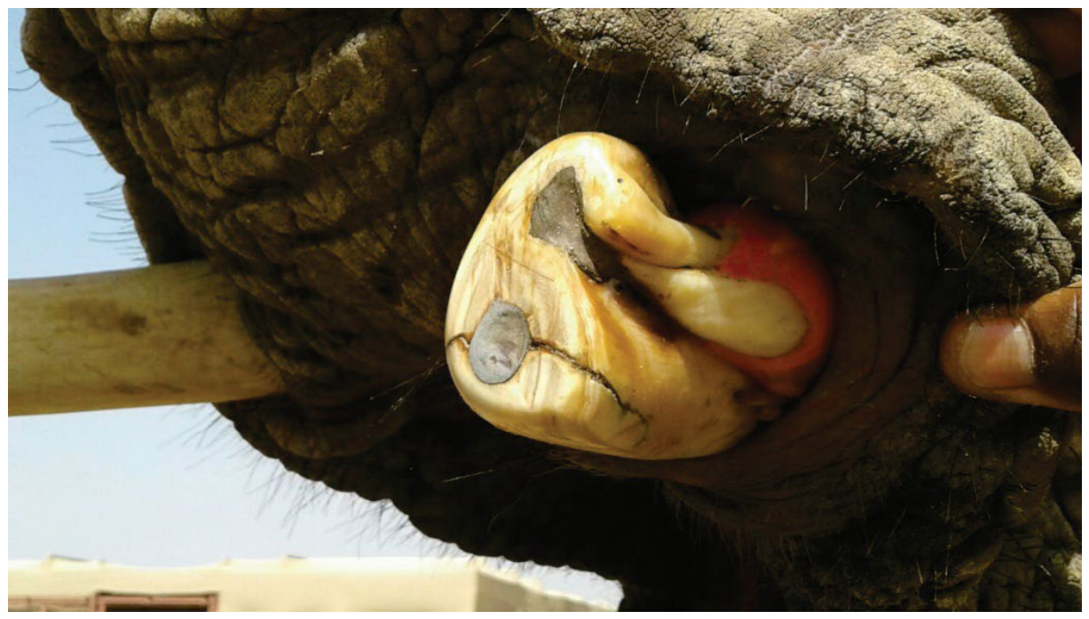
Figure 2.
(A) A small, needle-like tusk protruding through the skin. (B) The two larger, more laterally positioned supernumerary tusks attached to the primary tusk.
Figure 2.
(A) A small, needle-like tusk protruding through the skin. (B) The two larger, more laterally positioned supernumerary tusks attached to the primary tusk.
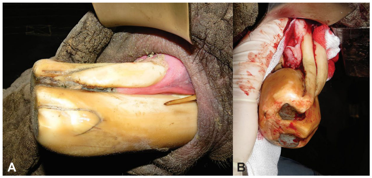
Figure 3.
The two larger supernumerary tusks after sectioning and removal. Pulp tissue removed from the more dorsally positioned tusk is shown adjacent to it, while the lower structure retained its pulp tissue.
Figure 3.
The two larger supernumerary tusks after sectioning and removal. Pulp tissue removed from the more dorsally positioned tusk is shown adjacent to it, while the lower structure retained its pulp tissue.
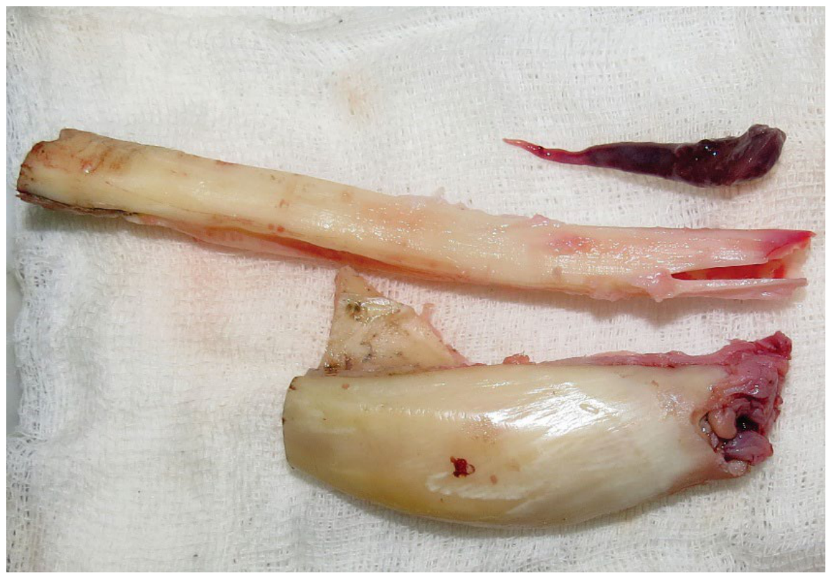
Figure 4.
The remaining primary tusk after extraction of the three supernumerary tusks in a young African elephant bull.
Figure 4.
The remaining primary tusk after extraction of the three supernumerary tusks in a young African elephant bull.
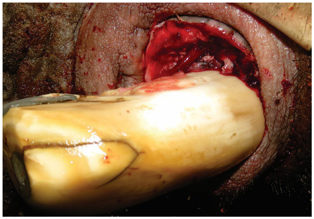
Figure 5.
(A) The epoxy resin remained visible on the coronal aspect of the growing tusk 24 months after extraction of the supernumerary tusks. (B) At 36 months, the epoxy resin was no longer visible and the shape of the tusk approximated that of the contralateral tusk.
Figure 5.
(A) The epoxy resin remained visible on the coronal aspect of the growing tusk 24 months after extraction of the supernumerary tusks. (B) At 36 months, the epoxy resin was no longer visible and the shape of the tusk approximated that of the contralateral tusk.
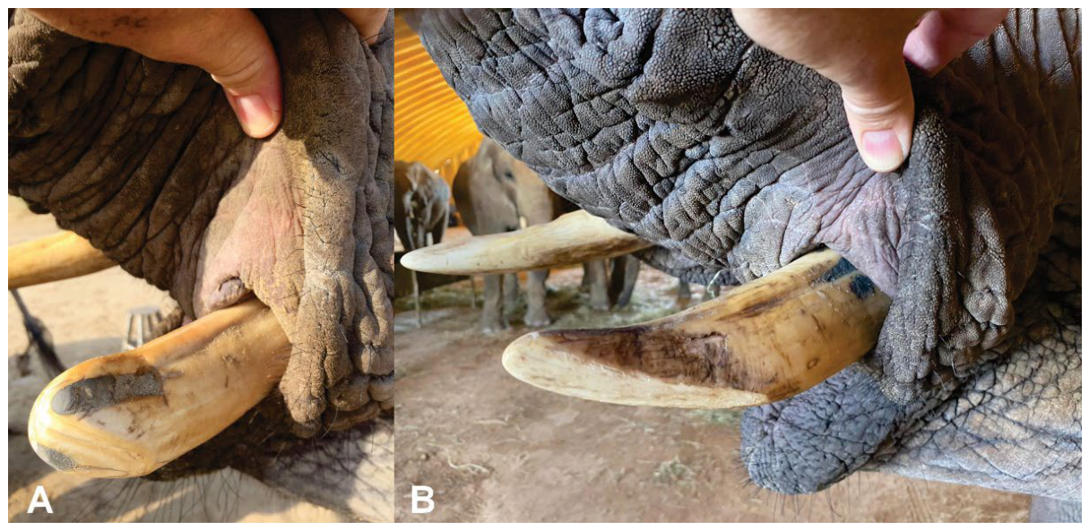
Figure 6.
An adult African elephant bull with three separate tusks present in the left alveolus.
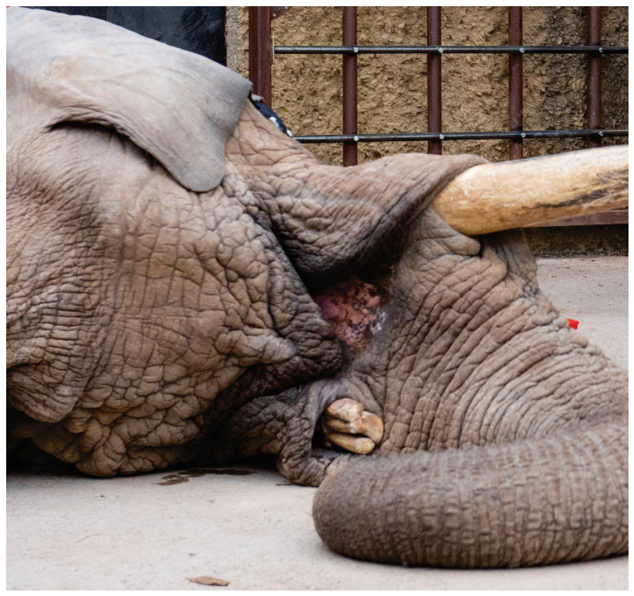
Figure 7.
Coronal sections of the three tusks present in the left alveolus of an adult African elephant bull after trimming.
Figure 7.
Coronal sections of the three tusks present in the left alveolus of an adult African elephant bull after trimming.
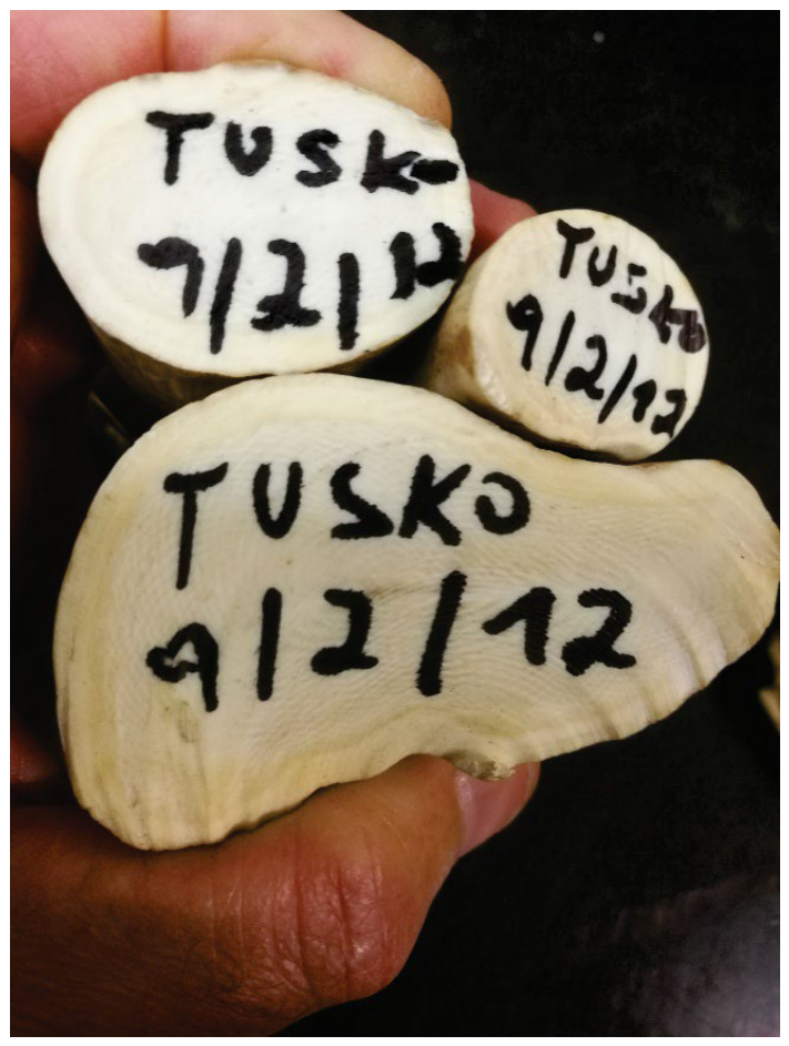
Figure 8.
Two tusks present in the left alveolus of an adult African elephant cow. The right tusk had normal length and curvature.
Figure 8.
Two tusks present in the left alveolus of an adult African elephant cow. The right tusk had normal length and curvature.
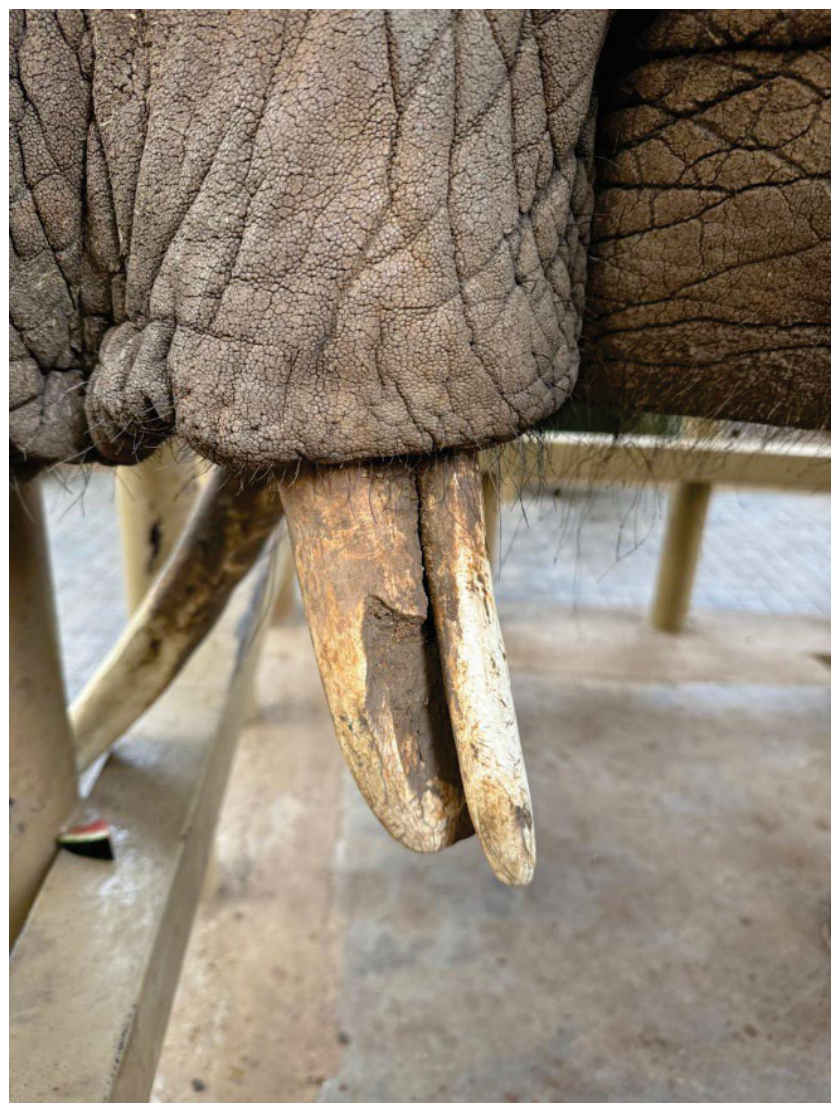
Figure 9.
Dorsal (A), apical (B), and coronal (C) views of the multiple tusks from an African elephant. Five distinct pulp canals are visible apically, while the tusks were united coronally. The five tusks are numbered for ease of reference.
Figure 9.
Dorsal (A), apical (B), and coronal (C) views of the multiple tusks from an African elephant. Five distinct pulp canals are visible apically, while the tusks were united coronally. The five tusks are numbered for ease of reference.
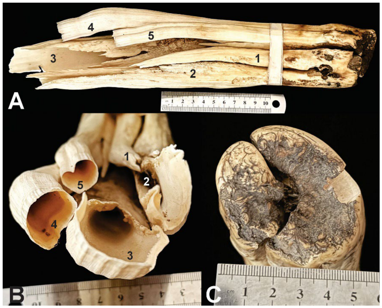

Table 1.
Summary of the four African savanna elephant cases and the available metadata. Age represents the interval from first to last observation.
Table 1.
Summary of the four African savanna elephant cases and the available metadata. Age represents the interval from first to last observation.
| Case | Age (years) | Sex | Tusk affected | Total tusks in affected alveolus |
|---|---|---|---|---|
| 1 | 7-10 | Male | Left | 4 |
| 2 | 3-31 | Male | Left | 3 |
| 3 | 29 | Female | Left | 2 |
| 4 | Unknown | Unknown | Unknown | 5 |
Disclaimer/Publisher’s Note: The statements, opinions and data contained in all publications are solely those of the individual author(s) and contributor(s) and not of MDPI and/or the editor(s). MDPI and/or the editor(s) disclaim responsibility for any injury to people or property resulting from any ideas, methods, instructions or products referred to in the content. |
© 2026 by the authors. Licensee MDPI, Basel, Switzerland. This article is an open access article distributed under the terms and conditions of the Creative Commons Attribution (CC BY) license (http://creativecommons.org/licenses/by/4.0/).
Copyright: This open access article is published under a Creative Commons CC BY 4.0 license, which permit the free download, distribution, and reuse, provided that the author and preprint are cited in any reuse.