

Submitted:
10 August 2026
Posted:
11 August 2026
You are already at the latest version
Abstract
Low temperature severely restricts the growth of Hemerocallis fulva, yet the tissue-specific molecular regulatory networks governing cold adaptation in H. fulva remain poorly understood. In this study, phenotypic observation and comparative transcriptome analyses were performed on cold-treated and control roots and leaves of H. fulva. Phenotypic data showed that cold stress significantly suppressed seedling growth, root biomass, and reproductive development of daylily. Transcriptome identification obtained 9504 DEGs in roots and 6916 DEGs in leaves, with only 3165 shared cold-responsive genes, indicating largely divergent transcriptional programs between underground and aerial organs. KEGG enrichment analysis demonstrated that hormone signal transduction, antioxidant pathways, and soluble sugar metabolism represented core modules of the cold response. Further tissue-specific expression profiling revealed distinct regulatory patterns of three key phytohormone pathways: ethylene biosynthesis and downstream ERF/DREB/COR genes were predominantly activated in roots or leaves; gibberellin pathway exhibited opposite transcriptional trends in roots and leaves, with cold triggering DELLA accumulation to restrain growth; ABA biosynthetic genes (CHY-β, ZEP, NCED, ABA2, ABA3) were universally upregulated in both tissues, while ABA signaling components displayed root-leaf divergent expression patterns. In addition, cold stress induced comprehensive transcriptional activation of enzymatic antioxidant genes (SOD, CAT, POD, GPX, GST, GSH) to eliminate excess reactive oxygen species, with stronger induction observed in roots. For osmoprotective carbohydrate metabolism, key biosynthetic genes of inulin-type fructan, raffinose family oligosaccharides (RFOs), and trehalose were significantly upregulated. In contrast, catabolic enzyme genes were repressed in both roots and leaves, jointly promoting soluble sugar accumulation to maintain osmotic homeostasis. Collectively, these findings reveal the tissue-specific transcriptional landscape of cold acclimation in H. fulva and provide a theoretical foundation for molecular breeding of cold-tolerant daylily plants.
Keywords:
Hemerocallis fulva
; cold stress
; transcriptome analysis
; phytohormone
; antioxidant defense
; soluble sugar
1. Introduction
Low temperature is one of the most destructive abiotic stresses that severely restricts the growth, biomass accumulation, flowering quality, and geographical distribution of plants [1,2]. Cold exposure impairs membrane fluidity, triggers massive burst of reactive oxygen species (ROS), disrupts photosynthetic carbon assimilation, disturbs endogenous hormone homeostasis, and blocks the biosynthesis of osmoprotective soluble carbohydrates [3,4]. Consequently, cold stress leads to leaf chlorosis, root growth inhibition, reduced flower bud differentiation, delayed anthesis, and even seedling death [5].
Phytohormones act as central signal mediators linking low-temperature perception to downstream cold-resistance gene expression, among which ethylene, gibberellin (GA) and abscisic acid (ABA) exert dominant regulatory functions [6]. Ethylene is a gaseous hormone that plays a crucial role in plant growth, development, and responses to multiple stresses, including cold [7,8]. In Arabidopsis thaliana, freezing stress tolerance is suppressed by ethylene signaling. Loss-of-function mutant ctr1-1, which constitutively activates ethylene cascades, as well as EIN3-overexpressing transgenic materials, exhibit impaired freezing resistance. Integrated genetic and biochemical analyses uncover that EIN3 mediates transcriptional repression of CBF genes [9]. Contrary to the negative regulatory role of ethylene in Arabidopsis freezing tolerance, ethylene positively boosts cold resistance in cotton [10]. Multiple genetic assays confirmed that the ethylene biosynthetic gene GhACO1 improves cold tolerance. Further molecular tests proved that GhDREB1/CBF directly transactivates GhACO1. Therefore, GhDREB1-GhACO1 module promotes ethylene accumulation to strengthen cotton cold tolerance via the CBF-dependent pathway [10]. Similarly, ethylene positively modulates cold tolerance in grapevine. Cold induces ethylene accumulation and activates the ethylene-responsive gene VaERF057. Ectopic expression of VaERF057 improves Arabidopsis cold resistance by boosting antioxidant capacity and upregulating CBF and other stress genes [11]. These existing studies have verified ethylene’s positive or negative role in cold acclimation.
Gibberellin (GA) homeostasis is balanced by synthetic and catabolic pathways, with three subfamilies of GA 2-oxidases responsible for GA degradation. In Arabidopsis thaliana, cold stress strongly induces AtGA2ox. Genetic evidence shows AtGA2ox9 mutants are freezing-sensitive, and positively regulate Arabidopsis freezing tolerance [12]. In rice, cold-induced OsWRKY53 represses anther GA biosynthesis by targeting GA synthetic gene promoters. Loss-of-function wrky53 mutants accumulate higher anther GA under cold treatment. GA counteracts SLR1-mediated inhibition of tapetum factors UDT1/TDR to sustain male fertility. Knocking out WRKY53 improves booting cold tolerance without yield penalties [13]. Generally, cold stress suppresses GA biosynthesis and activates DELLA accumulation to restrain excessive vegetative growth [14,15]. ABA is recognized as the master stress hormone governing cold tolerance [16,17]. Previous work has demonstrated that cold rapidly induces ABA accumulation in plants, for instance, the ABA synthetic gene OsNCED4 in rice is markedly upregulated under cold treatment. CRISPR/Cas9-generated osnced4 mutants exhibit cold hypersensitivity, attributed to reduced ABA levels and excessive ROS accumulation under low temperature [18]. Analogously, rice OsNAC5 confers cold resistance by transactivating OsABI5, which further orchestrates a suite of ABA- and ROS-responsive stress genes [19]. In pepper, the transcription factor CaNAC035 physically interacts with CaSnRK2.4 and directly targets the promoters of ABA biosynthetic genes CaNCED3 and CaAAO3, thereby driving ABA biosynthesis upon cold exposure [20]. Indeed, exogenous ABA enhances rice cold tolerance under cold stress and chilling acclimation. High-concentration ABA enhances seedling biomass and survival by promoting osmolyte accumulation, thereby alleviating membrane lipid peroxidation and maintaining water homeostasis. ABA also elevates SOD, POD, and CAT activities to eliminate excess H2O2 and O2− and strengthen ROS scavenging capacity [21].
Cold stress disrupts cellular electron transport chains and provokes excessive ROS production, which causes oxidative damage, lipid peroxidation, and protein denaturation [22]. Plants rely on two core antioxidant systems to scavenge toxic ROS: enzymatic antioxidant system and glutathione circulation system [23]. Key functional enzymes include SOD, CAT, POD, GPX, GST, and GSH. SOD converts superoxide anion into hydrogen peroxide, which is further decomposed by CAT and POD; glutathione-related enzymes eliminate lipid peroxides to alleviate secondary oxidative toxicity [6]. Under cold stress, cold-susceptible rice accumulates excessive ROS and MDA, whereas cold-tolerant rice elevates CAT and POD activities to clear ROS efficiently. OsCATC/OsPOX1 expression aligns with enzyme phenotypes, indicating CAT and POD are key ROS-scavenging factors for rice cold tolerance [24]. A cold-inducible NAC transcription factor gene DgNAC3 was cloned from chrysanthemum. Overexpressing DgNAC3 markedly enhances chrysanthemum cold tolerance. Transgenic plants accumulate less MDA, H2O2 and O2− under cold stress, accompanied by higher CAT, SOD, and POD activities and upregulated antioxidant genes (DgPOD, DgCAT, DgCu/ZnSOD). Therefore, DgNAC3 strengthens ROS scavenging capacity to enhance chrysanthemum cold resistance [25]. These reports indicated that the antioxidant system universally participates in cold stress mitigation.
Soluble carbohydrates function as critical compatible solutes to stabilize cell membranes, maintain cellular osmotic balance, and scavenge ROS under low temperature [26]. In Lavandula angustifolia, cold induces the expression of starch-degrading genes LaAMY, LaBAM1/3 and soluble sugar synthetic genes (LaRHM1, LaMUR4, LaUGD4), while repressing photosynthetic genes (LaPSAD1, LaPSAN, LaLHCB4.2 etc.). These genes correlate closely with starch hydrolysis, which enhances osmotic adjustment via soluble sugar accumulation [27]. During cold acclimation, dormant alfalfa sequentially accumulates starch and raffinose then re-synthesizes sucrose, accompanied by coordinated shifts in synthetase and hydrolase activities [28]. In Jatropha curcas, cold accelerates starch breakdown and soluble sugar buildup, with leaves and cotyledons showing more dramatic changes than stems and roots. Multiple sugar synthetic enzymes are activated; continuously rising invertase activity in all organs. Besides, elevated galactinol and raffinose alongside related enzyme activity confirm raffinose oligosaccharides contribute greatly to cold tolerance [29]. Cold-induced VaWRKY65 acts as a dual regulator in Vitis amurensis. It transcriptionally activates starch degradation gene VaBAM3 to accumulate soluble sugars, and directly upregulates antioxidant gene VaPOD3 to clear ROS, thereby improving cold tolerance [30]. These works lay a basis for uncovering sugar-mediated cold adaptation and signaling mechanisms.
As a core perennial ornamental crop belonging to Liliaceae, H. fulva (daylily) possesses high ornamental, ecological, and medicinal value [31]. However, low temperature in late spring and early autumn frequently restricts its vegetative propagation and reproductive development, seriously limiting its landscaping application in temperate and cold regions [32]. To adapt to cold environments, plants have evolved sophisticated multi-layered adaptive strategies, including tissue-specific transcriptional remodeling of hormone signaling, antioxidant defense, and soluble sugar metabolic pathways [33,34]. Roots and leaves execute distinct physiological functions: underground roots are the primary tissue sensing soil low temperature and responsible for water/nutrient absorption [35,36], while aerial leaves undertake photosynthesis and integrate light-temperature-hormone signals to regulate floral transition [37]. Cumulative physiological evidence has confirmed that roots and leaves deploy divergent cold acclimation strategies [38,39], yet the genome-wide tissue-specific transcriptional regulatory network of daylily under cold stress remains largely uncharacterized.
In the present study, we performed phenotypic observation and comparative RNA-seq transcriptome analysis on cold-treated and control roots and leaves of H. fulva. We systematically analyzed DEG distribution and KEGG pathway enrichment, then characterized tissue-specific transcriptional changes of ethylene, GA, ABA hormone pathways, antioxidant defense genes, and fructan/RFO/trehalose sugar metabolism genes. This work aims to reveal the tissue-specific molecular regulatory mechanisms of daylily cold acclimation, clarify the crosstalk network of cold signal, multiple phytohormones, ROS scavenging, and osmoprotective sugar metabolism, and provide theoretical basis for cold-tolerant breeding and cultivation management of daylily plants.
2. Materials & Methods
2.1. Plant Materials and Cold Stress Treatment
H. fulva uniform seedlings with consistent growth status were selected as experimental materials. Seedlings were cultivated in plastic pots filled with mixed substrate (peat soil: vermiculite = 2:1, v/v) in a greenhouse under 16 h light / 8 h dark photoperiod, 25 °C daytime / 20 °C nighttime, and 60%–70% relative humidity. Seedlings with 6 fully expanded leaves were divided into two groups: the control group (CK) and the cold stress treatment group (CD). For cold treatment, seedlings were transferred to an artificial climate incubator and subjected to continuous low temperature at 4 °C for 24 h under identical light and humidity conditions. CK seedlings were maintained at normal temperature (25 °C/20 °C day/night) without cold exposure. After treatment, fresh leaf tissues (CK-L, CD-L) and whole root systems (CK-R, CD-R) were rapidly harvested from each seedling. All samples were rinsed with sterile distilled water, wiped dry, immediately frozen in liquid nitrogen, and stored at −80 °C ultra-low temperature refrigerator for subsequent phenotypic observation and RNA-seq sequencing. Three independent biological replicates were set for each tissue group.
2.2. Phenotypic Observation and Photographing
Whole potted seedlings, complete root systems and single detached seedlings of CK and CD groups were photographed separately. Scale bars were marked uniformly in each picture: 20 cm for whole potted plants, 10 cm for root systems and single seedlings.
2.3. RNA Extraction, Library Construction and Illumina Sequencing
Total RNA was extracted from root and leaf frozen samples using the Trizol reagent kit (Invitrogen, USA) following the manufacturer’s instructions. RNA purity and integrity were detected by NanoDrop 2000 spectrophotometer and 1% agarose gel electrophoresis, respectively. RNA concentration was accurately quantified using Qubit 3.0 fluorometer. High-quality RNA samples with RIN > 6.5 were used for cDNA library construction. mRNA was enriched via oligo(dT) magnetic beads, fragmented randomly, and reverse-transcribed into first-strand cDNA using random hexamer primers. Second-strand cDNA was synthesized with DNA polymerase I, followed by end repair, A-tailing and adapter ligation. Fragments of 200–300 bp were screened by AMPure XP beads, PCR amplified, and purified to construct RNA-seq libraries. Finally, qualified libraries were sequenced on the Illumina NovaSeq 6000 platform to generate paired-end raw reads. Three biological replicates were performed.
2.4. Raw Data Quality Control and Genome Mapping
Raw sequencing reads were processed to remove low-quality reads, reads containing adapters and ambiguous N bases to obtain clean reads. Q20, Q30 and GC content of clean data were calculated to evaluate sequencing quality. The reference genome of Hemerocallis fulva was adopted as the mapping reference. Clean reads were aligned to the reference genome using HISAT2 software [40,41], and mapping statistics including total mapped reads and mapping ratio were summarized for each sample.
2.5. Differentially Expressed Genes (DEGs) Screening
Gene expression level was calculated as FPKM (Fragments Per Kilobase of transcript per Million mapped reads) based on read mapping results. Differential expression analysis between comparison groups (CK-R vs CD-R, CK-L vs CD-L) was performed using DESeq2 R package [42]. Genes satisfying |log2(fold change)| ≥ 1 and adjusted P-value (padj) < 0.01 were defined as significantly differentially expressed genes (DEGs).
2.6. Gene Functional Annotation Analyses
Functional annotation of differentially expressed genes (DEGs) was performed against three databases: the NCBI non-redundant protein sequence database (Nr, ftp://ftp.ncbi.nih.gov/blast/db/), the Gene Ontology (GO) database (http://www.geneontology.org/), and the Kyoto Encyclopedia of Genes and Genomes (KEGG) database (http://www.genome.jp/kegg/). KOBAS 2.0 software [43] was adopted to conduct KEGG enrichment analysis, while GO enrichment analysis for DEGs was implemented via the GOseq R package [44]. All DEGs were mapped to KEGG database orthology entries. Pathways with Q-value < 0.05 were regarded as significantly enriched pathways. Bubble plots were generated to visualize the top 20 significantly enriched pathways of root and leaf DEGs separately, with rich factor, gene number and Q-value as plotting parameters.
2.7. Data Processing and Visualization
Heat maps were drawn to display the gene expression patterns of the FPKM values by using the TBtools software [45]. Venn diagram was drawn using an online tool (https://www.bioinformatics.com.cn/static/others/jvenn/example.html) to count root-specific DEGs, leaf-specific DEGs and shared DEGs between two tissues.
3. Results
3.1. Phenotypic Responses of H. fulva to Low-Temperature Cold Exposure
To assess the morphological responses of H. fulva to cold stress, we systematically compared the aboveground and underground phenotypes between cold-treated group (CD) and non-stressed control group (CK) (Figure 1). For intact potted plants (Figure 1A), the CK seedlings exhibited a robust growth status, with abundant fully opened flowers, plump flower buds, and luxuriant green leaves. In contrast, cold-stressed CD plants displayed obvious growth retardation: fewer flower buds were formed, most floral primordia failed to bloom, and partial leaf tips presented slight withering and chlorosis compared with CK.
At the underground level (Figure 1B), the root architecture was markedly affected by low-temperature stress. The control CK possessed thicker rhizomes and a larger amount of fine fibrous roots with well-developed lateral roots. After cold exposure (CD), total root biomass was reduced, rhizome thickness decreased, and the quantity and elongation of fibrous roots were significantly suppressed. Consistent with whole-pot observations, single separated seedlings (Figure 1C) further verified cold-induced growth inhibition. The CK individuals showed expanded, stretched leaves and intact floral stalks, whereas CD seedlings presented shortened leaf length, inward-curling leaves and retarded scape development. Collectively, cold stress significantly inhibited the vegetative growth, root development and reproductive bud differentiation of H. fulva, resulting in suppressed biomass accumulation and abnormal floral development.
3.2. Transcriptome Profiles of H. fulva Plants Exposed to Cold Stress and Pathway Enrichment of DEGs
To systematically characterize the global transcriptional responses to cold stress in underground root and aboveground leaf tissues, we performed RNA-seq comparative transcriptome analysis between control and cold-treated samples, including roots (CK-R vs CD-R) and leaves (CK-L vs CD-L). On average, 27,397,800 paired-end reads were generated per sample, resulting in a total of 98 Gb of high-quality clean bases obtained from the 12 samples (Table S1). Using the H. fulva reference genome, the mapping ratio for 12 samples against the reference genome ranged from 59.75% to 82.46% (Table S2). The number of upregulated and downregulated DEGs exhibited distinct patterns between root and leaf tissues under cold treatment (Figure 2A). In root tissue (CK-R vs CD-R), a total of 9504 DEGs were detected, consisting of 4926 significantly upregulated genes and 4578 downregulated genes. In leaf tissue (CK-L vs CD-L), 6916 DEGs were identified, with 3975 genes induced and 2941 genes repressed upon cold exposure. These data indicated that cold stress triggers a broader transcriptional perturbation in roots than in leaves, and both tissues harbored more cold-activated transcripts than repressed transcripts.
A Venn diagram was constructed to dissect the overlap and tissue-specificity of cold-responsive DEGs between roots and leaves (Figure 2B). The results showed that 6339 genes were uniquely differentially expressed in cold-treated roots, while 3751 DEGs exclusively responded to cold stress in leaf tissue. 3165 genes were shared between the root and leaf comparison groups, suggesting that roots and leaves deploy largely divergent transcriptional programs to cope with low temperature, with limited conserved cold response modules across organs.
To uncover the core biological processes modulated by cold in roots, we performed KEGG enrichment bubble plot analysis for root DEGs (Figure 2C). The most significantly enriched pathways included starch and sucrose metabolism, glycolysis/gluconeogenesis, carbon metabolism, fructose and mannose metabolism. These pathways might coordinate carbohydrate catabolism to supply energy and compatible solutes for cold acclimation in roots. In addition, the pathways of plant hormone signal transduction, glutathione metabolism, alpha-linolenic acid metabolism, MAPK signaling pathway-plant were markedly enriched, which might underpin reactive oxygen species scavenging and cold stress signal transduction.
KEGG enrichment of leaf cold-responsive DEGs revealed a tissue-specific set of functional pathways distinct from those in roots (Figure 2D), including photosynthesis-associated metabolism, with carbon fixation in photosynthetic organisms being highly enriched. Plant hormone signal transduction and MAPK signaling pathway-plant, carbon metabolism, glycolysis/gluconeogenesis, fructose and mannose metabolism, and valine, leucine and isoleucine degradation remained core enriched pathways shared with roots, representing conserved cold signal transduction modules across organs. Additional stress-related pathways included alpha-linolenic acid metabolism, cysteine and methionine metabolism, and ubiquitin mediated proteolysis. These global transcriptomic differences laid a foundation for the tissue-specific regulation of hormone, reactive oxygen species scavenging, and carbon metabolism pathway genes characterized in the subsequent analyses.
3.3. Transcriptional Profiling of Ethylene Biosynthesis and Signaling Pathway in H. fulva Roots and Leaves Under Cold Stress
To characterize the transcriptional reprogramming of ethylene regulatory cascade upon cold exposure in H. fulva, transcript abundance of core genes involved in ethylene biosynthesis, signal transduction and downstream cold-responsive modules was analyzed (Figure 3). Previous investigations have established that ethylene is synthesized from S-adenosylmethionine (SAM) via two successive enzymatic reactions [46,47]. Briefly, SAM is catalyzed into 1-aminocyclopropane-1-carboxylic acid (ACC) by ACC synthase (ACS), followed by the oxidation of ACC to ethylene mediated by ACC oxidase (ACO) [48,49] (Figure 3A). The canonical ethylene signaling cascade has been well characterized in Arabidopsis thaliana. Core signaling constituents comprise endoplasmic reticulum-localized ethylene receptors (ETR1/2, ERS1, EIN4), the negative regulator CTR1, central membrane protein EIN2, master transcription factor EIN3, as well as downstream ERF transcription factors and functional target genes [1,50] (Figure 3A).
In the ethylene biosynthetic branch from SAM to ethylene, divergent expression patterns were observed among distinct ACS isoforms (Figure 3B). ACS1 was significantly upregulated in leaf tissues but suppressed in roots, whereas root-specific ACS7 was induced and ACS3 was markedly downregulated in leaves under cold treatment. Similarly, multiple ACO subfamily genes also show divergent expression patterns in both tissues after cold stress treatment (Figure 3C). For example, cold stress induced the expression of ACO1 in leaves, while the transcript levels of most ACO1 were reduced in roots, suggesting tissue- and isoform-specific fine-tuning of ACC and ethylene production in the daylily plant’s response to cold stress.
For ethylene signal transduction components, one ethylene receptor gene (ETR2) was transcriptionally decreased following cold stress (Figure 3D), accompanied by moderate upregulation of EIN2 in roots (Figure 3F). In addition, the ethylene signal pathway inhibitor CTR1 genes showed diverged expression profiles after cold stress, among which two genes were induced, while the other two genes were repressed in both tissues (Figure 3E). Notably, the master transcription factor gene EIN3 exhibited decreased transcript abundance (Figure 3G). Downstream targets of ethylene and cold signaling, including ERF, DREB1/2/3 and cold-regulated COR protein genes. Among them, most ERF genes were strongly upregulated under cold conditions in leaves but depressed in roots (Figure 3H). In contrast, the expression of four DREB1 and three DREB2 genes was induced in leaves, while divergent expression patterns of DREB2 genes were detected in roots (Figure 3I). The upregulation of DREB1 genes consequently facilitated the robust transcriptional activation of COR genes to initiate cold acclimation responses (Figure 3J). Collectively, cold stress might trigger isoform-specific transcription of genes involved in ethylene biosynthesis and signaling to achieve tissue-specific ethylene homeostasis, thereby improving cold tolerance of H. fulva.
3.4. Transcriptional Profiling of Gibberellin (GA) Biosynthesis and Signaling Pathway in H. fulva Roots and Leaves Under Cold Stress
GA was also involved in the cold stress response in plants [14,51]. To characterize the transcriptional reprogramming of the GA metabolic and signaling cascade in H. fulva under cold treatment, we first illustrated the full GA pathway framework, covering precursor synthesis, bioactive GA generation, GA deactivation, and downstream signal transduction (Figure 4A). The trans-geranyl-geranyl diphosphate (GGDP) is converted to ent-copalyl diphosphate (ent-CDP) by ent-copalyl diphosphate synthase (CPS), followed by successive catalysis of ent-kaurene synthase (KS), ent-Kaurene oxidase (KO), and ent-kaurenoic acid oxidase (KAO) to form GA12 [52], the universal intermediate of all GA species. Two divergent metabolic branches then synthesize bioactive GA1 (13-hydroxylation pathway) and GA4 (non-13-hydroxylation pathway) [53,54], while GA2 oxidase (GA2ox) mediated hydroxylation irreversibly converts active GAs into inactive forms (GA8, GA29, GA34, GA51) [55]. Bioactive GAs are recognized by the soluble receptor GID1; the GA-GID1 complex promotes the ubiquitination and proteasomal degradation of DELLA repressor proteins. Degradation of DELLA further releases downstream transcription factors PIF4 and DREB1 to modulate cold stress resistance [56,57].
For upstream GA biosynthetic enzymes, genes encoding KS (Figure 4B), KO (Figure 4C) and KAO (Figure 4D) exhibited distinct expression profiles between control and treatment groups. In root tissue, one KS gene was strongly upregulated under cold treatment (Figure 4B). Conversely, leaf tissue displayed the reverse pattern: constitutively high expression in CK-L was sharply suppressed in CD-L, indicating cold treatment exerts antagonistic transcriptional control over the first committed GA biosynthetic gene in underground versus aboveground organs. Three paralogs of the KO gene family were detected in roots, all induced by cold treatment (Figure 4C). Only one KO gene was expressed in leaves, and this gene was consistently induced by cold treatment in both roots and leaves, representing a universally cold-responsive upstream biosynthetic paralog. The expression levels of two KAO paralogs were significantly repressed in roots under cold treatment (Figure 4D). In leaf tissue, only Hfu02G027620 was expressed, with its expression pattern inverted relative to roots: strong induction under cold stress. This might highlight organ-specific subfunctionalization of duplicated KAO genes under stress.
Root-expressed one GA20ox gene was drastically downregulated by cold treatment (Figure 4E), suggesting weakened GA precursor synthesis in stressed roots. In leaves, three GA20ox paralogs were detected; most leaf GA20ox members accumulated higher transcripts under cold treatment compared with CK (Figure 4E), implying elevated GA biosynthesis in cold-challenged leaves. One GA2ox paralog was strongly induced in roots (Figure 4F), and in leaves, the expression of three GA2ox copies also increased after cold treatment, which would accelerate the conversion of bioactive GAs into inactive catabolites. The simultaneous activation of degrading GA enzymes in both tissues implied cold stress will exert an inhibitory effect on plant growth.
We next characterized the expression of GA receptor, DELLA repressor, and downstream transcription factor genes to evaluate cold-mediated perturbation of GA signal transduction. In roots, two GA receptor GID1C genes showed mild downregulation in roots (Figure 4G). In leaf tissue, the single detectable GID1C gene exhibited robust transcriptional induction under cold treatment. The DELLA-encoding GAI gene was moderately upregulated in root and leaf tissues (Figure 4H), indicating cold stress promoted the accumulation of the major GA signaling repressor in both roots and leaves, which would restrain GA-dependent growth responses. Two PIF4 paralogs were identified in roots: both were suppressed in roots. In leaves, one paralog was strongly and coordinately repressed by cold treatment (Figure 4I), indicating the relief of inhibitory effect on the repression of DREB1, thereby enhancing cold tolerance. Collectively, these tissue-divergent transcriptional signatures demonstrated that cold stress differentially modulates GA metabolism and signaling in underground root and aboveground leaf tissues, which likely underpins organ-specific morphological and physiological adaptation to cold exposure.
3.5. Transcriptional Profiling of Abscisic Acid (ABA) Biosynthesis and Signaling Pathway in H. fulva Roots and Leaves Under Cold Stress
ABA functions as a key stress phytohormone, and accumulating evidence reveals its essential role in plant cold tolerance through transcriptional regulation of specialized stress-responsive genes [17,58]. The biosynthesis of ABA originates from β-carotene via a series of enzymatic reactions, followed by signal transduction mediated by the PYR/PYL-PP2C-SnRK2 core module [59,60,61,62] (Figure 5A). To systematically reveal how cold stress remodels ABA pathway transcription in H. fulva, we performed transcript profiling analyses of all these genes in control and cold-treated root and leaf samples. Finally, in our data, seven ABA biosynthetic metabolic enzyme and signaling transduction genes, including β-carotene hydroxylase (CHY-β), zeaxanthin epoxidase (ZEP), 9-cis-epoxycarotenoid dioxygenase (NCED), xanthoxin dehydrogenase (ABA2), molybdenum cofactor sulfurase (ABA3), ABA receptor (PLY), and protein phosphatase 2C (PP2C) were detected (Figure 5B-H).
β-carotene hydroxylase (CHY-β) catalyzes the conversion of β-carotene to zeaxanthin, representing the first committed step of ABA precursor synthesis (Figure 5B). In root and leaf tissues, two CHY-β genes were significantly upregulated under cold treatment relative to control (Figure 5B), suggesting cold stress accelerates the synthesis of zeaxanthin precursors in roots and leaves. Four ZEP genes were detected in roots. Two ZEP transcripts accumulated to higher levels under cold treatment, while the other two copies showed reduced expression after cold exposure (Figure 5C). In leaves, two ZEP enzyme genes were identified and both were repressed after cold stress (Figure 5C). NCED catalyzes the rate-limiting step of ABA biosynthesis, cleaving 9-cis-violaxanthin and 9-cis-neoxanthin to generate xanthoxin. In roots and leaves, all three detected NCED genes displayed robust transcriptional upregulation under cold stress (Figure 5D). In addition, late ABA biosynthetic genes ABA2 (Figure 5E) and ABA3 (Figure 5F) were universally cold-induced across roots and leaves. The coordinated induction of these genes might greatly promote xanthoxin generation and ABA accumulation in underground and aboveground tissues.
The core ABA signaling cascade consists of PYR/PYL/RCAR receptors, PP2C negative regulators, and SnRK2 positive kinases, which jointly mediate downstream cold-responsive gene expression. In roots, one receptor gene (PYL4) was strongly induced by cold stress (Figure 5G), which improves the capacity of root cells to perceive endogenous ABA signals. In leaves, the only PYL3 paralog showed contrasting expression change: PYL3 was moderately downregulated (Figure 5G), revealing tissue-specific differentiation of ABA signal perception. Multiple PP2C homologous genes were detected (Figure 5H). The five detected PP2C genes exhibited significant downregulation in cold-treated roots. However, all PP2C transcripts were significantly elevated in cold-treated leaves. Overall, these tissue-specific transcriptional signatures demonstrate that cold stress differentially manipulates ABA metabolism and signal transduction in underground root and aboveground leaf tissues, which mediates distinct physiological adaptation strategies for roots and leaves during low-temperature exposure.
3.6. Transcriptional Responses of Antioxidant Defense-Related Genes in H. fulva Roots and Leaves Under Cold Stress
Under cold stress, excessive reactive oxygen species, particularly O2⋅−, are rapidly generated, leading to lipid peroxidation and the accumulation of secondary oxidative toxins [6,63]. The coordinated action of superoxide dismutase (SOD), catalase (CAT), and peroxidase (POD) constitutes the primary line of defense: SOD catalyzes the dismutation of O2⋅− into H2O2, which is subsequently detoxified to H2O and O2 by CAT and POD (Figure 6A). Concurrently, the glutathione system, including glutathione peroxidase (GPX), glutathione S-transferase (GST), and glutathione synthetase (GSH), mitigates cytotoxicity by scavenging lipid peroxides and secondary toxins [64]. In view of this, we investigated the transcriptomic responses of key genes involved in the antioxidant defense system under low-temperature stress. The reactive oxygen species (ROS) scavenging pathway, including enzymes such as SOD, CAT, POD, GPX, GST, and GSH, was significantly modulated by cold treatment (Figure 6).
Transcriptome analysis revealed distinct expression patterns of genes encoding these antioxidant enzymes between control (CK) and cold-treated (CD) samples (Figure 1B–G). For SOD, four genes showed upregulated expression levels in roots under cold stress (Figure 6B), while two genes exhibited contrasting expression profiles in leaves (Figure 6B). CAT-related genes were significantly upregulated in cold-treated roots, whereas leaf samples displayed downregulated expression patterns (Figure 6C). A large number of POD-encoding genes showed widespread upregulation in both root and leaf tissues under cold stress, but expressed at low levels in leaf (less than 10 fpkm), indicating a central role in H2O2 detoxification in roots (Figure 6D).
Among the GPX genes, their transcripts showed a significant increase in both roots and leaves after cold stress (Figure 6E). Genes involved in glutathione-mediated detoxification, including GST and GSH, showed predominantly induced expression in both tissues, suggesting enhanced capacity to neutralize lipid peroxides and oxidative toxins under cold conditions (Figure 6F, G). Collectively, these results demonstrate that cold stress triggers a comprehensive transcriptional reprogramming of the antioxidant defense system in H. fulva, with tissue-specific gene expression patterns that contribute to enhanced ROS scavenging and reduced oxidative cytotoxicity, mainly in roots.
3.7. Expression Patterns of Genes Related to Inulin Series Fructan Metabolism Pathway in H. fulva Roots and Leaves Under Cold Stress
Plants can increase their cold tolerance by accumulating the soluble sugars, such as fructans, raffinose family oligosaccharides or trehalose [65,66]. Fructans are fructose-rich polymers biosynthesized from sucrose under the catalysis of fructosyltransferases, mainly consisting of inulin-type and levan-type [67]. Inulin-type fructans are linked by β-2,1 glycosidic bonds and are widely distributed in Liliaceae and Asteraceae. By contrast, levan-type fructans are connected via β-2,6 glycosidic bonds and are predominantly present in Poaceae [68]. As a member of the Liliaceae, H. fulva preferentially accumulates inulin-type fructans [69]. Enzymes involved in the inulin series fructan biosynthesis have been identified in plant, including sucrose:sucrose 1-fructosyltransferase (1-SST), fructan:fructan 1-fructosyltransferase (1-FFT). 1-SST catalyzes the production of 1-kestose from sucrose [70]. 1-FFT is the enzyme responsible for chain elongation of inulin series fructans [71]. Inulin series fructan degradation is catalyzed by fructan 1 exohydrolases (1-FEH) (Figure 7A).
In our data, four candidate genes encoding 1-SST were generally upregulated in both roots and leaves after cold treatment, indicating that low temperature promoted the initiation step of fructan biosynthesis (Figure 7B). Two candidate genes encoding 1-FFT were significantly upregulated in both tissues under cold stress, suggesting that low temperature enhanced fructan chain elongation (Figure 7C). Conversely, two candidate genes encoding 1-FEH were generally downregulated in roots and leaves after cold treatment, implying that low temperature inhibited fructan degradation (Figure 7D). In summary, the overall fructan metabolism in H. fulva is regulated toward the biosynthetic direction under low-temperature stress: the key biosynthetic enzyme genes (1-SST and 1-FFT) are upregulated, while the catabolic enzyme gene (1-FEH) is downregulated. This coordinated regulation ultimately promotes the accumulation of inulin-type fructans, which might contribute to cold tolerance in H. fulva.
3.8. Expression Patterns of Genes Related to Raffinose Family Oligosaccharides (RFOs) Metabolism Pathway in H. fulva Roots and Leaves Under Cold Stress
RFOs, including raffinose, stachyose, and verbascose, are biosynthesized from sucrose and activated galactosyl moieties supplied by galactinol. Increasing studies have confirmed the essential participation of galactinol and RFOs in the regulation of plant cold stress responses [26,72,73,74,75]. The galactinol-dependent pathway for RFO biosynthesis has been identified (Figure 8A). First, galactose is phosphorylated into galactose-1-phosphate under the catalysis of galactokinase (GalK). Subsequently, galactose-1-phosphate uridylyltransferase (GalT) mediates the transfer of a uridine monophosphate (UMP) moiety from UDP-glucose to galactose-1-phosphate, which generates glucose-1-phosphate and UDP-galactose. In this metabolic pathway, galactinol synthase (GolS) catalyzes the synthesis of galactinol using UDP-galactose and L-myo-inositol as substrates, representing a pivotal reaction step. Raffinose synthase (RS) then catalyzes the transfer of galactosyl residues from galactinol to sucrose to produce raffinose. Similarly, stachyose synthase (STS) mediates the addition of galactosyl groups from galactinol to raffinose for stachyose biosynthesis. Verbascose is further synthesized when verbascose synthase (VES) catalyzes the galactosyl transfer from galactinol to stachyose. In addition, RFOs can be hydrolyzed into sucrose and free galactose by alkaline α-galactosidase (AGA).
Two GalK gene transcripts were detected in our data set, showing the induced expression patterns under cold stress treatment in H. fulva roots and leaves, respectively (Figure 8B). Three and one GolS genes presented higher transcript levels in response to cold stress in H. fulva roots and leaves, respectively (Figure 8C). One and two RS genes showed elevated expression levels under cold stress treatment in H. fulva roots and leaves, respectively (Figure 8D). The expression of one STS gene was markedly up-regulated after cold stress in H. fulva roots and leaves, respectively (Figure 8E). In contrast, eight AGA transcripts were detected. In both roots and leaves, these AGA genes showed down-regulated expression levels after cold stress treatment (Figure 8F). The above data showed that under low-temperature stress, the overall RFOs metabolic pathway in H. fulva roots and leaves was regulated toward RFOs accumulation. Among them, key enzyme genes involved in the RFOs biosynthesis pathway (GalK/GolS/RS/STS) are generally upregulated in roots and leaves, promoting the biosynthesis of stress-resistant oligosaccharides such as raffinose and stachyose. In contrast, the catabolic enzyme gene AGA is universally downregulated in roots and leaves, reducing the degradation and consumption of RFOs.
3.9. Expression Patterns of Genes Related to Trehalose Metabolism Pathway in H. fulva Roots and Leaves Under Cold Stress
Trehalose is a non-reducing disaccharide formed by two glucose units linked by an α,α-1,1-glycosidic bond, and trehalose alleviates cold stress in plants by manipulating the expression of trehalose metabolism-related genes [76,77]. The trehalose pathway is conserved in plants, and is produced from uridine diphosphate (UDP)-glucose and glucose-6-phosphate by trehalose-6-phosphate synthases (TPS) and trehalose-6-phosphate phosphatases (TPP), and degraded by trehalase (TRE) (Figure 9A). In our data set, the expression levels of six TPS, five TPP, and one TRE genes were detected. Notably, four and two TPS genes were strongly induced under cold stress in H. fulva roots and leaves, respectively (Figure 9B). Two and three TPP genes showed elevated expression levels in response to cold stress in H. fulva roots and leaves, respectively (Figure 9C). The expression of one TRE gene, however, was markedly down-regulated after cold stress in H. fulva roots and leaves, respectively (Figure 9D). These results indicated that low-temperature stress significantly induced the expression of the trehalose synthase genes TPS and TPP in H. fulva, with consistent response patterns observed in both roots and leaves, thereby promoting trehalose biosynthesis. Meanwhile, cold stress suppressed the expression of the trehalose-degrading enzyme gene TRE, reducing trehalose consumption. Collectively, these changes facilitate trehalose accumulation and help H. fulva withstand low-temperature-induced damage.
4. Discussion
To survive, plants have evolved numerous mechanisms to cope with suboptimal environments; these include triggering a series of signal transduction responses and accumulating compatible metabolic substances [74,78]. Cold stress severely restricts plant growth. Plants resist cold via multiple coordinated pathways: maintaining membrane fluidity through unsaturated lipids, eliminating excess antioxidants to relieve ROS oxidative damage, adjusting phytohormones and osmoprotectant levels, and activating the ICE1-CBF-COR genetic cascade to reprogram stress-related genes [79]. For instance, cold raised MDA and H2O2 in Bermudagrass, while 100 μM ABA reduced these oxidative indicators, elevated chlorophyll fluorescence and PSII activity. ABA modulated ABF1 and CBF1 expressions, conferring stronger cold tolerance, especially in the cold-resistant bermudagrass genotype [80]. Under cold conditions, Arabidopsis ABA receptor PYL11 inhibited seed germination through an ABA-dependent pathway involving HAB1, OST1 and ABI5. Meanwhile, PYL11 promoted ABA-independent stomatal closure and upregulates cold-responsive genes, thereby enhancing plant freezing tolerance [81]. Meanwhile, exogenous ABA enhanced the cold resistance of winter wheat by activating antioxidant systems. ABA lowered H2O2 and electrolyte leakage. At −10/−20 °C, ABA elevated CAT, SOD, POD and other antioxidant enzymes in leaves and rhizomes to strengthen cold tolerance [82]. In addition, early study identified 14 soybean GmDREB1 transcription factors, most of which are strongly cold-inducible. GmDREB1 proteins activate downstream cold-responsive genes by binding DRE cis-elements. GmDREB1B;1, a key homolog of Arabidopsis DREB1A, directly upregulates soybean stress genes including GmPYL21. It enhances ABRE-dependent gene expression in an ABA-independent manner, mediating soybean cold stress tolerance [83]. In grape, ABA and cold stress synergistically activate CBF/DREB1 pathways to facilitate grape bud cold acclimation, revealing the vital role of combined environmental signals in plant cold adaptation [84]. Root priming with 0.5 mM ABA effectively alleviates cold damage of tomato seedling. ABA decreased electrolyte leakage, lipid peroxidation and H2O2, elevates antioxidant enzyme activity, and ion absorption under cold stress [85]. In summary, ABA serves as a central signal orchestrating multiple physiological and genetic regulatory networks to mitigate cold stress damage, and exogenous ABA can effectively boost cold resistance in various plant species.
Cold stress disrupts cellular electron transport chains and triggers massive ROS burst, which causes irreversible lipid peroxidation and cell damage. Under cold stress, cold-tolerant soybean variety had stronger antioxidant enzyme activity, higher expression of photosynthetic, trehalose synthetic and cold marker genes, less H2O2 and MDA, and milder leaf damage compared with cold-sensitive variety [86]. In rice, cold-induced R2R3-MYB transcription factor OsMYB4P directly activates two target genes, OsT5H and OsLEA3-2. Activated OsT5H boosts serotonin synthesis, which elevates antioxidant enzyme activity to eliminate excess ROS. OsLEA3-2 also stabilizes ROS balance for improving rice cold tolerance [87]. In wild Vitis amurensis with strong cold resistance, a cold-induced GARP transcription factor gene VaAQUILO was isolated. Overexpression of VaAQUILO enhanced cold tolerance in transgenic Arabidopsis and grape calli by elevating antioxidant enzyme activity and ROS-scavenging genes [88]. Our transcriptome data revealed that the complete enzymatic antioxidant system was activated by cold, but with clear tissue preference. Genes encoding SOD, CAT and POD displayed much stronger induction in roots than in leaves. Roots continuously absorb water and mineral nutrients under cold soil conditions, and thus face more severe oxidative stress; the robust upregulation of antioxidant enzymes provides a powerful ROS scavenging system to maintain root cell viability.
The fact that low temperature significantly induced the accumulation of fructan, RFOs, galactinol, trehalose in plant roots and leaves to enhance osmotic tolerance had also been demonstrated. Cold stress triggers massive accumulation of galactinol and RFOs in plants, galactinol synthase (GolS), the enzyme catalyzing the first step in RFO biosynthesis [26]. In Arabidopsis, AtGolS3 is specifically induced by cold stress, which is considered as a target gene of the CBF regulon. Recombinant AtGolS3 protein exhibits galactinol synthase activity in vitro, which drives the biosynthesis of galactinol and raffinose to mediate plant cold stress responses [89]. In Vitis amurensis, a cold-induced GARP transcription factor could activate cold-responsive pathways and RFO biosynthetic genes including GolS and raffinose synthase (RS), directly modulating grape VvGolS to boost osmoprotective RFO accumulation for cold acclimation [88]. RS acts as the rate-limiting enzyme for raffinose production. Raffinose-deficient zmrs mutants show weakened cold resistance and less photosynthetic carbohydrates under cold stress. Cold-induced ZmDREB1A directly binds the DRE motif of the ZmRS promoter to activate its transcription, facilitating raffinose biosynthesis and cold tolerance in maize [90]. Moreover, cold-induced nuclear activator PtrERF108 from trifoliate orange (Poncirus trifoliata) directly targets PtrRS to boost raffinose synthesis, positively regulating cold tolerance via a novel ERF108-RS regulatory module [91]. Multi-omics analyses of cold-treated beets showed cold suppresses leaf photosynthesis and induces ROS in taproots. Raffinose and its synthetic metabolites accumulate sharply in tissues, and pith raffinose level positively correlates with frost tolerance, protecting taproots against freezing injury [92]. In cucumber, CsGolS4 overexpression elevated RFOs and reduced ROS, enhancing cold tolerance; RNAi lines show lower RFOs, excess ROS, and severe cold-induced leaf wilting [93]. In petunia, a zinc finger protein, PhZFP1, positively regulated cold resistance: its overexpression strengthened freezing tolerance, while silencing increased cold sensitivity. PhZFP1 directly activated PhGolS1-1 to boost galactinol and raffinose production, and upregulates ROS-scavenging genes [94]. In grape, class II trehalose 6-phosphate synthases (TPS) lack canonical TPS catalytic activity regulating cold-resistance responses was reported. They confirmed that grape VvTPS10 positively regulates cold tolerance. VvCBF1 transcriptionally activates VvTPS10, whereas VvPUB19 mediates the ubiquitination and degradation of VvTPS10. VvTPS10 enhances trehalose levels and cold resistance through the CBF pathway, sucrose metabolism and physical interaction with VvSnRK1, uncovering a new cold-regulatory mechanism of class II TPS [95]. Overall, these osmoprotective sugars governed by GolS, RS and TPS family proteins improve plant cold resistance not only via osmotic adjustment but also by alleviating ROS oxidative stress under low-temperature environments.
Cold stress restricts sustainable plant production and disrupts plant physiological metabolism and cellular structure. In tomato, a SlRAF-like B family gene, SlRAF7, was predominantly cold-inducible, and it positively regulated tomato cold tolerance. Genetically, SlRAF7 improves cold resistance by enhancing antioxidant enzyme activities and activating the conserved ICE1-CBF-COR signaling cascade [96]. In tobacco, upon cold exposure, pre-acclimated tobacco maintained stronger photoprotection, greatly alleviating PSII photoinhibition and membrane oxidative injury [97]. OsDREB1A and OsDREB1B are cold-induced rice DREB transcription factors. OsDREB1A overexpression in Arabidopsis boosts freezing tolerance by triggering cold-responsive genes [98]. Comparative physiological and transcriptomic analyses revealed that under cold stress, cold-tolerant maize maintained intact PSII function and accumulated more secondary metabolites. Transcriptome data revealed cold-tolerant maize exhibited higher expression of genes for photosystem protection, electron transport and unsaturated fatty acid synthesis, accumulating more unsaturated fatty acids to stabilize cell membranes and achieve stronger cold tolerance [99]. Cold stress damaged bean thylakoids, disordered photosynthetic complexes, and reduced fluidity. Light recovery partly repairs bean photochemistry but cannot restore its thylakoid suprastructure [100]. Collectively, plants deploy a suite of adaptive strategies against cold stress, including activating core ICE1-CBF-COR and DREB-mediated signaling pathways, preserving photosystem integrity, enhancing antioxidant capacity, and accumulating unsaturated fatty acids to stabilize cellular membranes.
Previous transcriptome research on H. fulva leaves under low-temperature stress identified 2457 DEGs, consisting of 1253 upregulated and 1204 downregulated genes. These DEGs were annotated to participate in plant hormone signal transduction, soluble sugar biosynthesis, and pathways related to SWEET transporter genes [101]. In addition, transcriptome analysis of H. fulva roots revealed that more DEGs were identified in roots of cold-tolerant genotypes than cold-sensitive counterparts upon low-temperature treatment. These DEGs were predominantly enriched in pathways associated with secondary metabolite biosynthesis. Genes involved in Ca2+ signaling, MAPK cascades, transcription factors, and heat shock proteins exhibited distinct expression patterns between genotypes, which may account for their divergent responses to cold stress. Such evidence highlights the vital functions of Ca2+ and MAPK signaling pathways in the cold adaptation of H. fulva [102]. Furthermore, previous research verified that HfSWEET2a and HfSWEET17 from H. fulva participate in low-temperature responses. Sugars serve as the primary photosynthate and fundamental energy resource in plants. As a recently discovered class of sugar efflux transporters, SWEET proteins participate in diverse plant physiological processes, including reproductive development, senescence, and responses to cold stresses [103,104]. Nevertheless, those studies failed to characterize the specific functional genes within these pathways as well as key enzyme-coding genes in detail, which highlights the innovation of the present research.
5. Conclusions
In summary, this study systematically clarified the tissue-specific molecular regulatory network of cold acclimation in H. fulva via comparative root-leaf transcriptome analysis. Cold stress induced global transcriptional reprogramming, with distinct divergence between underground roots and aerial leaves. Roots exhibited broader transcriptional changes, stronger antioxidant activation, and conserved soluble sugar accumulation. Leaves, on the other hand, relied on the ethylene signal cascade and photosystem adjustment to resist cold damage. Three core hormone pathways (ethylene, GA, ABA) formed an organ-differentiated crosstalk module to balance growth and cold defense, while fructan, RFO and trehalose pathways coordinately synthesized osmoprotective sugars to maintain cellular stability. Our findings provide a comprehensive transcriptomic resource for cold tolerance research in daylily. The key candidate genes identified in hormone signaling, antioxidant and soluble sugar pathways can be utilized for subsequent gene function verification and molecular marker development. This work also offers theoretical guidance for the cultivation management and cold-tolerant germplasm breeding of Hemerocallis and other perennial Liliaceae ornamental flowers. Future research can focus on the functional characterization of critical cold-responsive genes and the physiological verification of hormone-sugar crosstalk under low temperature.
Supplementary Materials
The following supporting information can be downloaded at the website of this paper posted on Preprints.org: Table S1: Characteristics of the RNA-sequencing data from 12 samples of daylily.; Table S2: The mapping results of RNA-seq clean reads from 12 daylily samples using the genome of Hemerocallis fulva.
Author Contributions
Z.C. and J.Z. conceived and designed this experiment. Z.C., X.G., Z.G. and W.L. collected samples and performed the study. Z.C., X.G., Z.G. and W.L. participated in the acquisition and analysis of the data. Z.C. wrote the manuscript. Z.C. and J.Z. participated in the discussion draft of the manuscript. Z.C. revised the final manuscript. All authors read and approved the final manuscript.
Funding
This work was supported by the Doctoral Research Initiation Foundation Project of Shanxi Datong University (2019-B-03), the Applied Basic Research Programs of Shanxi Province (202303021221171), and the Scientific and Technological Innovation Programs of Higher Education Institutions in Shanxi (2023L263). The funding bodies did not participate in the design of the study; the collection, analysis, or interpretation of the data; or in the writing of the manuscript.
Institutional Review Board Statement
Not applicable.
Informed Consent Statement
Not applicable.
Data Availability Statement
The RNA-Seq raw reads generated are available from the Genome Sequence Archive of the BIG Data Center of Sciences (https://bigd.big.ac.cn/) under accession number CRA005792.
Acknowledgments
We thank the editor and the reviewers for their helpful remarks, which improved this article.
Conflicts of Interest
The authors declare no conflicts of interest.
Abbreviations
DEGs: differentially expressed genes; FPKM, fragments per kilobase of transcript per million mapped fragments; H. fulva, Hemerocallis fulva; CK, control group; CD, cold-treated group; GO: Gene Ontology; KEGG: Kyoto Encyclopedia of Genes and Genomes; ROS, reactive oxygen species; ET, ethylene; SAM, S-adenosylmethionine; ACC, 1-aminocyclopropane-1-carboxylic acid; ACS, ACC synthase; ACO, ACC oxidase; ETR, ethylene receptor gene; GA, gibberellin; KS, ent-kaurene synthase; KO, ent-Kaurene oxidase; KAO, ent-kaurenoic acid oxidase; ABA, abscisic acid; CHY-β, β-carotene hydroxylase; ZEP, zeaxanthin epoxidase; NCED, 9-cis-epoxycarotenoid dioxygenase; ABA2, xanthoxin dehydrogenase; ABA3, molybdenum cofactor sulfurase; PLY, ABA receptor; PP2C, protein phosphatase 2C; POD, peroxidase; SOD, superoxide dismutase; CAT, catalase; GPX, glutathione peroxidase; GST, glutathione S-transferase; GSH, glutathione synthetase; ROS, reactive oxygen species; COR, cold-regulated protein; 1-SST, sucrose:sucrose 1-fructosyltransferase; 1-FFT, fructan:fructan 1-fructosyltransferase; 1-FEH, fructan 1 exohydrolases; RFOs, Raffinose family oligosaccharides; GalK, galactokinase; GalT, galactose-1-phosphate uridylyltransferase; GolS, galactinol synthase; RS, raffinose synthase; STS, stachyose synthase; VES, verbascose synthase; AGA, alkaline α-galactosidase; TPS, trehalose-6-phosphate synthases; TPP, trehalose-6-phosphate phosphatases; TRE, trehalase.
References
- Ding, Y.; Shi, Y.; Yang, S. Regulatory networks underlying plant responses and adaptation to cold stress. Annu. Rev. Genet. 2024, 58, 43–65. [Google Scholar] [CrossRef] [PubMed]
- Yu, M.; Luobu, Z.; Zhuoga, D.; Wei, X.; Tang, Y. Advances in plant response to low-temperature stress. Plant Growth Regul. 2025, 105, 167–185. [Google Scholar] [CrossRef]
- Bartas, M. Abiotic stresses in plants: From molecules to environment. Int. J. Mol. Sci. 2024, 25, 8072. [Google Scholar] [CrossRef] [PubMed]
- Aslam, M.; Fakher, B.; Ashraf, M.A.; Cheng, Y.; Wang, B.; Qin, Y. Plant low-temperature stress: Signaling and response. Agronomy 2022, 12, 702. [Google Scholar] [CrossRef]
- Jiang, Z.; van Zanten, M.; Sasidharan, R. Mechanisms of plant acclimation to multiple abiotic stresses. Commun. Biol. 2025, 8, 655. [Google Scholar] [CrossRef] [PubMed]
- Qian, Z.; He, L.; Li, F. Understanding cold stress response mechanisms in plants: An overview. Front. Plant Sci. 2024, 15. [Google Scholar] [CrossRef] [PubMed]
- Huang, J.; Zhao, X.; Bürger, M.; Chory, J.; Wang, X. The role of ethylene in plant temperature stress response. Trends Plant Sci. 2023, 28, 808–824. [Google Scholar] [CrossRef] [PubMed]
- Wang, X.; Wen, H.; Suprun, A.; Zhu, H. Ethylene signaling in regulating plant growth, development, and stress responses. Plants 2025, 14, 309. [Google Scholar] [CrossRef] [PubMed]
- Shi, Y.; Tian, S.; Hou, L.; Huang, X.; Zhang, X.; Guo, H.; Yang, S. Ethylene signaling negatively regulates freezing tolerance by repressing expression of CBF and Type-A ARR genes in Arabidopsis. Plant Cell 2012, 24, 2578–2595. [Google Scholar] [CrossRef] [PubMed]
- Dong, Y.; Ma, H.; Shen, Y.; Li, P.; Ge, C.; Shen, Q.; Li, J.; Liu, R.; Zhang, S.; Liu, S.; Pang, C. Ethylene enhances cold resistance through GhDREB1/CBF in cotton (Gossypium hirsutum L.). Plant J. 2025, 124, e70517. [Google Scholar] [CrossRef] [PubMed]
- Sun, X.; Zhao, T.; Gan, S.; Ren, X.; Fang, L.; Karungo, S.K.; Wang, Y.; Chen, L.; Li, S.; Xin, H. Ethylene positively regulates cold tolerance in grapevine by modulating the expression of ETHYLENE RESPONSE FACTOR 057. Sci. Rep. 2016, 6, 24066. [Google Scholar] [CrossRef] [PubMed]
- Lange, T.; Krämer, C.; Pimenta Lange, M.J. The Class III gibberellin 2-oxidases AtGA2ox9 and AtGA2ox10 contribute to cold stress tolerance and fertility. Plant Physiol. 2020, 184, 478–486. [Google Scholar] [CrossRef] [PubMed]
- Tang, J.; Tian, X.; Mei, E.; He, M.; Gao, J.; Yu, J.; Xu, M.; Liu, J.; Song, L.; Li, X.; Wang, Z.; Guan, Q.; Zhao, Z.; Wang, C.; Bu, Q. WRKY53 negatively regulates rice cold tolerance at the booting stage by fine-tuning anther gibberellin levels. Plant Cell 2022, 34, 4495–4515. [Google Scholar] [CrossRef] [PubMed]
- Lantzouni, O.; Alkofer, A.; Falter-Braun, P.; Schwechheimer, C. GROWTH-REGULATING FACTORS interact with DELLAs and regulate growth in cold stress. Plant Cell 2020, 32, 1018–1034. [Google Scholar] [CrossRef] [PubMed]
- Chen, H.-I.; Li, P.-F.; Yang, C.-H. NAC-like gene GIBBERELLIN SUPPRESSING FACTOR regulates the gibberellin metabolic pathway in response to cold and drought stresses in Arabidopsis. Sci. Rep. 2019, 9, 19226. [Google Scholar] [CrossRef] [PubMed]
- Guan, Y.; Hwarari, D.; Korboe, H.M.; Ahmad, B.; Cao, Y.; Movahedi, A.; Yang, L. Low temperature stress-induced perception and molecular signaling pathways in plants. Environ. Exp. Bot. 2023, 207, 105190. [Google Scholar] [CrossRef]
- Li, C.; Pan, Y.; Cui, J.; Lu, X.; Yu, W. Mechanism of ABA in plants exposed to cold stress. Agronomy 2025, 15, 403. [Google Scholar] [CrossRef]
- Xiang, Z.; Zhang, L.; Long, Y.; Zhang, M.; Yao, Y.; Deng, H.; Quan, C.; Lu, M.; Cui, B.; Wang, D. An ABA biosynthesis enzyme gene OsNCED4 regulates NaCl and cold stress tolerance in rice. Sci. Rep. 2024, 14, 26711. [Google Scholar] [CrossRef] [PubMed]
- Li, R.; Song, Y.; Wang, X.; Zheng, C.; Liu, B.; Zhang, H.; Ke, J.; Wu, X.; Wu, L.; Yang, R.; Jiang, M. OsNAC5 orchestrates OsABI5 to fine-tune cold tolerance in rice. J. Integr. Plant Biol. 2024, 66, 660–682. [CrossRef] [PubMed]
- Zhang, H.; Pei, Y.; Zhu, F.; He, Q.; Zhou, Y.; Ma, B.; Chen, X.; Guo, J.; Khan, A.; Jahangir, M.; Ou, L.; Chen, R. CaSnRK2.4-mediated phosphorylation of CaNAC035 regulates abscisic acid synthesis in pepper (Capsicum annuum L.) responding to cold stress. Plant J. 2024, 117, 1377–1391. [Google Scholar] [CrossRef] [PubMed]
- Adel khatab, A.; El-Badri, A.M.; Shuang, L.; Batool, M.; Sherif, A.; Ghidan, W.; Abo-Yaaousef, M.; Huang, L.; Ayaad, M.; Xie, G. Abscisic acid and chilling acclimation improve cold tolerance in rice seedling plants by modulating the antioxidative defense system. Plant Stress 2025, 18, 101088. [Google Scholar] [CrossRef]
- Roychowdhury, R.; Das, S.P.; Sarkar, P.; Khan, Z.; Kumar, A.; Sarker, U.; Sajeevan, R.S.S. Physiological, biochemical and molecular signaling basis of cold stress tolerance in plants. Front. Plant Sci. 2025, 16. [Google Scholar] [CrossRef] [PubMed]
- Dreyer, A.; Dietz, K.-J. Reactive oxygen species and the redox-regulatory network in cold stress acclimation. Antioxidants 2018, 7, 169. [Google Scholar] [CrossRef] [PubMed]
- Guo, Z.; Cai, L.; Liu, C.; Chen, Z.; Guan, S.; Ma, W.; Pan, G. Low-temperature stress affects reactive oxygen species, osmotic adjustment substances, and antioxidants in rice (Oryza sativa L.) at the reproductive stage. Sci. Rep. 2022, 12, 6224. [Google Scholar] [CrossRef] [PubMed]
- Zhang, X.; Feng, Y.; Wang, Y.; Xu, X.; Wang, Y.; Zhang, F.; Zhang, L.; Liu, Q. Genetic modulation of ROS scavenging by DgNAC3 improves cold stress tolerance in Chrysanthemum morifolium. Physiol. Plant. 2025, 177, e70593. [Google Scholar] [CrossRef] [PubMed]
- Tarkowski, Ł.P.; Van den Ende, W. Cold tolerance triggered by soluble sugars: A multifaceted countermeasure. Front Plant Sci. 2015, 6, 203. [Google Scholar] [CrossRef] [PubMed]
- Liang, Y.; Liu, Y.; Wang, Y.; Yang, R.; Yuan, Z.; Li, H. Investigation of the physiological and molecular regulatory mechanism of soluble sugar metabolism in Lavandula angustifolia Mill. under cold stress. Front. Plant Sci. 2025, 16. [Google Scholar] [CrossRef] [PubMed]
- Zhu, L.; Li, Z.; Zhang, X.; Yin, G.; Liu, S.; Zhao, J.; Yun, Y.; Guo, M.; Zhang, J. Unveiling the cold acclimation of Alfalfa: Insights into its starch-soluble sugar dynamic transformation. Plants 2025, 14, 1313. [Google Scholar] [CrossRef] [PubMed]
- Wang, H.; Gong, M.; Xin, H.; Tang, L.; Dai, D.; Gao, Y.; Liu, C. Effects of chilling stress on the accumulation of soluble sugars and their key enzymes in Jatropha curcas seedlings. Physiol. Mol. Biol. Plants 2018, 24, 857–865. [Google Scholar] [CrossRef] [PubMed]
- Meng, L.; Zhou, H.; Tan, L.; Li, Q.; Hou, Y.; Li, W.; Kafle, S.; Liang, J.; Aryal, R.; Liang, Z.; Xin, H. VaWRKY65 contributes to cold tolerance through dual regulation of soluble sugar accumulation and reactive oxygen species scavenging in Vitis amurensis. Hortic. Res. 2025, 12, uhae367. [Google Scholar] [CrossRef] [PubMed]
- Shalan, E.E.; Soliman, S.S.; Mahmoud, A.A.; Al-Khayri, J.M.; Alshamrani, S.M.; Safhi, F.A.; Jalal, A.S.; El-Moneim, D.A.; Hassanin, A.A. Micropropagation of daylily (Hemerocallis fulva) from crown-tip explants and assessment of somaclonal variation of in vitro-propagated plants using SCoT markers. Phyton-Int. J. Exp. Bot. 2023, 92, 2183–2196. [Google Scholar] [CrossRef]
- Liu, Y.; Gao, Y.; Yuan, L.; Zhang, Q. Molecular characterization and expression patterns of the HkSVP gene reveal distinct roles in inflorescence structure and floral organ development in Hemerocallis fulva. Int. J. Mol. Sci. 2021, 22, 12010. [Google Scholar] [CrossRef] [PubMed]
- Ti, Y.; Zhang, Y.; Ban, Y.; Wang, X.; Hou, Y.; Song, Z. Polysaccharide from Hemerocallis citrina Borani by subcritical water with different temperatures and investigation of its physicochemical properties and antioxidant activity. Front. Nutr. 2022, 9. [Google Scholar] [CrossRef] [PubMed]
- Li, W.; Sun, H.; Chen, Z.; Zhang, Y.; Zhao, J. Genome-wide identification of GRAS gene family in daylily (Hemerocallis citrina Baroni) and its expression profiles in development, hormone and biotic stress response. Biology 2025, 14, 770. [Google Scholar] [CrossRef] [PubMed]
- Bardgett, R.D.; Mommer, L.; De Vries, F.T. Going underground: Root traits as drivers of ecosystem processes. Trends Ecol. Evol. 2014, 29, 692–699. [Google Scholar] [CrossRef] [PubMed]
- Freschet, G.T.; Roumet, C.; Comas, L.H.; Weemstra, M.; Bengough, A.G.; Rewald, B.; Bardgett, R.D.; De Deyn, G.B.; Johnson, D.; Klimešová, J.; Lukac, M.; McCormack, M.L.; Meier, I.C.; Pagès, L.; Poorter, H.; Prieto, I.; Wurzburger, N.; Zadworny, M.; Bagniewska-Zadworna, A.; Blancaflor, E.B.; Brunner, I.; Gessler, A.; Hobbie, S.E.; Iversen, C.M.; Mommer, L.; Picon-Cochard, C.; Postma, J.A.; Rose, L.; Ryser, P.; Scherer-Lorenzen, M.; Soudzilovskaia, N.A.; Sun, T.; Valverde-Barrantes, O.J.; Weigelt, A.; York, L.M.; Stokes, A. Root traits as drivers of plant and ecosystem functioning: Current understanding, pitfalls and future research needs. New Phytol. 2021, 232, 1123–1158. [Google Scholar] [CrossRef] [PubMed]
- Rizwan Rashid, B.; Pharkphoom, P. The role of roots, stems, and leaves in plant function: Structural and physiological perspectives for optimized plant growth. Aust. Herb. Insight 2023, 6. [Google Scholar] [CrossRef]
- Chen, Q.; Luo, L.; Zhou, T.; Gan, J.; Liu, N.; Lu, R.; Xu, Q.; Hu, L.; Chen, G. Comparative transcriptome analysis of leaves and roots revealed organ-specific and cross-stress defense strategies of pearl millet under different abiotic stresses. Agronomy 2025, 15, 2707. [Google Scholar] [CrossRef]
- Adler, S.O.; Kitashova, A.; Bulović, A.; Nägele, T.; Klipp, E. Plant cold acclimation and its impact on sensitivity of carbohydrate metabolism. npj Syst. Biol. Appl. 2025, 11, 28. [Google Scholar] [CrossRef] [PubMed]
- Kim, D.; Langmead, B.; Salzberg, S.L. HISAT: A fast spliced aligner with low memory requirements. Nat. Methods 2015, 12, 357–360. [Google Scholar] [CrossRef] [PubMed]
- Pertea, M.; Kim, D.; Pertea, G.M.; Leek, J.T.; Salzberg, S.L. Transcript-level expression analysis of RNA-seq experiments with HISAT, StringTie and Ballgown. Nat. Protoc. 2016, 11, 1650–1667. [Google Scholar] [CrossRef] [PubMed]
- Love, M.I.; Huber, W.; Anders, S. Moderated estimation of fold change and dispersion for RNA-seq data with DESeq2. Genome Biol. 2014, 15, 550. [Google Scholar] [CrossRef]
- Mao, X.; Cai, T.; Olyarchuk, J.G.; Wei, L. Automated genome annotation and pathway identification using the KEGG Orthology (KO) as a controlled vocabulary. Bioinformatics 2005, 21, 3787–3793. [Google Scholar] [CrossRef] [PubMed]
- Young, M.D.; Wakefield, M.J.; Smyth, G.K.; Oshlack, A. Gene ontology analysis for RNA-seq: Accounting for selection bias. Genome Biol. 2010, 11, R14. [Google Scholar] [CrossRef] [PubMed]
- Chen, C.; Chen, H.; Zhang, Y.; Thomas, H.R.; Frank, M.H.; He, Y.; Xia, R. TBtools: An Integrative Toolkit Developed for Interactive Analyses of Big Biological Data. Mol. Plant 2020, 13, 1194–1202. [Google Scholar] [CrossRef] [PubMed]
- Yang, S.F.; Hoffman, N.E. Ethylene biosynthesis and its regulation in higher plants. Annu. Rev. Plant Physiol. 1984, 35, 155–189. [Google Scholar] [CrossRef]
- Wang, K.L.; Li, H.; Ecker, J.R. Ethylene biosynthesis and signaling networks. Plant Cell 2002, 14, S131–S151. [Google Scholar] [CrossRef] [PubMed]
- Ruduś, I.; Sasiak, M.; Kępczyński, J. Regulation of ethylene biosynthesis at the level of 1-aminocyclopropane-1-carboxylate oxidase (ACO) gene. Acta Physiol. Plant. 2013, 35, 295–307. [Google Scholar] [CrossRef]
- Pattyn, J.; Vaughan-Hirsch, J.; Van de Poel, B. The regulation of ethylene biosynthesis: A complex multilevel control circuitry. New Phytol. 2021, 229, 770–782. [Google Scholar] [CrossRef] [PubMed]
- Guo, H.; Ecker, J.R. The ethylene signaling pathway: New insights. Curr. Opin. Plant Biol. 2004, 7, 40–49. [Google Scholar] [CrossRef] [PubMed]
- Chen, Q.; Li, G.; Duan, J.; Tang, Z.; Song, Z.; Liu, H.; Shi, X. Gibberellin-induced germination enhancement in Chinese sour jujube seeds under cold stress. Plant Stress 2025, 17, 100936. [Google Scholar] [CrossRef]
- Richards, D.E.; King, K.E.; Ait-Ali, T.; Harberd, N.P. How gibberellin regulates plant growth and development: A molecular genetic analysis of gibberellin signaling. Annu Rev. Plant Physiol. Plant Mol. Biol. 2001, 52, 67–88. [Google Scholar] [CrossRef] [PubMed]
- Magome, H.; Nomura, T.; Hanada, A.; Takeda-Kamiya, N.; Ohnishi, T.; Shinma, Y.; Katsumata, T.; Kawaide, H.; Kamiya, Y.; Yamaguchi, S. CYP714B1 and CYP714B2 encode gibberellin 13-oxidases that reduce gibberellin activity in rice. Proc. Natl. Acad. Sci. USA 2013, 110, 1947–1952. [Google Scholar] [CrossRef] [PubMed]
- Yamaguchi, S. Gibberellin metabolism and its regulation. Annu Rev. Plant Biol. 2008, 59, 225–251. [Google Scholar] [CrossRef] [PubMed]
- Sakamoto, T.; Miura, K.; Itoh, H.; Tatsumi, T.; Ueguchi-Tanaka, M.; Ishiyama, K.; Kobayashi, M.; Agrawal, G.K.; Takeda, S.; Abe, K.; Miyao, A.; Hirochika, H.; Kitano, H.; Ashikari, M.; Matsuoka, M. An overview of gibberellin metabolism enzyme genes and their related mutants in rice. Plant Physiol. 2004, 134, 1642–1653. [Google Scholar] [CrossRef] [PubMed]
- Shahan, R. The cold never bothered me anyway: DELLA-interacting GROWTH REGULATING FACTORS mediate plant growth in cold stress. Plant Cell 2020, 32, 797–798. [Google Scholar] [CrossRef] [PubMed]
- Wang, F.; Chen, X.; Dong, S.; Jiang, X.; Wang, L.; Yu, J.; Zhou, Y. Crosstalk of PIF4 and DELLA modulates CBF transcript and hormone homeostasis in cold response in tomato. Plant Biotechnol. J. 2020, 18, 1041–1055. [Google Scholar] [CrossRef] [PubMed]
- Shi, Y.; Yang, S. ABA regulation of the cold stress response in plants. In Abscisic Acid: Metabolism, Transport and Signaling; Zhang, D.-P., Ed.; Springer: Dordrecht, The Netherlands, 2014; pp. 337–363. [Google Scholar]
- Jia, K.-P.; Mi, J.; Ali, S.; Ohyanagi, H.; Moreno, J.C.; Ablazov, A.; Balakrishna, A.; Berqdar, L.; Fiore, A.; Diretto, G.; Martínez, C.; de Lera, A.R.; Gojobori, T.; Al-Babili, S. An alternative, zeaxanthin epoxidase-independent abscisic acid biosynthetic pathway in plants. Mol. Plant 2022, 15, 151–166. [Google Scholar] [CrossRef] [PubMed]
- Endo, A.; Okamoto, M.; Koshiba, T. ABA Biosynthetic and Catabolic Pathways. In Abscisic Acid: Metabolism, Transport and Signaling; Zhang, D.-P., Ed.; Springer: Dordrecht, The Netherlands, 2014; pp. 21–45. [Google Scholar]
- Huang, Y.; Jiao, Y.; Xie, N.; Guo, Y.; Zhang, F.; Xiang, Z.; Wang, R.; Wang, F.; Gao, Q.; Tian, L.; Li, D.; Chen, L.; Liang, M. OsNCED5, a 9-cis-epoxycarotenoid dioxygenase gene, regulates salt and water stress tolerance and leaf senescence in rice. Plant Sci. 2019, 287, 110188. [Google Scholar] [CrossRef] [PubMed]
- Li, H.; Liu, L.; Kong, X.; Wang, X.; Si, A.; Zhao, F.; Huang, Q.; Yu, Y.; Chen, Z. Time-course transcriptomics analysis reveals molecular mechanisms of salt-tolerant and salt-sensitive cotton cultivars in response to salt stress. Int. J. Mol. Sci. 2025, 26, 329. [Google Scholar] [CrossRef] [PubMed]
- Bhattacharya, A. Lipid metabolism in plants under low-temperature stress: A review. In Physiological Processes in Plants Under Low Temperature Stress; Bhattacharya, A., Ed.; Springer: Singapore, 2022; pp. 409–516. [Google Scholar]
- Zhang, H.; Dong, J.; Zhao, X.; Zhang, Y.; Ren, J.; Xing, L.; Jiang, C.; Wang, X.; Wang, J.; Zhao, S.; Yu, H. Research progress in membrane lipid metabolism and molecular mechanism in peanut cold tolerance. Front. Plant Sci. 2019, 10. [Google Scholar] [CrossRef] [PubMed]
- Meng, L.; Zhou, H.; Tan, L.; Li, Q.; Hou, Y.; Li, W.; Kafle, S.; Liang, J.; Aryal, R.; Liang, Z.; Xin, H. VaWRKY65 contributes to cold tolerance through dual regulation of soluble sugar accumulation and reactive oxygen species scavenging in Vitis amurensis. Hortic. Res. 2025, 12. [Google Scholar] [CrossRef] [PubMed]
- Márquez-López, R.E.; Uc-Chuc, M.A.; Loyola-Vargas, V.M.; Santiago-García, P.A.; López, M.G. Fructosyltransferases in plants: Structure, function and application: A review. Carbohydr. Polym. Technol. Appl. 2023, 6, 100343. [Google Scholar] [CrossRef]
- Livingston, D.P.; Hincha, D.K.; Heyer, A.G. Fructan and its relationship to abiotic stress tolerance in plants. Cell. Mol. Life Sci. 2009, 66, 2007–2023. [Google Scholar] [CrossRef] [PubMed]
- Valluru, R.; Van den Ende, W. Plant fructans in stress environments: Emerging concepts and future prospects. J. Exp. Bot. 2008, 59, 2905–2916. [Google Scholar] [CrossRef] [PubMed]
- Yoshida, M. Fructan structure and metabolism in overwintering plants. Plants 2021, 10. [Google Scholar] [CrossRef] [PubMed]
- Li, Y.; Liang, G.; Lu, S.; Wang, H.; Zeng, F.; Nai, G.; Mao, J.; Chen, B. Overexpression of VaSS4 negatively regulates cold tolerance by disturbing ROS balance and decreasing soluble sugar content. Sci. Hortic. 2024, 337, 113590. [Google Scholar] [CrossRef]
- Vergauwen, R.; Van Laere, A.; Van den Ende, W. Properties of fructan:fructan 1-fructosyltransferases from chicory and globe thistle, two Asteracean plants storing greatly different types of inulin. Plant Physiol. 2003, 133, 391–401. [Google Scholar] [CrossRef] [PubMed]
- Gu, H.; Lu, M.; Zhang, Z.; Xu, J.; Cao, W.; Miao, M. Metabolic process of raffinose family oligosaccharides during cold stress and recovery in cucumber leaves. J. Plant Physiol. 2018, 224–225, 112–120. [Google Scholar] [CrossRef] [PubMed]
- Sanyal, R.; Kumar, S.; Pattanayak, A.; Kar, A.; Bishi, S.K. Optimizing raffinose family oligosaccharides content in plants: A tightrope walk. Front. Plant Sci. 2023, 14. [Google Scholar] [CrossRef] [PubMed]
- Yan, S.; Liu, Q.; Li, W.; Yan, J.; Fernie, A.R. Raffinose family oligosaccharides: Crucial regulators of plant development and stress responses. Crit. Rev. Plant Sci. 2022, 41, 286–303. [Google Scholar] [CrossRef]
- Liu, H.; Wang, F.; Liu, B.; Kong, F.; Fang, C. Significance of Raffinose Family Oligosaccharides (RFOs) metabolism in plants. Adv. Biotechnol. 2024, 2, 13. [Google Scholar] [CrossRef]
- Eh, T.-J.; Jiang, Y.; Jiang, M.; Li, J.; Lei, P.; Ji, X.; Kim, H.-I.; Zhao, X.; Meng, F. The role of trehalose metabolism in plant stress tolerance. J. Adv. Res. 2025, 76, 57–72. [Google Scholar] [CrossRef] [PubMed]
- Hassan, M.U.; Nawaz, M.; Shah, A.N.; Raza, A.; Barbanti, L.; Skalicky, M.; Hashem, M.; Brestic, M.; Pandey, S.; Alamri, S.; Mostafa, Y.S.; Sabagh, A.E.L.; Qari, S.H. Trehalose: A key player in plant growth regulation and tolerance to abiotic stresses. J. Plant Growth Regul. 2023, 42, 4935–4957. [Google Scholar] [CrossRef]
- ElSayed, A.I.; Rafudeen, M.S.; Golldack, D. Physiological aspects of raffinose family oligosaccharides in plants: Protection against abiotic stress. Plant Biol. 2014, 16, 1–8. [Google Scholar] [CrossRef] [PubMed]
- Zhou, L.; Ullah, F.; Zou, J.; Zeng, X. Molecular and physiological responses of plants that enhance cold tolerance. Int. J. Mol. Sci. 2025, 26, 1157. [Google Scholar] [CrossRef] [PubMed]
- Huang, X.; Shi, H.; Hu, Z.; Liu, A.; Amombo, E.; Chen, L.; Fu, J. ABA is involved in regulation of cold stress response in Bermudagrass. Front. Plant Sci. 2017, 8. [Google Scholar] [CrossRef] [PubMed]
- Lim, C.W.; Lee, S.C. ABA-dependent and ABA-independent functions of RCAR5/PYL11 in response to cold stress. Front. Plant Sci. 2020, 11. [Google Scholar] [CrossRef] [PubMed]
- Yu, J.; Cang, J.; Lu, Q.; Fan, B.; Xu, Q.; Li, W.; Wang, X. ABA enhanced cold tolerance of wheat ‘dn1’ via increasing ROS scavenging system. Plant Signal. Behav. 2020, 15, 1780403. [Google Scholar] [CrossRef] [PubMed]
- Kidokoro, S.; Watanabe, K.; Ohori, T.; Moriwaki, T.; Maruyama, K.; Mizoi, J.; Myint Phyu Sin Htwe, N.; Fujita, Y.; Sekita, S.; Shinozaki, K.; Yamaguchi-Shinozaki, K. Soybean DREB1/CBF-type transcription factors function in heat and drought as well as cold stress-responsive gene expression. Plant J. 2015, 81, 505–518. [Google Scholar] [CrossRef] [PubMed]
- Rubio, S.; Noriega, X.; Pérez, F.J. Abscisic acid (ABA) and low temperatures synergistically increase the expression of CBF/DREB1 transcription factors and cold-hardiness in grapevine dormant buds. Ann. Bot. 2019, 123, 681–689. [Google Scholar] [CrossRef] [PubMed]
- Elkelish, A.; Qari, S.H.; Mazrou, Y.S.A.; Abdelaal, K.A.A.; Hafez, Y.M.; Abu-Elsaoud, A.M.; Batiha, G.E.; El-Esawi, M.A.; El Nahhas, N. Exogenous ascorbic acid induced chilling tolerance in tomato plants through modulating metabolism, osmolytes, antioxidants, and transcriptional regulation of catalase and heat shock proteins. Plants 2020, 9, 431. [Google Scholar] [CrossRef] [PubMed]
- Hussain, M.A.; Li, S.; Gao, H.; Feng, C.; Sun, P.; Sui, X.; Jing, Y.; Xu, K.; Zhou, Y.; Zhang, W.; Li, H. Comparative analysis of physiological variations and genetic architecture for cold stress response in soybean germplasm. Front. Plant Sci. 2023, 13. [Google Scholar] [CrossRef] [PubMed]
- Liu, B.; Yang, W.; Zheng, C.; Hu, C.; Wang, L.; Wei, Y.; Wang, H.; Sun, L.; Yin, J.; Xu, X.; Tan, Y.; Fotopoulos, V.; Zhang, C.; Shu, Q.; Jiang, M. OsMYB4P regulates serotonin biosynthesis and ROS homeostasis to confer cold tolerance in rice. New Phytol. 2026, 250, 1993–2000. [Google Scholar] [CrossRef] [PubMed]
- Sun, X.; Matus, J.T.; Wong, D.C.J.; Wang, Z.; Chai, F.; Zhang, L.; Fang, T.; Zhao, L.; Wang, Y.; Han, Y.; Wang, Q.; Li, S.; Liang, Z.; Xin, H. The GARP/MYB-related grape transcription factor AQUILO improves cold tolerance and promotes the accumulation of raffinose family oligosaccharides. J. Exp. Bot. 2018, 69, 1749–1764. [Google Scholar] [CrossRef] [PubMed]
- Taji, T.; Ohsumi, C.; Iuchi, S.; Seki, M.; Kasuga, M.; Kobayashi, M.; Yamaguchi-Shinozaki, K.; Shinozaki, K. Important roles of drought- and cold-inducible genes for galactinol synthase in stress tolerance in Arabidopsis thaliana. Plant J. Cell Mol. Biol. 2002, 29, 417–426. [Google Scholar] [CrossRef] [PubMed]
- Han, Q.; Qi, J.; Hao, G.; Zhang, C.; Wang, C.; Dirk, L.M.A.; Downie, A.B.; Zhao, T. ZmDREB1A regulates RAFFINOSE SYNTHASE controlling raffinose accumulation and plant chilling stress tolerance in maize. Plant Cell Physiol. 2020, 61, 331–341. [Google Scholar] [CrossRef] [PubMed]
- Khan, M.; Hu, J.; Dahro, B.; Ming, R.; Zhang, Y.; Wang, Y.; Alhag, A.; Li, C.; Liu, J.-H. ERF108 from Poncirus trifoliata (L.) Raf. functions in cold tolerance by modulating raffinose synthesis through transcriptional regulation of PtrRafS. Plant J. Cell Mol. Biol. 2021, 108, 705–724. [Google Scholar] [CrossRef] [PubMed]
- Keller, I.; Müdsam, C.; Rodrigues, C.M.; Kischka, D.; Zierer, W.; Sonnewald, U.; Harms, K.; Czarnecki, O.; Fiedler-Wiechers, K.; Koch, W.; Neuhaus, H.E.; Ludewig, F.; Pommerrenig, B. Cold-triggered induction of ROS- and raffinose metabolism in freezing-sensitive taproot tissue of sugar beet. Front. Plant Sci. 2021, 12, 715767. [Google Scholar] [CrossRef] [PubMed]
- Ma, S.; Lv, J.; Li, X.; Ji, T.; Zhang, Z.; Gao, L. Galactinol synthase gene 4 (CsGolS4) increases cold and drought tolerance in Cucumis sativus L. by inducing RFO accumulation and ROS scavenging. Environ. Exp. Bot. 2021, 185, 104406. [Google Scholar] [CrossRef]
- Zhang, H.; Sun, Z.; Feng, S.; Zhang, J.; Zhang, F.; Wang, W.; Hu, H.; Zhang, W.; Bao, M. The C2H2-type zinc finger protein PhZFP1 regulates cold stress tolerance by modulating galactinol synthesis in Petunia hybrida. J. Exp. Bot. 2022, 73, 6434–6448. [Google Scholar] [CrossRef] [PubMed]
- Zhao, M.; Wang, L.; Feng, M.; Li, X.; Huang, C.; Lian, S.; Sun, M.; Li, Y.; Zhang, C. Trehalose-6-phosphate synthase VvTPS10 regulates cold tolerance by participating in the CBF pathway and VvPUB19-mediated ubiquitination in grapevine. Plant Cell Environ. 2025, 48, 8086–8102. [Google Scholar] [CrossRef] [PubMed]
- Li, J.; Li, Q.; Wang, F.; Ding, R.; Shang, Y.; Hu, X.; Hu, S. Analysis of the SlRAF-like B gene family in tomato and the molecular mechanism of SlRAF7 in regulating cold stress resistance. Plant Sci. 2025, 355, 112475. [Google Scholar] [CrossRef] [PubMed]
- Wei, Y.; Chen, H.; Wang, L.; Zhao, Q.; Wang, D.; Zhang, T. Cold acclimation alleviates cold stress-induced PSII inhibition and oxidative damage in tobacco leaves. Plant Signal. Behav. 2022, 17, 2013638. [Google Scholar] [CrossRef] [PubMed]
- Dubouzet, J.G.; Sakuma, Y.; Ito, Y.; Kasuga, M.; Dubouzet, E.G.; Miura, S.; Seki, M.; Shinozaki, K.; Yamaguchi-Shinozaki, K. OsDREB genes in rice, Oryza sativa L., encode transcription activators that function in drought-, high-salt- and cold-responsive gene expression. Plant J. 2003, 33, 751–763. [Google Scholar] [CrossRef] [PubMed]
- Li, M.; Sui, N.; Lin, L.; Yang, Z.; Zhang, Y. Transcriptomic profiling revealed genes involved in response to cold stress in maize. Funct. Plant Biol. 2019, 46, 830–844. [Google Scholar] [CrossRef] [PubMed]
- Mazur, R.; Gieczewska, K.; Kowalewska, Ł.; Kuta, A.; Proboszcz, M.; Gruszecki, W.I.; Mostowska, A.; Garstka, M. Specific composition of lipid phases allows retaining an optimal thylakoid membrane fluidity in plant response to low-temperature treatment. Front. Plant Sci. 2020, 11, 723. [Google Scholar] [CrossRef] [PubMed]
- Huang, D.; Chen, Y.; Bai, L.; Ni, D.; Xu, Y.; Zhang, Z.; Qin, Q. Transcriptome analysis of Hemerocallis fulva leaves respond to low temperature stress. Bull. Bot. Res. 2022, 42, 424–436. [Google Scholar] [CrossRef]
- Huang Changbing, C.P., Yang Shaozong, Zhang Huanchao, Jiang Zhengzhi, Jin Limin, Transcriptome analysis of Hemerocallis fulva under low temperature stress. Acta Agric. Zhejiangensis 2021, 33, 1445–1460.
- Dongmei, H.; Haitao, X.; Zhiguo, Z.; Lu, B.A.I.; Qiaoping, Q.I.N. Cloning and expression analysis of a Hemerocallis fulva HfSWEET2a and its expression under low temperature stress. J. Technol. 2020, 20, 367–374. [Google Scholar] [CrossRef]
- Huang, D.-M.; Chen, Y.; Liu, X.; Ni, D.-A.; Bai, L.; Qin, Q.-P. Genome-wide identification and expression analysis of the SWEET gene family in daylily (Hemerocallis fulva) and functional analysis of HfSWEET17 in response to cold stress. BMC Plant Biol. 2022, 22, 211. [Google Scholar] [CrossRef] [PubMed]
Figure 1.
Phenotypic differences of H. fulva under cold treatment (CD) versus normal control (CK). (A) Whole-plant morphology of potted H. fulva; scale bar = 20 cm. (B) Root system architecture; scale bar = 10 cm. (C) Morphology of single seedling; scale bar = 10 cm. CK, untreated control; CD, cold-stressed treatment.
Figure 1.
Phenotypic differences of H. fulva under cold treatment (CD) versus normal control (CK). (A) Whole-plant morphology of potted H. fulva; scale bar = 20 cm. (B) Root system architecture; scale bar = 10 cm. (C) Morphology of single seedling; scale bar = 10 cm. CK, untreated control; CD, cold-stressed treatment.
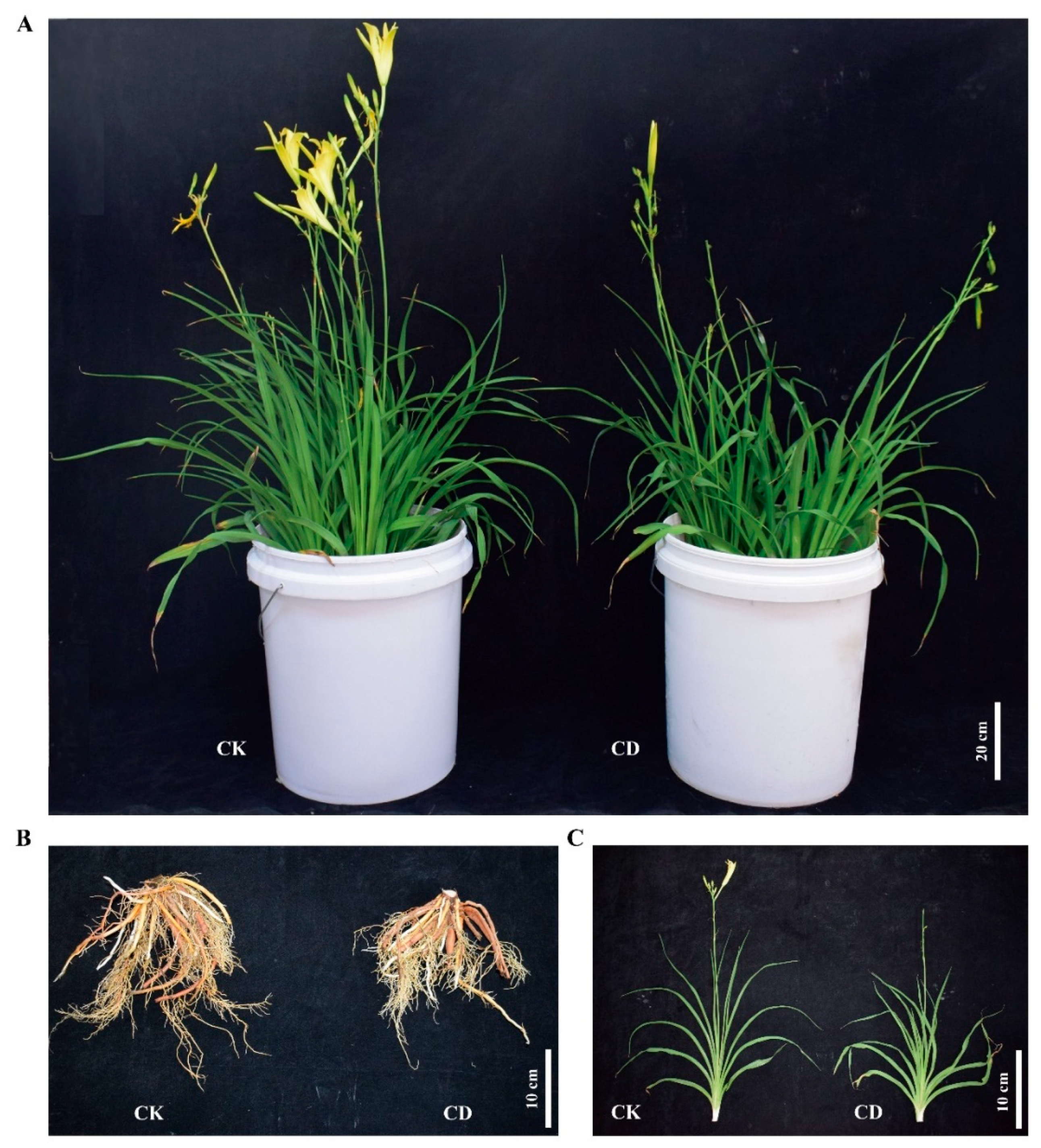
Figure 2.
Identification of DEGs and KEGG pathway analysis of enriched DEGs. (A) Number of DEGs between the comparisons of CK-R vs CD-R, and CK-L vs CD-L groups. (B) Venn diagram illustrating the overlap and tissue-specificity of cold-responsive DEGs between root and leaf samples. The numbers represent counts of root-specific DEGs, leaf-specific DEGs, and shared DEGs, respectively. (C) Top 20 pathways of significantly enriched DEGs from CK-R vs CD-R; (D) Top 20 pathways of significantly enriched DEGs from CK-L vs CD-L. The x-axis represents the rich factor; dot color corresponds to q-value (red = highly significant enrichment), dot size indicates the number of enriched DEGs in each pathway. CK-L and CD-L represent the control group and low-temperature treatment group of H. fulva leaves, respectively. CK-R and CD-R represent the control group and low-temperature treatment group of H. fulva roots, respectively.
Figure 2.
Identification of DEGs and KEGG pathway analysis of enriched DEGs. (A) Number of DEGs between the comparisons of CK-R vs CD-R, and CK-L vs CD-L groups. (B) Venn diagram illustrating the overlap and tissue-specificity of cold-responsive DEGs between root and leaf samples. The numbers represent counts of root-specific DEGs, leaf-specific DEGs, and shared DEGs, respectively. (C) Top 20 pathways of significantly enriched DEGs from CK-R vs CD-R; (D) Top 20 pathways of significantly enriched DEGs from CK-L vs CD-L. The x-axis represents the rich factor; dot color corresponds to q-value (red = highly significant enrichment), dot size indicates the number of enriched DEGs in each pathway. CK-L and CD-L represent the control group and low-temperature treatment group of H. fulva leaves, respectively. CK-R and CD-R represent the control group and low-temperature treatment group of H. fulva roots, respectively.
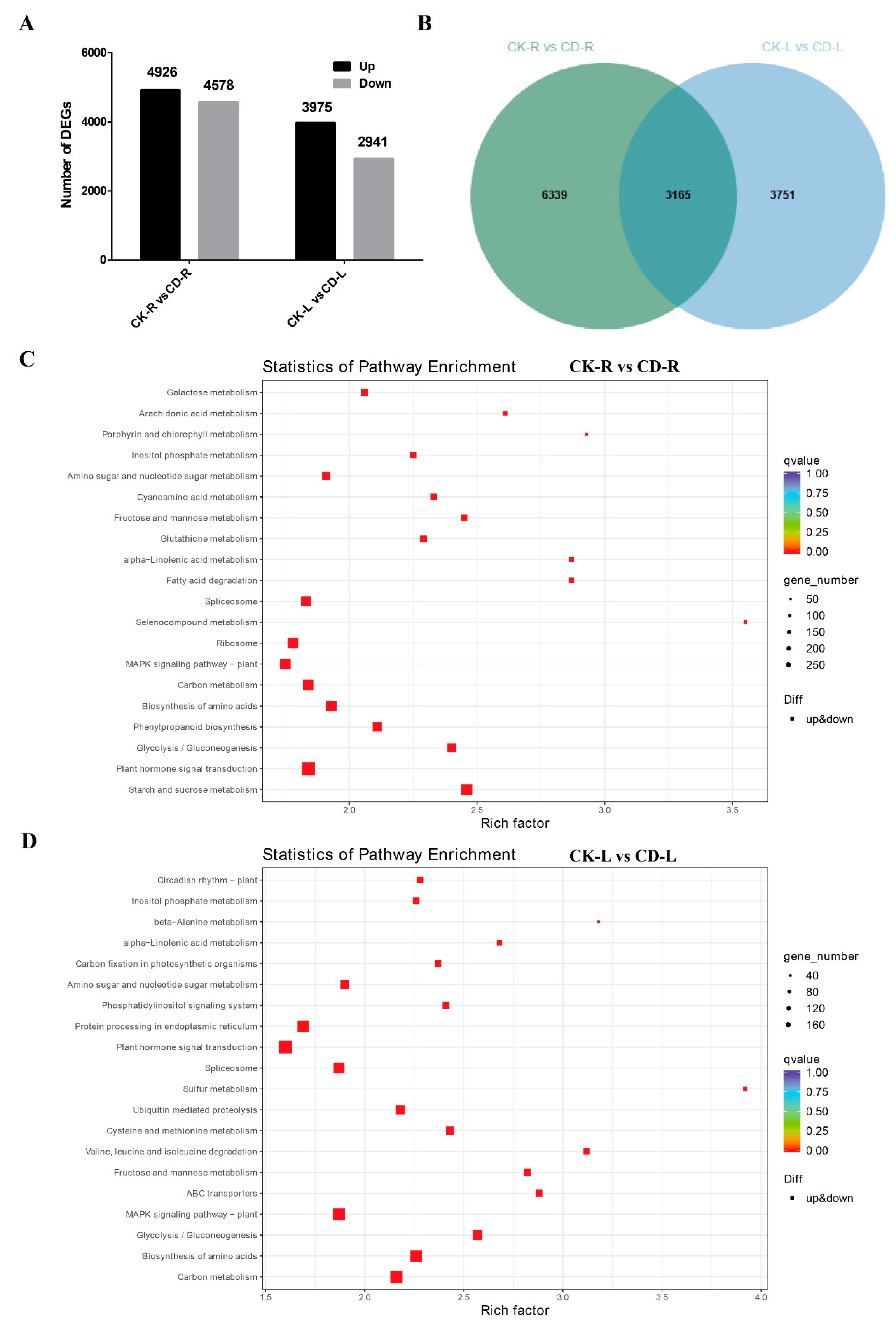
Figure 3.
Expression of genes related to ethylene biosynthesis and signaling pathway in H. fulva under cold stress. (A) Schematic overview of ethylene biosynthesis and signaling pathway. Expression patterns of ethylene biosynthesis, signal transduction and downstream transcription factors as well as functional target genes in H. fulva roots and leaves under cold stress, including ACS genes (B), and ACO genes (C), ETR2 genes (D), CTR1 genes (E), EIN2 genes (F), EIN3 genes (G), ERF genes (H), DREB genes (I) and COR genes (J). Note: CK-L and CD-L represent the control group and low-temperature treatment group of H. fulva leaves, respectively. CK-R and CD-R represent the control group and low-temperature treatment group of H. fulva roots, respectively. Each row represents a single gene, and the columns represent samples from different groups. Colors indicate gene expression levels as FPKM values. Lower levels of expression are represented in blue, higher expression is indicated in yellow, and the highest expression is indicated in red.
Figure 3.
Expression of genes related to ethylene biosynthesis and signaling pathway in H. fulva under cold stress. (A) Schematic overview of ethylene biosynthesis and signaling pathway. Expression patterns of ethylene biosynthesis, signal transduction and downstream transcription factors as well as functional target genes in H. fulva roots and leaves under cold stress, including ACS genes (B), and ACO genes (C), ETR2 genes (D), CTR1 genes (E), EIN2 genes (F), EIN3 genes (G), ERF genes (H), DREB genes (I) and COR genes (J). Note: CK-L and CD-L represent the control group and low-temperature treatment group of H. fulva leaves, respectively. CK-R and CD-R represent the control group and low-temperature treatment group of H. fulva roots, respectively. Each row represents a single gene, and the columns represent samples from different groups. Colors indicate gene expression levels as FPKM values. Lower levels of expression are represented in blue, higher expression is indicated in yellow, and the highest expression is indicated in red.
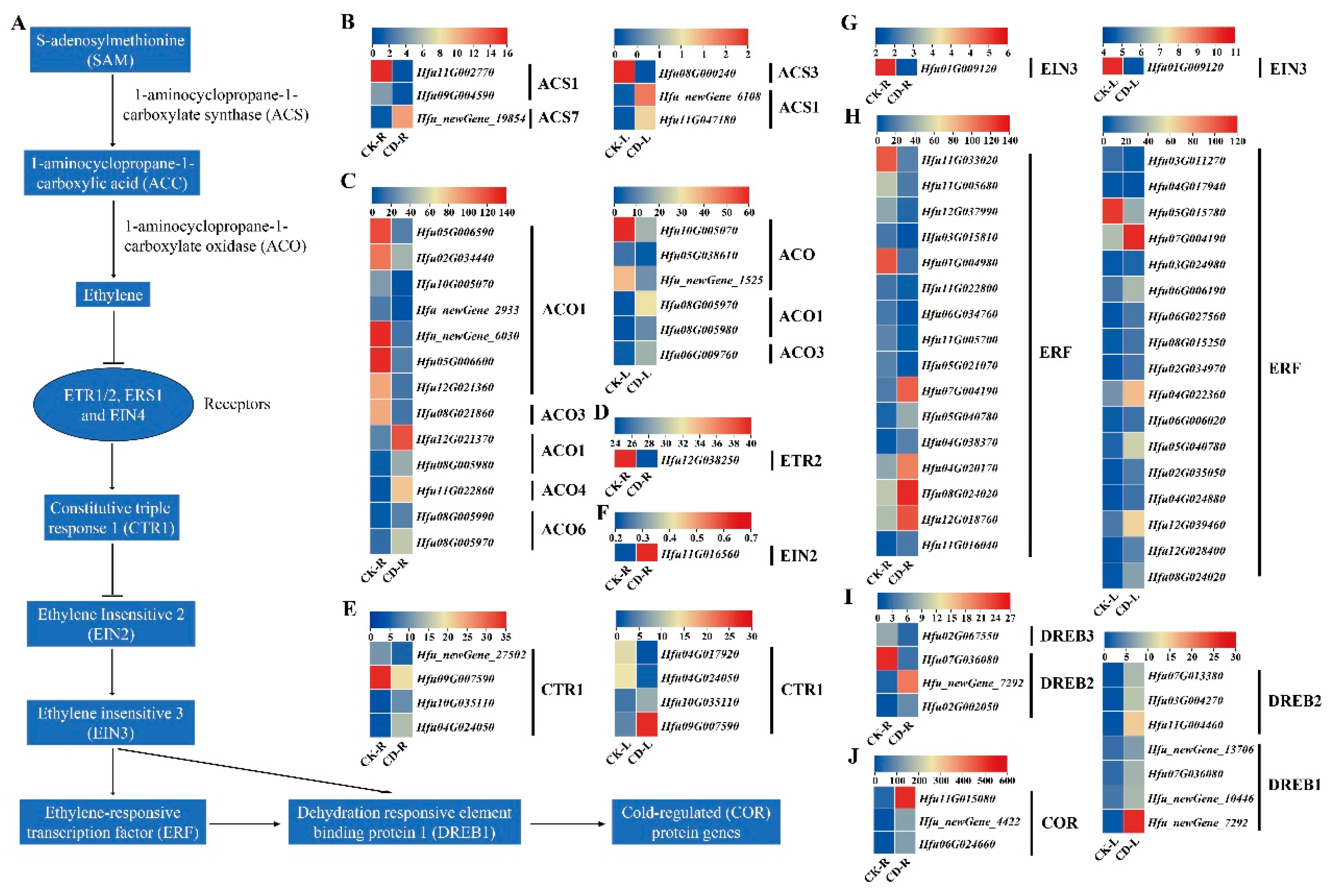
Figure 4.
Expression of genes related to GA biosynthesis and signaling pathway in H. fulva under cold stress. (A) Schematic overview of GA biosynthesis and signaling pathway. Expression patterns of GA biosynthesis, signal transduction and downstream genes in H. fulva roots and leaves under cold stress, including KS genes (B), KO genes (C), KAO genes (D), GA20ox genes (E), GA2ox genes (F), GID1C genes (G), GAI genes (H), and PIF4 genes (I). Note: CK-L and CD-L represent the control group and low-temperature treatment group of H. fulva leaves, respectively. CK-R and CD-R represent the control group and low-temperature treatment group of H. fulva roots, respectively. Each row represents a single gene, and the columns represent samples from different groups. Colors indicate gene expression levels as FPKM values. Lower levels of expression are represented in blue, higher expression is indicated in yellow, and the highest expression is indicated in red.
Figure 4.
Expression of genes related to GA biosynthesis and signaling pathway in H. fulva under cold stress. (A) Schematic overview of GA biosynthesis and signaling pathway. Expression patterns of GA biosynthesis, signal transduction and downstream genes in H. fulva roots and leaves under cold stress, including KS genes (B), KO genes (C), KAO genes (D), GA20ox genes (E), GA2ox genes (F), GID1C genes (G), GAI genes (H), and PIF4 genes (I). Note: CK-L and CD-L represent the control group and low-temperature treatment group of H. fulva leaves, respectively. CK-R and CD-R represent the control group and low-temperature treatment group of H. fulva roots, respectively. Each row represents a single gene, and the columns represent samples from different groups. Colors indicate gene expression levels as FPKM values. Lower levels of expression are represented in blue, higher expression is indicated in yellow, and the highest expression is indicated in red.
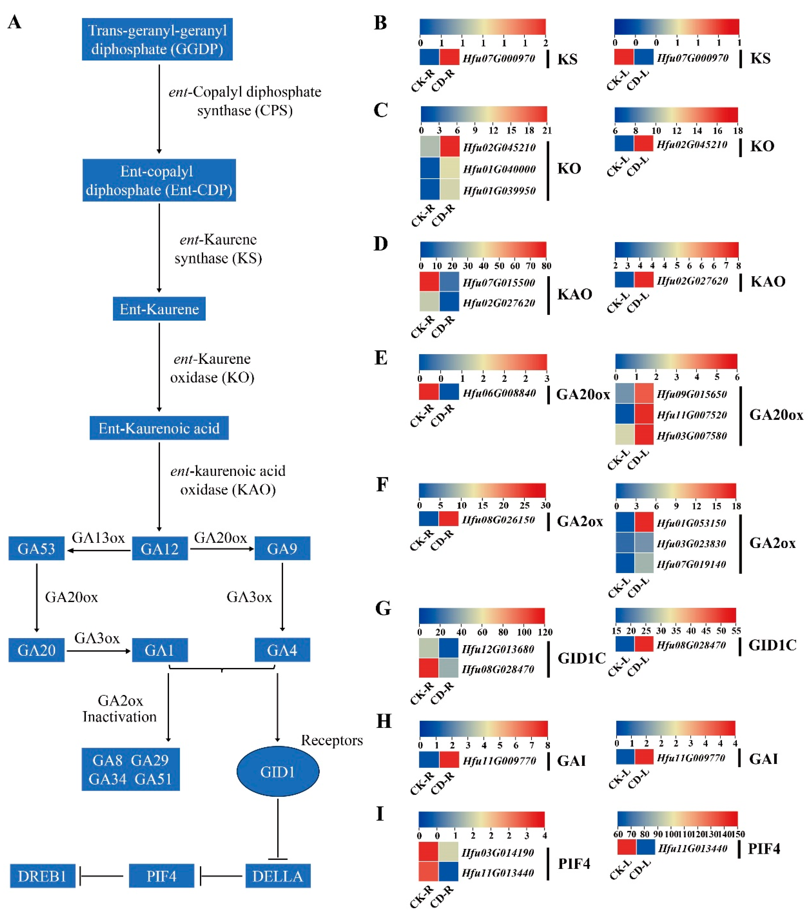
Figure 5.
Expression of genes related to ABA biosynthesis and signaling pathway in H. fulva under cold stress. (A): Schematic overview of ABA biosynthesis and signaling pathway. Expression patterns of ABA biosynthesis, signal transduction, and downstream genes in H. fulva roots and leaves under cold stress, including CHY-β genes (B), ZEP genes (C), NCED genes (D), ABA2 genes (E), ABA3 genes (F), PLY receptor genes (G), and PP2C genes (H). Note: CK-L and CD-L represent the control group and low-temperature treatment group of H. fulva leaves, respectively. CK-R and CD-R represent the control group and low-temperature treatment group of H. fulva roots, respectively. Each row represents a single gene, and the columns represent samples from different groups. Colors indicate gene expression levels as FPKM values. Lower levels of expression are represented in blue, higher expression is indicated in yellow, and the highest expression is indicated in red.
Figure 5.
Expression of genes related to ABA biosynthesis and signaling pathway in H. fulva under cold stress. (A): Schematic overview of ABA biosynthesis and signaling pathway. Expression patterns of ABA biosynthesis, signal transduction, and downstream genes in H. fulva roots and leaves under cold stress, including CHY-β genes (B), ZEP genes (C), NCED genes (D), ABA2 genes (E), ABA3 genes (F), PLY receptor genes (G), and PP2C genes (H). Note: CK-L and CD-L represent the control group and low-temperature treatment group of H. fulva leaves, respectively. CK-R and CD-R represent the control group and low-temperature treatment group of H. fulva roots, respectively. Each row represents a single gene, and the columns represent samples from different groups. Colors indicate gene expression levels as FPKM values. Lower levels of expression are represented in blue, higher expression is indicated in yellow, and the highest expression is indicated in red.
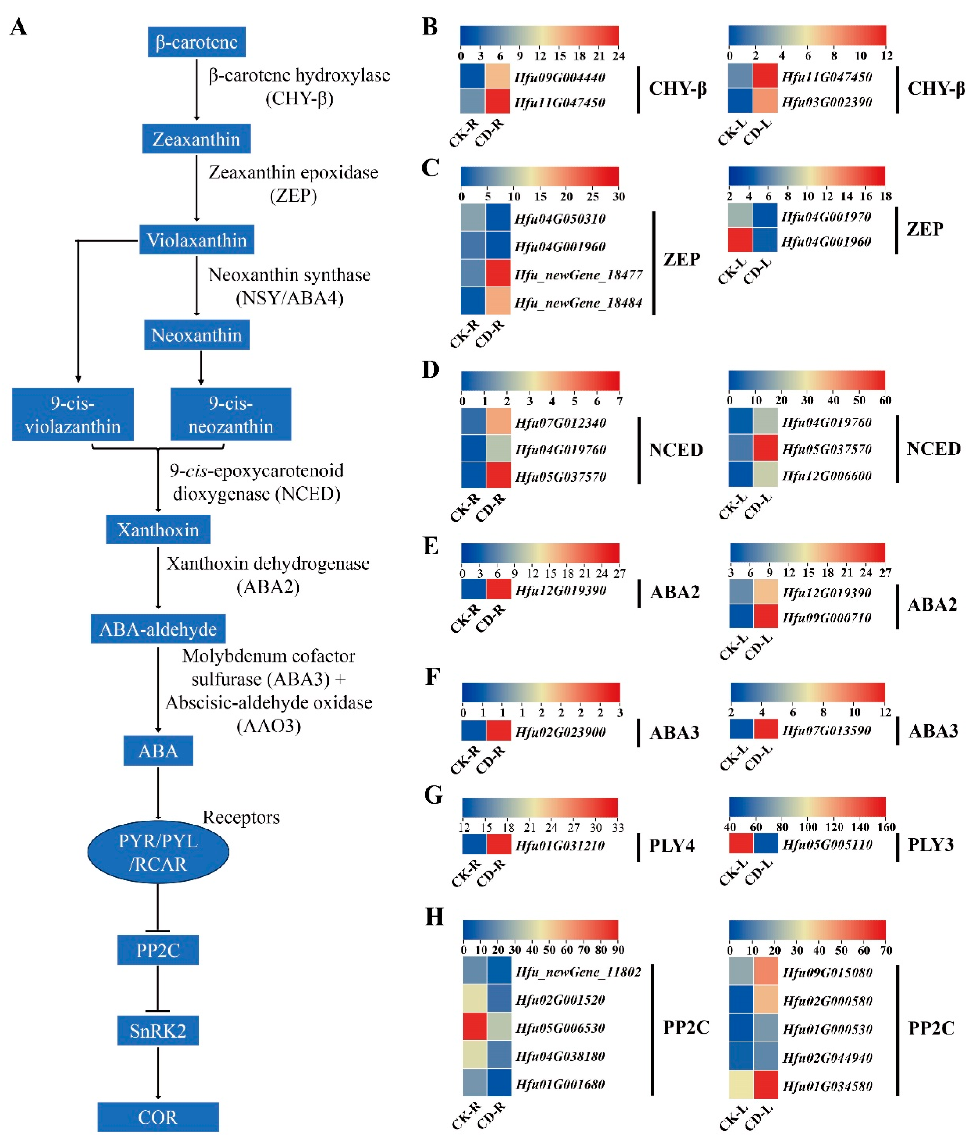
Figure 6.
Transcriptional regulation of the antioxidant defense pathway in H. fulva under cold stress. (A) Schematic overview of the cold-induced reactive oxygen species (ROS) scavenging pathway. Cold stress triggers the accumulation of O2⋅− and lipid peroxides, which are detoxified by sequential actions of superoxide dismutase (SOD), catalase (CAT), peroxidase (POD), glutathione peroxidase (GPX), glutathione S-transferase (GST), and glutathione synthetase (GSH), ultimately reducing oxidative cytotoxicity. (B–G) Heatmaps showing the expression profiles of genes encoding key antioxidant enzymes in roots (CK-R, CD-R) and leaves (CK-L, CD-L) under control (CK) and cold treatment (CD), including SOD genes (B); CAT genes (C); POD genes (D); GPX genes (E); GST genes (F); and GSH genes (G). Each row represents a single gene, and the columns represent samples from different groups. Colors indicate gene expression levels as FPKM values. Lower levels of expression are represented in blue, higher expression is indicated in yellow, and the highest expression is indicated in red.
Figure 6.
Transcriptional regulation of the antioxidant defense pathway in H. fulva under cold stress. (A) Schematic overview of the cold-induced reactive oxygen species (ROS) scavenging pathway. Cold stress triggers the accumulation of O2⋅− and lipid peroxides, which are detoxified by sequential actions of superoxide dismutase (SOD), catalase (CAT), peroxidase (POD), glutathione peroxidase (GPX), glutathione S-transferase (GST), and glutathione synthetase (GSH), ultimately reducing oxidative cytotoxicity. (B–G) Heatmaps showing the expression profiles of genes encoding key antioxidant enzymes in roots (CK-R, CD-R) and leaves (CK-L, CD-L) under control (CK) and cold treatment (CD), including SOD genes (B); CAT genes (C); POD genes (D); GPX genes (E); GST genes (F); and GSH genes (G). Each row represents a single gene, and the columns represent samples from different groups. Colors indicate gene expression levels as FPKM values. Lower levels of expression are represented in blue, higher expression is indicated in yellow, and the highest expression is indicated in red.
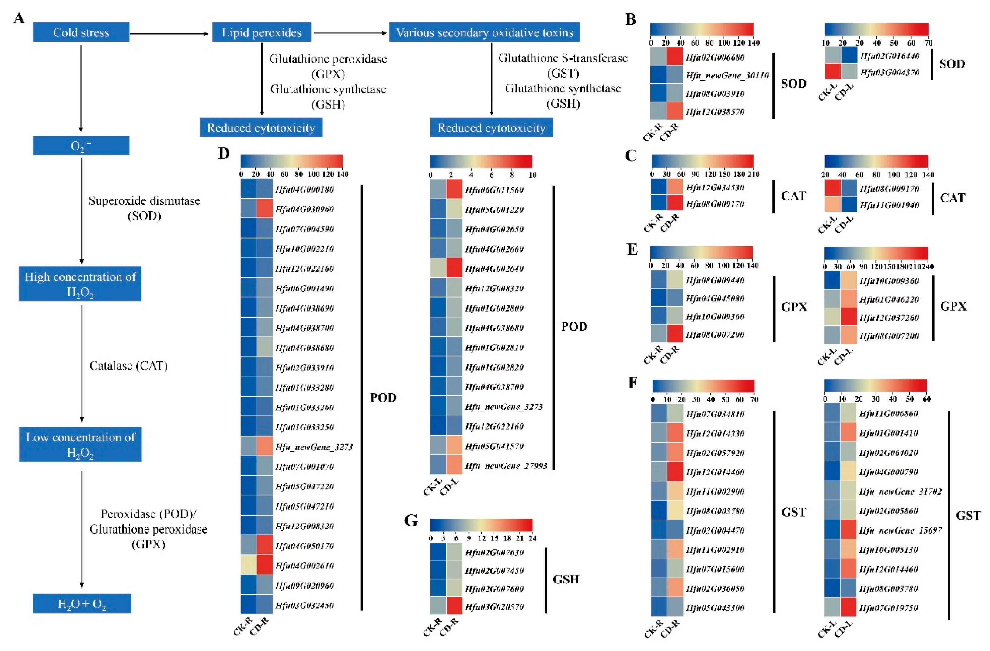
Figure 7.
Expression of genes related to inulin series fructan metabolism in H. fulva under cold stress. (A) Schematic overview of the inulin series fructan biosynthesis and degradation pathway. Expression patterns of inulin series fructan biosynthesis and degradation genes in H. fulva roots and leaves under cold stress, including 1-SST genes (B), 1-FFT genes (C), and 1-FEH genes (D). Note: CK-L and CD-L represent the control group and low-temperature treatment group of H. fulva leaves, respectively. CK-R and CD-R represent the control group and low-temperature treatment group of H. fulva roots, respectively. Each row represents a single gene, and the columns represent samples from different groups. Colors indicate gene expression levels as FPKM values. Lower levels of expression are represented in blue, higher expression is indicated in yellow, and the highest expression is indicated in red.
Figure 7.
Expression of genes related to inulin series fructan metabolism in H. fulva under cold stress. (A) Schematic overview of the inulin series fructan biosynthesis and degradation pathway. Expression patterns of inulin series fructan biosynthesis and degradation genes in H. fulva roots and leaves under cold stress, including 1-SST genes (B), 1-FFT genes (C), and 1-FEH genes (D). Note: CK-L and CD-L represent the control group and low-temperature treatment group of H. fulva leaves, respectively. CK-R and CD-R represent the control group and low-temperature treatment group of H. fulva roots, respectively. Each row represents a single gene, and the columns represent samples from different groups. Colors indicate gene expression levels as FPKM values. Lower levels of expression are represented in blue, higher expression is indicated in yellow, and the highest expression is indicated in red.
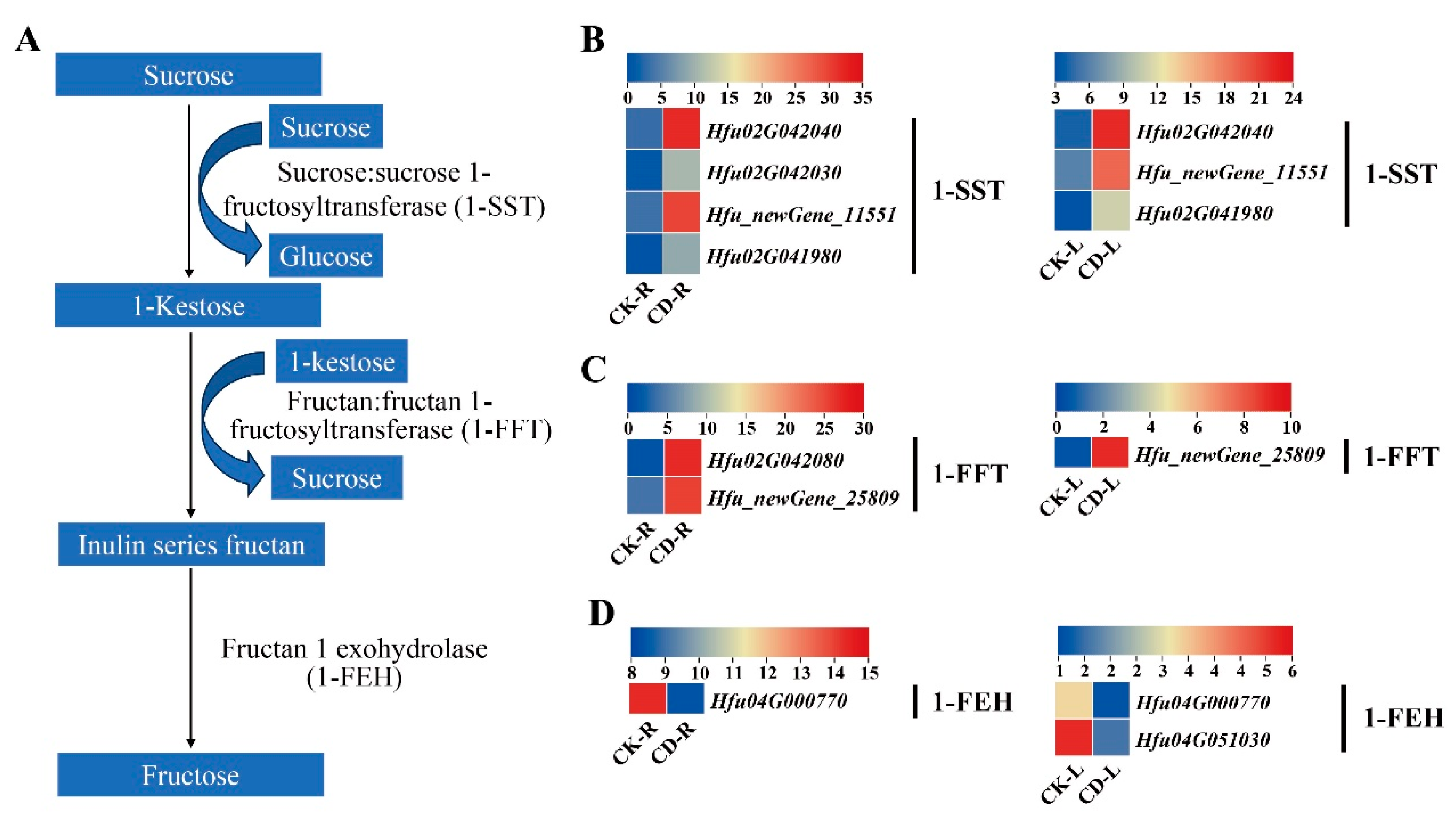
Figure 8.
Expression of genes related to raffinose family oligosaccharides (RFOs) metabolism in H. fulva under cold stress. (A) Schematic overview of RFOs biosynthesis and degradation pathway. Expression patterns of RFOs biosynthesis and degradation genes in H. fulva roots and leaves under cold stress, including GalK genes (B), GolS genes (C), RS genes (D) STS genes (E) and AGA genes (F). Note: CK-L and CD-L represent the control group and low-temperature treatment group of H. fulva leaves, respectively. CK-R and CD-R represent the control group and low-temperature treatment group of H. fulva roots, respectively. Each row represents a single gene, and the columns represent samples from different groups. Colors indicate gene expression levels as FPKM values. Lower levels of expression are represented in blue, higher expression is indicated in yellow and the highest expression is indicated in red.
Figure 8.
Expression of genes related to raffinose family oligosaccharides (RFOs) metabolism in H. fulva under cold stress. (A) Schematic overview of RFOs biosynthesis and degradation pathway. Expression patterns of RFOs biosynthesis and degradation genes in H. fulva roots and leaves under cold stress, including GalK genes (B), GolS genes (C), RS genes (D) STS genes (E) and AGA genes (F). Note: CK-L and CD-L represent the control group and low-temperature treatment group of H. fulva leaves, respectively. CK-R and CD-R represent the control group and low-temperature treatment group of H. fulva roots, respectively. Each row represents a single gene, and the columns represent samples from different groups. Colors indicate gene expression levels as FPKM values. Lower levels of expression are represented in blue, higher expression is indicated in yellow and the highest expression is indicated in red.
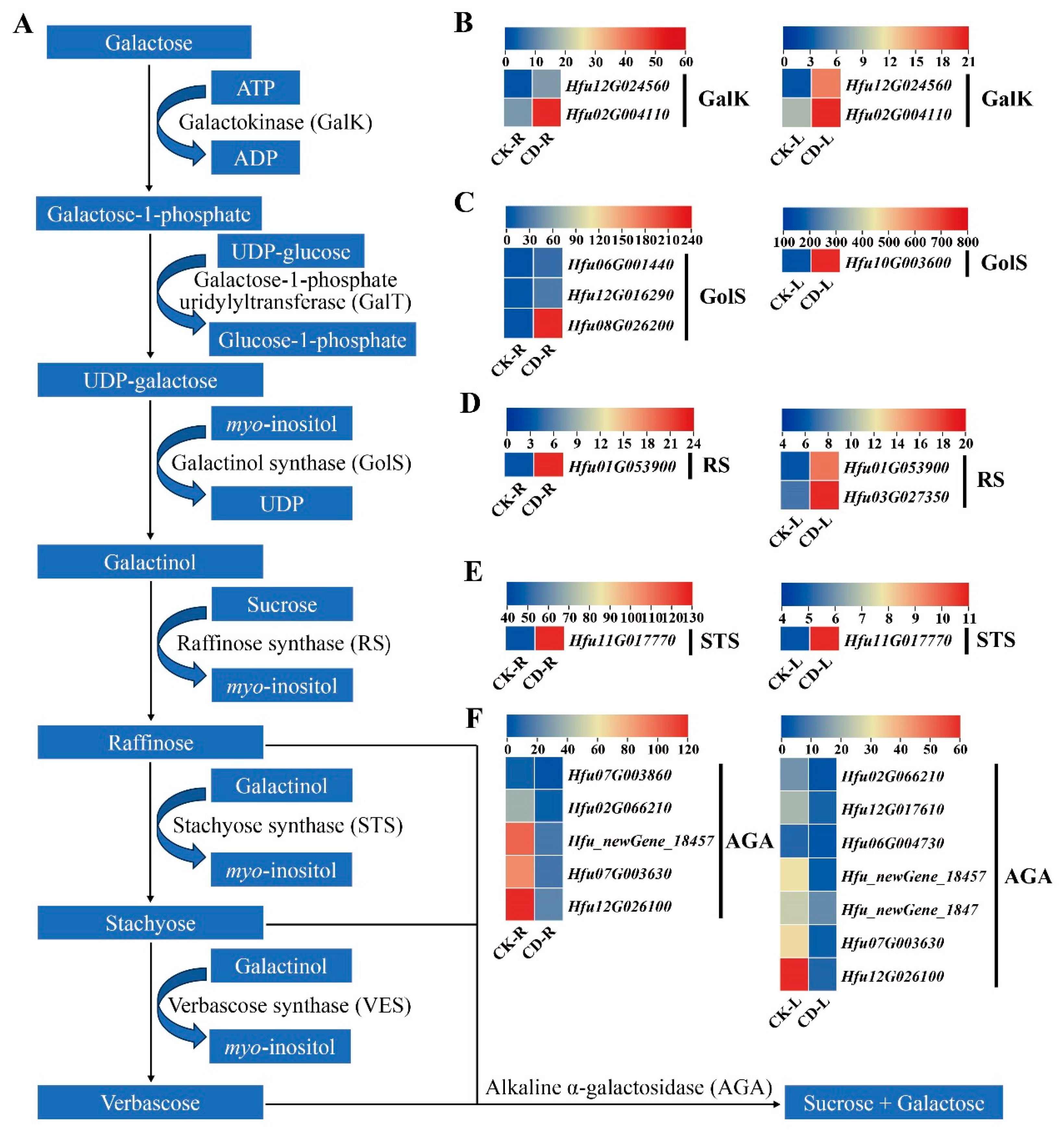
Figure 9.
Expression of genes related to trehalose metabolism in H. fulva under cold stress. (A) Schematic overview of trehalose biosynthesis and degradation pathway. Expression patterns of trehalose biosynthesis and degradation genes in H. fulva roots and leaves under cold stress, including TPS genes (B), TPP genes (C), and TRE genes (D). Note: CK-L and CD-L represent the control group and low-temperature treatment group of H. fulva leaves, respectively. CK-R and CD-R represent the control group and low-temperature treatment group of H. fulva roots, respectively. Each row represents a single gene, and the columns represent samples from different groups. Colors indicate gene expression levels as FPKM values. Lower levels of expression are represented in blue; higher expression is indicated in yellow, and the highest expression is indicated in red.
Figure 9.
Expression of genes related to trehalose metabolism in H. fulva under cold stress. (A) Schematic overview of trehalose biosynthesis and degradation pathway. Expression patterns of trehalose biosynthesis and degradation genes in H. fulva roots and leaves under cold stress, including TPS genes (B), TPP genes (C), and TRE genes (D). Note: CK-L and CD-L represent the control group and low-temperature treatment group of H. fulva leaves, respectively. CK-R and CD-R represent the control group and low-temperature treatment group of H. fulva roots, respectively. Each row represents a single gene, and the columns represent samples from different groups. Colors indicate gene expression levels as FPKM values. Lower levels of expression are represented in blue; higher expression is indicated in yellow, and the highest expression is indicated in red.
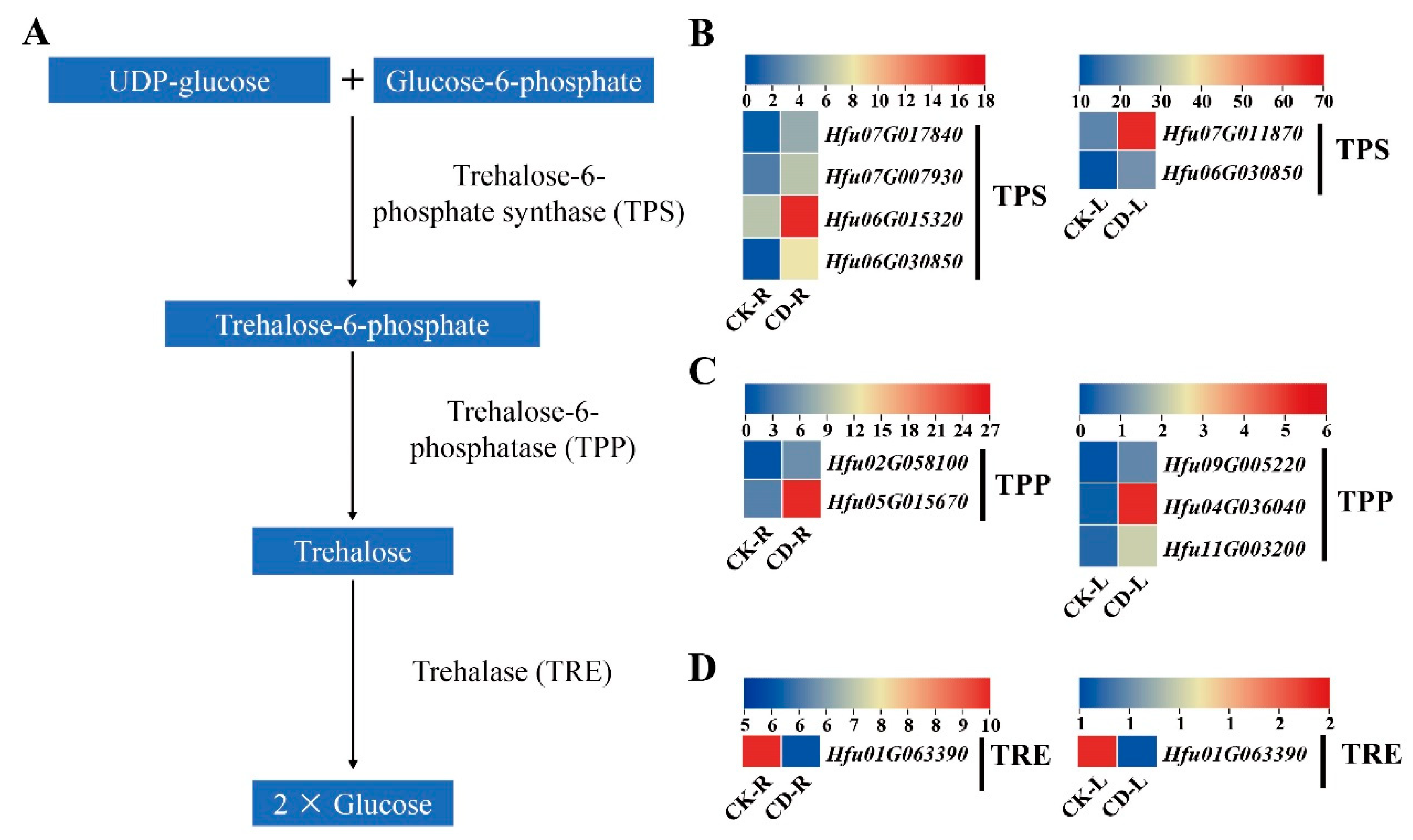
Disclaimer/Publisher’s Note: The statements, opinions and data contained in all publications are solely those of the individual author(s) and contributor(s) and not of MDPI and/or the editor(s). MDPI and/or the editor(s) disclaim responsibility for any injury to people or property resulting from any ideas, methods, instructions or products referred to in the content. |
© 2026 by the authors. Licensee MDPI, Basel, Switzerland. This article is an open access article distributed under the terms and conditions of the Creative Commons Attribution (CC BY) license (http://creativecommons.org/licenses/by/4.0/).
Copyright: This open access article is published under a Creative Commons CC BY 4.0 license, which permit the free download, distribution, and reuse, provided that the author and preprint are cited in any reuse.