

Submitted:
17 July 2026
Posted:
11 August 2026
You are already at the latest version
Abstract
Ziziphus lotus (L.) Lam (wild jujube) leaves are traditionally used in Moroccan medicine to treat hypertension. The present study evaluated the aqueous extract of Z. lotus leaves (ZLAqExt) for its vasorelaxant effects, mechanisms of action, acute toxicity, antioxidant potential, and phytochemical profile. In phenylephrine-precontracted rat aortic rings, the ZLAqExt induced concentration-dependent relaxation (Emax = 79.25 ± 2.77% at 10⁻¹ mg/mL). The mechanism of action was investigated using specific antagonists and blockers. Complete inhibition by endothelium denudation, and preincubation with L-NAME, hydroxocobalamin, ODQ, and thapsigargin confirmed endothelium-dependent nitric ox-ide (NO)/cyclic GMP (cGMP)/sarco/endoplasmic reticulum Ca²⁺-ATPase (SERCA) path-way involvement. A partial inhibition by BaCl₂, indomethacin, calmidazolium, and KCl indicated cyclooxygenase (COX), endothelium-derived hyperpolarizing factor (EDHF), and inward-rectifier potassium (Kir) channel contributions. Acute oral toxicity did not show any mortality at 2 g/kg BW in mice. The extract displayed potent antioxidant activity (DPPH IC₅₀: 18.12 ± 1.49 µg/mL; β-carotene bleaching IC₅₀: 8.3 ± 0.014 µg/mL). UHPLC-ESI-MS identified rutin as the major compound, followed by 3',5'-di-C-β-glucopyranosyl phloretin with high docking affinity for guanylate cyclase and SERCA pump. The presence of flavonoids in the ZLAqExt might have mediated its vaso-relaxant effects, supporting its traditional use as an antihypertensive. Our results high-light that Z. lotus is a potential natural therapeutic agent against vascular dysfunc-tion-related cardiovascular disorders.
Keywords:
Ziziphus lotus
; hypertension
; vasorelaxation
; endothelial signaling pathway
; molecular docking analysis
1. Introduction
Cardiovascular diseases rank as the top global killer, surpassing other pathological states [1,2]. Hypertension constitutes a principal determinant in this mortality burden [3]. Although various categories of antihypertensive pharmaceutical agents are accessible [4], their long-term administration frequently elicits adverse reactions that impact 20% to 97% of treated individuals [5]. This realization accentuates the need for antihypertensive strategies characterized by improved safety and durability profiles. Based on an international scale, the botanical remedies are utilized for the management of numerous pathologies due to their inherent bioactive characteristics. It is estimated that almost 80% of the inhabitants within developing countries rely on traditional therapeutic modalities as foundational healthcare provisions [6,7]. Phytochemical-based derivatives comprise at least 25% of the pharmacopoeia in industrialized countries and facilitate the advancement of innovative therapeutic strategies [8]. Appropriate modulation of vascular smooth muscle preserves the blood pressure homeostasis, however, the irregularities affecting this modulatory capacity may precipitate hypertensive conditions [9]. Natural derivative agents present encouraging prospects for vascular regulation directed at hypertension prevention and management [10].
Ziziphus lotus (L.) Lam. (Rhamnaceae) is a shrub indigenous to the Mediterranean basin. It is widely distributed across North Africa and the Middle East territories and is valued for its therapeutic properties [11]. In the traditional medicine of Morocco, Algeria, and Mauritania, it serves as a common remedy for hypertension [12,13,14]. Pharmacological studies confirm that the extracts from its fruit, leaves, roots, and other parts exhibit antioxidant, anti-inflammatory, analgesic, antimicrobial, antidiabetic, antihyperlipidemic, antitumoral, antiulcerogenic, antispasmodic, and antilithiatic properties [15]. Based on the current state of knowledge, however, the effects of its aqueous leaf extract on vascular tone regulation have not yet been investigated. The present research, therefore, focused on assessing the vasorelaxant effects and underlying mechanisms of the aqueous leaf extract of Z. lotus (L.) Lam (ZLAqExt), along with its antioxidant activities, acute toxicity, and phytochemical profile. Molecular docking simulations further explored the molecular interactions between the ZLAqExt's key compounds and their vasorelaxant targets.
2. Results
2.1. Vasorelaxation Response in Normotensive Rat Aorta
2.1.1. Endothelial Modulation of ZLAqExt-Induced Vasorelaxation in PHE-Constricted Aortic Segments
The aqueous extract of Z. lotus (L.) Lam. (ZLAqExt) elicited notable vasorelaxation in PHE-precontracted aortic rings from rats (n = 6) with an intact endothelium. The aortic rings exhibited a concentration-dependent relaxation in response to ZLAqExt at concentrations of 10⁻³, 10⁻², and 10⁻¹ mg/mL. The extract yielded an effective concentration (EC₅₀) of 0.036 ± 0.012 mg/mL against PHE-induced contraction, with a corresponding Emax of 79.25 ± 2.77% at 10-1 mg/mL (Figure 1 [A, B]). However, the relaxing effect of CCH and ZLAqExt on isolated aortic rings was completely abolished after mechanical disruption of the endothelium.
2.1.2. Involvement of the Ca2+-Calmodulin /NO/sGC Pathway and Muscarinic Receptors in ZLAqExt-Induced Vasorelaxation
The vasorelaxant effect of ZLAqExt was completely abolished in endothelium-intact aortic rings pretreated with the eNOS inhibitor L-NAME, the sGC inhibitor ODQ, or the NO scavenger hydroxocobalamin (Figure 2 and 3A). Conversely, when tissues were pre-treated with the selective muscarinic receptor antagonist atropine, a similar vasorelaxant effect was maintained, with an Emax of 82.08 ± 2.19% (p > 0.05, n = 6). However, the inhibition was less pronounced with the Ca2+-calmodulin antagonist calmidazolium (Emax = 61.22 ± 2.64%; p < 0.001, n = 6) (Figure 2 and 3B).
2.1.3. Involvement of the COX and EDHF Pathways in ZLAqExt-Induced Vasorelaxation
Pre-incubation with indomethacin (a non-selective COX inhibitor) significantly blunted the ZLAqExt-induced vasorelaxation, limiting the maximal response to 38.75 ± 3.84% (p < 0.001, n = 6). Furthermore, the relaxation was partially impaired in the presence of KCl, suggesting a role for endothelium-derived hyperpolarizing factors (Emax = 59.32 ± 2.69%; p < 0.001, n = 6) (Figure 2 and 3B).
2.1.4. Involvement of Potassium Channels (KIR, KV, KCa, and KATP) in ZLAqExt-Induced Vasorelaxation
The vasorelaxant response induced by ZLAqExt was significantly attenuated following pretreatment with BaCl2 (Emax = 52.87 ± 1.62%; p < 0.001, n = 6) (Figure 4. [A, B]). In contrast, no significant changes were observed after the application of 4-AP (Emax = 71.19 ± 2.15%), TEA (Emax = 80.52 ± 2.29%), or glibenclamide (Emax = 71.20 ± 1.06%).
2.1.5. Involvement of VOC Channels and the SERCA Pump in ZLAqExt-Induced Vasorelaxation
The vasorelaxant activity of ZLAqExt was completely suppressed following the pretreatment with thapsigargin, a SERCA pump inhibitor (Figure 4. [C, D]). In contrast, upon inhibition of VOC channels by verapamil, the relaxant effect remained largely unchanged, with a maximal response of 71.53 ± 2.02% (p > 0.05, n = 6).
2.2. Acute Toxicity
No mortality, clinical signs of toxicity, or behavioral changes were observed in mice treated with ZLAqExt compared to the control group receiving tap water. Furthermore, no evidence of organ hypertrophy, tissue inflammation, mucosal erosion, or significant body weight loss was recorded during the observation period. Based on these results, ZLAqExt is considered non-toxic, with a median lethal dose (LD50) exceeding 2000 mg/kg BW.
2.3. Antioxidant Properties
ZLAqExt demonstrated concentration-dependent inhibitory activity against DPPH free radicals (Figure 5A). The highest scavenging effect was observed at 0.4 mg/mL, with a maximum value of 93.94%. The median inhibitory concentration (IC50) of ZLAqExt was determined to be 18.12 ± 1.49 µg/mL. For ascorbic acid, its activity was significant, characterized by an Emax of 98.71% and an IC50 value of 7.73 ± 0.83 µg/mL.
The inhibition of linoleic acid-induced β-carotene oxidation presented comparable efficacy in ZLAqExt (Emax = 72.60 ± 1.06%) and the standard ascorbic acid (Emax = 77.09 ± 2.78%) (Figure 5B). The IC50 value of the extract in this essay was 8.3 ± 0.014 µg/mL.
2.4. Phytochemical Profiling
2.4.1. Quantification of Total Phenolic and Tannin Constituents
ZLAqExt contained significant amounts of total polyphenols and tannins (Figure 6), with estimated contents of 82.04 ± 4.86 mg GAE/g Ext and 31.83 ± 1.53 mg GAE/g Ext, respectively. These results clearly show that, within the overall total polyphenol content, 50.21 ± 3.33 mg GAE/g Ext corresponds to non-tannin compounds. It can therefore be inferred that the aqueous leaf extract of Z. lotus (L.) Lam. contains more non-tannin phenolic compounds than tannins.
2.4.2. UHPLC-ESI-MS Profile of ZLAqExt
The UHPLC-ESI-MS chromatogram and chemical composition of ZLAqExt are presented in Figure 7 and Table 1, respectively. The peaks were identified and assigned based on the matching of retention times and mass spectral data with those reported in the literature. Finally, the standards of the identified compounds were injected under the same conditions. The results indicated that our extract predominantly contained rutin (major peak), along with 3’,5’-di-C-β-glucopyranosyl phloretin, myricetin-3-O-rutinoside, and kaempferol-3-O-rutinoside (nicotiflorin).
2.5. Molecular Docking Analysis
To explore the potential molecular interactions underlying the vasorelaxant activity of ZLAqExt, the major phytoconstituents identified in the extract were computationally evaluated against the key proteins involved in the NO/GC/SERCA signaling pathway. This pathway plays a central role in vascular relaxation through the coordinated regulation of nitric oxide signaling, cyclic GMP production, and intracellular calcium homeostasis. Endothelial nitric oxide synthase (eNOS) was not included in the present docking analysis due to its highly complex activation mechanisms involving multiple signaling cascades and regulatory protein interactions [19].
Prior to virtual screening, the docking protocol was validated using experimentally resolved holo-structures of guanylate cyclase (GC) to assess the reliability of the predicted ligand-binding poses. As reference compounds, riociguat and CDN1163 were used as benchmark ligands for GC and SERCA, respectively. CDN1163 has been previously reported as a SERCA-binding ligand associated with modulation of Ca2+ homeostasis [20]. Because no co-crystallized agonist was available for the SERCA structure, blind docking was performed for this receptor.
Docking accuracy was evaluated by comparing the experimental and predicted binding conformations of the co-crystallized GC ligand, yielding an RMSD below 2.0 Å. This level of agreement supports the reliability of the predicted binding poses generated by the DiffDock framework, as illustrated in Figure 8. Following docking validation, MM-PB(GB)SA rescoring was performed to estimate ligand–receptor binding free energies and improve the reliability of affinity prediction.
Among the investigated phytoconstituents, 3’,5’-di-C-β-glucopyranosyl phloretin (CGP) exhibited the most favorable predicted binding free energies toward both GC and SERCA compared with the other tested compounds, including the reference ligand CDN1163 (Figure 9 [A, B]). Interaction analysis further suggested that CGP established multiple stabilizing interactions within the binding pockets of both targets, including hydrogen bonding and hydrophobic contacts with key amino acid residues. The high density of hydroxyl groups present in the CGP structure may contribute to enhanced polar interactions and stabilization of the ligand–protein complexes.
Although these computational findings provide valuable structural insights into the potential interactions between ZLAqExt phytoconstituents and vasorelaxation-associated targets, further molecular dynamics simulations and experimental biophysical validation are required to confirm the stability and functional relevance of the predicted complexes.
3. Discussion
In traditional Moroccan medicine, Ziziphus lotus (L.) Lam. is used for hypertension management [12]. This study evaluated the vasorelaxant effects, phytochemical composition, and antioxidant activity of its leaf aqueous extract (ZLAqExt). Results showed no acute toxicity in mice, and experiments in isolated rat aorta indicated endothelium-dependent vasorelaxation. The primary mechanisms involve NO pathway activation and SERCA pump stimulation, with partial contributions from COX metabolites, EDHF, and inward-rectifier K⁺ channels.
ZLAqExt (10⁻³–10⁻¹ mg/mL) induced concentration-dependent relaxation of phenylephrine-precontracted aortic rings with functional endothelium (EC₅₀ = 0.036 ± 0.012 mg/mL; Emax = 79.25 ± 2.77%). The concentration of 10⁻¹ mg/mL produced the greatest vasorelaxation, surpassing the effects reported for the aqueous extract of Garcinia cowa Roxb. ex Choisy leaves [21]. Furthermore, ZLAqExt demonstrated significantly greater effectiveness than extracts of Chenopodium ambrosioides L. [22] and Artemisia campestris L. [23], which produced only 13.4% and 15% relaxation at 10⁻¹ mg/mL, respectively, under the same experimental protocol.
Carbachol is known to promote vascular relaxation by activating muscarinic receptors, which trigger NO release from endothelial cells. This NO diffuses into the vascular smooth muscle cells, activating soluble guanylate cyclase (sGC) and elevating cGMP levels [24]. These findings support the primary goal of this study, which was to elucidate the underlying mechanisms of ZLAqExt-induced vasorelaxation by investigating various cellular signaling pathways, including both endothelium-dependent and endothelium-independent routes.
The vascular endothelium plays a critical role in sustaining vascular tone by releasing three key vasodilators: NO, prostacyclin (PGI2), and EDHF [25,26]. According to our data, ZLAqExt was found to be endothelium-dependent, as its impact was completely abolished following the denudation process. An investigation was also conducted to determine the contribution of endothelial mediators and to confirm the observed vasorelaxant response.
Numerous receptors, including muscarinic receptors, are known to be involved in NO production in endothelial cells. This is brought about by CaM complex-induced activation of eNOS [27,28,29]. Our findings demonstrated that the suppression of muscarinic receptors by atropine had no impact on the vasorelaxation induced by ZLAqExt. However, when the aortic rings were exposed to L-NAME (a NOS inhibitor), hydroxocobalamin (a NO scavenger), or ODQ (an inhibitor of soluble guanylyl cyclase), the observed response was completely inhibited. In contrast, calmidazolium (a Ca2+-calmodulin binding blocker) led to a slight attenuation in vasorelaxation. These results suggest that the primary mechanism by which ZLAqExt induces its vasorelaxant effects is the release of NO from endothelial cells.
Additionally, our results indicated that the activation of NOS, which is likely triggered by a brief elevation in intracellular calcium, does not involve muscarinic receptors. Instead, other receptors may be implicated, such as bradykinin B2 receptors [30,31] or β2-adrenoceptors, which can regulate eNOS activity through a protein kinase B (Akt)-dependent pathway [32]. Beyond NO, PGI2 (a COX product) also mediates endothelium-derived relaxation by stimulating adenylate cyclase and increasing cAMP levels [33]. Our data suggests that ZLAqExt stimulates prostanoid production, as pretreatment with the COX inhibitor indomethacin significantly reduced the vasorelaxant effects.
Likewise, vascular tone is influenced by EDHF, which induces hyperpolarization in smooth muscle by opening potassium (K+) channels, such as Kca and KATP channels [34]. ZLAqExt- induced relaxation was partially attenuated when the EDHF response was inhibited by high extracellular potassium (30 mM), which alters the electrochemical gradient [35,36]. This suggests that PGI2 may partially mediate vasodilation by stimulating EDHF release, possibly through the AC/cAMP-dependent pathway.
Since K+ channels are the primary factor determining the potential of the cell membrane, their activation plays a major role in vascular tone regulation. Endothelium-dependent vasodilators open the K+ ion channels in the membrane, permitting K+ ions to exit. This lowers the membrane potential and induces hyperpolarization, which in turn closes voltage-gated Ca2+ channels in the cell membrane, ultimately leading to vascular muscle relaxation [34,37]. The contribution of K+ channels was examined using various blockers. Our findings highlight the partial involvement of inwardly rectifying potassium (Kir) channels. This suggests that the aqueous extract may not directly activate Kir channels, but may facilitate the conditions conducive to NO release, which in turn influences channel activity.
One of the important factors controlling the contractile state of vascular smooth muscle is the cytosolic calcium concentration ([Ca2+]i). Voltage-operated calcium (VOC) channels, which are precisely regulated by the membrane potential, are activated in response to changes in [Ca2+]i [38,39]. The influx of extracellular calcium is necessary to control the smooth muscle vasoconstriction capacity and preserve the equilibrium of ions inside and outside the cell. The sarcoplasmic reticulum Ca2+-ATPase (SERCA) pump is one of the key regulators of the sarcoplasmic reticulum function, which is essential for the storage and release of calcium [40]. Based on our findings, the VOC inhibitor verapamil did not alter the effect of ZLAqExt, while the response was completely suppressed after pretreatment with thapsigargin, an inhibitor of the SERCA pump. This suggests a complex interplay between NO and the SERCA pump. It also indicates that the aqueous extract may not directly activate the SERCA pump; rather, its action may likely result from the release of NO from endothelial cells. Furthermore, the literature supports the notion that NO can influence SERCA function, thereby facilitating vasorelaxation [41]. Thus, while both NO and SERCA are critical for the extract's effects, it appears that NO primarily mediates these responses, with SERCA activation being a downstream effect of NO signaling.
In summary, ZLAqExt exhibits significant endothelium-dependent vasorelaxant activity. This effect primarily involves the NO pathway and SERCA pump activation, with partial contributions from COX, EDHF, and Kir channel opening (Figure 10).
The oral acute toxicity of the extract was evaluated to ensure the safety of ZLAqExt. The results showed an LD50 higher than 2 g/kg BW, which is comparable to that reported for Algerian Z. lotus (L.) Lam. leaves [42]. It is well-recognized that a correlation exists between antioxidant potential and vasorelaxation. Increased production of reactive oxygen species (ROS), often associated with cardiovascular risk factors such as hypertension, can reduce NO bioavailability. This, in turn, impairs vasodilation and increases vascular tone, contributing to the development of atherosclerosis and other cardiovascular diseases [43,44]. Moreover, ROS participate in signaling pathways influencing vascular cell proliferation, inflammation, and apoptosis, the processes central to vascular remodeling and the pathogenesis of hypertension [44,45].
Consequently, a complementary study was conducted to evaluate the capacity of the extract to scavenge DPPH free radicals and protect against β-carotene oxidation in vitro. ZLAqExt was characterized by a significant antioxidant potential (IC50 = 0.01 mg/mL), comparable to values obtained from leaves collected in Tunisia [46] and significantly more potent than the aqueous extract from Algerian leaves (IC50 = 0.342 mg/mL) [47].
Phytochemical studies revealed that ZLAqExt is rich in polyphenols, particularly non-tannin compounds. UHPLC-ESI-MS analysis confirmed the presence of flavonoids, with quercetin-3-O-rutinoside as the major compound, followed by 3’,5’-di-C-β-glucopyranosylphloretin, kaempferol-3-O-rutinoside, and myricetin-3-O-rutinoside. These findings are consistent with the data reported by Rached et al. (2019) on the phytochemical profile of wild jujube from western Algeria, where leaves and fruit predominantly contained flavonoid derivatives [16]. Notably, 3’,5’-di-C-β-glucopyranosylphloretin was identified for the first time among the main flavonoids in leaves from the eastern region of Morocco. However, when the aqueous extract of leaves collected from the Sidi Sliman region of Morocco was prepared using the same extraction method (infusion), its phytochemical analysis revealed that phenolic acids were the main compounds. Gallic acid, vanillic acid, caffeic acid, pyrogallol, and chlorogenic acid were found to be the predominant compounds, along with p-hydroxybenzoic acid, p-coumaric acid, naringin, and trace amounts of rutin [48]. Furthermore, rutin was identified as the major constituent in the methanolic and water extracts of Algerian Ziziphus lotus leaves [49]. Myricetin 3-O-rutinoside and kaempferol 3-O-rutinoside were also reported in leaf samples from Western Algeria [16].
There is ample evidence that the flavonoids found in ZLAqExt, especially rutin, possess potent endothelium-dependent vasorelaxant properties mediated through the NO/sGC pathway and the activation of KATP channels [50,51]. Myricetin, the aglycone of myricetin-3-O-rutinoside, has been shown to interact with sGC and promote the overexpression of SERCA [52,53], suggesting that it may also contribute to the observed vasorelaxant effects. Additionally, kaempferol-3-O-rutinoside has been reported to elicit endothelium-independent relaxation (Emax = 76%) in isolated rat aorta via a cholinergic pathway [54].
The MM-PB(GB)SA analysis provided additional insight into the predicted binding affinities of the investigated phytoconstituents toward the selected protein targets. Among the evaluated compounds, 3’,5’-di-C-β-glucopyranosyl phloretin (CGP) displayed the most favorable predicted binding free energies against both GC and SERCA as compared to other tested molecules. These findings suggest that CGP may establish more stable ligand–protein interactions within the binding regions of these targets. Guanylate cyclase plays a central role in nitric oxide-mediated vasodilation through cGMP generation, whereas SERCA is critically involved in intracellular calcium homeostasis in vascular smooth muscle cells [55]. Therefore, the predicted interaction profile of CGP with both targets may partially contribute to the vasorelaxant effects observed experimentally for ZLAqExt.
The MM-PB(GB)SA rescoring approach further strengthened the interpretation of the docking results by providing complementary estimations of ligand–receptor binding free energies. This method integrates molecular mechanics energy terms with implicit solvation models based on Poisson–Boltzmann and generalized Born formalisms, allowing improved evaluation of protein–ligand interaction stability at the molecular level [56]. The favorable ΔG values obtained for CGP support the docking predictions generated using the DiffDock framework. In addition, the interaction analysis suggested that CGP established multiple stabilizing interactions within the binding pockets of GC and SERCA, including hydrogen bonding and hydrophobic contacts with key amino acid residues. The high number of hydroxyl groups present in the CGP structure may contribute to enhanced polar interactions and stabilization of the ligand–protein complexes.
Taken together, the ex vivo vascular experiments and in silico findings suggest the presence of bioactive phytoconstituents within ZLAqExt capable of interacting with proteins associated with vascular relaxation pathways. Nevertheless, these computational predictions should be interpreted cautiously, as molecular docking and MM-PB(GB)SA analyses alone cannot confirm the functional modulation of the investigated targets. Additional molecular dynamics simulations, biochemical assays, and in vivo investigations will therefore be necessary to validate the stability, specificity, and physiological relevance of the predicted ligand–protein interactions.
Overall, the present study provides preliminary structural insight into the possible contribution of Z. lotus phytoconstituents to vascular relaxation and supports further investigation of CGP as a potential bioactive compound involved in endothelial-dependent vasorelaxant mechanisms.
4. Materials and Methods
4.1. Chemicals
The pharmacological agents employed in this study consisted of (R)-(-)-phenylephrine hydrochloride [Phe] (procured from Sigma Aldrich, Germany), 1H-[1,2,4]oxadiazolo[4,3-a]quinoxalin-1-one [ODQ] (supplied by Cayman Chemical, USA), 4-aminopyridine [4-AP] (obtained from Alfa Aesar, Germany), atropine and verapamil hydrochloride (sourced respectively from Sigma Aldrich, China), indomethacin (acquired from Sigma Aldrich-Fluka, Italy), Nω-Nitro-L-arginine methyl ester hydrochloride [L-NAME] (provided by Sigma Aldrich, Switzerland), hydroxocobalamin hydrochloride (supplied by Fluka, USA), thapsigargin (procured from Sigma Aldrich, Israel), barium chloride dihydrate [BaCl2] (obtained from VWR, Belgium), calmidazolium, glibenclamide, tetraethylammonium chloride hydrate [TEA], and carbamylcholine chloride [carbachol, CCH] (all procured from Sigma Aldrich, USA).
Stock solutions of ODQ and thapsigargin were prepared in dimethyl sulfoxide (DMSO), whereas ethanol served as the carrier for indomethacin preparation. All other supplementary reagents were dissolved in distilled water.
4.2. Plant Material
In April 2022, samples of Z. lotus (L.) Lam. leaves, locally known as "Sedra," were gathered in the vicinity of Figuig City in southeastern Morocco (coordinates 32°06'10.6"N 1°14'41.0" W). Professor Mostapha ELACHOURI from the Biology Department of Sciences Faculty at University Mohamed First in Oujda, Morocco, processed and registered the specimen in the department's herbarium for scientific reference under voucher number (HUMPOM428). The plant’s name was verified and validated as suggested by http://www.worldfloraonline.org.
4.3. Aqueous Extract Preparations
The aqueous extract was prepared based on traditional Moroccan practices. Twenty grams of crushed dried leaves were infused for 30 minutes in 200 mL of boiled distilled water. The aqueous extract (AqExt) was subjected to filtration, and the filtrate was concentrated in vacuo at 50 °C using a rotary evaporator (EYELA N-1300, Japan). The yield of the extraction was 15%. The crude extract was conserved in a plastic bottle in the freezer at -20 ºC until use.
4.4. Experimental Animals
The present study involved male Wistar rats (average weight 300 g), and albino mice, weighing 25–35 g. The animals were bred, housed, and acclimatized to regular environmental conditions in the animal facility of the Faculty of Sciences, Oujda, Morocco. The animals were fed a specifically tailored rat chow, had unrestricted access to water, and were exposed to a regulated 12-hour light-dark cycle. The recommendations, given in the "Guide for the Care and Use of Laboratory Animals" [57], were strictly followed throughout the experimental period. Ethical approval for all the experimental protocols was granted by the Faculty of Sciences, Mohamed First University, Oujda, Morocco (Authorization No. 11/2023-LBBES).
4.5. Preparation of Thoracic Aortic Rings
Male Wistar rats (n = 20) were anesthetized through intraperitoneal administration of urethane (20%, 1 mL/100 g BW) to perform a thoracotomy. The thoracic aorta was swiftly and delicately removed and was rapidly placed in cold oxygenated Krebs-Henseleit solution (composition in mM: NaCl 119, KCl 4.7, CaCl2 2.6, MgSO4 1.2, KH2PO4 1.2, NaHCO3 25, and glucose 11). Subsequently, the aorta was thoroughly cleaned under a binocular loupe to remove fat and connective tissue adhesions. Afterwards, the isolated aorta was segmented into rings approximately 3–4 mm in length. The rings were delicately mounted between two parallel stainless-steel hooks, which were closed in opposition to each other and suspended in an organ chamber (Emka Technologies, France) filled with 11 mL of Krebs solution (95% O2/5% CO2, 37°C, pH 7.4). One hook was attached to an isometric force transducer (Emka Technologies, France), which detects the vasomotricity of the aortic rings. An amplifier (EMKA Technologies, France) directly amplified the generated voltage signal, which was subsequently acquired by usbACQ (EMKA Technologies, France) and was recorded by the data visualization and analysis software (IOX2). The rings were stretched to a basal tension of 1 g and were left to stabilize for 30 min. Endothelial integrity was confirmed when carbachol (10⁻⁴ M) induced ≥60% relaxation following phenylephrine (10⁻⁶ M)-induced contraction. Endothelium-denuded rings were prepared by gentle mechanical abrasion of the intimal surface; denudation was verified by the absence of carbachol-induced relaxation. Before experiments, the rings were rinsed twice and re-equilibrated for 30 min. The vasorelaxant effect of ZLAqExt and its endothelium dependence were assessed using the rings with intact or denuded endothelium. After stabilization and phenylephrine-induced contraction, cumulative concentrations of ZLAqExt (10⁻³, 10⁻², 10⁻¹ mg/mL) were applied once the contractile response reached a steady state.
4.6. Mechanism of Action Involved in the Vascular Effect
To clarify the mechanisms underlying ZLAqExt-induced vasorelaxation, endothelium-intact aortic rings were preincubated for 20 min with various pharmacological inhibitors [23,52]: L-NAME (10⁻⁴ M, NOS inhibitor), ODQ (10⁻⁵ M, sGC inhibitor), hydroxocobalamin (3×10⁻⁵ M, NO scavenger), indomethacin (10⁻⁵ M, COX inhibitor), BaCl2 (10⁻⁴ M, Kir blocker), 4-AP (10⁻⁴ M, Kv blocker), TEA (10⁻² M, KCa blocker), glibenclamide (10⁻⁵ M, KATP blocker), calmidazolium (10⁻³ µM, Ca²⁺-CaM/eNOS binding inhibitor), KCl (30 mM, EDHF inhibitor), atropine (10⁻⁶ M, muscarinic antagonist), verapamil (10⁻⁵ M, VOC inhibitor), and thapsigargin (10⁻⁷ M, SERCA inhibitor). Following preincubation, the cumulative concentrations of ZLAqExt (10⁻³–10⁻¹ mg/mL) were added after phenylephrine-induced contraction. Responses were compared to control conditions (ZLAqExt alone). Each ring was tested under a single condition (inhibitor or control) to preserve tissue integrity and avoid desensitization caused by repeated exposures; all experiments were performed independently on the rings from separate rats.
4.7. Acute Toxicity
Acute toxicity assessment was undertaken according to OECD guidelines 423 [58] with some modifications. The study was carried out on 12 albino mice (5 females and 7 males, weighing 25-35 g), assigned to control and test groups (n = 6 per group). The control group received distilled water as a vehicle, whereas the test group was given ZLAqExt. The animals were fasted for 16 h before the initiation of the experiment. After weighing, they received a single dose of 2 g/kg BW of ZLAqExt via gastric intubation (10 mL/kg). The mice were subjected to continuous individual observation for 30 min immediately after administration, followed by 24 h, with specific emphasis on the first 4 h. This monitoring continued for 14 days, enabling the recording of any instances of mortality or manifestations of general signs and symptoms of toxicity. Upon completion of treatment, the surviving animals were euthanized. Subsequently, a macroscopic examination was conducted on the internal organs, including the heart, lungs, liver, and kidneys.
4.8. DPPH Free Radical Neutralization Assay
One milliliter of a 0.1 mM 2,2-diphenyl-1-picrylhydrazyl (DPPH) solution prepared in methanol was combined with an identical 1 mL volume of the extract dilutions at concentrations of 5, 10, 20, 50, 100, 200, 400, and 2000 µg/mL. All the dilutions were previously dissolved in methanol, according to de la Rosa et al. (2011) [59], incorporating selected adjustments. Ascorbic acid served as the positive control and was evaluated at equivalent concentrations to ZLAqExt. Pure methanol provided the blank reference. Triplicate preparations were performed for every concentration examined. The reaction mixtures underwent a 30 min incubation phase at room temperature, shielded from light. Upon completion of incubation time, the absorbance was measured at 515 nm. The percentage of DPPH free radicals neutralized by the extract was calculated employing the formula:
wherein Ac designates the absorbance of the control and At signifies the absorbance of the test sample.
Neutralization (%) = [(Ac - At) / Ac] × 100
4.9. β-Carotene Oxidation Protection Assay
An emulsified formulation comprising β-carotene and linoleic acid was prepared, following the method of Bekkouch et al. (2019) [60], albeit with certain procedural variations. Briefly, 10 mL of β-carotene solution (solubilized in chloroform at 0.2 mg/mL) was combined with 20 mg of linoleic acid and 200 mg of Tween 40. Following vigorous mixing, the chloroform was removed under vacuum at 40 °C. Subsequently, 100 mL of distilled water was incorporated under robust agitation to form the emulsion. Serial dilutions of ZLAqExt (50, 100, 200, 400, 1000, and 2000 µg/mL) and the standard ascorbic acid solution were prepared in methanol. A volume of 200 µL of either the extract or the reference was added to 5 mL of the emulsion. The reaction mixtures were incubated in a water bath at 50 °C for 2 h. The absorbance was measured at 490 nm immediately upon addition of the emulsion (A0) and following 120 min of incubation (At).
The degree of β-carotene preservation was determined using the expression: β-carotene preservation (% P) = (At/A0) × 100, where At and A0 represent the absorbance values at 120 min and 0 min, respectively.
4.10. Polyphenolic Constituents’ Quantification
The Folin-Ciocalteu colorimetric assay was employed to determine the total phenolic content of ZLAqExt, as described for 96-well microplates [61]. Briefly, a 20 µL aliquot of the extract (3.33 mg/mL) was transferred to each well and mixed with 100 µL of Folin-Ciocalteu reagent (10%). Thereafter, 80 µL of sodium carbonate solution (106 g/L) was added to the mixture. Following 5 min of orbital shaking, the absorbance was measured at 760 nm using a SPECTROstar Nano spectrophotometer, with measurements recorded every 15 min for 90 min. The negative control followed the same procedure, substituting the extract with 20 µL of distilled water. The phenolic concentration was calculated from a gallic acid standard curve (0, 6.25, 12.5, 25, 50, and 100 mg/mL) and is presented as milligrams of gallic acid equivalents per gram of dry extract (mg GAE/g).
4.11. Tannic Constituents’ Quantification
Total tannin content in ZLAqExt was measured using the hide-powder method [61], which involves adsorbing tannins with hide powder and quantifying the resulting polyphenol difference. A 500 µL aliquot of the extract (3.33 mg/mL) was incubated with 10 mg of non-chromated hide powder for 1 h, and then centrifuged (4350 rpm, 13 min). The polyphenols in the supernatant were determined following the procedure mentioned in section 2.10. Total tannin content was calculated as the difference between initial and post-adsorption polyphenol levels and expressed as mg GAE/g.
4.12. UHPLC-ESI-MS Phytochemical Profiling
An Acquity UPLC H-Class system (Waters, Guyancourt, France) equipped with a photodiode array detector (PDA) coupled to a QDa ESI-quadrupole mass spectrometer was employed. Data acquisition, instrument control, and analysis were managed using Empower 3 software. Chromatographic separation was performed using a multi-step elution gradient: 10–80% solvent B (0–9 min), followed by an increase to 100% B (9.01–10.49 min), and returning to 10% B (11.5–14 min) for system re-equilibration. The binary mobile phase system comprised 0.1% (v/v) aqueous formic acid (Phase A) and acetonitrile supplemented with 0.1% formic acid (Phase B). Samples (2 µL), dissolved at 1 mg/mL in analytical-grade MeOH and filtered through a 0.4 µm PTFE membrane, were injected for analysis.
4.13. Molecular Docking
The 3D structures of the candidate ligands investigated in this study included quercetin 3-O-rutinoside (QUE, PubChem ID: 5280805), myricetin-3-O-rutinoside (MYR, PubChem ID: 21577860), and kaempferol 3-O-rutinoside (KAE, PubChem ID: 5318767). These structures were obtained from the PubChem chemical repository https://pubchem.ncbi.nlm.nih.gov/. while the configuration for 3’,5’-di-C-β-glucopyranosyl phloretin (CGP), which was unlisted in the PubChem archive, was constructed de novo using MarvinSketch software. Regarding the receptor targets, the 3D protein structures for guanylate cyclase (GC, PDB ID: 7D9R) and sarco-endoplasmic reticulum calcium²⁺-ATPase (SERCA, PDB ID: 4H1W) were retrieved from the RCSB Protein Data Bank https://www.rcsb.org/.
Protein structures were prepared by removing crystallographic water molecules except those involved in ligand stabilization. Hydrogen atoms and Kollman/AMBER charges were added, and protonation states of ionizable residues were assigned at physiological pH. Energy minimization was subsequently performed to relieve steric clashes prior to docking. To further improve the structural quality, receptor models were refined using the SWISS-MODEL server to correct structural gaps and optimize the local geometry. Stereochemical validation of the refined protein structures was subsequently performed using the PROCHECK program according to previously established protocols [62,63].
Initial ligand binding poses were generated using the DiffDock framework, a diffusion-based deep learning approach for protein–ligand pose prediction [64]. Reference ligands, including riociguat for GC and CDN1163 for SERCA, were used as control compounds to benchmark the docking performance. A DiffDock confidence score (DDS) greater than 0 was used to identify the most reliable binding poses. Docking protocol validation for GC was achieved through redocking of the co-crystallized ligand, yielding a root mean square deviation (RMSD) value below 2.0 Å between experimental and predicted poses. Because no co-crystallized agonist was available for the SERCA structure, blind docking was performed for this receptor.
To improve the reliability of binding affinity estimation, docking poses were subsequently refined and rescored using the MM-PB(GB)SA (molecular mechanics/Poisson–Boltzmann/generalized Born surface area) to implicit solvation approach implemented in the fastDRH server [65]. Ligand–receptor binding free energies were expressed as ΔGpbsa and ΔGgbsa values.
4.14. Statistics
Data is reported as mean ± SEM, where n is the number of independent experiments. Results were analysed with GraphPad Prism 5 using one-way and two-way ANOVA, followed by Dunnett’s or Bonferroni’s post-hoc tests. Statistical significance was established at p < 0.05.
5. Conclusions
This study provides the first comprehensive demonstration that the aqueous leaf extract of Ziziphus lotus induces significant, endothelium-dependent vasorelaxation in isolated rat aortic rings. Mechanistic investigations using pharmacological inhibitors establish that this effect is principally mediated through the activation of the NO/sGC/cGMP signaling pathway, coupled with stimulation of SERCA pumps. Partial contributions from COX-derived prostacyclin, EDHF, and Kir channels were also identified. The aqueous extract demonstrated a favorable acute safety profile in mice (LD₅₀ > 2 g/kg BW) and potent antioxidant activity, which may synergistically support the vascular protection by preserving NO bioavailability. UHPLC-ESI-MS profiling revealed a flavonoid-rich composition, with rutin as the predominant compound. Notably, AI-based molecular docking combined with MM-PB(GB)SA scoring identified 3',5'-di-C-β-glucopyranosyl phloretin as a high-affinity dual ligand for both GC and SERCA, suggesting that it may be a key bioactive contributor to the observed vasorelaxant effects. Collectively, these findings validate the traditional Moroccan use of Z. lotus leaves for hypertension management and position ZLAqExt as a promising natural therapeutic candidate for vascular dysfunction, warranting further in vivo efficacy studies and isolated compound investigations.
Author Contributions
Conceptualization, S.S. and A.Z.; methodology, Z.L.E.A.; formal analysis, C.A., Z.L.E.A., A.M. and N.E.; investigation, C.A., Z.L.E.A., A.M. and N.E.; resources, C.A., A.A., I.D., H.M., A.L. and A.Z.; data curation, C.A., Z.L.E.A., A.M. and N.E.; molecular docking analysis, C.A., M.H.Y. and S.Sh.; writing—original draft preparation, Z.L.E.A.; writing—review and editing, C.A., A.A., I.D., H.M., A.L., S.S. and A.Z.; supervision, S.S. and A.Z.; project administration, S.S. and A.Z.; funding acquisition, A.Z. All authors have read and agreed to the published version of the manuscript.
Funding
Funding for this research was offered by the CNRST's Research Excellence Grants Program (awards: 1UMP2022 for ALLA Chaimae; 19UMP2022 for MEHIOU Afaf). Additional support was provided by the 2nd VPMA 2020/5 project on aromatic and medicinal plants (2020-2024), a collaboration between the Ministry of Higher Education, the National Agency for Aromatic and Medicinal Plants in Taounate, and Mohammed First University Presidency.
Institutional Review Board Statement
The animal research received ethical clearance from the Faculty of Sciences, Mohamed First University (Oujda, Morocco). Procedures were fully adhered to the Ethical Standards guidelines under approval number 11/2023-LBBES.
Informed Consent Statement
Not applicable.
Data Availability Statement
The data can be made available on request from the corresponding author.
Acknowledgments
We convey our sincere gratitude to the CNRST, the Ministry of Higher Education, the National Agency for Medicinal and Aromatic Plants in Taounate, and the Presidency of Mohammed First University for supplying the necessary funding to conduct this work. Gratitude is likewise extended to the Faculty of Sciences in Oujda for the provision of essential logistical and financial support.
Conflicts of Interest
The contributing authors attest that no competing interests exist, including financial or personal affiliations that could influence the nature of the study presented herein.
Abbreviations
The following abbreviations are used in this manuscript:
| 4-AP | 4-aminopyridine |
| BaCl₂ | Barium chloride |
| BCB | β-carotene bleaching |
| [Ca²⁺]i | Intracellular free calcium concentration |
| CaM | Calmodulin |
| CCH | Carbachol |
| cGMP | Cyclic guanosine monophosphate |
| COX | Cyclooxygenase |
| DPPH | 2,2-diphenyl-1-picrylhydrazyl |
| EDHF | Endothelium-derived hyperpolarizing factor |
| Emax | Maximal effect |
| eNOS | Endothelial nitric oxide synthase |
| GC | Guanylate cyclase |
| IC50 | Half-maximal inhibitory concentration |
| LD50 | Median lethal dose |
| L-NAME | Nω-Nitro-L-arginine methyl ester |
| MM-PB(GB)SA | Molecular mechanics Poisson-Boltzmann generalized Born surface area |
| NO | Nitric oxide |
| ODQ | 1H-[1,2,4]oxadiazolo[4,3-a]quinoxalin-1-one |
| PGI2 | Prostacyclin |
| PHE | Phenylephrine |
| SERCA | Sarco-endoplasmic reticulum Ca²⁺-ATPase |
| TEA | Tetraethylammonium chloride |
| UHPLC-ESI-MS | Ultra-high-performance liquid chromatography-electrospray ionization mass spectrometry |
| VOC | Voltage-operated calcium channels |
| ZLAqExt | Ziziphus lotus (L.) Lam. aqueous extract. |
References
- Sun D.; Liu J.; Xiao L.; Liu Y.; Wang Z.; Li C.; Jin Y.; Zhao Q.; Wen S. Recent development of risk-prediction models for incident hypertension: An updated systematic review. PloS one. 12(10), e0187240 (2017).
- Gaidai O.; Cao Y.; Loginov S. Global cardiovascular diseases death rate prediction. Current problems in cardiology. 48(5), 101622 (2023).
- Poulter NR.; Prabhakaran D.; Caulfield M. Hypertension. The Lancet. 386(9995), 801-12 (2015). [CrossRef]
- Al-Makki A.; DiPette D.; Whelton PK.; Murad MH.; Mustafa RA.; Acharya S.; Beheiry HM.; Champagne B.; Connell K.; Cooney MT. Hypertension pharmacological treatment in adults: a World Health Organization guideline executive summary. Hypertension. 79(1), 293-301 (2022).
- Tedla YG.; Bautista LE. Drug Side Effect Symptoms and Adherence to Antihypertensive Medication. American journal of hypertension. 29(6), 772-9 (2016). [CrossRef]
- Elachouri M.; Kharchoufa L.; Fakchich J.; Lorigooini Z.; Subhasis P.; Subhash M. Ancestral phytotherapeutic practices in Morocco: regards on history, current state, regulatory and safety of commonly used herbal medicine.Arabian Journal of Chemical and Environmental Research. 8(1), 133-49 (2021).
- WHO. Estrategia de la OMS sobre medicina tradicional 2002-2005. (2002).
- Chen S-L.; Yu H.; Luo H-M.; Wu Q.; Li C-F.; Steinmetz A. Conservation and sustainable use of medicinal plants: problems, progress, and prospects. Chinese Medicine. 11(1), 37 (2016). [CrossRef]
- Laurent S. Antihypertensive drugs. Pharmacological research. 124, 116-25 (2017).
- Ahmadi P.; Honardoost M.; Janzadeh A.; Taherkhani S. Flavonoids and Their Influence on the Gut Microbiome: Implications for Cardiovascular Health. Nutrition Bulletin. (2026).
- Alla C.; Ali A.; Mehiou A.; Salhi Y.; Bouanani N.; Legssyer A.; Ziyyat A. Phytochemical Composition of Ziziphus lotus (L.) Lam and Its Impact on the Metabolic Syndrome: A Review. Advances in Pharmacological and Pharmaceutical Sciences. 2025(1), 8276090 (2025). [CrossRef]
- Idm’hand E.; Msanda F.; Cherifi K. Ethnopharmacological documentation of medicinal plants used in the traditional treatment of hypertension in Tarfaya province, Morocco. International Journal of Pharmacology, Phytochemistry and Ethnomedicine. 14, 16-39 (2019).
- Yebouk C.; Redouan FZ.; Benítez G.; Bouhbal M.; Kadiri M.; Boumediana AI.; Molero-Mesa J.; Merzouki A. Ethnobotanical study of medicinal plants in the Adrar Province, Mauritania. Journal of Ethnopharmacology. 246, 112-217. (2020). [CrossRef]
- Chermat S.; Gharzouli R. Ethnobotanical study of medicinal flora in the North East of Algeria-An empirical knowledge in Djebel Zdimm (Setif). Journal of Materials Science and Engineering. 5, 50-9 (2015).
- Bencheikh N.; Radi FZ.; Fakchich J.; Elbouzidi A.; Ouahhoud S.; Ouasti M.; Bouhrim M.; Ouasti I.; Hano C.; Elachouri M. Ethnobotanical, Phytochemical, Toxicological, and Pharmacological Properties of Ziziphus lotus (L.) Lam.: A Comprehensive Review. Pharmaceuticals. 16(4), 575 (2023).
- Rached W.; Barros L.; Ziani BE.; Bennaceur M.; Calhelha RC.; Heleno SA.; Alves MJ.; Marouf A.; Ferreira IC. HPLC-DAD-ESI-MS/MS screening of phytochemical compounds and the bioactive properties of different plant parts of Zizyphus lotus (L.) Desf. Food & function 10(9), 5898-909 (2019).
- Abdoul-Azize S. Potential benefits of jujube (Zizyphus Lotus L.) bioactive compounds for nutrition and health. Journal of nutrition and metabolism. 2016(1), 2867470 (2016).
- Cacciola A.; D'Angelo V.; Raimondo FM.; Germanò MP.; Braca A.; De Leo M. Ziziphus lotus (L.) Lam. as a Source of Health Promoting Products: Metabolomic Profile, Antioxidant and Tyrosinase Inhibitory Activities. Chem Biodivers. 19(6), e202200237 (2022).
- Dudzinski DM.; Michel T. Life history of eNOS: partners and pathways. Cardiovascular research. 75(2), 247-60 (2007).
- Nogami Ki.; Maruyama Y.; Sakai-Takemura F.; Motohashi N.; Elhussieny A.; Imamura M.; Miyashita S.; Ogawa M.; Noguchi S.; Tamura Y. Pharmacological activation of SERCA ameliorates dystrophic phenotypes in dystrophin-deficient mdx mice. Hum Mol Genet. 30(11), 1006-19 (2021).
- Yorsin S.; Sriwiriyajan S.; Chongsa W. Vasorelaxing effect of Garcinia cowa leaf extract in rat thoracic aorta and its underlying mechanisms. Journal of traditional and complementary medicine. 13(3), 219-25 (2023). [CrossRef]
- Assaidi A.; Dib I.; Tits M.; Angenot L.; Bellahcen S.; Bouanani N.; Legssyer A.; Aziz M.; Mekhfi H.; Bnouham M.; Frederich M.; Ziyyat A. Chenopodium ambrosioides induces an endothelium-dependent relaxation of rat isolated aorta. Journal of Integrative Medicine. 17(2), 115-24 (2019). [CrossRef]
- Dib I.; Tits M.; Angenot L.; Wauters JN.; Assaidi A.; Mekhfi H.; Aziz M.; Bnouham M.; Legssyer A.; Frederich M.; Ziyyat A. Antihypertensive and vasorelaxant effects of aqueous extract of Artemisia campestris L. from Eastern Morocco. Journal of Ethnopharmacology. 206, 224-35 (2017). [CrossRef]
- Skiker M.; Mekhfi H.; Aziz M.; Benyounes H.; Saad L.; Abdelkhaleq L.; Bnouham M.; Ziyyat A. Artemisia herba-alba Asso relaxes the rat aorta through activation of NO/cGMP pathway and KATP channels. Journal of Smooth Muscle Research. 46, 165-74 (2010). [CrossRef]
- Moncada S. Chapter 1 - The Vascular Endothelium. In: Da Luz PL, Libby P, Chagas ACP, Laurindo FRM, editors. Endothelium and Cardiovascular Diseases. Academic Press; p. 5-10 (2018).
- Sandoo A.; van Zanten JJ.; Metsios GS.; Carroll D.; Kitas GD. The endothelium and its role in regulating vascular tone. The open cardiovascular medicine journal. 4, 302-12 (2010). [CrossRef]
- To-on K.; Chatturong U.; Panklai T.; Palang I.; Inchan A.; Wisutthathum S.; Paracha TU.; Apaikawee P.; Chootip K. Isolation of Intrapulmonary Artery and Smooth Muscle Cells to Investigate Vascular Responses. JoVE. (184), e63686 (2022). [CrossRef]
- DeFeudis FV. Mechanism of endothelium-dependent vasorelaxation. Medical Hypotheses. 17(4), 363-74 (1985). [CrossRef]
- Vanhoutte PM.; Zhao Y.; Xu A.; Leung SW. Thirty years of saying NO: sources, fate, actions, and misfortunes of the endothelium-derived vasodilator mediator. Circul Res. 119(2), 375-96 (2016).
- Igarashi J.; Bernier SG.; Michel T. Sphingosine 1-phosphate and activation of endothelial nitric-oxide synthase: differential regulation of Akt and MAP kinase pathways by EDG and bradykinin receptors in vascular endothelial cells. J Biol Chem. 276(15), 12420-6 (2001).
- Su Y. Regulation of endothelial nitric oxide synthase activity by protein-protein interaction. Curr Pharm Des. 20(22), 3514-20 (2014). [CrossRef]
- Ciccarelli M.; Cipolletta E.; Santulli G.; Campanile A.; Pumiglia K.; Cervero P.; Pastore L.; Astone D.; Trimarco B.; Iaccarino G. Endothelial β2 adrenergic signaling to AKT: role of Gi and SRC. Cell Signal. 19(9), 1949-55 (2007).
- Mitchell JA.; Kirkby NS. Eicosanoids, prostacyclin and cyclooxygenase in the cardiovascular system. Br J Pharmacol. 176(8), 1038-50 (2019).
- Ding H.; Triggle CR. Contribution of EDHF and the role of potassium channels in the regulation of vascular tone. Drug Dev Res. 58(1), 81-9 (2003).
- Mathewson AM.; Dunn WR. A comparison of responses to raised extracellular potassium and endothelium-derived hyperpolarizing factor (EDHF) in rat pressurised mesenteric arteries. PloS one. 9(11), e111977 (2014).
- McGuire JJ.; Ding H.; Triggle CR. Endothelium-derived relaxing factors: a focus on endothelium-derived hyperpolarizing factor (s). Can J Physiol Pharmacol. 79(6), 443-70 (2001).
- Gao Y. Endothelium-derived factors. Biology of Vascular Smooth Muscle: Vasoconstriction and Dilatation. Springer, 131-152; p. 131-52 (2022).
- Amberg GC.; Navedo MF. Calcium dynamics in vascular smooth muscle. Microcirculation. 20(4), 281-9 (2013). [CrossRef]
- Hill-Eubanks DC.; Werner ME.; Heppner TJ.; Nelson MT. Calcium signaling in smooth muscle. Cold Spring Harbor perspectives in biology. 3(9), a004549 (2011). [CrossRef]
- Wu K-D.; Bungard D.; Lytton J. Regulation of SERCA Ca2+ pump expression by cytoplasmic [Ca2+] in vascular smooth muscle cells. American Journal of Physiology-Cell Physiology. 280(4), 843-51 (2001).
- Cohen RA.; Weisbrod RM.; Gericke M.; Yaghoubi M.; Bierl C.; Bolotina VM. Mechanism of nitric oxide-induced vasodilatation: refilling of intracellular stores by sarcoplasmic reticulum Ca2+ ATPase and inhibition of store-operated Ca2+ influx. Circul Res. 84 2, 210-9 (1999).
- Hani AF.; Zaouani M.; Mimoune N.; Ainouz L.; Djellout B.; Remichi H.; Boudjellaba S.; Bouchoucha A. Evaluation of Anti-inflammatory and Antidiarrhoeal Activity of Leaf Aqueous Extracts of Zizyphus Lotus (L) in Albino Wistar Rats. Bulletin of the University of Agricultural Sciences and Veterinary Medicine Cluj-Napoca Veterinary Medicine. 77(1), 53-60. (2020). [CrossRef]
- Higashi Y. Roles of Oxidative Stress and Inflammation in Vascular Endothelial Dysfunction-Related Disease. Antioxidants. 11(10), 1958 (2022).
- Sena CM.; Leandro A.; Azul L.; Seiça R.; Perry G. Vascular Oxidative Stress: Impact and Therapeutic Approaches. Frontiers in Physiology. 9, 1-11 (2018). [CrossRef]
- Beaudeux JL.; Delattre J.; Therond P.; Bonnefont-Rousselot D.; Legrand A.; Peynet J. Le stress oxydant, composante physiopathologique de l'athérosclérose. Immuno-analyse & Biologie Spécialisée. 21(3), 144-50 (2006). [CrossRef]
- Elaloui M.; Ennajah A.; Ghazghazi H.; Youssef IB.; Othman NB.; Hajlaoui MR.; Khouja A.; Laamouri A. Quantification of total phenols, flavonoides and tannins from Ziziphus jujuba (mill.) and Ziziphus lotus (l.)(Desf). Leaf extracts and their effects on antioxidant and antibacterial activities. International Journal of Secondary Metabolite. 4(1), 18-26. (2017). [CrossRef]
- Bekkar NEH.; Meddah B.; Cakmak YS.; Keskin B. Phenolic composition, antioxidant and antimicrobial activities of zizyphus lotus l. And ruta chalepensis l. Growing in mascara (Western Algeria). Journal of Microbiology, Biotechnology and Food Sciences. 10(5), e3004 (2021).
- Marmouzi I.; Kharbach M.; El Jemli M.; Bouyahya A.; Cherrah Y.; Bouklouze A.; Vander Heyden Y.; Faouzi MEA. Antidiabetic, dermatoprotective, antioxidant and chemical functionalities in Zizyphus lotus leaves and fruits. Industrial Crops and Products. 132, 134-9 (2019). [CrossRef]
- Benslama A.; Harrar A.; Gul F.; Demirtaş I. Phenolic Compounds, Antioxidant and Antibacterial Activities of Zizyphus lotus L. Leaves Extracts. The Natural Products Journal. 7(4), 316-22 (2017).
- da Rocha Lapa F.; Soares KC.; Rattmann YD.; Crestani S.; Missau FC.; Pizzolatti MG.; Marques MCA.; Rieck L.; Santos ARS. Vasorelaxant and hypotensive effects of the extract and the isolated flavonoid rutin obtained from Polygala paniculata L. J Pharm Pharmacol. 63(6), 875-81 (2011).
- Xia M-l.; Yao H.; Wei E-q.; Zhou X-m.; Bruce IC.; Xia Q. Rutin-induced endothelium-dependent vasorelaxation in rat aortic Rings and the underlying mechanism. IEEE Engineering in Medicine and Biology 27th Annual Conference. 5595-7 (2006).
- Hassan NA.; Abdelfattah MA.; Mandour YM.; El-Shazly AM.; Sobeh M.; Mahmoud MF. Vasorelaxant effects of Syzygium samarangense (Blume) Merr. and LM perry extract are mediated by NO/cGMP pathway in isolated rat thoracic aorta. Pharmaceuticals. 15(11), 1349 (2022).
- Viskupicova J.; Rezbarikova P. Natural Polyphenols as SERCA Activators: Role in the Endoplasmic Reticulum Stress-Related Diseases. Molecules. 27(16), 5095 (2022).
- Shahlehi S.; Azizi A.; Tengah A.; Amdani SN.; Petalcorin MI. Anti-hypertensive vasodilatory action of Gynura procumbens mediated by kaempferol 3-O-rutinoside. F1000Research. 9(1226), 1226 (2020).
- Patel D.; Lakhkar A.; Wolin MS. Redox mechanisms influencing cGMP signaling in pulmonary vascular physiology and pathophysiology. Pulmonary Vasculature Redox Signaling in Health and Disease. 227-40 (2017).
- Tuccinardi T. What is the current value of MM/PBSA and MM/GBSA methods in drug discovery? Expert opinion on drug discovery. 16(11), 1233-7 (2021).
- NIH. Care and use of laboratory animals. In: National Research Council (US) Committee for the Update of the Guide for the Care and Use of Laboratory Animals, eighth ed. National. Academies Press (US), Washington DC, p. 218 (2011).
- OECD. Test No. 423: Acute Oral toxicity - Acute Toxic Class Method, OECD Guidelines for the Testing of Chemicals, Section 4, Éditions OCDE, Paris. 2002.
- de la Rosa LA.; Alvarez-Parrilla E.; Shahidi F. Phenolic compounds and antioxidant activity of kernels and shells of Mexican pecan (Carya illinoinensis). J Agric Food Chem. 59(1), 152-62 (2011).
- Bekkouch O.; Harnafi M.; Touiss I.; Khatib S.; Harnafi H.; Alem C.; Amrani S. In Vitro Antioxidant and In Vivo Lipid-Lowering Properties of Zingiber officinale Crude Aqueous Extract and Methanolic Fraction: A Follow-Up Study. Evidence-Based Complementary and Alternative Medicine. 2019(1), 9734390 (2019).
- Alla C.; Mehiou A.; Akissi ZLE.; Berraaouan A.; Ali A.; Mekhfi H.; Legssyer A.; Sahpaz S.; Ziyyat A. Hypotensive and potent vasorelaxant effects of the methanolic extract of Ziziphus lotus (L.) Lam: Involvement of NO/SERCA Pathway and Activation of Kir Channels. Scientific African. 29, e02872 (2025). [CrossRef]
- Shityakov S.; Dandekar T. Lead expansion and virtual screening of Indinavir derivate HIV-1 protease inhibitors using pharmacophoric - shape similarity scoring function. Bioinformation. 4(7), 295-9 (2010). [CrossRef]
- Shityakov S.; Fischer A.; Su K-P.; Hussein AA.; Dandekar T.; Broscheit J. Novel approach for characterizing propofol binding affinities to serum albumins from different species. ACS omega. 5(40), 25543-51 (2020).
- Corso G.; Stärk H.; Jing B.; Barzilay R.; Jaakkola T. Diffdock: Diffusion steps, twists, and turns for molecular docking. arXiv preprint arXiv:221001776. (2022).
- Wang Z.; Pan H.; Sun H.; Kang Y.; Liu H.; Cao D.; Hou T. fastDRH: a webserver to predict and analyze protein–ligand complexes based on molecular docking and MM/PB(GB)SA computation. Briefings in Bioinformatics. 23(5), 1-10 (2022). [CrossRef]
Figure 1.
Panel A: Concentration-response curves illustrating the vasorelaxant effect of ZLAqExt on endothelium-intact and -denuded aortic rings pre-contracted with PHE (10⁻⁶ M). Results are reported as mean ± standard error mean (n = 6), analysed via two-way ANOVA/Bonferroni, *** p<0.001 vs. intact rings. Panel B: Original recordings showing the vascular effect of ZLAqExt on intact (B1) and denuded (B2) aortic rings.
Figure 1.
Panel A: Concentration-response curves illustrating the vasorelaxant effect of ZLAqExt on endothelium-intact and -denuded aortic rings pre-contracted with PHE (10⁻⁶ M). Results are reported as mean ± standard error mean (n = 6), analysed via two-way ANOVA/Bonferroni, *** p<0.001 vs. intact rings. Panel B: Original recordings showing the vascular effect of ZLAqExt on intact (B1) and denuded (B2) aortic rings.
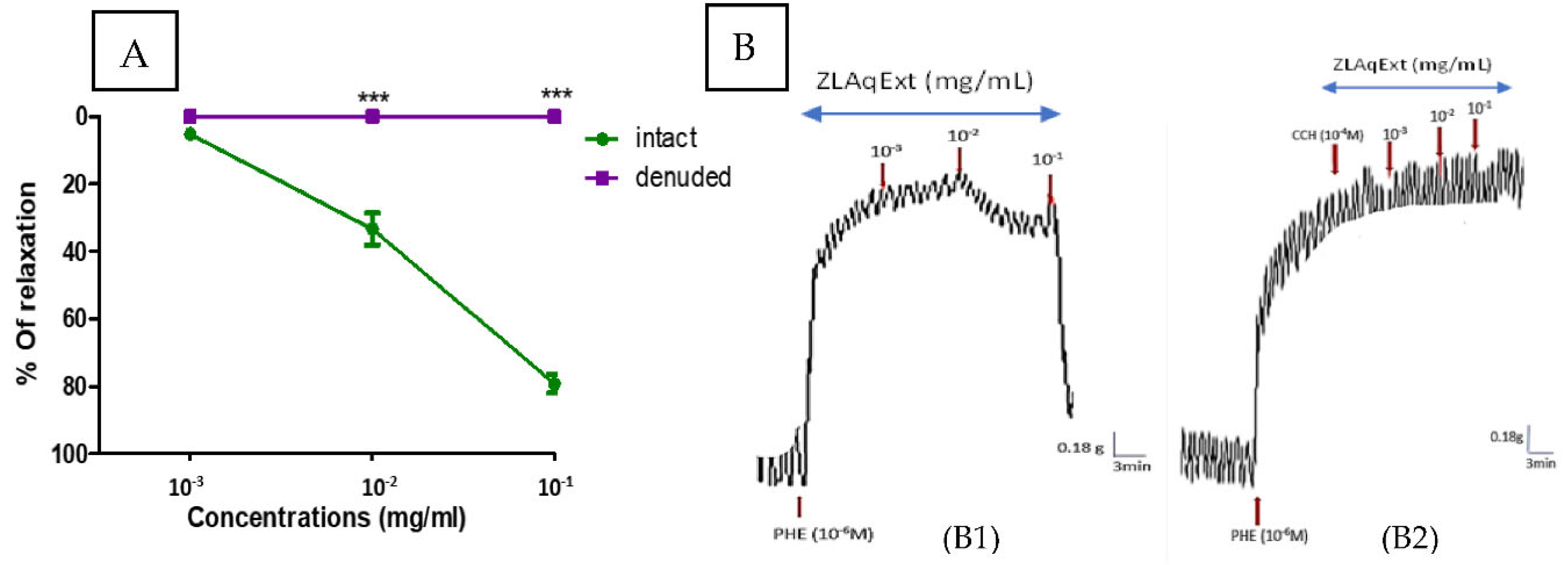
Figure 2.
Original recordings demonstrate the vascular effect of ZLAqExt in intact aortic rings treated with (A) L-NAME, (B) ODQ, (C) calmidazolium, (D) hydroxocobalamin, (E) KCl, (F) atropine, and (G) indomethacin.
Figure 2.
Original recordings demonstrate the vascular effect of ZLAqExt in intact aortic rings treated with (A) L-NAME, (B) ODQ, (C) calmidazolium, (D) hydroxocobalamin, (E) KCl, (F) atropine, and (G) indomethacin.
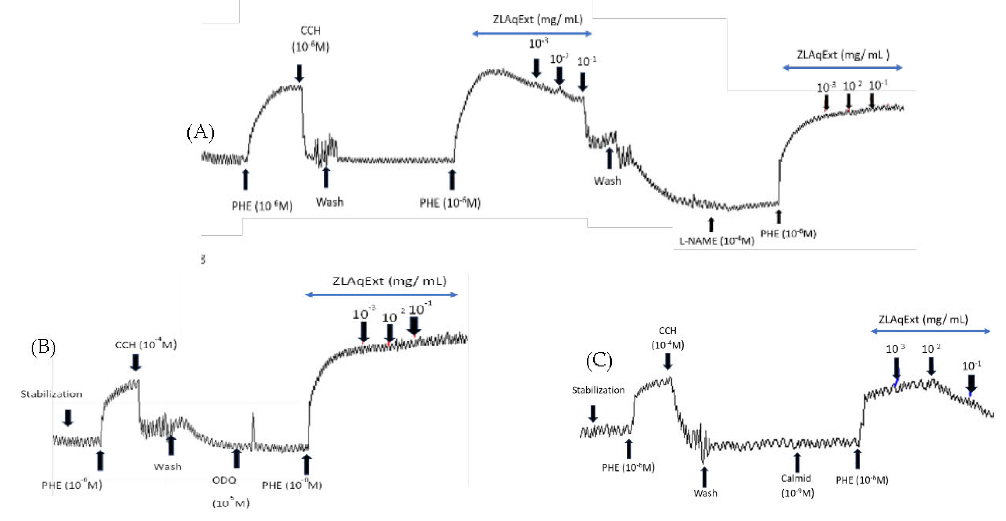
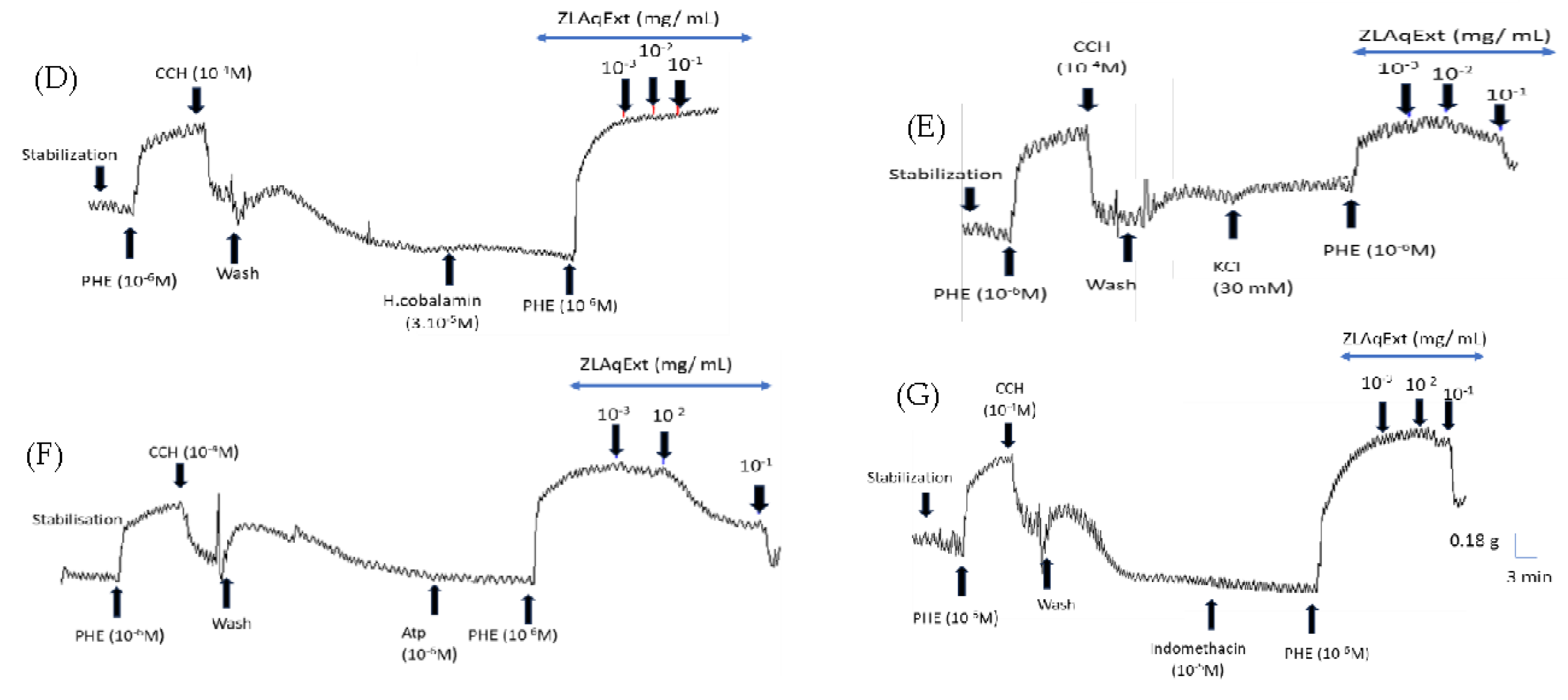
Figure 3.
Vasorelaxant-response curves of ZLAqExt in the presence of various pharmacological inhibitors: (Panel A) L-NAME, ODQ, hydroxocobalamin, and atropine; (Panel B) calmidazolium, KCl, and indomethacin. Results are presented as mean ± SEM (n = 6 aortic rings /12 rats). Statistical significance was assessed using two-way ANOVA with Bonferroni post-hoc test. ***p < 0.001 vs. control (endothelium-intact, E+).
Figure 3.
Vasorelaxant-response curves of ZLAqExt in the presence of various pharmacological inhibitors: (Panel A) L-NAME, ODQ, hydroxocobalamin, and atropine; (Panel B) calmidazolium, KCl, and indomethacin. Results are presented as mean ± SEM (n = 6 aortic rings /12 rats). Statistical significance was assessed using two-way ANOVA with Bonferroni post-hoc test. ***p < 0.001 vs. control (endothelium-intact, E+).
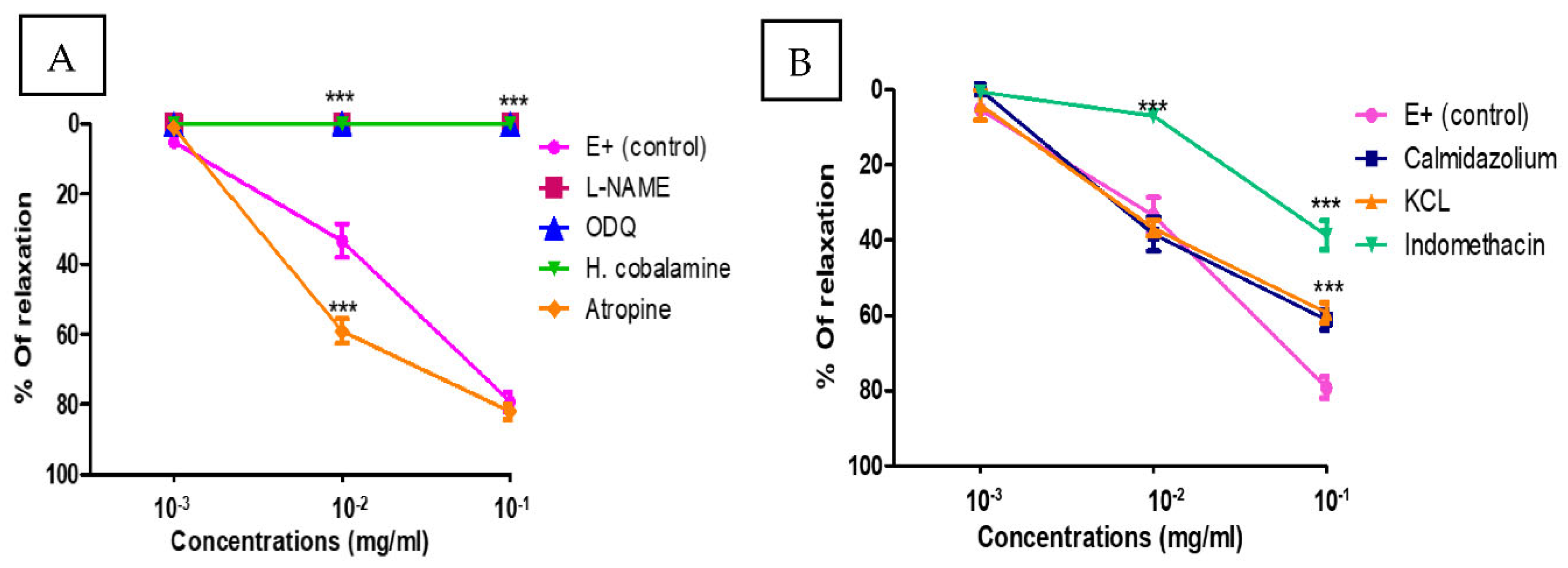
Figure 4.
Cumulative ZLAqExt-induced relaxation in aortic rings pre-constricted with PHE (10⁻⁶ M) in the presence of various inhibitors. Panels A and C: Graphs showing the effect of ZLAqExt in the presence of BaCl₂, glibenclamide, 4-AP, TEA, verapamil, and thapsigargin. Values are expressed as mean ± SEM (n = 6 aortic rings/ 8 rats). Statistical significance was determined using two-way ANOVA with Bonferroni’s post-hoc analysis; ***p < 0.001, *p < 0.05 vs. control. Panels B and D: Representative original recordings showing the vascular effect of ZLAqExt following incubation of intact aortic rings with: (B1) BaCl₂, (B2) glibenclamide, (B3) 4-AP, (B4) TEA, (D1) verapamil, (D2) thapsigargin.
Figure 4.
Cumulative ZLAqExt-induced relaxation in aortic rings pre-constricted with PHE (10⁻⁶ M) in the presence of various inhibitors. Panels A and C: Graphs showing the effect of ZLAqExt in the presence of BaCl₂, glibenclamide, 4-AP, TEA, verapamil, and thapsigargin. Values are expressed as mean ± SEM (n = 6 aortic rings/ 8 rats). Statistical significance was determined using two-way ANOVA with Bonferroni’s post-hoc analysis; ***p < 0.001, *p < 0.05 vs. control. Panels B and D: Representative original recordings showing the vascular effect of ZLAqExt following incubation of intact aortic rings with: (B1) BaCl₂, (B2) glibenclamide, (B3) 4-AP, (B4) TEA, (D1) verapamil, (D2) thapsigargin.
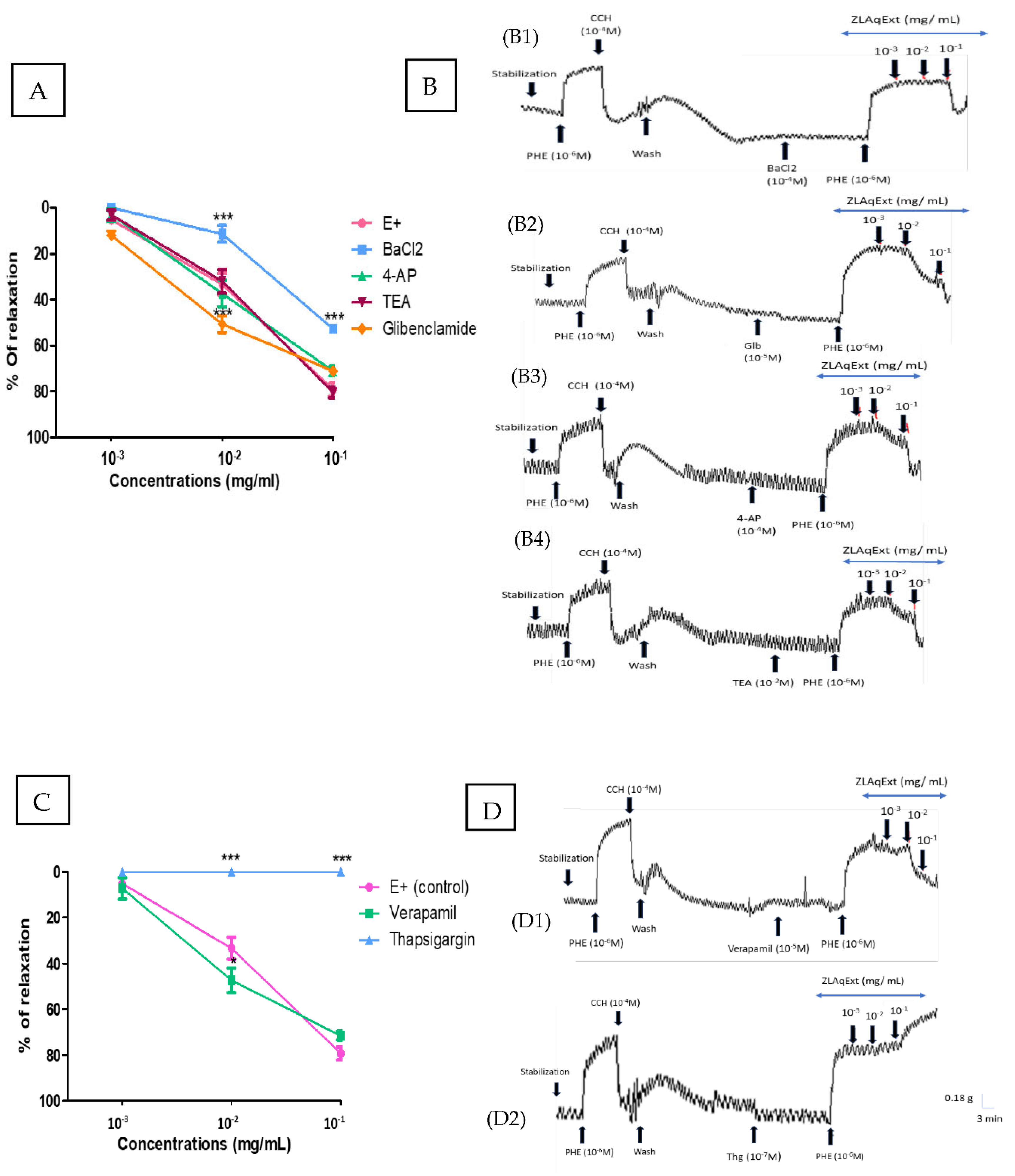
Figure 5.
Antioxidant performance of ZLAqExt and ascorbic acid evaluated through (A) DPPH free radical neutralization activity and (B) inhibition of β-carotene oxidative discoloration.
Figure 5.
Antioxidant performance of ZLAqExt and ascorbic acid evaluated through (A) DPPH free radical neutralization activity and (B) inhibition of β-carotene oxidative discoloration.
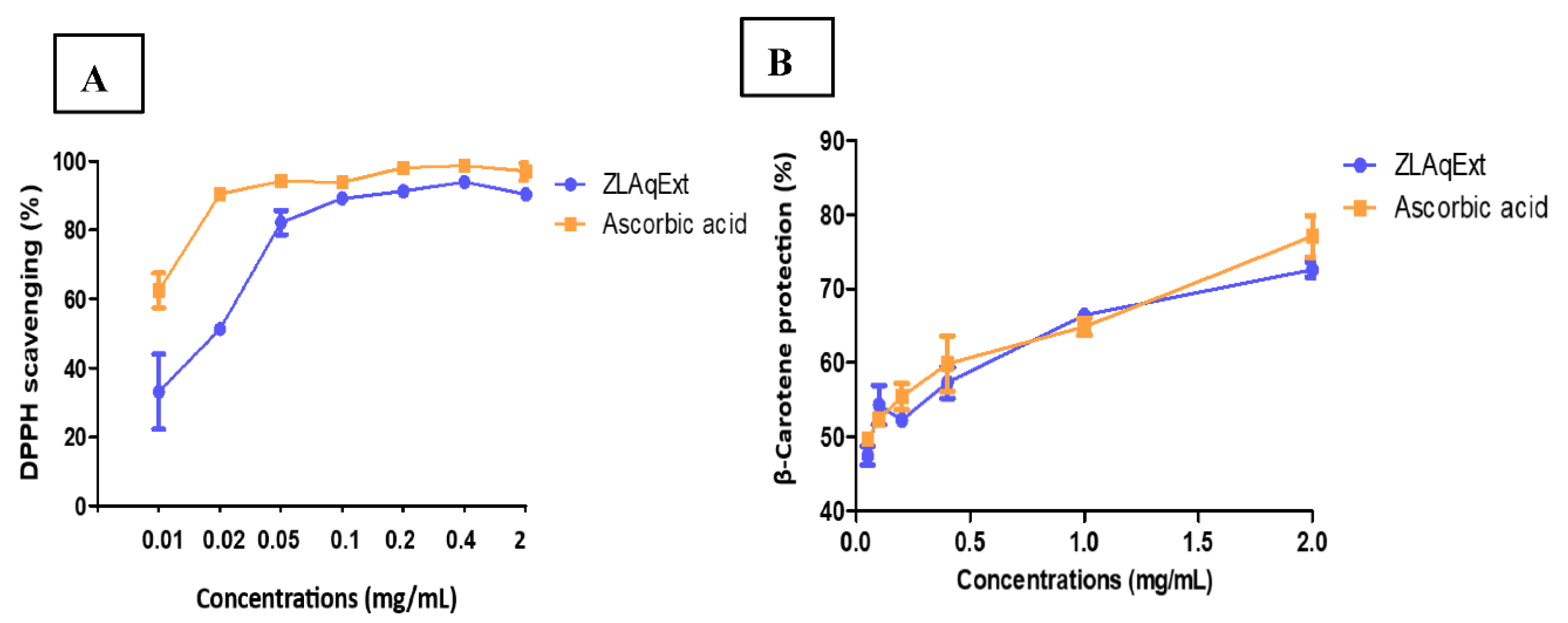
Figure 6.
Total polyphenol and tannin contents in ZLAqExt.
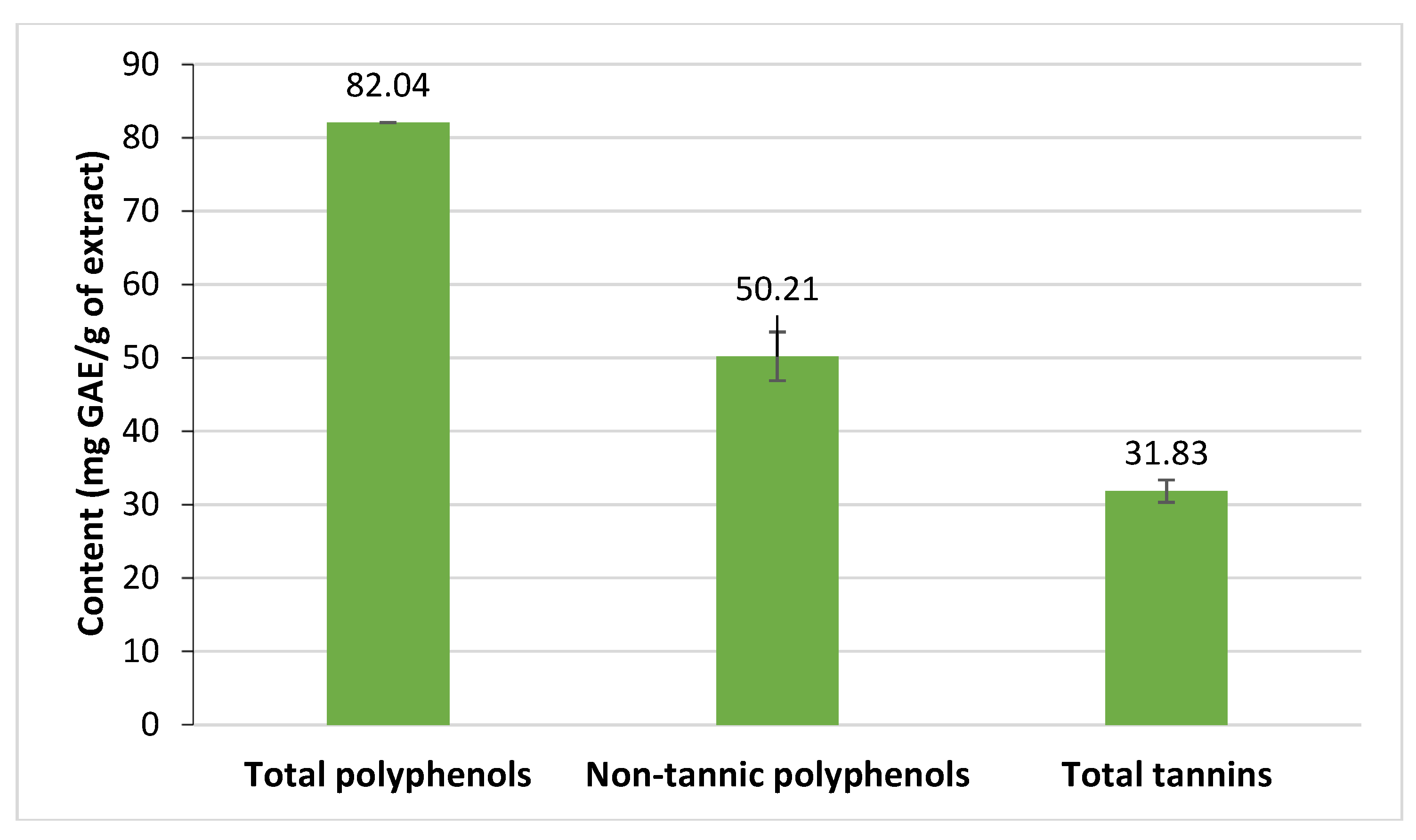
Figure 7.
Ultra-high-performance liquid chromatography (UHPLC) chromatogram of ZLAqExt monitored at 286 nm. Peak (1): myricetin-3-O-rutinoside; peak (2): rutin; peak (3): 3’,5’-di-C-β-glucopyranosyl phloretin; peak (4): nicotiflorin.
Figure 7.
Ultra-high-performance liquid chromatography (UHPLC) chromatogram of ZLAqExt monitored at 286 nm. Peak (1): myricetin-3-O-rutinoside; peak (2): rutin; peak (3): 3’,5’-di-C-β-glucopyranosyl phloretin; peak (4): nicotiflorin.
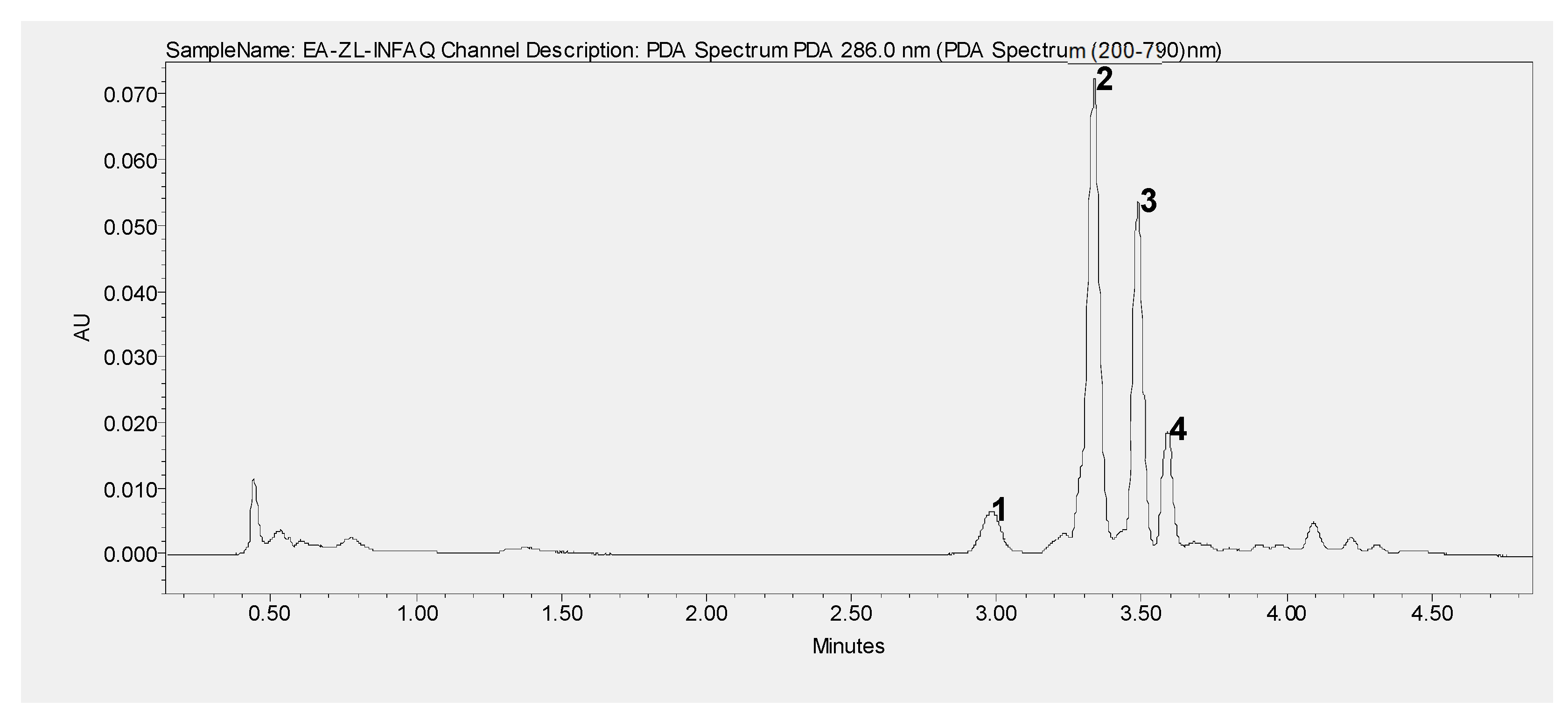
Figure 8.
Validation (RMSD< 2.0 Å) of reverse diffusion docking for guanylate cyclase. Protein backbones are shown as cartoons, ligands as sticks (colored by element). X-ray ligand conformations in grey; predicted poses in magenta. Hydrogens omitted for clarity.
Figure 8.
Validation (RMSD< 2.0 Å) of reverse diffusion docking for guanylate cyclase. Protein backbones are shown as cartoons, ligands as sticks (colored by element). X-ray ligand conformations in grey; predicted poses in magenta. Hydrogens omitted for clarity.
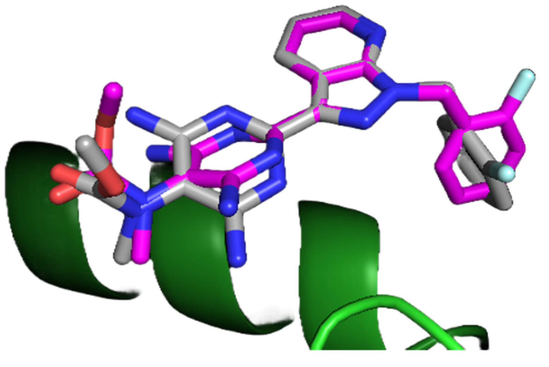
Figure 9.
Outcomes generated from molecular docking assessments focused upon guanylate cyclase (A) and sarco-endoplasmic reticulum calcium²⁺-adenosine triphosphatase (B) protein receptor targets. The energetic demarcation threshold appears signified via a broken horizontal line indicator. The presented data corresponds to mean values accompanied by standard deviation calculations arising from implementation of PBSA (n = 2) combined with GBSA (n = 6) energetic scoring functions.
Figure 9.
Outcomes generated from molecular docking assessments focused upon guanylate cyclase (A) and sarco-endoplasmic reticulum calcium²⁺-adenosine triphosphatase (B) protein receptor targets. The energetic demarcation threshold appears signified via a broken horizontal line indicator. The presented data corresponds to mean values accompanied by standard deviation calculations arising from implementation of PBSA (n = 2) combined with GBSA (n = 6) energetic scoring functions.
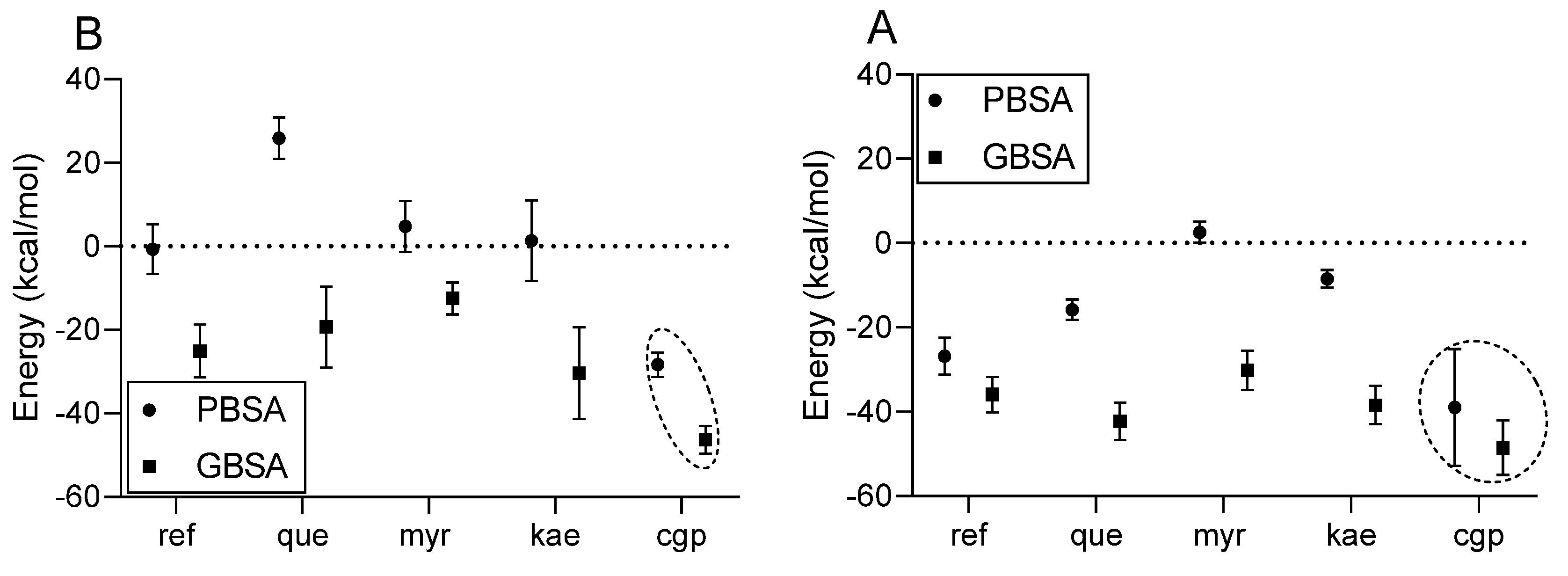
Figure 10.
Schematic representation of the signaling pathways potentially involved in ZLAqExt's vasodilator effect on isolated rat aortic rings.
Figure 10.
Schematic representation of the signaling pathways potentially involved in ZLAqExt's vasodilator effect on isolated rat aortic rings.
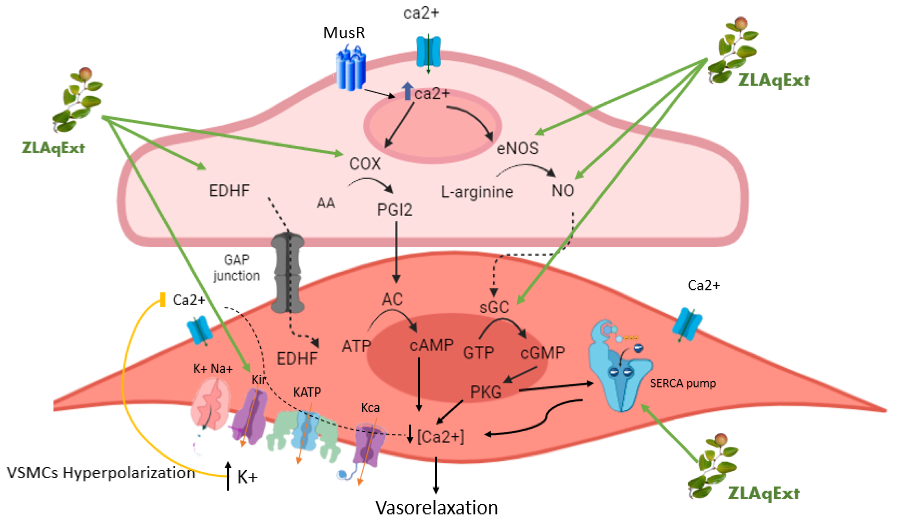

Table 1.
Ultra high-performance liquid chromatography (UHPLC) and electrospray ionization–mass spectrometry (ESI-MS) negative ion analysis of aqueous extract compounds of Z. lotus leaves.
Table 1.
Ultra high-performance liquid chromatography (UHPLC) and electrospray ionization–mass spectrometry (ESI-MS) negative ion analysis of aqueous extract compounds of Z. lotus leaves.
| Peak | RT (min) | m/z [M-H]- | Compound | Reference | Chemical structure |
| 1 | 2.980 | 625.34 | Myricetin 3-O-rutinoside | [16] | 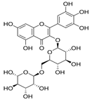 |
| 2 | 3.335 | 609.33 | Quercetin 3-O-rutinoside (Rutin) |
[17,18] | 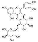 |
| 3 | 3.486 | 597.40 | 3’,5’-di-C-β-glucopyranosyl phloretin | [15] | 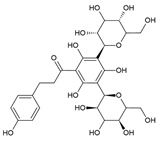 |
| 4 | 3.587 | 593.38 | Kaempferol 3-O-rutinoside (Nicotiflorin) | [16] | 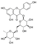 |
Disclaimer/Publisher’s Note: The statements, opinions and data contained in all publications are solely those of the individual author(s) and contributor(s) and not of MDPI and/or the editor(s). MDPI and/or the editor(s) disclaim responsibility for any injury to people or property resulting from any ideas, methods, instructions or products referred to in the content. |
© 2026 by the authors. Licensee MDPI, Basel, Switzerland. This article is an open access article distributed under the terms and conditions of the Creative Commons Attribution (CC BY) license (http://creativecommons.org/licenses/by/4.0/).
Copyright: This open access article is published under a Creative Commons CC BY 4.0 license, which permit the free download, distribution, and reuse, provided that the author and preprint are cited in any reuse.