

Submitted:
10 August 2026
Posted:
10 August 2026
You are already at the latest version
Abstract
Rhipicephalus microplus represents an important challenge for cattle farming due to economic losses and growing resistance to chemical acaricides. In this context, this study evaluated the temporal dynamics of survival of engorged females of R. microplus exposed to different concentrations of a commercial formulation of Steinernema carpocapsae. Three independent bioassays were conducted, in which survival was analyzed by Kaplan–Meier curves, Cox proportional hazards model and TL50 and TL90 estimates. The infectivity of nematodes recovered from engorged females was subsequently evaluated in Tenebrio molitor. The more concentrated suspensions promoted a faster reduction in survival, shorter lethal times and higher risk ratios, while the 130 mL dilution showed a later response. Despite the variation in lethal times between bioassays, a consistent pattern of response was observed between the concentrations evaluated. The recovered nematodes remained infectious, causing mortality in T. molitor. The results demonstrate that the analysis of the temporal dynamics of mortality provides a more comprehensive characterization of the activity of S. carpocapsae on R. microplus, expanding the understanding of its application as a biological control agent.
Keywords:
biological control
; ticks
; entomopathogenic nematodes
; acaricide resistance
; survival analysis
; Steinernema carpocapsae
1. Introduction
The tick Rhipicephalus microplus is one of the main ectoparasites in cattle farming, causing negative impacts on both animal health and the economy. Estimates indicate that losses, adding direct and indirect losses, could exceed billions of dollars annually [1]. In addition to the damage caused by direct parasitism, R. microplus acts as a biological vector for the causative agents of bovine parasitic sadness (BPS), a disease associated with high costs for medicines and herd treatments, in addition to the loss of animals, especially young ones [2,3].
Direct parasitism significantly compromises herd productivity. Continuous blood intake by engorged females negatively impacts milk production, with estimated losses of approximately 10 mL of milk for each teleogine present in the animal. Similarly, weight gain is impaired, with reductions of about 1 gram of live weight per parasite, with the female being primarily responsible for this damage due to her high blood consumption and her ability to expand her body size by up to 200 times during the parasitic phase [4,5].
The control of this ectoparasite is mainly based on the use of chemical acarides. However, the intensive use of these products favored the selection of resistant populations, reducing their effectiveness to levels below 50% on many properties. As a result, more frequent applications become necessary, increasing production costs and the risks of environmental contamination and residues in meat and milk [5,6]. A recent integrative review, based on 46 studies published between 2004 and 2025, highlighted the occurrence of R. microplus populations resistant to different classes of acaricides, including pyrethroids, organophosphates, and formamidines, which reinforces the importance of monitoring resistance and adopting alternatives to chemical control [7].
Given the limitations of chemical control, research aimed at developing sustainable biological control strategies has intensified in recent decades. In this context, entomopathogenic nematodes (EPNs) stand out for their ability to locate, infect and cause mortality in several arthropods of agricultural and veterinary importance. This process depends on the mutualistic association with bacteria of the genera Xenorhabdus and Photorhabdus, released into the hemocoel after penetration of the infecting juveniles (IJ), where they produce toxins, enzymes and antimicrobial compounds that contribute to the death of the host, often in a short period after infection. In addition to their control capacity, EPNs are considered safe for vertebrates and plants, characteristics that favor their use in integrated management programs [8,9].
Studies have demonstrated the potential of EPNs for the control of R. microplus under different experimental conditions. [10], evaluating Steinernema rarum in vitro, observed mortality close to 100% and inhibition of posture between 84% and 95%. Souza (2025) [11], when evaluating isolates of the genera Heterorhabditis and Steinernema, reported efficacy greater than 80% under laboratory conditions, in addition to the maintenance of pathogenic activity in substrate and under simulated field conditions and changes in the reproductive tissues of treated females. Monteiro (2009) [12], evaluating different isolates of EPNs under laboratory and simulated field conditions, demonstrated high activity on R. microplus, in addition to persistence in soil and compatibility with chemical and biological agents.
Although these studies highlight the potential of EPNs against R. microplus, much of the research focuses on parameters such as final mortality, postural inhibition, and treatment efficacy. The importance of the time factor in this interaction has already been demonstrated by Carvalho et al. (2010) [13], who observed differences in infection and treatment efficacy depending on the period of exposure of engorged females of R. microplus to Steinernema glaseri. However, the assessment of survival throughout the experimental period allows characterizing not only the occurrence of mortality, but also its temporal distribution and the speed with which different concentrations affect engorged females. This approach may provide complementary information to conventional measures of final mortality and contribute to a more comprehensive understanding of the response of R. microplus to entomopathogenic nematodes.
Given the above, the present work aimed to evaluate the survival pattern of engorged females of Rhipicephalus microplus exposed to different concentrations of Steinernema carpocapsae in three independent bioassays, characterizing the temporal response of mortality and its consistency between experiments, in addition to evaluating the infective capacity of nematodes recovered from exposed females.
2. Materials and Methods
2.1. Location and Time of the Experiment
The experiment was conducted at the Entomology/Phytosanity Laboratory of the Federal Institute of Santa Catarina (IFSC), Canoinhas Campus, located in the municipality of Canoinhas, state of Santa Catarina, southern Brazil, between January and February 2026.
2.2. Collection of Ectoparasites
Engorged females of R. microplus were collected on a dairy cattle farm located in the rural area of the municipality of Major Vieira, state of Santa Catarina, southern Brazil (26°27’19.3’’S, 50°17’48.7’’W), approximately 38 km from the bioassay site.
The property covers approximately 10 ha, with a predominance of perennial pastures managed in a traditional silvopastoral system, characterized by the association between pastures and native tree vegetation, in addition to areas designated for the cultivation of annual winter and summer forage. The herd consisted of approximately 30 dairy cows, kept predominantly on pasture.
The collection was carried out manually in the early hours of the morning, according to Gomes (2010) [14]. Three independent collections were carried out to carry out the bioassays, in which approximately 100, 200 and 180 teleogine were obtained, respectively, defined as engorged females over 4 mm in length. The animals used in the collections had not received tick treatment in the previous 30 days. The ticks were packaged in plastic bottles with perforated ventilation caps and transported in a Styrofoam box to the laboratory for approximately 1 hour. In the laboratory, females were washed with distilled water, dried on absorbent paper and subsequently selected for use in the respective bioassays.
2.3. In Vitro Bioassay Under Controlled Conditions
Three independent bioassays were conducted at different times. The first was exploratory in nature and was carried out to establish the experimental procedure, initially evaluate the activity of S. carpocapsae on engorged females of R. microplus and define the dilutions used in subsequent experiments. Based on the results obtained, the second was conducted with an increase in the number of treatments, experimental units and evaluation period, allowing the characterization of the females’ response to a wider range of nematode concentrations. The third bioassay used the same dilutions evaluated in the second and was conducted as an initial step in an assay aimed at recovering nematodes from infected females and subsequently evaluating their infective capacity.
In all bioassays, engorged females of R. microplus that presented normal physiological conditions, without apparent morphological changes, were selected. Females were individualized in Petri dishes (90 mm in diameter × 15 mm in height), containing sterile filter paper previously moistened with 2 mL of autoclaved distilled water.
To expose engorged females to S. carpocapsae, the commercial product Terranem® [15] was used, formulated based on this entomopathogenic nematode and containing, according to the manufacturer’s specification, at least 2.2 × 10⁶ infective juveniles (IJ) g⁻¹ of product. Upon receipt, the product was kept under refrigeration, at approximately 5 °C and protected from light until the time of use. Before preparing the suspensions, it remained at room temperature for approximately 30 min. Subsequently, the product was hydrated in distilled water and homogenized.
The dilution of 1 g of the product in 10 mL of distilled water corresponded to the proportion indicated for preparing the premix (100 g of product to 1 L of water). In the exploratory stage, five dilutions of Terranem® were evaluated, prepared by keeping 1 g of the product and varying the volume of distilled water to 10, 40, 70, 100 and 130 mL. Based on the results of this stage, the number of dilutions was increased, with the inclusion of intermediate volumes. Thus, in the second and third bioassays, dilutions of 10, 30, 50, 70, 90, 100 and 130 mL were analyzed, maintaining 1 g of the product in each preparation. Increasing water volume resulted in suspensions with decreasing nominal nematode concentrations.
The nominal concentrations of IJ were estimated by the ratio between the minimum number of IJ present in 1 g of the product, and the volume of water used in the preparation of each suspension. In each experimental unit, 1 mL of the suspension corresponding to the treatment was applied; therefore, the nominal concentration estimated in IJ mL⁻¹ corresponds numerically to the nominal number of IJ applied per plate. No direct counting of the IJ’s in the suspensions was performed.
As a positive control in the first and second bioassays, the same commercial acaricide was used, made available by the property of origin of the engorged females, formulated as an emulsifiable concentrate based on dichlorvos (450 g L⁻¹) and cypermethrin (50 g L⁻¹). The solution was prepared in the ratio 1:400 indicated by the manufacturer, using 250 μL of the product in 100 mL of distilled water. Distilled water was used as a negative control in all three stages. The third bioassay did not include the positive control. In all treatments and controls, 1 mL was applied per experimental unit, keeping the application volume constant between groups.
The experimental design was completely randomized, with each experimental unit consisting of an engorged female of R. microplus individualized in a Petri dish. In the first stage, seven treatments were evaluated, consisting of five dilutions of Terranem® (10, 40, 70, 100 and 130 mL), negative control and positive control, with 10 replicates per treatment, totaling 70 experimental units. In the second bioassay, nine treatments were evaluated, corresponding to the seven Terranem® dilutions (10, 30, 50, 70, 90, 100 and 130 mL), negative control and positive control, with 20 replicates per treatment, totaling 180 experimental units. In the third bioassay, the same seven dilutions and the negative control were used, without including the positive control, totaling eight treatments, with 20 replication per treatment and 160 experimental units. The composition of the treatments, nominal IJ concentrations and their distribution among the three bioassays are presented in Table 1.
After applying the treatments, the plates were identified and kept in a B.O.D incubation chamber at 28 °C, approximately 80% relative humidity and under continuous illumination (24 hours of light). Incubation conditions were maintained in the three bioassays, differing only in the follow-up period for females. In the first and third bioassays, evaluations were carried out for 10 days (240 hours), while in the second they were carried out for 15 days (360 hours).
2.4. Mortality Assessment and Confirmation of Infection
The mortality of female R. microplus was assessed at 24 hour intervals during the respective follow-up periods of each bioassay, recording the time until death of each individual. Death was determined by the absence of movement of the female, including the absence of movement of the appendages. After death, morphological changes were recorded, including darkening, body distension, dryness and, in some individuals exposed to S. carpocapsae, cuticle rupture with extravasation of internal fluid.
To confirm S. carpocapsae infection, females killed in nematode-containing treatments were individually dissected under a stereoscopic magnifying glass through a small incision in the dorsal region. A sample of internal fluid was collected, transferred to a microscopy slide, covered with a coverslip, and examined under an optical microscope. The presence of live nematodes in the collected fluid was used as a criterion for confirming infection.
2.5. Assessment of the Infectious Capacity of Recovered Nematodes
In the third bioassay, in addition to confirming the infection, nematodes present inside R. microplus females killed after exposure to S. carpocapsae were recovered to evaluate their infectious capacity using Tenebrio molitor larvae as host. Each female was dissected individually under a stereoscopic magnifying glass, through a small incision in the dorsal region. Internal fluid was collected with a micropipette and transferred to a beaker containing distilled water. After homogenization, 1 mL of the obtained suspension was applied to a Petri dish containing a T. molitor larva. After each procedure, the remaining suspension was discarded and the beaker sanitized before processing the next female, avoiding mixing of material between individuals.
Larvae were individualized in Petri dishes (90 mm in diameter × 15 mm in height), containing sterile filter paper previously moistened with 2 mL of distilled water. Eight treatments were evaluated, corresponding to the seven dilutions of Terranem® origin used in the third bioassay (10, 30, 50, 70, 90, 100 and 130 mL) and the negative control, with 20 replicates per treatment, totaling 160 experimental units.
After application, the plates were kept in a B.O.D incubation chamber at 28 °C and approximately 80% relative humidity, in the dark, for 10 days (240 hours). Larval mortality was assessed at 24 hours intervals, recording the time until death of each individual. After death, the larvae were examined for the presence of nematodes, which was used to confirm the infection.
2.6. Statistical Analysis
Time to death data were subjected to survival analysis, with death being considered as the event of interest. The analyses were performed separately for each of the three bioassays with R. microplus and for the assay with T. molitor. Individuals who remained alive at the end of the respective follow-up period were treated as right-censored.
Survival functions were estimated by the Kaplan–Meier method [16], and the differences between treatments were evaluated by log-rank test. When an overall difference was identified between treatments, pairwise comparisons were made between survival curves, with adjustment of p values using the Benjamini–Hochberg method to control the false discovery rate (FDR).
The lethal times for 50% (TL50) and 90% (TL90) of the individuals were estimated nonparametrically from Kaplan–Meier curves, with respective 95% confidence intervals. Additionally, Cox proportional hazards models (Cox, 1972) [17] were adjusted to estimate hazard ratios (hazard ratios, HR) and their respective 95% confidence intervals, using negative control as the reference category. The proportional hazards assumption was assessed using the Schoenfeld residuals-based test.
In all analyses, a significance level of 5% (p < 0.05) was adopted. Statistical analyses were performed in R software, using packages survival and survminer.
3. Results
3.1. Bioassay 1
In the first bioassay, Kaplan-Meier survival curves differed significantly between treatments (log-rank test, χ2 = 66.56; gl = 6; p < 0.0001). Females exposed to S. carpocapsae showed different survival patterns throughout the 240 h of evaluation (Figure 1). At dilutions of 10, 40, and 70 mL, the decline in survival was concentrated in the first evaluations, with a marked reduction in the probability of survival up to approximately 72 hours. In the 100 mL treatment, the reduction occurred later and lasted for a longer period. At 130 mL, the curve showed a slower and more gradual decline, maintaining a higher proportion of live females in the initial evaluations compared to the other dilutions, presenting mortality only after 168 hours. The negative control maintained the highest probability of survival throughout the experimental period.
Differences in the temporal pattern of mortality were evidenced by the TL50 and TL90 estimates. The lowest TL50 was observed at treatment 40 mL (48 hours), followed by treatments 10 mL (60 hours) and 70 mL (72 hours), which did not differ significantly from each other in pairwise comparisons of survival curves. In the 100 mL treatment, TL50 increased to 108 hours, accompanied by a significant difference in relation to the 10, 40 and 70 mL treatments. The latest response among S. carpocapsae treatments was observed at 130 mL, with TL50 only reached within 240 hours and TL90 not reached during the experimental period. In contrast, treatments 10, 40, 70, and 100 mL reached TL90 between 72 and 136.5 hours.
The positive control had a TL50 of 204 hours, but did not reach 90% mortality during the experimental period. In the negative control, the 50% and 90% mortality thresholds were not reached. In pairwise comparisons, the 130 mL treatment did not differ significantly from both controls, while the positive control also did not differ from the 100 mL treatment (Table 2).
The Cox proportional hazards model, using negative control as a reference, also showed differences in mortality risk between treatments (Figure 2). Regarding the negative control, the greatest risks were observed in treatments with the most concentrated suspensions of S. carpocapsae, with emphasis on 10, 40 and 70 mL. The risk decreased in the most diluted treatments, with the lowest values observed for 130 mL and for the positive control, following the later pattern of mortality observed in the survival curves and lethal times.
3.2. Bioassay 2
Kaplan–Meier survival curves differed significantly between treatments (log-rank test, p < 0.001), evidencing distinct survival patterns of engorged R. microplus females throughout the 360 h of evaluation. Treatments 10 and 30 mL showed the sharpest reductions in the probability of survival in the first evaluations, reaching values below 50% in the first 48 hours. At dilutions of 50, 70, 90, and 100 mL, the decline occurred more gradually, with the probability of survival reducing to approximately 50% between 72 and 96 hours. The positive control and the T130 mL treatment showed similar behavior, characterized by a slow decline in survival and greater stability of the curves during the first hours of evaluation, requiring a period greater than 168 hours to reach 50% mortality, while the negative control maintained the highest probability of survival and remained above 50% at the end of the 360 hours of evaluation (Figure 3).
Estimates of lethal times allowed quantifying the temporal differences observed between treatments. The lowest TL50 values were recorded at dilutions of 10 and 30 mL, both at 48 hours. In treatments 50, 90 and 100 mL, the TL50 was 96 h, while in the 70 mL dilution this threshold was reached in 72 hours. Treatment 130 mL and the positive control showed a later response, with TL50 of 192 and 180 hours, respectively. For o TL90, the lowest values were observed in the treatments 10 mL (84 hours), 30 and 70 mL (96 hours), followed by 50 mL (120 hours), 100 mL (156 hours) and 90 mL (168 hours). Treatment 130 mL and or positive control reached 90% mortality in 300 and 312 hours, respectively. No negative control, the thresholds of 50% and 90% mortality were not reached during 360 hours of evaluation.
Pairwise comparisons between survival curves showed that treatments 10 and 30 mL did not differ significantly from each other. Among the other dilutions, 50, 90 and 100 mL also did not show significant differences between them, while the 70 mL treatment showed intermediate behavior, without differing from 30, 50, 90 and 100 mL. The 130 mL treatment did not differ significantly from the positive control, but both differed from the other S. carpocapsae dilutions. The negative control, in turn, differed significantly from all other treatments (Table 3).
The Cox proportional hazards model showed differences in the magnitude of mortality risk ratios between treatments, considering the negative control as a reference. The highest risk estimates of death were observed at dilutions of 10 and 30 mL, followed by treatment of 70 mL. Dilutions of 50, 90, and 100 mL had lower risk ratios, while treatment 130 mL and the positive control had the lowest risk estimates among the treatments evaluated (Figure 4).
3.3. Bioassay 3
Kaplan–Meier survival curves differed significantly between treatments (log-rank test, χ² = 97.39; gl = 7; p < 0.0001), evidencing distinct survival patterns of engorged R. microplus females throughout the 240 hours of evaluation. In the first 96 hours, a marked reduction in survival was observed in all dilutions of S. carpocapsae, a period in which most deaths occurred in these treatments. After 96 hours, the probability of survival remained low at dilutions of 10, 30, 50, 70, 90, and 100 mL, while the 130 mL treatment showed a more prolonged decline, maintaining approximately 50% survival until 144 hours and individuals alive until the end of the evaluation. In the negative control, 75% of females remained alive at the end of the 240 hours, representing the highest survival among the treatments evaluated (Figure 5).
Estimates of lethal times allowed quantifying the temporal differences observed between treatments. TL50 was reached in 72 hours at dilutions of 10, 30, 50, 90 and 100 mL, while at treatments 70 and 130 mL this threshold was reached at 96 and 120 hours, respectively. For TL90, the lowest values were recorded at dilutions of 10 and 50 mL, both at 96 hours, followed by treatments 30, 70 and 90 mL, at 108 hours, and 100 mL, at 120 hours. The later response was observed in the 130 mL treatment, which reached TL90 only within 240 hours. In the negative control, the 50% and 90% mortality thresholds were not reached during the 240 hours of evaluation.
Pairwise comparisons between survival curves showed that treatments 10, 30, 50, 90, and 100 mL did not differ significantly from each other. The 70 mL treatment differed from the 10 and 50 mL dilutions, but did not show a significant difference in relation to 30, 90 and 100 mL. The 130 mL dilution differed from treatments 10, 30, 50, 90, and 100 mL, but did not differ from 70 mL. The negative control differed significantly from all S. carpocapsae dilutions (Table 4).
The Cox proportional hazards model showed differences in the magnitude of the risk of death between the dilutions evaluated. Females exposed to treatments 10, 30 and 50 mL had the highest mortality risks, while lower estimates were observed at dilutions of 70, 90 and 100 mL. The 130 mL treatment presented the lowest risk of mortality among S. carpocapsae dilutions, remaining far from the estimates observed in the more concentrated treatments (Figure 6).
3.4. Infectivity of S. carpocapsae Recovered from Female R. microplus
Exposure of Tenebrio molitor larvae to nematodes recovered from female R. microplus resulted in different survival patterns throughout the 240 hours of evaluation. The greatest reductions in the probability of survival occurred in dilutions between 10 and 100 mL, with a sharp decline mainly in the first 72 hours of evaluation. After this period, the curves remained at low survival levels until the end of the experiment, with emphasis on the 10 mL treatment, in which accumulated mortality reached 95%. In contrast, the 130 mL treatment showed a smaller reduction, with a mortality rate of 40% and approximately 60% of individuals remaining alive at the end of 240 hours, close to that observed in the negative control, whose mortality rate was 45% (Figure 7).
The Cox proportional hazards model showed differences in the magnitude of the risk of death of T. molitor between treatments. The highest hazard ratio was observed at treatment 10 mL, followed by dilutions of 50, 30, and 70 mL. Treatments 90 and 100 mL presented lower magnitude estimates, while treatment 130 mL presented the lowest hazard ratio (HR = 1.12) and did not differ significantly from the negative control (p = 0.8140) (Figure 8).
Females exposed to S. carpocapsae presented, after death, morphological changes such as darkening and body distension and, in some cases, cuticle rupture with extravasation of internal fluid (Figure 9). Dissection of dead females in nematode treatments allowed observation of live nematodes in the internal fluid, confirming S. carpocapsae infection.
In the bioassay with T. molitor, individuals killed after exposure to nematodes recovered from female R. microplus showed reduced body consistency during manipulation. Dissection of the dead individuals and microscopic examination of the internal contents allowed the presence of nematodes to be observed, complementing the survival results and providing additional evidence of the maintenance of infectivity after recovery in females (Figure 10).
4. Discussion
The results demonstrated that exposure to S. carpocapsae significantly altered the survival dynamics of engorged R. microplus females, with differences in the rate of mortality occurrence between treatments. In the three bioassays, the negative control maintained a high probability of survival throughout the experimental period, while treatments with S. carpocapsae, especially in the most concentrated suspensions, showed a sharp decline in survival in the first evaluations, with a large proportion of deaths concentrated up to approximately 72 hours. In contrast, the positive control in the bioassays in which it was included showed a later response, with a greater reduction in survival after approximately 200 hours. This pattern was accompanied by estimates of lethal times and Cox hazard ratios, which indicated a faster response and a higher risk of mortality in the more concentrated suspensions. Despite this general trend, no uniform ordering of the response was observed among all concentrations evaluated, indicating that mortality dynamics did not vary strictly linearly with dilution.
Analysis of the temporal dynamics of mortality allowed us to distinguish responses between treatments that would not be fully characterized solely by the mortality observed at the end of the experimental period. Treatments capable of resulting in similar final mortalities can differ considerably in the rate at which this effect is established, an aspect particularly relevant in engorged females of R. microplus, whose non-parasitic phase comprises processes directly related to reproduction. Thus, the earlier occurrence of mortality may reduce the period available for the progression of these processes, although their effects on reproductive parameters were not evaluated in this study. In this context, the association of Kaplan–Meier curves with TL50 and TL90 estimates and the Cox proportional hazards model allowed characterizing the distribution of mortality over time and distinguishing treatments in terms of speed and risk of death.
Estimates of lethal times reinforced the pattern observed in survival curves, although TL50 and TL90 values varied among bioassays. In general, treatments prepared with lower dilution volumes and, consequently, a higher concentration of infective juveniles reached mortality thresholds earlier, while the 130 mL dilution showed a consistently later response. This trend was particularly evident for TL50, which occurred between 48 and 72 h in the most concentrated treatments in the three bioassays, while in 130 mL it was reached between 120 and 240 hours. Although the estimated times were not identical between experiments, they remained at temporally close intervals and maintained a similar overall pattern of response.
The importance of the temporal component in the interaction between entomopathogenic nematodes and R. microplus was also demonstrated by Carvalho et al. (2010) [12], who evaluated engorged females exposed to S. glaseri CCA for different periods. Although only 2 hours of exposure was sufficient to establish infection, efficacy increased with prolonged exposure, requiring at least 24 hours to obtain values greater than 90%, demonstrating that prolonged contact between the nematode and the host intensified the response to treatment. In the present study, although exposure time was not used as an experimental factor, the response also presented an evident temporal component, since the differences between concentrations were reflected not only in the occurrence of mortality, but mainly in the speed with which it was established. The lower TL50 observed in treatments with a higher concentration of JIs indicate that, under these conditions, the effect on females occurred earlier, while the more diluted suspensions required a longer period to reach the same mortality threshold. This behavior highlights differences in the speed with which mortality is established between the concentrations evaluated, an aspect particularly evident in the late response observed in the 130 mL dilution.
The response observed at a dilution of 130 mL reinforces this interpretation. In this treatment, the sharpest decline in survival occurred later, accompanied by the longest lethal times among S. carpocapsae suspensions in the three bioassays. In Bioassay 2, conducted for 360 hours, mortality progressed over the period until reaching all individuals, with TL50 of 192 hours and TL90 of 300 hours. In bioassays with a shorter evaluation period, some females remained alive at the end of the follow-up, making it impossible to determine whether mortality would continue to occur later. Thus, the behavior observed in 130 mL appears to be primarily related to a slower response rather than the absence of S. carpocapsae activity.
Another relevant aspect was the similarity in the temporal pattern observed between the positive control and the 130 mL dilution in the two bioassays in which the chemical treatment was included. Both showed later reduction in survival and longer lethal times compared to more concentrated suspensions of S. carpocapsae, in addition to not differing significantly from each other in pairwise comparisons. This trend was also accompanied by the risk ratios estimated by the Cox model. In both bioassays, the highest mortality risks were associated with the most concentrated suspensions, while the 130 mL dilution presented the lowest risk among treatments with S. carpocapsae. The positive control also presented a comparatively lower risk, following the later response observed for these treatments in the survival curves and lethal times.
The later response observed in the positive control also deserves attention given the occurrence of resistance of R. microplus to chemical acaricides in Brazil. Teixeira et al. (2026) [7] identified a wide occurrence of resistance to pyrethroids and organophosphates, classes to which cypermethrin and dichlorvos, active ingredients present in the product used as a positive control in this study, belong, respectively. For pyrethroids, the authors recorded resistant populations including in municipalities in Santa Catarina, including Canoinhas and Itaiópolis. This scenario makes it relevant to consider the possibility of lower susceptibility of the population evaluated to chemical treatment as one of the possible explanations for the later response and lower risk reasons for death observed in the positive control. However, as the susceptibility of the population used in the present study was not previously characterized by specific resistance tests, it is not possible to attribute the behavior observed in the positive control to acaricide resistance.
In addition to the effects on survival, females exposed to S. carpocapsae presented morphological changes after death, such as darkening, body distension and, in some individuals, rupture of the integument with extravasation of the internal contents. Infection with EPNs can also promote internal changes in R. microplus, as observed by Souza (2025) [10], who reported histological changes and impairment of reproductive tissues in females exposed to different nematode isolates. These effects are consistent with the mechanism of action of EPNs, in which infecting juveniles release symbiotic bacteria into the host whose multiplication and production of toxins and enzymes contribute to tissue death and degradation [18]. Observation of nematodes inside females during dissection confirmed the establishment of S. carpocapsae infection.
The infective capacity of nematodes after their passage through female R. microplus was evidenced in the bioassay with T. molitor. Nematodes recovered from females from treatments of between 10 and 100 mL remained associated with reduced larval survival, with a higher occurrence of deaths in the first evaluations and significantly higher mortality risks than the negative control. These results demonstrate that, after infection of the females and subsequent recovery, the nematodes maintained the ability to infect and cause mortality in a new host. In contrast, nematodes from the 130 mL dilution produced a more discreet response, with a survival curve close to that of the control and a hazard ratio that did not differ significantly from the reference. The observation of nematodes in the internal contents of the dead larvae complemented this evidence, confirming the infection of T. molitor by the recovered nematodes.
In addition to the evidence of activity and infectivity of S. carpocapsae, the use of a commercial formulation gives an applied character to the results obtained. Although the fastest responses occurred in suspensions with the highest concentration of IJs, intermediate treatments also showed high mortality throughout the experimental period, demonstrating that nematode activity was maintained at different product dilutions. The evaluation of a commercial formulation brings the results closer to a perspective of practical use and highlights the importance of studies aimed at defining concentrations that reconcile speed of action, effectiveness and use of the product.
Despite the demonstrated potential under laboratory conditions, the use of EPNs in the management of R. microplus depends on their ability to remain viable and infectious under environmental conditions. These conditions can influence the survival and activity of IJs, posing challenges for their application in the field. Even so, characteristics such as low toxicity to vertebrates and reduced environmental impact favor the use of these organisms as biological control agents [9]. In this sense, the results obtained provide support for further studies aimed at evaluating the persistence, efficacy and application strategies of S. carpocapsae under field conditions, aiming at its possible integration into the management of R. microplus and the reduction of exclusive dependence on chemical acaricides.
5. Conclusions
This section is not mandatory, but can be added to the manuscript if the discussion is unusually long or complex.
6. Patents
Steinernema carpocapsae promoted a reduction in the survival of engorged females of Rhipicephalus microplus in the bioassays performed. Survival analyses, associated with lethal time estimates and the Cox proportional hazards model, demonstrated that the differences between concentrations were not restricted to the occurrence of mortality, but mainly involved its temporal dynamics, with greater speed of action and greater risk of death in the most concentrated suspensions.
Nematodes recovered from females maintained infective capacity in Tenebrio molitor, confirming their viability after passage through the host. Taken together, the results reinforce the potential of the commercial formulation of S. carpocapsae for the biological control of R. microplus and demonstrate that the analysis of the temporal dynamics of mortality constitutes a useful approach to characterize the activity of entomopathogenic nematodes, providing a basis for future evaluations under field conditions.
Author Contributions
Conceptualization, A.N. and J.P.P.P.; Methodology, A.N. and J.P.P.P.; Investigation, A.N. and L.K.C.; Resources, J.P.P.P.; Data curation A.N. and L.K.C.; Formal analysis, J.P.P.P.; Original draft preparation, A.N.; Writing-review and editing, A.N., L.K.C. and J.P.P.P.; Supervision, J.P.P.P. All authors have read and agreed to the published version of the manuscript.
Funding
This research was supported by the Federal Institute of Santa Catarina (IFSC) through its Institucional Scientific Initiation Program. Grant number: nº 03/2024/PROPPI/DAE.
Institutional Review Board Statement
Not applicable.
Informed Consent Statement
Not applicable.
Data Availability Statement
The data presented in this study are available from the corresponding author upon reasonable request.
Conflicts of interest
The authors declare no conflict of interest.
Abbreviations
The following abbreviations are used in this manuscript:
IJ Infective juveniles
EPN Entopathogenic nematodes
HR Hazard ratios
TL Lethal times
IC Confidence interval
NR Lethal time not reached during the assessment period
References
- Grisi, L.; Leite, R.C.; Martins, J.R.S.; Barros, A.T.M.; Andreotti, R.; Cançado, P.H.D.; León, A.A.P.; Pereira, J.B.; Villela, H.S. Reassessment of the potential economic impact of cattle parasites in Brazil. Rev. Bras. Parasitol. Vet. 2014, 23, 150–156. [CrossRef]
- Brito, L.G.; Madruga, C.R.; et al. Transmissão Congênita de Babesia bovis e Anaplasma marginale na Epidemiologia da Tristeza Parasitária Bovina; Embrapa Amazônia Oriental: Belém, PA, Brazil, 2019.
- Silva, T.F.; Alves-Sobrinho, A.V.; Lima, L.F.S.; Ziemniczak, H.M.; Ferraz, H.T.; Lopes, D.T.; Silva, V.L.D.; Braga, Í.A.; Saturnino, K.C.; Ramos, D.G.S. Tristeza parasitária bovina: revisão. Res. Soc. Dev. 2021, 10, e15410111631. [CrossRef]
- Carvalho, L.B.; Furlong, J.; Prata, M.C.A.; Reis, É.S.; Batista, E.S.P.; Faza, A.P.; Leite, R.C. Evaluation in vitro of the infection times of engorged females of Rhipicephalus (Boophilus) microplus by the entomopathogenic nematode Steinernema glaseri CCA strain. Cienc. Rural 2010, 40, 939–943. [CrossRef]
- ANDREOTTI, R.; GARCIA, M. V.; KOLLER, W. W. Controle estratégico dos carrapatos nos bovinos. In: KOLLER, W. W. et al. (org.). Carrapatos na cadeia produtiva de bovinos no Brasil. Brasília, DF: Embrapa, 2019. cap. 9, p. 125–135.
- KKlafke, G.M.; Webster, A.; Dall’Agnol, B.; Pradel, E.; Silva, J.; de La Canal, L.H.; Becker, M.; Osório, M.F.; Mansson, M.; Barreto, R.; Scheffer, R.; Souza, U.A.; Corassini, V.B.; dos Santos, J.; Reck, J.; Martins, J.R. Multiple resistance to acaricides in field populations of Rhipicephalus (Boophilus) microplus from the state of Rio Grande do Sul, Southern Brazil. Ticks Tick Borne Dis. 2017, 8, 73–80. [CrossRef]
- Teixeira, T.M.; Miloca, L.M.; Sakane, K.T.; Rodrigues, S.A.; Marchetti, P.H.L.; Piau Junior, R.; Ribeiro, C.E.; Gazim, Z.C. Occurrence of acaricide resistance in Rhipicephalus microplus in Brazilian municipalities: An integrative review. Cienc. Anim. Bras. 2026, 27, e81941E. [CrossRef]
- NEGRISOLI JUNIOR, A. S. et al. Produção e armazenamento de nematoides entomopatogênicos. Brasília, DF: Embrapa, 2015.
- TARASCO, E. et al. Entomopathogenic nematodes and their symbiotic bacteria: from lab to field applications for sustainable pest control. Frontiers in Insect Science, Lausanne, v. 3, 2023. [CrossRef]
- ARANTES, L. J. et al. Testes in vitro com nematoide Steinernema rarum para o controle biológico do carrapato Rhipicephalus (Boophilus) microplus. In: CONGRESSO INTERINSTITUCIONAL DE INICIAÇÃO CIENTÍFICA, 2022, Campinas. Campinas: Instituto Agronômico de Campinas, 2022.
- SOUZA, E. R. Avaliação de nematoides entomopatogênicos para o controle do carrapato Rhipicephalus (Boophilus) microplus (Acari: Ixodidae). 2025. Dissertação (Mestrado em Sanidade, Segurança Alimentar e Ambiental no Agronegócio) – Instituto Biológico, Agência Paulista de Tecnologia dos Agronegócios, São Paulo, 2025. Available in: http://repositoriobiologico.com.br/jspui/bitstream/123456789/1243/1/elianai-souza.pdf.
- MONTEIRO, C. M. O.; PRATA, M. C. A.; SILVA, M. V.; LEAL, I. R.; FAZA, A. P.; CARVALHO, L. B.; LEITE, R. C. Entomopathogenic nematodes in the control of Rhipicephalus microplus: a review. Ticks and Tick-borne Diseases, v. 11, n. 1, p. 101313, 2020. Available in: https://pubmed.ncbi.nlm.nih.gov/31704209/. [CrossRef]
- CARVALHO, L. B.; FURLONG, J.; PRATA, M. C. A.; REIS, É. S.; BATISTA, E. S. P.; FAZA, A. P.; LEITE, R. C. Evaluation in vitro of the infection times of engorged females of Rhipicephalus (Boophilus) microplus by the entomopathogenic nematode Steinernema glaseri CCA strain. Ciência Rural, Santa Maria, v. 40, n. 4, p. 939-943, 2010. [CrossRef]
- GOMES, C. C. G. Instruções para coleta e envio de material para teste de sensibilidade aos carrapaticidas ou biocarrapaticidograma. Comunicado Técnico. Bagé, RS: Embrapa Pecuária Sul, 2010.
- KOPPERT BIOLOGICAL SYSTEMS. Terranem®: bula do produto. Piracicaba, 2023. Available in: https://www.koppert.com.br/content/brasil/Produtos/Terranem/Bula_Produto_Terranem_01_06_2023.pdf.
- Kaplan, E.L.; Meier, P. Nonparametric estimation from incomplete observations. J. Am. Stat. Assoc. 1958, 53, 457–481. [CrossRef]
- Cox, D.R. Regression models and life-tables. J. R. Stat. Soc. Ser. B 1972, 34, 187–220. [CrossRef]
- NEGRISOLI JUNIOR, A. S. et al. Produção e armazenamento de nematoides entomopatogênicos. Brasília, DF: Embrapa, 2015.
Figure 1.
Kaplan-Meier survival curves of ingurgitated females of Rhipicephalus microplus exposed to different dilutions of Steinernema carpocapsae and control treatments in Bioassay 1. The survival curves differed significantly between the treatments by the log-rank test (χ2 = 66,56; gl = 6; p < 0,0001).
Figure 1.
Kaplan-Meier survival curves of ingurgitated females of Rhipicephalus microplus exposed to different dilutions of Steinernema carpocapsae and control treatments in Bioassay 1. The survival curves differed significantly between the treatments by the log-rank test (χ2 = 66,56; gl = 6; p < 0,0001).
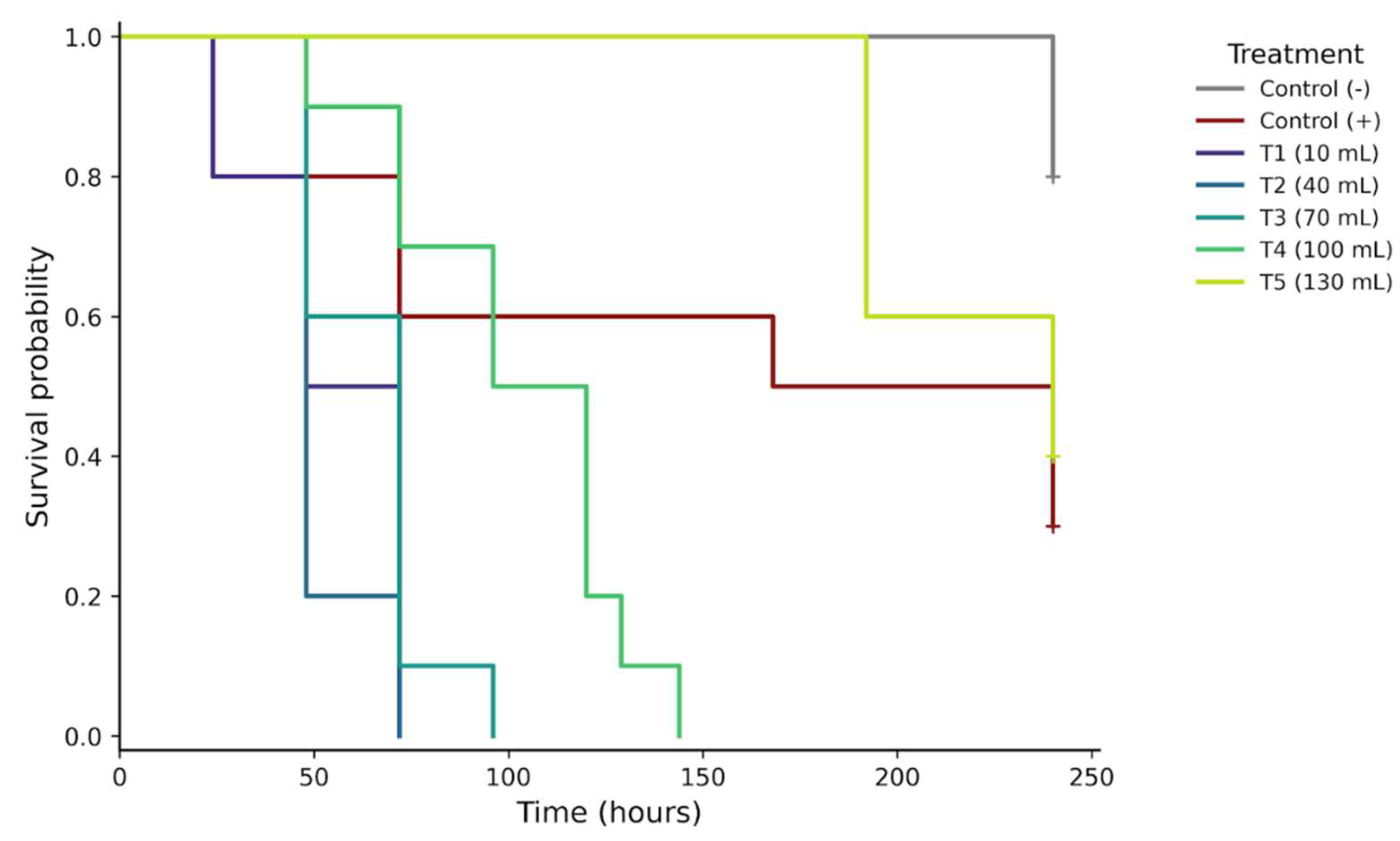
Figure 2.
Hazard ratios (hazard ratios, HR) of mortality of engorged females of Rhipicephalus microplus subjected to the treatments evaluated in Bioassay 1, estimated by the Cox proportional hazards model. The negative control was used as the reference category (HR = 1). The points represent the risk ratio estimates and the horizontal bars their respective 95% confidence intervals. The dashed vertical line represents HR = 1, corresponding to the reference category.
Figure 2.
Hazard ratios (hazard ratios, HR) of mortality of engorged females of Rhipicephalus microplus subjected to the treatments evaluated in Bioassay 1, estimated by the Cox proportional hazards model. The negative control was used as the reference category (HR = 1). The points represent the risk ratio estimates and the horizontal bars their respective 95% confidence intervals. The dashed vertical line represents HR = 1, corresponding to the reference category.
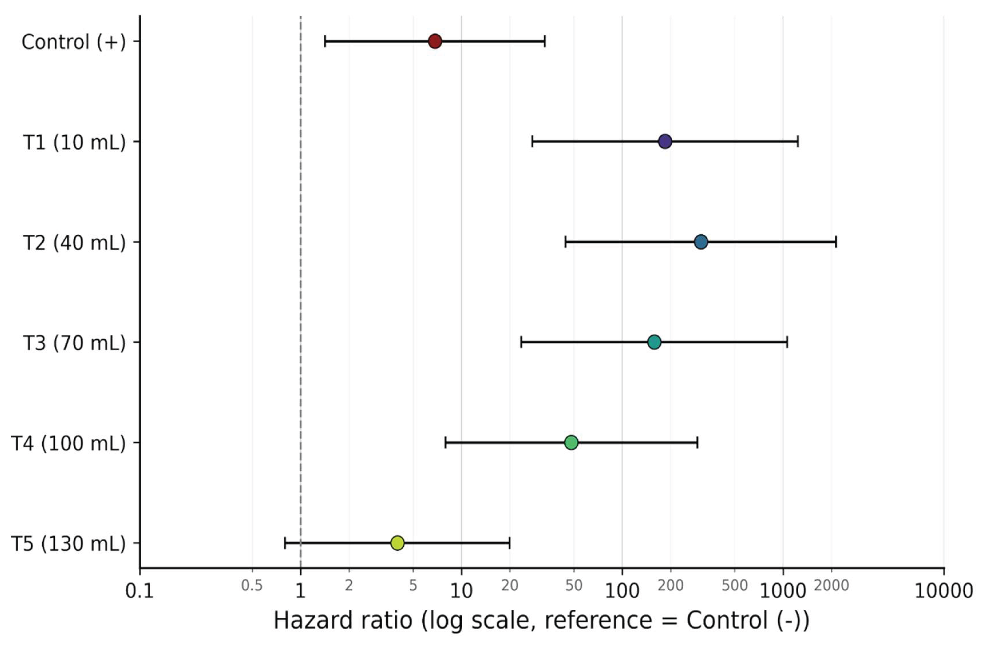
Figure 3.
Kaplan–Meier survival curves of Rhipicephalus microplus exposed to different dilutions of Steinernema carpocapsae and control tretments in Bioassay 2. Survival curves differed significantly between treatments using the log-rank test (p < 0.001).
Figure 3.
Kaplan–Meier survival curves of Rhipicephalus microplus exposed to different dilutions of Steinernema carpocapsae and control tretments in Bioassay 2. Survival curves differed significantly between treatments using the log-rank test (p < 0.001).
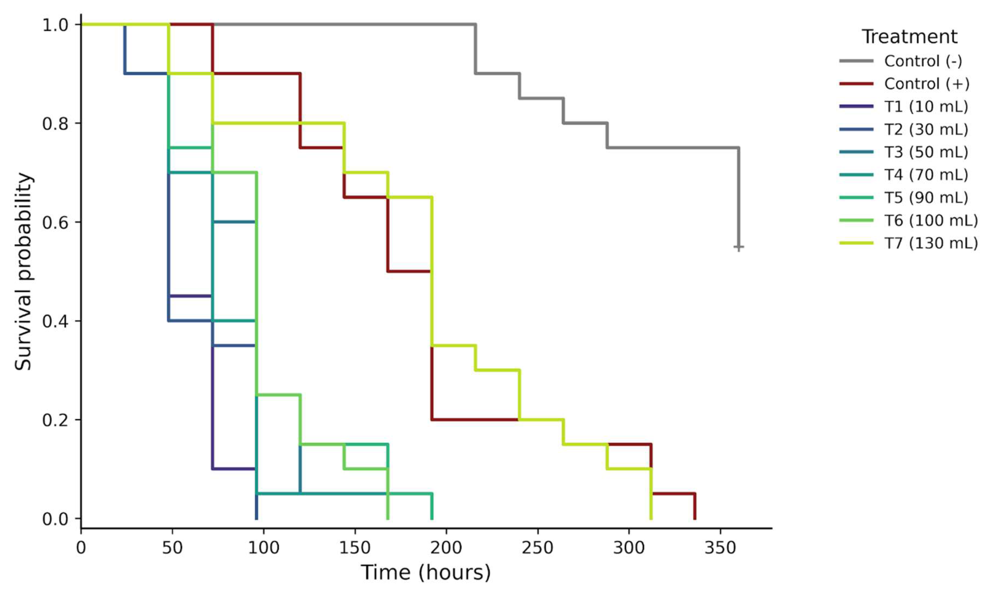
Figure 4.
Hazard ratios (hazard ratios, HR) of mortality of engorged females of Rhipicephalus microplus subjected to the treatments evaluated in Bioassay 2, estimated by the Cox proportional hazards model. The negative control was used as the reference category (HR = 1). The points represent the risk ratio estimates and the horizontal bars their respective 95% confidence intervals. The dashed vertical line represents HR = 1, corresponding to the reference category.
Figure 4.
Hazard ratios (hazard ratios, HR) of mortality of engorged females of Rhipicephalus microplus subjected to the treatments evaluated in Bioassay 2, estimated by the Cox proportional hazards model. The negative control was used as the reference category (HR = 1). The points represent the risk ratio estimates and the horizontal bars their respective 95% confidence intervals. The dashed vertical line represents HR = 1, corresponding to the reference category.
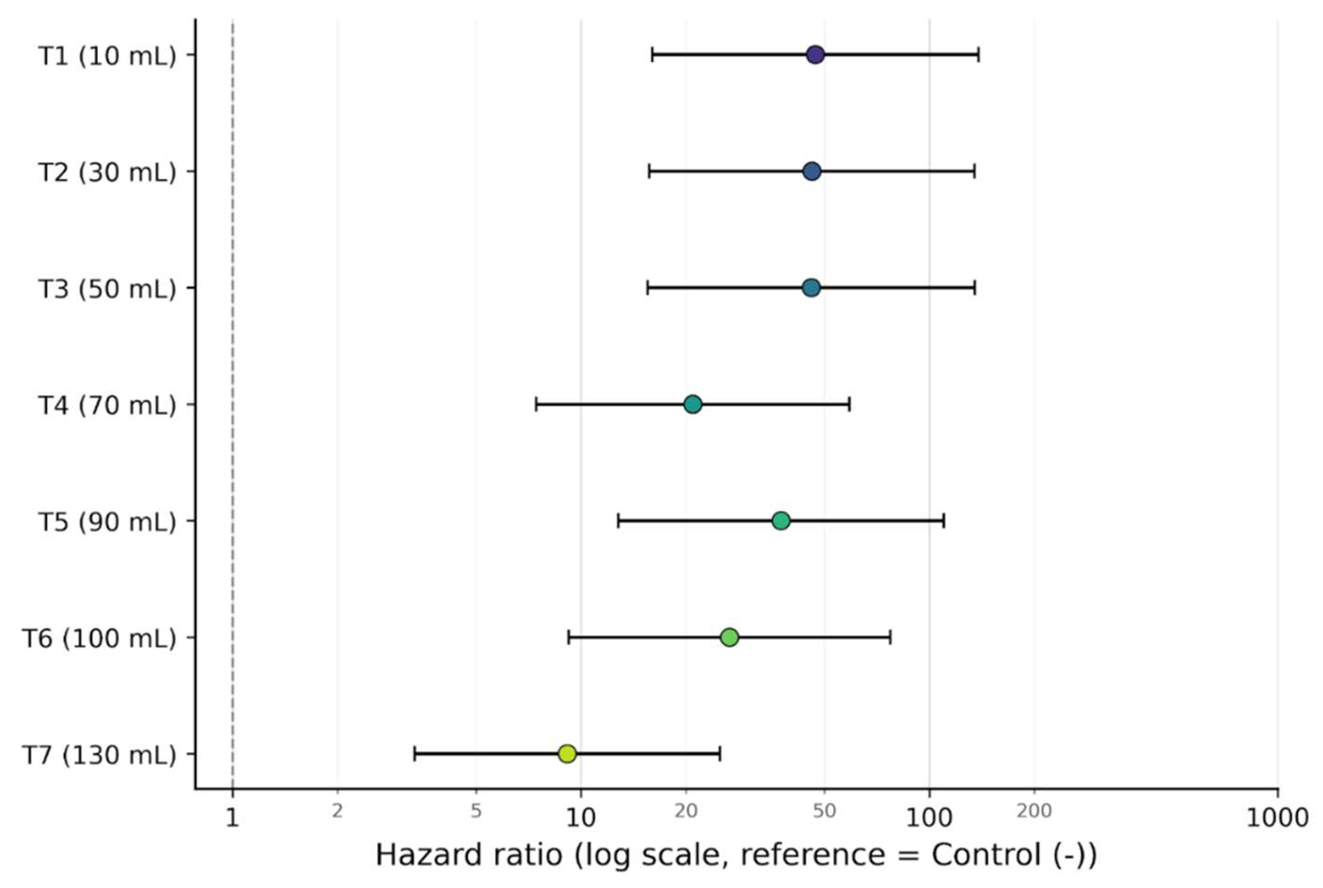
Figure 5.
Kaplan–Meier survival curves of engorged females of Rhipicephalus microplus exposed to different dilutions of Steinernema carpocapsae in Bioassay 3. Survival curves differed significantly between treatments using the log-rank test (χ² = 97.39; gl = 7; p < 0.0001).
Figure 5.
Kaplan–Meier survival curves of engorged females of Rhipicephalus microplus exposed to different dilutions of Steinernema carpocapsae in Bioassay 3. Survival curves differed significantly between treatments using the log-rank test (χ² = 97.39; gl = 7; p < 0.0001).
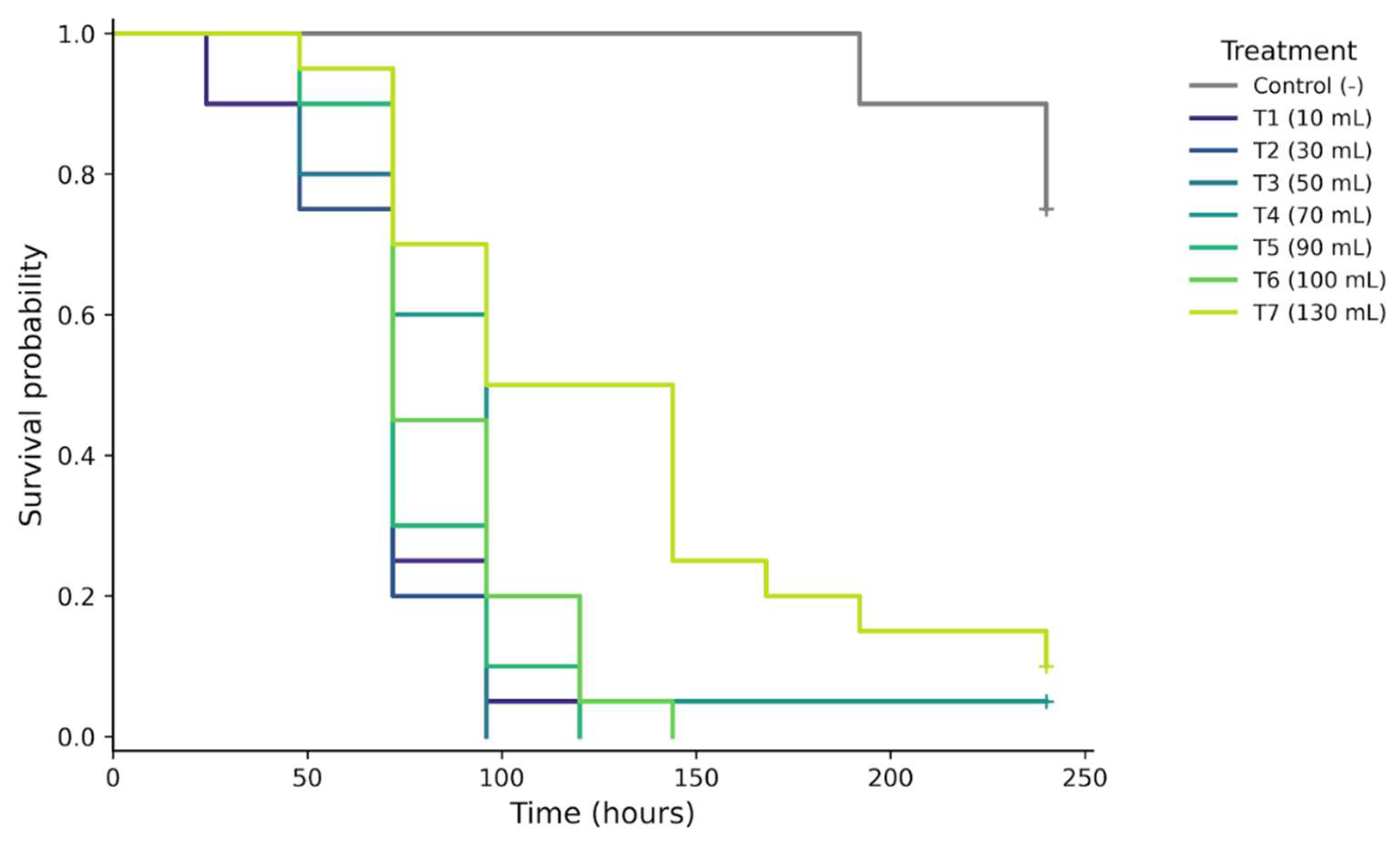
Figure 6.
Hazard ratios (hazard ratios, HR) of mortality of engorged females of Rhipicephalus microplus subjected to the treatments evaluated in Bioassay 3, estimated by the Cox proportional hazards model. The negative control was used as the reference category (HR = 1). The points represent the risk ratio estimates and the horizontal bars their respective 95% confidence intervals. The dashed vertical line represents HR = 1, corresponding to the reference category.
Figure 6.
Hazard ratios (hazard ratios, HR) of mortality of engorged females of Rhipicephalus microplus subjected to the treatments evaluated in Bioassay 3, estimated by the Cox proportional hazards model. The negative control was used as the reference category (HR = 1). The points represent the risk ratio estimates and the horizontal bars their respective 95% confidence intervals. The dashed vertical line represents HR = 1, corresponding to the reference category.
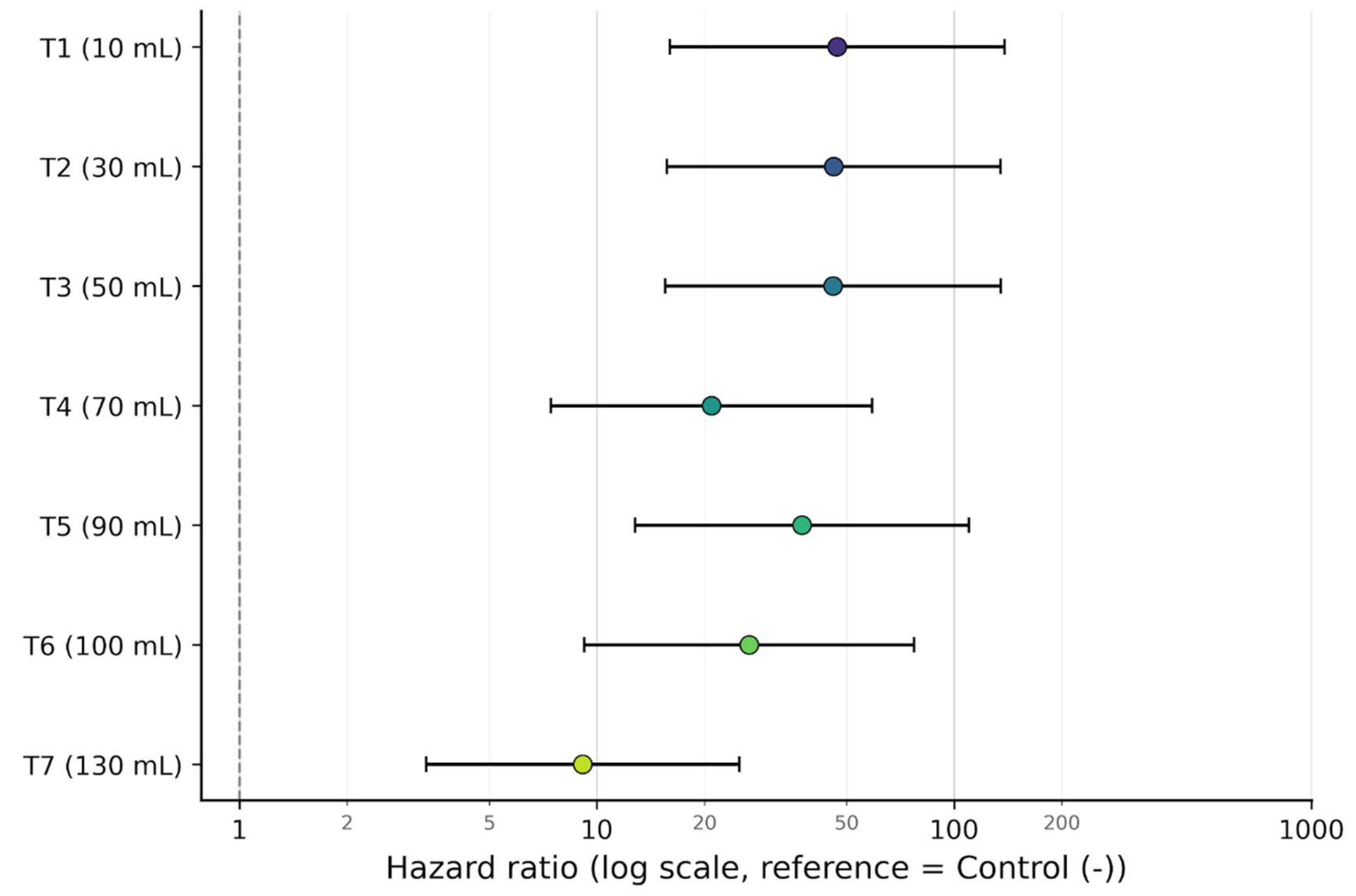
Figure 7.
Kaplan-Meier survival curves of Tenebrio molitor larvae exposed to Steinernema carpocapsae recovered from ingurgitated females of Rhipicephalus microplus. The survival curves differed significantly between treatments by the log-rank test (χ2 = 46.05; gl = 7; p < 0.0001).
Figure 7.
Kaplan-Meier survival curves of Tenebrio molitor larvae exposed to Steinernema carpocapsae recovered from ingurgitated females of Rhipicephalus microplus. The survival curves differed significantly between treatments by the log-rank test (χ2 = 46.05; gl = 7; p < 0.0001).
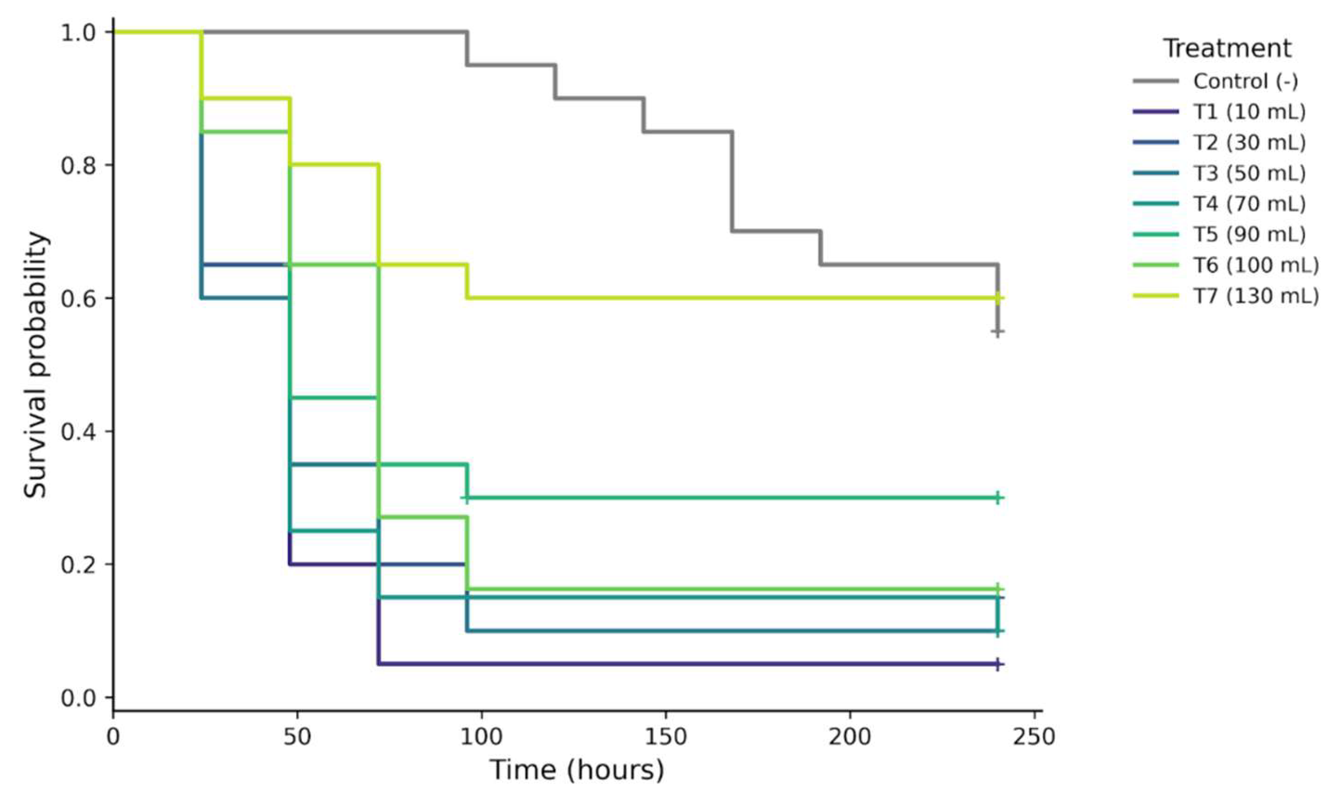
Figure 8.
Hazard ratios (hazard ratios, HR) of mortality of Tenebrio molitor larvae exposed to Steinernema carpocapsae recovered from engorged females of Rhipicephalus microplus, estimated by the Cox proportional hazards model. The negative control was used as the reference category (HR = 1). The points represent the risk ratio estimates and the horizontal bars their respective 95% confidence intervals. The dashed vertical line represents HR = 1, corresponding to the reference category.
Figure 8.
Hazard ratios (hazard ratios, HR) of mortality of Tenebrio molitor larvae exposed to Steinernema carpocapsae recovered from engorged females of Rhipicephalus microplus, estimated by the Cox proportional hazards model. The negative control was used as the reference category (HR = 1). The points represent the risk ratio estimates and the horizontal bars their respective 95% confidence intervals. The dashed vertical line represents HR = 1, corresponding to the reference category.
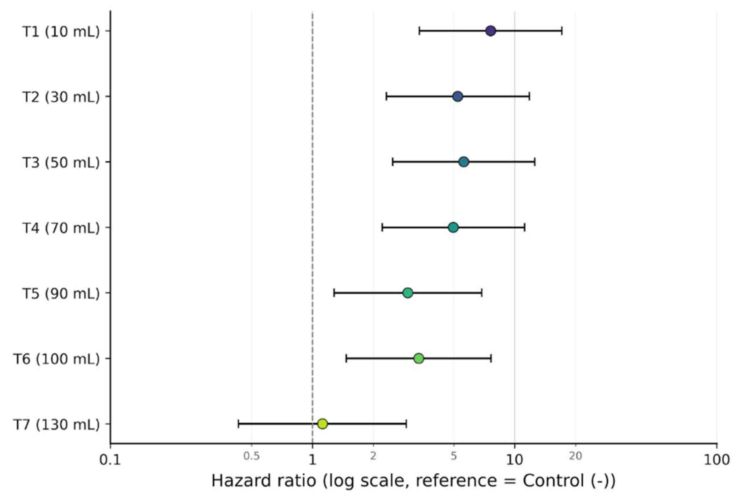
Figure 9.
Morphological aspects of engorged females of Rhipicephalus microplus after exposure to Steinernema carpocapsae and evidence of the presence of the nematode. (A) Female of the negative control. (B) Female exposed to S. carpocapsae, presenting darkening and body distension after death. (C) Female exposed to S. carpocapsae, presenting cuticle rupture and extravasation of internal fluid. (D) Nematodes observed in the internal fluid obtained after dissection of a female exposed to S. carpocapsae.
Figure 9.
Morphological aspects of engorged females of Rhipicephalus microplus after exposure to Steinernema carpocapsae and evidence of the presence of the nematode. (A) Female of the negative control. (B) Female exposed to S. carpocapsae, presenting darkening and body distension after death. (C) Female exposed to S. carpocapsae, presenting cuticle rupture and extravasation of internal fluid. (D) Nematodes observed in the internal fluid obtained after dissection of a female exposed to S. carpocapsae.
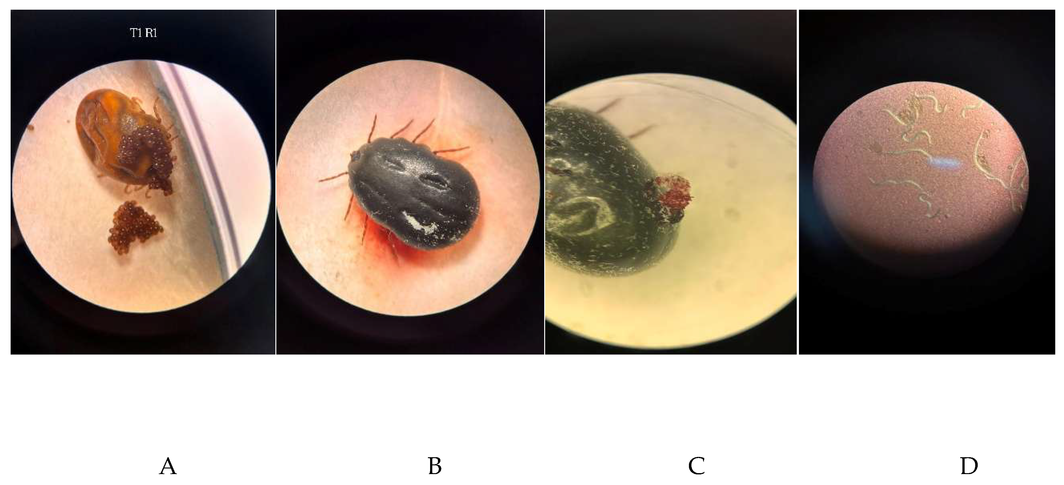
Figure 10.
Evidence for the infectivity of Steinernema carpocapsae in Tenebrio molitor larvae exposed to nematodes recovered from female Rhipicephalus microplus. (A) T. molitor larva killed after exposure to recovered nematodes. (B) Nematode observed by microscopy in the internal contents obtained after dissection of dead T. molitor larva.
Figure 10.
Evidence for the infectivity of Steinernema carpocapsae in Tenebrio molitor larvae exposed to nematodes recovered from female Rhipicephalus microplus. (A) T. molitor larva killed after exposure to recovered nematodes. (B) Nematode observed by microscopy in the internal contents obtained after dissection of dead T. molitor larva.
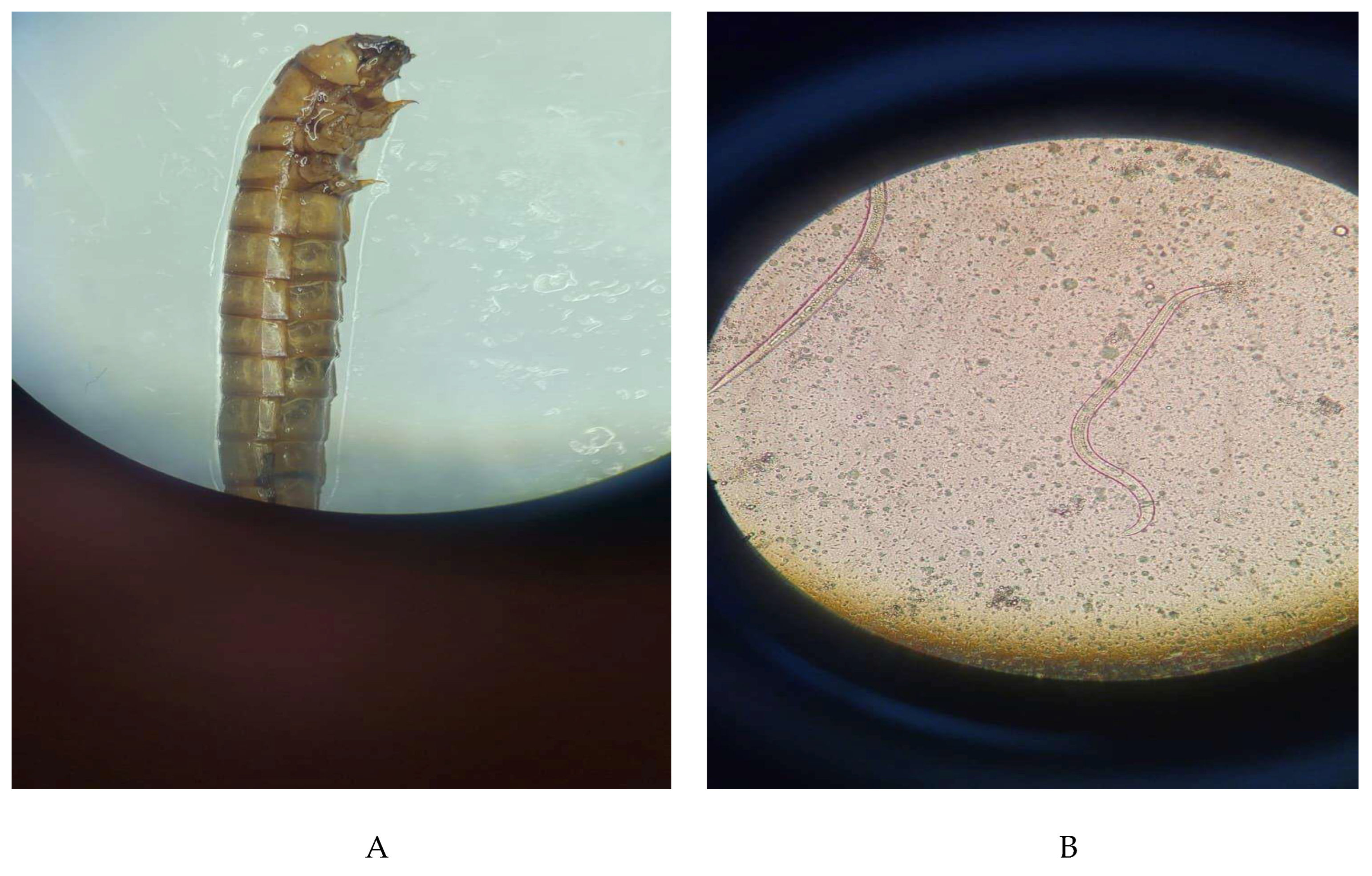

Table 1.
Experimental design and nominal concentrations of infective juveniles (IJ) of Steinernema carpocapsae in the three bioassays.
Table 1.
Experimental design and nominal concentrations of infective juveniles (IJ) of Steinernema carpocapsae in the three bioassays.
| Treatment | Preparation of the suspension | Nominal concentration (IJ mL⁻¹) | Bioassay 1 | Bioassay 2 | Bioassay 3 |
|---|---|---|---|---|---|
| Negative control | Distilled water | – | X | X | X |
| Positive control | Dichlorvos + cypermethrin, 1:400 | – | X | X | – |
| 10 mL | 1 g in 10 mL | 2,20 × 10⁵ | X | X | X |
| 30 mL | 1 g in 30 mL | 7,33 × 10⁴ | – | X | X |
| 40 mL | 1 g in 40 mL | 5,50 × 10⁴ | X | – | – |
| 50 mL | 1 g in 50 mL | 4,40 × 10⁴ | – | X | X |
| 70 mL | 1 g in 70 mL | 3,14 × 10⁴ | X | X | X |
| 90 mL | 1 g in 90 mL | 2,44 × 10⁴ | – | X | X |
| 100 mL | 1 g in 100 mL | 2,20 × 10⁴ | X | X | X |
| 130 mL | 1 g in 130 mL | 1,69 × 10⁴ | X | X | X |
Note: X = treatment included in the bioassay; – = treatment not included. Nominal concentrations were estimated based on the minimum content of 2.2 × 10⁶ JI g⁻¹ declared by the manufacturer, without direct counting of infective juveniles in the suspensions.
Table 2.
Lethal times (TL50 and TL90) and pairwise comparisons of survival curves of engorged females of Rhipicephalus microplus subjected to the treatments evaluated in Bioassay 1.
Table 2.
Lethal times (TL50 and TL90) and pairwise comparisons of survival curves of engorged females of Rhipicephalus microplus subjected to the treatments evaluated in Bioassay 1.
| Treatment | TL50 (h) | IC 95% | TL90 (h) | IC 95% | Grupo (log-rank) |
|---|---|---|---|---|---|
| Negative control | NR | – | NR | – | a |
| Positive control | 204 | 72–NR | NR | – | bc |
| 10 mL | 60 | 48–NR | 84 | 72–NR | d |
| 40 mL | 48 | 48–NR | 72 | 48–NR | d |
| 70 mL | 72 | 48–NR | 84 | 72–NR | d |
| 100 mL | 108 | 72–NR | 136,5 | 120–NR | b |
| 130 mL | 240 | 192–NR | NR | – | ac |
Note: TL50 and TL90 = lethal times for 50% and 90% of individuals, respectively; IC 95% = 95% confidence interval; NR = lethal time not reached during the evaluation period (240 hours); – = not applicable. Treatments that share at least one letter do not differ significantly from each other in pairwise comparisons of survival curves using the log-rank test, with Benjamini–Hochberg adjustment (p ≥ 0.05).
Table 3.
Lethal times (TL50 and TL90) and pairwise comparisons of survival curves of engorged females of Rhipicephalus microplus subjected to the treatments evaluated in Bioassay 2.
Table 3.
Lethal times (TL50 and TL90) and pairwise comparisons of survival curves of engorged females of Rhipicephalus microplus subjected to the treatments evaluated in Bioassay 2.
| Treatment | TL50 (h) | IC 95% | TL90 (h) | IC 95% | Group (log-rank) |
|---|---|---|---|---|---|
| Negative control | NR | – | NR | – | b |
| Positive control | 180 | 144–192 | 312 | 192–NR | a |
| 10 mL | 48 | 48–72 | 84 | 72–NR | c |
| 30 mL | 48 | 48–NR | 96 | NR–NR | cd |
| 50 mL | 96 | 72–120 | 120 | 120–NR | e |
| 70 mL | 72 | 72–96 | 96 | 96–NR | de |
| 90 mL | 96 | 96–120 | 168 | 120–NR | e |
| 100 mL | 96 | 96–120 | 156 | 120–NR | e |
| 130 mL | 192 | 168–240 | 300 | 240–NR | a |
Note: TL50 e TL90 = lethal times for 50% and 90% of individuals, respectively; IC 95% = 95% confidence interval; NR = lethal time not reached during the evaluation period (360 hours); – = not applicable. Treatments that share at least one letter do not differ significantly from each other in pairwise comparisons of survival curves using the log-rank test, with Benjamini–Hochberg adjustment (p ≥ 0.05).
Table 4.
Lethal times (TL50 and TL90) and pairwise comparisons of the survival curves of engorged females of Rhipicephalus microplus subjected to the treatments evaluated in Bioassay 3.
Table 4.
Lethal times (TL50 and TL90) and pairwise comparisons of the survival curves of engorged females of Rhipicephalus microplus subjected to the treatments evaluated in Bioassay 3.
| Treatment | TL50 (h) | IC 95% | TL90 (h) | IC 95% | Group (log-rank) |
|---|---|---|---|---|---|
| Negative control | NR | – | NR | – | a |
| 10 mL | 72 | 72–96 | 96 | 96–NR | b |
| 30 mL | 72 | 72–72 | 108 | 72–NR | bc |
| 50 mL | 72 | 72–NR | 96 | NR–NR | b |
| 70 mL | 96 | 72–96 | 108 | 96–NR | cd |
| 90 mL | 72 | 72–96 | 108 | 96–NR | bc |
| 100 mL | 72 | 72–96 | 120 | 96–NR | bc |
| 130 mL | 120 | 96–168 | 240 | 168–NR | d |
Note: TL50 e TL90 = lethal times for 50% and 90% of individuals, respectively; IC 95% = 95% confidence interval; NR = lethal time not reached during the assessment period (240 hours); – = not applicable. Treatments that share at least one letter do not differ significantly from each other in pairwise comparisons of survival curves using the log-rank test, with Benjamini–Hochberg adjustment (p ≥ 0.05).
Disclaimer/Publisher’s Note: The statements, opinions and data contained in all publications are solely those of the individual author(s) and contributor(s) and not of MDPI and/or the editor(s). MDPI and/or the editor(s) disclaim responsibility for any injury to people or property resulting from any ideas, methods, instructions or products referred to in the content. |
© 2026 by the authors. Licensee MDPI, Basel, Switzerland. This article is an open access article distributed under the terms and conditions of the Creative Commons Attribution (CC BY) license.
Copyright: This open access article is published under a Creative Commons CC BY 4.0 license, which permit the free download, distribution, and reuse, provided that the author and preprint are cited in any reuse.