

Submitted:
07 August 2026
Posted:
10 August 2026
You are already at the latest version
Abstract
Three viruses, Telosma mosaic virus (TeMV), East Asian passiflora virus (EAPV), and Cucumber mosaic virus (CMV), are widespread in Chinese passion fruit growing regions. Co-infection by these three viruses was confirmed in symptomatic field samples collected from Guilin. To evaluate the effectiveness of shoot tip culture alone for virus elimination, in vitro organogenesis was examined, and both direct (DOR) and indirect (IOR) regeneration pathways were observed. In the DOR pathway, organogenic buds and nodules were induced at a rate of 40.83%, and 14.86% of explants produced adventitious buds longer than 1 cm. In the IOR pathway, callus was induced at a rate of 26.67%, and callus-derived meristemoids yielded a bud induction rate of 7.46%. Semi-quantitative PCR analysis showed that only a few virus-free plantlets were obtained, and no regenerated plantlets were found to simultaneously eliminate two or three viruses. No significant difference in median virus elimination efficiency was detected between the DOR and IOR pathways (TeMV: 0% vs. 16.67%; EAPV: 16.67% vs. 0%; CMV: 33.33% vs. 16.67%). SSR analysis confirmed genetic stability in the DOR pathway, whereas a polymorphism rate of 27.27% was observed in the IOR pathway. These findings indicate that the DOR pathway maintains genetic stability while achieving elimination of certain viruses in a small number of co-infected plantlets, providing a basis for subsequent secondary virus elimination and combined treatments.
Keywords:
Passiflora edulis
; shoot tips
; organogenesis
; RT-PCR
; virus elimination
1. Introduction
Passion fruit is a perennial evergreen vine widely cultivated in tropical and subtropical regions [1,2]. The pulp of passion fruit, characterized by its intense aroma and high nutritional content is not only consumed fresh but also widely utilized in processed products such as juice, ice cream, cakes, and wine due to its distinctive flavor profile [3]. Passion fruit exhibits a rapid growth cycle, reaching harvest maturity within 4-6 months of planting, which ensures early production and high economic returns for farmers.
China has a large area dedicated to the cultivation of passion fruit, with the major production regions located in Guangxi, Guangdong, Yunnan, Guizhou and Hainan. As the cultivation area continues to expand, the demand for high-quality seedlings has grown substantially. However, passion fruit is predominantly propagated vegetatively through cuttings or grafting. Such long-term asexual reproduction may facilitate viral accumulation, leading to cultivar degeneration, compromised disease, and reduced yield [4]. Current research indicated at least seven virus diseases infecting passion fruit had been discovered in the major passion fruit cultivation areas of Guizhou Province, China [5]. These included Telosma mosaic virus (TeMV), East Asian passiflora virus (EAPV), Cucumber mosaic virus (CMV), Euphorbia leaf curl virus (ELCV), Passiflora latent virus (PLV), Turnip mosaic virus (TuMV) and Passiflora virus Y (PaVY). The infection rates, ordered in descending order, were as following: 50%, 19%, 15%, 6%, 1%, 0.6% and 0.3%. Furthermore, a high proportion of mixed infections with TeMV, CMV and EAPV had also been identified in this province.
By eliminating viruses, the yield and quality of economic crops can be effectively restored. Currently, in vitro shoot tip culture, either alone or in combination with thermotherapy, cryotherapy, or chemotherapy, is a common method of eliminating plant viruses [6]. These technologies had been applied in various plants such as strawberry [7], kiwifruit [8], potato [9] and grapevine [10]. In kiwifruit, the elimination rate of Actinidia chlorotic ringspot-associated virus was 23.3%, when 0.5 mm shoot tips were used in a secondary meristem culture assay. In contrast, when 1.0 mm shoot tips were excised following thermotherapy (alternating 36/32 °C for 20 d) or chemotherapy (15 µg/mL ribavirin for 2 months), the virus elimination rate reached 100% [8].
Few studies had addressed virus eradication in passion fruit. Ogata and Yamanaka [11] developed an in vivo micrografting for PLV-free plants with low survival due to temperature, whereas Ribeiro et al. [12] evaluated shoot tip culture for virus elimination in hybrid seed matrices with low efficiency. After shoot tips regenerated leaf primordia, explants on MS medium with 4.43 µM 6-BA for 30 d produced shoots > 6 mm, but only one early leaf primordium sample of the CPGA1 genotype tested virus-free. Managing explants during the transition to adventitious buds was critical, highlighting challenges including low rooting rates, difficulty in elongating clustered leaf primordia, and their misidentification as buds [13]. Although widely used, shoot tip culture in Passiflora yields low regeneration from leaf primordia, affected by PGR ratios, explant size, and species [12], suggesting virus elimination efficiency is linked to regeneration capacity.
In vitro regeneration of Passiflora species had been documented from various explants. P. cincinnata regenerated from root and leaf discs via both direct and indirect organogenesis [14], while P. edulis root segments regenerated only directly, maintaining ploidy stability [15]. In P. edulis, buds induced from root, hypocotyl, and leaf explants with 4.44 µM 6-BA followed both pathways, except for leaf explants, which were restricted to direct organogenesis, as characterized by histology, scanning electron microscopy (SEM), and transmission electron microscopy (TEM) [16,17]. Nevertheless, the organogenic pathway and detailed cellular characterization of shoot tip explants in passionfruit remain poorly understood.
To investigate whether different organogenesis patterns affect virus elimination efficiency in purple passionfruit shoot tips, three viruses commonly infecting passionfruit in Guangxi were firstly detected and identified. In vitro organogenesis of P. edulis shoot tip explants was then characterized, and cells in each regeneration pattern were examined microstructurally. Virus elimination efficiency was subsequently compared between the two patterns in regenerated lines by semi-quantitative analysis. Finally, SSR markers were used to evaluate genetic stability between mother plants and regenerated lines. This study provides new insights into the use of shoot tip organogenesis for virus elimination in passionfruit.
2. Materials and Methods
2.1. Major Virus Detection in Passion Fruit
The purple passion fruit line ‘Guizao’ (GZ) originated from a wild plant collected from a high-altitude area (1,800 m) in Ruili, Yunnan Province of China. It was subsequently developed through artificial breeding as a dwarf line and introduced to the greenhouse of the Guangxi Institute of Botany (Yanshan district, Guilin, Guangxi, China). This line, which was characterized by cold tolerance, early maturity, slow lateral branching, and suitability for high-density planting, was selected as the plant material (Supplementary Figure S1). Leaf samples exhibiting typical viral symptoms (such as leaf curling and crinkling) were collected from symptomatic passion fruit plants (Supplementary Figure S2). The samples were performed at the pre-flowering stage, with the third to fifth fully expanded functional leaves collected for analysis, and then immediately frozen in liquid nitrogen, as well as kept at −80 °C until RNA extraction.
Total RNA was extracted using the RNAprep Pure plant RNA extraction kit (DP432, Tiangen Biotech, Beijing, China). RNA concentration, purity, and integrity were assessed using a NanoDrop 2000c spectrophotometer and 1.0% agarose (1×TBE) gel electrophoresis. First-strand cDNA was synthesized using the PrimeScript® RT reagent kit (DRR047A, TaKaRa, Japan) according to the manufacturer’s instructions, and the cDNA was stored at −20 °C.
Reverse-transcription polymerase chain reaction (RT-PCR) was used for virus detection. cDNA was used as the template, PCR was performed with specific primers (Supplementary Table S1) and Pfu DNA polymerase (50 μL reaction system in Supplementary Table S2). Primers were designed using Primer Premier 5.0 based on references [5,18]. The PCR program was as follows: 94 °C for 3 min; 35 cycles of 94 °C for 30 s, 51–58 °C for 30 s, and 72 °C for 1 min; followed by 72 °C for 10 min. The products were separated on a 1.5% agarose (1×TAE) gel, purified using a DNA gel recovery kit (B110092, Diamond, Shanghai, China), and ligated into the pMD™ 18-T vector. Positive colonies were sent to Sangon Biotech (Shanghai) Co., Ltd. for sequencing.
2.2. Phylogenetic Analysis
The reported sequences of TeMV, EAPV and CMV were downloaded from the NCBI database, and MEGA 5.0 was used to build the Neighbor-Joining (NJ) tree. In the phylogenetic analysis, the SoMV (Soybean mosaic virus) isolate WS200 (FJ548849.1), SMV (Soybean mosaic virus) isolate NP-L (HQ166266.1) and PSV (Peanut stunt virus) line ER (U15730.1) were regarded as outgroups, respectively [5,19]. The phylogenetic tree was evaluated using the bootstrap method with 1,000 replicates. Information on the isolates is provided in parentheses following the taxon names.
2.3. The Primary Culture of Shoot Tip Explants
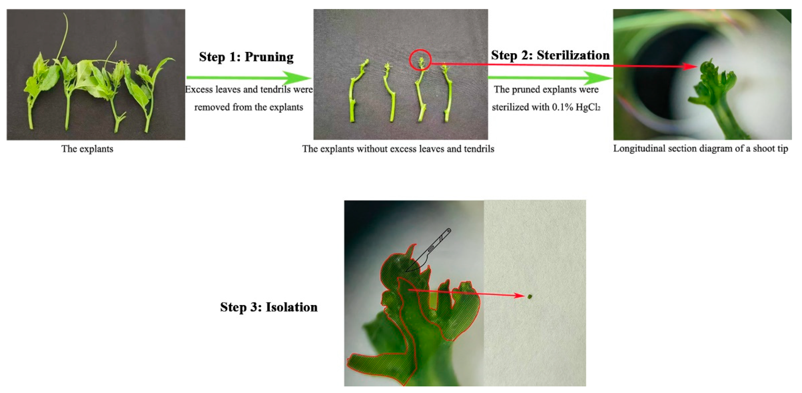
Plants exhibiting distinct infection symptoms in the field, which were subsequently confirmed by RT-PCR to be co-infected with TeMV, EAPV, and CMV, were selected as mother plants (Supplementary Figure S2). Semi-lignified cuttings were collected in mid-April and transplanted to the greenhouse. Terminal shoots (2-3 cm in length) were collected after two months of growth, and then all leaves and tendrils were removed. The pruned shoot tips were surface sterilized with 0.1% (w/v) HgCl2 for 6–8 min, followed by three rinses with sterile distilled water. They were then blotted dry on sterile filter paper and used for explants. Then shoot tips (1.0-1.5 mm in length) were excised (Figure 1) and inoculated on the primary culture medium (MS + 4.43 µM 6-BA + 3% sucrose + 0.8% agar). The ambient temperature was maintained at 26 ± 3 °C, with alternating 12 h light and dark periods.
Figure 1.
The preparation of shoot tip explants, including pruning, sterilization and isolation. A longitudinal section schematic showed the shoot tip (1.0-1.5 mm in length).
Figure 1.
The preparation of shoot tip explants, including pruning, sterilization and isolation. A longitudinal section schematic showed the shoot tip (1.0-1.5 mm in length).
2.4. In Vitro Induction of Organogenesis
After 30 d of primary culture, early leaf primordia from shoot tip explants were transferred to OR medium (MS + 0.44 µM 6-BA + 3% sucrose + 0.8% agar). For another 40 d, explants regenerating organogenic structures (buds, nodules, or calli) were recorded. Based on the previous study, during direct organogenesis (DOR), these structures arose from meristemoids in the pericycle region, while during indirect organogenesis (IOR), they originated from meristemoids at the callus periphery [17]. Each bottle contained 5–7 explants, and 40 explants constituted one biological replicate with three replicates. The organogenic structure induction rate (%) = (number of explants with organogenic structures / total number of explants inoculated) × 100.
All regenerating structures were then transferred to fresh OR medium for adventitious bud induction, with medium renewal every 40 d. For DOR pathway, any calli surrounding the organogenic nodules were removed, and only newly formed buds and nodules were transferred. For IOR pathway, yellow-brown senescent calli were removed, and only newly formed calli were transferred. After two transfers (for additional 80 d), adventitious bud induction rates were compared between the two pathways. Buds > 1.0 cm were considered adventitious buds; those <1.0 cm were classified as leaf primordia. Each bottle was inoculated with 5 to 7 explants. For each set of 40 explants, the data was regarded as one biological replicate, with three biological replicates in total. Adventitious bud induction rate (%) = (number of explants with adventitious buds / number of explants with organogenic structures) × 100.
Adventitious buds were excised and sub-cultured every 60 d on MS medium until most reached > 3 cm. Those > 3 cm were then excised and transferred to rooting medium (1/2 MS + 0.5 µM IBA + 2% sucrose + 0.8% agar) for 30 d to obtain the rooting plantlets.
2.5. Microscopy Sample Preparation
For the in vitro organogenesis process of P. edulis shoot tip explants (growing on OR medium for a total of 120 d, samples were collected at 80 d or 100 d), tissue samples participating in different regeneration patterns were collected for microstructural analysis. The long axis of the samples ranged from 0.5 to 1.0 cm. Firstly, one portion of the samples was fixed in FAA solution for over 24 h, rinsed with 50% ethanol, and then processed through dehydration, clearing, paraffin infiltration, and embedding for conventional paraffin sectioning. Transverse and longitudinal sections (8 μm) were cut using a rotary microtome (PM 24, Servicebio, China), stained with safranin–fast green, and observed under a light microscope (SWE-CX63, Servicebio, China). Secondly, another portion of the samples was examined and photographed under a stereomicroscope (SZX2-FOF, Olympus, Japan). Moreover, an additional portion of the samples was fixed overnight in 2.5% glutaraldehyde (prepared in 0.1 M phosphate buffer, pH 7.2) at 4 °C. They were then rinsed with 0.1 M phosphate buffer (pH 7.2), dehydrated through a graded ethanol series (50%, 75%, 85%, 95%, and 100%, each grade for 15 min), critical point dried with CO2, and sputter coated with gold. Observations and photomicrographs were taken using a scanning electron microscope (EVO 18, Zeiss, Germany) at magnifications of 30 × to 45 ×.
2.6. Relative Virus Amounts
Semi-quantitative PCR (sq-PCR) was used to compare the relative virus amounts among samples [20]. In order to evaluate the effect of a single round shoot tip regeneration on virus elimination in plants co-infected with TeMV, EAPV and CMV, RNA was extracted from two young leaves of regenerated plantlets (height > 3 cm) after two sub-cultures (80 d). The undiluted cDNA solution obtained via reverse transcription was diluted 5-fold and used as the template. Based on the housekeeping gene selection study in Passiflora [21], PeEF1α was used as the internal reference. The primer sequences are listed in Supplementary Table S1. Amplification was performed using 20 μL (Supplementary Table S2) PCR reaction system with 2× SanTaq PCR Mix (Sangon Biotech, Shanghai, China). The PCR program was as follows: initial denaturation at 95 °C for 3 min; followed by 26 cycles (for PeEF1α) or 33 cycles (for TeMV, EAPV, and CMV) of 95 °C for 30 s, 50–60 °C for 30 s, and 72 °C for 30 s; and a final extension at 72 °C for 10 min. The products were separated with 1.5% agarose (1×TAE) gel electrophoresis. For each set of 6 regenerated plantlets, the data was regarded as one biological replicate, with three biological replicates in total. Virus elimination efficiency of regenerated plantlets was compared between the two pathways. Virus elimination efficiency (%) = (Number of virus-free individuals / Total number of detected individuals) × 100.
2.7. Genetic Fidelity Assessment
Mother plants were selected from stem cutting propagated plants, either without or with calli at the base. Progeny lines were regenerated via direct or indirect organogenesis from the shoot tip explants of mother plant. Genomic DNA was extracted from fresh leaves of eight randomly selected regenerated progeny lines and one mother plant using a modified CTAB method. Genetic fidelity was assessed using SSR markers. A total of six SSR primers were tested and three were selected in the analysis [22]. PCR was performed as follows: 95 °C for 2 min; 26 cycles of 94 °C for 40 s, 56 °C for 45 s, and 72 °C for 30 s; and 72 °C for 5 min (PCR reaction system in Supplementary Table S3). PCR products (1 µL) were separated on a 9% non-denaturing polyacrylamide gel (Supplementary Table S4) by electrophoresis in 0.5 × TBE buffer. After electrophoresis, the gel was stained with 1.0 g·L−1¹ Ag3O₃ for 10–15 min, developed in 20 −1·L⁻¹ NaOH containing 10 mL formaldehyde for 5–8 min, rinsed twice with water, and photographed on a light box. Wells were marked with pipette tips prior to staining. Amplified bands for each primer were scored as present (1) or absent (0). Only clear and unambiguous bands were scored. The total number of bands, number of polymorphic bands, and percentage of polymorphic bands were calculated.
2.8. Statistical Analysis
Microsoft Excel and SPSS software were used for statistical analysis to compare the two regeneration patterns or the three viruses. Normality and homogeneity of variance were tested, and percentage data were log-transformed (LN) before analysis. For data that met the assumptions of normality and homogeneity of variance, such as the induction rates of organogenic structures and adventitious buds, independent samples t-tests were applied (P < 0.05). Data were presented as means ± standard deviations (SD) from three biological replicates, and histograms were generated with GraphPad Prism 7 software.
For data that were still not normally distributed after transformation, such as virus elimination efficiency, the non-parametric Mann-Whitney U test (between the two regeneration patterns) or Kruskal-Wallis H test (among the three viruses) of independent samples was used (P < 0.05), and the median (M) was reported to describe the central tendency and dispersion. Image processing was performed using Adobe Photoshop CS6.
3. Results
3.1. Partial Nucleotide Sequences Amplification
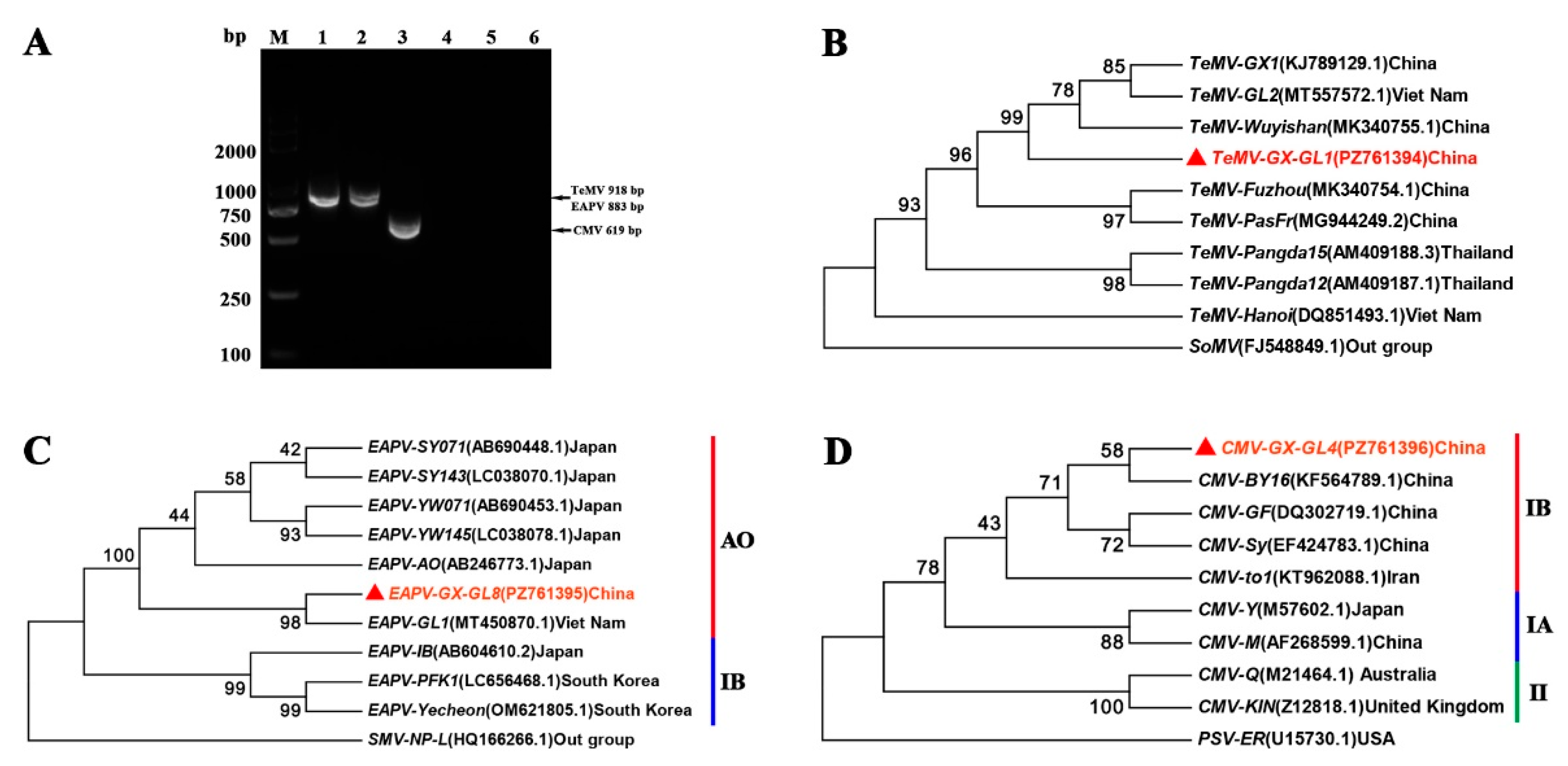
After amplification using specific primers, the partial nucleotide sequences of TeMV, EAPV and CMV obtained were 918 bp, 883 bp and 619 bp, respectively (Figure 2A). Then the above sequences were analyzed by BLAST against the NCBI database. TeMV product showed 99.46% identity to isolate ‘RJ’ (ON932195.1) from Jiangxi, China; the EAPV product showed 99.77% identity to isolate ‘GL1’ (MT450870.1) from Vietnam; and the CMV product showed 97.74% identity to isolate ‘yang-ss3-2’ (MK778782.1) from Henan, China. These results confirmed the amplified fragments as partial sequences of the three passion fruit infecting viruses, TeMV, EAPV, and CMV, respectively.
3.2. Phylogenetic Analysis of TeMV, EAPV and CMV
Phylogenetic analysis showed that the amplified TeMV-GX-GL1 (PZ761394) and eight other partial TeMV isolates clustered together, with the TeMV-Wuyishan isolate (MK340755.1) from China, a relationship supported by a high bootstrap value (Figure 2B). For EAPV, the phylogenetic tree was divided into two major clades, corresponding to the AO and IB lines. The AO line exhibited clear regional clustering: all Japanese isolates formed a subclade, whereas the cloned isolates EAPV-GX-GL8 (PZ761395) and EAPV-GL1 (MT450870.1) grouped together (Figure 2C), suggesting that the EAPV isolates infecting passion fruit in Guangxi are closely related to that from Viet Nam. The CMV phylogenetic tree was divided into three major clades, designated as subgroups IB, IA, and II. The cloned isolates CMV-GX-GL4 (PZ761396) and CMV-BY16 (KF564789.1) clustered together (Figure. 2D), indicating a close genetic relationship between the CMV isolates from Guangxi and Hubei in China.
Figure 2.
(A) Specificity of the three primer pairs used for RT-PCR detection of TeMV, EAPV, and CMV, analyzed by agarose gel electrophoresis. Lanes 1–3: TeMV (918 bp), EAPV (883 bp), and CMV (619 bp), respectively; Lanes 4–6: corresponding negative controls; Lane M: Trans 2000 Plus DNA marker; (B-D) Phylogenetic analysis of partial nucleotide sequences from TeMV, EAPV and CMV. Neighbor-joining phylogenetic trees were constructed based on the partial nucleotide sequences of TeMV-GX-GL1 (B), EAPV-GX-GL8 (C), and CMV-GX-GL4 (D), with SoMV, SMV-NP-L, and PSV-ER designated as outgroups, respectively. EAPV was divided into two lines, AO and IB, whereas CMV was categorized into three subgroups, namely IA, IB and II.
Figure 2.
(A) Specificity of the three primer pairs used for RT-PCR detection of TeMV, EAPV, and CMV, analyzed by agarose gel electrophoresis. Lanes 1–3: TeMV (918 bp), EAPV (883 bp), and CMV (619 bp), respectively; Lanes 4–6: corresponding negative controls; Lane M: Trans 2000 Plus DNA marker; (B-D) Phylogenetic analysis of partial nucleotide sequences from TeMV, EAPV and CMV. Neighbor-joining phylogenetic trees were constructed based on the partial nucleotide sequences of TeMV-GX-GL1 (B), EAPV-GX-GL8 (C), and CMV-GX-GL4 (D), with SoMV, SMV-NP-L, and PSV-ER designated as outgroups, respectively. EAPV was divided into two lines, AO and IB, whereas CMV was categorized into three subgroups, namely IA, IB and II.
3.3. Morphogenetic Responses of Shoot Tip
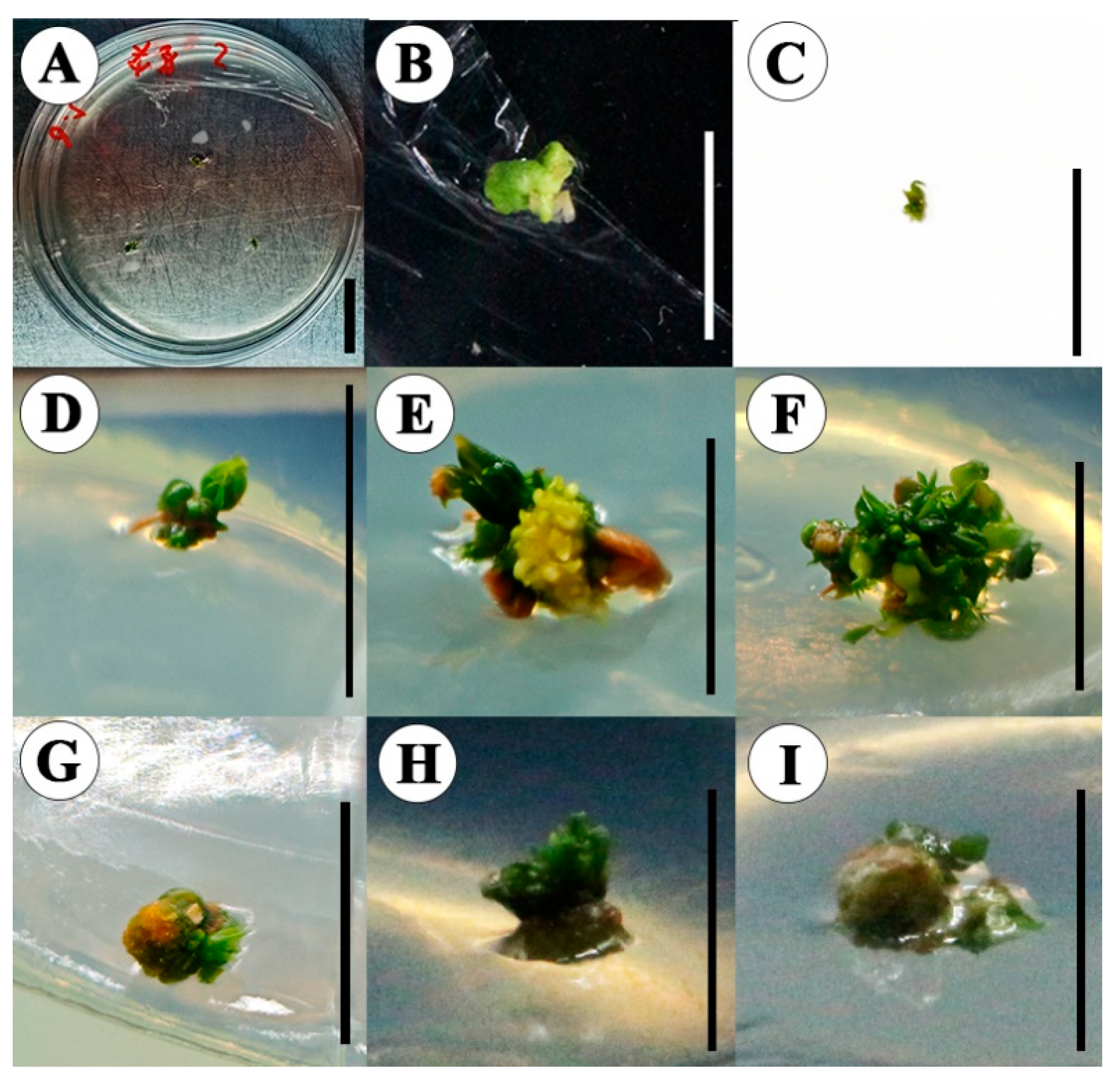
Shoot tip explants were cultured on primary medium supplemented with 1 mg·L−1¹ 6-BA. After 30 d, early leaf primordia (0.2–0.3 cm in length) were initiated (Figure 3A–C). These early leaf primordia were then transferred to OR medium (supplemented with 0.1 mg−1L⁻¹ 6-BA). After another 40 d, both direct and indirect organogenesis pathways were induced.
For direct organogenesis, the induction rate of organogenic buds and nodules was 40.83% ± 3.82% (Figure 5A). Organogenic nodules formed at the base of the early leaf primordia, and new leaf primordia differentiated on their surface (Figure 3D–F). Some of these clustered leaf primordia subsequently elongated into adventitious shoots.
For indirect organogenesis, the early leaf primordia developed through a distinct callus intermediate stage (Figure 3G–I). During the early induction stage, calli proliferated faster than clustered leaf primordia formed. The calli induction rate was 26.67% ± 5.20% (Figure 5A), which was significantly lower than that of organogenic buds and nodules in the direct pathway. These results indicated that shoot tip explants regenerated mainly through direct organogenesis, with indirect organogenesis occurring at a lower frequency. In addition, organogenic buds and nodules or calli at different developmental stages were observed, suggesting that the initiation of regeneration among different meristematic cell clusters on the same explant may not be synchronized.
Furthermore, approximately 30% of the shoot tip explants successfully initiated organogenic buds and nodules, or calli during the early stage of regeneration culture. However, these organogenic structures gradually browned and failed to regenerate.
Figure 3.
In vitro organogenesis of Passiflora edulis shoot tips explants. Explants developed into leaf primordia following 0 d (A), 15 d (B), 30 d (C) of cultivation on MS medium supplemented with 1.0 mg/L 6-benzyladenine (6-BA); Organogenic buds and nodules (D–F) formed after 40 d of incubation on MS medium supplemented with 0.1 mg·L−1¹ 6-BA, designated as direct organogenesis (DOR); calli formed after 40 d (G–I) of incubation on MS medium supplemented with 0.1 mg−1L⁻¹ 6-BA, designated as indirect organogenesis (IOR). Scale Bars = 1 cm.
Figure 3.
In vitro organogenesis of Passiflora edulis shoot tips explants. Explants developed into leaf primordia following 0 d (A), 15 d (B), 30 d (C) of cultivation on MS medium supplemented with 1.0 mg/L 6-benzyladenine (6-BA); Organogenic buds and nodules (D–F) formed after 40 d of incubation on MS medium supplemented with 0.1 mg·L−1¹ 6-BA, designated as direct organogenesis (DOR); calli formed after 40 d (G–I) of incubation on MS medium supplemented with 0.1 mg−1L⁻¹ 6-BA, designated as indirect organogenesis (IOR). Scale Bars = 1 cm.
After additional 80 d on OR medium, the induction rate of adventitious buds (longer than 1.0 cm) was 14.86% ± 2.89% in the direct organogenesis pathway, compared to 7.46% ± 2.56% in the indirect pathway (Figure 5B). In the indirect pathway, buds mainly originated from compact green calli, and their regeneration was less efficient than those from direct organogenesis. Adventitious buds at different stages were also observed (Figure 4A–D, 4E–H), which may be due to asynchronous differentiation among different organogenic nodules or callus cell clusters on the same explant. Finally, these buds were transferred to MS medium for proliferation (Figure 4I–K), and adventitious buds over 3 cm were rooted (Figure 4L).
3.4. Origin of Direct and Indirect Organogenesis in P. edulis
Three types of cell masses were observed on OR medium. Type I (DOR, 80 d) appeared as green, compact clusters with protruding organogenic nodules and incipient leaf primordia (Figure 6A). Type II (IOR, 80 d) consisted of intermingled pale green and pale yellow regions, where green areas were covered with meristemoids, and yellow regions were compact, translucent calli (Figure 6B). Type III (both DOR and IOR, 100 d) appeared as pale green clusters with densely distributed leaf primordia, meristemoids, and large organogenic nodules with aged calli at the base (Figure 6C).
Microscopic observations revealed that Type I cell masses contained multiple meristemoids and organogenic nodules at various developmental stages. These consisted of small, compact polygonal cells with dense cytoplasm and a nucleus occupying over one-third of the cell volume (Figure 6D, G). Hemispherical meristematic protuberances were visible rather than a smooth surface (Figure 6J). Notably, highly vacuolated parenchyma cells of varying sizes were present around the organogenic nodules, supporting the proliferation of occasional calli (Arrows in Figure 6D, G). Type II cell masses, composed of intermingled pale green and pale yellow regions, displayed clustered meristemoids on the surface (Figure 6E). These meristemoids could differentiate into new leaf primordia that broke through the epidermal layer, while also producing parenchyma cells to maintain callus proliferation (Figure 6H). In some regions, outlines of early shoot primordia were observed, without a loose, fragmented surface (Figure 6K). Type III cell masses showed meristemoids, early shoot primordia, and organogenic nodules on the callus surface (Figure 6F). Distinct adventitious buds and a vascular cylinder were visible near the central top region (Figure 6I, L), with stained red vascular tissue precursors locally (Figure 6I). These features indicated that such cell masses had initiated structural preparations for organ differentiation and possessed high differentiation potential.
Figure 4.
Adventitious buds induction. (A–D) Adventitious buds from organogenic buds and nodules at different developmental stages after another 80 d on OR medium (refreshed every 40 d, two times); (E–H) Adventitious buds from calli at different developmental stages after another 80 d on OR medium (refreshed every 40 d, two times); (I–K) Shoot proliferation and elongation on MS medium at 0, 18, and 60 d; (L) Rooting culture after 30 d. Scale bars = 1 cm.
Figure 4.
Adventitious buds induction. (A–D) Adventitious buds from organogenic buds and nodules at different developmental stages after another 80 d on OR medium (refreshed every 40 d, two times); (E–H) Adventitious buds from calli at different developmental stages after another 80 d on OR medium (refreshed every 40 d, two times); (I–K) Shoot proliferation and elongation on MS medium at 0, 18, and 60 d; (L) Rooting culture after 30 d. Scale bars = 1 cm.
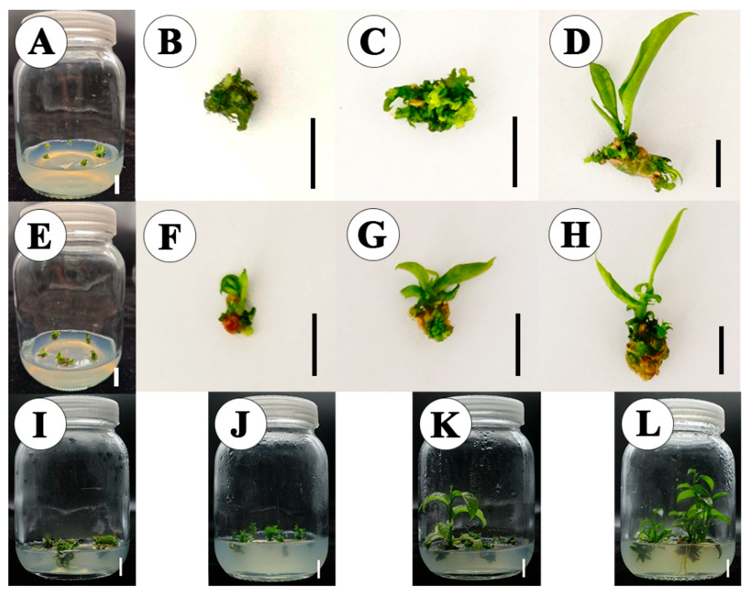
Figure 5.
Regeneration of P. edulis shoot tip explants occurred via both direct (DOR) and indirect organogenesis (IOR). Organogenic structures included organogenic buds and nodules, as well as calli. Buds longer than 1.0 cm were considered as adventitious buds. Data were presented as the mean ± standard deviation (SD) from three biological replicates. Different lowercase letters a-b denoted significant differences (t-test, P<0.05).
Figure 5.
Regeneration of P. edulis shoot tip explants occurred via both direct (DOR) and indirect organogenesis (IOR). Organogenic structures included organogenic buds and nodules, as well as calli. Buds longer than 1.0 cm were considered as adventitious buds. Data were presented as the mean ± standard deviation (SD) from three biological replicates. Different lowercase letters a-b denoted significant differences (t-test, P<0.05).
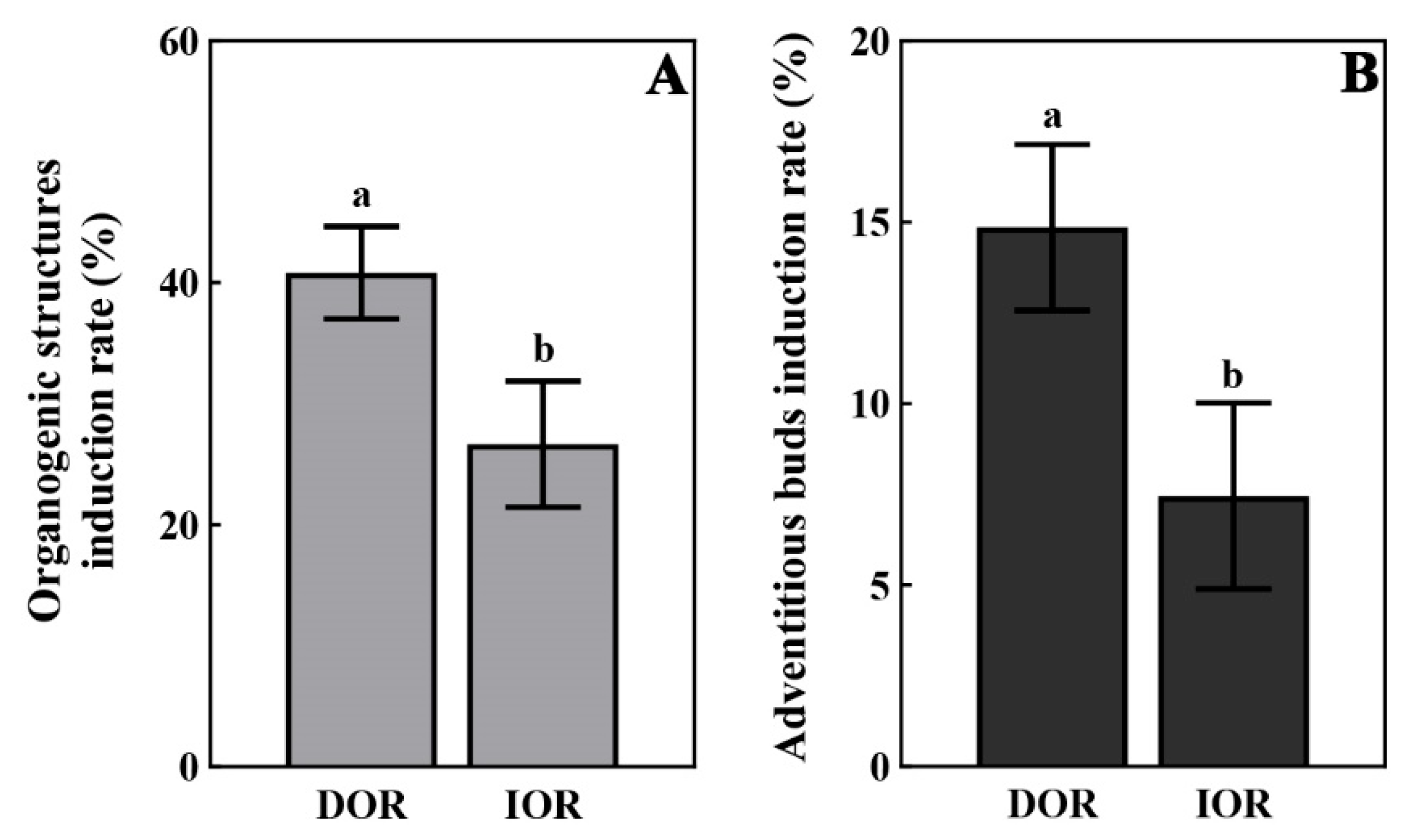
Figure 6.
Microstructural analysis of P. edulis shoot tip participating in different regeneration patterns. (A–C) Representative stereo microscope images; (D–I) Longitudinal paraffin sections stained with safranin-fast green; (J–L) scanning electron micrographs. (A, D, G, J) Direct organogenesis (for 80 d), demonstrated the development of meristemoids and organogenic nodules in the pericycle region. The arrows indicated parenchyma cell clusters of varying sizes, which supported the proliferation of occasional calli; (B, E, H, K) Indirect organogenesis (for 80 d), showed the early development of leaf primordia at the periphery of calli. Organogenic sections could develop into meristemoids and leaf primordia; (C, F, I, L) Direct and indirect organogenesis coexisted (for 100 d), showed the development of adventitious buds on the calli and the distribution of the vascular cylinder. ca, calli; on, organogenic nodule; me, meristemoid; vc, vascular cylinder; lp, leaf primordium. Scale bars: 1 mm in (A–C); 100 µm in (D–L).
Figure 6.
Microstructural analysis of P. edulis shoot tip participating in different regeneration patterns. (A–C) Representative stereo microscope images; (D–I) Longitudinal paraffin sections stained with safranin-fast green; (J–L) scanning electron micrographs. (A, D, G, J) Direct organogenesis (for 80 d), demonstrated the development of meristemoids and organogenic nodules in the pericycle region. The arrows indicated parenchyma cell clusters of varying sizes, which supported the proliferation of occasional calli; (B, E, H, K) Indirect organogenesis (for 80 d), showed the early development of leaf primordia at the periphery of calli. Organogenic sections could develop into meristemoids and leaf primordia; (C, F, I, L) Direct and indirect organogenesis coexisted (for 100 d), showed the development of adventitious buds on the calli and the distribution of the vascular cylinder. ca, calli; on, organogenic nodule; me, meristemoid; vc, vascular cylinder; lp, leaf primordium. Scale bars: 1 mm in (A–C); 100 µm in (D–L).
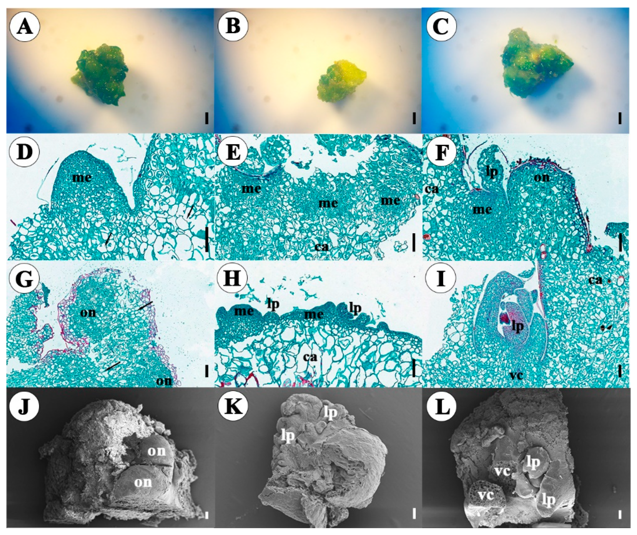
In summary, all three types of cell masses retained strong meristematic activity. In DOR pathway, clustered leaf primordia likely originated from meristemoids and organogenic nodules in the pericycle region. In IOR pathway, they likely arose from meristemoids at the callus periphery. During mid-to-late development, large parenchyma cell masses proliferated around the organogenic nodules, indicating that organogenic nodules could coexist with calli.
3.5. Analysis of TeMV, EAPV and CMV Viral Amounts by Semi-qPCR
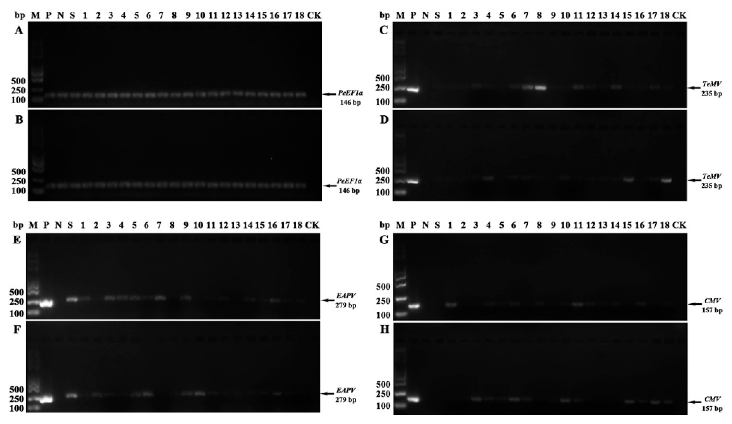
Transcript levels of TeMV (235 bp, Figure 7C, D), EAPV (279 bp, Figure 7E, F), and CMV (157 bp, Figure 7G, H) were detected in regenerated plantlets (height > 3 cm, no obvious viral symptoms were observed) after two sub-cultures (80 d) via sq-PCR. Only a few lines derived from one single meristem tip culture process showed no detectable target bands, indicated that the relative virus amounts of all three viruses were markedly reduced. Based on the Mann-Whitney U test, the median virus elimination efficiencies for TeMV, EAPV, and CMV in the DOR and IOR pathways were 0% vs. 16.67%, 16.67% vs. 0%, and 33.33% vs. 16.67%, respectively, no significant difference in the median (M) virus elimination efficiency was observed between the two regeneration pathways for any of the three viruses (P = 0.46, 0.35, and 0.35, respectively; all > 0.05). Similarly, according to the Kruskal-Wallis H test, no significant difference was found among the median (M) elimination efficiencies of TeMV, EAPV, and CMV within either the DOR or the IOR pathway (P = 0.35 and 0.33, respectively, all > 0.05). These results indicated that the regeneration mode did not significantly affect virus elimination efficiency, and that different virus types did not affect virus elimination efficiency under the same regeneration pathway. Furthermore, no regenerated plantlets were found to simultaneously eliminate two or three viruses.
Figure 7.
Transcript levels of TeMV (235 bp), EAPV (279 bp), and CMV (157 bp) genes in regenerated plantlets were analyzed by sq-PCR with agarose gel electrophoresis. Lane M: Trans 2000 Plus DNA marker; Lane P: Positive control (leaf samples co-infected with three viruses); Lanes N: Negative control (symptomless seedlings); Lanes S: Plantlets propagated in vitro from symptomless stem segments; Lane CK: Blank controls. Lanes 1-18: Leaf samples from regenerated plantlets obtained through direct (A, C, E, G) or indirect (B, D, F, H) organogenesis pathways using shoot tip explants, respectively. PeEF1α (146 bp) was used as an internal reference for sq-PCR (A-B), and the cycle number of housekeeping gene was set to 26, and the cycle number of TeMV (C-D), EAPV (E-F), and CMV (G-H) was set to 33.
Figure 7.
Transcript levels of TeMV (235 bp), EAPV (279 bp), and CMV (157 bp) genes in regenerated plantlets were analyzed by sq-PCR with agarose gel electrophoresis. Lane M: Trans 2000 Plus DNA marker; Lane P: Positive control (leaf samples co-infected with three viruses); Lanes N: Negative control (symptomless seedlings); Lanes S: Plantlets propagated in vitro from symptomless stem segments; Lane CK: Blank controls. Lanes 1-18: Leaf samples from regenerated plantlets obtained through direct (A, C, E, G) or indirect (B, D, F, H) organogenesis pathways using shoot tip explants, respectively. PeEF1α (146 bp) was used as an internal reference for sq-PCR (A-B), and the cycle number of housekeeping gene was set to 26, and the cycle number of TeMV (C-D), EAPV (E-F), and CMV (G-H) was set to 33.
3.6. Genetic Fidelity
Initailly, six pairs of SSR primers were tested for amplification. Among them, three pairs produced clear and distinct DNA bands in both the mother plant and regenerated progeny lines (Figure 8). A total of 10–11 amplified bands were scored, ranging from 100 to 500 bp in length, with an average of three bands per primer pair. In DOR pathway, no polymorphic bands were detected among the 10 amplified bands, so there was no molecular difference between the mother plant and regenerated progeny lines. In IOR pathway, 3 of the 11 amplified bands (arrows indicated) were polymorphic, corresponding to a genetic variation rate of 27.27% between the mother plant and regenerated progeny lines (Table 1).
4. Discussion
Organogenic nodules, also termed ‘organized callus’ [23], represent a transitional state between undifferentiated and differentiated tissues. Unlike ordinary callus composed of large parenchyma cells, they can directly regenerate shoots, roots, and leaf-like structures under suitable conditions [24]. In poplar, organogenic nodules develop indirectly via a callus phase through three stages: small cell clusters, single-centered nodules, and multi-centered nodules [25]. At the single-centered stage, parenchyma cells with fewer plastids form inside the plastid-rich cortex adjacent to the vascular center. In most species, both auxin and cytokinin are required for organogenic nodule formation, whereas in root and hypocotyl explants of P. edulis, organogenic nodules were induced by the cytokinin 6-BA, and both direct and indirect organogenesis pathways have been observed [16,17]. In this study, shoot tip organogenesis differed from the above patterns. When cultured for an extended period on MS medium with 4.44 µM 6-BA, early leaf primordia were induced but failed to elongate and later developed abnormalities, including vitrified shoots, morphological deformities, and excessive callus on organogenic nodules. After transfer to medium with 0.44 µM BA and removal of abnormal tissues, elongated adventitious shoots were obtained via both direct and indirect pathways. This agrees with the general finding that organogenic nodules in most plants regenerate more effectively on media with low concentrations of plant growth regulators [26,27]. To address two issues observed in direct and indirect shoot organogenesis from shoot tip explants, namely the occasional callus formation around organogenic nodules in DOR and the unclear origin of regenerated clustered leaf primordia in IOR, microstructural observations were performed. In DOR, large parenchyma cell masses were found adjacent to organogenic nodules in the pericycle region, supporting occasional callus proliferation. Therefore, the visibly callused cell masses were excised, and only organogenic buds and nodules were transferred. In IOR, leaf primordia, meristemoids, and organogenic nodules were all observed at the periphery of calli. Since regenerated leaf primordia could originate from either organogenic nodules or calli, visibly browned callus was removed, and only newly formed calli, organogenic nodules, and leaf primordia were transferred to fresh medium.
Organogenic nodules retained stable genetic characteristics and strong differentiation capacity even after prolonged proliferation, ensuring uniformity between regenerated progeny and the mother plant [28,29]. In contrast, amitosis with nuclear fragmentation is common in peripheral cells of callus, and passage through a callus intermediate stage can lead to genetic instability in regenerated plantlets [16]. In P. xishuangbannaensis, the highest callus induction and shoot formation were achieved on 1/2 MS medium with 4.44 μM 6-BA and 1.08 μM NAA using stem nodes, petioles, or tendrils as explants. ISSR analysis revealed 13.95% genetic variation in indirectly regenerated plantlets [30], consistent with the SSR analysis in this study. No molecular differences were found between the mother plant and regenerated progeny lines in DOR, whereas 27.27% genetic variation was observed in IOR. Thus, shoot tip explants maintained genetic stability during direct organogenesis but not during indirect organogenesis. Therefore, during shoot tip organogenesis, excising occasional ordinary callus derived from organogenic nodules is essential to maintain genetic stability of regenerated plants.
RT-PCR was widely employed to detect RNA viruses in passion fruit, such as Cowpea aphid-borne mosaic virus (CAMBV) in Passiflora species [31] and viruses associated with woodiness symptoms [32]. Shoot tip culture following in vitro propagation was a conventional method for virus elimination, yet its application in Passiflora remained limited. One previous study reported successful virus eradication in P. edulis via shoot tip culture, yielding 26 plants free of detectable Passion fruit woodiness virus (PWV), indicating that virus elimination could be achieved through shoot tip culture alone [33]. However, conventional RT-PCR had limitations in detecting low-abundance viruses. When viral amounts were extremely low, amplified products might be insufficient for visible bands on agarose gels. Moreover, endpoint electrophoresis could not quantify the copy number of initial templates, making it difficult to distinguish virus-free samples from those with residual low viral amounts, thus reducing reliability. In the present study, sq-PCR was used to analyze the relative expression levels of three target viruses. Only a few lines showed no detectable bands. However, because the cycle number for all three viral targets was set at 33, false-negative results might have occurred in samples with low viral abundance.
The distribution of viruses within plant tissues was heterogeneous. Shoot tips were commonly selected for virus elimination because apical meristems exhibited lower viral loads [34] and stem cells in the shoot apical meristem possessed broad-spectrum antiviral defense mechanisms [35]. Thus, shoot tip culture was widely used for virus eradication. However, shoot tips smaller than 0.5 mm demonstrated high elimination efficacy but were difficult to excise and had low survival rates in vitro [34,36]. Virus elimination efficiency might be related to plant species, explant size, and virus type. For example, Tobacco mosaic virus and Potato virus X could be eliminated with smaller tips (0.1–0.3 mm), whereas Cucumber mosaic virus, Passion fruit woodiness virus, and Potato virus Y required larger tips (1–2 mm) [37]. In chrysanthemum, small meristems (0.1–0.2 mm) led to callus formation, while larger meristems (>0.3 mm) reduced elimination efficiency [38]. In this study, passion fruit shoot tip explants were relatively large (1.0–1.5 mm), so some virus residues might have been present. Only a few virus-free plants were obtained through shoot tip culture alone, and the regeneration mode did not significantly affect elimination efficiency. No regenerated plant eliminated more than two viruses simultaneously. RT-PCR results showed differences in positive-control band brightness among the three viruses (all at 33 cycles), suggesting that viral loads in the shoot tip tissue of the infected mother plants varied among virus species. For instance, CMV had weak positive-control band and high median virus elimination efficiency, although this difference was not statistically significant. These findings indicated that the difficulty of virus elimination differed among virus species. These results suggested that the secondary shoot tip regeneration combined with additional treatments such as thermotherapy or chemotherapy might be necessary to improve the elimination efficiency of passion fruit shoot tip explants
5. Conclusions
Three viral sequences were isolated from symptomatic passion fruit plants in Guilin and identified as TeMV, EAPV, and CMV. Shoot tip culture was performed to evaluate virus elimination. After transfer from high to low 6-BA for 40 d, both direct and indirect organogenesis pathways were induced, though about 30% of explants browned and failed to regenerate. The direct pathway achieved a higher adventitious shoot (>1 cm) induction rate (14.67%) than the indirect pathway (7.67%). Notably, the indirect pathway showed lower regeneration efficiency and a somaclonal variation rate of 27.27%. During direct organogenesis, extensive parenchyma cell proliferation was observed around some organogenic nodules, suggesting that these organogenic nodules might coexist with the occasional calli; therefore, excision of such calli might be necessary to maintain genetic uniformity. Most of the larger shoot tip explants (1.0–1.5 mm) still carried a detectable viral amount, making complete virus elimination difficult via shoot tip culture alone. No significant difference in the median virus elimination efficiency was found between the two regeneration pathways. Besides, no regenerated plantlets were found to simultaneously eliminate two or three viruses. These findings demonstrate that direct shoot tip organogenesis preserves genetic stability, while the indirect pathway introduces somaclonal variation, and that regeneration pattern does not differentially affect virus elimination, supporting the excision of occasional calli to maintain progeny uniformity.
Supplementary Materials
The following supporting information can be downloaded at the website of this paper posted on Preprints.org, Figure S1: Morphological characteristics of the flower, floral anatomy, fruit and seeds in Passiflora edulis line ‘Guizao’ (GZ); Figure S2: Phenotypes of representative shoot tips and leaves from symptomatic and symptomless purple passion fruit plants; Table S1: Virus-specific primers used in the RT-PCR assay; Table S2: PCR reaction system for sequence amplification; Table S3: PCR reaction system for SSR marker detection; Table S4: Non-denatured polyacrylamide gel formulation.
Author Contributions
J. H. and N. H. designed the experiments. K. X. and C. F. performed the plant materials preparation, microscopic structure observation and genetic fidelity assessment. J.-H.S., K.X. and B. L. carried out to the major virus detection, in vitro induction of organogenesis, semi-qPCR experiment and statistical analyses. B.L. and J.S. wrote the manuscript. All authors reviewed the manuscript.
Funding
This research was supported by the Guangxi Science and Technology Program (cultivation of young sci-tech talents, GKAD23026013), the basic research fund of Guangxi Academy of Sciences (No. CQZ-E-1910); the fund of Guangxi Key Laboratory of Plant Conservation and Restoration Ecology in Karst Terrain (No. 22-035-26); the Guilin Science Research and Technology Development Program Project (No. [2024] 17).
Data Availability Statement
The nucleotide sequences of the three viruses, TeMV-GX-GL1, EAPV-GX-GL8 and CMV-GX-GL4, have been deposited in NCBI, with accession number PZ761394, PZ761395 and PZ761396, respectively.
Acknowledgments
We would like to express our sincere gratitude to Professor Huang and Professor He for their invaluable guidance, insightful suggestions, and constant support throughout this work. Moreover, we thank Sun Deyi and Qin Xuxiong for line breeding technical support. In addition, we also express our gratitude to all colleagues and friends who provided helpful feedback.
Conflicts of Interest
The authors declare no conflicts of interest. The funders had no role in the design of the study; in the collection, analyses, or interpretation of data; in the writing of the manuscript; or in the decision to publish the results”.
Abbreviations
The following abbreviations are used in this manuscript:
| 6-BA | N-(Phenylmethyl)-9H-purin-6-amine |
| TBE | Tris-Borate-EDTA |
| cDNA | complementary DNA |
| TAE | Tris-Acetate-EDTA |
| MS | Murashige and Skoog medium |
| FAA | Formalin-Acetic Acid-Alcohol |
| IBA | Indole-3-butyric acid |
| CTAB | Hexadecyl trimethyl ammonium Bromide |
| SSR | Simple Sequence Repeats |
References
- Garcia, E.; Koh, J.; Wu, X.B.; Sarkhosh, A.; Liu, T. Tissue-specific proteome profile analysis reveals regulatory and stress responsive networks in passion fruit during storage. Sci. Rep. 2024, 14(1), 3564. [Google Scholar] [CrossRef] [PubMed]
- He, X.R.; Luan, F.; Yang, Y.; Wang, Z.; Zhao, Z.F.; Fang, J.C.; Wang, M.; Zuo, M.H.; Li, Y.S. Passiflora edulis: An insight into current researches on Phytochemistry and Pharmacology. Front. Pharmacol. 2020, 11, 617. [Google Scholar] [CrossRef] [PubMed]
- Samyor, D.; Deka, S.C.; Das, A.B. Physicochemical and phytochemical properties of foam mat dried passion fruit (Passiflora edulis Sims) powder and comparison with fruit pulp. J. Food Sci. Technol. 2021, 58(2), 787–796. [Google Scholar] [CrossRef] [PubMed]
- Wu, W.H.; Ma, F.N.; Zhang, X.Y.; Tan, Y.X.; Han, T.; Ding, J.; Wu, J.Y.; Xing, W.T.; Wu, B.; Huang, D.M.; et al. Research progress on viruses of Passiflora edulis. Biology 2024, 13(10), 839. [Google Scholar] [CrossRef] [PubMed]
- Fu, X.D.; Jiang, J.M.; Yang, Q.; Niu, L.L.; Wang, Y.; Long, X.Q.; Malichan, S.; Xie, X. Occurrence and distribution of major viruses infecting passion fruit in Guizhou province, China, and molecular characterization of two potyviruses. Plant Dis. 2023, 107(8), 2307–2312. [Google Scholar] [CrossRef] [PubMed]
- Panattoni, A.; Luvisi, A.; Triolo, E. Review. Elimination of viruses in plants: twenty years of progress. Span. J. Agric. Res. 2013, 11(1), 173–188. [Google Scholar] [CrossRef]
- Quiroz, K.A.; Berríos, M.; Carrasco, B.; Retamales, J.B.; Caligari, P.D.S.; García-Gonzáles, R. Meristem culture and subsequent micropropagation of Chilean strawberry (Fragaria chiloensis (L.) Duch.). Biol. Res. 2017, 50(1), 20. [Google Scholar] [CrossRef] [PubMed]
- Wang, Y.C.; Sun, J.X.; Wang, J.K.; Sujata, S.; Huang, Q.R.; Hou, C.T.; Wu, Y.F.; Zhao, L. Efficient elimination of Actinidia chlorotic ringspot-associated virus from infected kiwifruit shoots cultured in vitro. Plant Dis. 2023, 107(1), 34–37. [Google Scholar] [CrossRef] [PubMed]
- Bettoni, J.C.; Mathew, L.; Pathirana, R.; Wiedow, C.; Hunter, D.A.; McLachlan, A.; Khan, S.; Tang, J.; Nadarajan, J. Eradication of Potato Virus S, Potato Virus A, and Potato Virus M from infected in vitro-grown potato shoots using in vitro therapies. Front Plant Sci. 2022, 13, 878733. [Google Scholar] [CrossRef] [PubMed]
- Gambino, G.; Bondaz, J.; Gribaudo, I. Detection and elimination of viruses in callus, somatic embryos and regenerated plantlets of grapevine. Eur. J. Plant. Pathol. 2006, 114, 397–404. [Google Scholar] [CrossRef]
- Ogata, T.; Yamanaka, S. In vivo micrografting to eliminate passiflora latent virus from infected passion fruit plants. Hort. J. 2021, 90(3), 280–285. [Google Scholar] [CrossRef]
- Ribeiro, E.D.C.; Abreu, E.F.M.; Silva-Cardoso, I.M.D.A.; Vidal, A.H.; Ribeiro, S.D.G.; Souza, A.L.X.D.; Scherwinski-Pereira, J.E. Viral diagnosis and effectiveness of shoot tip culture for virus eradication in passion fruit (Passiflora edulis Sims) matrices used for hybrid seed production. Biotechnol. Res. Innov. 2024, 8(1), e2024003. [Google Scholar] [CrossRef]
- Trevisan, F.; Mendes, B.M.J.; Maciel, S.C.; Vieira, M.L.C.; Meletti, L.M.M.; Rezende, J.A.M. Resistance to passion fruit woodiness virus in transgenic passionflower expressing the virus coat protein gene. Plant Dis. 2006, 90(8), 1026–1030. [Google Scholar] [CrossRef] [PubMed]
- Lombardi, S.P.; Passos, I.R.D.S.; Nogueira, M.C.S.; Appezzato-da-Gloria, B. In vitro shoot regeneration from roots and leaf discs of Passiflora cincinnata Mast. Brazil. Arch. Biol. Technol. 2007, 50(2), 239–247. [Google Scholar] [CrossRef]
- Silva, C.V.D.; Oliveira, L.S.D.; Loriato, V.A.P.; Silva, L.C.D.; Campos, J.M.S.D.; Viccini, L.F.; Oliveira, E.J.D.; Otoni, W.C. Organogenesis from root explants of commercial populations of Passiflora edulis Sims and a wild passionfruit species, P. cincinnata Masters. Plant Cell Tissue Organ Cult. 2011, 107, 407–416. [Google Scholar] [CrossRef]
- Fernando, J.A.; Vieira, M.L.C.; Machado, S.R.; Appezzato-da-Gloria, B. New insights into the in vitro organogenesis process: the case of Passiflora. Plant Cell Tissue Organ Cult. 2007, 91(1), 37–44. [Google Scholar] [CrossRef]
- Rocha, D.I.; Vieira, L.M.; Tanaka, F.A.O.; Silva, L.C.D.; Otoni, W.C. Anatomical and ultrastructural analyses of in vitro organogenesis from root explants of commercial passion fruit (Passiflora edulis Sims). Plant Cell Tissue Organ Cult. 2012, 111(1), 69–78. [Google Scholar] [CrossRef]
- Zhang, L.J.; Wu, F.; Huang, C.C.; Xie, J.; Li, J.C.; Shan, B.; Lu, Q.Y. Establishment and application of a multiplex RT-PCR detection method for virus in passion fruit. J. Agric. Res. Appl. 2024, 37(3-4), 198–206. Available online: https://link.cnki.net/doi/10.20191/j.cnki.2095-0764.2024.03.002.
- Roossinck, M.J.; Zhang, L.; Hellwald, K.H. Rearrangements in the 5′ nontranslated region and phylogenetic analyses of cucumber mosaic virus RNA 3 indicate radial evolution of three subgroups. J. Virol. 1999, 73(8), 6752–6758. [Google Scholar] [CrossRef] [PubMed]
- Sade, D.; Sade, N.; Shriki, O.; Lerner, S.; Gebremedhin, A.; Karavani, A.; Brotman, Y.; Osorio, S.; Fernie, A.R.; Willmitzer, L.; et al. Water balance, hormone homeostasis, and sugar signaling are all involved in tomato resistance to tomato Yellow leaf curl virus. Plant Physiol. 2014, 165(4), 1684–1697. [Google Scholar] [CrossRef] [PubMed]
- Zhao, M.Q.; Fan, H.; Tu, Z.H.; Cai, G.J.; Zhang, L.M.; Li, A.D.; Xu, M. Stable reference gene selection for quantitative real-time PCR normalization in passion fruit (Passiflora edulis Sims). Mol. Biol. Rep. 2022, 49(7), 5985–5995. [Google Scholar] [CrossRef] [PubMed]
- Araya, S.; Martins, A.M.; Junqueira, N.V.; Costa, A.M.; Faleiro, F.G.; Ferreira, M.E. Microsatellite marker development by partial sequencing of the sour passion fruit genome (Passiflora edulis Sims). BMC Genom. 2017, 18(1), 549. [Google Scholar] [CrossRef] [PubMed]
- George, E.F.; Hall, M.A.; Klerk, G.J.D. The Background. In Plant Propagation by Tissue Culture, 3rd ed.; Springer: Netherlands, Dordrecht, 2008; Volume 1, pp. 1–28. [Google Scholar] [CrossRef]
- Salaj, J.; Petrovská, B.; Obert, B.; Pret’ová, A. Histological study of embryo-like structures initiated from hypocotyl segments of flax (Linum usitatissimum L.). Plant Cell Rep. 2005, 24(10), 590–595. [Google Scholar] [CrossRef] [PubMed]
- McCown, B.H.; Zeldin, E.L.; Pinkalla, H.A.; Dedolph, R.R. Nodule culture: a developmental pathway with high potential for regeneration, automated micropropagation and plant metabolite production from woody plants. In Genetic Manipulation of Woody Plants; Hanover, J.W., Keathley, D.E., Wilson, C.M., Kuny, G., Eds.; Springer: Boston, MA, 1988; Volume 44, pp. 149–166. [Google Scholar] [CrossRef]
- Batista, D.; Ascensäo, L.; Sousa, M.J.; Pais, M.S. Adventitious shoot mass production of hop (Humulus lupulus L.) var. eroica in liquid medium from organogenic nodule cultures. Plant Sci. 2000, 151(1), 47–57. [Google Scholar] [CrossRef]
- Schestibratov, K.A.; Mikhailov, R.V.; Dolgov, S.V. Plantlet regeneration from subculturable nodular callus of Pinus radiata. Plant Cell Tissue Organ Cult. 2003, 72, 139–146. [Google Scholar] [CrossRef]
- Kongbangkerd, A.; Köpf, A.; Allacher, P.; Wawrosch, C.; Kopp, B. Micropropagation of squill (Charybdis numidica) through nodule culture. Plant Cell Rep. 2005, 23(10), 673–677. [Google Scholar] [CrossRef] [PubMed]
- Alves, G.M.; Dal Vesco, L.L.; Guerra, M.P. Micropropagation of the Brazilian endemic bromeliad Vriesea reitzii through nodule clusters culture. Sci. Hortic. 2006, 110(2), 204–207. [Google Scholar] [CrossRef]
- Meng, Y.Y.; Song, S.J.; Landrein, S. In vitro organogenesis and plant regeneration of Passiflora xishuangbannaensis, a species with extremely small populations. Glob. Ecol. Conserv. 2021, 31, e01836. [Google Scholar] [CrossRef]
- Gonçalves, Z.S.; Lima, L.K.S.; Soares, T.L.; Abreu, E.F.M.; Barbosa, C.D.J.; Cerqueira-Silva, C.B.M.; Jesus, O.N.D.; Oliveira, E.J.D. Identification of Passiflora spp. genotypes resistant to Cowpea aphid-borne mosaic virus and leaf anatomical response under controlled conditions. Sci. Hortic. 2018, 231, 166–178. [Google Scholar] [CrossRef]
- Riska; Sato, Y.; Inudo, K.; Nakamura, M.; Fukumoto, T.; Takushi, T.; Fuji, S.; Iwai, H. East Asian Passiflora distortion virus: A novel potyvirus species causing deformation of passionfruits in Japan. J. Gen. Plant Pathol. 2019, 85(3), 221–231. [Google Scholar] [CrossRef]
- Prammanee, S.; Thumjamras, S.; Chiemsombat, P.; Pipattanawong, N. Efficient shoot regeneration from direct apical meristem tissue to produce virus-free purple passion fruit plants. Crop Prot. 2011, 30(11), 1425–1429. [Google Scholar] [CrossRef]
- Bettoni, J.C.; Wang, M.R.; Li, J.W.; Fan, X.D.; Fazio, G.; Hurtado-Gonzales, O. P.; Volk, G.M.; Wang, Q.C. Application of biotechniques for in vitro virus and viroid elimination in pome fruit crops. Phytopathol. 2024, 114(5), 930–954. [Google Scholar] [CrossRef] [PubMed]
- Wu, H.J.; Qu, X.Y.; Dong, Z.C.; Luo, L.J.; Shao, C.; Froner, J.; Lohmann, J. U.; Su, M.; Xu, M.C.; Liu, X.B.; et al. WUSCHEL triggers innate antiviral immunity in plant stem cells. Science 2020, 370(6513), 227–231. [Google Scholar] [CrossRef] [PubMed]
- Miljanić, V.; Rusjan, D.; Škvarč, A.; Chatelet, P.; Štajner, N. Elimination of eight viruses and two viroids from preclonal candidates of six grapevine varieties (Vitis vinifera L.) through in vivo thermotherapy and in vitro meristem tip micrografting. Plants 2022, 11(8), 1064. [Google Scholar] [CrossRef] [PubMed]
- Mori, K.; Hosokawa, D. Localization of viruses in apical meristem and production of virus-free plants by means of meristem and tissue culture. Acta Hortic. 1977, 78, 389–396. [Google Scholar] [CrossRef]
- Verma, N.; Ram, R.; Hallan, V.; Kumar, K.; Zaidi, A.A. Production of Cucumber mosaic virus-free chrysanthemum by meristem tip culture. Crop Prot. 2004, 23(5), 462–473. [Google Scholar] [CrossRef]
Figure 8.
SSR amplification products were produced using primers 1 (A, D), 2 (B, E) and 3 (C, F), and separated on a 9% non-denaturing polyacrylamide gel (native PAGE). M: 50 bp DNA Ladder, Lane 1: mother plant: in vitro propagated plantlets from stem segments with (D–F)/without (A–C) a callus phase; Lanes 2–9: in vitro raised plantlets (A–C: DOR; D–F: IOR). The arrow indicated the polymorphic amplification band.
Figure 8.
SSR amplification products were produced using primers 1 (A, D), 2 (B, E) and 3 (C, F), and separated on a 9% non-denaturing polyacrylamide gel (native PAGE). M: 50 bp DNA Ladder, Lane 1: mother plant: in vitro propagated plantlets from stem segments with (D–F)/without (A–C) a callus phase; Lanes 2–9: in vitro raised plantlets (A–C: DOR; D–F: IOR). The arrow indicated the polymorphic amplification band.
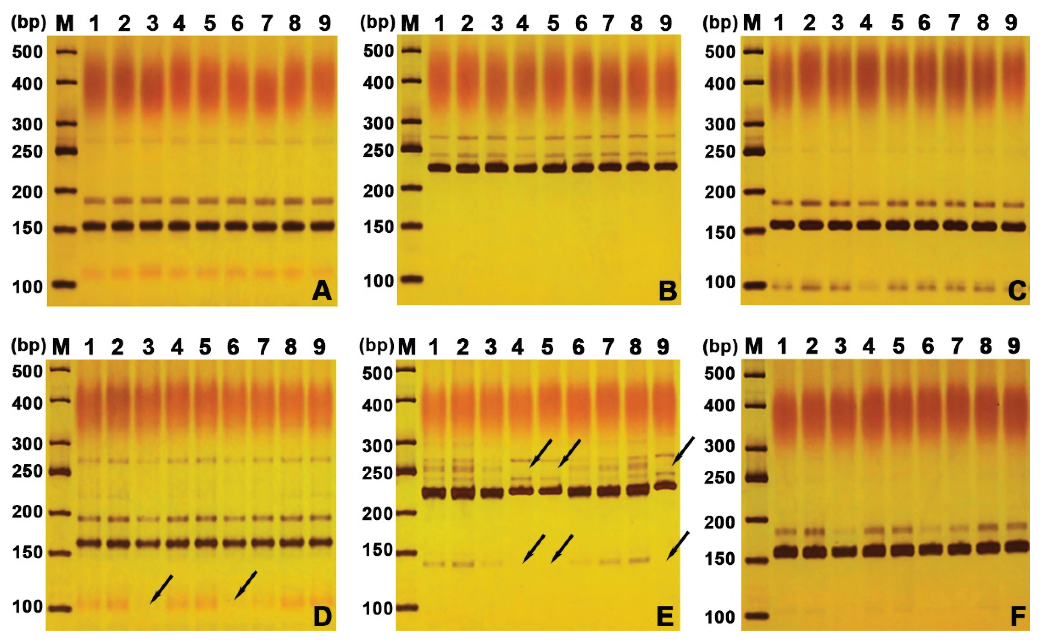

Table 1.
Band statistics of SSR primers used for assessing genetic fidelity in P. edulis.
| Primer ID | Sequences (5′–3′) | Tm1(°C) | TB2– DOR |
PB3– DOR |
TB2– IOR |
PB3– IOR |
|---|---|---|---|---|---|---|
| 1–F | TTGCACAATGACCAATGTTGT | 56.9 | 4 | 0 | 4 | 1 |
| 1–R | CTGAGCACCTTGTCAAAATACA | 55.7 | ||||
| 2–F | CCTGTGGTGAAAATGGAACC | 57.4 | 3 | 0 | 5 | 2 |
| 2–R | GAGCCCTGGACTGACACATT | 56.9 | ||||
| 3–F | CATGCATTCATTTGTTTTTCTTG | 58.1 | 3 | 0 | 2 | 0 |
| 3–R | GATGCTGGGAAAAAGAGTGC | 57.3 | ||||
| Total | 10 | 0 | 11 | 3 |
1 Tm (°C) annealing temperature, 2 TB total bands, 3 PB polymorphic bands. Band sizes ranged from 100 to 500 bp per primer.
Disclaimer/Publisher’s Note: The statements, opinions and data contained in all publications are solely those of the individual author(s) and contributor(s) and not of MDPI and/or the editor(s). MDPI and/or the editor(s) disclaim responsibility for any injury to people or property resulting from any ideas, methods, instructions or products referred to in the content. |
© 2026 by the authors. Licensee MDPI, Basel, Switzerland. This article is an open access article distributed under the terms and conditions of the Creative Commons Attribution (CC BY) license (http://creativecommons.org/licenses/by/4.0/).
Copyright: This open access article is published under a Creative Commons CC BY 4.0 license, which permit the free download, distribution, and reuse, provided that the author and preprint are cited in any reuse.