

Submitted:
06 August 2026
Posted:
07 August 2026
You are already at the latest version
Abstract
Valproic acid (VPA) is a widely used anticonvulsant and mood stabilizer and a known teratogen associated with neural tube defects and neurodevelopmental disorders, including autism spectrum disorder (ASD). Prenatal VPA exposure is widely used to model ASD-like behaviors in rodents; however, most studies employ a single dose, and direct comparisons between commonly used regimens are lacking. We therefore compared two widely used VPA doses (300 and 600 mg/kg) administered to pregnant mice on embryonic day 12.5 (E12.5). Male and female offspring were evaluated longitudinally for early neurological development, ultrasonic vocalizations, locomotor activity, anxiety-like behavior, sociability, and working memory. Prenatal VPA exposure produced non-linear, dose- and sex-dependent effects. Early neurodevelopment was largely preserved, with only mild and transient delays that were most evident after 300 mg/kg. In contrast, later behavioral alterations differed according to dose, sex, age, and behavioral measure. Sociability deficits were stable across doses in males but became more pronounced with age in females, whereas working memory impairment predominated after 600 mg/kg during adolescence and after 300 mg/kg in adulthood. These findings demonstrate that increasing prenatal VPA dose does not simply increase behavioral severity but produces distinct neurodevelopmental trajectories that depend on dose, sex, and developmental stage.
Keywords:
prenatal
; valproate
; ASD model
; dose-effect
; sex differences
; neurodevelopment
1. Introduction
VPA, an anticonvulsant and mood stabilizer, is one of the more potent teratogens associated with the etiology of Neural Tube Defects (NTDs in humans. When taken during early pregnancy it increases the risk of NTDs by 10-20 folds, to 1-2% and the general risk of congenital malformations to 8-10% [1]. VPA use during pregnancy is also associated with a high rate of spontaneous abortions, reduced neonatal weight, and various brain malformations that can lead to severe neurodevelopmental problems [2,3]. Although VPA is a causative factor for only a limited number of NTDs in humans, it serves as a useful model for studying both NTD and Autism Spectrum Disorder (ASD) etiology, pathogenesis and prevention [2,4]. Being an histone deacetylases (HDAC) inhibitor, VPA modulates neurotransmission and regulates gene expression by epigenetic chromatin remodeling [5,6,7]. Prenatal exposure to VPA causes transient hyperacetylation of H3 and H4 histones in the embryonic mouse brain [8] and the changes can be epigenetically transmitted, at least in rodents, to the second and even third generation [9].
Prenatal VPA exposure in mice show that the neurobehavioral consequences are sensitive to sex and developmental timing. The critical windows of VPA administration in pregnancy to produce NTD (exencephaly) in mice are 8 and 9 gestational days and to produce offspring with ASD-like phenotypes is embryonic day 12.5 (E 12.5), after neural tube closure [10,11,12]. Even a single dose of VPA resulted in most ASD like symptoms [10,13,14] and anxiety, a most common comorbid sign in autistic patients [15]. Kataoka et al., demonstrated that prenatal 500 mg/kg VPA on E12.5, but not at E9 or E14.5, induces autism-like behaviors in mice [8]. VPA exposure at E9 resulted in a high rate of malformed pups (30–40%), but the surviving normal pups did not exhibit behavioral abnormalities or autism-like traits. Moreover, VPA exposure at E14.5 did not result in behavioral abnormalities, histological changes, or neuronal loss, indicating that this stage is less sensitive to VPA-induced effects compared to E12.5 [8].
Most reproducible behavioral alterations across mouse strains appear to occur within a dosing range of approximately 300–600 mg/kg. A single prenatal VPA dose of 600 mg/kg administered around E12.5 is one of the most widely used protocols in rodent ASD models. This regimen consistently produces ASD-like behavioral phenotypes, including reduced sociability in three-chamber tests, increased repetitive grooming, anxiety-like behavior in the open field or elevated plus maze, and impairments in hippocampal-dependent memory tasks [8,16,17,18,19,20].
Intermediate doses of VPA (500 mg/kg) also produce ASD-like phenotypes with prominent reduction in dendritic spine density in the prefrontal cortex and hippocampus [21,22]. Lower doses of VPA (300–400 mg/kg) administered at GD12.5 have been less frequently studied but appear to produce social interaction behavior deficit and social memory impairment [23]. Studies using 400 mg/kg in Swiss or ICR strains demonstrated alterations in repetitive behavior and anxiety-like responses, although the magnitude of these effects varied among strains [24].
Higher doses have also been examined under different experimental conditions. For example, 800 mg/kg VPA mixed with peanut butter to facilitate oral consumption, administered on E11 resulted in early developmental delays, impaired olfactory discrimination, altered social behavior, accompanied by molecular changes in BDNF and NMDA receptor subunits [25].
The available reviews of rodent VPA models suggest that different prenatal doses produce different behavioral and molecular patterns, not necessarily graded by severity [10,13,14,26]. Higher doses (600–800 mg/kg) generally produce more consistent phenotypes, whereas lower and intermediate doses (300–500 mg/kg) often result in selective or sex-dependent alterations. Despite both 300 and 600 mg/kg VPA are widely used in mouse models, we were unable to identify studies directly comparing these doses within the same strain and experimental framework.
In addition to dose, sex also appears to influence VPA-induced neurobehavioral outcomes. ASD is diagnosed approximately three to four times more frequently in males than females [27], but sex-specific effects in rodent VPA models remain inconsistent. Some studies report greater vulnerability in males, whereas others describe comparable or even stronger effects in females. [17,28,29]. Behavioral abnormalities occur in offspring of both sexes, including reduced sociability, anxiety-like behavior, and repetitive movements. However, social deficits are typically more pronounced in males, whereas anxiety and repetitive phenotypes occur in both sexes [8,29]. These inconsistencies may partly reflect differences in VPA dose, mouse strain, behavioral paradigms, and the developmental stage at assessment.
Mouse strains used to induce ASD like behavior by VPA also varied among reported studies. In outbred ICR mice, ASD-like behavioral alterations are observed in both sexes following prenatal VPA exposure, including after 300 mg/kg at E12.5 [17,30]. In contrast, many studies using inbred C57BL/6 mice employ doses around 600 mg/kg to obtain robust behavioral phenotypes, often accompanied by greater embryonic loss [19,20,31].
Thus, it remains unclear whether increasing VPA dose simply increases behavioral severity or instead produces distinct age- and sex-dependent neurodevelopmental trajectories. To address this question, we directly compared two commonly used prenatal VPA regimens (300 and 600 mg/kg) administered at E12.5 in ICR mice. We aimed to determine whether increasing VPA dose results in a simple increase in behavioral severity or produces distinct neurodevelopmental and behavioral trajectories across developmental stages and between sexes. Male and female offspring were evaluated longitudinally from early postnatal development through adulthood using measures of neurological maturation, ultrasonic vocalizations, locomotor and anxiety-like behavior, sociability, and working memory.
2. Materials and Methods
Animals: All animal experiments were approved by the Institutional Animal Care and Use Committee of Ariel University, Israel (approval # AU-IL-2309-112 and AU-IL2511-115), and were conducted in accordance with institutional guidelines and the NIH Guide for the Care and Use of Laboratory Animals for the humane use of animals in research. The animals were housed on a 12 h light/dark cycle, at 23 ± 2 °C ambient humidity and had free access to standard laboratory food and water throughout testing.
Breeding: Adult male and female ICR mice were obtained at 7–8 weeks of age from Harlan (Israel) and randomly assigned to breeding pairs and all male mice used for breeding were approximately the same age as the females. The breeding pairs were monitored at early morning for the presence of vaginal plugs, which is considered as gestational day (GD) 0.5. Dams with noticed plugs were weighed starting on GD7. Progressive increase in body weight was taken as confirmation of pregnancy.
Embryonic/fetal mortality rate/resorptions. Fetuses from 6 dams exposed to saline, 6 dams exposed to VPA (300 mg/kg) and 5 dams exposed to VPA (600 mg/kg) were evaluated at E16.0 by counting the number of viable and non-viable fetuses per litter.
Experimental design:
Experimental chart flow
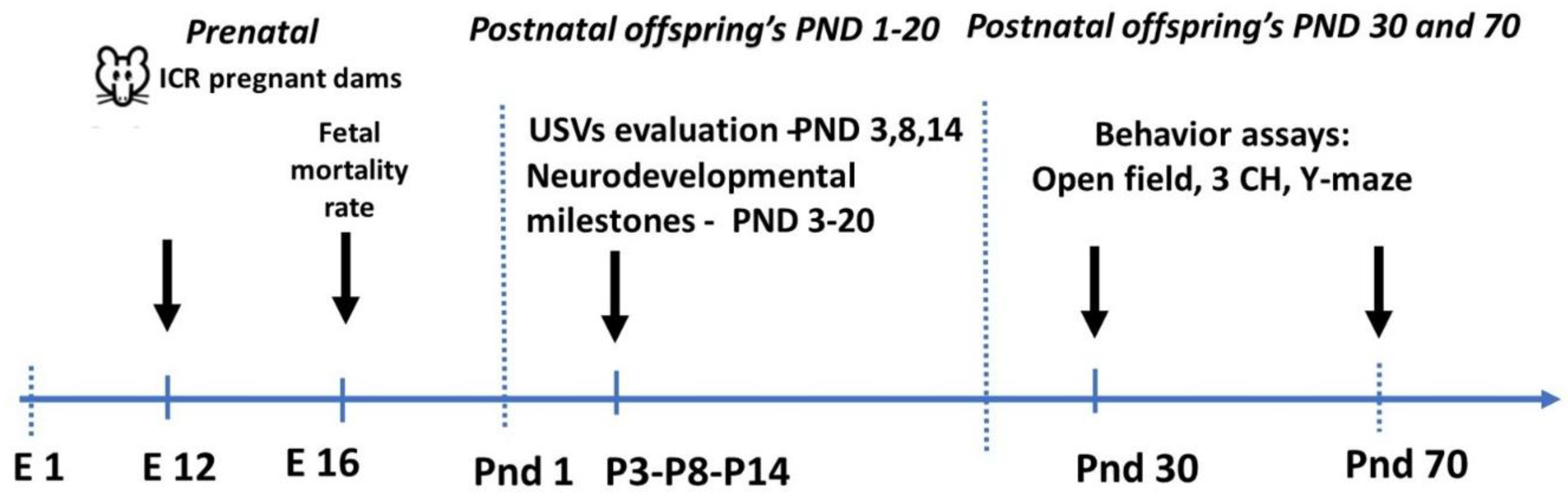
On E12.5, pregnant females (n=24) were randomly divided to three experimental groups (each comprise at least 6 Dams) and received a single subcutaneous injection of 300 mg/kg or 600 mg/kg VPA (Cat# P4543, Merk, Israel) dissolved in saline. Control females received an equal volume of Saline only. The day of birth was recorded as postnatal day (PND) 0. Each Dam was kept in a single cage with their offspring until weaning. Offspring were marked with a non-toxic identification foot tattoo (Ketchum MFG, NY) and returned to the litter. Dams and their litters were housed together until weaning on PND 21, when juvenile mice were weighed, ear-notched for permanent identification, and housed in same-sex groups.
Neurological and behavioral development: Mouse neurodevelopmental and behavioral patterns, particularly those related to autistic core symptoms, were evaluated from postnatal day (PND) 3 to PND70 using a battery of neurobehavioral tests. All offspring used for neurodevelopmental and behavioral assessments originated from at least four dams in each experimental group. The number of offspring included in each behavioral test varied and is reported for each analysis. The tests were conducted in the following order: neonatal ultrasonic vocalizations (PND3–14), neurodevelopmental milestones (PND3–20), open-field testing (locomotor activity and anxiety-like behavior), Y-maze testing (working memory; PND30 and PND70), and the three-chamber social interaction test (PND31 and PND71). All behavioral tests were recorded and analyzed using the Noldus XT computerized video-tracking system (Noldus Information Technology, The Netherlands). In addition to the neurodevelopmental and behavioral assessments, body weight was measured every two days from PND3 to PND20 to monitor postnatal growth.
Ultrasinic vocalizations (USVs) recording and analysis: USV recording and analysis were performed according to the method published by us previously [28]. The emitted USVs were measured at postnatal days 3, 8 and 14. The pups were separated from their mothers for 30 minutes in individual chambers and after 30 minutes of isolation were subjected to USV recordings for 3 minutes. Each pup was placed in a plastic rectangle chamber (11x6 cm, height of the walls 5 cm) located in a sound-insulating Styrofoam box for 3 minutes of the recording [28]. USVs recordings were performed by using the Ultravox 2.0 microphone (Noldus Information Technology, the Netherlands) placed 10 cm above the pup in the Styrofoam box, at the sample rate of 250 kHz. After the recording, pups returned to the home cage with the mother. The acquired data was filtered using lower cut-off 20 kHz filter to diminish the effects of the background noise. After the data was collected, a filtered file was exported in WAV format from the software. Then, registered sonograms from pups within experimental groups were evaluated using a machine-learning algorithm [32] and the total number of calls, call’s types and their acoustic parameters were calculate .
We adopted the classification described by Grimsley et al., [33], modified by Fonseca et al. [32] as described in our previous publication [28], including standard categories such as short, flat, upward, downward, complex, one frequency step up and step down, chevron syllables, reverse chevron, multiple frequency steps, two frequency steps frequency-modulated calls. Spectrogram plotting was performed using platform RStudio 4.2.1 (Posit, USA, RStudio Team http://www.rstudio.com/.) utilizing the following packages: Seewave [34], tuneR [35], ggplot2 [36], viridis [37], grid [38] and gridExtra [39]. The main part of code of spectrogram plotting was taken from the study of the winner of the spectro2018 R competition, Joshua Flickinger, which was published on the open source (https://rug.mnhn.fr/seewave/spec.html) and modified by us. Proceeded files were subjected to custom-written macros for R to extract the numerical values of the characteristics of interest.
Neurodevelopmentalmilestones: The following developmental milestones were evaluated in the offspring from PND3 to PND20 [40,41].
1. Surface righting reflex was measured as time in seconds until the mouse pup turns back onto all four paws when placed on the back. The max latency to turn back was defined to 30 sec and scored as 0, when pups time to turn back was more or equal to 30 sec, and scored 1, when pups time to turn back was less than 30 sec.
2. Cliff aversion was measured as time in seconds for pups positioned with forepaws and snout over the edge of a shelf to turn and begin to crawl away from the edge. Like in the surface righting test, a pup which took over 30 seconds to complete the task was considered to have failed
3. Negative geotaxis was measured as time in seconds for pup placed on an inclined plane (45 degrees) to turn 180 degrees and face upwards is recorded. This tests proprioception of the pups. Like in the surface righting test, a pup which took over 30 seconds to complete the task was considered to have failed
5. Eye opening - first day that both eyes are opened.
Behavioral tests:
Open field (OF) test.The OF test is used to estimate locomotor, exploration activity and repetitive behavior [42]. Each mouse is placed individually in the center of the apparatus and evaluated for 10 min. Distance moved, number of entries and time spent in the arena center and outer zone are analyzed to estimate curiosity to novelty exploration. The locomotor activity is determined by distance moved and the anxiety is determined by time and frequency spent in the periphery and central zones.
Y-Maze: The Y-maze spontaneous alternation test has been used to assess both repetitive behavior and spatial working memory [16]. The Y-maze was constructed of Plexiglas with three identical arms (42-cm long, 12-cm tall walls, illumination 30 lx). Each mouse was placed at the end of one arm and allowed to freely explore the maze for 6 min. The sequence of arm entries was recorded using the Noldus XT video-tracking system. An arm entry was scored when all four paws entered an arm. Spontaneous alternation was defined as consecutive entries into all three arms without repetition and was calculated as the percentage of alternations using the following formula:
Spontaneous alternation (%) = (Number of alternations / (Total arm entries − 2)) × 100.
Reduced spontaneous alternation indicates impaired spatial working memory.
Three chamber social interaction tests: Standard Crawley’s social interaction tests are used to evaluate social interaction [11]. Testing occurs in two sessions within a three-chambered box, with openings between the chambers. After habituation, the subject encounters a never-before-met stranger under one metal mesh buckets and an empty one in the “sociability” session. The time spent in each chamber, the time of sniffing each bucket was measured. Sociability index is reflected in the preference for social interaction for an empty chamber. It was calculated by dividing the time spent in the social (stranger) vs. non-social (empty) zone by the total time spent in both, expressed as tStranger/(tStranger + tEmpty) x100. Increased time spent exploring never-before-met stranger indicates a high degree of sociability in the test animal.
Statistical analysis: Statistical analysis of the experimental data was performed using Prism 11 (GraphPad Software, USA). For behavioral evaluations, statistical significance was analyzed between prenatally treated offspring groups and compared to saline treated one. Males and females were analyzed separately in all experiments. Comparisons involving treatment, sex, and age, two-way repeated-measures ANOVA was applied, as appropriate. When repeated-measures data were incomplete, a mixed-effects model fitted by restricted maximum likelihood (REML) was used, as implemented in GraphPad Prism. Significant main effects or interactions were followed by post-hoc multiple comparisons using Bonferroni’s correction. For non-normally distributed or proportional outcomes, non-parametric analyses were performed using the Kruskal–Wallis test, followed by Dunn’s post hoc multiple comparisons test. In addition, for milestone’s acquisition or maximal response of pups performing the task was defined as the first postnatal day on which each pup performed the tested task. Acquisition curves were analyzed separately in male and female offspring, and day-specific analyses were used when marked divergence between groups was confirmed, to capture both global and transient effects using the Fisher test. Statistical significance was set at P < 0.05.
3. Results
3.1. Effect of Prenatal VPA 300 mg/kg and 600 mg/kg Treatment on Embryonic Mortality Rate
Fetuses from 6 dams exposed to saline, VPA (300 mg/kg) and 5 dams exposed to VPA (600 mg/kg) were evaluated at E16.0 by counting the number of viable and non-viable fetuses per litter. Prenatal exposure resulted in a dose-dependent increase in embryonic mortality, most pronounced at the higher dose. Mortality was minimal in controls (0.95%), increased to 8.5% at 300 mg/kg, and reached 33.7% at 600 mg/kg. Kruskal–Wallis analysis, followed by post-hoc Dunn’s multiple comparisons, showed a significant group effect (p = 0.006), emerged by the difference between controls and the higher dose (p = 0.0091), while the intermediate dose did not differ from either group. Fisher’s test direct comparison between doses confirmed a marked increase in lethality at 600 mg/kg (p = 0.0026). Litter size and number of viable embryos were comparable, indicating that the effect reflects increased embryonic death rather than altered fertility.

Table 1.
Effect of Prenatal VPA treatment on embryonic mortality.
| Treatment | Saline | VPA 300 mg/kg | VPA 600 mg/kg |
| Number of fetuses | 73 | 75 | 61 |
| Alive | 72 | 69 | 44 |
| Dead | 1 | 6 | 17 |
| Mean Litter per Dam | 12.2 | 12.5 | 12.2 |
| Mortality rate (%) | 0.95 | 8.5 | 33.7 |
| Kruskal-Wallis test | p=0.006 | ||
| Dunn’s multiple comparisons test | VPA300 vs Sal (p=0.8) | ||
| VPA600 vs Sal (p=0.009) | |||
| VPA300 vs VPA600 (p=0.1) | |||
3.2. Effect of Prenatal VPA 300 mg/kg and 600 mg/kg Treatment on Postnatal Weight Gain and Eyes Opening in Male and Female Offspring in Comparison to Controls Treated with Saline
We assessed the weight gain in both male and female offspring from PND 3 to 20. Across all groups, a progressive and robust increase in body weight was observed, consistent with normal developmental growth trajectories (Figure 1). These findings indicate that prenatal VPA exposure at both doses, does not impair early postnatal growth in either sex under the tested conditions. The mean day of eye opening was similar in all experimental groups, occurring at approximately PND15 under all conditions. These findings indicate preserved somatic maturation with both doses of VPA.
3.3. Sex-Specific Effects of Prenatal VPA 300 mg/kg and 600 mg/kg Treatment on Ultrasonic Vocalization Quantity and Call-type Composition
In male offspring, the number of vocalizations in 3 min recorded sonogram followed the expected developmental trajectory, with a peak at PND8 and decline by PND14. Mixed-effects model demonstrated a significant postnatal age effect (F (1.448, 47.78) = 29.89; p < 0.0001), whereas no treatment effect or interaction was detected, and groups did not differ at any time point (Figure 2A). Bonferroni-corrected post-hoc comparisons confirmed that call numbers did not differ significantly between saline (n=20), VPA 300 (n=34), and VPA 600 (n=23) groups at any postnatal day (PND3, PND8, or PND14). Call-type distribution was similarly preserved (Figure 3A).
In female offspring, mixed-effects analysis revealed an age-dependent effect on call number (F (1.660, 76.07) =22.78, p<0.0001), reflecting the expected developmental peak at PND8 followed by a marked decline at PND14 (Figure 2B). A significant main effect of treatment was also detected (F (1.903, 87.55) =5.313, p=0.0075), whereas the treatment × age interaction was not significant (F (3.128, 28.15) = 1.427p = 0.26). Post-hoc Bonferroni comparisons showed significant reductions in call number only at PND8 where both doses showed reduced call numbers relative to saline controls (Saline (n=25) vs. VPA300 (n=37): p=0.048; Saline vs. VPA 600 (n=30): p=0.016), with no difference between moderate and high doses. Qualitative analysis of USV call-type composition confirmed a broad attenuation across categories at PND8, rather than selective loss of specific calls. (Figure 3B).
These results indicate that vocalization deficits were transient, female-specific, and dose-independent, indicating selective vulnerability of early communication.
3.4. Sex- and Dose-Dependent Effects of Prenatal VPA 300 mg/kg and 600 mg/kg Exposure on Early Neurodevelopmental (neurological) Milestones
To evaluate the impact of prenatal VPA exposure on early sensorimotor maturation, we assessed the acquisition of three core neurodevelopmental milestones: surface righting (SR), negative geotaxis (NG) and cliff aversion (CA), in male and female offspring from postnatal day 4 to 15 (Figure 4).
3.4.1. a.1 Surface Righting Acquisition
Surface righting acquisition was largely preserved in both sexes, with all pups reaching maximal performance within a narrow postnatal window. However, day-specific Fisher’s exact analysis revealed a transient early delay in both sexes, with some sex-dependent differences in duration. At PND4, the proportion of responders was significantly reduced in both VPA300 and VPA600 groups compared to saline in males (p=0.006 and p=0.0008, respectively) and females (p=0.043 and p=0.035, respectively) (Figure 4 and Figure 5). By PND5, males in both dose groups and females in the high-dose group no longer differed from controls, whereas the delay persisted for another day only in VPA300 females (p=0.041) (Table 2). These results indicate a brief shift in the onset of reflex acquisition with rapid recovery and convergence across groups, slightly more persistent in females at the moderate dose.
Table 2.
Surface righting acquisition following prenatal VPA exposure.
| Male | |||||||
| Treatment | PND | Day 4 | Day 5 | Day 6 | Day 7 | Day 8 | Day 9 |
| Saline n=28 | N of responders | 26 of 28 | 26 of 28 | 28 of 28 | 28 of 28 | 28 of 28 | 28 of 28 |
| % of response | 92.9 | 92.9 | 100 | 100 | 100 | 100 | |
| VPA 300 mg/kg n=34 | N of responders | 21 of 34 | 26 of 34 | 31 of 34 | 33 of 34 | 33 of 34 | 34 of 34 |
| % of response | 61.8 | 76.5 | 91.2 | 97.1 | 97.1 | 100 | |
| Fisher’s exact test | p=0.0064 | p>0.1 | p>0.1 | p>0.1 | |||
| VPA 600 mg/kg n=27 | N of responders | 14 of 27 | 22 of 27 | 24 of 27 | 26 of 27 | 27 of 27 | 27 of 27 |
| % of response | 51.9 | 81.5 | 88.9 | 96.3 | 100 | 100 | |
| Fisher’s exact test | p=0.0008 | p>0.1 | p>0.1 | p>0.1 | |||
| RM-ANOVA | effect of treatment | F(1, 9) = 3.1 | p>0.1 | ||||
| effect of time | F(8,16) = 6.6 | p = 0.0007 | |||||
| Female | |||||||
| Treatment | PND | Day 4 | Day 5 | Day 6 | Day 7 | Day 8 | Day 9 |
| Saline n=34 | N of responders | 28 of 34 | 31 of 34 | 33 of 34 | 33 of 34 | 34 of 34 | 34 of 34 |
| % of response | 82.4 | 91.2 | 97.1 | 97.1 | 100 | 100 | |
| VPA 300 mg/kg n=41 | N of responders | 24 of 41 | 29 of 41 | 36 of 41 | 40 of 41 | 41 of 41 | 41 of 41 |
| % of response | 58.5 | 70.7 | 87.8 | 97.6 | 100 | 100 | |
| Fisher’s exact test | p=0.043 | p=0.041 | p>0.1 | p>0.1 | |||
| VPA 600 mg/kg n=53 | N of responders | 32 of 53 | 40 of 53 | 44 of 53 | 49 of 53 | 51 of 53 | 53 of 53 |
| % of response | 60.4 | 75.5 | 83 | 92.5 | 96.2 | 100 | |
| Fisher’s exact test | p=0.035 | p=0.03 | p=0.098 | p>0.1 | |||
| RM-ANOVA | effect of treatment | F(1, 11) = 3.9 | p>0.1 | ||||
| effect of time | F (8, 16) = 14.86 | p < 0.0001 | |||||
Table 2. Surface righting acquisition following prenatal VPA exposure. Upper panel: males; lower panel: females. For each postnatal day, the number and percentage of pups achieving the reflex are shown for saline, VPA 300 mg/kg, and VPA 600 mg/kg groups. RM-ANOVA indicated a strong effect of time in both males and females, consistent with normal developmental progression, with no significant overall treatment effect. Day-specific differences in responder proportions between treatment groups and saline controls were assessed using Fisher’s exact test, with p values indicated for each comparison. These data illustrate the temporal progression of reflex acquisition and highlight early, transient delays in VPA-exposed groups relative to controls.
3.4.2. a.2 Negative Geotaxis Acquisition
In contrast to surface righting, negative geotaxis showed subtle sex-dependent effects with largely preserved acquisition in both sexes. However, day-specific Fisher’s exact analysis revealed a mild, transient early delay, restricted to females at PND4 in the VPA300 group (p=0.015), with no differences at later time points (Table 3, Figure 4C–D). Consistent with these findings, analysis of the mean day of acquisition (Figure 6) showed minimal variation across groups in both sexes, supporting the absence of sustained impairment. Therefore, prenatal VPA exposure induces a subtle and transient delay in negative geotaxis onset, detectable only at early postnatal stages and primarily in females at the moderate dose, with rapid normalization thereafter.
Table 3.
Negative Geotaxis acquisition following prenatal VPA exposure.
| Male | |||||||
| Treatment | PND | Day 4 | Day 5 | Day 6 | Day 7 | Day 8 | Day 9 |
| Saline n=28 | N of responders | 23 of 28 | 23 of 28 | 28 of 28 | 28 of 28 | 28 of 28 | 28 of 28 |
| % of response | 82.1 | 82.1 | 100 | 100 | 100 | 100 | |
| VPA 300 mg/kg n=34 | N of responders | 27of 34 | 29 of 34 | 32 of 34 | 32 of 34 | 33 of 34 | 34 of 34 |
| % of response | 79.4 | 85.3 | 94.1 | 94.1 | 100 | 100 | |
| Fisher’s exact test | p>0.1 | p>0.1 | p>0.1 | p>0.1 | |||
| VPA 600 mg/kg n=27 | N of responders | 25 of 27 | 25of 27 | 25 of 27 | 26 of 27 | 27 of 27 | 27 of 27 |
| % of response | 92.6 | 92.6 | 92.6 | 96.3 | 100 | 100 | |
| Fisher’s exact test | p>0.1 | p>0.1 | p=0.098 | p>0.1 | |||
| RM-ANOVA | effect of treatment | F (1, 11) = 1 | p>0.1 | ||||
| effect of time | F(8,16) = 9.8 | p < 0.0001 | |||||
| Female | |||||||
| Treatment | PND | Day 4 | Day 5 | Day 6 | Day 7 | Day 8 | Day 9 |
| Saline n=34 | N of responders | 32 of 34 | 34 of 34 | 34 of 34 | 34 of 34 | 34 of 34 | 34 of 34 |
| % of response | 94.1 | 100 | 100 | 100 | 100 | 100 | |
| VPA 300 mg/kg n=39 | N of responders | 28 of 39 | 35 of 39 | 38 of 39 | 39 of 39 | 39 of 39 | 39 of 39 |
| % of response | 71.8 | 89.7 | 97.4 | 100 | 100 | 100 | |
| Fisher’s exact test | p=0.015 | p>0.1 | p>0.1 | p>0.1 | |||
| VPA 600 mg/kg n=35 | N of responders | 32 of 35 | 34 of 35 | 34 of 35 | 35 of 35 | 35 of 35 | 35 of 35 |
| % of response | 91.4 | 97.1 | 97.1 | 100 | 100 | 100 | |
| Fisher’s exact test | p>0.1 | p>0.1 | p=0.098 | p>0.1 | |||
| RM-ANOVA | effect of treatment | F (1, 8) = 2 | p>0.1 | ||||
| effect of time | F(8,16) = 4 | p = 0.009 | |||||
Table 3 Negative Geotaxis acquisition following prenatal VPA exposure. Upper panel: males; lower panel: females. For each postnatal day, the number and percentage of pups achieving the reflex are shown for saline, VPA 300 mg/kg, and VPA 600 mg/kg groups. RM-ANOVA demonstrated a significant effect of time in both males and females with no significant main effect of treatment indicating overall normal developmental progression. Day-specific differences in responder proportions between treatment groups and saline controls were assessed using Fisher’s exact test, with p values indicated for each comparison. These data show the mild and transient delay in the onset of negative geotaxis in female exposed to the moderate dose in early postnatal stages (PND4).
3.4.3. a.3 Cliff Aversion Acquisition
Cliff aversion showed the most robust and consistent effects of prenatal VPA exposure, although acquisition was ultimately preserved in all groups, with pups reaching maximal performance within the expected developmental window. In males, a modest delay was detected in VPA300 group compared to saline (p=0.015), while VPA600 did not differ from controls (Figure 4E). In females, the effect was more pronounced, with a significant reduction in performance in the VPA300 group (p=0.0016), and no difference in VPA600 (Figure 4F).
Day-specific Fisher’s exact analysis confirmed these findings, showing a transient delay in males at PND5 (p=0.03), whereas in females the delay was more sustained, persisting across PND5–PND7 (p=0.03, 0.013, and 0.046, respectively). No significant effects were observed in the high-dose group at any time point. Thus, these findings indicate a dose- and sex-dependent delay in cliff aversion acquisition, most evident at 300 mg/kg and more pronounced and prolonged in females, followed by eventual convergence across groups.
Table 4.
Cliff aversion acquisition following prenatal VPA exposure.
| Male | |||||||
| Treatment | PND | Day 4 | Day 5 | Day 6 | Day 7 | Day 8 | Day 9 |
| Saline n=28 | N of responders | 27 of 28 | 28 of 28 | 28 of 28 | 28 of 28 | 28 of 28 | 28 of 28 |
| % of response | 96.4 | 100 | 100 | 100 | 100 | 100 | |
| VPA 300 mg/kg n=34 | N of responders | 29 of 34 | 30 of 34 | 31 of 34 | 31 of 34 | 33 of 34 | 34 of 34 |
| % of response | 85.3 | 88.2 | 91.2 | 91.2 | 97.1 | 100 | |
| Fisher’s exact test | p=0.01 | p=0.03 | p>0.1 | p>0.1 | |||
| VPA 600 mg/kg n=27 | N of responders | 27 of 27 | 27 of 27 | 27 of 27 | 27 of 27 | 27 of 27 | 27 of 27 |
| % of response | 100 | 100 | 100 | 100 | 100 | 100 | |
| Fisher’s exact test | p>0.1 | p>0.1 | p>0.1 | p>0.1 | |||
| RM ANOVA | effect of treatment | F (8, 16) =1 | p>0.1 | ||||
| effect of time | F (2, 16) =7 | p=0.005 | |||||
| Bonferroni’s multiple comparisons test | Saline vs. VPA 300 mg/kg | p=0.015 | |||||
| Saline vs. VPA 600 mg/kg | p>0.1 | ||||||
| VPA300 mg/kg vs. VPA 600 mg/kg | p=0.01 | ||||||
| Female | |||||||
| Treatment | PND | Day 4 | Day 5 | Day 6 | Day 7 | Day 8 | Day 9 |
| Saline n=34 | N of responders | 32 of 34 | 33 of 34 | 34 of 34 | 34 of 34 | 34 of 34 | 34 of 34 |
| % of response | 94.1 | 97.1 | 100 | 100 | 100 | 100 | |
| VPA 300 mg/kg n=40 | N of responders | 31 of 40 | 32 of 40 | 33 of 40 | 33 of 40 | 36 of 40 | 37 of 40 |
| % of response | 77.5 | 80 | 82.5 | 82.5 | 90 | 92.5 | |
| Fisher’s exact test | p=0.045 | p=0.0332 | p=0.013 | p=0.04 | p>0.1 | p>0.1 | |
| VPA 600 mg/kg n=53 | N of responders | 50 of 53 | 52 of 53 | 52 of 53 | 53 of 53 | 53 of 53 | 53 of 53 |
| % of response | 94.3 | 98.1 | 98.1 | 100 | 100 | 100 | |
| Fisher’s exact test | p>0.1 | p>0.1 | p>0.1 | p>0.1 | |||
| RM-ANOVA | effect of treatment | F (1, 8) = 3 | p=0.0007 | ||||
| effect of time | F (8, 16) = 3 | p=0.02 | |||||
| Bonferroni’s multiple comparisons test | Saline vs. VPA 300 mg/kg | p=0.002 | |||||
| Saline vs. VPA 600 mg/kg | p>0.1 | ||||||
| VPA 300 mg/kg vs. VPA 600 mg/kg | p=0.002 | ||||||
Table 4. Cliff aversion acquisition following prenatal VPA exposure. Upper panel: male; lower panel: females. For each postnatal day, the number and percentage of pups achieving the reflex are shown for saline, VPA 300 mg/kg, and VPA 600 mg/kg groups. RM-ANOVA revealed a significant effect of time in both sexes, indicating normal developmental progression, with stronger treatment-related effects in females. Post-hoc analyses demonstrated a dose- and sex-dependent delay predominantly in the VPA300 group. Day-specific differences in responder proportions between treatment groups and saline controls were assessed using Fisher’s exact test, with p values indicated for each comparison. These data show dose- and sex-dependent delay in cliff aversion with more robust and sustained effect in females.
All these results showed that prenatal VPA exposure resulted in transient, domain-specific delays in early neurodevelopment, with the most pronounced effects observed in cliff aversion, particularly at the moderate dose (300 mg/kg) and more evident in females. Surface righting and negative geotaxis were largely preserved, showing only brief or minimal significant delays at early days. Importantly, all groups reached full milestone acquisition, indicating delayed maturation rather than persistent neurological impairment.
3.5. Sex- and Dose-Dependent Effects of Prenatal VPA 300 mg/kg and 600 mg/kg Exposure on Behavioral Traits Measured at Postnatal days 30 (Adolescence) and 70 (Adulthood)
3.5.1. a.1 Locomotion and Anxiety-Like Behavior Assessed by Open Field
Locomotor activity showed sex- and dose-dependent effects of prenatal VPA exposure. In males, two-way ANOVA revealed an effect of age (F(1,50) = 5.72, p = 0.020), reflecting increased locomotion in saline controls, with no effect of treatment and no differences between groups (Figure 5A). In females, two-way ANOVA revealed a main effect of treatment (F(2,62) = 14.45, p < 0.0001) and a treatment × age interaction (F(2,62) = 19.9, p < 0.0001), with no effect of age. At PND30, both VPA300 (p = 0.0026, n = 17) and VPA600 (p < 0.0001, n = 32) groups showed reduced locomotion compared with saline (n = 31) (Figure 5B). At PND70, hypoactivity persisted only in the VPA300 group (p < 0.0001), while VPA600 no longer differed from controls, indicating divergent trajectories across doses.
Center exploration also showed sex-dependent effects. In males, two-way ANOVA revealed effects of treatment (F(2,58) = 3.927, p = 0.02) and age (F(1,58) = 7.6, p = 0.0075). At PND30, center time was reduced only in the VPA600 group (p = 0.03), with no differences at PND70 (Figure 5C). In females, two-way ANOVA revealed a strong effect of treatment (F(2,77) = 16.74, p < 0.0001), with no effect of age or interaction. At PND30, center time was reduced only in the VPA600 group compared with saline (p < 0.0001) and VPA300 (p = 0.001). At PND70, both VPA300 (p = 0.04) and VPA600 (p = 0.0001) groups showed reduced center exploration compared with saline, indicating an age-dependent onset of the effect (Figure 5D).
These results indicate a pronounced sex-dependent vulnerability to prenatal VPA exposure, characterized by relatively preserved behavioral outcomes in males and dynamic, dose- and age-dependent alterations in females, supporting a non-linear developmental effect that differentially impacts locomotor and anxiety-related domains.
Scheme 30. and PND70 (Figure 6A). Two-way ANOVA revealed a strong main effect of treatment (F(2,59)=32.19, p<0.0001), with no effect of age or interaction, indicating sociability deficits at both ages across doses. Bonferroni post-hoc comparisons confirmed significantly reduced social interaction in both VPA300 (n=16) and VPA600 (n=13) groups versus saline controls (n=42) at PND30 (p<0.0001 and p=0.0062, respectively) and PND70 (p<0.0001 and p=0.0004, respectively), with no difference between VPA doses.
In females two-way-ANOVA revealed significant main effects of treatment (F(2,72)=21.01, p<0.0001) and age (F (1,72) =7.96, p=0.0062), as well as a significant treatment × age interaction (p≈0.017), indicating divergent developmental trajectories across exposure groups (Figure 6B). Post-hoc comparisons revealed age-dependent changes in social interaction. At PND30, a reduction was observed only in the moderate dose group (vs saline p < 0.0001). By PND70, both the moderate dose (VPA300 mg/kg; p = 0.0006, n=17) and higher dose (VPA600 mg/kg; p = 0.0002, n=23) groups exhibited significant deficits relative to saline controls (n = 35). In addition, the higher dose group showed a significant decline in sociability across development (PND30 vs PND60, p = 0.0011).
Both sexes exhibited significant treatment-related impairments in sociability, with different developmental trajectory. Males showed stable deficits across adolescence and adulthood, whereas females demonstrated age-dependent changes, primarily reflecting worsening sociability in the higher dose group.
3.5.2. a.3 Spontaneous Alternation Assessed by Y-Maze Test
In males, two-way ANOVA revealed a significant main effect of treatment (F(2,67) = 6.69, p = 0.002) and a significant age × treatment interaction (F(2,67) = 4.54, p = 0.01), with no main effect of age (Figure 7A). Post-hoc Bonferroni comparisons showed that alternation performance was reduced only in the higher dose (VPA600 mg/kg; p = 0.046, n=19) compared with the moderate dose group (VPA300 mg/kg; p = 0.004; n=10;) and saline controls (n=44). While the moderate dose group did not differ from controls. By PND70, this pattern reversed. The significant impairment was observed in the moderate dose group (p = 0.0002), whereas higher dose did not differ from saline. Within-group analysis indicated a developmental decline in the VPA600 group between PND30 and PND70 (p = 0.0118). These findings indicate a dose-dependent, age-specific shift in spatial working memory deficits, with early impairment at the higher dose and later impairment at the intermediate dose.
In females, two-way ANOVA revealed a strong main effect of prenatal treatment (F(2,64) = 24.9, p < 0.0001), while neither the effect of age nor interaction was significant (Figure 7B). Bonferroni post-hoc comparisons showed reduced alternation in both VPA300 (p = 0.001, n = 11) and VPA600 (p = 0.04, n = 20) groups compared with Saline (n = 34) at PND30, with similar deficits persisting at PND70 (Saline vs VPA300 p = 0.0003; Saline vs VPA600 p = 0.02). These finding indicate an early-onset, stable impairment in spatial working memory in females that persists across development.
These results reveal a marked sex-dependent pattern, with dynamic, dose- and age-specific shifts in males contrasted by a stable, treatment-dependent deficit in females, supporting a non-linear effect of prenatal VPA exposure on cognitive function.
In summary, prenatal VPA exposure produced non-linear, dose-, sex-, and behavior-specific neurodevelopmental effects. Early postnatal development was largely preserved, with only mild and transient delays that were most evident after the 300 mg/kg dose. In contrast, later behavioral alterations varied according to dose, sex, age, and behavioral measure. The 600 mg/kg dose was associated with greater embryonic mortality and broader behavioral abnormalities, whereas the 300 mg/kg dose preferentially affected early neurodevelopment and produced persistent deficits in selected behavioral measures. These findings indicate that increasing VPA does not simply increase behavioral severity but leads to distinct developmental trajectories. A comparison of the effects of 300 and 600 mg/kg VPA is summarized in Table 5.
4. Discussion
The present study provides a longitudinal characterization of neurological development and behavioral outcomes following prenatal exposure to intermediate and high dose of valproate administered on day 12.5 of gestation. The higher dose was associated with substantially greater intrauterine mortality, indicating a markedly increased embryonic toxicity. Among surviving offspring, we assessed early communication (ultrasonic vocalizations), neurodevelopmental milestones, and later behavioral functions, including locomotion, anxiety-like behavior, sociability, and working memory. We found that prenatal VPA exposure did not produce a uniform increase in severity with higher dose. Instead, the effects differed between behavioral outcomes, sex, and changed with age. Early developmental processes, particularly sensorimotor milestones, were more sensitive to the intermediate dose (300 mg/kg). Whereas later behavioral changes involved both doses and varied between sexes. This pattern indicates that increasing dose does not simply intensify the same effect but alters the type, severity and timing of the outcome (Table 4).
In contrast, later behavioral functions changed over time and differed between behavioral outcomes and between sexes. Thus, increasing VPA dose did not simply increase behavioral severity but produced distinct behavioral profiles across development.
Table 6.
Dose comparison of prenatal VPA exposure effects across domains.
| Domain |
VPA300 mg/kg Effect |
VPA600 mg/kg Effect |
Dose Relationship |
| Embryonic mortality | ↓ Mild | ↑ Strong | 600 > 300 |
|
USV (Early communication) |
↓ calls at PND8 transient (♀ only) |
↓ calls at PND8 transient (♀ only) |
300 ≈ 600 |
| Surface righting | Minimal / absent | Minimal / absent | 300 ≈ 600 |
| Negative geotaxis | Mild transient delay (♀ only) |
Absent | 300 > 600 |
| Cliff aversion | Clear delay (both ♂ & ♀) |
Absent | 300 > 600 |
|
Open field: Locomotion |
Persistent effects (♀ only) |
Strong at PND30 (♀ only), |
600 > 300 (PND30) 300 > 600 (PND70) |
|
Open field: Anxiety-like behavior |
No effect at PND30 Mild at PND70 (♀ only) |
Persistent (both ♂ & ♀) |
600 > 300 |
| 3ChT: Sociability | Strong ((both ♂ & ♀) | Strong (both ♂ & ♀) | 300 ≈ 600 |
|
Y-maze: Working memory |
PND70 impairment (♂) Stable (♀) |
PND30 impairment (♂) Stable (♀) |
Male: Temporal shift (600 early, 300 late.) Female 300 ≈ 600 |
Early 8. that normalized by PND 14. The effect was dose-independent, indicating that the two VPA doses exceeded a threshold sufficiently to perturb early communication-related neural networks without further escalation at higher exposure levels. These findings extend previous reports by demonstrating that VPA-induced alterations in early communication may be temporally restricted and sex-specific [20,28,43,44,45,46,47,48,49]. Similarly, early neurodevelopmental milestones were largely preserved, with only selective and transient delays, most consistently at the intermediate dose, particularly in cliff aversion. These findings suggest that prenatal VPA exposure delayed early neurological maturation. However, all groups eventually achieved comparable neurodevelopmental milestones, consistent with previous mouse studies [50,51,52].
Previous studies in mice using single-dose, single-time-point designs report robust ASD-like phenotypes following prenatal VPA exposure [8,20,24,53,54,55,56,57]. Whereas the present findings demonstrate that the behavioral alterations follow behavior-specific, sex-dependent, and developmentally dynamic trajectories, revealing dose sensitivities that are not captured in conventional experimental paradigms. However, it should be noted that the present study does not assess all behavioral domains relevant to ASD, such as repetitive behaviors [58]. Therefore, it was not intended to define a complete ASD-like phenotype but to characterize specific neurobehavioral alterations induced by prenatal VPA exposure.
The present study also demonstrated that the neurobehavioral consequences of prenatal VPA exposure were influenced by sex, with females exhibiting stronger or more persistent alterations than males in several behavioral tests. Sex hormones play a central role in brain sexual differentiation by regulating neuronal proliferation, migration, synaptogenesis, and circuit maturation during prenatal and early postnatal development [59,60]. Consequently, male and female brains may differ in their susceptibility to prenatal environmental insults, including VPA exposure [61,62]. Although these mechanisms were not investigated in the present study, they provide a biological basis for the sex-dependent behavioral outcomes observed in our offspring.
An important finding of the present study was that prenatal VPA affected early neurological maturation and later behavioral functions differently. Early developmental processes, particularly sensorimotor milestones, were more sensitive to the intermediate VPA dose (300 mg/kg), whereas later behavioral functions were altered by both doses and followed distinct age- and sex-dependent trajectories.
Early neurodevelopmental milestones, including surface righting, negative geotaxis, and cliff aversion, depend on the rapid maturation of brainstem and cerebellar circuits integrating vestibular, proprioceptive, tactile, and spinal motor pathways [60,61,62,63]. In rodents, this early postnatal period corresponds to late gestation in humans and is characterized by intense circuit formation and refinement [63,64]. Because these neural systems mature within a relatively narrow developmental window, even subtle prenatal disturbances may transiently delay sensorimotor development [64]. This concept is consistent with reports describing cerebellar abnormalities [54,65,66,67,68,69,70,71] and altered inhibitory signaling following prenatal VPA exposure [51,65,66].
In contrast, later behaviors, including anxiety-like behavior, sociability, and working memory depend on higher-order cortical and cortico-subcortical networks that continue to mature throughout postnatal development. Disruption of these circuits during prenatal life may therefore remain clinically silent until they reach functional maturity. This developmental pattern is also recognized in human neurodevelopmental disorders, in which abnormalities in early brain development are often observed clinically only during childhood or adolescence [67,68].
Experimental studies have demonstrated that prenatal VPA exposure alters cortical development, synaptic organization, excitation-inhibition balance, and microglial function [29,46,69,70,71,72]. Although these mechanisms were not examined in the present study, they provide a possible explanation for why several behavioral abnormalities emerged only during adolescence and adulthood. Interestingly, several behavioral alterations observed at the intermediate dose (300 mg/kg) were no longer evident in adulthood. This suggests that some early behavioral changes may diminish as development progresses. Whether this reflects delayed maturation, developmental compensation, or other adaptive mechanisms remains unknown.
A key question arising from our findings is why intermediate dose (300 mg/kg) preferentially affects early development, whereas higher dose (600 mg/kg) produces later stronger behavioral impairments. This apparent paradox likely reflects the involvement of multiple interacting mechanisms rather than a simple dose - response relationship, or may be related to brain maturation.
One possible explanation relates to the greater embryotoxicity associated with higher VPA doses. Higher VPA doses are known to cause greater embryonic loss [73,74], a finding that was also observed in the present study. The offspring surviving higher doses may represent a selected population that differs from those surviving after intermediate-dose exposure. Therefore, early behavioral alterations may be more readily detected after the intermediate dose, whereas higher doses may result in more variable behavioral outcomes among the surviving offspring. However, this possibility was not directly examined in the present study.
This interpretation is supported by previous studies demonstrating a non-linear, threshold-like dose-response to VPA toxicity [75]. In ICR mice, even a small increase in VPA dose (from 550 to 600 mg/kg) during mid-gestation produced a marked shift from partial embryonic loss to near-complete embryonic lethality, illustrating that modest dose increases can lead to substantially different developmental outcomes [75]. Similarly, high VPA doses (500–600 mg/kg) in E12.5 mouse models result in both embryonic loss and persistent neurobehavioral abnormalities among surviving offspring, supporting a non-linear relationship between dose and developmental outcome [8]. Further support comes from Podgorac et al.l’s study that examined multiple prenatal VPA dosing regimens and reported that behavioral outcomes varied according to dose, sex, and behavioral measure, instead of a dose related increase in severity [50,55]. Although their exposure paradigm differed from the present study, their findings are consistent with our observation that different VPA doses produce distinct behavioral profiles. Together, these studies suggest that increasing VPA dose change the pattern of neurodevelopmental effects instead of simply intensifying the same phenotype.
5. Conclusions
Our findings demonstrate that prenatal VPA exposure produces dose-, sex-, and age-dependent neurodevelopmental effects. Early neurological maturation was only mildly and transiently affected, whereas later behavioral functions showed persistent, behavior-specific alterations. Importantly, the commonly used VPA doses of 300 and 600 mg/kg should not be regarded as differing simply in the severity of their effects. Instead, they produce distinct neurobehavioral profiles, with the intermediate dose preferentially affecting early developmental processes and the higher dose resulting in broader behavioral alterations that emerge later in development. These findings have important implications for selecting appropriate VPA paradigms in experimental studies of neurodevelopmental disorders. Future studies integrating behavioral, neuroanatomical, biochemical, and gene expression analyses will be required to elucidate the mechanisms underlying these dose-specific effects.
Author Contributions
Asher Ornoy: Conceptualization, Data curation, Formal analysis, Investigation, Methodology, Project administration, Resources, Supervision, Writing – review and editing. Maria Becker: Conceptualization, Formal analysis, Investigation, Methodology, Project administration, Resources, Software, Supervision, Validation, Visualization, Writing – original draft, Writing – review and editing. Denis Gorobets: USVs, Behavioral data curation, Formal analysis, Investigation, Methodology, Software. Boniface Ezenwata Echefu: Behavior data curation, Formal analysis, Akiva Juroviesky: Early neurodevelopment and behavior data curation, Formal analysis, Investigation. Anna Beylis: Behavior data curation, Formal analysis. Aviv Sela: Embryonic toxicity data curation, Formal analysis, Investigation, Methodology.
Funding
This research received no external funding.
Institutional Review Board Statement
The study was conducted in accordance with institutional guidelines and the NIH Guide for the Care and Use of Laboratory and approved by the Institutional Animal Care and Use Committee of Ariel University, Israel (approval # AU-IL-2309-112 and AU-IL2511-115).
Informed Consent Statement
.
Data Availability Statement
Data will be made available on request.
Conflicts of Interest
The authors declare that they have no known competing financial interests or personal relationships that could have appeared to influence the work reported in this paper.:
Acknowledgments
.
Abbreviations
The following abbreviations are used in this manuscript:
| ASD | Autism Spectrum Disorder |
| CA | Cliff aversion |
| E12.5 | Embryonic day 12.5 |
| NG | Negative geotaxis |
| NTDs | Neural Tube Defects |
| PND | Postnatal day |
| SR | Surface righting |
| USVs | Ultrasinic vocalizations |
| VPA | Valproic acid |
References
- Tomson, T.; Xue, H.; Battino, D. Major Congenital Malformations in Children of Women with Epilepsy. Seizure 2015, 28, 46–50. [Google Scholar] [CrossRef] [PubMed]
- Diav-Citrin, O.; Shechtman, S.; Bar-Oz, B.; Cantrell, D.; Arnon, J.; Ornoy, A. Pregnancy Outcome after in Utero Exposure to Valproate: Evidence of Dose Relationship in Teratogenic Effect. CNS Drugs 2008, 22(no. 4), 325–34. [Google Scholar] [CrossRef] [PubMed]
- Perucca, E.; Tomson, T. Prenatal Exposure to Antiepileptic Drugs. Lancet 2006, 367(no. 9521), 1467–9. [Google Scholar] [CrossRef] [PubMed]
- Ornoy, A.; Echefu, B.; Becker, M. Valproic Acid in Pregnancy Revisited: Neurobehavioral, Biochemical and Molecular Changes Affecting the Embryo and Fetus in Humans and in Animals: A Narrative Review. Int. J. Mol. Sci. 2023, 25(no. 1). [Google Scholar] [CrossRef] [PubMed]
- Göttlicher, Martin; Minucci, Saverio; Zhu, Ping; Krämer, Oliver H.; Schimpf, Annemarie; Giavara, Sabrina; Sleeman, Jonathan P.; Coco, Francesco Lo; Nervi, Clara; Pelicci, Pier Giuseppe; Heinzel, Thorsten. Valproic Acid Defines a Novel Class of Hdac Inhibitors Inducing Differentiation of Transformed Cells. Embo 2001, j 20(no. 24), 6969–78. [Google Scholar] [CrossRef] [PubMed]
- Wiltse, J. Mode of Action: Inhibition of Histone Deacetylase, Altering Wnt-Dependent Gene Expression, and Regulation of Beta-Catenin--Developmental Effects of Valproic Acid. Crit. Rev. Toxicol. 2005, 35(no. 8-9), 727–38. [Google Scholar] [CrossRef] [PubMed]
- Tung, Emily W. Y.; Winn, Louise M. Epigenetic Modifications in Valproic Acid-Induced Teratogenesis. Toxicol. Appl. Pharmacol. 2010, 248(no. 3), 201–09. [Google Scholar] [CrossRef] [PubMed]
- Kataoka, Shunsuke; Takuma, Kazuhiro; Hara, Yuta; Maeda, Yuko; Ago, Yukio; Matsuda, Toshio. Autism-Like Behaviours with Transient Histone Hyperacetylation in Mice Treated Prenatally with Valproic Acid. Int. J. Neuropsychopharmacol. 2013, 16(no. 1), 91–103. [Google Scholar] [CrossRef] [PubMed]
- Choi, Chang Soon; Gonzales, Edson Luck; Kim, Ki Chan; Yang, Sung Min; Kim, Ji-Woon; Mabunga, Darine Froy; Cheong, Jae Hoon; Han, Seol-Heui; Bahn, Geon Ho; Shin, Chan Young. The Transgenerational Inheritance of Autism-Like Phenotypes in Mice Exposed to Valproic Acid During Pregnancy. Sci. Rep. 2016, 6(no. 1), 36250. [Google Scholar] [CrossRef] [PubMed]
- Larner, O.; Roberts, J.; Twiss, J.; Freeman, L. A Need for Consistency in Behavioral Phenotyping for Asd: Analysis of the Valproic Acid Model. Autism Res. Treat. 2021, 2021, 8863256. [Google Scholar] [CrossRef] [PubMed]
- Ornoy, Asher; Weinstein-Fudim, Liza; Tfilin, Matanel; Ergaz, Zivanit; Yanai, Joseph; Szyf, Moshe; Turgeman, Gadi. S-Adenosyl Methionine Prevents Asd Like Behaviors Triggered by Early Postnatal Valproic Acid Exposure in Very Young Mice. Neurotoxicology Teratol. 2019, 71, 64–74. [Google Scholar] [CrossRef] [PubMed]
- Echefu, Boniface; Becker, Maria; Bazylevich, Andrii; Ornoy, Asher. Brain Malformations, Oxidative and Nitrosative Stress Induced Prenatally by Vpa in Icr Mouse Fetuses Are Counteracted by Concomitant Administration of S-Adenosyl Methionine. Reprod. Toxicol. 2026, 142, 109228. [Google Scholar] [CrossRef] [PubMed]
- Nicolini, Chiara; Fahnestock, Margaret. The Valproic Acid-Induced Rodent Model of Autism. Exp. Neurol. 2018, 299, 217–27. [Google Scholar] [CrossRef] [PubMed]
- Zarate-Lopez, D.; Torres-Chávez, A. L.; Gálvez-Contreras, A. Y.; Gonzalez-Perez, O. Three Decades of Valproate: A Current Model for Studying Autism Spectrum Disorder. Curr. Neuropharmacol. 2024, 22(no. 2), 260–89. [Google Scholar] [CrossRef] [PubMed]
- Vasa, Roma A.; Mazurek, Micah O. An Update on Anxiety in Youth with Autism Spectrum Disorders. Curr. Opin. Psychiatry 2015, 28(no. 2), 83–90. [Google Scholar] [CrossRef] [PubMed]
- Campolongo, Marcos; Kazlauskas, Nadia; Falasco, German; Urrutia, Leandro; Salgueiro, Natalí; Höcht, Christian; Depino, Amaicha Mara. Sociability Deficits after Prenatal Exposure to Valproic Acid Are Rescued by Early Social Enrichment. Mol. Autism 2018, 9, 36–36. [Google Scholar] [CrossRef] [PubMed]
- Guo, Q.; Yin, X.; Qiao, M.; Jia, Y.; Chen, D.; Shao, J.; Lebaron, T. W.; Gao, Y.; Shi, H.; Jia, B. Hydrogen-Rich Water Ameliorates Autistic-Like Behavioral Abnormalities in Valproic Acid-Treated Adolescent Mice Offspring. Front Behav. Neurosci. 2018, 12, 170. [Google Scholar] [CrossRef] [PubMed]
- Al-Amin, Md Mamun; Rahman, Md Mahbubur; Khan, Fazlur Rahman; Zaman, Fahmida; Reza, Hasan Mahmud. Astaxanthin Improves Behavioral Disorder and Oxidative Stress in Prenatal Valproic Acid-Induced Mice Model of Autism. Behav. Brain Res. 2015, 286, 112–21. [Google Scholar] [CrossRef] [PubMed]
- Yuan, Yong-Xue; Liu, Yang; Zhang, Jing; Bing, Yan-Hua; Chen, Chao-Yue; Li, Guang-Gao; Chu, Chun-Ping; Yin, Ming-Ji; Qiu, De-Lai. Gestational Valproic Acid Exposure Enhances Facial Stimulation-Evoked Cerebellar Mossy Fiber-Granule Cell Transmission Via Glun2a Subunit-Containing Nmda Receptor in Offspring Mice. Transl. Psychiatry 2024, 14(no. 1), 272. [Google Scholar] [CrossRef] [PubMed]
- Gandal, Michael J.; Edgar, J. Christopher; Ehrlichman, Richard S.; Mehta, Mili; Roberts, Timothy P. L.; Siegel, Steven J. Validating Γ Oscillations and Delayed Auditory Responses as Translational Biomarkers of Autism. Biol. Psychiatry 2010, 68(no. 12), 1100–06. [Google Scholar] [CrossRef] [PubMed]
- Hara, Y.; Ago, Y.; Taruta, A.; Hasebe, S.; Kawase, H.; Tanabe, W.; Tsukada, S.; Nakazawa, T.; Hashimoto, H.; Matsuda, T.; Takuma, K. Risperidone and Aripiprazole Alleviate Prenatal Valproic Acid-Induced Abnormalities in Behaviors and Dendritic Spine Density in Mice. Psychopharmacology (Berl) 2017, 234(no. 21), 3217–28. [Google Scholar] [CrossRef] [PubMed]
- Yamaguchi, Hiroshi; Hara, Yuta; Ago, Yukio; Takano, Erika; Hasebe, Shigeru; Nakazawa, Takanobu; Hashimoto, Hitoshi; Matsuda, Toshio; Takuma, Kazuhiro. Environmental Enrichment Attenuates Behavioral Abnormalities in Valproic Acid-Exposed Autism Model Mice. Behav. Brain Res. 2017, 333, 67–73. [Google Scholar] [CrossRef]
- Fu, P.; Luo, S.; Liu, Z.; Furuhara, K.; Tsuji, T.; Higashida, H.; Yokoyama, S.; Zhong, J.; Tsuji, C. Oral Supplementation with Maca Improves Social Recognition Deficits in the Valproic Acid Animal Model of Autism Spectrum Disorder. Brain Sci. 2023, 13(no. 2). [Google Scholar] [CrossRef] [PubMed]
- Kim, Ji-Woon; Seung, Hana; Kwon, Kyung Ja; Ko, Mee Jung; Lee, Eun Joo; Oh, Hyun Ah; Choi, Chang Soon; Kim, Ki Chan; Gonzales, Edson Luck; You, Jueng Soo; Choi, Dong-Hee; Lee, Jongmin; Han, Seol-Heui; Yang, Sung Min; Cheong, Jae Hoon; Shin, Chan Young; Bahn, Geon Ho. Subchronic Treatment of Donepezil Rescues Impaired Social, Hyperactive, and Stereotypic Behavior in Valproic Acid-Induced Animal Model of Autism. PLoS ONE 2014, 9(no. 8), e104927. [Google Scholar] [CrossRef] [PubMed]
- Roullet, F. I.; Wollaston, L.; Decatanzaro, D.; Foster, J. A. Behavioral and Molecular Changes in the Mouse in Response to Prenatal Exposure to the Anti-Epileptic Drug Valproic Acid. Neuroscience 2010, 170(no. 2), 514–22. [Google Scholar] [CrossRef] [PubMed]
- Błaszczyk, B.; Miziak, B.; Pluta, R.; Czuczwar, S. J. Epilepsy in Pregnancy-Management Principles and Focus on Valproate. Int. J. Mol. Sci. 2022, 23(no. 3). [Google Scholar] [CrossRef] [PubMed]
- Ornoy, A.; Weinstein-Fudim, L.; Becker, M.; Gorobets, D.; Szyf, M. Gender Specific Neurobehavioral and Gene Expression Changes in a Valproic Acid (Vpa)–Induced Mouse Model of Autistic Like Behavior and Correction by S-Adenosylmethionine (Same). In Sex, Gender, and Epigenetics: From Molecule to Bedside; Marianne Legato, J., Feldberg, Dov, Glezerman, Marek, Eds.; eBook ISBN: 9780128239384: Academic Press, Elsevier, 2023; pp. 180–97. [Google Scholar]
- Becker, M.; Gorobets, D.; Shmerkin, E.; Weinstein-Fudim, L.; Pinhasov, A.; Ornoy, A. Prenatal Same Treatment Changes Via Epigenetic Mechanism/S Usvs in Young Mice and Hippocampal Monoamines Turnover at Adulthood in a Mouse Model of Social Hierarchy and Depression. Int. J. Mol. Sci. 2023, 24(no. 13). [Google Scholar] [CrossRef] [PubMed]
- Kazlauskas, Nadia; Seiffe, Araceli; Campolongo, Marcos; Zappala, Cecilia; Depino, Amaicha Mara. Sex-Specific Effects of Prenatal Valproic Acid Exposure on Sociability and Neuroinflammation: Relevance for Susceptibility and Resilience in Autism. Psychoneuroendocrinology 2019, 110, 104441. [Google Scholar] [CrossRef] [PubMed]
- Ornoy, Asher; Weinstein-Fudim, Liza; Becker, Maria; Gorobets, Denis; Szyf, Moshe. Chapter 14 - Gender Specific Neurobehavioral and Gene Expression Changes in a Valproic Acid (Vpa)–Induced Mouse Model of Autistic Like Behavior and Correction by S-Adenosylmethionine (Same). In Sex, Gender, and Epigenetics; Legato, Marianne J., Feldberg, Dov, Glezerman, Marek, Eds.; Academic Press, 2023; pp. 163–80. [Google Scholar]
- Zhang, Yaodong; Zhao, Xiaotong; Gao, Chao; Shi, Shengli; Chen, Mengyuan; Guo, Bin; Hu, Shunan; Mei, Daoqi; Duan, Xujun; Wang, Xiaona. Deficiency of Calretinin in Prefrontal Cortex Causes Behavioral Deficits Relevant to Autism Spectrum Disorder in Mice. Mol. Brain 2025, 18(no. 1), 61. [Google Scholar] [CrossRef] [PubMed]
- Fonseca, Joao; Douzas, Georgios; Bacao, Fernando. Increasing the Effectiveness of Active Learning: Introducing Artificial Data Generation in Active Learning for Land Use/Land Cover Classification. Remote Sens. 2021, 13(no. 13), 2619. [Google Scholar] [CrossRef]
- Grimsley, Jasmine M. S.; Monaghan, Jessica J. M.; Wenstrup, Jeffrey J. Development of Social Vocalizations in Mice. PLoS ONE 2011, 6(no. 3), e17460. [Google Scholar] [CrossRef] [PubMed]
- Sueur, Jerome; Aubin, Thierry; Simonis, Caroline. Seewave, a Free Modular Tool for Sound Analysis and Synthesis. Bioacoustics 2008, 18(no. 2), 213–26. [Google Scholar] [CrossRef]
- Ligges, Uwe; Krey, Sebastian; Mersmann, Olaf; Schnackenberg, Sarah. Tuner: Analysis of Music and Speech. 2023. Available online: https://CRAN.R-project.org/package= tuneR.
- Wickham, Hadley. Ggplot2: Elegant Graphics for Data Analysis, 2nd ed.; 2016 ed, Use R! Springer International Publishing: Imprint: Springer: Cham, 2016. [Google Scholar]
- Sjmgarnier/Viridis: Cran Release V0.6.2 Version v0.6.0pre. Zenodo. Available online: https://zenodo.org/record/5579397.
- Murrell, Paul; Graphics, R. R Graphics, 1st edition ed; Computer Science, Mathematics & Statistics; Chapman and Hall/CRC: New York, 2005. [Google Scholar]
- Auguie, Baptiste; Antonov, A. “Gridextra: Miscellaneous Functions for” Grid” Graphics. R Package Version 2.3.” In Computer software. 2017. Available online: https://CRAN.R-project.org/package=gridExtra.
- Nguyen, Antoinette T; Armstrong, Edward A; Yager, Jerome Y. Neurodevelopmental Reflex Testing in Neonatal Rat Pups. J. Vis. Exp. JoVE 2017, no. 122, 55261. [Google Scholar]
- Becker, Maria; Abaev, Karin; Pinhasov, Albert; Ornoy, Asher. S-Adenosyl-Methionine Alleviates Sociability Aversion and Reduces Changes in Gene Expression in a Mouse Model of Social Hierarchy. Behav. Brain Res. 2022, 427, 113866. [Google Scholar] [CrossRef] [PubMed]
- Roubertoux, P. L.; Tordjman, S.; Caubit, X.; di Cristopharo, J.; Ghata, A.; Fasano, L.; Kerkerian-Le Goff, L.; Gubellini, P.; Carlier, M. Construct Validity and Cross Validity of a Test Battery Modeling Autism Spectrum Disorder (Asd) in Mice. Behav. Genet 2020, 50(no. 1), 26–40. [Google Scholar] [CrossRef] [PubMed]
- Roh, S. H.; Mendez-Vazquez, H.; Sathler, M. F.; Doolittle, M. J.; Zaytseva, A.; Brown, H.; Sainsbury, M.; Kim, S. Prenatal Exposure to Valproic Acid Reduces Synaptic Δ-Catenin Levels and Disrupts Ultrasonic Vocalization in Neonates. Neuropharmacology 2024, 253, 109963. [Google Scholar] [CrossRef] [PubMed]
- Tsuji, Chiharu; Fujisaku, Tomoaki; Tsuji, Takahiro. Oxytocin Ameliorates Maternal Separation-Induced Ultrasonic Vocalisation Calls in Mouse Pups Prenatally Exposed to Valproic Acid. J. Neuroendocrinol. 2020, 32(no. 4), e12850. [Google Scholar] [CrossRef] [PubMed]
- Agarwalla, S.; Yuvarani, M. S.; Bandyopadhyay, S. Alterations in the Ultrasonic Vocalization Sequences in Pups of an Autism Spectrum Disorder Mouse Model: A Longitudinal Study over Age and Sex. Prog. Neuropsychopharmacol. Biol. Psychiatry 2025, 139, 111372. [Google Scholar] [CrossRef] [PubMed]
- Kuo, Hsiao-Ying; Liu, Fu-Chin. Valproic Acid Induces Aberrant Development of Striatal Compartments and Corticostriatal Pathways in a Mouse Model of Autism Spectrum Disorder. FASEB J. 2017, 31(no. 10), 4458–71. [Google Scholar] [CrossRef] [PubMed]
- Ornoy, A.; Echefu, B.; Becker, M. Animal Models of Autistic-Like Behavior in Rodents: A Scoping Review and Call for a Comprehensive Scoring System. Int. J. Mol. Sci. 2024, 25(no. 19). [Google Scholar] [CrossRef] [PubMed]
- Hofer, M. A.; Shair, H. N.; Brunelli, S. A. Ultrasonic Vocalizations in Rat and Mouse Pups. Curr Protoc. Neurosci Chapter 8 2002, Unit 8.14. [Google Scholar]
- Wöhr, Markus; Schwarting, Rainer K. W. Affective Communication in Rodents: Ultrasonic Vocalizations as a Tool for Research on Emotion and Motivation. Cell Tissue Res. 2013, 354(no. 1), 81–97. [Google Scholar] [CrossRef] [PubMed]
- Podgorac, Jelena; Pešić, Vesna; Pavković, Željko; Martać, Ljiljana; Kanazir, Selma; Filipović, Ljupka; Sekulić, Slobodan. Early Physical and Motor Development of Mouse Offspring Exposed to Valproic Acid Throughout Intrauterine Development. Behav. Brain Res. 2016, 311, 99–109. [Google Scholar] [CrossRef] [PubMed]
- Hou, Q.; Wang, Y.; Li, Y.; Chen, D.; Yang, F.; Wang, S. A Developmental Study of Abnormal Behaviors and Altered Gabaergic Signaling in the Vpa-Treated Rat Model of Autism. Front Behav. Neurosci. 2018, 12, 182. [Google Scholar] [CrossRef] [PubMed]
- Mihalj, D.; Laszlo, K.; Havranek, T.; Voros, D.; Kupkova, K.; Bacova, Z.; Bakos, J. Prenatal Valproate Exposure Affects Cortical Neurite Branching, Gabaergic Markers, Motor Reflexes and Ultrasonic Vocalizations in the Male Rat Pups. J. Neurochem 2025, 169(no. 8), e70184. [Google Scholar] [CrossRef] [PubMed]
- Tanizaki, M.; Matsui, T.; Sugiyama, R.; Kiriyama, N.; Komada, M. Mouse Model of Prenatal Valproic Acid Exposure: Effects on Cortical Morphogenesis and Behavioral Outcomes across Environmental Conditions. Toxicol. Lett. 2025, 413, 111719. [Google Scholar] [CrossRef] [PubMed]
- Bouargane, Z.; Lamghari Moubarrad, F. Z.; Anouar, Y.; Boukhzar, L.; Bennis, M.; Ba-M’Hamed, S. Hippocampal Morphological Alterations and Oxidative Stress in Autism Spectrum Disorder Model Induced by Prenatal Exposure to Valproic Acid in Male and Female Mice. Hippocampus 2025, 35(no. 4), e70024. [Google Scholar] [CrossRef] [PubMed]
- Podgorac, J.; Sekulić, S.; Petković, B.; Stojadinović, G.; Martać, L.; Pešić, V. The Influence of Continuous Prenatal Exposure to Valproic Acid on Physical, Nociceptive, Emotional and Psychomotor Responses During Adolescence in Mice: Dose-Related Effects within Sexes. Front Behav. Neurosci. 2022, 16, 982811. [Google Scholar] [CrossRef] [PubMed]
- Mehta, Mili V.; Gandal, Michael J.; Siegel, Steven J. Mglur5-Antagonist Mediated Reversal of Elevated Stereotyped, Repetitive Behaviors in the Vpa Model of Autism. PLoS ONE 2011, 6(no. 10), e26077. [Google Scholar] [CrossRef] [PubMed]
- Moldrich, Randal X.; Leanage, Gayeshika; She, David; Dolan-Evans, Elliot; Nelson, Michael; Reza, Nargis; Reutens, David C. Inhibition of Histone Deacetylase in Utero Causes Sociability Deficits in Postnatal Mice. Behav. Brain Res. 2013, 257, 253–64. [Google Scholar] [CrossRef] [PubMed]
- Echefu, B.; Becker, M.; Stein, D.; Ornoy, A. Methods for Assessing Neurodevelopmental Disorders in Mice: A Critical Review of Behavioral Tests and Methodological Considerations Searching to Improve Reliability. NeuroSci 2025, 6(no. 2). [Google Scholar] [CrossRef] [PubMed]
- Roux, Sébastien; Bailly, Yannick; Bossu, Jean L. Regional and Sex-Dependent Alterations in Purkinje Cell Density in the Valproate Mouse Model of Autism. Neuroreport 2019, 30(no. 2), 82–88. [Google Scholar] [CrossRef] [PubMed]
- McCarthy, M. M.; Nugent, B. M. At the Frontier of Epigenetics of Brain Sex Differences. Front Behav. Neurosci. 2015, 9, 221. [Google Scholar] [CrossRef] [PubMed]
- Weinstein – Fudim, L.; Ergaz, Z.; Turgeman, G.; Yanai, J.; Szyf, M.; Ornoy, A. Gender Related Changes in Gene Expression Induced by Valproic Acid in a Mouse Model of Autism and the Correction by S-Adenosyl Methionine. Does It Explain the Gender Differences in Autistic Like Behavior? Int. J. Mol. Sci. 2019, 20(21), 5278. [Google Scholar] [CrossRef] [PubMed]
- Ornoy, Asher; Gorobets, Denis; Weinstein-Fudim, Liza; Becker, Maria. Sex-Related Changes in the Clinical, Genetic, Electrophysiological, Connectivity, and Molecular Presentations of Asd: A Comparison between Human and Animal Models of Asd with Reference to Our Data. Int. J. Mol. Sci. 2023, 24(no. 4), 3287–87. [Google Scholar] [CrossRef] [PubMed]
- Rice, D.; Barone, S., Jr. Critical Periods of Vulnerability for the Developing Nervous System: Evidence from Humans and Animal Models. Environ. Health Perspect. 2000, 108(no. Suppl 3) Suppl 3, 511–33. [Google Scholar] [CrossRef] [PubMed]
- Nguon, K.; Ladd, B.; Sajdel-Sulkowska, E. M. Exposure to Altered Gravity During Specific Developmental Periods Differentially Affects Growth, Development, the Cerebellum and Motor Functions in Male and Female Rats. Adv. Space Res. 2006, 38(no. 6), 1138–47. [Google Scholar] [CrossRef] [PubMed]
- Hollestein, V.; Poelmans, G.; Forde, N. J.; Beckmann, C. F.; Ecker, C.; Mann, C.; Schäfer, T.; Moessnang, C.; Baumeister, S.; Banaschewski, T.; Bourgeron, T.; Loth, E.; Dell’Acqua, F.; Murphy, D. G. M.; Puts, N. A.; Tillmann, J.; Charman, T.; Jones, E. J. H.; Mason, L.; Ambrosino, S.; Holt, R.; Bölte, S.; Buitelaar, J. K.; Naaijen, J. Excitatory/Inhibitory Imbalance in Autism: The Role of Glutamate and Gaba Gene-Sets in Symptoms and Cortical Brain Structure. Transl. Psychiatry 2023, 13(no. 1), 18. [Google Scholar] [CrossRef] [PubMed]
- Ma, S. Y.; Kwan, K. M. Size Anomaly and Alteration of Gabaergic Enzymes Expressions in Cerebellum of a Valproic Acid Mouse Model of Autism. Behav. Brain Res. 2022, 428, 113896. [Google Scholar] [CrossRef] [PubMed]
- Courchesne, Eric; Redcay, Elizabeth; Morgan, John T.; Kennedy, Daniel P. Autism at the Beginning: Microstructural and Growth Abnormalities Underlying the Cognitive and Behavioral Phenotype of Autism. Dev. Psychopathol. 2005, 17(no. 3), 577–97. [Google Scholar] [CrossRef] [PubMed]
- Girault, J. B.; Piven, J. The Neurodevelopment of Autism from Infancy through Toddlerhood. Neuroimaging Clin. N Am. 2020, 30(no. 1), 97–114. [Google Scholar] [CrossRef] [PubMed]
- Gogolla, N.; Leblanc, J. J.; Quast, K. B.; Südhof, T. C.; Fagiolini, M.; Hensch, T. K. Common Circuit Defect of Excitatory-Inhibitory Balance in Mouse Models of Autism. J. Neurodev. Disord. 2009, 1(no. 2), 172–81. [Google Scholar] [CrossRef] [PubMed]
- Kroon, T.; Sierksma, M. C.; Meredith, R. M. Investigating Mechanisms Underlying Neurodevelopmental Phenotypes of Autistic and Intellectual Disability Disorders: A Perspective. Front Syst. Neurosci. 2013, 7, 75. [Google Scholar] [CrossRef] [PubMed]
- Wang, S. S.; Kloth, A. D.; Badura, A. The Cerebellum, Sensitive Periods, and Autism. Neuron 2014, 83(no. 3), 518–32. [Google Scholar] [CrossRef] [PubMed]
- Qi, C.; Chen, A.; Mao, H.; Hu, E.; Ge, J.; Ma, G.; Ren, K.; Xue, Q.; Wang, W.; Wu, S. Excitatory and Inhibitory Synaptic Imbalance Caused by Brain-Derived Neurotrophic Factor Deficits During Development in a Valproic Acid Mouse Model of Autism. Front Mol. Neurosci. 2022, 15, 860275. [Google Scholar] [CrossRef] [PubMed]
- Brown, L. T. L.; Cull, M. E.; Xue, L.; Winn, L. M. Intralitter Variability Influences the Developmental Impact of Valproic Acid Exposure in Cd-1 Mice. J. Appl. Toxicol. 2025, 45(no. 12), 2624–40. [Google Scholar] [CrossRef] [PubMed]
- Jackson, Brianna L.; Shafique, Sidra; Natale, Bryony V.; Natale, David R. C.; Winn, Louise M. Investigating the Effects of Valproic Acid on Placental Epigenetic Modifications and Development in the Cd-1 Mouse Model. Reprod. Toxicol. 2024, 124, 108551. [Google Scholar] [CrossRef] [PubMed]
- Ohdo, Shigehiro; Watanabe, Hirokazu; Ogawa, Nobuya; Yoshiyama, Yuji; Sugiyama, Takashi. Chronotoxicity of Sodium Valproate in Pregnant Mouse and Embryo. Jpn. J. Pharmacol. 1996, 70(no. 3), 253–58. [Google Scholar] [CrossRef] [PubMed]
Figure 1.
Postnatal weight gain in male and female pups following prenatal exposure to VPA. Weight gain (% relative to birth weight) was measured in male (left) and female (right) pups on postnatal days (PND) 3 to 21. No significant sex-specific differences in weight gain were observed between treatment groups. Data are expressed as mean ± SD.
Figure 1.
Postnatal weight gain in male and female pups following prenatal exposure to VPA. Weight gain (% relative to birth weight) was measured in male (left) and female (right) pups on postnatal days (PND) 3 to 21. No significant sex-specific differences in weight gain were observed between treatment groups. Data are expressed as mean ± SD.
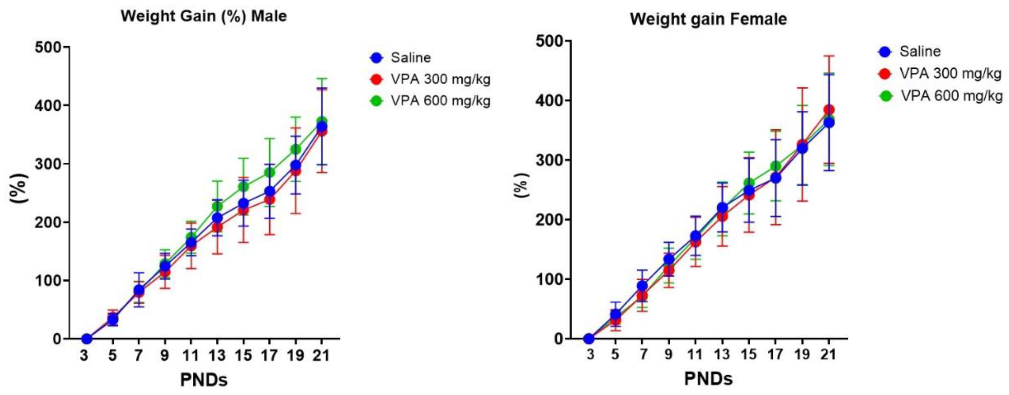
Figure 2.
Ultrasonic vocalization (USV) showing call numbers in male and female mouse pups at postnatal days 3, 8, and 14 following prenatal exposure to VPA recorded for 3 minutes. Male (left panel) and female (right panel) offspring were evaluated for the number of ultrasonic vocalizations (USVs) at PND3, PND8, and PND14. Dams received saline (blue), VPA at 300 mg/kg (red), or VPA at 600 mg/kg (green) during pregnancy. In males, all groups exhibited a peak in USV calls at PND 8 followed by a sharp decline by PND14, with no statistically significant group differences observed. In contrast, female pups showed a significant reduction in USV calls in the VPA-treated groups at PND8 compared to saline controls (*p < 0.05), indicating a sex-specific vulnerability to prenatal VPA exposure. Data are presented as mean ± SEM.
Figure 2.
Ultrasonic vocalization (USV) showing call numbers in male and female mouse pups at postnatal days 3, 8, and 14 following prenatal exposure to VPA recorded for 3 minutes. Male (left panel) and female (right panel) offspring were evaluated for the number of ultrasonic vocalizations (USVs) at PND3, PND8, and PND14. Dams received saline (blue), VPA at 300 mg/kg (red), or VPA at 600 mg/kg (green) during pregnancy. In males, all groups exhibited a peak in USV calls at PND 8 followed by a sharp decline by PND14, with no statistically significant group differences observed. In contrast, female pups showed a significant reduction in USV calls in the VPA-treated groups at PND8 compared to saline controls (*p < 0.05), indicating a sex-specific vulnerability to prenatal VPA exposure. Data are presented as mean ± SEM.
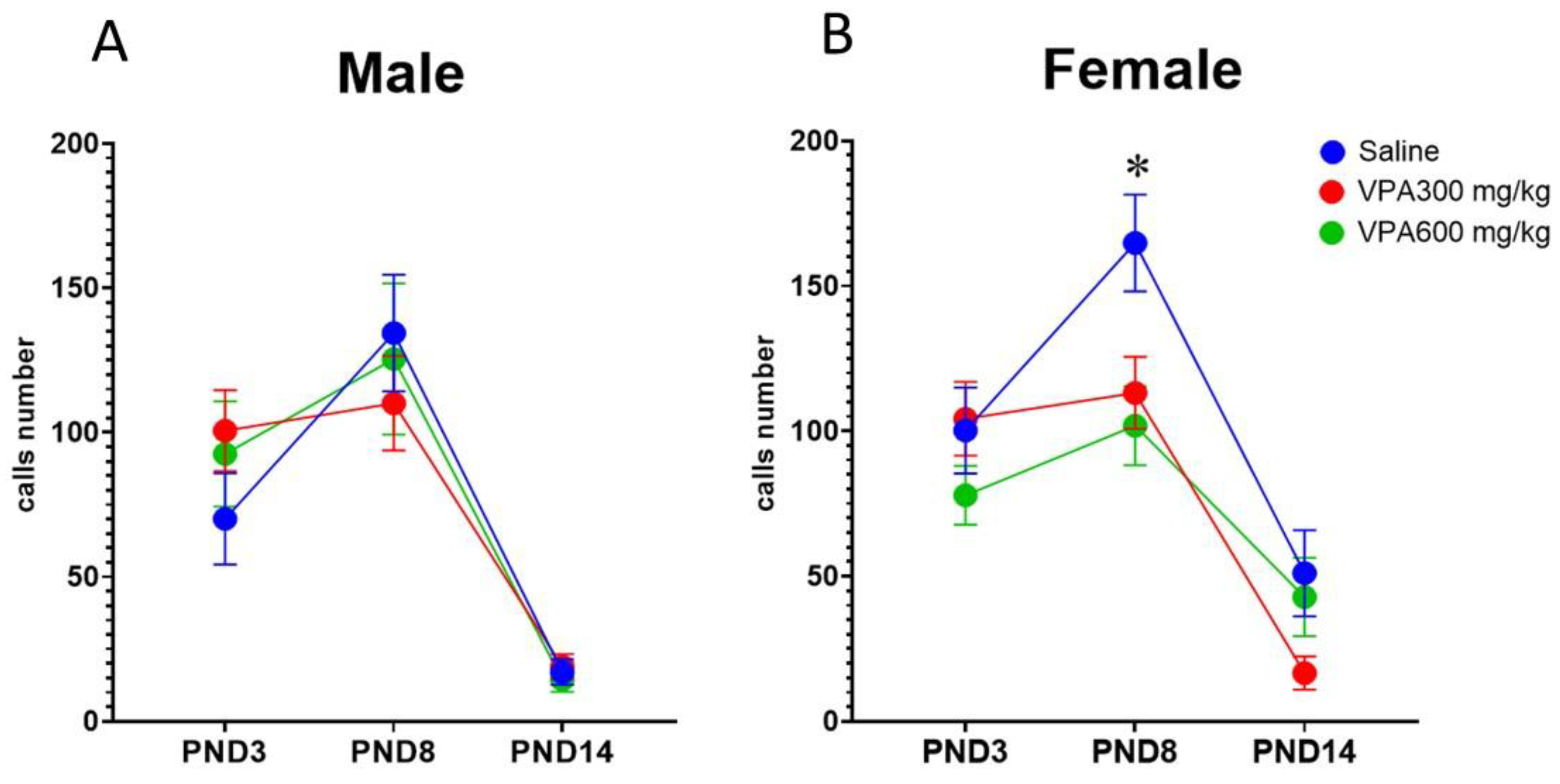
Figure 3.
Sex-specific effects of prenatal valproate exposure on ultrasonic vocalization call-type composition. Heatmap representation of ultrasonic vocalization (USV) call categories in male (A) and female (B) offspring at PND3, PND8, and PND14 following prenatal exposure to saline, VPA 300 mg/kg, or VPA 600 mg/kg. Rows indicate individual call types, and columns represent treatment groups within each postnatal age. Color intensity reflects the relative number of calls within each category (lighter to darker blue). In males (A), call-type distributions exhibited age-dependent maturation but were largely preserved across treatment groups at all postnatal days, consistent with the absence of treatment effects on total call number. In females (B), prenatal VPA exposure was associated with a transient reduction in call-type intensity and diversity at PND8, affecting multiple dominant call categories rather than a single call class. By PND14, call-type distributions converged across all treatment groups in both sexes. These qualitative patterns parallel the quantitative mixed-effects analysis, which identified a sex-specific and temporally restricted reduction in USV call number in VPA treated females, with no persistent effects or alterations in males.
Figure 3.
Sex-specific effects of prenatal valproate exposure on ultrasonic vocalization call-type composition. Heatmap representation of ultrasonic vocalization (USV) call categories in male (A) and female (B) offspring at PND3, PND8, and PND14 following prenatal exposure to saline, VPA 300 mg/kg, or VPA 600 mg/kg. Rows indicate individual call types, and columns represent treatment groups within each postnatal age. Color intensity reflects the relative number of calls within each category (lighter to darker blue). In males (A), call-type distributions exhibited age-dependent maturation but were largely preserved across treatment groups at all postnatal days, consistent with the absence of treatment effects on total call number. In females (B), prenatal VPA exposure was associated with a transient reduction in call-type intensity and diversity at PND8, affecting multiple dominant call categories rather than a single call class. By PND14, call-type distributions converged across all treatment groups in both sexes. These qualitative patterns parallel the quantitative mixed-effects analysis, which identified a sex-specific and temporally restricted reduction in USV call number in VPA treated females, with no persistent effects or alterations in males.
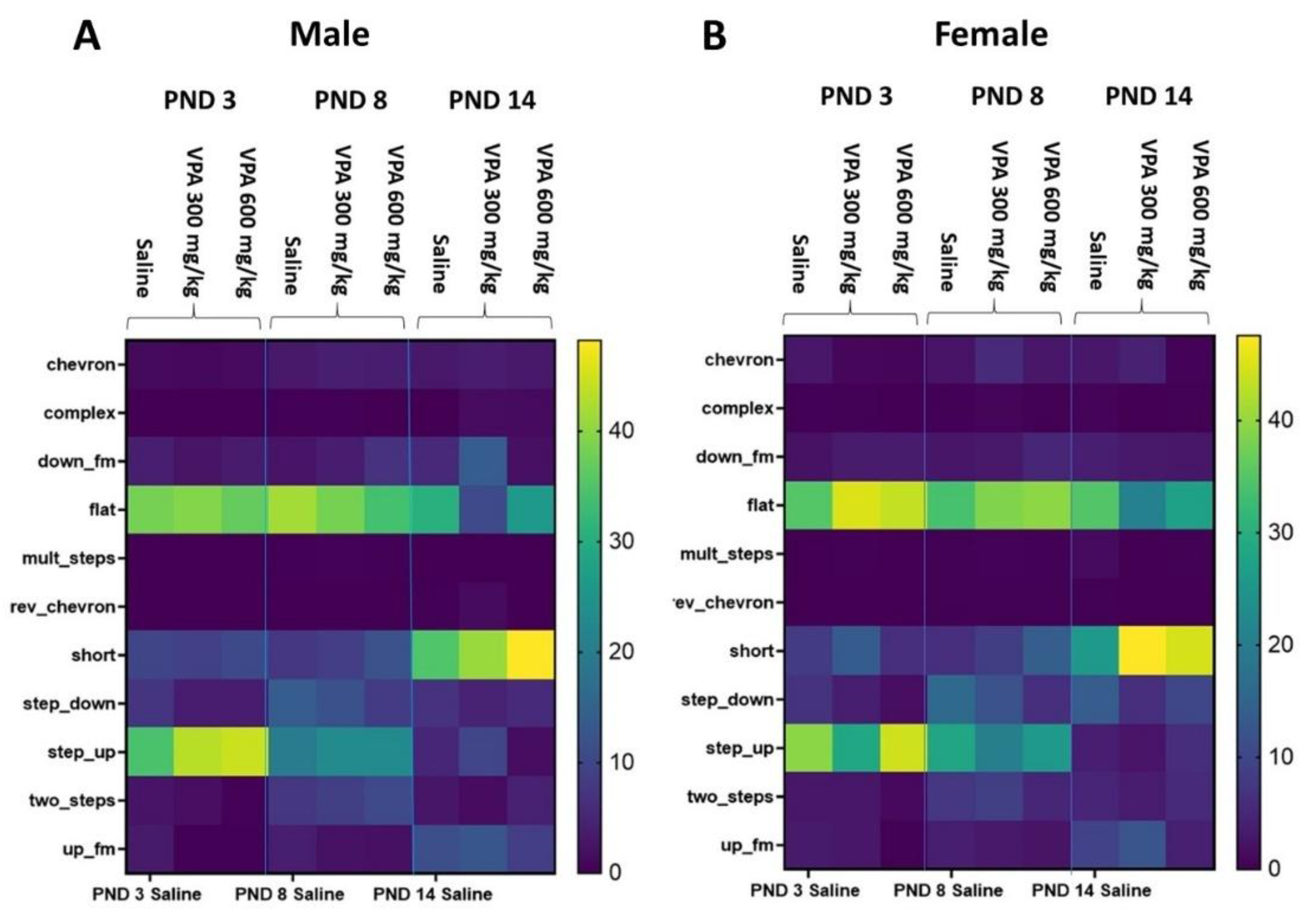
Figure 4.
Sex-specific effects of prenatal valproate exposure on early milestone acquisition. Developmental acquisition curves of surface righting (A, B), negative geotaxis (C, D) and cliff aversion (E, F) represent the cumulative proportion of pups achieving the milestone across postnatal days. Curves were generated using nonlinear sigmoidal fitting for visualization. Data are shown separately for males (A, C, E) and females (B, D, F) offspring. VPA-treated groups exhibit early delays, evident at PND4–5, particularly in VPA300, but all groups reach near-complete acquisition (plateau) by postnatal day 6. Negative geotaxis is largely preserved, with only minor, transient effects. Cliff aversion shows the most pronounced delay in both sexes, especially in VPA300. Asterisks indicate significant differences vs saline measured by Fisher’s exact test: *p < 0.05, **p < 0.01, ***p < 0.001.
Figure 4.
Sex-specific effects of prenatal valproate exposure on early milestone acquisition. Developmental acquisition curves of surface righting (A, B), negative geotaxis (C, D) and cliff aversion (E, F) represent the cumulative proportion of pups achieving the milestone across postnatal days. Curves were generated using nonlinear sigmoidal fitting for visualization. Data are shown separately for males (A, C, E) and females (B, D, F) offspring. VPA-treated groups exhibit early delays, evident at PND4–5, particularly in VPA300, but all groups reach near-complete acquisition (plateau) by postnatal day 6. Negative geotaxis is largely preserved, with only minor, transient effects. Cliff aversion shows the most pronounced delay in both sexes, especially in VPA300. Asterisks indicate significant differences vs saline measured by Fisher’s exact test: *p < 0.05, **p < 0.01, ***p < 0.001.
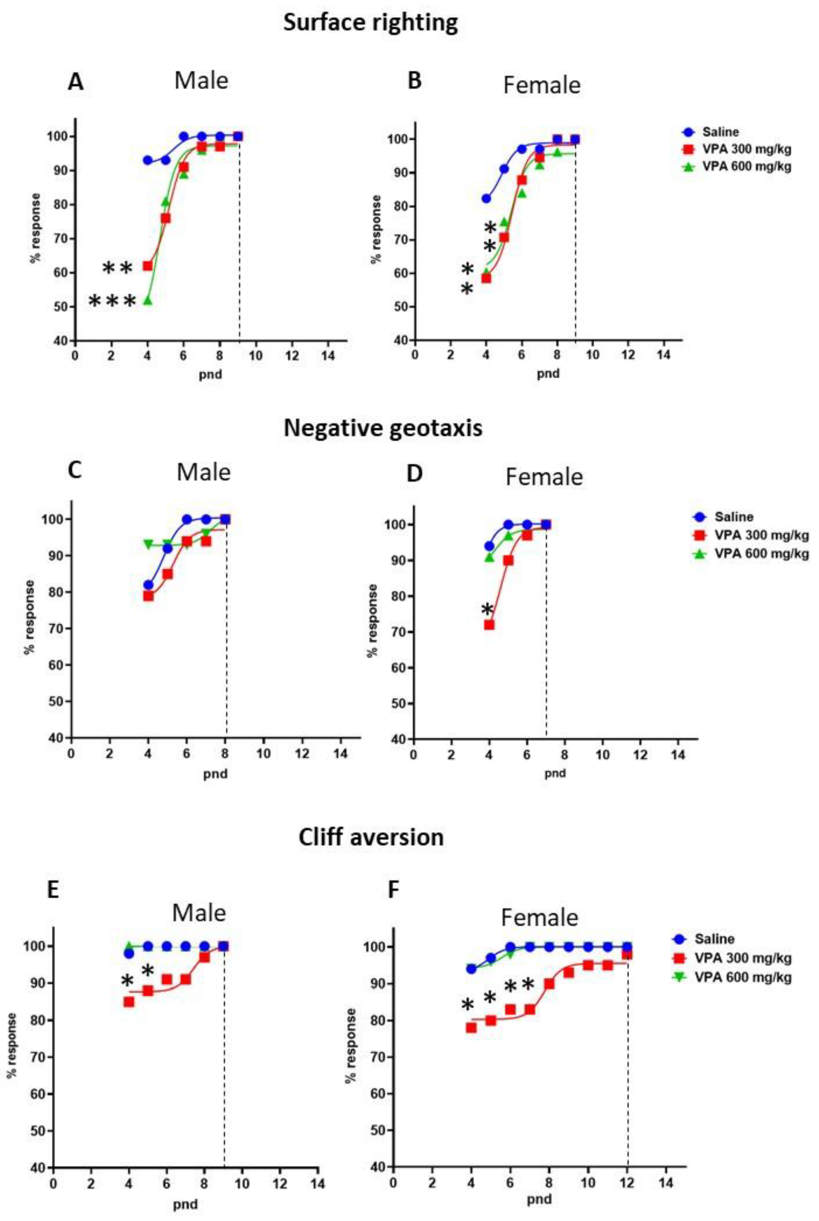
Figure 5.
Sex-dependent effects of prenatal VPA exposure on locomotor activity and anxiety-like behavior in the open-field test. Open-field performance was assessed at PND30 and PND70 in male and female offspring prenatally exposed to saline (blue), VPA 300 mg/kg (red), or VPA 600 mg/kg (green). Top panels show total distance moved (cm) as an index of locomotor activity; bottom panels show time spent in the center of the arena (%) as a measure of anxiety-like behavior. Data are presented as individual values with mean ± SEM. Asterisks indicate significant pairwise differences (*p < 0.05, **p < 0.01, ***p < 0.001, ****p < 0.0001). In males, locomotor activity increased modestly with age in saline controls, while VPA-treated groups did not show this developmental trend but remained comparable to controls. In contrast, center exploration was reduced only in the VPA600 group at both PND30 and PND60, indicating a persistent, dose-specific effect. In females, locomotor activity was lower at PND30 but changed with age, while center exploration was reduced only in the VPA600 group at PND30 and in both VPA groups by PND70.
Figure 5.
Sex-dependent effects of prenatal VPA exposure on locomotor activity and anxiety-like behavior in the open-field test. Open-field performance was assessed at PND30 and PND70 in male and female offspring prenatally exposed to saline (blue), VPA 300 mg/kg (red), or VPA 600 mg/kg (green). Top panels show total distance moved (cm) as an index of locomotor activity; bottom panels show time spent in the center of the arena (%) as a measure of anxiety-like behavior. Data are presented as individual values with mean ± SEM. Asterisks indicate significant pairwise differences (*p < 0.05, **p < 0.01, ***p < 0.001, ****p < 0.0001). In males, locomotor activity increased modestly with age in saline controls, while VPA-treated groups did not show this developmental trend but remained comparable to controls. In contrast, center exploration was reduced only in the VPA600 group at both PND30 and PND60, indicating a persistent, dose-specific effect. In females, locomotor activity was lower at PND30 but changed with age, while center exploration was reduced only in the VPA600 group at PND30 and in both VPA groups by PND70.
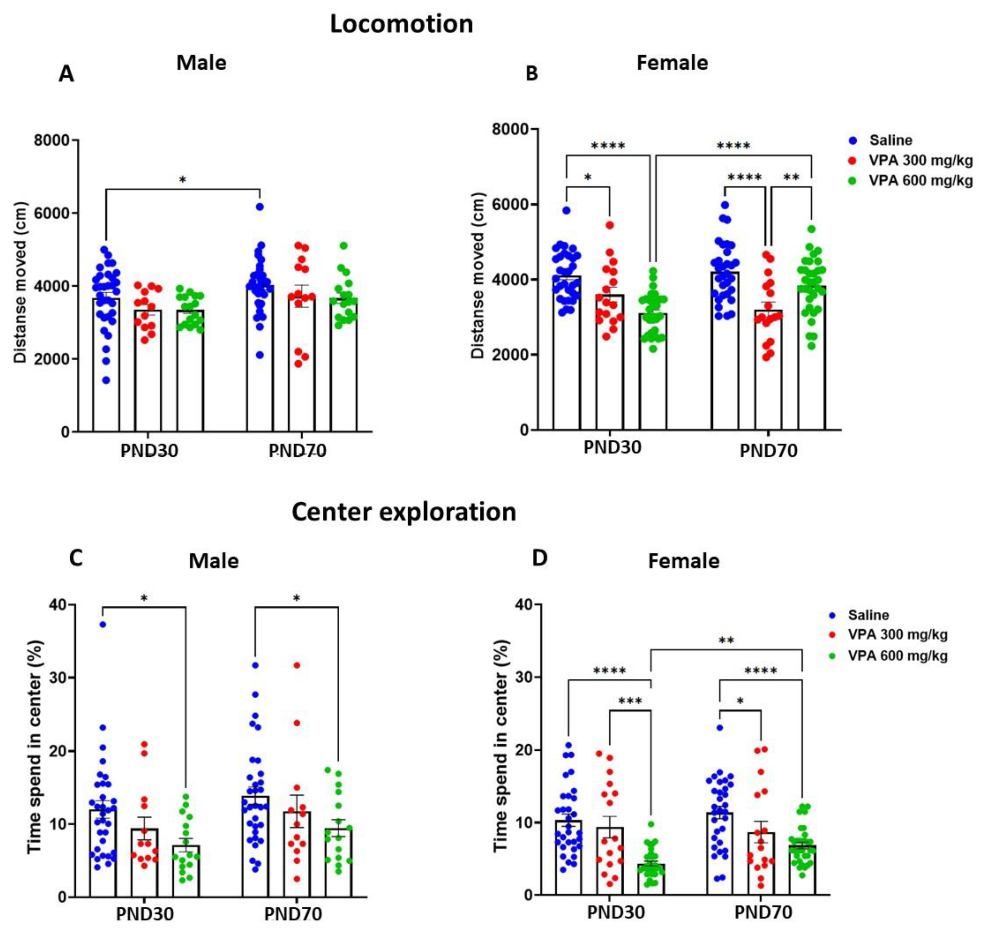
Figure 6.
Prenatal VPA exposure impairs sociability in the three-chamber test in a sex- and age-dependent manner. Prenatal VPA exposure produced sex-dependent effects on sociability, with males (A) showing stable, dose-independent deficits across ages, whereas females (B) exhibited an age-dependent pattern, with early impairment at the moderate dose and later deficits at both doses. Asterisks indicate significant pairwise differences (*p < 0.05, **p < 0.01, ***p < 0.001, ****p < 0.0001).
Figure 6.
Prenatal VPA exposure impairs sociability in the three-chamber test in a sex- and age-dependent manner. Prenatal VPA exposure produced sex-dependent effects on sociability, with males (A) showing stable, dose-independent deficits across ages, whereas females (B) exhibited an age-dependent pattern, with early impairment at the moderate dose and later deficits at both doses. Asterisks indicate significant pairwise differences (*p < 0.05, **p < 0.01, ***p < 0.001, ****p < 0.0001).
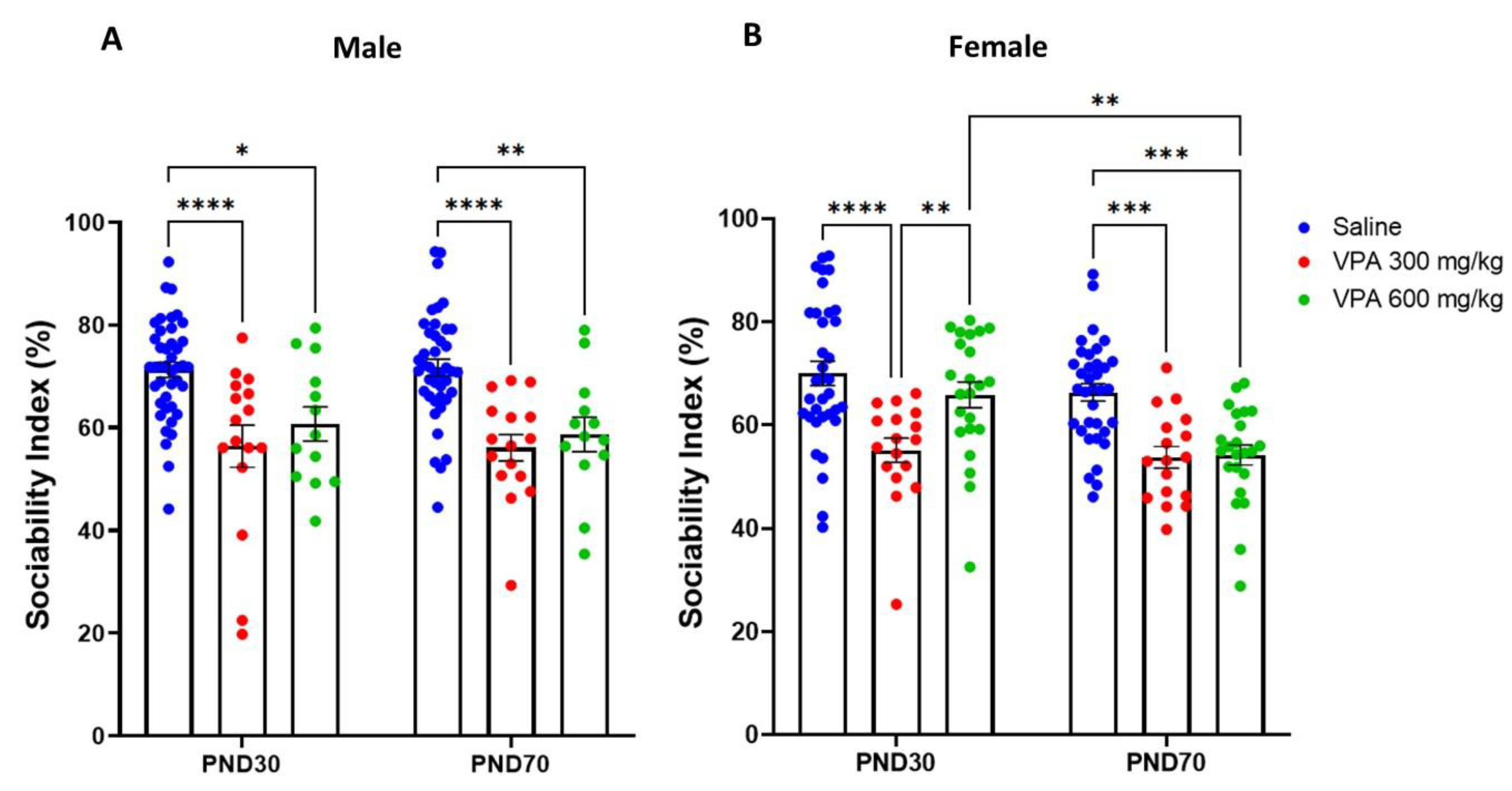
Figure 7.
Prenatal VPA exposure produces sex- and age-dependent effects on Y-maze spontaneous alternation. Males (A) showed a significant impairment in VPA 600 mg/kg on PND30. At PND70, working memory impairment was evident in the VPA300 mg/kg group relative to saline, indicating a developmental shift in dose sensitivity. In contrast, female offspring (B) demonstrated stable reduced alternation in both VPA300 mg/kg and VPA600 mg/kg groups compared with saline controls at PND30 and PND70.
Figure 7.
Prenatal VPA exposure produces sex- and age-dependent effects on Y-maze spontaneous alternation. Males (A) showed a significant impairment in VPA 600 mg/kg on PND30. At PND70, working memory impairment was evident in the VPA300 mg/kg group relative to saline, indicating a developmental shift in dose sensitivity. In contrast, female offspring (B) demonstrated stable reduced alternation in both VPA300 mg/kg and VPA600 mg/kg groups compared with saline controls at PND30 and PND70.
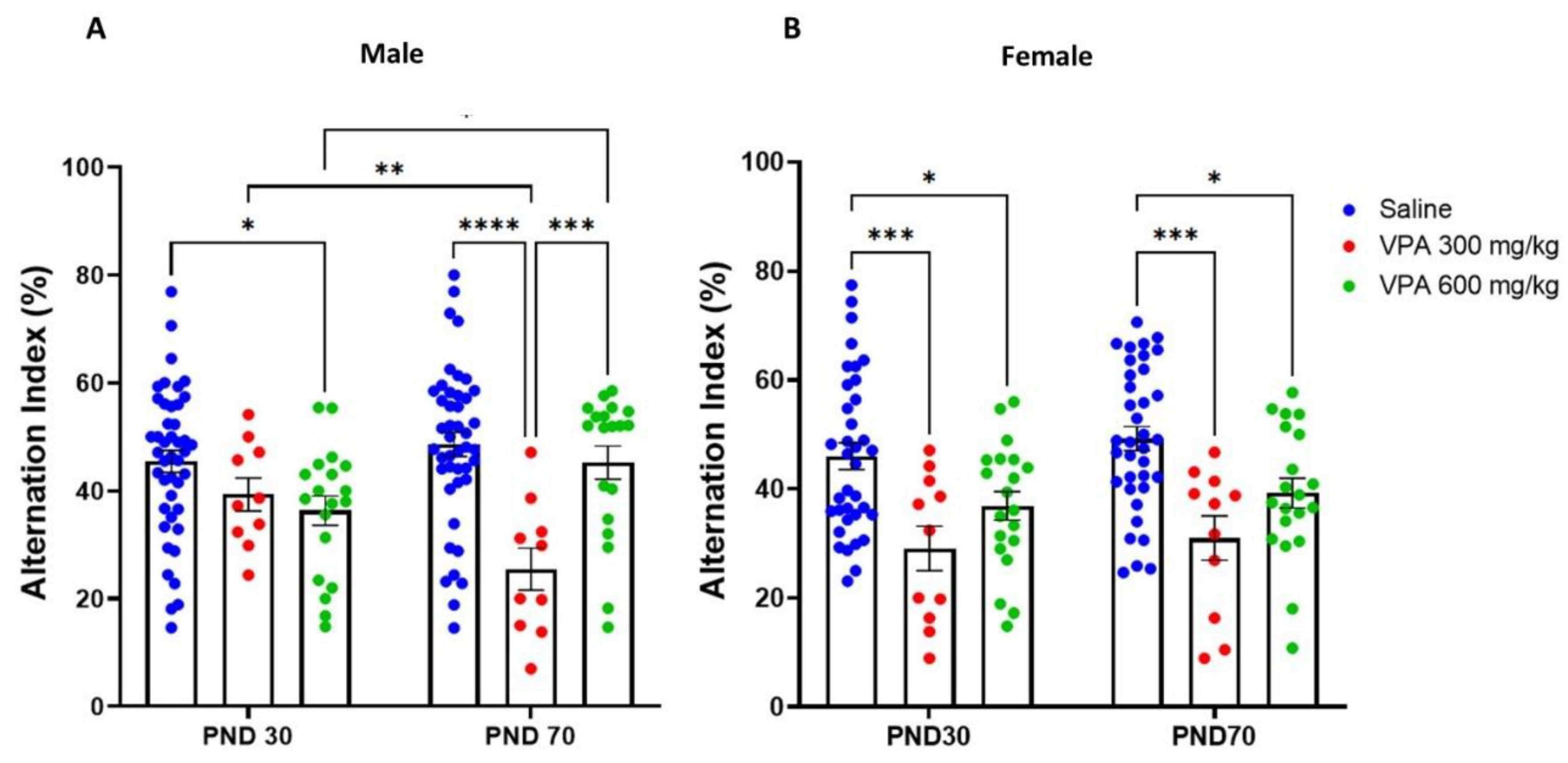
Table 5.
Summary of dose and sex-dependent effects of prenatal VPA exposure across domains.
| Domain | Males | Females | Dose effect |
| VPA300 vs VPA600 | VPA300 vs PA600 | ||
| Fetal death rate | Increased in VPA 600 (Not sex specific, not assessed) | Dose dependent 600 > 300 | |
| Ultrasonic vocalizations number (PND3–14) | No effect at either dose; normal developmental trajectory preserved | Transient reduction at PND8 at both doses; recovery by PND14 | Dose-independent, transient effect (female-specific) |
| USV call-type composition | Preserved across both doses | Broad attenuation at PND8 in both doses | Dose-independent (female-specific); |
| Body weight gain/ Eye opening | No effect | No effect | No dose effects |
| Surface righting | Mild, transient early delay at PND4 at both doses; rapid normalization | VPA300 mg/kg shows slightly longer delay (PND4–5) vs rapid recovery in VPA600 | Intermediate dose more effective |
| Negative geotaxis | No effect | Mild delay mainly at VPA300 at PND4 | Selective effect at intermediate dose (Female sensitivity) |
| Cliff aversion | Transient delay at 300 mg/kg (PND 4-5) | Robust and sustained delay at 300 mg/kg (PND 4–7) | Strongest effect at intermediate dose |
| Open field (locomotion) | no treatment effect | PND30: Reduced activity at both doses; PND70: persistent only at 300 mg/kg, recovery at 600 mg/kg | Divergent dose-dependent pattern; persistent only at intermediate dose in females |
| Open field anxiety (center time) | Mild, transient reduction at 600 mg/kg (PND30 only) | PND30: reduction only at 600 mg/kg; PND70: at both doses | Early 600, later both; High dose drives persistent effect |
| Sociability (3-chamber) | Strong deficits at both doses, at PND30 and PND70 | PND30: Early deficit at 300 mg/kg; PND70: Both doses impaired (600 mg/kg worsening) | Males: dose-independent; Females: dose-progressive |
| Working memory (Y-maze) | PND30: deficit at VPA600 mg/kg; PND70 deficit at VPA300 mg/kg | Persistent impairment at both doses across ages | Males: Early VPA600, later VPA300; Females: dose-independent Stable deficit |
Disclaimer/Publisher’s Note: The statements, opinions and data contained in all publications are solely those of the individual author(s) and contributor(s) and not of MDPI and/or the editor(s). MDPI and/or the editor(s) disclaim responsibility for any injury to people or property resulting from any ideas, methods, instructions or products referred to in the content. |
© 2026 by the authors. Licensee MDPI, Basel, Switzerland. This article is an open access article distributed under the terms and conditions of the Creative Commons Attribution (CC BY) license (http://creativecommons.org/licenses/by/4.0/).
Copyright: This open access article is published under a Creative Commons CC BY 4.0 license, which permit the free download, distribution, and reuse, provided that the author and preprint are cited in any reuse.