Preprint
Article

This version is not peer-reviewed.

Genomic Characterization of Mastitis-Associated Escherichia coli from Sardinia and Comparative Analysis with Isolates from Mainland Italy

Submitted:

06 August 2026

Posted:

07 August 2026

You are already at the latest version

Abstract

Escherichia coli is a leading environmental cause of bovine mastitis, and continued genomic research in specialized dairy systems remains essential for understanding population dynamics and adaptive strategies. In this study, we analyzed 622 milk samples from clinical and subclinical mastitis quarters collected in the Arborea district (Sardinia, Italy) by bacteriological culture and MALDI-TOF MS. E. coli was the most prevalent pathogen (21.54% of positive samples), followed by Streptococcus uberis (12.31%) and Staphylococcus chromogenes (8.72%). Antimicrobial resistance (AMR) was limited to tetracycline (21.43%) and ampicillin (14.29%). Thirty-nine mastitis-associated E. coli (MAEC) obtained from six positive farms were subjected to Nanopore long-read whole-genome sequencing to characterize their population structure, virulome, resistome, and plasmidome. Genomic analysis revealed a polyclonal population with extensive sequence type (ST) diversity and no farm-specific clustering. Seven isolates carried acquired antimicrobial resistance genes (ARGs) (17.95%). Virulence analysis highlighted a conserved core repertoire for extraintestinal survival (adherence, motility, and iron acquisition), with a marked variation in accessory determinants. Comparison with E. coli genomes from mastitis cases and healthy cows isolated in Lombardy showed lineage interspersion rather than disease-specific clustering. Our data are in line with a model in which coliform mastitis in modern dairy farms is driven by heterogeneous environmental lineages sharing common colonization and iron sequestration traits.
Keywords: 
;  ;  ;  ;  ;  ;  ;  

1. Introduction

Escherichia coli is a leading cause of bovine mastitis in intensive dairy systems. This environmental pathogen is mainly responsible for clinical infections that compromise animal health, milk quality, and farm productivity [1,2]. E. coli mastitis is well documented at the clinical level and the reasons behind its diffusion, impact and pathogenicity are the subject of an active field of research.
Current evidence suggests that E. coli mastitis is driven by genetically heterogeneous strains that exploit the mammary environment opportunistically by adapting to local ecological and management contexts, rather than being driven by a limited set of broadly shared, specialized pathogenic lineages. MAEC are therefore described as an ecotype rather than a true pathotype. Their success in the udder depends on a broad repertoire of fitness attributes for survival and rapid adaptation, combined with cow factors [3,4,5]. Consequently, MAEC isolates are often phylogenetically diverse, resemble commensal strains, and possess variable combinations of traits including serum survival, fimbrial adhesion, biofilm formation, and iron acquisition that are neither universally present nor individually sufficient to cause disease [5,6,7,8,9,10].
Understanding the population biology of MAEC is essential for interpreting disease epidemiology, identifying reservoirs and sources, and devising effective control strategies. Genomic approaches now allow high-resolution characterization of core genome structure, accessory gene content, and mobile genetic elements, including plasmids that may carry virulence or antimicrobial resistance (AMR) determinants [11]. Whole-genome sequencing (WGS) approaches provide the resolution needed to characterize the core genome and the accessory elements that may contribute to mammary colonization, persistence, and disease expression. Comparing MAEC populations across distinct dairy ecosystems can help understand local drivers of strain adaptation from broader evolutionary trends, improving our understanding of their population structures, transmission dynamics, and pathogenic potential.
In Italy, dairy systems range from highly specialized, geographically isolated districts to large, intensively managed production basins [12]. The Arborea dairy district in Sardinia is a geographically isolated but highly specialized Holstein dairy system with modern management practices, implementing selective dry-cow therapy, biosecurity measures, and responsible antimicrobial use. Although E. coli is a prominent cause of mastitis in this region, its high-resolution genomic population structure, plasmid architecture, and molecular epidemiology have remained insufficiently characterized. Within this setting, E. coli isolates recovered from cows with clinical and subclinical mastitis provide an opportunity to examine the genomic features of MAEC strains in a defined, territorially constrained dairy environment. The comparison with Lombardy, the main intensive dairy region in Italy, can be useful to investigate local adaptation and the circulation of pathogenic lineages across geographically and production-distinct basins. Both areas are characterized by large dairy farms and highly standardized, modern management systems; however, they differ in important ecological respects, including the absence of dense livestock production farms (pigs, poultry, veal calves) in the Sardinian setting, significantly lower anthropic pressure, and distinct climatic conditions with less humid and warmer winters.
In this study, we carried out the detailed molecular characterization of Sardinian MAEC isolates by long-read whole genome sequencing. To place these findings in a broader national context, we integrated our dataset with publicly available Italian MAEC and commensal bovine E. coli genomes [11]. This comparative approach allowed us to investigate whether Sardinian isolates exhibit distinct genomic features driven by geographic isolation and whether MAEC strains cluster according to clinical status across dairy district with different characteristics.

2. Materials and Methods

2.1. Study Area, Herd Enrolment, and Milk Sampling

The study was carried out in the Arborea dairy district (Oristano, Sardinia, Italy), a geographically delimited area covering approximately 95 km² (with around 6,000 hectares of intensive irrigated agricultural land) originating from a marshland reclamation process completed around a century ago. The district is characterized by a high density of Holstein dairy farms, comprising approximately 150 specialized intensive farms managing over 31,000 dairy cattle, all under continuous veterinary oversight and routine milk-quality monitoring. Twelve dairy farms with a documented previous history of clinical mastitis episodes were prospectively enrolled with the support of four field veterinarians and according to farmer availability. Quarter milk samples were collected from Holstein dairy cows presenting with clinical mastitis, defined as presence of visible udder or milk alterations, or subclinical mastitis, defined as increased somatic cell count (SCC) or positive California Mastitis Test (CMT). Milk sampling was performed according to National Mastitis Council (NMC) guidelines using standard aseptic procedures [13]. For each sampled animal, a standardized clinical record was completed at the time of collection. This form included animal identification, age, herd code, reason for sampling, clinical findings at the quarter level, mammary gland consistency, and macroscopic milk appearance. Clinical data included the presence of local inflammatory signs and other udder abnormalities. Quarter consistency was classified as normal, sclerotic, edematous, or atrophic. Milk appearance was recorded as normal, serous, hemorrhagic, containing flakes, or characterized by absent secretion.

2.2. Milk Culture, Bacterial Identification, and Isolate Selection

Upon arrival at the laboratory, all milk samples were cultured on blood agar according to NMC standards [13] and incubated at 37°C for 18 h. For the detection of Prototheca spp., incubation was extended by an additional 24 h. Only plates with up to two morphologically distinct colony types were considered suitable for interpretation, whereas plates with three or more colony morphotypes were classified as mixed microbial flora and excluded from isolate-based interpretation. To investigate the presence of mycoplasmas, milk samples were also inoculated onto modified Hayflick agar supplemented with 8% horse serum and incubated at 37°C for 5–7 days in a humid chamber. Plates were then examined under an inverted light microscope for typical mycoplasma colony growth and film-and-spot production. Bacterial identification was performed by MALDI-TOF MS. Briefly, cloned colonies grown overnight on blood agar at 37°C were spotted onto a MALDI target plate, air-dried, and overlaid with 1 μl of matrix consisting of α-cyano-4-hydroxycinnamic acid (HCCA) dissolved in acetonitrile, trifluoroacetic acid (TFA), and deionized water. Spectra were acquired in positive linear mode using a Microflex MALDI Biotyper instrument (Bruker Daltonics GMBH, Bremen, Germany). Identification was performed using the MALDI Biotyper reference library and software package (Bruker Daltonics). Isolates with identification log scores lower than 2.00 were processed with the extended direct transfer method and re-analyzed. After subculture on blood agar, colonies were cloned and stored with the CryoBead System at −20°C.

2.3. Antimicrobial Susceptibility Testing

Antimicrobial susceptibility of all E. coli isolates was assessed by the Kirby–Bauer disk diffusion method according to EUCAST and CLSI recommendations [13,14,15]. Results were interpreted as susceptible, susceptible with increased exposure, or resistant according to the applicable criteria. The following antibiotics were tested: Ampicillin (10 µg), Cefoperazone (30 µg), Ceftiofur (30 µg), Enrofloxacin (5 µg), Tetracycline (30 µg). To investigate resistance to third-generation cephalosporins and related β-lactam phenotypes, the following antimicrobial agents were also tested: aztreonam (30 µg), cefotaxime (5 µg), cefpodoxime (10 µg), ceftazidime (10 µg), ceftriaxone (30 µg), meropenem (30 µg), piperacillin–tazobactam (110 µg), and cefepime (30 µg). Each isolate was tested in duplicate to confirm reproducibility of the results. Escherichia coli ATCC 25922 and Klebsiella pneumoniae ATCC 700603 were used as quality control strains.

2.4. Pulsed-Field Gel Electrophoresis

Clonal relatedness among E. coli isolates was first investigated by pulsed-field gel electrophoresis (PFGE) using a protocol based on the PulseNet standard operating procedure for E. coli. Briefly, a single colony from each isolate was inoculated into Luria broth and incubated overnight at 37°C under agitation. After spectrophotometric standardization, bacterial cells were embedded in agarose plugs and lysed in proteinase K-containing buffer. The plugs were then washed repeatedly and stored in TE buffer until digestion. Genomic DNA embedded in plugs was digested with XbaI. Restricted fragments were separated in a CHEF Mapper system using electrophoretic conditions optimized for macrorestriction analysis. Salmonella enterica serotype Braenderup H9812 digested with XbaI and Lambda ladder were used as molecular size markers. Gels were stained with ethidium bromide, visualized, and photographed. PFGE was primarily used as an initial assessment of genomic relatedness and population heterogeneity rather than as the main basis for final phylogenetic inference.

2.5. Sequencing Platform and Library Preparation

MAEC isolates were subjected to whole-genome sequencing using Oxford Nanopore Technologies (ONT) long-read sequencing. Isolates were first cultured on LB agar at 37°C for 24 h. For each isolate, a single colony was inoculated into 5 ml of LB broth and incubated at 37°C for 18 h under agitation. After centrifugation, genomic DNA was extracted from the washed bacterial pellet using the Quick-DNA HMW MagBead kit (Zymo Research Corporation, Orange, CA, USA) according to the manufacturer’s protocol for Gram-negative bacteria. DNA concentration was measured using a Qubit fluorometer. Isolates were sequenced using the ONT long-read platform (MinION) using a R10.4.1 flow cell and the rapid barcoding sequencing kit 24 V14. The sequencing run was monitored in real time, and base quality and yield were assessed during the run.

2.6. Basecalling and Quality Control

Raw nanopore signal data were basecalled with Dorado using the super-accurate (SUP) model dna_r10.4.1_e8.2_400bps_sup@v5.2.0. Reads were demultiplexed by barcode, and adapter and barcode sequences were trimmed during basecalling. Read quality control and length/quality filtering were carried out within the wf-bacterial-genomes workflow prior to assembly, and samples with insufficient read yield were excluded from downstream analysis. The final analysis was performed on curated, checksum-validated FASTQ pass reads for each isolate.

2.7. Genome Assembly and Polishing

Production assemblies were generated with the ONT wf-bacterial-genomes workflow v2.0.2, using Flye for de novo assembly and Medaka for consensus polishing. Following quality-control review, three assemblies required targeted correction. F2M23PDSubA and F2M23PDSubB were reassembled from the frozen FASTQ-pass datasets with Flye using the --nano-hq mode, an expected genome size of 5.4 Mb, 32 threads, and two iterations, followed by Medaka polishing; standard and metagenomic Flye modes were also compared for F2M23PDSubB. The F8M744AS assembly was curated by removing one exact duplicated 3654-bp contig while preserving all unique sequence content. Corrected assemblies were assessed using assembly statistics, QUAST, read-mapping coverage, and sequence-level checksum validation before downstream analyses.

2.8. Genome Annotation

Genomes were annotated with Bakta v1.12.0 using the full database v6.0. Annotation included coding sequences, ribosomal and transfer RNA genes, and other supported functional elements. The three corrected assemblies (F2M23PDSubA, F2M23PDSubB, and F8M744AS) were reannotated with the same Bakta version and database used for the definitive genome panel. Antimicrobial-resistance calling was performed separately with AMRFinderPlus.

2.9. Bioinformatic Analysis

Assembled genomes underwent a sequential in silico characterization workflow. MLST was performed under both the Pasteur (ecoli) and Achtman (ecoli_achtman_4) schemes; sequence-type labels are therefore reported together with the corresponding scheme. Serotypes were predicted with EcTyper v2.0.0. Acquired resistance genes and selected point mutations were detected with AMRFinderPlus v4.2.7 (database 2026-03-24.1). ABRicate was used with ResFinder, CARD, PlasmidFinder, VFDB, and ecoli_vf [14]; BacMet2 screening was run on individual contigs and interpreted as exploratory homology evidence. Phylogenetic relationships among isolates were reconstructed from core-genome SNPs identified with Snippy v4.6.0 against F9M342PD as the analytical reference. Maximum-likelihood inference was performed with IQ-TREE2 under the GTR+G+ASC model with 1,000 ultrafast bootstrap replicates. The combined pangenome and core alignment were generated with Panaroo v1.6.0 in strict mode from standardized Bakta annotations, and the definitive phylogeny was inferred with IQ-TREE2 v2.4.0. Trees and pangenome outputs were visualized with iTOL and Phandango [15]. The input files required for interactive visualization are available at Zenodo under DOI 10.5281/zenodo.21139332. Virulence-associated genes were analyzed through two complementary frameworks using ABRicate. Genes were organized according to the functional categories defined by the Virulence Factor Database (VFDB). Alluvial graphs were created with RAWgraphs 2.0 (www.rawgraphs.io).

2.10. Comparative Analysis of Regional Datasets

The Sardinian dataset was integrated with Lombardy genomes from BioProject PRJNA1242576 [11]. All genomes were annotated with Bakta v1.12.0 and database v6.0. The 85-input panel contained 39 Sardinian inputs, 23 Lombardy mastitis isolates, and 23 Lombardy healthy cow isolates. Panaroo v1.6.0 was run in strict mode and IQ-TREE2 v2.4.0 inferred a maximum-likelihood tree from the filtered core-gene alignment with 1,000 ultrafast bootstrap replicates.

3. Results

3.1. Bacteriological Profile of Quarter Milk Samples

Between February and August 2025, 622 quarter milk samples were collected from 12 Holstein dairy farms located in the Arborea dairy district in Sardinia, Italy. Of these, 195 (31.35%) yielded one or two bacterial species, 67 (10.77%) were classified as mixed microbial flora, and 360 (57.88%) were culture-negative. In total, 37 bacterial species were identified by MALDI-TOF MS. E. coli was the most frequently identified one among all culture-positive samples (n = 42, 21.54%), followed by Streptococcus uberis (n = 24, 12.31%) and Staphylococcus chromogenes (n = 17, 8.72%). Contagious mastitis pathogens were uncommon as only S. aureus was detected, only in one herd and only once. Neither Streptococcus agalactiae nor Mycoplasma spp. were detected. Therefore, environmental and opportunistic pathogens were the most relevant etiological agents of mastitis in this dairy district.

3.2. Distribution of E. coli-Positive Farms and Quarters

Among the 12 enrolled herds, 6 had at least one quarter milk sample positive for E. coli. Cases were distributed as follows: Herd 2, 18 positive quarters; Herd 4, 4 positive quarters, Herd 6, 1 positive quarter; Herd 8, 3 positive quarters; Herd 9, 13 positive quarters; Herd 11, 3 positive quarters. The characteristics of the six dairy farms are detailed in Supplementary File S1.

3.3. Clinical–Bacteriological Associations

Quarter-level comparison between clinical records and microbiological results showed that E. coli was among the bacterial species displaying a recognizable association with clinical presentation. Together with other Gram-negative organisms, E. coli was more often linked to marked alterations in milk appearance, especially flakes, clots, or absent secretion. By contrast, Gram-positive isolates more commonly corresponded to serous milk and inflammatory udder changes such as edema, heat, redness, and pain. Not all bacterial species displayed a consistent pattern, therefore statistically supported associations could not be identified. However, the recurrent association between Gram-negative pathogens and overtly abnormal milk secretion indicates that coliform mastitis in this setting was generally characterized by an evident clinical presentation at the quarter level.

3.4. Phenotypic Antimicrobial Susceptibility of MAEC Isolates

The great majority of E. coli isolates were phenotypically susceptible to the tested compounds, including third-generation cephalosporins. Of 42 isolates, nine showed phenotypical resistance to at least one of the tested molecules (21.43%). Specifically, three were phenotypically resistant to tetracycline and six to ampicillin and tetracycline.

3.5. PFGE Analysis of MAEC Isolates

A total of 39 viable isolates could be recovered from the frozen archive for molecular analysis, distributed as follows: 16, 4, 1, 3, 12, and 3 isolates from Herds 2, 4, 6, 8, 9, and 11, respectively. PFGE revealed marked genetic heterogeneity among MAEC. As this observation suggested that multiple genetically distinct E. coli lineages were present in the samples and were able to cause intramammary infection under field conditions, all 39 MAEC were subjected to whole-genome sequencing (WGS).

3.6. Whole-Genome Sequencing and Genome Characteristics

Long-read ONT sequencing yielded assemblies for all 39 Escherichia coli study isolates. The mean assembly length was 5,032,320 bp (range 4,681,359–5,429,632 bp), with a mean GC content of 50.70%. The median number of contigs was 4 (range 1–87), and the mean N50 was 4,222,443 bp (range 83,722–5,131,528 bp). Complete per-isolate assembly statistics are provided in Supplementary File S2.

3.7. Phylogenetic Structure and Sequence Type (ST) Diversity

Core-genome SNP phylogenomic analysis showed a polyclonal population structure, with broad dispersion of isolates across the unrooted tree and no evidence of farm-specific clustering (Figure 1). Isolates from the same farm occurred in phylogenetically distant clades, consistent with the environmental epidemiology of coliform mastitis and with the absence of a single contagious epidemic clone circulating within or between the sampled farms. Under the Achtman scheme, 37/39 received exact assignments and two remained unassigned or non-exact; ST10 as the most prevalent with 9/39. Under the Pasteur scheme, 17/39 study isolates received exact ST assignments and 22 were unassigned or non-exact; ST302 was most frequent (four isolates). EcTyper-predicted serotypes were also very diverse. The overall pangenome structure and gene presence/absence patterns across all isolates are visualized in Supplementary File S3.
Replicome analysis revealed a high prevalence of extrachromosomal elements, with 89.74% of isolates carrying at least one plasmid replicon. The IncF family served as the primary plasmid backbone, dominated by IncFIB, IncFIC, and IncFIA variants, occurring both independently and within multi-replicon complexes. The isolates exhibited substantial genomic heterogeneity. Isolates carrying a single replicon represented the largest group (31.6%), predominantly carrying IncY or IncFIB backbones. Multi-plasmid carrying strains were also highly prevalent: 23.7% harbored three co-resident replicons, typically combining IncF variants with small colicinogenic plasmids (ColRNAI, Col(pHAD28)). A distinct subset of high-complexity isolates (13.2%, including F8M744AS and F2M23PDSubB) accumulated 4 to 5 distinct replicons (co-hosting IncF, IncX1, IncI1-Alpha, and Col-like elements). Only four isolates (10.5%) were entirely devoid of detectable plasmid replicons.

3.8. Virulence Profile

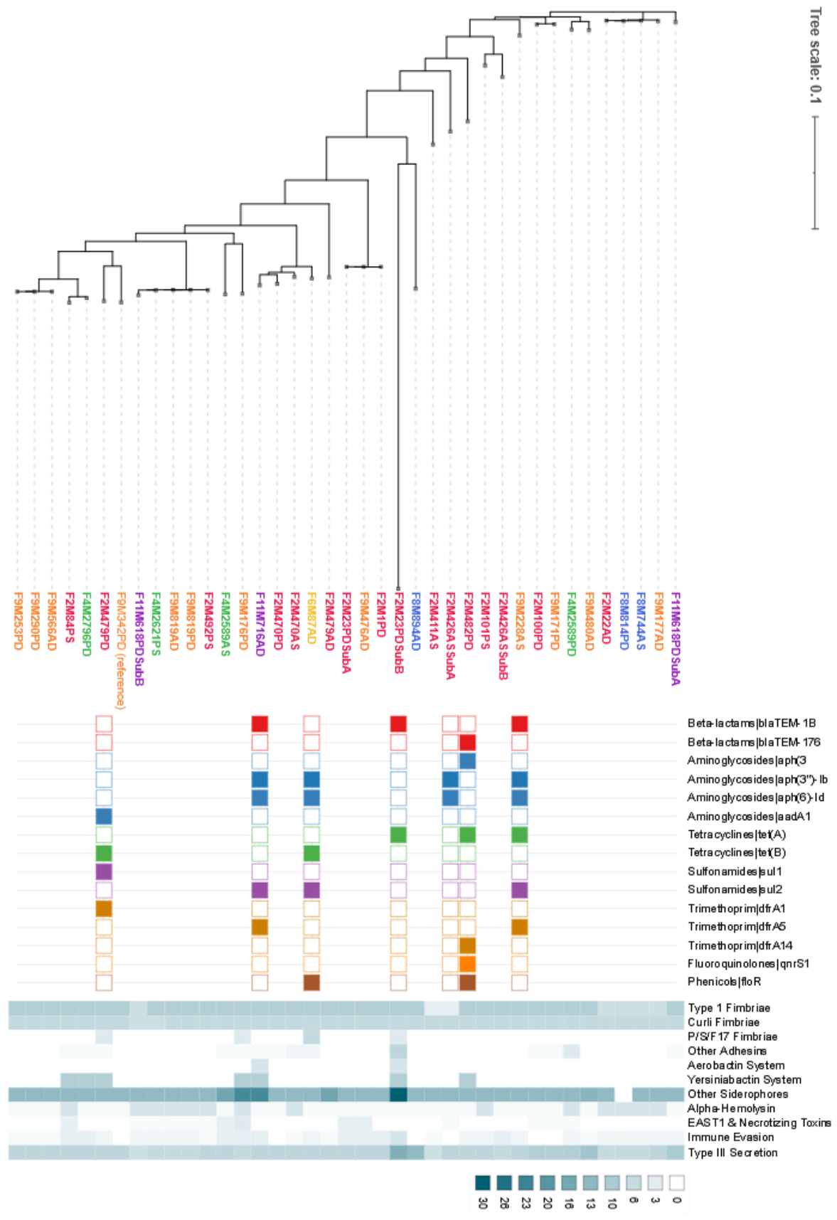
Analysis of the E. coli virulome revealed a highly conserved core of genes associated with adherence and colonization. The results are visualized as heatmap of the main virulence categories in Figure 2 and detailed in Supplementary File S4. Most isolates harbored the genes encoding for Type 1 fimbriae (fimA-I cluster, 32/39) and curli fibers (csgA-G, 37/39). All harbored the flagellar apparatus (flg, flh, and fli operons). Iron acquisition systems were widely present across the dataset. The enterobactin biosynthesis and transport operons (ent and fep clusters) were detected in all isolates but one, and several isolates also carried additional genes for yersiniabactin (fyuA, irp1/2, ybt) and salmochelin (iroB-N). Genes associated with serum survival were also highly prevalent. In particular, ompA and the capsule synthesis-associated gene kpsM were detected in almost all isolates (38/39 and 37/39, respectively). In contrast, classical exotoxin-associated determinants, including Shiga toxins, cytotoxic necrotizing factor 2 (cnf2), cytolethal distending toxins (cdt), and the colibactin pathogenicity island (clb), were largely absent or detected at low frequency. A variable but significant presence of Type III Secretion Systems (T3SS) was detected in a subset of isolates.

3.9. Antimicrobial Resistance Profile

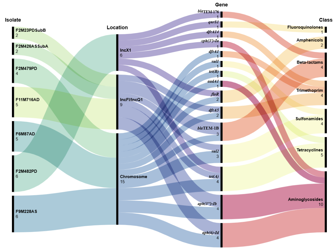
Overall, the genomic profile of the Sardinian MAEC was characterized by a low AMR burden combined with high lineage diversity (Figure 3). Most sequenced mastitis isolates lacked major acquired resistance gene (ARG) determinants, while seven (17,95%) carried at least one acquired ARG. blaTEM genes were detected in five of the six isolates phenotyped as ampicillin-resistant, while tet genes were detected in five of the nine isolates classified as tetracycline-resistant. The resistome architecture of the MAEC isolates carrying ARGs is depicted in Figure 3.
Five isolates met the criteria for potential genomic Multidrug Resistance (MDR) [16]. Isolate F2M482PD carried blaTEM-176, qnrS1, tet(A), floR, dfrA14, and aph(3')-Ia on an IncX1 plasmid contig. Isolate F11M716AD carried multiple resistances on an IncFIA/IncFIB/IncFIC/IncQ1 multireplicon plasmid. In other potential MDR isolates (F9M228AS, F6M87AD, and F2M479PD), the acquired resistance determinants occurred on chromosomal contigs. Finally, the two ARGs of isolates F2M23PDSubB and F2M426ASSubA were located in an IncFIA/IncFIB/IncFIC/IncQ1 multireplicon plasmid.

3.10. Sardinia-Lombardy Comparative Genomic Analysis

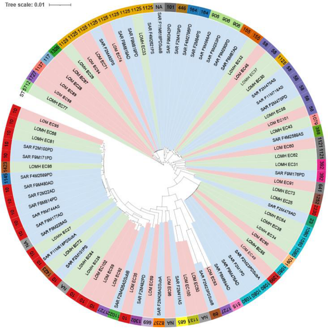
Comparative genomic analysis of the Sardinian mastitis isolates with the Lombardy mastitis and healthy cow collections [11] showed a highly diverse E. coli population with no large clusters that could be considered broadly mastitis-specific across regions (Figure 4). The combined pangenome comprised 14,865 gene families: 2,700 core, 925 soft-core, 1,822 shell, and 9,418 cloud genes. The cloud fraction represented 63.4% of the pangenome, indicating marked accessory-genome expansion and substantial genomic plasticity across the three populations (Supplementary File S5).
Overall, the phylogenomic and pangenomic data indicate that MAEC in this study arised from a broad bovine-associated reservoir rather than from dissemination of successful clones. The increase in accessory-genome diversity after inclusion of the healthy-cow genomes reflects the expected heterogeneity of bovine E. coli populations and does not reveal a sharp mastitis-versus-healthy partition. The phylogeny indicates that mastitis and healthy isolates are interspersed across the same clades and do not form disease-specific lineages.
Phylogenetic analysis revealed a diverse distribution of Sequence Types (STs) among the analyzed isolates originating from Lombardy and Sardinia. The overall most predominant lineage was ST10, forming a major continuous clade represented in both geographic regions. Other prominent shared clones included ST1125, ST58, ST1080, and ST155, indicating widespread dissemination of these STs across both regions. In contrast, several STs exhibited region-specific localization. Among the Sardinia-exclusive lineages, ST906 formed the largest unique cluster (n = 4), accompanied by ST164 (n = 2) and several single-isolate STs (including ST101, ST446, ST388, ST23, ST1091, ST69, ST1131, ST609, and ST11022). Conversely, lineages restricted to Lombardy were characterized by minor clusters such as ST1121 and ST392 (n = 2 each), as well as a series of region-specific singletons (including ST1079, ST641, ST1252, ST2230, ST1399, ST515, ST1727, ST685, ST8237, and ST1302). These findings highlight a dual epidemiological pattern defined by a core of widely circulating shared STs together with localized, region-restricted clones.

4. Discussion

The bacteriological profile of milk from clinical and subclinical dairy cow mastitis cases observed in the Arborea dairy district is broadly consistent with the etiological pattern described in modern specialized dairy systems, in which environmental and opportunistic mastitis agents predominate and classical contagious pathogens have become comparatively less prominent under current control programs [1]. E. coli was the most frequently identified species, followed by S. uberis and S. chromogenes. The prominence of this NASM species is also coherent with recent worldwide findings [2,17,18]. Classical contagious pathogens were uncommon overall. S. agalactiae and Mycoplasma spp. were not detected, and S. aureus was detected only once, further supporting the usefulness of the active policies for controlling contagious pathogens and the sustained impact of farmer-training initiatives in the Arborea district. The epidemiological picture observed in Sardinia is therefore coherent with the broader epidemiological trends emerging from MALDI-based mastitis surveillance studies conducted in Lombardy [2,19]. In this perspective, the Arborea district does not appear epidemiologically anomalous despite its geographic insularity but rather resembles other modern dairy production contexts.
The genomic characterization results are in line with the current hypothesis that MAEC act as an ecotype rather than a distinct pathotype [3]. Our analysis of the population structure revealed remarkable genomic heterogeneity among the Sardinian MAEC isolates. The core-genome phylogeny, combined with the wide diversity of serotypes and ST, demonstrated a polyclonal population with no evidence of farm-specific clustering or epidemic clonal dissemination. Our inter-regional comparative analysis including MAEC genomes from mastitis and healthy cows in Lombardy [11] showed no strict disease-specific lineages. STs found in clinical cases in Arborea were also circulating in mastitis and healthy animals in Northern Italy.
Virulence profiling showed that MAEC isolated in this study share a conserved set of colonization and fitness-associated traits. The uniform presence of Type 1 fimbriae, curli-associated genes, and flagellar operons suggests that adhesion to host surfaces and motility represent a common functional colonization behavior in these strains. These determinants may not be specific to MAEC, but their consistent conservation indicates that they could provide the basic phenotypic features useful for persistence in the bovine mammary environment.
Iron acquisition systems were widespread. The enterobactin-related ent and fep clusters were ubiquitous, and several isolates carried additional siderophore-associated determinants linked to yersiniabactin and salmochelin, indicating some redundancy in iron scavenging capacity [20]. In the inflamed udder, bacterial growth occurs in a nutritionally restricted environment with abundant host iron sequestration mechanisms such as lactoferrin [21]. Therefore, the abundance of siderophore systems may provide a selective advantage by improving competitive fitness during IMI and facilitate persistence under inflammatory conditions.
Serum survival genes were also highly prevalent. The consistent detection of ompA, ompT, and kpsM suggests that resistance to complement-mediated killing and other host defense mechanisms may represent another core component of the MAEC adaptive repertoire [22,23]. Classical exotoxin-associated determinants, including cnf2, cdt, and the clb island, were absent or only sporadically detected. The variable presence of T3SS-related genes in a subset of isolates suggests that, in at least some strains, host-cell interaction may be mediated through secretion-associated mechanisms [24].
This conservation of colonization determinants, iron-acquisition systems, and limited classical toxigenic content, further indicates that pathogenic success in these isolates may depend more on ecological fitness within the mammary gland than on extensive toxin-mediated virulence, consistent with the MAEC concept [3]. This low-toxin/high-adherence and secretion phenotype is coherent with a tissue damage driven mostly by the host inflammatory response to tissue colonization rather than by direct, toxin-mediated cytotoxicity [25,26], typical of Gram-negatives but also observed in Gram-positive udder pathogens such as Streptococcus uberis [27].
In terms of AMR, the Arborea MAEC population was characterized by an overall low burden of acquired ARGs, combined with a separation between virulence and resistance traits. Most isolates lacked major acquired resistance determinants, with seven out of 39 isolates (17.94%) harboring acquired ARGs. When the Lombardy genomes were re-analyzed under the same acquired-ARG criteria applied here, acquired resistance did not follow disease status, being detected in 1 of 23 (4.3%) Lombardy mastitis isolates and in 5 of 23 (21.7%) isolates from apparently healthy Lombardy cows [11]. The acquired-ARG burden of the Sardinian mastitis isolates (7 of 39, 17.9%) was not lower than that of Northern-Italian mastitis isolates. However, our results suggest that national-level generalizations on AMR prevalence in MAEC should be made cautiously, because resistance patterns may vary considerably even between dairy farms and dairy systems with comparable management practices, likely reflecting the influence of local animal health institution initiatives, ecological and epidemiological context, and AMU policies.
Of the seven Sardinian isolates harboring acquired resistance genes, five exhibited a potential MDR profile. The genomic localization of these resistance determinants was different among isolates. In some MDR isolates, the multi-resistance cassettes were integrated into the bacterial chromosome, minimizing the fitness cost associated with plasmid carriage. Other isolates harbored mega-plasmids, including the multireplicon IncF and IncX1 structures that carried MDR cassettes [28]. Notably, IncX1 harbored the plasmid-mediated quinolone resistance (PMQR) gene qnrS1 together with the β-lactamase gene blaTEM-176, the aminoglycoside-inactivating enzyme aph(3’)Ia, and the florfenicol efflux pump floR. The detection of mobilizable resistance against fluoroquinolones, which are classified as Highest Priority Critically Important Antimicrobials (HP-CIA) for human medicine [29], is of some concern. The detection of these hybrid virulence-resistance plasmids indicates that these traits can rapidly spread horizontally within the herd and highlights the importance of continuous surveillance even on farm with low overall AMR prevalence.
Finally, these findings have epidemiological and management implications. Given that coliform mastitis is driven by a vast, heterogeneous reservoir of environmental E. coli rather than a single contagious lineage, control strategies based on tracking specific clones are unlikely to be effective. Instead, mastitis management must remain focused on limiting environmental exposure, optimizing milking hygiene, and supporting host immunity. Furthermore, the detection of chromosomal MDR integrations and complex resistance plasmids in a minority of isolates highlights the ongoing need for continued strict antimicrobial stewardship to prevent the selection and local expansion of these resilient, hard-to-treat opportunistic lineages. Future research should explore the specific environmental niches on farms that select for these accessory traits and investigate the host-pathogen interactions that allow low-virulence strains to elicit robust clinical symptoms.
This study has several limitations that should be acknowledged. First, although designed as a prospective survey across 12 dairy farms, E. coli was isolated from only six herds. Therefore, a sampling bias was present regarding the distribution of isolates across farms, as the majority originated from two herds. This uneven representation may impact estimates of local clonal diversity. Nevertheless, the identification of multiple, distinct STs within these individual farms further supports the observation of an intra-herd polyclonal epidemiology. Second, the comparative analysis with Lombardy relied on publicly available genomes [11] generated and curated by an independent group. Although all datasets were re-annotated and processed using a uniform bioinformatic pipeline, underlying differences in original sampling strategies, clinical metadata granularity, and assembly quality between the two regional cohorts may have influenced the observed patterns of lineage interspersion. Future studies integrating larger, multi-region sampling efforts with standardized metadata collection will be needed to expand upon on these findings.

5. Conclusions

This study provides a comprehensive high-resolution genomic characterization of MAEC in a specialized Italian dairy district. The remarkable phylogenetic diversity, the variety of sequence types, and the sharing of lineages between mastitis and healthy animals across geographically distinct regions is in line with the concept that coliform mastitis in modern dairy settings is caused by a vast reservoir of opportunistic environmental strains. Our virulome analysis further supports this environment-driven etiology; however, a highly conserved foundational fitness core comprising adherence, motility, and serum survival factors, combined with variable, redundant iron-acquisition systems, may favor the success of these isolates in the udder. The overall low prevalence of acquired antimicrobial resistance in the Arborea district is reassuring, but the sporadic detection of chromosomal MDR integrations and mobilizable multireplicon plasmids warrants ongoing surveillance to prevent the silent spread of resistant determinants.
As a final consideration, the integration of WGS-based epidemiological surveillance into modern dairy systems offers valuable perspectives. In the context of MAEC, the long-read WGS approach enabled the high-resolution tracking of opportunistic environmental lineages and the detailed investigation of their virulence and resistance profiles, opening the way to targeted, herd-specific surveillance and management strategies.

Supplementary Materials

The following supporting information can be downloaded at Preprints.org. S1. Management characteristics of the six dairy herds originating the MAEC isolates characterized in this study. S2. Genome-assembly characteristics of the 39 biological MAEC study isolates. S3. Pangenome structure and virulence gene distribution of E. coli isolates. S4. Detail of the virulence genes detected on the 39 MAEC isolates upon interrogation of the coli_vf database. S5. Pangenome structure of the 85-input panel.

Author Contributions

Conceptualization, M.F.A., A.R., and S.T.; methodology, M.F.A., A.G., S.T.; software, M.F.A., A.G.; formal analysis, M.F.A., A.G., L.S., E.A., C.L., F.C., M.P.F., S.B., M.C., A.C.; resources, M.F.A., A.G., S.T., A.R.; data curation, M.F.A., A.G., S.T.; writing—original draft preparation, M.F.A.; writing—review and editing, all authors; visualization, M.F.A., A.G.; supervision, M.F.A., A.R., S.T.; project administration, A.R.; funding acquisition, M.F.A., A.R., S.T. All authors have read and agreed to the published version of the manuscript.

Funding

This research was funded by the Italian Ministry of Health under the Call “Progetti di Ricerca Corrente 2024”, Project ID IZS SA 03/24 “Caratterizzazione del fenotipo, del genotipo e dei profili di antibioticoresistenza di Escherichia coli associati a mastite bovina in Sardegna”. High-performance computing resources were provided by CINECA (Leonardo HPC system) under the REMOVES allocation (project account uMI25_Removes).

Institutional Review Board Statement

Ethical approval for the collection of samples within the broader project framework was granted by the Ethics Review Committee of the Istituto Zooprofilattico Sperimentale della Sardegna (Minutes no. 4, December 29, 2025).

Data Availability Statement

The study is registered in NCBI BioProject PRJNA1494036, with 39 BioSamples registered for the biological cohort. As of 3 August 2026, genome-assembly and raw-read accessions were undergoing NCBI processing and were not yet publicly linked; they will be linked upon release. Pangenome files are available at https://doi.org/10.5281/zenodo.21139332.

Acknowledgments

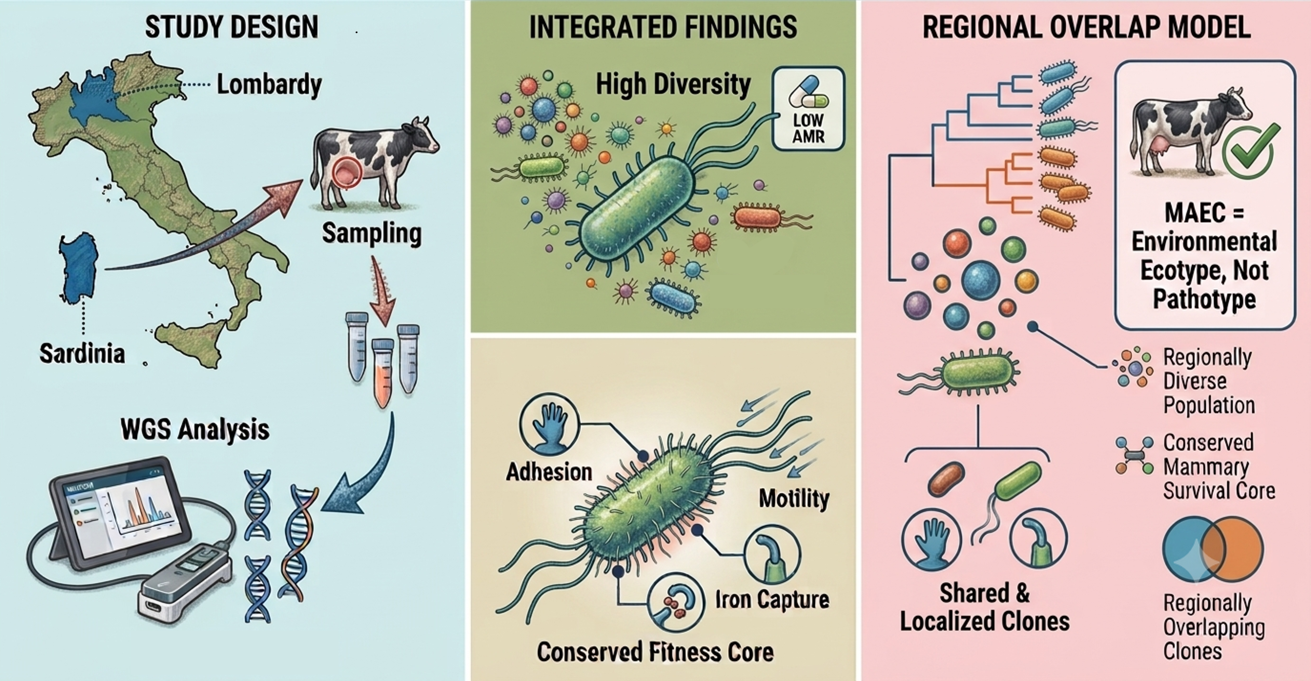
The authors thank Prof. Paolo Moroni for insightful discussions and suggestions provided at the beginning of the project. We acknowledge that during the preparation of this manuscript Google Gemini (Version 3.6 Flash) was used for language refinement and for graphical abstract drafting but not for generating scientific content, interpreting data, drafting results, or making methodological decisions. All study design, data collection, analysis, interpretation, and final editorial decisions were performed and verified by the authors, who take full responsibility for the content of the manuscript.

Conflicts of Interest

The authors declare no conflicts of interest. The funders had no role in the design of the study; in the collection, analyses, or interpretation of data; in the writing of the manuscript; or in the decision to publish the results.

Abbreviations

The following abbreviations are used in this manuscript:
AMR Antimicrobial Resistance
ARG Antimicrobial Resistance Gene
CDS Coding Sequence
CLSI Clinical and Laboratory Standards Institute
CMT California Mastitis Test
ESBL Extended-Spectrum Beta-Lactamase
EUCAST European Committee on Antimicrobial Susceptibility Testing
HCCA α-cyano-4-hydroxycinnamic acid
HP-CIA Highest Priority Critically Important Antimicrobials
HPC High Performance Computing
MAEC Mastitis-Associated Escherichia coli
MALDI-TOF MS Matrix-Assisted Laser Desorption/Ionization Time-Of-Flight Mass Spectrometry
MDR Multidrug-Resistant
MLST Multi-Locus Sequence Typing
NASM Non-aureus staphylococci and mammaliicocci
NMC National Mastitis Council
ONT Oxford Nanopore Technologies
PFGE Pulsed-Field Gel Electrophoresis
PMQR Plasmid-Mediated Quinolone Resistance
SCC Somatic Cell Count
SNP Single Nucleotide Polymorphism
ST Sequence Type
T3SS Type III Secretion System
TFA Trifluoroacetic acid
VF Virulence Factor
VFDB Virulence Factor Database
WGS Whole-Genome Sequencing

References

  1. Ruegg, P.L. A 100-Year Review: Mastitis Detection, Management, and Prevention. J. Dairy Sci. 2017, 100, 10381–10397. [Google Scholar] [CrossRef] [PubMed]
  2. Addis, M.F.; Locatelli, C.; Penati, M.; Poli, S.F.; Monistero, V.; Zingale, L.; Rota, N.; Gusmara, C.; Piccinini, R.; Moroni, P.; et al. Non-Aureus Staphylococci and Mammaliicocci Isolated from Bovine Milk in Italian Dairy Farms: A Retrospective Investigation. Vet. Res. Commun. 2023, 1–8. [Google Scholar] [CrossRef]
  3. Germon, P.; Foucras, G.; Smith, D.G.E.; Rainard, P. Invited Review: Mastitis Escherichia Coli Strains—Mastitis-Associated or Mammo-Pathogenic? J. Dairy Sci. 2025, 108, 4485–4507. [Google Scholar] [CrossRef] [PubMed]
  4. Leimbach, A.; Poehlein, A.; Vollmers, J.; Görlich, D.; Daniel, R.; Dobrindt, U. No Evidence for a Bovine Mastitis Escherichia Coli Pathotype. BMC Genom. 2017, 18, 359. [Google Scholar] [CrossRef]
  5. Liu, Y.; Liu, G.; Liu, W.; Liu, Y.; Ali, T.; Chen, W.; Yin, J.; Han, B. Phylogenetic Group, Virulence Factors and Antimicrobial Resistance of Escherichia Coli Associated with Bovine Mastitis. Res. Microbiol. 2014, 165, 273–277. [Google Scholar] [CrossRef] [PubMed]
  6. Lambertini, E.; Karns, J.S.; Van Kessel, J.A.S.; Cao, H.; Schukken, Y.H.; Wolfgang, D.R.; Smith, J.M.; Pradhan, A.K. Dynamics of Escherichia Coli Virulence Factors in Dairy Herds and Farm Environments in a Longitudinal Study in the United States. Appl. Env. Microbiol. 2015, 81, 4477–4488. [Google Scholar] [CrossRef] [PubMed]
  7. Fernandes, J.B.C.; Zanardo, L.G.; Galvão, N.N.; Carvalho, I.A.; Nero, L.A.; Moreira, M.A.S. Escherichia Coli from Clinical Mastitis: Serotypes and Virulence Factors. J. Vet. Diagn. Invest 2011, 23, 1146–1152. [Google Scholar] [CrossRef] [PubMed]
  8. Zaatout, N. An Overview on Mastitis-Associated Escherichia Coli: Pathogenicity, Host Immunity and the Use of Alternative Therapies. Microbiol. Res. 2022, 256, 126960. [Google Scholar] [CrossRef] [PubMed]
  9. Keane, O.M. Genetic Diversity, the Virulence Gene Profile and Antimicrobial Resistance of Clinical Mastitis-Associated Escherichia Coli. Res. Microbiol. 2016, 167, 678–684. [Google Scholar] [CrossRef] [PubMed]
  10. Blum, S.E.; Leitner, G. Genotyping and Virulence Factors Assessment of Bovine Mastitis Escherichia Coli. Vet. Microbiol. 2013, 163, 305–312. [Google Scholar] [CrossRef] [PubMed]
  11. Laterza, G.; Meroni, G.; Soggiu, A.; Martino, P.A.; Sora, V.M.; Zaghen, F.; Bonizzi, L.; Colombo, L.; Zecconi, A. Bovine Clinical E. Coli Mastitis in Italian Dairy Herds Is Not Associated with a Specific Pathotype. Pathogens 2025, 14. [Google Scholar] [CrossRef] [PubMed]
  12. Masi, M.; Vecchio, Y.; Pauselli, G.; Pasquale, J.D.; Adinolfi, F. A Typological Classification for Assessing Farm Sustainability in the Italian Bovine Dairy Sector. Sustainability 2021, 13. [Google Scholar] [CrossRef]
  13. Adkins, P.R.F.; Middleton, J.R.; Fox, L.K.; Pighetti, G.; Petersson-Wolfe, C. National Mastitis Council Laboratory Handbook on Bovine Mastitis; National Mastitis Council: New Prague, 2017. [Google Scholar]
  14. Liu, B.; Zheng, D.; Zhou, S.; Chen, L.; Yang, J. VFDB 2022: A General Classification Scheme for Bacterial Virulence Factors. Nucleic Acids Res. 2022, 50, D912–D917. [Google Scholar] [CrossRef] [PubMed]
  15. Hadfield, J.; Croucher, N.J.; Goater, R.J.; Abudahab, K.; Aanensen, D.M.; Harris, S.R. Phandango: An Interactive Viewer for Bacterial Population Genomics. Bioinformatics 2018, 34, 292–293. [Google Scholar] [CrossRef] [PubMed]
  16. Magiorakos, A.-P.; Srinivasan, A.; Carey, R.B.; Carmeli, Y.; Falagas, M.E.; Giske, C.G.; Harbarth, S.; Hindler, J.F.; Kahlmeter, G.; Olsson-Liljequist, B.; et al. Multidrug-Resistant, Extensively Drug-Resistant and Pandrug-Resistant Bacteria: An International Expert Proposal for Interim Standard Definitions for Acquired Resistance. Clin. Microbiol. Infect. 2012, 18, 268–281. [Google Scholar] [CrossRef] [PubMed]
  17. De Buck, J.; Ha, V.; Naushad, S.; Nobrega, D.B.; Luby, C.; Middleton, J.R.; De Vliegher, S.; Barkema, H.W. Non-Aureus Staphylococci and Bovine Udder Health: Current Understanding and Knowledge Gaps. Front. Vet. Sci. 2021, 8, 1–16. [Google Scholar] [CrossRef] [PubMed]
  18. Freu, G.; Gioia, G.; Gross, B.; Biscarini, F.; Virkler, P.; Watters, R.; Addis, M.F.; Franklin-Guild, R.J.; Runyan, J.; Masroure, A.J.; et al. Frequency of Non-Aureus Staphylococci and Mammaliicocci Species Isolated from Quarter Clinical Mastitis: A 6-Year Retrospective Study. J. Dairy Sci. 2024, 107, 3813–3823. [Google Scholar] [CrossRef] [PubMed]
  19. Penati, M.; Ulloa, F.; Locatelli, C.; Monistero, V.; Pavesi, L.F.; Santandrea, F.; Piccinini, R.; Moroni, P.; Bronzo, V.; Addis, M.F. An Update on Non-Aureus Staphylococci and Mammaliicocci in Cow Milk: Unveiling the Presence of Staphylococcus Borealis and Staphylococcus Rostri by MALDI-TOF MS. Vet. Res. Commun. 2024, 48, 2555–2561. [Google Scholar] [CrossRef] [PubMed]
  20. Garcia, E.C.; Brumbaugh, A.R.; Mobley, H.L.T. Redundancy and Specificity of Escherichia Coli Iron Acquisition Systems during Urinary Tract Infection. Infect. Immun. 2011, 79, 1225–1235. [Google Scholar] [CrossRef] [PubMed]
  21. Kawai, K.; Hagiwara, S.; Anri, A.; Nagahata, H. Lactoferrin Concentration in Milk of Bovine Clinical Mastitis. Vet. Res. Commun. 1999, 23, 391–398. [Google Scholar] [CrossRef] [PubMed]
  22. Prasadarao, N.V.; Blom, A.M.; Villoutreix, B.O.; Linsangan, L.C. A Novel Interaction of Outer Membrane Protein A with C4b Binding Protein Mediates Serum Resistance of Escherichia Coli K1. J. Immunol. 2002, 169, 6352–6360. [Google Scholar] [CrossRef] [PubMed]
  23. Abreu, A.G.; Barbosa, A.S. How Escherichia Coli Circumvent Complement-Mediated Killing. Front. Immunol. 2017, 8. [Google Scholar] [CrossRef] [PubMed]
  24. Slater, S.L.; Sågfors, A.M.; Pollard, D.J.; Ruano-Gallego, D.; Frankel, G. The Type III Secretion System of Pathogenic Escherichia Coli. Curr. Top. Microbiol. Immunol. 2018, 416, 51–72. [Google Scholar] [CrossRef] [PubMed]
  25. Günther, J.; Petzl, W.; Bauer, I.; Ponsuksili, S.; Zerbe, H.; Schuberth, H.-J.; Brunner, R.M.; Seyfert, H.-M. Differentiating Staphylococcus Aureus from Escherichia Coli Mastitis: S. Aureus Triggers Unbalanced Immune-Dampening and Host Cell Invasion Immediately after Udder Infection. Sci. Rep. 2017, 7, 4811. [Google Scholar] [CrossRef] [PubMed]
  26. Burvenich, C.; Van Merris, V.; Mehrzad, J.; Diez-Fraile, A.; Duchateau, L. Severity of E. Coli Mastitis Is Mainly Determined by Cow Factors. Vet. Res. 2003, 34, 521–564. [Google Scholar] [CrossRef] [PubMed]
  27. Archer, N.; Egan, S.A.; Coffey, T.J.; Emes, R.D.; Addis, M.F.; Ward, P.N.; Blanchard, A.M.; Leigh, J.A. A Paradox in Bacterial Pathogenesis: Activation of the Local Macrophage Inflammasome Is Required for Virulence of Streptococcus Uberis. Pathogens 2020, 9, 1–23. [Google Scholar] [CrossRef] [PubMed]
  28. Dobiasova, H.; Dolejska, M. Prevalence and Diversity of IncX Plasmids Carrying Fluoroquinolone and β-Lactam Resistance Genes in Escherichia Coli Originating from Diverse Sources and Geographical Areas. J. Antimicrob. Chemother. 2016, 71, 2118–2124. [Google Scholar] [CrossRef] [PubMed]
  29. Critically Important Antimicrobials for Human Medicine : 6th Revision. Available online: https://www.who.int/publications/i/item/9789241515528 (accessed on 4 July 2026).
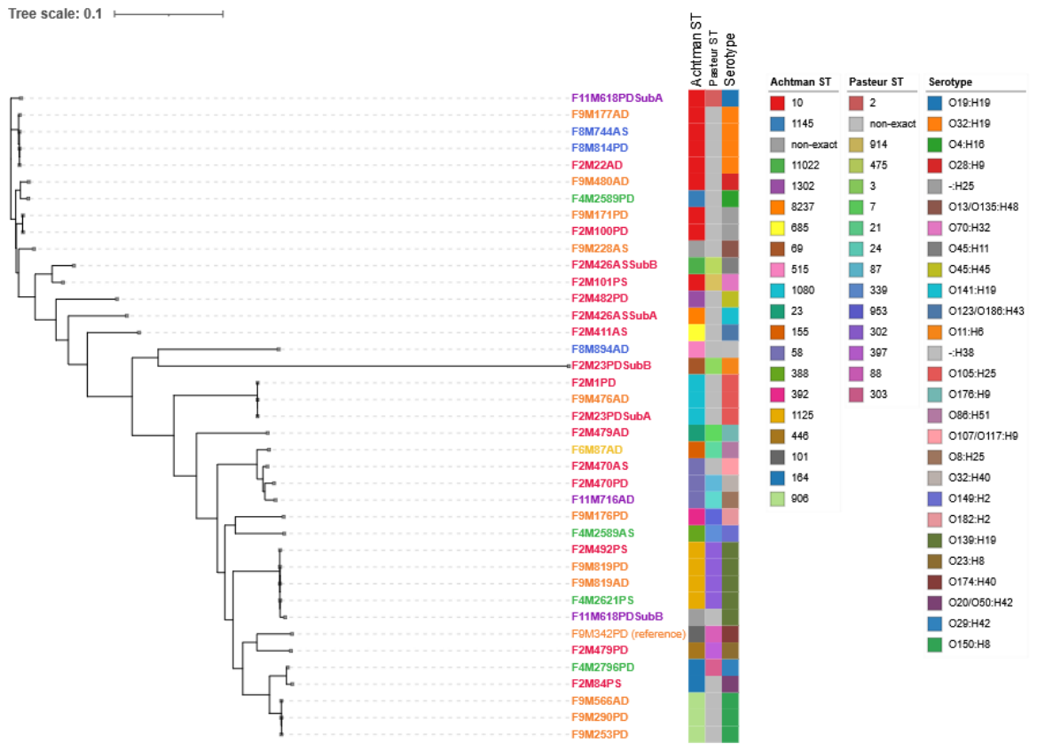
Figure 1. Unrooted maximum-likelihood core-SNP phylogeny of 39 sequenced MAEC isolates. The tree illustrates the predicted multi-locus sequence typing (MLST) result according to the Achtman scheme, Pasteur scheme, and the EcTyper-predicted serotype. Farms are color-coded and labels indicate the farm (F), source (M), animal, quarter, and bacterial subculture, when present. Isolate F9M342PD was used as the internal reference.
Preprints 227181 g001
Figure 2. Antimicrobial resistance and virulence of the 39 sequenced MAEC isolates. The solid color squares illustrate the antimicrobial resistance genes detected in the isolates according to their phylogenetic classification. The antibiotic molecule class and the respective genes are indicated in the label flanking each category. The heatmap illustrates the virulence category profile of the isolates, with intensity according to the operon density as indicated in the legend. Farms are color-coded and labels indicate the farm (F), source (M), animal, quarter, and bacterial subculture, when present. Isolate F9M342PD was used as the internal reference.
Preprints 227181 g002
Figure 3. Alluvial diagram illustrating the flow from bacterial isolates to genomic contexts and resistance genes detected in the sequenced MAEC isolates. Flows represent individual acquired antibiotic resistance genes (ARGs) mapping from left to right: bacterial isolate identifier (Isolate), genomic/plasmid location (Location), specific resistance gene allele (Gene), and target antimicrobial class (Class). Node heights and flow stream widths are proportional to the number of detected resistance determinants, indicated under each category label.
Preprints 227181 g003
Figure 4. Core-genome phylogeny of 85 Escherichia coli isolates from Sardinia and Lombardy. The tree includes the 39 Sardinian isolates sequenced in this study, plus 23 Lombardy mastitis and 23 Lombardy healthy-cow publicly available genomes [11]. Clades are colored according to the isolate source, as follows: blue, mastitis isolates from Sardinia; pink, mastitis isolates from Lombardy; green, commensal isolates from Lombardy. The outer strip indicates the Achtman sequence type (ST) of each isolate. NA: ST not available/not assigned. ST color scheme as in Figure 1.
Preprints 227181 g004
Disclaimer/Publisher’s Note: The statements, opinions and data contained in all publications are solely those of the individual author(s) and contributor(s) and not of MDPI and/or the editor(s). MDPI and/or the editor(s) disclaim responsibility for any injury to people or property resulting from any ideas, methods, instructions or products referred to in the content.
Copyright: This open access article is published under a Creative Commons CC BY 4.0 license, which permit the free download, distribution, and reuse, provided that the author and preprint are cited in any reuse.