

Submitted:
05 August 2026
Posted:
06 August 2026
You are already at the latest version
Abstract
Streptomyces sp. HR58, isolated from the hop rhizosphere, exhibits antifungal activity against a variety of fungal phytopathogens affecting many crops. HR58 was characterized using phenotypic and genomic approaches. Growth tolerance and enzymatic activities were characterized, while whole-genome sequencing enabled biosynthetic gene cluster prediction. The strain HR58 displayed morphological and biochemical traits characteristic of the genus Streptomyces growing from pH 5 to pH 12 and salt tolerance up to 5% NaCl. Genome analysis revealed a size of 8,270,372 bp with a GC content of 71.76% and the presence of gene clusters associated with antifungal activity. Although ANI analysis showed 96.6% identity with Streptomyces anulatus ATCC 11523, the dDDH value of 66.5% fell below the species delineation threshold of 70%, indicating that HR58 cannot be confidently assigned to any currently described Streptomyces species. A biosynthetic gene cluster encoding the antifungal compound cycloheximide was located in its genome. Cycloheximide production was confirmed in liquid cultures. Silver nanoparticle (AgNPs) biosynthesis by strain HR58 was confirmed and their antimicrobial activity was evaluated. The ability of strain HR58 to biosynthesize antifungal nanoparticles further enhances its potential applications in sustainable agriculture, offering a promising alternative for the development of eco-friendly plant disease control strategies.
Keywords:
antifungal activity
; biological control
; biosynthetic gene clusters
; phytopathogenic fungi
; secondary metabolites
; silver nanoparticles
1. Introduction
Fungal phytopathogens are among the major constraints to agricultural productivity worldwide, causing severe yield losses and threatening the sustainability of cropping systems. Due to climate change, its incidence is expected to continue increasing in the coming decades [1]. Conventional chemical control remains limited by increasing regulatory restrictions, environmental concerns, and the rapid emergence of fungicide resistance [2], highlighting the need for novel, effective, and sustainable disease management strategies.
Microbial antagonists (biocontrol agents) have emerged as promising alternatives within integrated pest management by providing effective and environmentally sustainable control of plant pests and pathogens while reducing dependence on chemical pesticides [3].
Among them, the genus Streptomyces stands out due to its exceptional biosynthetic versatility. Streptomyces spp. are well known for their ability to produce a variety of bioactive compounds with antifungal, antibacterial, and plant growth–promoting properties [4,5]. Secondary metabolites produced by streptomycetes can be produced during death, stationary, or exponential growth phases because of the depletion of essential nutrients [6]. Many species also behave as beneficial endophytes, enhancing plant health through antibiosis, nutrient mobilization, enzymatic degradation of fungal cell walls, and modulation of the rhizosphere microbiome [7]. Specifically, rhizosphere streptomycetes can protect plant roots by inhibiting the growth of fungal pathogens [8].
Advances in whole-genome sequencing and comparative genomics have substantially improved the taxonomic resolution and functional characterization of Streptomyces species, enabling the identification of genome features, phylogenetic relationships, and biosynthetic gene clusters (BGCs) involved in secondary metabolite production [9]. In addition to their well-known ability to synthesize bioactive compounds, some Streptomyces species have recently been explored as biological factories for the green synthesis of metallic nanoparticles, linking microbial biotechnology with nanotechnology for agricultural and biomedical applications.
Nanoparticles are at the leading edge of the rapidly developing field of nanotechnology, and they are currently produced by chemical, physical and biological methods. Different elements such as magnesium, zinc, copper, gold, platinum and silver are used for synthesis nanoparticles. However, unique properties of silver nanoparticles (AgNPs) at nano-scales such as high surface plasmon resonance (SPR), high stability, good conductivity, photoelectrochemical activity, and high activity against pathogenic microbes provide more biological applications [10].
Several reports are available for biosynthesis of Ag-NPs using different Streptomyces species such as Streptomyces atrovirens [11], Streptomyces glaucus [12], Streptomyces griseorubens [13] Streptomyces griseoplanus [14], Streptomyces hygroscopicus [15], Streptomyces catenulae [16], Streptomyces capillispiralis, Streptomyces zaomyceticus, and Streptomyces pseudogriseolus [10]
In this study, we performed an extensive characterization of Streptomyces sp. HR58, a promising biocontrol agent, including its phenotypic traits, phylogenomic placement, and biosynthetic potential. We also evaluated its antifungal (AF) activity against a broad panel of agriculturally relevant fungal pathogens. Furthermore, we investigated the production of antifungals and the strain’s ability to synthesize silver nanoparticles, as well as the antifungal properties of the resulting AgNPs. Overall, this work provides an integrated assessment of the biocontrol potential of HR58 and highlights its value as a source of bioactive metabolites with applications in sustainable plant disease management.
2. Materials and Methods
2.1. Strains and Growth Conditions
Streptomyces sp. HR58 strain was isolated from the rhizosphere of hop plants (Nugget cultivar) cultivated under conventional agronomic practices in a field located in León, Spain [17]. Rhizosphere soil samples were collected from five individual plants by carefully excavating the surrounding root systems. Soil in direct contact with the roots was extracted using a sterile spatula, transferred into sterile 50 mL Falcon tubes, and stored in an icebox prior to preservation at 4 ºC until further processing. Culturable Streptomycetes were isolated on starch casein agar (SCA) [18] and International Streptomyces Project 2 (ISP2) agar medium [19]. The strain was routinely cultivated on MEY (Maltose Yeast Extract) medium at 30 ºC and maintained at 4 ºC [20].
Alternaria alternata, Cadophora luteo-olivacea, Dactylonectria macrodydima, Diplodia seriata, Fusarium fujikuroi, F. oxysporum, Phaeomoniella chlamydospora, Pleurostoma richardsiae, Rhizoctonia solani and Thelonectria olida were isolated from diseased grapevines. Verticillium dahliae D0913 and V. dahliae ND1113 were kindly provided by Dr. Leire Molinero [21] and V. dahliae V937I were kindly provided by Dr. Jesús Mercado [22] isolated from olive trees. Fungal isolates were routinely cultivated on PDA (Potato Dextrose agar) medium at 25 ºC and maintained at 4 ºC.
Pure cultures of bacteria (Bacillus subtilis, Enterococcus faecalis, Escherichia coli and Salmonella typhimurium) were grown in Nutrient Broth medium at 37 ºC on a rotary shaker at 200 rpm. Yeasts (Candida albicans and Saccharomyces cerevisiae) were grown in YEPD medium at 28 ºC, on a rotary shaker at 180 rpm.
For long-term stored, spore or cells suspensions were preserved in 40% glycerol at −20 ºC.
2.2. Phenotypic Characterization of Streptomyces sp. HR58
The cultural characteristics of the strain HR58 were evaluated following the phenotypic characterization procedure described by Calvo-Peña et al. [23] The strain was grown on a range of solid media, including ISP 1 (DSMZ 1764), ISP 2 (DSMZ 987), ISP 3 (DSMZ 609), ISP 4 (DSMZ 547), ISP 5 (DSMZ 993), ISP 6 (DSMZ 1269), ISP 7 (DSMZ 1619) [19], nutrient agar (DSMZ 1) (Scharlab S.L., Sentmenat, Spain), Bennett’s agar (DSMZ 548; HiMedia Laboratories GmbH, Modautal, Germany), trypticase soy agar (TSA; DSMZ 535; Scharlab S.L.), R5 agar [20] and GYM medium (DSMZ 65). Cultures were incubated at 28 ºC for 7 days before colony morphology was examined. Growth was assessed at temperatures ranging from 4 to 45 °C and at pH values between 5.0 and 12.0 using GYM medium (glucose 4 g/L; yest extract, 4 g/L; malt extract, 10 g/L; CaCO3, 2 g/L; agar, 20 g/L; pH 7.2). Salt tolerance was determined on ISP2 medium supplemented with NaCl concentrations ranging from 2.5% to 7.5% (w/v). Colony pigmentation, including the colour of aerial and substrate mycelia and the production of diffusible pigments, was recorded using a RAL colour chart. All tests were conducted in triplicate.
The biochemical and enzymatic properties of the HR58 strain were characterized using API-ZYM and API 20NE test strips (bioMerieux, Lyon, France). For API 20NE, inoculation of the cupules was performed at 28 ºC with a bacterial suspension standardized to a turbidity of 5 on the McFarland scale, and results were recorded at 24 and 48 h. API-ZYM assays were conducted at 28 ºC instead of the standard 37 ºC, with an incubation period of 24 h. Both tests were conducted in duplicate.
2.3. Genome Features and Phylogenomic Analysis
Whole-genome sequencing of strain HR58 was performed by Macrogen Inc. (Seoul, Republic of Korea) using PacBio Revio technology to generate long reads. Raw sequencing reads were assembled de novo using Canu v2.2 [24] with default parameters optimized for long-read data. Assembly quality metrics were assessed using QUAST v5.2.0 [25] and genome completeness was further evaluated with BUSCO v5.4.7 [26] using the actinobacteria_odb10 dataset. Functional genome annotation was performed with the RASTtk (RAST, Rapid Annotation using Subsystem Technology) server [27].
The 16S rRNA gene sequence was extracted from the assembled genome and compared against type strain sequences in the NCBI RefSeq database using BLASTN v2.15.0+ [28]. Whole-genome relatedness was assessed through Average Nucleotide Identity (ANI) calculations using FastANI v1.34 [29] and digital DNA–DNA hybridization (dDDH) using the Type (Strain) Genome Server (TYGS) [30]. Thresholds of 95% ANI [29] and 70% dDDH were applied for species delineation [31]. Phylogenomic analysis was conducted within the TYGS framework using the Genome BLAST Distance Phylogeny (GBDP) method [32], with branch support estimated by 100 pseudo-bootstrap replicates.
2.4. Analysis of Bioactive Compound Biosynthetic Gene Clusters
Biosynthetic gene clusters (BGCs) were identified using antiSMASH version 7.1.0 [33] with all optional annotation modules enabled, including ClusterBlast, KnownClusterBlast, SubClusterBlast, and ActiveSiteFinder. Predicted clusters were examined to determine cluster boundaries and similarity to previously characterized biosynthetic pathways.
Genes encoding enzymes potentially involved in fungal cell wall degradation were identified by screening the RAST annotation using keyword- and EC number-based searches. Terms included “chitinase”, “chitin-binding protein”, “β-glucosidase”, “glucanase”, “cellulase”, and “protease”. All predicted proteins associated with chitin and N-acetylglucosamine utilization subsystems were extracted and manually curated. Putative enzymes lacking formal EC annotations were retained when their conserved domain architecture (e.g., GH18 endochitinase-like signatures) supported their functional assignment. Signal peptide prediction was performed using SignalP version 6.0 with the “other organisms” model to identify proteins potentially secreted via the Sec or Tat pathways [34].
2.5. Antifungal Activity of Streptomyces sp. HR58
In order to quantify the AF activity of Streptomyces sp. HR58 against fungal phytopathogens they were individually tested on MEY agar plates forming a circle at 1 cm from the edge of the plate. An agar plug containing the fungal pathogen was deposited in the center of the plate [35]. Plates were incubated at 25 ºC up to 10 days. The inhibition index (I index) was calculated as follows: I index (%) = [(Rc − R)/Rc] × 100, where R is the radius of the fungal colony in the presence of the bacterial isolate, and Rc is the maximum radius of the fungal colony (control). All tests were performed in triplicate for each fungus tested.
2.6. Purification and Identification of Cycloheximide production
To assess cycloheximide production by the strain Streptomyces HR58, an ethyl acetate extract was obtained from 100 mL of liquid culture following the procedure described by Das et al. [36] and the crude extract was solved in 250 µl of 80% methanol.
AF activity was evaluated using in vitro plate assay against V. dahliae V937I strain on PDA agar plates containing 0.8% of agar (w/v) was performed as described by Calvo-Peña [35]. Briefly, we embedded 5.0 x 10⁴ fungal spores/mL in the agar media before plating. Wells that were 6mm in diameter, were made on the agar plates and each well was inoculated with 60 µl of crude extract. Plates were incubated at 25 ºC for 4 days and measured the inhibition halos. A total of 600 µg of commercial cycloheximide was added to one well as a control.
The remaining HR58 culture extract was filtered through Corning® Costar® Spin-X® centrifuge tube filters (0.45 μm pore size; Merck KGaA, Darmstadt, Germany) and stored at −20 ºC until further analysis. High-Performance Liquid Chromatography analysis was performed using an Agilent 1200 Series Gradient HPLC System (Agilent Technologies), following the chromatographic method described by Awla et al. [37]. The system was equipped with a quaternary pump delivery system (G1311A), a preparative autosampler (G1329A), a diode array multi-wavelength detector (G7115A), and an analytical fraction collector (G1364F) with an Autosampler Thermostat (G1330B). A 10 μL sample was injected and resolved using an analytical LiChrospher RP-18 column (40 × 250 mm; 5 μm) (Teknokroma, San Cugat del Vallés, Spain).
2.7. Microbial Synthesis and Characterization of AgNPs
Nanoparticles were synthesized according to Sadhasivam et al. [15] and Vijayabharathi et al. [14]. Briefly, Streptomyces sp. HR58 was cultured in ISP2 broth at 28 °C with shaking at 200 rpm for 72 h. The culture was subsequently centrifuged at 10,000 rpm for 15 min to remove the biomass, and the resulting supernatant was passed through a 0.2 µm membrane filter to obtain a sterile cell-free filtrate. For AgNP biosynthesis, the cell-free filtrate was mixed with a 1 mM AgNO₃ solution (Thermo Scientific) to obtain a final filtrate concentration of 10% (v/v). The reaction mixture was incubated at 28 °C and 200 rpm for 72 h in the absence of light.
The progress of nanoparticle formation was monitored every 24 h by UV–visible spectrophotometry (UV-1600PC, VWR, Leuven, Belgium). Absorption spectra were recorded between 300 and 800 nm using aliquots collected throughout the 72 h incubation period. Solutions containing only 1 mM AgNO₃ or 10% (v/v) cell-free filtrate served as negative controls. Upon completion of the reaction, the synthesized AgNPs were recovered by centrifugation, thoroughly washed with nanopure water to remove unreacted components, freeze-dried, and stored for subsequent characterization and biological assays.
TEM specimens were prepared by placing a drop of the AgNP suspension onto a Formvar-coated copper grid and allowing it to air-dry at room temperature. The size and morphology of the biosynthesized AgNPs was examined using a transmission electron microscope (TEM 1010, JEOL, Japan) operated at an accelerating voltage of 90 kV [38].
2.8. Antibiotic Activity of AgNPs
The antimicrobial activity of the microbiologically synthesized AgNPs against pathogenic organisms such as gram-positive bacteria (Bacillus subtilis and Enterococcus faecalis), gram negative bacteria (Escherichia coli and Salmonella typhimurium), yeasts (Candida albicans and Saccharomyces cerevisiae) and fungal phytopathogens (Fusarium oxysporum, Alternaria alternata and Verticillium dahliae) was measured using well-diffusion method. Agar plates were prepared by adding 1mL of an overnight culture of each microorganism in the medium, just before pouring into the plates. Wells were made on the agar and 20µL of the biosynthesized AgNPs solution was added into each well. After incubation, the diameter of the inhibition zone was measured. All tests were performed by triplicate.
3. Results
3.1. Phenotypic Characterization of Streptomyces sp. HR58
Morphological studies revealed that Streptomyces sp. HR58 grows well on different agar media showing sporulation on most of the ISP media tested (Figure 1). As summarized in Supplementary Material (Table S1), the strain exhibited a narrow growth temperature range, growing well between 4 °C and 28 °C, but showing markedly reduced growth at 37 °C. It could grow at all pH values tested, from pH 5 to pH 12, indicating a broad tolerance. Regarding salt tolerance, HR58 tolerated NaCl concentrations of 2.5% and 5%, whereas no growth was observed at 7.5% NaCl. According to the RAL code, the strain exhibits a distinctive chromatic profile, with hues intermediate between papyrus white and green-beige, which contributes to its easy recognition among related taxa.
The biochemical profile obtained with the API 20NE and API-ZYM systems is detailed in Supplementary Material (Table S2). In the API 20NE test, strong activity was observed for urease, esculin hydrolysis, gelatin hydrolysis and for the assimilation of glucose, N-acetyl-glucosamine, potassium gluconate, or malic acid. According to the API-ZYM test, Streptomyces sp. HR58 showed high activity for alkaline phosphatase, lipase (C14), leucine arylamidase, trypsin, α-chymotrypsin and α-glucosidase.
3.2. Genome Features and Phylogenomic Analysis
PacBio Revio long-read sequencing followed by Canu assembly generated a single contig of 8,270,372 bp with a GC content of 71.76%. QUAST analysis confirmed the structural integrity of the assembly. Genome annotation using RAST identified 7,299 predicted protein-coding sequences, 84 RNA genes and 306 functional subsystems. BUSCO analysis indicated a high level of completeness for both the assembly and the predicted gene set (98.25%) (Table 1).
The 16S rRNA gene sequence of strain HR58 showed 100% identity to members of the Streptomyces anulatus group, including S. chrysomallus and S. citreofluorescens. Average Nucleotide Identity (ANI) analysis revealed 96.6% nucleotide identity with S. anulatus, whereas digital DNA–DNA hybridization (dDDH) similarity between HR58 and S. anulatus ATCC 11523 (formerly S. chrysomallus) was 66.5%, with a confidence interval from 63.5 to 69.3%, just below the 70% threshold used to distinguish between closely species.
Genome-based phylogenomic analysis inferred from whole-genome sequences supported the affiliation of HR58 with the Streptomyces anulatus clade (Figure 2). In the GBDP-based phylogram, HR58 formed a distinct branch relative to the type strain and other closely related taxa, supported by high pseudo-bootstrap values (average branch support of 85.5%).
3.3. Analysis of Bioactive Compound Biosynthetic Gene Clusters
AntiSMASH v7.1.0 predicted a total of 35 biosynthetic gene clusters (BGCs) in the genome of Streptomyces sp. HR58, including NRPS, PKS, hybrid NRPS–PKS systems, RiPPs, terpenes, siderophores, and other specialized metabolite pathways (Table 2; Supplementary Material Table S3).
Ten BGCs showed 100% similarity to known clusters, including those most similar to geosmin, griseobactin, naringenin, ectoine, desferrioxamine B, melanin, AmfS, 2-methylisoborneol and SGR polycyclic tetramate macrolactams. Four additional BGCs exhibited high similarity (75–99%) to characterized pathways, including clusters related to skyllamycin D/E and warkmycin CS1/CS2. Among them, only naringenin (Region 4) and SGR PTM (Region 28) are described as antifungals [39,40]
A trans-AT polyketide synthase (PKS) region showing 94% similarity to the cycloheximide biosynthetic gene cluster was identified (Region 34, Table S3). The cluster contained the core biosynthetic genes typically associated with cycloheximide biosynthesis in Streptomyces, with minor differences in gene content and organization.
Three BGCs displayed moderate similarity (30–70%) to known pathways, whereas fourteen showed low similarity (1–29%). Four predicted BGCs showed no detectable similarity to characterized clusters and were classified as cryptic (Table 2).
The genome of Streptomyces sp. HR58 encodes enzymes predicted to target fungal cell wall components (Table 3). The chitinolytic repertoire includes eight chitinases (six annotated as EC 3.2.1.14 and two putative endochitinases), four chitin-binding proteins and two β-N-acetylglucosaminidases (EC 3.2.1.52). Four endo-1,4-β-glucanases were also identified. SignalP-6.0 predicted Sec/SPI signal peptides consistent with extracellular localization in seven of the eight chitinases.
In addition, HR58 encodes 62 predicted proteases, including 38 genes exceeding 1,000 bp in length. SignalP analysis predicted secretion signals for 23 proteases via Sec or Tat pathways. Genes encoding LysM-domain proteins, AA10/AA11 lytic polysaccharide monooxygenases or canonical chitin-catabolic regulators (dasR, chiR and nagR) were not detected.
3.4. Antifungal Activity of Streptomyces sp. HR58
AF activity based on dual cultures growth, was tested against eleven fungal phytopathogens affecting many different crops: A. alternata, C. luteo-olivacea, D. macrodydima, D.seriata, F. fujikuroi, F. oxysporum, P. chlamydospora, P. richardsiae, R. solani, T. olida; and V. dahliae. The Inhibition index (I index) ranged from 25.7% against Thelonectria olida to 80.9% against Verticillium dahliae ND1113 (Figure 3).
The radial growth of A. alternata was reduced by 60.5%; C. luteo-olivacea by 54.76%; D. macrodydima by 44.7%; D. seriata by 36.4%; and F. fujikuroi by 41.7%. In the case of F. oxysporum, one of the most ubiquitous fungal phytopathogens, Streptomyces HR58 was able to reduce fungal growth by almost 50 %. The growth of P. chlamydospora, P. richardsiae and R. solani was reduced by 46.5, 61.8 and 68% respectively. Different V. dahliae strains showed different degrees of inhibition. In the case of the most aggressive pathogenic strain (V937I isolated from olive trees), growth inhibition reached 65.7%. In strains isolated from sunflowers, however, inhibition values ranged from 78.2% for the defoliating strain (D0913) to 80.9% for the non-defoliating strain (ND1113).
3.5. Purification and Identification of Cycloheximide production
The antiSMASH analysis revealed a putative cycloheximide BGC in the genome of strain HR58. This data might suggest that the AF activity observed could be attributed (at least in part) to the production of this compound.
To evaluate a putative biosynthesis of this AF compound, liquid cultures were developed and culture supernatants analysed by HPLC. HPLC chromatograms of commercial cycloheximide and the HR58 extract were compared to identify a peak in the chromatogram with a retention time identical to that of commercial cycloheximide (retention time of 6.93 min) (Figure 4A). The confirmation that the peak corresponded to cycloheximide was made by analysis of its absorption spectrum at 220 nm, its spectrum being identical to that of the commercial antifungal and exhibiting a maximum absorption around 210 nm (Figure 4B)
The fraction corresponding to the peak of interest was collected 30 times. Subsequently it was extracted with ethyl acetate and the residue was dissolved in 85 µl of 80% methanol. AF activity test was performed using a plate bioassay. The fraction exhibited a strong growth inhibition against V. dahliae, supporting the AF activity of the collected fraction (Figure 5).
3.6. Synthesis and Characterization of AgNPs
The synthesis of silver nanoparticles (AgNPs) by the cell-free filtrate of Streptomyces sp. HR58 was monitored over a 72 h incubation period through visual inspection and UV–visible spectroscopy at 24 h intervals. A progressive change in the reaction mixture from colorless to a yellowish-brown coloration was observed, indicating the reduction of silver ions and the formation of AgNPs. In contrast, no visible color change occurred in the control samples containing either AgNO₃ or the cell-free filtrate alone.
The UV–visible spectra corroborated these observations (Figure 6). At the beginning of the experiment (0 h), no characteristic absorption peak associated with AgNP formation was detected in any of the reaction mixtures, including the controls and the AgNO₃ solution supplemented with the cell-free filtrate. After 72 h of incubation, however, the AgNO₃-treated cell-free filtrate exhibited a broad surface plasmon resonance (SPR) band centered between 410 and 420 nm, confirming the successful biosynthesis of AgNPs.
Morphological analysis of the synthesized biogenic AgNPs, performed using transmission electron microscopy, revealed their topology and size (Figure 7). The TEM micrographs demonstrated the relatively spherical shape of the AgNPs with aggregation. The overage size of the nanoparticles was 23.65 nm, with a standard deviation of 4.26 nm.
3.7. Antibiotic Activity of AgNPs
The antibiotic activity of AgNPs synthesized from the cell-free supernatant of Streptomyces sp. HR58 was measured using the well-diffusion method (Table 4). Inhibition was observed against Bacillus subtilis, Enterococcus faecalis and Salmonella typhimurium whereas no inhibition was observed against Escherichia coli or yeasts (Candida albicans and Saccharomyces cerevisiae). AgNPs were found to inhibit the growth of fungal phytopathogens, Alternaria alternata, Fusarium fujikuroi, F. oxysporum and Verticillium dahliae producing an inhibition halo of 10.81, 2.62, 3.4 and 14 mm respectively.
The best results were obtained for V. dahliae reaching an inhibition halo of 14±3.2 with 190 µg of HR58 AgNPs. No inhibition was observed below 1 µg of HR58 AgNPs (Figure 8).
4. Discussion
Several rhizobacteria have been reported as biological control agents (BCAs), with Streptomyces being one of the most important genera because of their prolific production of bioactive compounds [41]. Identifying Streptomyces species represents a major challenge due to the complexity of their taxonomy and the large number of species within the genus, one of the largest groups in the Bacteria domain, with more than 810 species validly named (https://lpsn.dsmz.de/genus/streptomyces; accessed on 7 May 2026). In this study we have identified and characterised the strain Streptomyces HR58, a promising biocontrol agent isolated from hop rhizosphere.
Morphological traits such as spore colour, hyphal morphology, pigment production or the utilisation of different sugars as a carbon source in physiological assays are considered consistent and reliable characteristics in taxonomic classifications. The International Streptomyces Project (ISP) published descriptions of type strains for 458 Streptomyces species [19]. Phenotypic Characterization of Streptomyces sp. HR58 revealed a characteristic pigmentation with colors close to papyrus white, which has also been reported in other species like S. shenzhenensis [42] or S. albiflaviniger [43]. The growth pattern of the strain across temperature conditions indicates a preference for low to moderate temperatures. In contrast, it tolerated a wide pH range in DSMZ 65 medium. As reported by Kontro et al.[44], growth media containing complex nutrients such as yeast extract may support a wider pH tolerance in Streptomyces.
At the genomic level, long-read sequencing revealed that HR58 has a single-contig genome of 8.27 Mb with a GC content of 71.76%, consistent with the large, high-GC genomes typically observed in the genus Streptomyces [45]. The recovery of a complete genome provides a robust framework for comparative genomics and genome mining analyses, minimizing fragmentation-associated uncertainties in biosynthetic gene cluster prediction.
Genome-based phylogenomic analyses placed HR58 within the Streptomyces anulatus clade. ANI calculations revealed 96.6% nucleotide identity with S. anulatus, whereas dDDH values was 66.5%, remaining slightly below the classical 70% species delineation threshold [46]. Although ANI values above 95% are commonly considered compatible with species-level assignment [29], recent studies have shown that, within the genus Streptomyces, a 70% dDDH value corresponds more closely to approximately 96.7% ANI rather than to the general bacterial ANI threshold [42]. Moreover, these authors proposed that strains displaying genome-relatedness values close to these cut-offs should be interpreted using an integrated taxonomic framework combining phylogenomics, phenotypic characterization and chemotaxonomic evidence.
Therefore, although HR58 is clearly affiliated with the S. anulatus lineage, the slight discordance observed between ANI and dDDH values suggests that the strain should be conservatively regarded as belonging to the S. anulatus species complex/clade rather than being unequivocally assigned to this species. Similar borderline genome relatedness values have been reported in other closely related Streptomyces taxa and likely reflect the complex evolutionary history and extensive genomic conservation characteristic of this genus.
AntiSMASH analysis of the Streptomyces HR58 genome identified 35 BGCs, including NRPS, PKS, hybrid NRPS–PKS systems, RiPPs, terpenes and siderophores, with 14 clusters showing more than 75% similarity with characterized BGCs. The presence of four cryptic clusters without detectable similarity to known BGCs in HR58 suggests that this strain may harbor additional biosynthetic potential that remains unexplored under specific environmental conditions [47]. This is consistent with studies where biotic interactions between Streptomyces and fungi have induced the expression of otherwise silent BGCs [5,48].
Among the predicted antifungal-related pathways, at least a trans-AT PKS region showing high similarity (94%) to the cycloheximide biosynthetic gene cluster was detected. Cycloheximide is a well-known glutarimide antibiotic that inhibits eukaryotic protein synthesis at the elongation step [49] and has been implicated in the AF activity of several Streptomyces strains used as BCAs [50,51]. In Streptomyces HR58, this genomic prediction was supported by HPLC–DAD analysis and bioassays against V. dahliae: the HR58 extract showed a peak with the same retention time and UV absorption profile as a commercial cycloheximide standard, and the corresponding collected fraction displayed AF activity. These results provide convergent evidence supporting the production of a cycloheximide-like antifungal compound by HR58. However, definitive structural confirmation would require complementary LC-MS/MS or NMR-based analyses, and the direct contribution of this compound to the overall antagonistic phenotype should be further confirmed through targeted chemical or genetic approaches.
Beyond antibiotic production, our genomic analysis suggests that HR58 encodes an extensive repertoire of enzymes potentially involved in fungal cell wall degradation, including eight chitinases, four chitin-binding proteins, two β-N-acetylglucosaminidases, four endo-β-1,4-glucanases and at least 62 proteases, many of which are predicted to be secreted. Similar combinations of chitinolytic and proteolytic enzymes have been associated with the degradation of fungal cell walls and microsclerotia, enhancing the biocontrol efficacy of Streptomyces and other antagonistic bacteria under both in vitro and in planta conditions [52,53,54]. HR58 may combine specialized metabolite production with hydrolytic activities potentially targeting fungal structural cell wall components. Such multi-component modes of action are considered advantageous for durable biocontrol, as they may reduce the likelihood of resistance development compared with single-target fungicides [55].
Streptomyces spp. are consistently referred in the literature as potential BCAs against a large number of fungal pathogens including Alternaria [56], Cadophora [57], Dactylonectria [58] Diplodia [59] , Fusarium [60,61], Phaeomoniella [58,62], Rhizoctonia [63] or Verticillium [35]. However, to the best of our knowledge, no studies have investigated the antifungal activity of Streptomyces against Pleurostoma or Thelonectria. In our study, we assessed the activity of the strain HR58 against 13 different isolates of fungal phytopathogens affecting different crops. Our findings indicate that Streptomyces HR58 has a strong AF activity against Alternaria alternata, Pleurostoma richardsiae, Rhizoctonia solani and Verticillium dahliae with inhibition indexes over 60%. Díaz-Díaz et al. [64] reported that six Streptomyces strains evaluated against V. dahliae showed moderate mycelial growth inhibition in double culture, ranging from 33.6% to 44.4% (against isolates V323 (D pathotype) and V004 (ND pathotype), while Streptomyces HR58 exhibit a inhibition index of 80% against V. dahliae (D and ND pathotypes) indicating that antagonistic performance within this genus can depend on the strain and target structure.
In addition to its biocontrol traits, Streptomyces HR58 demonstrated the ability to mediate the extracellular biosynthesis of silver nanoparticles (AgNPs) using cell-free culture filtrates. The resulting AgNPs displayed a characteristic surface plasmon resonance band between 410 and 420 nm and an average particle size of approximately 24 nm with predominantly spherical morphology. Similar results were obtained for Streptomyces hygroscopicus [15] with a SPR of 420-425 nm and an overage size of 20-30 nm or S. griseoplanus [14] with a SPR of 413-417 nm and diameter of 19.5-20.9 nm. Although numerous studies have reported broad-spectrum antimicrobial activity for silver nanoparticles (AgNPs) synthesized by Streptomyces spp., the AgNPs produced by strain HR58 displayed a more restricted antimicrobial profile. In our study, significant antifungal activity was only observed against Alternaria alternata, Fusarium fujikuroi, F. oxysporum, and Verticillium dahliae. In contrast, no significant inhibitory effects were detected against the yeast or bacterial strains tested, although a slight antibacterial activity was observed against Bacillus subtilis, Enterococcus faecalis, and Salmonella typhimurium. These findings differ from previous reports describing pronounced antibacterial activity of Streptomyces-derived AgNPs, suggesting that the antimicrobial spectrum of biologically synthesized nanoparticles may vary depending on factors such as the producing strain, nanoparticle physicochemical properties, or the experimental conditions employed[16,65,66]. It is known that release of silver ions following the interaction between the fungal cell surface and the AgNPs, disrupt cell replication, intracellular homeostasis and hyphae vacuolation, leading to cell death [67]. A plausible explanation is that the size, surface chemistry, and capping biomolecules associated with the HR58 secretome-derived nanoparticles may confer a certain degree of specificity toward fungal hyphae, likely due to differences in cell wall composition. However, this hypothesis requires further physicochemical characterization.
The dual capacity of HR58 to function as a biocontrol agent and as a biological factory for AgNPs opens interesting avenues for integrated strategies that combine microbial inoculants with nanotechnology-based tools in plant disease management. Future studies should therefore focus on testing HR58 in greenhouse and field trials on susceptible crops, both as a free-living rhizosphere inoculant and, potentially, as part of bioformulations tailored for fungal-infested soils. Combining RNA-seq and untargeted metabolomics under co-culture conditions with fungal pathogens could help to clarify which BGCs and hydrolytic enzymes are expressed in planta and how they contribute to disease suppression. Together, these efforts would enable a more comprehensive assessment of Streptomyces HR58 as a versatile component of sustainable, multi-modal strategies for the management of Verticillium wilt and other soil-borne diseases.
Supplementary Materials
The following supporting information can be downloaded at https://www.mdpi.com/article/doi/s1, Table S1: Phenotypic properties of Streptomyces sp. HR58; Table S2: Biochemical and enzymatic properties of the strain OR58 according to API 20NE and API-ZYM tests, Table S3: Biosynthetic gene clusters (BGCs) predicted by AntiSMASH v 7.1 in the Streptomyces sp. HR58 genome.
Author Contributions
Conceptualization, R.C. and J.J.R.C.; methodology, C.C.-P., M.R.-M., S.G., S.C. and R.C.; bioinformatics analysis, M.R.-M.; investigation, C.C.-P., M.R.-M., S.G., S.C. and R.C.; writing—original draft preparation, C.C.-P, M.R.-M., R.C. and J.J.R.C.; writing—review and editing R.C. and J.J.R.C.; project administration, J.J.R.C.; funding acquisition, J.J.R.C. All authors have read and agreed to the published version of the manuscript.
Funding
This work was financed through an AGROECOLOGY Partnership 2024 grant (CoolFarmLab Project) as a part of the PCI2025-163206 action funded by the Ministerio de Ciencia, Innovación y Universidades (MICIU), the Agencia Estatal de Investigación (AEI), (MICIU/AEI/10.13039/501100011033), and the European Union (EU). Carla Calvo-Peña was supported by a postdoctoral contract financed through the aforementioned PCI2025-163206 action
Data Availability Statement
The genome sequences of strainHR58 have been deposited in the GenBank database under the accession number SUB15943355. The associated BioProject accession number is PRJNA1230872.
Conflicts of Interest
The authors declare no conflicts of interest.
References
- Savary, S.; Willocquet, L.; Pethybridge, S.J.; Esker, P.; McRoberts, N.; Nelson, A. The Global Burden of Pathogens and Pests on Major Food Crops. Nat. Ecol. Evol. 2019, 2019 3:3 3, 430–439. [Google Scholar] [CrossRef] [PubMed]
- Fisher, M.C.; Hawkins, N.J.; Sanglard, D.; Gurr, S.J. Worldwide Emergence of Resistance to Antifungal Drugs Challenges Human Health and Food Security. Science (1979) 2018, 360, 739–742. [Google Scholar] [CrossRef] [PubMed]
- Negi, R.; Sharma, B.; Kaur, S.; Kaur, T.; Khan, S.S.; Kumar, S.; Ramniwas, S.; Rustagi, S.; Singh, S.; Rai, A.K.; et al. Microbial Antagonists: Diversity, Formulation and Applications for Management of Pest–Pathogens. Egypt. J. Biol. Pest Control 2023 2023, 33 33, 105. [Google Scholar] [CrossRef]
- Barka, E.A.; Vatsa, P.; Sanchez, L.; Gaveau-Vaillant, N.; Jacquard, C.; Klenk, H.-P.; Clément, C.; Ouhdouch, Y.; van Wezel, G.P. Taxonomy, Physiology, and Natural Products of Actinobacteria. Microbiol. Mol. Biol. Rev. 2016, 80, 1–43. [Google Scholar] [CrossRef] [PubMed]
- van der Meij, A.; Worsley, S.F.; Hutchings, M.I.; van Wezel, G.P. Chemical Ecology of Antibiotic Production by Actinomycetes. FEMS Microbiol. Rev. 2017, 41, 392–416. [Google Scholar] [CrossRef] [PubMed]
- Harir, M.; Bendif, H.; Bellahcene, M.; Pogni, Z.F.; Harir, R.; Bendif, M.; Bellahcene, H.; Pogni, M.Z.F.; Streptomyces, R. Secondary Metabolites. Basic Biol. Appl. Actinobacteria 2018. [Google Scholar] [CrossRef]
- Viaene, T.; Langendries, S.; Beirinckx, S.; Maes, M.; Goormachtig, S. Streptomyces as a Plant’s Best Friend? FEMS Microbiol. Ecol. 2016, 92, fiw119. [Google Scholar] [CrossRef] [PubMed]
- Selim, M.S.M.; Abdelhamid, S.A.; Mohamed, S.S. Secondary Metabolites and Biodiversity of Actinomycetes. J. Genet. Eng. Biotechnol. 2021, 19. [Google Scholar] [CrossRef] [PubMed]
- Mohite, O.S.; Jørgensen, T.S.; Booth, T.J.; Charusanti, P.; Phaneuf, P. V.; Weber, T.; Palsson, B.O. Pangenome Mining of the Streptomyces Genus Redefines Species’ Biosynthetic Potential. Genome Biol. 2025, 26. [Google Scholar] [CrossRef] [PubMed]
- Fouda, A.; Hassan, S.E.D.; Abdo, A.M.; El-Gamal, M.S. Antimicrobial, Antioxidant and Larvicidal Activities of Spherical Silver Nanoparticles Synthesized by Endophytic Streptomyces spp. Biol. Trace Elem. Res. 2020, 195, 707–724. [Google Scholar] [CrossRef] [PubMed]
- Subbaiya, R.; Saravanan, M.; Priya, A.R.; Shankar, K.R.; Selvam, M.; Ovais, M.; Balajee, R.; Barabadi, H. Biomimetic Synthesis of Silver Nanoparticles from Streptomyces Atrovirens and Their Potential Anticancer Activity against Human Breast Cancer Cells. IET Nanobiotechnol 2017, 11, 965. [Google Scholar] [CrossRef] [PubMed]
- Tsibakhashvili, N.Y.; Kirkesali, E.I.; Pataraya, D.T.; Gurielidze, M.A.; Kalabegishvili, T.L.; Gvarjaladze, D.N.; Tsertsvadze, G.I.; Frontasyeva, M.V.; Zinicovscaia, I.I.; Wakstein, M.S.; et al. Microbial Synthesis of Silver Nanoparticles by Streptomyces glaucus and Spirulina platensis. Adv. Sci. Lett. 2011, 4, 3408–3417. [Google Scholar] [CrossRef]
- Baygar, T.; Ugur, A. Biosynthesis of Silver Nanoparticles by Streptomyces griseorubens Isolated from Soil and Their Antioxidant Activity. IET Nanobiotechnol 2017, 11, 286–291. [Google Scholar] [CrossRef] [PubMed]
- Vijayabharathi, R.; Sathya, A.; Gopalakrishnan, S. Extracellular Biosynthesis of Silver Nanoparticles Using Streptomyces griseoplanus SAI-25 and Its Antifungal Activity against Macrophomina phaseolina, the Charcoal Rot Pathogen of Sorghum. Biocatal. Agric. Biotechnol. 2018, 14, 166–171. [Google Scholar] [CrossRef]
- Sadhasivam, S.; Shanmugam, P.; Yun, K.S. Biosynthesis of Silver Nanoparticles by Streptomyces hygroscopicus and Antimicrobial Activity against Medically Important Pathogenic Microorganisms. Colloids Surf. B Biointerfaces 2010, 81, 358–362. [Google Scholar] [CrossRef] [PubMed]
- Khalil, M.A.; El-Shanshoury, A.E.-R.R.; Alghamdi, M.A.; Sun, J.; Ali, S.S. Streptomyces catenulae as a Novel Marine Actinobacterium Mediated Silver Nanoparticles: Characterization, Biological Activities, and Proposed Mechanism of Antibacterial Action. Front. Microbiol. 2022, 13, 833154. [Google Scholar] [CrossRef] [PubMed]
- Ghoreshizadeh, S.; Calvo-Peña, C.; Ruiz-Muñoz, M.; Dobrajc, M.; Radišek, S.; Coque, J.J.R.; Cobos, R. Isolation and Characterization of Pseudomonas sp. HX1, Streptomyces luteogriseus HR40, and Streptomyces flavofungini HR77 as Promising Biocontrol Agents Against Verticillium Wilt in Hops Affected by Verticillium nonalfalfae. Horticulturae 2025, 11. [Google Scholar] [CrossRef]
- Küster, E.; Williams, S.T. Selection of Media for Isolation of Streptomycetes. Nature 1964, 2024(202), 928–929. [Google Scholar] [CrossRef] [PubMed]
- Shirling, E.B.; Gottlieb, D. Methods for Characterization of Streptomyces Species. Int. J. Syst. Bacteriol. 1966, 16, 313–340. [Google Scholar] [CrossRef]
- Kieser, Tobias.; Bibb, M.J.; Buttner, M.J.; Chater, K.F.; Hopwood, D.A. Practical Streptomyces Genetics; John Innes Foundation, 2000; ISBN 0708406238. [Google Scholar]
- Martín-Sanz, A.; Rueda, S.; García-Carneros, A.B.; González-Fernández, S.; Miranda-Fuentes, P.; Castuera-Santacruz, S.; Molinero-Ruiz, L. Genetics, Host Range, and Molecular and Pathogenic Characterization of Verticillium dahliae From Sunflower Reveal Two Differentiated Groups in Europe. Front. Plant Sci. 2018, 9, 1–13. [Google Scholar] [CrossRef] [PubMed]
- Collado-Romero, M.; Mercado-Blanco, J.; Olivares-García, C.; Valverde-Corredor, A.; Jiménez-Díaz, R.M. Molecular Variability Within and Among Verticillium dahliae Vegetative Compatibility Groups Determined by Fluorescent Amplified Fragment Length Polymorphism and Polymerase Chain Reaction Markers. Phytopathology 2006, 96, 485–495. [Google Scholar] [CrossRef] [PubMed]
- Calvo-Peña, C.; Ruiz-Muñoz, M.; Nouioui, I.; Kirstein, S.; Neumann-Schaal, M.; Sánchez-López, J.M.; Ghoreshizadeh, S.; Cobos, R.; Coque, J.J.R. Rapamycin Plays a Pivotal Role in the Potent Antifungal Activity Exhibited Against Verticillium dahliae by Streptomyces iranensis OE54 and Streptomyces lacaronensis sp. Nov. Isolated from Olive Roots. Microorganisms 2025, 13, 1622. [Google Scholar] [CrossRef] [PubMed]
- Koren, S.; Walenz, B.P.; Berlin, K.; Miller, J.R.; Bergman, N.H.; Phillippy, A.M. Canu: Scalable and Accurate Long-Read Assembly via Adaptive k-Mer Weighting and Repeat Separation. Genome Res. 2017, 27, 722–736. [Google Scholar] [CrossRef] [PubMed]
- Gurevich, A.; Saveliev, V.; Vyahhi, N.; Tesler, G. QUAST: Quality Assessment Tool for Genome Assemblies. Bioinformatics 2013, 29, 1072–1075. [Google Scholar] [CrossRef] [PubMed]
- Manni, M.; Berkeley, M.R.; Seppey, M.; Simão, F.A.; Zdobnov, E.M. BUSCO Update: Novel and Streamlined Workflows along with Broader and Deeper Phylogenetic Coverage for Scoring of Eukaryotic, Prokaryotic, and Viral Genomes. Mol. Biol. Evol. 2021, 38, 4647–4654. [Google Scholar] [CrossRef] [PubMed]
- Brettin, T.; Davis, J.J.; Disz, T.; Edwards, R.A.; Gerdes, S.; Olsen, G.J.; Olson, R.; Overbeek, R.; Parrello, B.; Pusch, G.D.; et al. RASTtk: A Modular and Extensible Implementation of the RAST Algorithm for Building Custom Annotation Pipelines and Annotating Batches of Genomes. Sci. Rep. 2015, 5:1 5, 8365. [Google Scholar] [CrossRef] [PubMed]
- Camacho, C.; Coulouris, G.; Avagyan, V.; Ma, N.; Papadopoulos, J.; Bealer, K.; Madden, T.L. BLAST+: Architecture and Applications. BMC Bioinform. 2009, 10. [Google Scholar] [CrossRef] [PubMed]
- Jain, C.; Rodriguez-R, L.M.; Phillippy, A.M.; Konstantinidis, K.T.; Aluru, S. High Throughput ANI Analysis of 90K Prokaryotic Genomes Reveals Clear Species Boundaries. Nat. Commun. 2018, 9. [Google Scholar] [CrossRef] [PubMed]
- Meier-Kolthoff, J.P.; Göker, M. TYGS Is an Automated High-Throughput Platform for State-of-the-Art Genome-Based Taxonomy. Nat. Commun. 2019 10:1 2019, 10, 1–10. [Google Scholar] [CrossRef] [PubMed]
- Wayne, L.G.; Brenner, D.J.; Colwell, R.R.; Grimont, P.A.D.; Kandler, O.; Krichevsky, M.I.; Moore, L.H.; Moore, W.E.C.; Murray, R.G.E.; Stackebrandt, E.; et al. Report of the Ad Hoc Committee on Reconciliation of Approaches to Bacterial Systematics. Int. J. Syst. Evol. Microbiol. 1987, 37, 463–464. [Google Scholar] [CrossRef]
- Meier-Kolthoff, J.P.; Auch, A.F.; Klenk, H.P.; Göker, M. Genome Sequence-Based Species Delimitation with Confidence Intervals and Improved Distance Functions. BMC Bioinform. 2013, 14:1 14, 60. [Google Scholar] [CrossRef] [PubMed]
- Blin, K.; Shaw, S.; Augustijn, H.E.; Reitz, Z.L.; Biermann, F.; Alanjary, M.; Fetter, A.; Terlouw, B.R.; Metcalf, W.W.; Helfrich, E.J.N.; et al. AntiSMASH 7.0: New and Improved Predictions for Detection, Regulation, Chemical Structures and Visualisation. Nucleic Acids Res. 2023, 51, W46–W50. [Google Scholar] [CrossRef] [PubMed]
- Teufel, F.; Almagro Armenteros, J.J.; Johansen, A.R.; Gíslason, M.H.; Pihl, S.I.; Tsirigos, K.D.; Winther, O.; Brunak, S.; von Heijne, G.; Nielsen, H. SignalP 6.0 Predicts All Five Types of Signal Peptides Using Protein Language Models. Nat. Biotechnol. 2022, 40, 1023–1025. [Google Scholar] [CrossRef] [PubMed]
- Calvo-Peña, C.; Cobos, R.; Sánchez-López, J.M.; Ibañez, A.; Coque, J.J.R. Albocycline Is the Main Bioactive Antifungal Compound Produced by Streptomyces sp. OR6 against Verticillium dahliae. Plants 2023, 12, 3612. [Google Scholar] [CrossRef] [PubMed]
- Das, R.; Romi, W.; Das, R.; Sharma, H.K.; Thakur, D. Antimicrobial Potentiality of Actinobacteria Isolated from Two Microbiologically Unexplored Forest Ecosystems of Northeast India. BMC Microbiol. 2018, 18, 71. [Google Scholar] [CrossRef] [PubMed]
- Awla, H.K.; Rashid, T.S. HPLC Fractionation: A Comparative Analysis of Anti-Fungal Compounds from Different Streptomyces Isolates Inhibiting Colletotrichum acutatum. Biocatal. Agric. Biotechnol. 2020, 27, 101688. [Google Scholar] [CrossRef]
- Sambangi, P.; Gopalakrishnan, S. Streptomyces-Mediated Synthesis of Silver Nanoparticles for Enhanced Growth, Yield, and Grain Nutrients in Chickpea. Biocatal. Agric. Biotechnol. 2023, 47, 102567. [Google Scholar] [CrossRef]
- Duda-Madej, A.; Stecko, J.; Sobieraj, J.; Szymańska, N.; Kozłowska, J. Naringenin and Its Derivatives—Health-Promoting Phytobiotic against Resistant Bacteria and Fungi in Humans. Antibiotics 2022, 11, 1628. [Google Scholar] [CrossRef] [PubMed]
- Luo, Y.; Huang, H.; Liang, J.; Wang, M.; Lu, L.; Shao, Z.; Cobb, R.E.; Zhao, H. Activation and Characterization of a Cryptic Polycyclic Tetramate Macrolactam Biosynthetic Gene Cluster. Nat. Commun. 2013, 4, 2894. [Google Scholar] [CrossRef] [PubMed]
- Watve, M.; Tickoo, R.; Jog, M.; Bhole, B. How Many Antibiotics Are Produced by the Genus Streptomyces? Arch. Microbiol. 2001, 176, 386–390. [Google Scholar] [CrossRef] [PubMed]
- Hu, H.; Lin, H.P.; Xie, Q.; Li, L.; Xie, X.Q.; Sun, M.; Hong, K. Streptomyces Shenzhenensis Sp. Nov., a Novel Actinomycete Isolated from Mangrove Sediment. Antonie Van. Leeuwenhoek Int. J. General. Mol. Microbiol. 2011, 100, 631–637. [Google Scholar] [CrossRef] [PubMed]
- Goodfellow, M.; Kumar, Y.; Labeda, D.P.; Sembiring, L. The Streptomyces Violaceusniger Clade: A Home for Streptomycetes with Rugose Ornamented Spores. Antonie Van. Leeuwenhoek Int. J. General. Mol. Microbiol. 2007, 92, 173–199. [Google Scholar] [CrossRef] [PubMed]
- Kontro, M.; Lignell, U.; Hirvonen, M.R.; Nevalainen, A. PH Effects on 10 Streptomyces Spp. Growth and Sporulation Depend on Nutrients. Lett. Appl. Microbiol. 2005, 41, 32–38. [Google Scholar] [CrossRef] [PubMed]
- Nikolaidis, M.; Hesketh, A.; Frangou, N.; Mossialos, D.; Van de Peer, Y.; Oliver, S.G.; Amoutzias, G.D. A Panoramic View of the Genomic Landscape of the Genus Streptomyces. Microb. Genom. 2023, 9. [Google Scholar] [CrossRef] [PubMed]
- Wayne, L.G.; Brenner, D.J.; Colwell, R.R.; Grimont, P.A.D.; Kandler, O.; Krichevsky, M.I.; Moore, L.H.; Moore, W.E.C.; Murray, R.G.E.; Stackebrandt, E.; et al. Report of the Ad Hoc Committee on Reconciliation of Approaches to Bacterial Systematics. Int. J. Syst. Evol. Microbiol. 1987, 37, 463–464. [Google Scholar] [CrossRef]
- Nett, M.; Ikeda, H.; Moore, B.S. Genomic Basis for Natural Product Biosynthetic Diversity in the Actinomycetes. Nat. Prod. Rep. 2009, 26, 1362. [Google Scholar] [CrossRef] [PubMed]
- Nicault, M.; Zaiter, A.; Dumarcay, S.; Chaimbault, P.; Gelhaye, E.; Leblond, P.; Bontemps, C. Elicitation of Antimicrobial Active Compounds by Streptomyces-Fungus Co-Cultures. Microorganisms 2021, 9, 1–13. [Google Scholar] [CrossRef] [PubMed]
- Schneider-Poetsch, T.; Ju, J.; Eyler, D.E.; Dang, Y.; Bhat, S.; Merrick, W.C.; Green, R.; Shen, B.; Liu, J.O. Inhibition of Eukaryotic Translation Elongation by Cycloheximide and Lactimidomycin. Nat. Chem. Biol. 2010, 6, 209–217. [Google Scholar] [CrossRef] [PubMed]
- Liang, N.; Charron, J.-B.; Jabaji, S. Comparative Transcriptome Analysis Reveals the Biocontrol Mechanism of Bacillus velezensis E68 against Fusarium graminearum DAOMC 180378, the Causal Agent of Fusarium Head Blight. PLoS ONE 2023, 18, e0277983. [Google Scholar] [CrossRef] [PubMed]
- Stulberg, E.R.; Lozano, G.L.; Morin, J.B.; Park, H.; Baraban, E.G.; Mlot, C.; Heffelfinger, C.; Phillips, G.M.; Rush, J.S.; Phillips, A.J.; et al. Genomic and Secondary Metabolite Analyses of Streptomyces Sp. 2AW Provide Insight into the Evolution of the Cycloheximide Pathway. Front. Microbiol. 2016, 7, 187425. [Google Scholar] [CrossRef]
- Fróes, A.; Macrae, A.; Rosa, J.; Franco, M.; Souza, R.; Soares, R.; Coelho, R. Selection of a Streptomyces Strain Able to Produce Cell Wall Degrading Enzymes and Active against Sclerotinia sclerotiorum. J. Microbiol. 2012, 50, 798–806. [Google Scholar] [CrossRef] [PubMed]
- Quecine, M.C.; Araujo, W.L.; Marcon, J.; Gai, C.S.; Azevedo, J.L.; Pizzirani-Kleiner, A.A. Chitinolytic Activity of Endophytic Streptomyces and Potential for Biocontrol. Lett. Appl. Microbiol. 2008, 47, 486–491. [Google Scholar] [CrossRef] [PubMed]
- Mahadevan, B.; Crawford, D.L. Properties of the Chitinase of the Antifungal Biocontrol Agent Streptomyces lydicus WYEC108. Enzym. Microb. Technol. 1997, 20, 489–493. [Google Scholar] [CrossRef]
- Boubekri, K.; Soumare, A.; Mardad, I.; Lyamlouli, K.; Ouhdouch, Y.; Hafidi, M.; Kouisni, L. Multifunctional Role of Actinobacteria in Agricultural Production Sustainability: A Review. Microbiol. Res. 2022, 261. [Google Scholar] [CrossRef] [PubMed]
- Cai, L.; Zhang, H.; Deng, Y.; Tian, W.; Fan, G.; Sun, X. Antifungal Activity of Streptomyces hygroscopicus JY-22 against Alternaria alternata and Its Potential Application as a Biopesticide to Control Tobacco Brown Spot. Agronomy 2023, 13. [Google Scholar] [CrossRef]
- Di Francesco, A.; Jabeen, F.; Di Foggia, M.; Zanon, C.; Cignola, R.; Sadallah, A.; Tugnoli, V.; Ermacora, P.; Martini, M. Study of the Efficacy of Bacterial Antagonists against Cadophora luteo-olivacea of Kiwifruit. Biol. Control 2023, 180. [Google Scholar] [CrossRef]
- Álvarez-Pérez, J.M.; González-García, S.; Cobos, R.; Olego, M.Á.; Ibañez, A.; Díez-Galán, A.; Garzón-Jimeno, E.; Coque, J.J.R. Use of Endophytic and Rhizosphere Actinobacteria from Grapevine Plants To Reduce Nursery Fungal Graft Infections That Lead to Young Grapevine Decline. Appl. Environ. Microbiol. 2017, 83. [Google Scholar] [CrossRef] [PubMed]
- Buzón-Durán, L.; Martín-Gil, J.; Pérez-Lebeña, E.; Ruano-Rosa, D.; Revuelta, J.L.; Casanova-Gascón, J.; Ramos-Sánchez, M.C.; Martín-Ramos, P. Antifungal Agents Based on Chitosan Oligomers, ε-Polylysine and Streptomyces Spp. Secondary Metabolites against Three Botryosphaeriaceae Species. Antibiotics 2019, 8, 99. [Google Scholar] [CrossRef] [PubMed]
- Amini, J.; Agapoor, Z.; Ashengroph, M. Evaluation of Streptomyces spp. against Fusarium oxysporum f. sp. ciceris for the Management of Chickpea Wilt. J. Plant Prot. Res. 2016, 56, 257–264. [Google Scholar] [CrossRef]
- Wang, X.; Du, Z.; Chen, C.; Guo, S.; Mao, Q.; Wu, W.; Wu, R.; Han, W.; Xie, P.; Zeng, Y.; et al. Antifungal Effects and Biocontrol Potential of Lipopeptide-Producing Streptomyces against Banana Fusarium Wilt Fungus Fusarium oxysporum f. sp. cubense. Front. Microbiol. 2023, 14. [Google Scholar] [CrossRef] [PubMed]
- Martínez-Diz, M. del P.; Díaz-Losada, E.; Andrés-Sodupe, M.; Bujanda, R.; Maldonado-González, M.M.; Ojeda, S.; Yacoub, A.; Rey, P.; Gramaje, D. Field Evaluation of Biocontrol Agents against Black-foot and Petri Diseases of Grapevine. Pest Manag. Sci. 2020, 77, 697–708. [Google Scholar] [CrossRef] [PubMed]
- Ebrahimi-Zarandi, M.; Bonjar, G.H.S.; Riseh, R.S.; El-Shetehy, M.; Saadoun, I.; Barka, E.A. Exploring Two Streptomyces Species to Control Rhizoctonia solani in Tomato. Agronomy 2021, 11. [Google Scholar] [CrossRef]
- Díaz-Díaz, M.; Antón-Domínguez, B.I.; Raya, M.C.; Bernal-Cabrera, A.; Medina-Marrero, R.; Trapero, A.; Agustí-Brisach, C. Streptomyces spp. Strains as Potential Biological Control Agents against Verticillium Wilt of Olive. J. Fungi 2024, 10. [Google Scholar] [CrossRef] [PubMed]
- Bakhtiari-Sardari, A.; Mashreghi, M.; Eshghi, H.; Behnam-Rasouli, F.; Lashani, E.; Shahnavaz, B. Comparative Evaluation of Silver Nanoparticles Biosynthesis by Two Cold-Tolerant Streptomyces Strains and Their Biological Activities. Biotechnol. Lett. 2020, 42, 1985–1999. [Google Scholar] [CrossRef] [PubMed]
- Fouda, A.; Hassan, S.E.D.; Abdo, A.M.; El-Gamal, M.S. Antimicrobial, Antioxidant and Larvicidal Activities of Spherical Silver Nanoparticles Synthesized by Endophytic Streptomyces spp. Biol. Trace Elem. Res. 2019, 195, 707–724. [Google Scholar] [CrossRef] [PubMed]
- Li, L.; Pan, H.; Deng, L.; Qian, G.; Wang, Z.; Li, W.; Zhong, C. The Antifungal Activity and Mechanism of Silver Nanoparticles against Four Pathogens Causing Kiwifruit Post-Harvest Rot. Front. Microbiol. 2022, 13. [Google Scholar] [CrossRef] [PubMed]
Figure 1.
Cultural and morphological properties of HR58 on MEY (left plate) and several culture media after 7 days at 28 °C.
Figure 1.
Cultural and morphological properties of HR58 on MEY (left plate) and several culture media after 7 days at 28 °C.
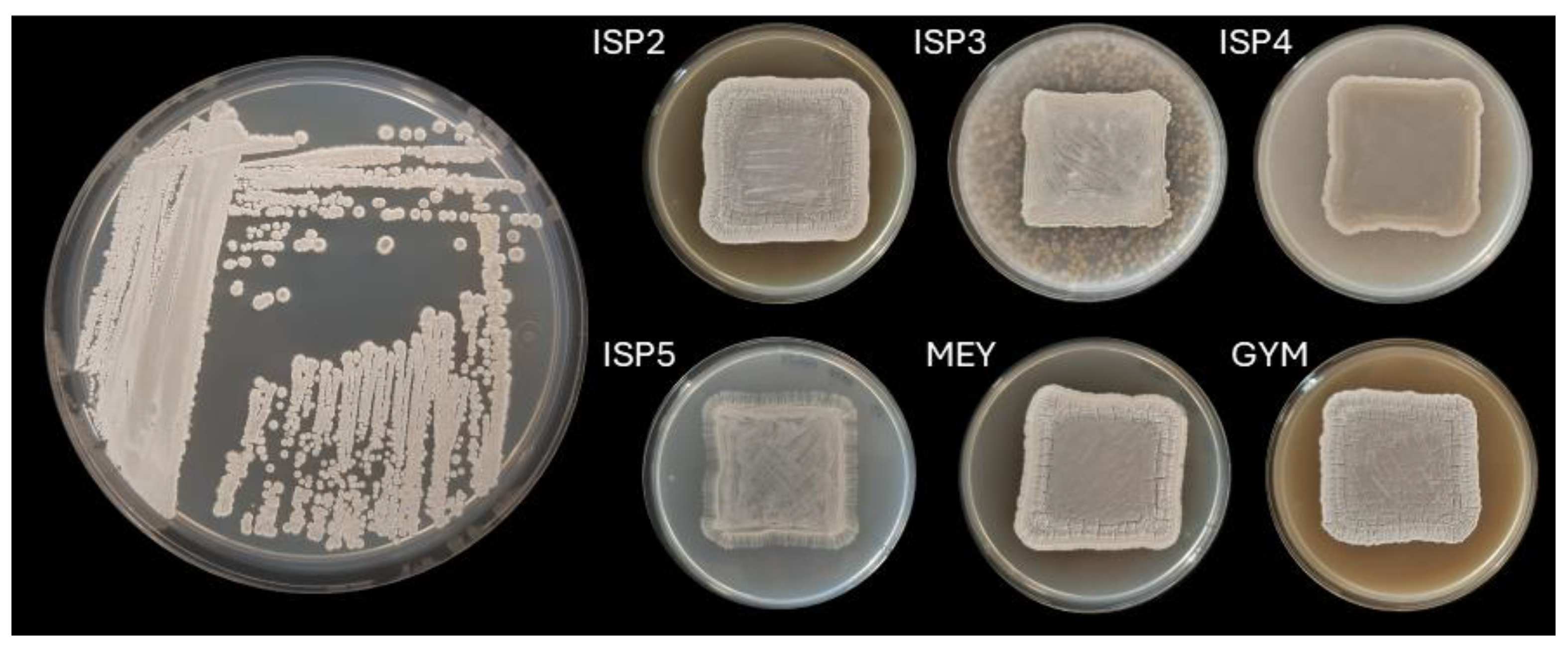
Figure 2.
Whole-genome phylogenomic tree inferred using TYGS based on Genome BLAST Distance Phylogeny (GBDP) distances. Numbers at nodes indicate pseudo-bootstrap support values.
Figure 2.
Whole-genome phylogenomic tree inferred using TYGS based on Genome BLAST Distance Phylogeny (GBDP) distances. Numbers at nodes indicate pseudo-bootstrap support values.
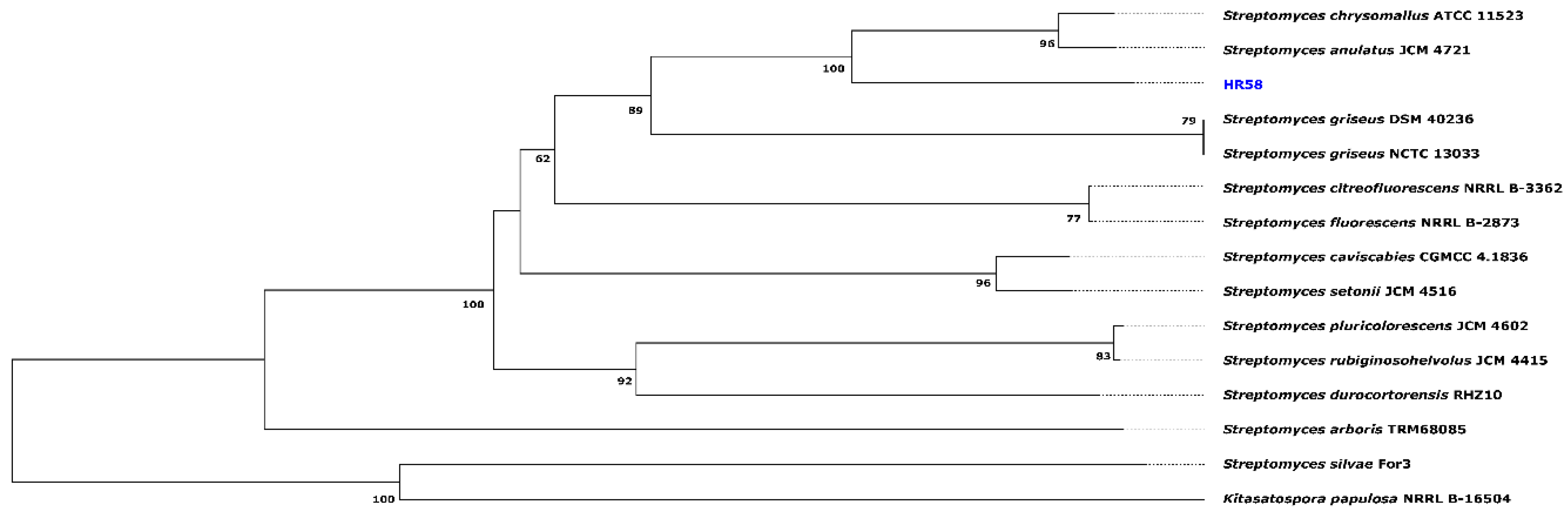
Figure 3.
Antifungal activity of Streptomyces sp. HR58 against different fungal phytopathogens. (a) Fungal growth in presence/absence of Streptomyces sp. HR58. (b) Quantification of AF activity based on calculation of the Inhibition index.
Figure 3.
Antifungal activity of Streptomyces sp. HR58 against different fungal phytopathogens. (a) Fungal growth in presence/absence of Streptomyces sp. HR58. (b) Quantification of AF activity based on calculation of the Inhibition index.
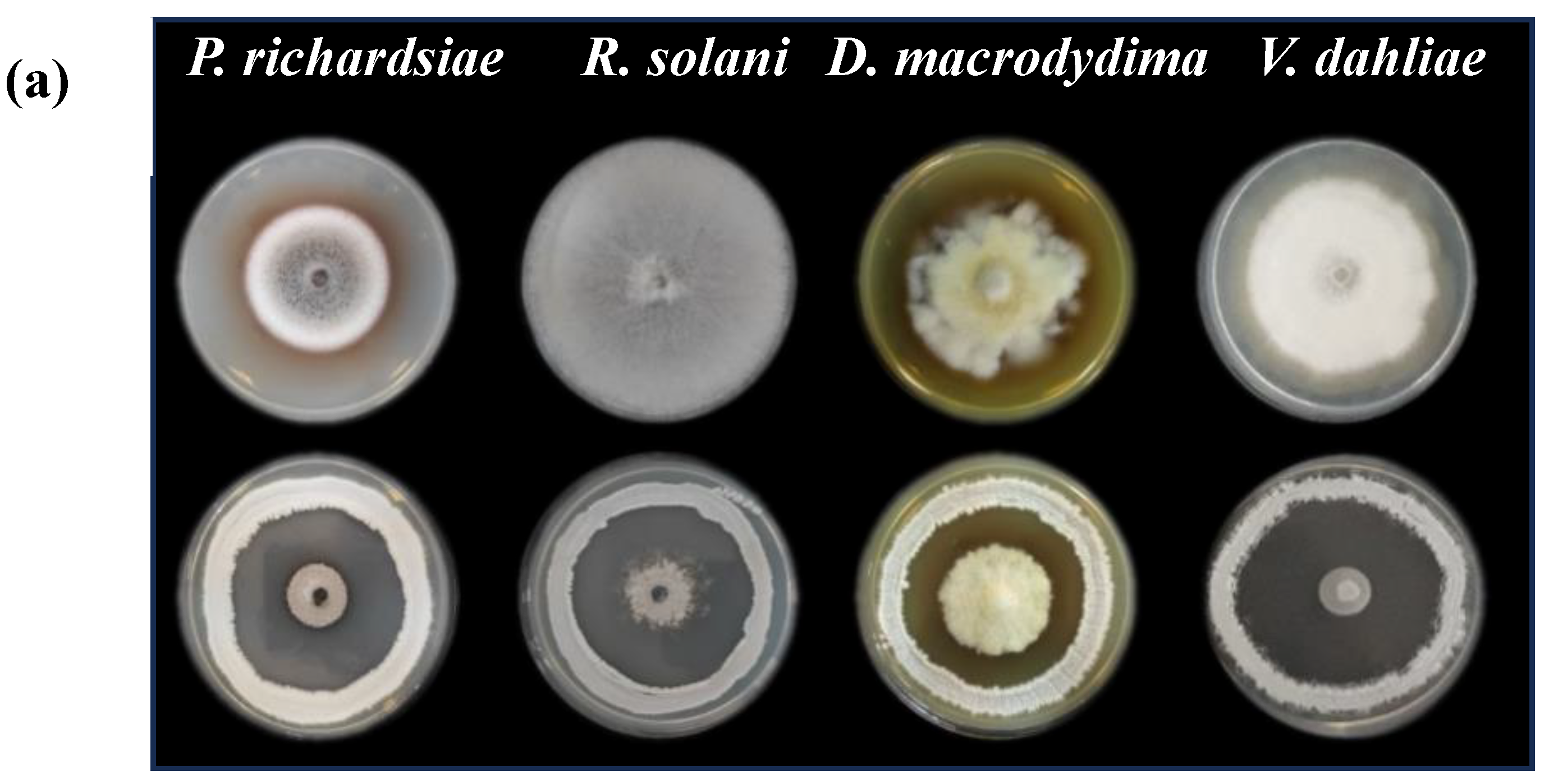
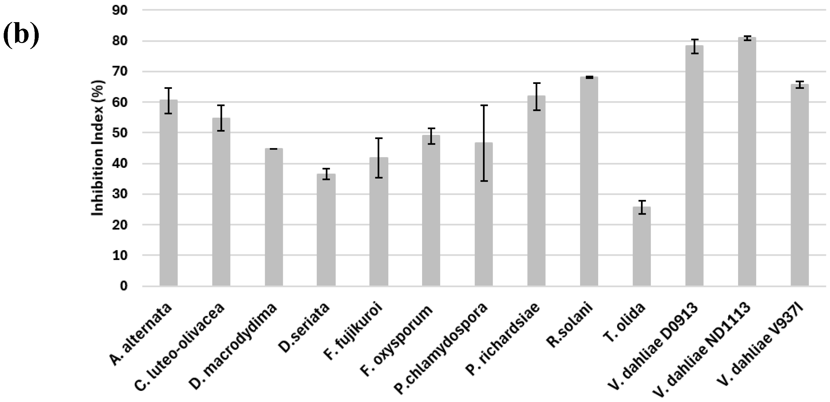
Figure 4.
(a) HPLC chromatograms of a 10 mg/ml cycloheximide standard (green trace) and HR58 extract (blue trace). (b) UV spectrum of cycloheximide.
Figure 4.
(a) HPLC chromatograms of a 10 mg/ml cycloheximide standard (green trace) and HR58 extract (blue trace). (b) UV spectrum of cycloheximide.
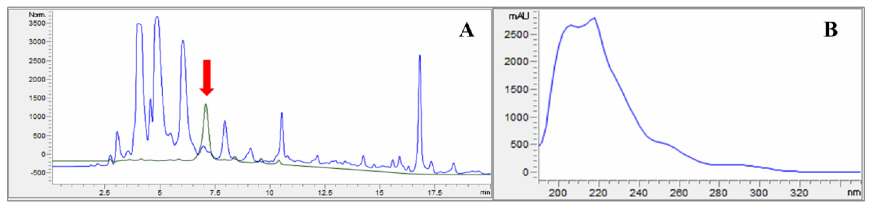
Figure 5.
(a) Antifungal activity against Verticillium dahliae of a 10 mg/ml commercial cycloheximide solution (600 µg) (b) a HPLC fraction collected at 6.93-min corresponding to a culture supernatant of a liquid culture of HR58 strain; and (c) same fraction collected and concentrated.
Figure 5.
(a) Antifungal activity against Verticillium dahliae of a 10 mg/ml commercial cycloheximide solution (600 µg) (b) a HPLC fraction collected at 6.93-min corresponding to a culture supernatant of a liquid culture of HR58 strain; and (c) same fraction collected and concentrated.
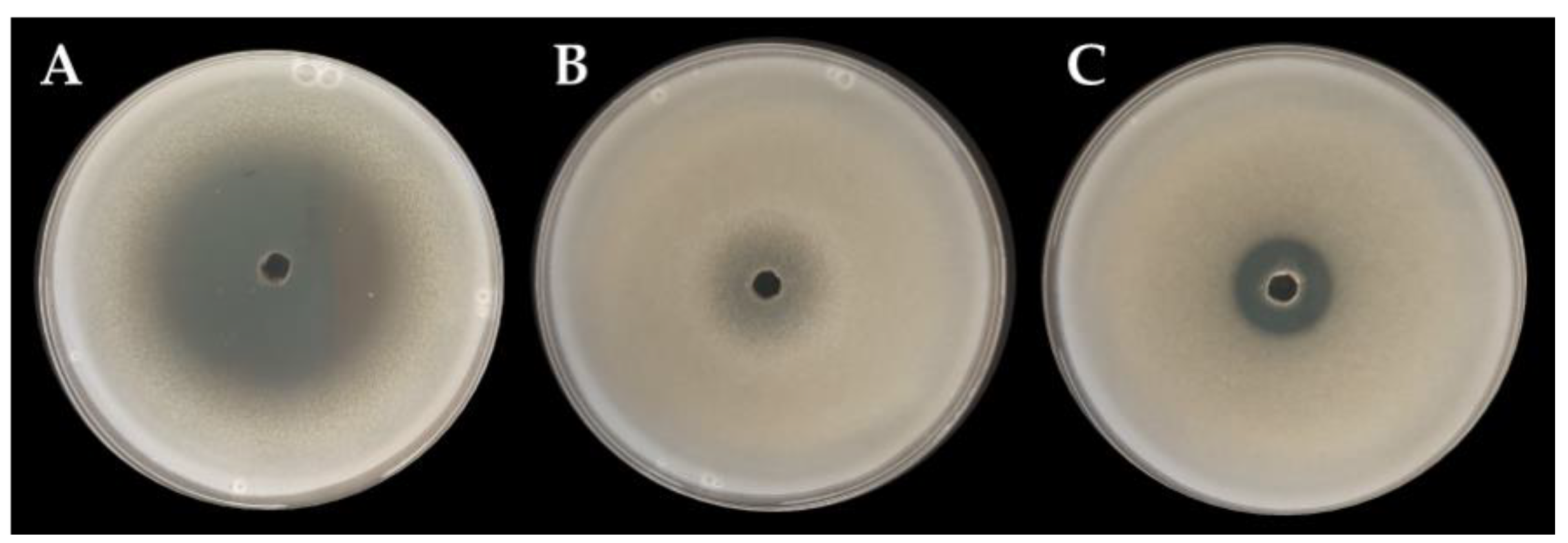
Figure 6.
UV–visible spectroscopy of biogenic AgNPs formed from the extracellular extracts of the Streptomyces sp. HR58 strain.
Figure 6.
UV–visible spectroscopy of biogenic AgNPs formed from the extracellular extracts of the Streptomyces sp. HR58 strain.
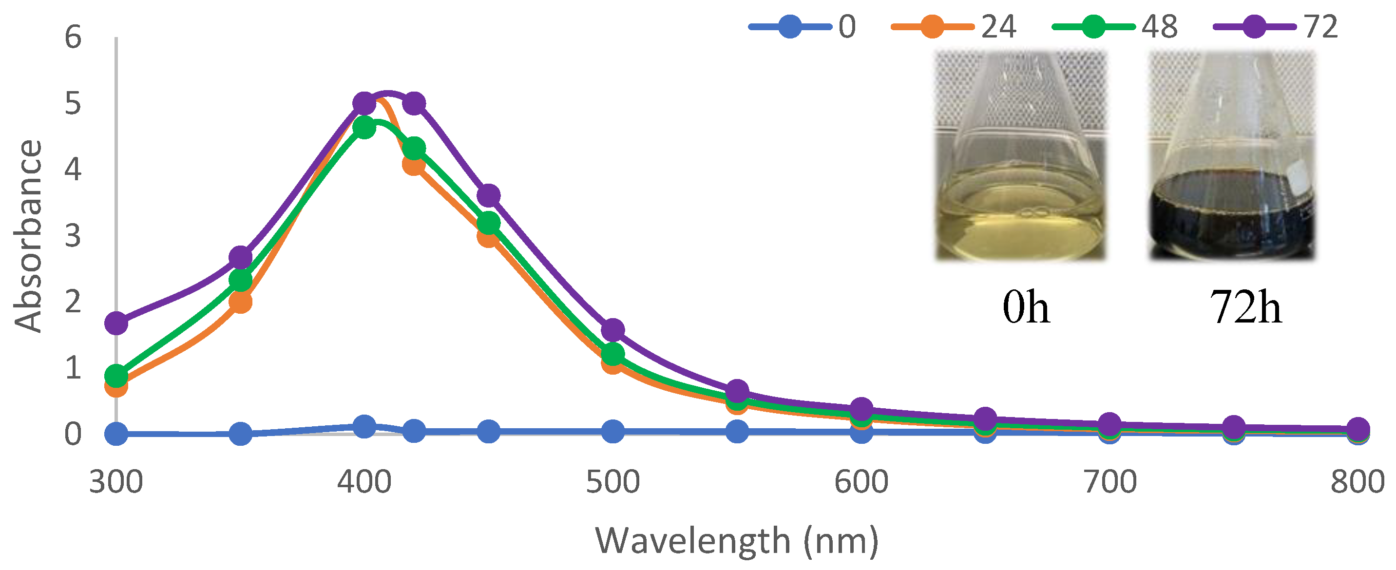
Figure 7.
Transmission electron microscope imagen of biogenic AgNPs formed from the extracellular extracts of the Streptomyces sp. HR58 strain.
Figure 7.
Transmission electron microscope imagen of biogenic AgNPs formed from the extracellular extracts of the Streptomyces sp. HR58 strain.
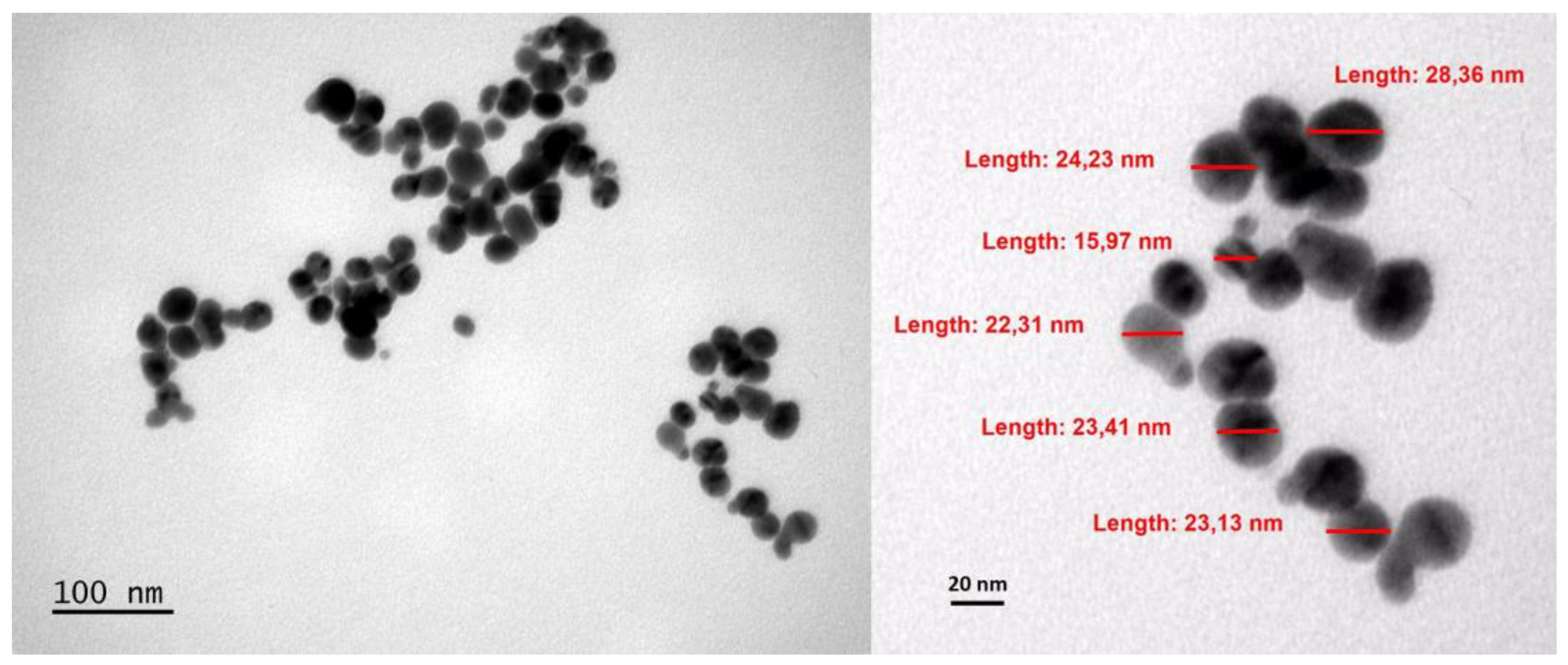
Figure 8.
Antifungal activity of Streptomyces sp. HR58 AgNPs against V. dahliae.
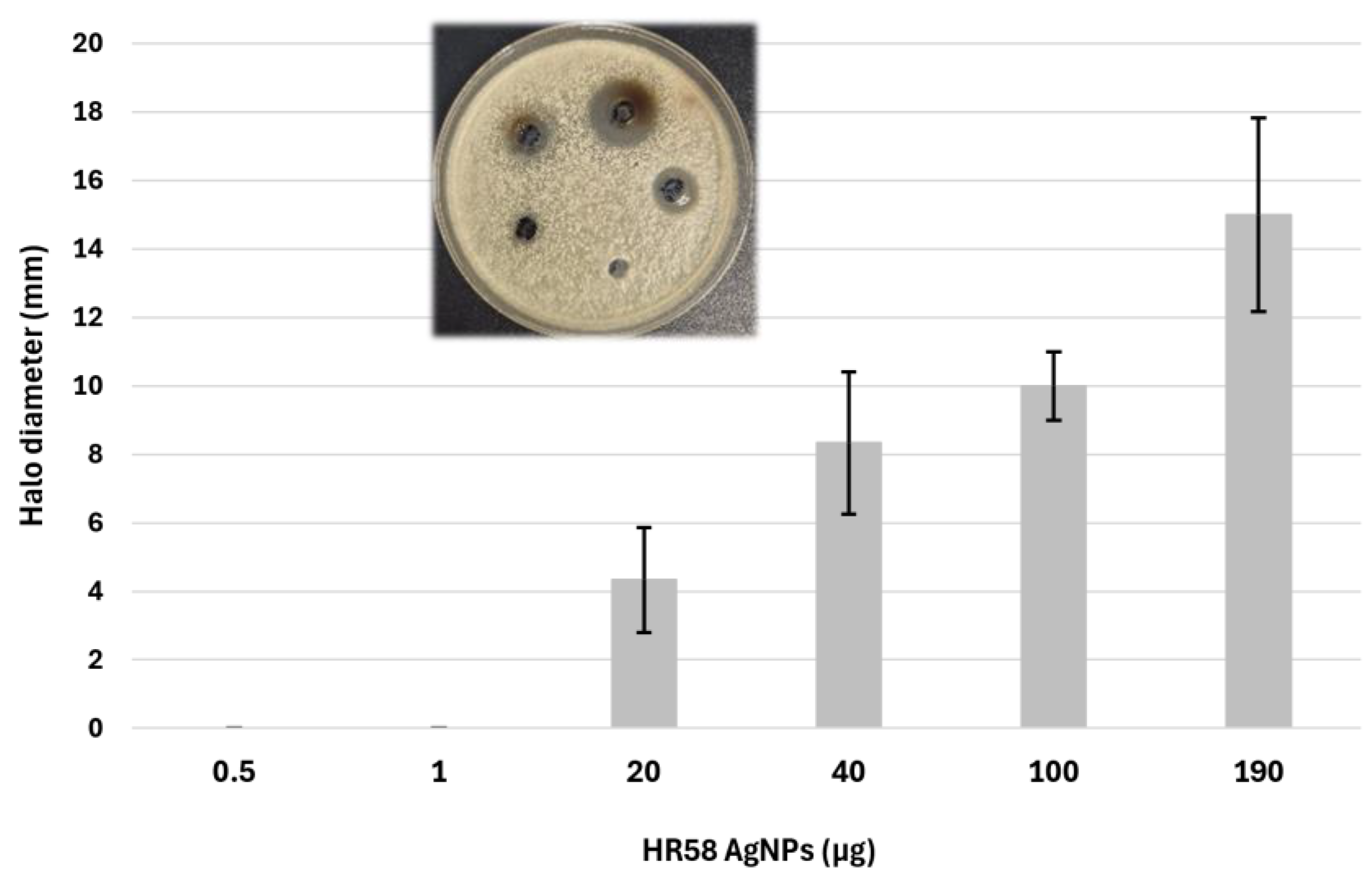

Table 1.
Genome features of Streptomyces sp. HR58.
| Feature | Value |
|---|---|
| Number of contigs | 1 |
| Genome size (bp) | 8,270,372 |
| GC content (%) | 71.76 |
| Protein-coding sequences (CDS) | 7,299 |
| RNA genes | 84 |
| Functional subsystems (RAST) | 306 |
| Biosynthetic gene clusters (BGCs) | 35 |
Table 2.
Distribution of biosynthetic gene clusters in Streptomyces sp. HR58 according to similarity with known clusters.
Table 2.
Distribution of biosynthetic gene clusters in Streptomyces sp. HR58 according to similarity with known clusters.
| BGC similarity to known clusters | Number of BGCs |
|---|---|
| 100% similarity | 10 |
| 75–99% similarity | 4 |
| 30–70% similarity | 3 |
| 1–29% similarity | 14 |
| No detectable similarity (cryptic) | 4 |
| Total | 35 |
Table 3.
Enzymes associated with fungal cell wall degradation in Streptomyces sp. HR58.
| Enzyme group | Number of genes | Relevant features |
|---|---|---|
| Chitinases | 8 | 7 predicted secreted (SignalP-6.0) |
| Chitin-binding proteins | 4 | Putative CBPs involved in substrate recognition |
| β-N-acetylglucosaminidases | 2 | Terminal enzymes of chitin degradation |
| Endo-1,4-β-glucanases | 4 | Putative glucan-degrading enzymes |
| Proteases | 62 | 23 predicted secreted; 38 >1000 bp |
Table 4.
Antibiotic activity of Streptomyces sp. HR58 AgNPs (190µg).
| Microorganisms | Type | Halo (mm) |
|---|---|---|
| Bacillus subtilis | Gram + | 6.85±1.62 |
| Enterococcus faecalis | Gram + | 8.75±1.06 |
| Escherichia coli | Gram - | 0 |
| Salmonella typhimurium | Gram - | 7.75±2.47 |
| Candida utilis | 0 | |
| Saccharomyces cerevisiae | 0 | |
| Alternaria alternata | 10.81±1.42 | |
| Fusarium fujikuroii | 2.62±0.38 | |
| Fusarium oxysporum | 3.4±0.73 | |
| Verticillium dahliae | 14±3.22 |
Disclaimer/Publisher’s Note: The statements, opinions and data contained in all publications are solely those of the individual author(s) and contributor(s) and not of MDPI and/or the editor(s). MDPI and/or the editor(s) disclaim responsibility for any injury to people or property resulting from any ideas, methods, instructions or products referred to in the content. |
© 2026 by the authors. Licensee MDPI, Basel, Switzerland. This article is an open access article distributed under the terms and conditions of the Creative Commons Attribution (CC BY) license (http://creativecommons.org/licenses/by/4.0/).
Copyright: This open access article is published under a Creative Commons CC BY 4.0 license, which permit the free download, distribution, and reuse, provided that the author and preprint are cited in any reuse.