

Submitted:
05 August 2026
Posted:
06 August 2026
You are already at the latest version
Abstract
Leishmaniasis continues to be a major neglected tropical disease, particularly in endemic areas where reliable diagnosis remains challenging. Many existing diagnostic methods are invasive, lack sufficient sensitivity, or require equipment and expertise that may not be available in resource-limited settings. Parasite-derived spliced leader (SL) RNA has recently gained attention as a promising molecular marker because it may enable the sensitive detection of viable Leishmania parasites and help track changes in infection over time. Extracellular vesicles (EVs) are also of growing interest because they protect and transport stable biomolecules that may provide valuable diagnostic information and influence interactions between the parasite and its host. In this study, we evaluated the feasibility of detecting Leishmania-derived SL-RNA within EV-associated samples. EVs were isolated from parasite-conditioned media (PCM) and infected THP-1 macrophage (I-MP) cell-conditioned medium (CCM), followed by nanoparticle tracking analysis for EV characterization. RNA was extracted from EV preparations, reverse transcribed into cDNA and analyzed for SL-RNA amplification using quantitative polymerase chain reaction (qPCR) and droplet digital PCR (ddPCR) approaches. Successful amplification of SL-RNA was detected in PCM and I-MP CCM EV samples, whereas uninfected controls showed no specific amplification. ddPCR analysis demonstrated sensitive detection across infected samples which supports the potential utility of this approach for low-abundance target detection. These findings support the feasibility of EV-associated SL-RNA as a molecular biomarker for Leishmania infection.
Keywords:
visceral leishmaniasis
; extracellular vesicles
; spliced leader RNA
; biomarker diagnostics
; EV‐based diagnosis
1. Introduction
Leishmaniasis is a neglected tropical disease caused by protozoan parasites of the genus Leishmania and transmitted through the bite of infected female phlebotomine sand flies [1,2,3]. The disease remains a major global health burden, especially in tropical and subtropical regions where access to healthcare is often limited. The disease disproportionately affects vulnerable and underserved populations living in low-resource settings [4,5,6,7]. It is estimated that hundreds of thousands of new visceral leishmaniasis (VL) and cutaneous leishmaniasis (CL) cases occur annually, with disease endemicity reported across regions of Asia, Africa, South America, and the Mediterranean basin [8,9,10].
Despite advances in disease surveillance and treatment strategies, accurate diagnosis of leishmaniasis remains a major clinical challenge [11]. Conventional parasitological methods, including microscopy, tissue biopsy, and splenic or bone marrow aspiration, remain important diagnostic approaches but are invasive, require skilled personnel, and often show reduced sensitivity in low-parasite or chronic infections [12,13,14]. Serological assays such as recombinant K39 antigen-based rapid diagnostic test (rK39 RDT), indirect fluorescent antibody test (IFAT), enzyme-linked immunosorbent assay (ELISA), and direct agglutination test (DAT) provide less invasive alternatives for VL diagnosis. However, their sensitivity and specificity can vary depending on geographic region, infecting species, and host immune status [15,16,17]. Molecular methods such as polymerase chain reaction (PCR) and quantitative PCR (qPCR) have significantly improved analytical sensitivity and specificity for Leishmania detection [11]. However, DNA persistence following parasite death may limit their utility for monitoring active infection or treatment response [18].
Recently, RNA-based molecular targets have emerged as promising tools for detecting viable Leishmania parasites, with spliced leader RNA (SL-RNA) the gaining attention due to its conserved nature, high abundance, and association with metabolically active organisms [19]. SL-RNA, also known as mini-exon RNA, is a short non-coding RNA that plays an important role in the maturation of parasite mRNAs [20,21]. In Leishmania, genes are transcribed as long polycistronic transcripts, and mature mRNAs are generated through trans-splicing, during which the capped spliced leader sequence is added to the 5′ end of each mRNA [20,21]. The mini-exon/spliced leader region is highly conserved across Leishmania species but remains distinct enough from related kinetoplastid parasites to be useful as a pan-Leishmania detection target [22,23]. Because SL-RNA is abundant and linked to active parasite RNA expression, SL-RNA-based detection may offer a more sensitive and biologically relevant approach than DNA-based methods, especially for identifying active infection and monitoring treatment response [19]. However, the application of SL-RNA detection within extracellular vesicle-associated samples remains poorly explored.
Extracellular vesicles (EVs) are lipid bilayer membrane-delimited particles released by nearly all cell types and play central roles in intercellular communication through the transfer of proteins, lipids, metabolites, and nucleic acids [24,25]. EVs include several subclasses, such as exosomes and microvesicles, which differ in their biogenesis, size, and molecular composition [24,26]. EVs play an important role in host-pathogen communication during infectious diseases by transporting virulence factors, immunomodulatory molecules, and regulatory RNAs that can alter immune responses and support pathogen survival [27,28,29,30]. In leishmaniasis, Leishmania-derived EVs have been shown to modulate macrophage signaling, cytokine production, and immune evasion pathways, while recent studies also highlight their potential as stable, minimally invasive biomarkers for diagnostic applications in visceral leishmaniasis [31,32,33,34,35,36].
Although previous studies have identified parasite-derived proteins and nucleic acids in Leishmania EVs, limited information is available regarding the detection of EV-associated parasite RNA biomarkers in infected host-derived samples, and standardized workflows for clinical diagnostic applications are still lacking [11,31,37]. In this study, we evaluated the feasibility of detecting Leishmania-derived SL-RNA in EV-associated samples using qPCR and droplet digital PCR, supporting the potential of EV-derived parasite RNA as a minimally invasive biomarker for leishmaniasis diagnosis.
2. Materials and Methods
Parasite and Cell Culture
L. tropica (MHOM/AF/87/RUPERT), L. infantum (MHOM/TR/3/Adana7), transgenic L. donovani (MHOM/SD/62/1S-CL2D) constitutively expressing mCherry, and transgenic L. major (MHOM/JL/80/Friedlin) constitutively expressing mCherry [38] were maintained as promastigotes at 27 °C in M199 (MP Biomedicals) under standard culture conditions following previously published protocols [39,40]. Axenic amastigotes were generated from L. donovani log-stage promastigotes in a modified RPMI-1640 medium which was pH adjusted to 5.5 as previously described [39].
Trypanosoma cruzi Dm28c epimastigotes and Trypanosoma brucei brucei 427 29:13 WT procyclic forms were cultured under standard conditions in LIT medium supplemented with fetal bovine serum (FBS) and in SDM-79 medium supplemented with FBS and GlutaMAX, respectively [41].
Human monocytic THP-1 cells (ATCC TIB-202) were cultured at 37 °C in a humidified 5% CO₂ atmosphere using RPMI-1640 medium (Corning) supplemented with 10% FBS and 1% penicillin–streptomycin (Corning). THP-1 monocytes were differentiated into macrophage-like cells by treatment with 150 nM phorbol 12-myristate 13-acetate (PMA) for 24 h prior to infection experiments.
THP-1 Cell Line Infection
Metacyclic promastigotes were isolated from stationary-phase parasite cultures using a Ficoll-400 density gradient as previously described [42]. Differentiated THP-1 macrophages were infected with metacyclic parasites at a multiplicity of infection (MOI) of 10:1 parasite-to-macrophage ratio for 6 h. Following infection, macrophages were washed five times with phosphate-buffered saline (PBS) to remove extracellular parasites and incubated with fresh complete RPMI-1640 medium. Infected cultures were then maintained for an additional 72 h. Cytospin slides were prepared following the 6 h incubation from each infection experiment and stained using a three-step Hematology Quick Stain (VWR) for visual analysis by light microscopy. Infected macrophages (100 total) were counted to calculate the infection rate (% infected) and the parasite indices (# parasites per 100 cells) for each infection sample. Culture supernatants were collected from other wells for downstream EV isolation and molecular analyses, consistent with previously established protocols [43].
Mice Infection Model
6–8-week-old female wild type C57Bl/6 mice were bred and housed in accordance with IACUC protocols (#24-02-8390) in Friemann Life Science Center at the University of Notre Dame. For infections, 1x10⁵ L. major metacyclic promastigotes in 20 µL of PBS were injected intradermally into the outside surface of the ear as previous described [44] and serum was collected 6-8 weeks post infection and stored at -20° until use.
Isolation of EVs by Precipitation Method
EVs were isolated from parasite-conditioned medium (PCM), infected (I-MP) and uninfected (U-MP) THP-1 macrophages cell-conditioned medium (CCM), and serum from experimentally infected mice. Samples were first pre-cleared by centrifugation at 2,000 × g for 30 min to remove cells and cellular debris. EVs were then isolated using a polymer-based precipitation approach with the Total Exosome Isolation kit for cell culture media or the Total Exosome Isolation kit for serum, according to the manufacturer’s instructions (Invitrogen). Isolated EV pellets were subsequently processed for downstream characterization and molecular analyses.
Characterization of EVs by Nanoparticle Tracking Analysis
To obtain sufficient volume for EV characterization, a portion of each EV-enriched preparation was reserved and EV preparations from three independent collections were pooled for each sample prior to nanoparticle tracking analysis (NTA). Pooling of EV preparations for downstream characterization has been described in previous EV studies when sample yield is limiting [45]. The pooled EV-enriched samples were diluted 10- to 100-fold and analyzed by NTA using a NanoSight LM10 equipped with an sCMOS camera and a 532-nm laser. The camera level was set to 12, the screen gain was set to 10, and the focus was adjusted manually. For each sample, 500 µL was manually injected into the laser chamber, and five technical replicate videos were recorded for 45 s each. The laser chamber was washed with distilled water between samples. Particle size distribution and concentration were analyzed using NanoSight NTA software version 3.1.
EV-RNA Extraction and cDNA Synthesis
Total RNA was extracted from isolated EV pellets using the Monarch Total RNA Miniprep Kit (New England Biolabs) according to the manufacturer’s instructions. RNA was eluted in nuclease-free water and stored at -80 °C for downstream analysis. RNA concentration and purity were assessed using a NanoDrop spectrophotometer.
Complementary DNA (cDNA) was synthesized from purified RNA using SuperScript III Reverse Transcriptase (Invitrogen) following the manufacturer’s protocol. The resulting cDNA was either used immediately for downstream PCR analyses or stored at -20 °C for future characterization.
Polymerase Chain Reaction (PCR) and Quantitative PCR (qPCR)
Detection of Leishmania SL-RNA was performed using the previously published pan-Leishmania SL-RNA primer set [23]. Conventional PCR was performed using GoTaq G2 Green Master Mix (Promega) according to the manufacturer’s recommendations. Amplified products were analyzed by agarose gel electrophoresis prior to sequencing to confirm product size. qPCR was carried out using Luna Universal qPCR Master Mix (New England Biolabs). Reactions were performed under the following cycling conditions: 50 °C for 2 min, followed by an initial denaturation at 95 °C for 2 min, and 40 cycles consisting of 95 °C for 20 s, 45.7 °C for 30 s, and 68 °C for 30 s. Melt curve analysis was subsequently performed using the following dissociation program: 95 °C for 15 s, 47 °C for 15 s, and 95 °C for 15 s. No-template controls (NTCs) and samples derived from uninfected cells or animals were included as negative controls in each experiment to assess contamination and assay specificity.
Droplet Digital PCR (ddPCR)
Absolute quantification of Leishmania SL-RNA was performed using the QX200 Droplet Digital PCR (ddPCR) System (Bio-Rad Laboratories). ddPCR reactions were prepared using QX200 ddPCR EvaGreen Supermix (Bio-Rad) according to the manufacturer’s instructions. The reaction mix was partitioned into droplets using the QX200 Droplet Generator (Bio-Rad). Thermal cycling was performed under the following conditions: an initial enzyme activation step at 95 °C for 3 min, followed by 40 cycles of 95 °C for 30 s, 49.3 °C for 30 s, and 72 °C for 1 min. Following amplification, reactions were incubated at 4 °C for 5 min and 90 °C for 5 min before being held at 4 °C until analysis. Amplified droplets were analyzed using the QX200 Droplet Reader (Bio-Rad), and data were processed using QuantaSoft Analysis Pro Software (Bio-Rad). Positive and negative droplet populations were identified by applying a fluorescence threshold based on NTCs and negative control samples included in each run.
Agarose Gel Electrophoresis and Sequencing Validation
PCR amplicons generated using the Leishmania SL-RNA primer set were analyzed by agarose gel electrophoresis to confirm amplification of products of the expected size. To verify amplicon identity, selected PCR products were cloned into the pGEM-T Vector System (Promega) according to the manufacturer’s instructions. Recombinant plasmids were transformed into competent bacterial cells, and putative positive colonies were screened by colony PCR using both SL-RNA primers and M13 forward and reverse primers. Plasmid DNA was extracted from positive colonies, quantified using a NanoDrop spectrophotometer, and re-amplified by PCR to confirm the presence of the target insert. A total of six confirmed clones were submitted for Sanger sequencing using M13 forward and reverse primers at the Notre Dame Genomics & Bioinformatics Core Facility, University of Notre Dame, Notre Dame, IN, USA (RRID: SCR_025555). Sequence identity was verified by comparison with published Leishmania spliced leader RNA sequences.
3. Results
3.1. Characterization of Extracellular Vesicles and RNA Extraction
Pooled EV-enriched preparations obtained from PCM, I- and U-MP CCM, and mouse serum were characterized by NTA to determine particle size and concentration (Table 1). EV-enriched preparations had mean particle diameters below 200nm, consistent with the size range associated with small EVs. Corresponding NTA particle-size distribution plots are provided in Supplementary Figure S1.
3.2. Standard Curve Assessment of the SL-RNA qPCR Assay
Total RNA was recovered from all EV-enriched preparations and used for cDNA synthesis. RNA concentrations are reported in Supplementary Table S2. The analytical performance of the SL-RNA qPCR assay was evaluated before its application to biological samples. A standard curve was generated using seven four-fold serial dilutions of Leishmania donovani cDNA, covering concentrations from approximately 0.20 to 800 ng/µL. Mean Ct values decreased progressively with increasing cDNA concentration, from approximately 40.0 at the lowest input concentration to 27.2 at the highest concentration (Figure 1). Linear regression of mean Ct against log₁₀-transformed cDNA concentration produced the equation y = 38.41 − 3.838x, with a coefficient of determination of R² = 0.9777. The regression slope corresponded to an amplification efficiency of 82.2%. Overall, the standard curve demonstrated a consistent concentration-dependent response across the approximately 3.6-log₁₀ input range. A formal limit of detection was not determined.
3.3. Detection of EV-Associated Leishmania SL-RNA Across Experimental Models
3.3.1. SL-RNA Detection in EV-Enriched Preparations from Four Leishmania Species and Axenic Amastigotes
EV-enriched preparations obtained from PCM were analyzed by RT-qPCR. SL-RNA was detected in EV-associated RNA isolated from all four tested Leishmania species, including L. donovani (Ld), L. major (Lm), L. infantum (Li), and L. tropica (Lt). For each promastigote-derived EV preparation, three independent biological collections were analyzed, with each biological sample evaluated in three technical qPCR replicates. Technical replicates were averaged prior to analysis.
EV-associated SL-RNA was consistently detected in all biological collections analyzed. The mean Ct values were 23.07 ± 5.28 for Ld, 16.05 ± 0.50 for Lm, 17.46 ± 1.94 for Li, 22.43 ± 10.46 for Lt (Figure 2A). In addition, EV-enriched preparations generated from L. donovani axenic amastigotes (LdAA) demonstrated detectable SL-RNA amplification (Ct = 20.12, n=1), indicating that SL-RNA detection was maintained in an alternative parasite developmental stage. The corresponding qPCR amplification plots are presented in Supplementary Figure S2.
Specific amplification was confirmed by melt-curve analysis, with positive reactions producing a single melt peak corresponding to the validated SL-RNA amplicon. In contrast, EV-enriched preparations derived from fresh M199 culture media and NTCs showed no detectable amplification (Ct = 40/ND).
3.3.2. Detection of SL-RNA in EVs from Leishmania-Infected Macrophages
Differentiated THP-1 macrophages were infected with L. infantum metacyclic promastigotes, and EV-enriched preparations from I-MP and U-MP CCM were analyzed by RT-qPCR. Parasite indices were confirmed using light microscopy at the time of CCM collection. Parasite indices for the three independent biological collections were 282, 193 and 178, respectively. SL-RNA was detected in EV-enriched preparations from all three I-MP collections. Each biological collection was analyzed in three technical replicates, yielding mean Ct values of 17.29 ± 0.03, 27.52 ± 0.60, and 30.41 ± 0.93 for collections 1, 2, and 3, respectively (Figure 2B). Amplification was undetermined in EV-enriched preparations from U-MP. Representative amplification plots are provided in Supplementary Figure S4.
3.3.3. Detection of SL-RNA in Serum EVs from Leishmania-Infected Mice
EV-enriched preparations isolated from the serum of L. major-infected B6 mice were analyzed by qPCR. Target-specific amplification was detected in all eight infected mice (Figure 3). Mean Ct values, calculated from the three technical replicates for each mouse ranged from 28.82 to 33.36, with a median of 31.19. In contrast, amplification was undetermined in all three technical replicates of the uninfected serum EV preparation and NTC. Amplification plots are presented in Supplementary Figure S3.
3.4. Validation of SL-RNA Detection by ddPCR
To further confirm the presence of SL-RNA detected by qPCR, RNA derived from I-MP was analyzed by ddPCR. A PCM EV-enriched sample was included as a positive control, while RNA from U-MP and a NTC served as negative controls. Positive droplets corresponding to SL-RNA were detected in both the I-MP and PCM. In contrast, no positive droplets were observed in the U-MP sample (Figure 4).
3.5. Sequence Confirmation of the Amplified SL-RNA
The identity of the amplified SL target was confirmed by Sanger sequencing. All six colony PCR-positive clones contained the expected 39-nt Leishmania SL sequence, with representative bidirectional reads showing 100% identity to the reference sequence (Supplementary Figure S5).
3.6. Specificity of the Leishmania SL Assay
The specificity of the SL RT-qPCR assay was assessed using parasite-derived RNA from L. major PCM and the closely related non-target trypanosomatids, T. brucei brucei (Tbb) and T. cruzi (Tc). Two independent biological samples from each non-target species were analyzed, with three technical replicates per sample. The L. major PCM sample produced strong and consistent amplification in all three replicates, with a mean Ct of 16.62 ± 0.23 (Figure 5). In contrast, the replicates of Tbb and Tc produced Ct values more than 22 cycles later than the mean L. major signal. RT-qPCR amplification plots are presented in Supplementary Figure S4.
4. Discussion
This study tested the hypothesis that Leishmania-derived SL-RNA is detectable in EV-enriched preparations from PCM, I-MP, and infected mice models. Detection across four Leishmania species and life stages, infected macrophages, and infected mouse serum supports this hypothesis. The absence of target-specific amplification in the corresponding controls, together with confirmation by ddPCR and Sanger sequencing, provides proof-of-concept evidence for investigating EV-associated SL-RNA as a minimally invasive molecular biomarker.
Previous studies have identified SL-RNA as a sensitive pan-Leishmania marker in clinical samples, experimentally infected animals, vectors, and reservoir hosts [19,23]. Its high abundance and association with active parasite transcription may offer an advantage over DNA targets that can persist after parasite death. Our findings extend this work by demonstrating that SL-RNA recovery from EV-enriched fractions rather than only from total blood, tissue, or cellular nucleic acids. Detection in the axenic amastigotes is also relevant to mammalian infection and has been validated by intracellular amastigotes. Differences in Ct values among species should not be interpreted quantitatively because parasite input, particle recovery, and RNA yield were not standardized for cross-species comparisons.
SL-RNA was detected in all I-MP preparations and all eight infected-mouse serum samples, but not in their respective controls. These findings indicate extracellular release during intracellular infection. This is consistent with evidence that EVs carry parasite- and host-derived molecules with diagnostic potential [31,32,33,34,35,36,37]. An EV-based blood assay could eventually reduce reliance on invasive tissue sampling. However, the present study demonstrates detectability rather than clinical accuracy.
The RT-qPCR assay demonstrated a concentration-dependent response with an efficiency of 82.2%. Quantification limits were not established therefore Ct values support target detection but not comparisons of SL-RNA abundance across sample types. ddPCR provided independent confirmation, although its advantage over qPCR requires further evaluation. The negative results for Tbb and Tc were consistent with the specificity reported for the pan-Leishmania SL-RNA assay [23]. However, occasional amplification indicates that a broader specificity panel and predefined positivity threshold are still needed.
NTA supported the presence of extracellular particles, but the preparations may also contain non-vesicular particles or RNA-protein complexes. Therefore, “EV-enriched preparations” remains the appropriate term [24]. Additional EV characterization and RNase treatment with and without membrane disruption would help determine the location of the detected SL-RNA.
The main limitations of this study were the low and variable RNA yield, incomplete EV characterization, and absence of human samples. The qPCR assay lacked formal detection limits and was tested against only two non-target species. The clinical phase will evaluate EV-enriched SL-RNA in patients with confirmed leishmaniasis and appropriate control groups, compare its performance with established diagnostic methods and assess changes during treatment using longitudinal samples.
5. Conclusions
This study demonstrates that Leishmania SL-RNA can be detected in EV-enriched preparations across multiple experimental models. Confirmation by ddPCR and sequencing supports its potential as a minimally invasive molecular marker, although its vesicular localization and clinical performance remain to be established. The planned evaluation of patient samples will determine whether this approach can support leishmaniasis detection and treatment monitoring.
Supplementary Materials
The following supporting information can be downloaded at the website of this paper posted on Preprints.org, Figure S1: title; Table S1: title; Video S1: title.
Author Contributions
Conceptualization, M.S.S.O., D.A.S., and M.A.M.; Data curation, M.S.S.O., J.P.G., and D.J.M-L.; Formal analysis, M.S.S.O.; Funding acquisition, C.K., and M.A.M.; Investigation, M.S.S.O., J.P.G., and D.J.M-L.; Methodology, M.S.S.O., J.P.G., and D.J.M-L.; Project administration, M.S.S.O., D.A.S., and M.A.M.; Resources, C.K., and M.A.M.; Supervision, D.A.S., C.K., and M.A.M.; Visualization, M.S.S.O.; Writing- original draft, M.S.S.O., and M.A.M.; Writing- reviewing and editing, M.S.S.O., J.P.G., D.J.M-L, D.A.S, C.K., and M.A.M. All authors have read and agreed to the published version of the manuscript.
Funding
This research received no external funding.
Institutional Review Board Statement
Not applicable.
Informed Consent Statement
Not applicable.
Data Availability Statement
The raw data supporting the conclusions of this manuscript will be made available by the authors on request.
Acknowledgments
The authors acknowledge the support of the University of Notre Dame Biological Sciences core facility and Jeffrey S. Schorey Laboratory for access to NTA equipment and Alvaro Acosta Serrano Laboratory for providing the Trypanosoma brucei cell line. Open AI was used for sentence revision.
Conflicts of Interest
The authors declare no conflicts of interest.
Abbreviations
The following abbreviations are used in this manuscript:
| SL-RNA | Spliced Leader RNA |
| EV | Extracellular Vesicles |
| ddPCR | Digital Droplet PCR |
| NTA | Nanoparticle Tracking Analysis |
| qPCR | Quantitative PCR |
| NTC | No-Template Control |
| CCM | Cell-Conditioned Medium |
| U-MP | Uninfected Macrophages |
| I-MP | Infected Macrophages |
| PCM | Parasite-Conditioned Medium |
References
- CDC Leishmaniasis. Available online: https://www.cdc.gov/dpdx/leishmaniasis/index.html.
- Dostálová, A.; Volf, P. Leishmania development in sand flies: parasite-vector interactions overview. Parasites Vectors 2012, 5, 276. [Google Scholar] [CrossRef] [PubMed]
- Mann, S.; Frasca, K.; Scherrer, S.; Henao-Martínez, A.F.; Newman, S.; Ramanan, P.; Suarez, J.A. A review of leishmaniasis: current knowledge and future directions. Curr. Trop. Med. Rep. 2021, 8, 121–132. [Google Scholar] [CrossRef] [PubMed]
- Engels, D.; Zhou, X.-N. Neglected tropical diseases: an effective global response to local poverty-related disease priorities. Infect. Dis. Poverty 2020, 9, 9–17. [Google Scholar] [CrossRef] [PubMed]
- Mitra, A.K.; Mawson, A.R. Neglected tropical diseases: epidemiology and global burden. Trop. Med. Infect. Dis. 2017, 2, 36. [Google Scholar] [CrossRef] [PubMed]
- Zheng, J.-X.; Liu, Y.; Guan, S.-Y.; Guo, Z.-Y.; Duan, L.; Lv, S.; Zhang, S.-X.; Li, S.-Z.; Zhou, X.-N.; Feng, X.-Y. Global, regional, and national burden of neglected tropical diseases and malaria in the general population. J. Adv. Res. 2025. [Google Scholar] [CrossRef] [PubMed]
- Ochola, E.A.; Karanja, D.M.; Elliott, S.J. The impact of Neglected Tropical Diseases (NTDs) on health and wellbeing in sub-Saharan Africa (SSA): A case study of Kenya. PLoS Neglected Trop. Dis. 2021, 15, e0009131. [Google Scholar] [CrossRef] [PubMed]
- Alvar, J.; Vélez, I.D.; Bern, C.; Herrero, M.; Desjeux, P.; Cano, J.; Jannin, J.; Boer, M.d.; Team, W.L.C. Leishmaniasis worldwide and global estimates of its incidence. PLoS ONE 2012, 7, e35671. [Google Scholar] [CrossRef] [PubMed]
- Zhang, S.-X.; Yang, G.-B.; Sun, J.-Y.; Li, Y.-J.; Yang, J.; Wang, J.-C.; Deng, Y. Global, regional, and national burden of Visceral leishmaniasis, 1990–2021: findings from the global burden of disease study 2021. Parasites Vectors 2025, 18, 157. [Google Scholar] [CrossRef] [PubMed]
- Wamai, R.G.; Kahn, J.; McGloin, J.; Ziaggi, G. Visceral leishmaniasis: a global overview. J. Glob. Health Sci. 2020, 2. [Google Scholar] [CrossRef]
- Gow, I.; Smith, N.C.; Stark, D.; Ellis, J. Laboratory diagnostics for human Leishmania infections: a polymerase chain reaction-focussed review of detection and identification methods. Parasites Vectors 2022, 15, 412. [Google Scholar] [CrossRef] [PubMed]
- Abass, E.; Mansour, D.; Abdalla, Z.; Altaher, H.; Sliman, M.; Osman, H.; Babiker, R.A.; Salah, M.; Omer, E.; Elamin, E. Diagnostic performance of a novel point-of-care test for the diagnosis of visceral leishmaniasis in Sudan: A Comparative Accuracy Study. PLoS Neglected Trop. Dis. 2025, 19, e0012905. [Google Scholar] [CrossRef] [PubMed]
- de Larminat, J.; Dahane, N.; Argy, N.; Rifai, M.; Ferrua, O.H.; Pomares, C.; Ravonjena, N.R.; Dupont, D.; Lemoine, J.-P.; Dutron, S. Diagnosis and Management of Visceral Leishmaniasis in Children: A French Retrospective Study. In Proceedings of the Open Forum Infectious Diseases, 2025; p. ofaf708. [Google Scholar]
- Ma, F.; Zhang, R.; He, M.; Li, J.; Bai, M.; Liang, G.; Liu, X. Case Report: Exclusive localization of Leishman-Donovan bodies in neutrophils on peripheral blood smear in a patient with systemic lupus erythematosus. Front. Med. 2025, 12, 1693178. [Google Scholar] [CrossRef]
- Cota, G.F.; De Sousa, M.R.; Demarqui, F.N.; Rabello, A. The diagnostic accuracy of serologic and molecular methods for detecting visceral leishmaniasis in HIV infected patients: meta-analysis. PLoS Neglected Trop. Dis. 2012, 6, e1665. [Google Scholar] [CrossRef]
- Getnet, M.; Minaye Dejen, A.; Abebaw, D.; Fentahun, G.G.; Birhanu, E. Diagnostic accuracy of serological rk-39 test for visceral Leishmaniasis: Systematic review and meta-analysis. PLoS Neglected Trop. Dis. 2024, 18, e0011938. [Google Scholar] [CrossRef] [PubMed]
- Mohebali, M.; Keshavarz, H.; Shirmohammad, S.; Akhoundi, B.; Borjian, A.; Hassanpour, G.; Mamishi, S.; Mahmoudi, S. The diagnostic accuracy of direct agglutination test for serodiagnosis of human visceral leishmaniasis: a systematic review with meta-analysis. BMC Infect. Dis. 2020, 20, 946. [Google Scholar] [CrossRef] [PubMed]
- Aleem, M.T.; Shaukat, A.; Hussain, J.; Ali, H.; Zaman, M.; Saeed, Z.; Abbas, R.; Ijaz, A.; Shahid, Z.; Mohsin, M. A review on recent advancement in the molecular diagnostics of Leishmania. J. Hell. Vet. Med. Soc. 2023, 74, 5547–5564. [Google Scholar]
- Hendrickx, R.; Melkamu, R.; Tadesse, D.; Teferi, T.; Feijens, P.-B.; Vleminckx, M.; van Henten, S.; Alves, F.; Shibru, T.; van Griensven, J. Spliced-leader RNA as a dynamic marker for monitoring viable Leishmania parasites during and after treatment. J. Infect. Dis. 2024, 230, 183–187. [Google Scholar] [CrossRef] [PubMed]
- Miller, S.I.; Wirth, D.F. trans splicing in Leishmania enriettii and identification of ribonucleoprotein complexes containing the spliced leader and U2 equivalent RNAs. Mol. Cell. Biol. 1988, 8, 2597–2603. [Google Scholar] [CrossRef]
- Zeiner, G.M.; Sturm, N.R.; Campbell, D.A. The Leishmania tarentolae spliced leader contains determinants for association with polysomes. J. Biol. Chem. 2003, 278, 38269–38275. [Google Scholar] [CrossRef] [PubMed]
- Bitar, M.; Boroni, M.; Macedo, A.M.; Machado, C.R.; Franco, G.R. The spliced leader trans-splicing mechanism in different organisms: molecular details and possible biological roles. Front. Genet. 2013, 4, 199. [Google Scholar] [CrossRef] [PubMed]
- Eberhardt, E.; Van den Kerkhof, M.; Bulté, D.; Mabille, D.; Van Bockstal, L.; Monnerat, S.; Alves, F.; Mbui, J.; Delputte, P.; Cos, P. Evaluation of a pan-Leishmania spliced-leader RNA detection method in human blood and experimentally infected Syrian golden hamsters. J. Mol. Diagn. 2018, 20, 253–263. [Google Scholar] [CrossRef] [PubMed]
- Welsh, J.A.; Goberdhan, D.C.; O’Driscoll, L.; Buzas, E.I.; Blenkiron, C.; Bussolati, B.; Cai, H.; Di Vizio, D.; Driedonks, T.A.; Erdbrügger, U. Minimal information for studies of extracellular vesicles (MISEV2023): From basic to advanced approaches. J. Extracell. Vesicles 2024, 13, e12404. [Google Scholar] [CrossRef] [PubMed]
- Thakur, A.; Wei, Z.; Chen, H.J. Extracellular vesicles and cell-cell communication in normal cellular processes and cancer. 2023, 10, 1172797. [Google Scholar] [CrossRef] [PubMed]
- Elsner, C.; Ergün, S.; Wagner, N. Biogenesis and release of endothelial extracellular vesicles: Morphological aspects. Ann. Anat.-Anat. Anz. 2023, 245, 152006. [Google Scholar] [CrossRef] [PubMed]
- Wu, Z.; Wang, L.; Li, J.; Wang, L.; Wu, Z.; Sun, X. Extracellular vesicle-mediated communication within host-parasite interactions. Front. Immunol. 2019, 9, 3066. [Google Scholar] [CrossRef] [PubMed]
- Kumar, M.A.; Baba, S.K.; Sadida, H.Q.; Marzooqi, S.A.; Jerobin, J.; Altemani, F.H.; Algehainy, N.; Alanazi, M.A.; Abou-Samra, A.-B.; Kumar, R. Extracellular vesicles as tools and targets in therapy for diseases. Signal Transduct. Target. Ther. 2024, 9, 27. [Google Scholar] [CrossRef] [PubMed]
- Keshtkar, S.; Soleimanian, S.; Kaviani, M.; Sarvestani, F.S.; Azarpira, N.; Asvar, Z.; Pakbaz, S. Immune cell-derived extracellular vesicles in the face of pathogenic infections. Front. Immunol. 2022, 13, 906078. [Google Scholar] [CrossRef] [PubMed]
- Buzas, E.I. The roles of extracellular vesicles in the immune system. Nat. Rev. Immunol. 2023, 23, 236–250. [Google Scholar] [CrossRef] [PubMed]
- Torres, A.; Montero Calle, A.; Lozano-Rendal, M.; Sánchez, C.; Bernardo, L.; Solana, J.C.; San Martin, J.V.; Barderas, R.; Moreno, J.; Carrillo, E. Proteomics of plasma-derived extracellular vesicles from human patients identifies biomarkers for monitoring visceral leishmaniasis therapy. Front. Immunol. 2025, 16, 1646335. [Google Scholar] [CrossRef] [PubMed]
- Cruz, C.G.; Sodawalla, H.M.; Mohanakumar, T.; Bansal, S. Extracellular vesicles as biomarkers in infectious diseases. Biology 2025, 14, 182. [Google Scholar] [CrossRef] [PubMed]
- Nogueira, P.M.; de Menezes-Neto, A.; Borges, V.M.; Descoteaux, A.; Torrecilhas, A.C.; Xander, P.; Revach, O.-Y.; Regev-Rudzki, N.; Soares, R.P. Immunomodulatory properties of Leishmania extracellular vesicles during host-parasite interaction: differential activation of TLRs and NF-κB translocation by dermotropic and viscerotropic species. Front. Cell. Infect. Microbiol. 2020, 10, 380. [Google Scholar] [CrossRef] [PubMed]
- Zauli, R.C.; de Souza Perez, I.C.; de Morais, A.C.C.; Ciaccio, A.C.; Vidal, A.S.; Soares, R.P.; Torrecilhas, A.C.; Batista, W.L.; Xander, P. Extracellular vesicles released by Leishmania (leishmania) amazonensis promastigotes with distinct virulence profile differently modulate the macrophage functions. Microorganisms 2023, 11, 2973. [Google Scholar] [CrossRef] [PubMed]
- Dong, G.; Filho, A.L.; Olivier, M. Modulation of host-pathogen communication by extracellular vesicles (EVs) of the protozoan parasite Leishmania. Front. Cell. Infect. Microbiol. 2019, 9, 100. [Google Scholar] [CrossRef] [PubMed]
- Carneiro, F.M.; Da Cruz, A.B.; Maia, M.M.; Taniwaki, N.N.; Pereira, I.d.S.; Namiyama, G.M.; Gava, R.; Hiramoto, R.M.; Vicente, B.; Midlej, V. Extracellular vesicles from Leishmania (Leishmania) infantum contribute in stimulating immune response and immunosuppression in hosts with visceral leishmaniasis. Microorganisms 2024, 12, 270. [Google Scholar] [CrossRef] [PubMed]
- Xander, P.; de Oliveira, C.I. mGem: Extracellular vesicles in Leishmania—secret messengers driving infection and disease. mBio 2025, e03239-03224. [Google Scholar] [CrossRef] [PubMed]
- Vacchina, P.; Morales, M.A. In vitro screening test using Leishmania promastigotes stably expressing mCherry protein. Antimicrob. Agents Chemother. 2014, 58, 1825–1828. [Google Scholar] [CrossRef] [PubMed]
- Goyard, S.; Segawa, H.; Gordon, J.; Showalter, M.; Duncan, R.; Turco, S.J.; Beverley, S.M. An in vitro system for developmental and genetic studies of Leishmania donovani phosphoglycans. Mol. Biochem. Parasitol. 2003, 130, 31–42. [Google Scholar] [CrossRef] [PubMed]
- Geraci, N.S.; Tan, J.C.; McDowell, M.A. Characterization of micro RNA expression profiles in L eishmania-infected human phagocytes. Parasite Immunol. 2015, 37, 43–51. [Google Scholar] [CrossRef] [PubMed]
- Da Silva, L.P.; Camargo, E.P. Differentiation in the life cycle of trypanosomes. Rev. Do Inst. De Med. Trop. De Sao Paulo 1964, 6, 188–192. [Google Scholar]
- Späth, G.F.; Beverley, S.M. A lipophosphoglycan-independent method for isolation of infective Leishmania metacyclic promastigotes by density gradient centrifugation. Exp. Parasitol. 2001, 99, 97–103. [Google Scholar] [CrossRef] [PubMed]
- Shokouhy, M.; Sarvnaz, H.; Taslimi, Y.; Lajevardi, M.S.; Habibzadeh, S.; Mizbani, A.; Shekari, F.; Behbahani, M.; Torrecilhas, A.C.; Rafati, S. Isolation, characterization, and functional study of extracellular vesicles derived from Leishmania tarentolae. Front. Cell. Infect. Microbiol. 2022, 12, 921410. [Google Scholar] [CrossRef] [PubMed]
- Milon, G.; Titus, R.G.; Cerottini, J.C.; Marchal, G.; Louis, J.A. Higher frequency of Leishmania major-specific L3T4+ T cells in susceptible BALB/c as compared with resistant CBA mice. J. Immunol. 1986, 136, 1467–1471. [Google Scholar] [CrossRef]
- Turner, N.P.; Abeysinghe, P.; Kwan Cheung, K.A.; Vaswani, K.; Logan, J.; Sadowski, P.; Mitchell, M.D. A comparison of blood plasma small extracellular vesicle enrichment strategies for proteomic analysis. Proteomes 2022, 10, 19. [Google Scholar] [CrossRef] [PubMed]
Figure 1.
Standard curve and analytical performance of the L. donovani SL-RNA qPCR assay. Seven fourfold serial dilutions of L. donovani cDNA, ranging from approximately 0.20 to 800 ng/µL, were analyzed by qPCR. Mean Ct values were plotted against the log₁₀-transformed cDNA concentration. Black circles represent mean Ct values, error bars denote standard deviations from three technical replicates, and the blue line represents the least-squares linear regression fit.
Figure 1.
Standard curve and analytical performance of the L. donovani SL-RNA qPCR assay. Seven fourfold serial dilutions of L. donovani cDNA, ranging from approximately 0.20 to 800 ng/µL, were analyzed by qPCR. Mean Ct values were plotted against the log₁₀-transformed cDNA concentration. Black circles represent mean Ct values, error bars denote standard deviations from three technical replicates, and the blue line represents the least-squares linear regression fit.
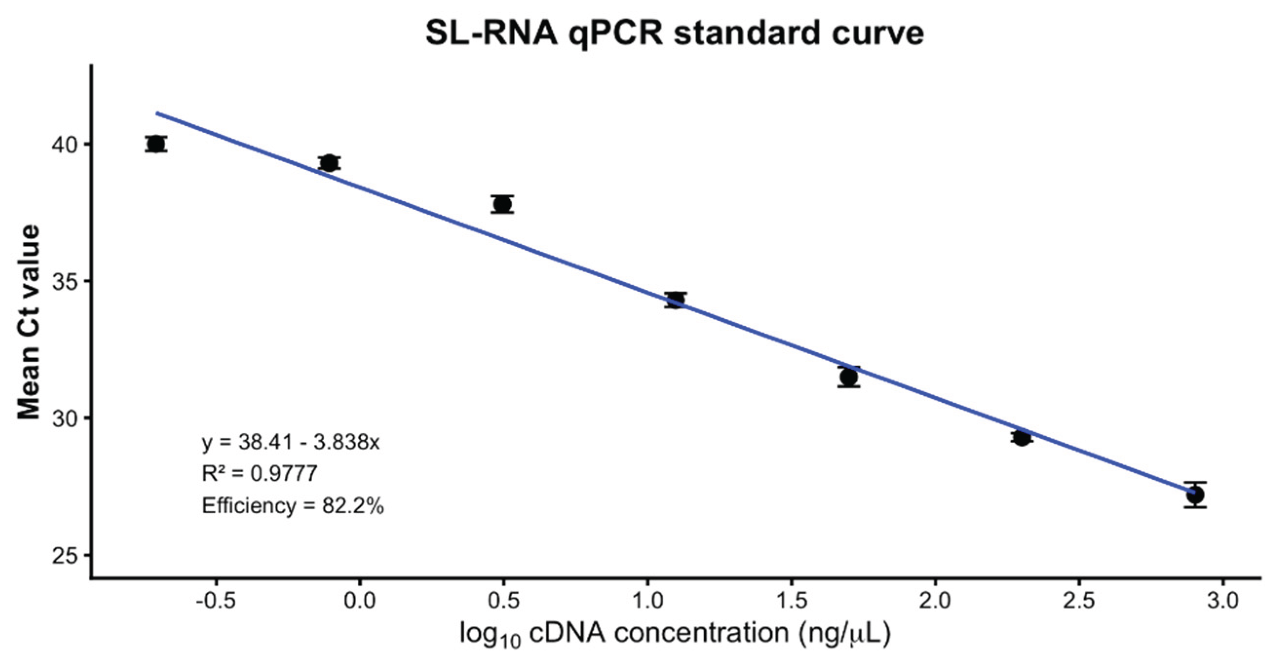
Figure 2.
qPCR detection of SL-RNA in EVs from multiple Leishmania models. (A) EV-enriched preparations from L. donovani (Ld), L. major (Lm), L. infantum (Li), L. tropica (Lt) promastigotes and L. donovani axenic amastigotes (LdAA) were analyzed for SL-RNA. Points represent means of three technical replicates, horizontal lines indicate means of three biological replicates ± SE. SL-RNA was detected in all parasite-derived EV preparations but not in media-derived EV. (B) EV-enriched preparations from uninfected THP-1 macrophages- (U-MP) and L. infantum-infected THP-1 macrophage- (Li-MP) conditioned media were analyzed for SL-RNA. Points represent technical replicates of three biological samples, horizontal lines indicate means ± SD. SL-RNA was detected in all Li-MP derived preparations, whereas U-MP derived preparations were undetermined. Parasite Index is presented in parenthesis above each replicate. UD (Undetermined = 40).
Figure 2.
qPCR detection of SL-RNA in EVs from multiple Leishmania models. (A) EV-enriched preparations from L. donovani (Ld), L. major (Lm), L. infantum (Li), L. tropica (Lt) promastigotes and L. donovani axenic amastigotes (LdAA) were analyzed for SL-RNA. Points represent means of three technical replicates, horizontal lines indicate means of three biological replicates ± SE. SL-RNA was detected in all parasite-derived EV preparations but not in media-derived EV. (B) EV-enriched preparations from uninfected THP-1 macrophages- (U-MP) and L. infantum-infected THP-1 macrophage- (Li-MP) conditioned media were analyzed for SL-RNA. Points represent technical replicates of three biological samples, horizontal lines indicate means ± SD. SL-RNA was detected in all Li-MP derived preparations, whereas U-MP derived preparations were undetermined. Parasite Index is presented in parenthesis above each replicate. UD (Undetermined = 40).
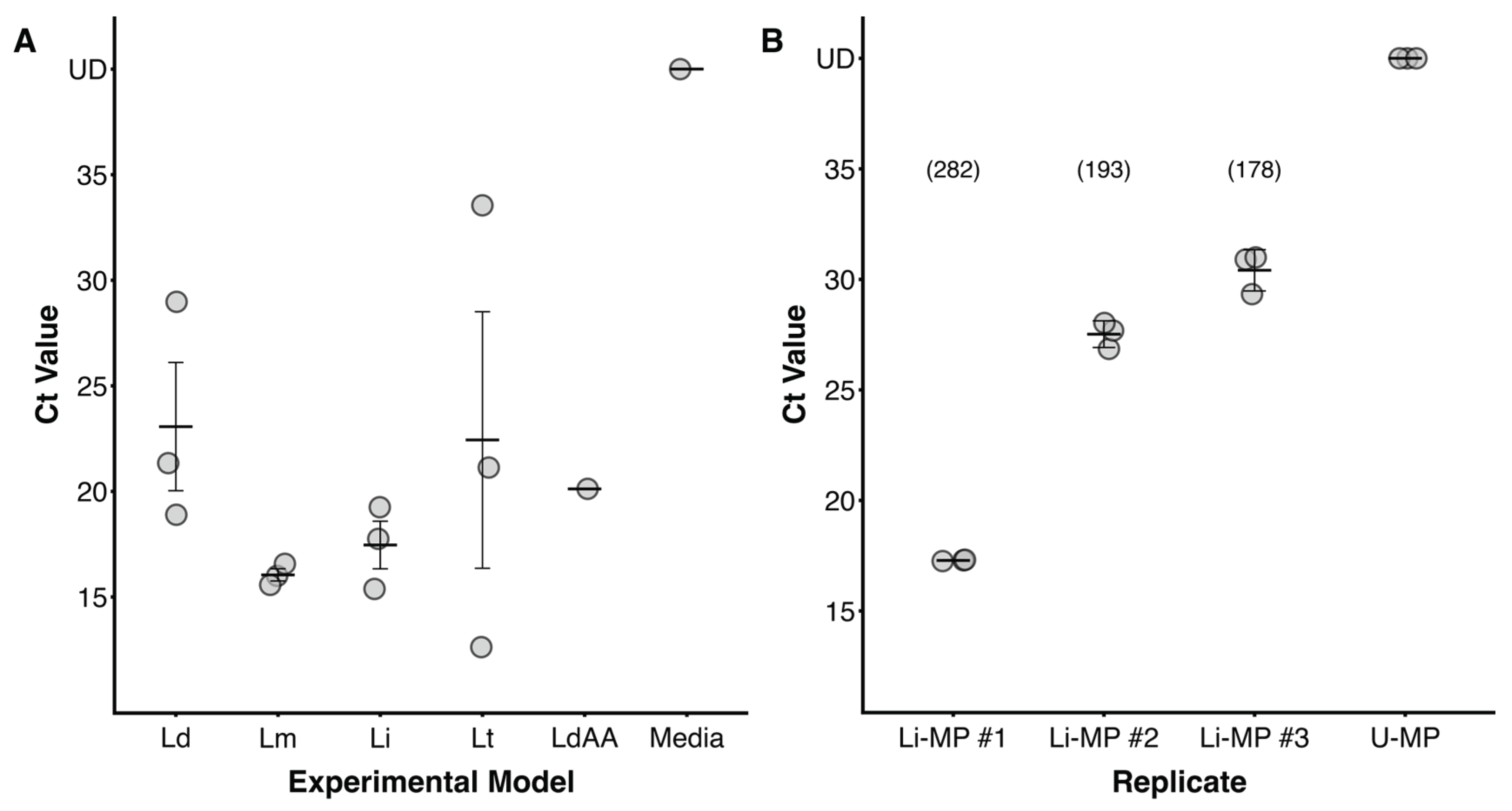
Figure 3.
RT-qPCR detection of SL-RNA in EVs from Leishmania infected mice. EV-enriched preparations from the serum of eight L. major-infected C57Bl/6 mice and one uninfected control mouse were analyzed for SL-RNA. Points represent means of three technical replicates, horizontal lines indicate means of three biological replicates ± SD. SL-RNA was detected in all eight infected mice derived EV preparations compared to no detection in the uninfected mouse (UI) control.
Figure 3.
RT-qPCR detection of SL-RNA in EVs from Leishmania infected mice. EV-enriched preparations from the serum of eight L. major-infected C57Bl/6 mice and one uninfected control mouse were analyzed for SL-RNA. Points represent means of three technical replicates, horizontal lines indicate means of three biological replicates ± SD. SL-RNA was detected in all eight infected mice derived EV preparations compared to no detection in the uninfected mouse (UI) control.
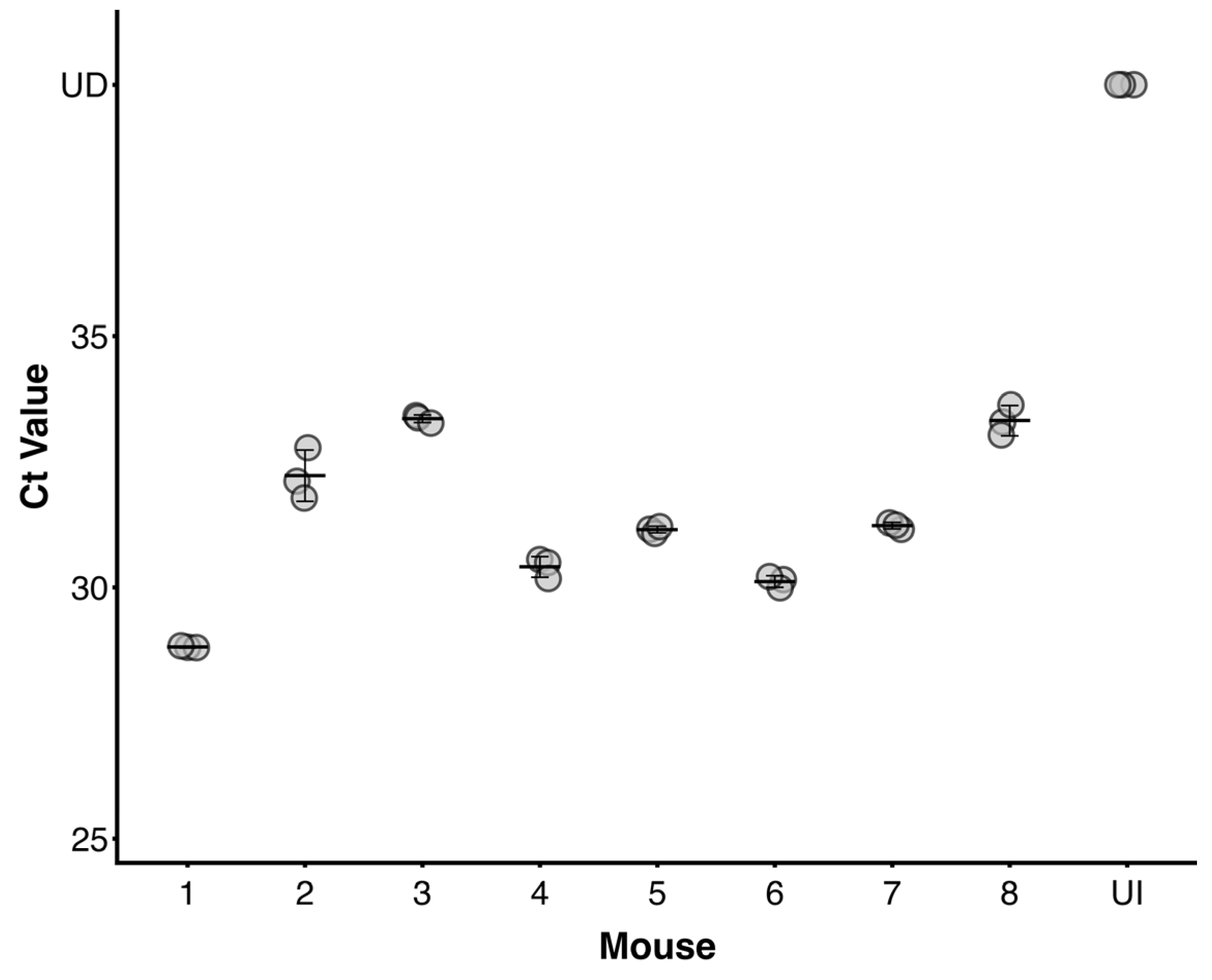
Figure 4.
Validation of SL-RNA in EVs by ddPCR. Droplet-amplitude plot of EV-enriched preparations from Li-MP #1-3, PCM, and U-MP CCM were analyzed for SL-RNA by ddPCR. Blue partitions represent positive droplets while grey partitions represent negative droplets. Positive SL-RNA signal was observed in the three Li-MP samples similar to PCM, while no signal was observed in U-MP CCM. Parasite Index is presented in parenthesis above each replicate.
Figure 4.
Validation of SL-RNA in EVs by ddPCR. Droplet-amplitude plot of EV-enriched preparations from Li-MP #1-3, PCM, and U-MP CCM were analyzed for SL-RNA by ddPCR. Blue partitions represent positive droplets while grey partitions represent negative droplets. Positive SL-RNA signal was observed in the three Li-MP samples similar to PCM, while no signal was observed in U-MP CCM. Parasite Index is presented in parenthesis above each replicate.
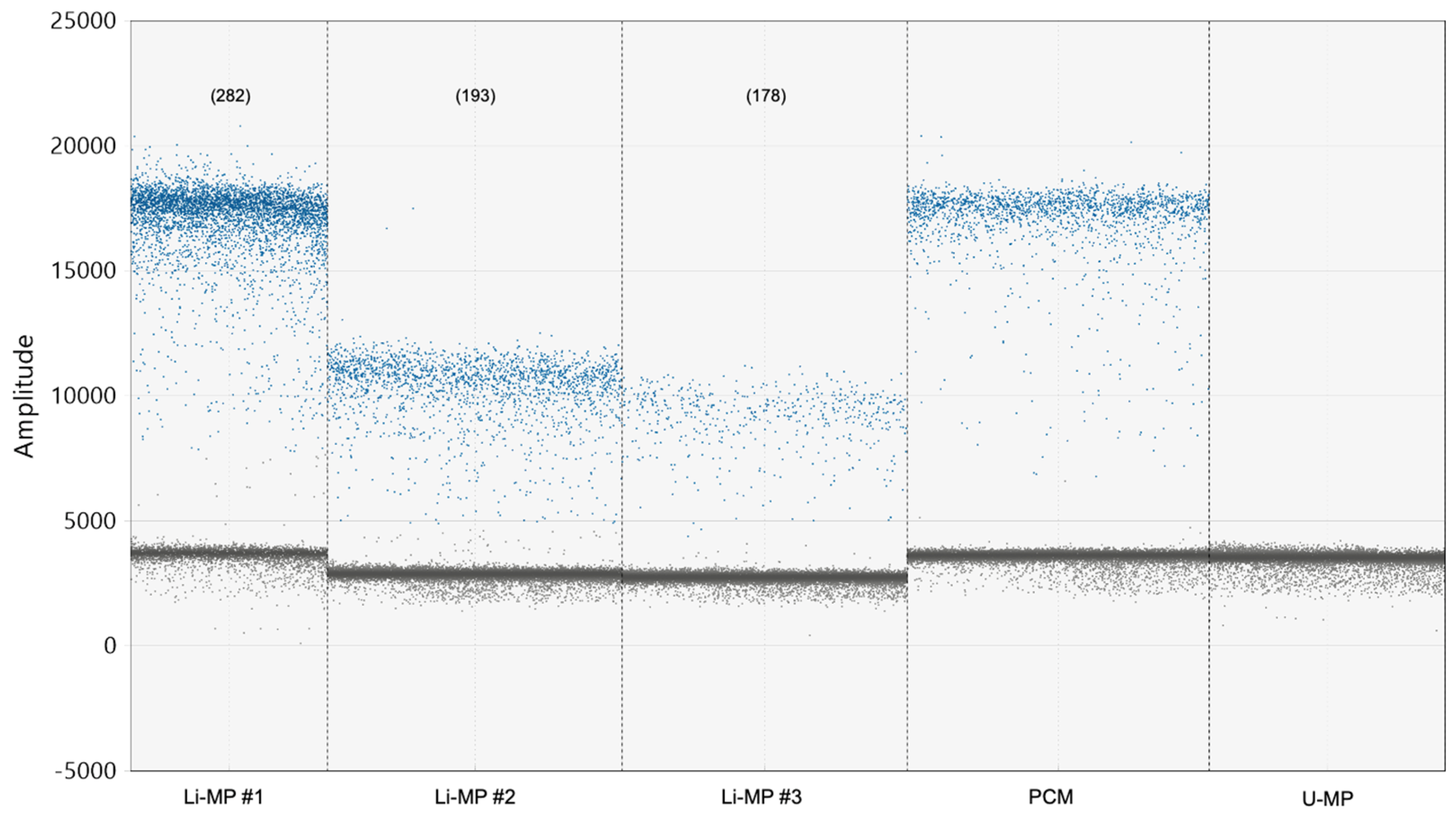
Figure 5.
Specificity of SL-RNA in non-target kinetoplasts. EV-enriched preparations from Lm-PCM, Tbb-PCM, and Tc-PCM were analyzed for SL-RNA by qPCR. Points represent technical replicates of Lm-PCM (n=1), Tbb-PCM (n=2), and Tc-PCM (n=2). SL-RNA was detected in Lm-PCM compared to no detection in Tbb-CCM and Tc-CCM.
Figure 5.
Specificity of SL-RNA in non-target kinetoplasts. EV-enriched preparations from Lm-PCM, Tbb-PCM, and Tc-PCM were analyzed for SL-RNA by qPCR. Points represent technical replicates of Lm-PCM (n=1), Tbb-PCM (n=2), and Tc-PCM (n=2). SL-RNA was detected in Lm-PCM compared to no detection in Tbb-CCM and Tc-CCM.
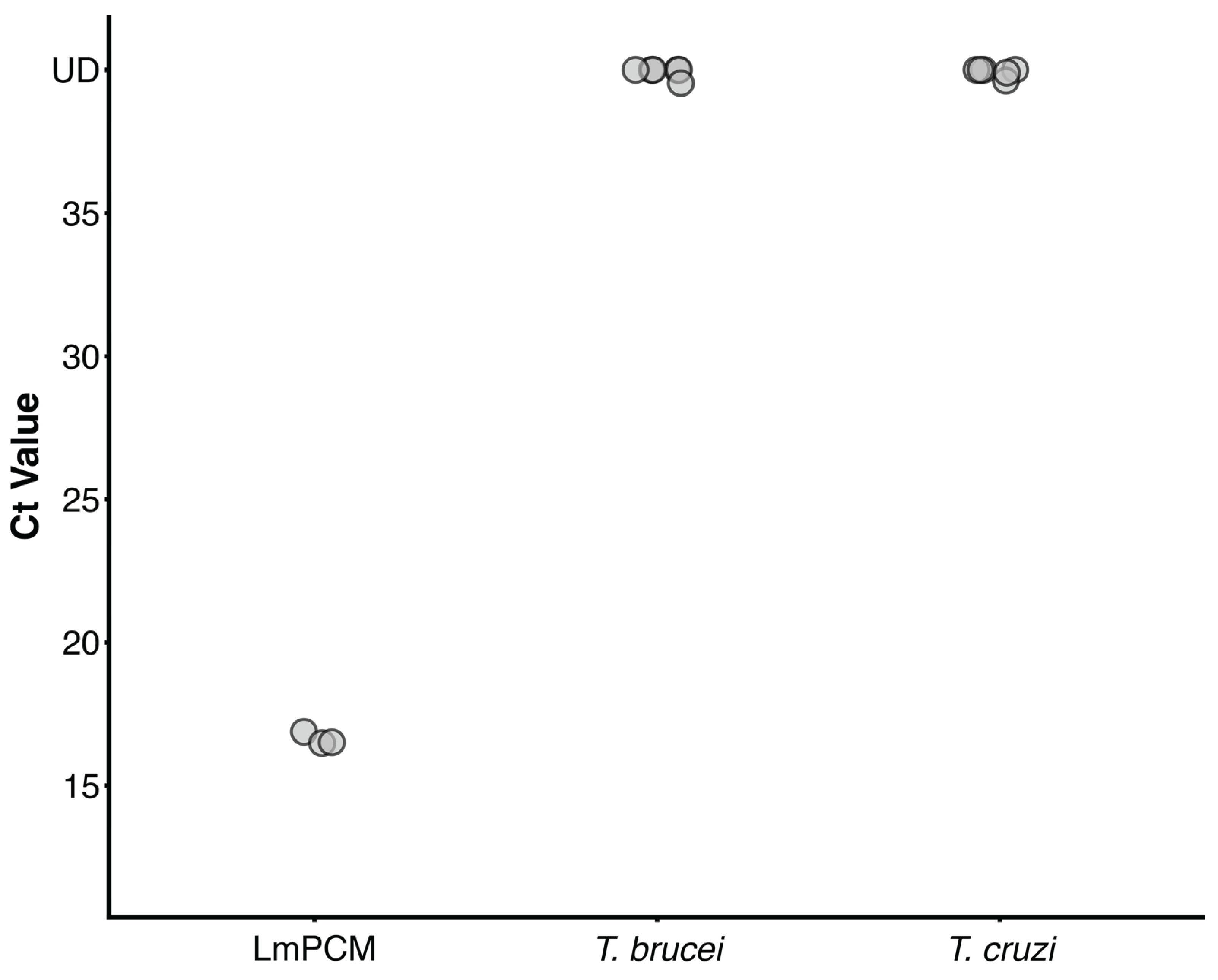

Table 1.
NTA of pooled EV-enriched preparations obtained from PCM, I- and U-MP CCM, mouse serum, and negative process controls.
Table 1.
NTA of pooled EV-enriched preparations obtained from PCM, I- and U-MP CCM, mouse serum, and negative process controls.
| Sample preparation | Mean particle diameter ± SE (nm) | Particle concentration ± SE (particles/mL) |
|---|---|---|
| L.donavani promastigote | 231.8 ± 2.0 | (2.06 ± 0.16) × 10⁹ |
|
L.major promastigote L.tropica promastigote L.infantum promastigotes L.donavani axenic amastigote T. brucei procyclic T.cruzi epimastigotes L.infantum-infected THP-1 MP Uninfected THP-1 MP L.major-infected mouse serum Uninfected mouse serum Fresh conditioned-medium PBS process control |
166.8 ± 2.1 163.0 ± 8.3 189.7 ± 4.3 165.3 ± 1.6 167.1 ± 2.8 182.3 ± 4.9 177.3 ± 3.2 172.9 ± 4.6 251.4 ± 10.3 200.2 ± 2.7 ND ND |
(6.37 ± 0.36) × 10⁹ (6.62 ± 0.53) × 10⁸ (4.65 ± 0.39) × 10⁹ (1.93 ± 0.04) × 10⁹ (1.11 ± 0.04) × 10⁹ (4.09 ± 0.08) × 10⁹ (2.10 ± 0.11) × 10⁹ (2.04 ± 0.19) × 10⁹ (2.39 ± 0.06) × 10⁹ (2.42 ± 0.31) × 10⁹ 2.87 × 10⁶ 1.43 × 10⁶ 1 |
1 Values are presented as the mean ± standard error of technical NTA measurements performed on each pooled EV-enriched preparation. Parasite-derived preparations consisted of three pooled biological collections unless otherwise indicated. Particle concentrations were corrected for the dilution applied before NTA. ND, not determined.
Disclaimer/Publisher’s Note: The statements, opinions and data contained in all publications are solely those of the individual author(s) and contributor(s) and not of MDPI and/or the editor(s). MDPI and/or the editor(s) disclaim responsibility for any injury to people or property resulting from any ideas, methods, instructions or products referred to in the content. |
© 2026 by the authors. Licensee MDPI, Basel, Switzerland. This article is an open access article distributed under the terms and conditions of the Creative Commons Attribution (CC BY) license (http://creativecommons.org/licenses/by/4.0/).
Copyright: This open access article is published under a Creative Commons CC BY 4.0 license, which permit the free download, distribution, and reuse, provided that the author and preprint are cited in any reuse.