

Submitted:
04 August 2026
Posted:
06 August 2026
You are already at the latest version
Abstract
Plants cannot flee unfavorable conditions, so they must sense and respond to physical and biotic stress from within a single cell. Biomolecular condensates — protein- and RNA-rich assemblies formed by phase separation — have emerged as a unifying mechanism that lets plant cells convert continuous physical cues such as heat, drought and pathogen attack into discrete, reversible biochemical switches. This mini review synthesizes recent structural, biophysical and genetic evidence on how plant condensates assemble, how they sense the environment, how they are exploited and subverted during infection, and how they organize RNA processing and membrane remodeling.
Keywords:
biomolecular condensates
; plant signaling
; plant membrane biology
; signal transduction
; membrane remodeling
; dynamic regulatory mechanism
; plant development and stress responses
Biophysical Principles of Condensation in Plant Cells
Biomolecular condensates are membraneless assemblies that concentrate proteins and nucleic acids into a dense phase that coexists with a dilute surrounding cytoplasm or nucleoplasm. In plants, as in other eukaryotes, condensation is most often driven by liquid–liquid phase separation (LLPS): multivalent, individually weak contacts between intrinsically disordered regions (IDRs), low-complexity domains and folded interaction modules cooperatively outcompete the entropic cost of remixing once local concentration or interaction valency crosses a threshold [1,3,14] (Figure 1a).
A recurring theme in the plant literature is that the resulting assemblies are not a single physical state but occupy a continuum of material properties. Nuclear condensates formed by the transcriptional regulator SEUSS behave as liquid-like droplets whose formation depends on conformational compaction of its first intrinsically disordered region (IDR1) under hyperosmotic stress [5]. In contrast, the RNA-binding protein FCA assembles into a discrete, stable oligomeric core — resolved by single-particle tracking and SEC-MALS — onto which further condensation is built, illustrating that a defined oligomeric scaffold can underlie an otherwise liquid-like condensate [11]. At the more rigid end of the spectrum, the chloroplast RNA-editing factor MORF8 forms condensates with solid-like material properties specifically under heat stress, and this rigidity is what allows the condensate to trap and inhibit editing factors rather than simply concentrating them for reaction [15].
Large-scale, cell-free biochemical screens have shown that the capacity to phase separate is a widespread, quantifiable property of the plant proteome rather than a rare curiosity restricted to a handful of well-studied proteins, providing a resource for identifying new condensate-forming factors [16]. Two features distinguish plant condensate biology conceptually from its counterparts in other kingdoms. First, because plants are sessile, many of their condensates are wired directly to abiotic and biotic cues at the cell-autonomous level, rather than depending on systemic hormonal or neuronal responses. Second, plant genomes and life histories have generated condensate-relevant machinery — for example, chloroplast RNA-editing complexes and plant-specific endocytic adaptors — that have no direct animal counterpart [15,17]. The remainder of this mini review considers, in turn, how plant condensates sense the physical environment, how they participate in immunity, and how they organize RNA metabolism and membrane remodeling (Figure 1b).
Condensates as Sensors of Temperature, Osmolarity and Water Status
Thermosensing is among the best-mechanistically resolved examples of condensate-based environmental sensing in plants. The intrinsically disordered protein FUST1 senses heat directly through its prion-like domain (PrLD): all-atom molecular dynamics simulations combined with experimental validation show that the PrLD encodes a thermo-switch that shifts from a compact, ‘locked’ conformation at ambient temperature to an open conformation once a threshold temperature is exceeded, nucleating condensation into heat-induced stress granules [4] (Figure 2a). This provides a mechanistic, physical explanation for how a continuous temperature increase is converted into an all-or-nothing assembly event, complementing an earlier proposal that phase separation of the circadian-clock component ELF3 can act as a molecular thermosensor in a conceptually similar manner [13]. A broader synthesis of how heat is sensed and decoded, from the plasma membrane to the nucleus, situates such condensate-based switches alongside membrane fluidity changes and calcium signaling as complementary layers of thermal perception [18].
Heat-induced condensation also has consequences well beyond the initial sensing event. Stress granules sequester components of the conserved target-of-rapamycin (TOR) growth-signaling complex, including RAPTOR1B and LST8; this sequestration suppresses TOR activity during heat stress and its reversal upon stress granule disassembly is required for the resumption of growth, revealing stress granules as a hub that couples acute stress sensing to the subsequent decision to resume proliferation [19]. Thermal history can even be inherited: the HSFA1B–HSP70-3 chaperone module modulates the stability of SGS3, a component of small-RNA-body condensates, to establish transgenerational thermomemory that adjusts flowering time and immune competence in the offspring of heat-stressed plants [20].
A parallel logic operates for osmotic and hydric stress. SEUSS senses increasing extracellular osmolarity through IDR1-dependent conformational compaction, triggering rapid coalescence into nuclear condensates that promote hyperosmotic stress tolerance [5]. More recently, a distinct sterile-alpha-motif (SAM)-domain protein, SAM8, was shown to sense cellular water potential itself, rather than solute concentration or osmotic pressure per se: SAM8 is strongly hydrated and soluble under normal water status, and loses its hydration shell as water potential falls, driving condensation that supports both hyperosmotic stress tolerance and seed germination [6]. Together, SEUSS and SAM8 indicate that plants have evolved at least two biophysically distinct condensate-based strategies for reading water status — one keyed to conformational change in an IDR, the other to loss of hydration — and a comprehensive framework for abiotic-stress sensing more broadly emphasizes that such biochemical and biophysical mechanisms operate alongside classical receptor-based signaling to set the sensitivity and speed of the whole-plant stress response [21] (Figure 2b).
A common design principle unifies these examples: an IDR-containing protein behaves as a tunable physicochemical switch whose set point (a temperature, an osmotic strength, a degree of hydration) is encoded in its own sequence and structure. Because assembly and disassembly require no new transcription or translation, condensate-based sensing is intrinsically faster than gene-expression-based responses, and because it is readily reversible, it allows the cell to track a fluctuating environment rather than committing irreversibly to a single stress response [22,23].
Condensates in Plant Immunity: A Molecular Battleground
Plant immune signaling is increasingly understood to rely on condensation, and pathogens have in turn evolved strategies to disable or repurpose these assemblies. In Arabidopsis guard cells, the RNA-recognition-motif protein STOMATAL IMMUNE RNA-BINDING PROTEIN 1 (SAIR1) forms pathogen-responsive condensates in response to the bacterial molecular pattern flg22. Perception of flg22 activates the MAP kinases MPK3 and MPK6, which phosphorylate SAIR1 and trigger its condensation; the resulting condensate fine-tunes local translation to help close stomata and restrict bacterial entry, defining a cell-autonomous, condensate-based arm of stomatal immunity [7] (Figure 3a).
Because a condensate’s assembly depends on a comparatively small number of weak, cooperative contacts, it presents an efficient target for a pathogen effector: disabling one or a few key interactions can collapse the entire assembly. The stripe rust fungus Puccinia striiformis f. sp. tritici secretes the haustorial effector Hasp170, which binds directly to the first intrinsically disordered region (IDR1) of the wheat nuclear protein TaPSTE (phase-separation protein targeted by effector). TaPSTE ordinarily forms LLPS-dependent condensates that support wheat immunity; by occupying IDR1, Hasp170 blocks TaPSTE condensation and thereby disables this defense, allowing infection to proceed [8]. This illustrates a general principle: pathogens can defeat a condensate-based defense not by rewiring an entire signaling pathway, but simply by preventing the phase transition itself (Figure 3b).
A second, more subtle mode of subversion is repurposing rather than dissolution. During infection with rice stripe virus, the viral disease-specific protein (SP) carries its own phase-separating IDR1, but rather than forming independent condensates, SP instead interferes with the phase separation of the host protein SERRATE (SE), which normally organizes nuclear dicing bodies for microRNA processing (Figure 3c). By altering SE-dependent condensation, the viral protein reshapes host microRNA biogenesis in a manner that favors viral pathogenesis, without needing to assemble a distinct viral condensate of its own [9]. Considered together, these examples show that condensates sit at a genuine host–pathogen interface: immune condensates such as SAIR1 and TaPSTE provide defense, while pathogen proteins that either dissolve (Hasp170) or hijack (RSV SP) host phase separation represent convergent counter-strategies exploiting the same underlying physical chemistry.
Condensates that Organize Nuclear RNA Processing
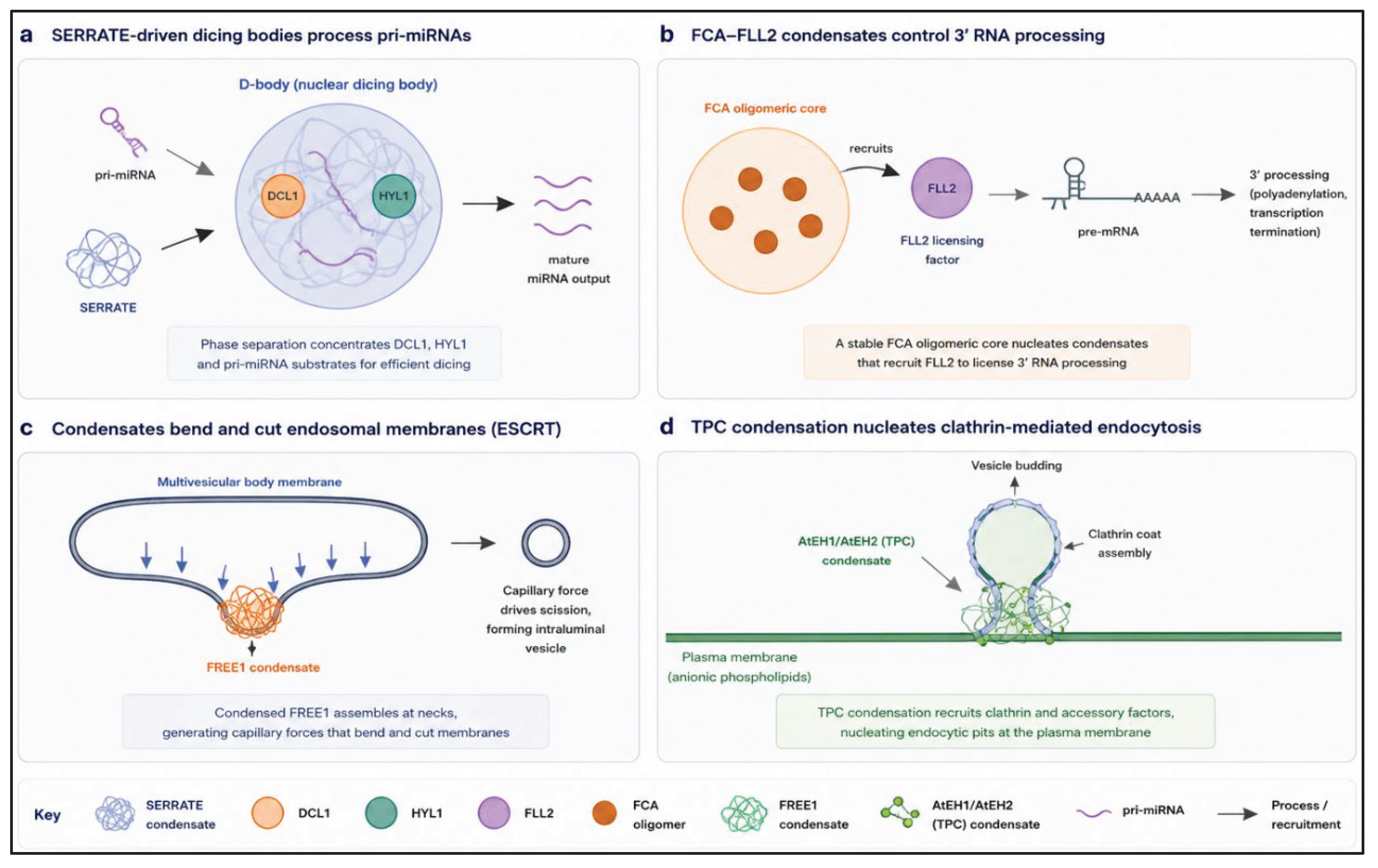
Some of the earliest and best-characterized plant condensates function in nuclear RNA metabolism, where concentrating an enzyme with its substrate inside a shared compartment increases the efficiency and specificity of processing. Nuclear dicing bodies (D-bodies), the site of microRNA (miRNA) biogenesis, form when the protein SERRATE (SE) undergoes phase separation: SE forms droplets in vitro and recruits DCL1, HYL1 and pri-/pre-miRNA substrates into the same droplets, and disrupting SE phase separation in vivo substantially reduces miRNA processing activity, directly linking condensation to the stepwise Dicing reaction [10] (Figure 4a).
A parallel condensate-based logic governs the 3′ end of transcription. The RNA-binding protein FCA is found in liquid-like nuclear condensates that promote proximal polyadenylation and transcription termination; single-particle tracking shows that FCA first assembles into a defined, stable oligomeric core, providing a discrete nucleation point onto which a more dynamic, liquid-like condensate is built [11]. This condensation is licensed by FLL2, identified through a genetic screen for regulators of FCA function as a positive regulator of FCA-dependent LLPS of the polyadenylation machinery, showing that condensate assembly in vivo can itself be a genetically tractable, regulated step rather than a purely spontaneous physical event [24] (Figure 4b).
Condensation also regulates RNA-modifying enzymes directly. Under heat stress, the chloroplast RNA-editing factor MORF8 forms solid-like condensates whose assembly is driven by its own IDR; because these condensates are rigid rather than liquid, they sequester and thereby inhibit other editing factors (including PPR proteins and additional MORF-family members), providing a mechanism by which heat directly suppresses chloroplast RNA editing and, downstream, photosynthetic efficiency [15]. Phase separation further intersects with photoreceptor signaling: the scaffold protein TZP undergoes LLPS to promote phosphorylation of the far-red photoreceptor phytochrome A by the PPK kinase family, coupling condensation to a specific post-translational modification rather than to transcription or splicing [25] (Box 2).
Finally, condensation can directly control a developmental decision: reactive-oxygen-species-dependent, redox-sensitive chaperoning of the transcription factor GBF1 governs its LLPS into condensates that repress the germination-promoting gene CathB3, linking a specific redox state to a phase transition that gates the seed-to-seedling transition [26]. Across these examples, phase separation acts less as a switch between ‘on’ and ‘off’ states of a pathway and more as a means of enforcing which molecular partners are allowed to meet, at what time and under what physiological condition [14,27] (Box 1).
Mechanical Roles: Condensates that Remodel Membranes
A distinct and more recently appreciated role for plant condensates is mechanical rather than biochemical: condensates can generate capillary-like forces sufficient to bend and cut lipid membranes. The plant ESCRT component FREE1 forms liquid-like condensates that associate with the membranes of multivesicular bodies (MVBs), the endosomal compartments that degrade membrane-bound cargo. Rather than acting solely as an adaptor for the canonical, ATP-consuming ESCRT machinery, FREE1 condensates themselves promote the invagination and eventual scission of MVB membranes to generate intraluminal vesicles, showing that a phase-separated assembly can substitute for or complement classical mechanoenzyme-driven membrane fission [12]. A conceptual synthesis of this and related examples across biology — termed ‘wet scissors’ — argues that the surface tension generated at the interface between a liquid-like condensate and a membrane is, in physical terms, directly analogous to the forces exerted by canonical fission machines such as dynamins, and can drive comparable topological membrane transitions using only the physics of phase separation [28] (Figure 4c).
Condensation also nucleates membrane remodeling at the cell surface during endocytosis. The evolutionarily ancient TSET–TPLATE complex (TPC) is essential for clathrin-mediated endocytosis in plants, and two of its highly disordered subunits, AtEH1 and AtEH2, function as condensation scaffolds. These subunits drive biomolecular condensation of the TPC that nucleates specifically at the plasma membrane through interactions with anionic phospholipids, and this condensation step is required for efficient recruitment and assembly of the downstream clathrin coat [17]. Taken together with the FREE1 example, these findings indicate that plant condensates are not confined to the nucleus or cytoplasm as sites of gene regulation, but operate directly at cellular membranes, where their physical properties — surface tension, wetting behavior and viscosity — are put to mechanical use in shaping and cutting the membranes with which they associate (Figure 4d).
Concluding Remarks and Future Directions
The studies synthesized here in the mini review converge on a picture in which biomolecular condensation is not a peripheral curiosity of plant cell biology but a core, repeatedly re-used mechanism for converting continuous physical and biotic cues into fast, reversible, spatially organized biochemical decisions [1,2]. The same design principle — an IDR-encoded, tunable switch built from weak multivalent contacts — recurs in thermosensing, osmosensing, immune signaling, RNA processing and membrane remodeling, suggesting that plant genomes have repeatedly co-opted a common physical mechanism for functionally unrelated purposes.
Several open questions follow directly from the work reviewed here. First, most condensates have so far been characterized by one protein at a time; a systems-level, quantitative understanding of how multiple condensates compete for shared components, or influence one another’s assembly, within a single cell remains largely unexplored. Second, although material state (liquid, gel or solid) is emerging as a functionally important variable, in-cell tools to measure it quantitatively, rather than qualitatively via FRAP, are still limited. Third, the discovery that pathogens can defeat plant immunity either by dissolving (Hasp170–TaPSTE) or by hijacking (rice stripe virus SP–SERRATE) host condensates [8,9] raises the possibility that condensate-stabilizing or condensate-restoring chemistry could represent a genuinely new axis for crop protection, complementary to conventional resistance breeding. Finally, given that condensation can be trans generationally inherited via chaperone-regulated stability of condensate components such as SGS3 [20], it will be important to determine how broadly condensate-encoded stress memory contributes to acclimation across plant generations in the field, beyond the controlled conditions used in current studies.

Table 1.
Summary of plant condensate systems.
| Process | Key condensate / protein | Physical cue or trigger | Biological outcome | Refs. |
| Osmotic stress sensing | SEUSS (nuclear) | Hyperosmolarity | Hyperosmotic stress tolerance | [5] |
| Water-potential sensing | SAM8 (nuclear) | Loss of hydration shell | Osmotic tolerance and seed germination | [6] |
| Thermosensing | FUST1 (stress granule) | Elevated temperature (PrLD switch) | Heat-induced stress granule formation | [4] |
| Growth–stress coupling | Stress granules / TOR complex | Heat stress | TOR suppression then growth recovery | [19] |
| Transgenerational memory | SGS3 (siRNA body) | Heat stress in parent generation | Transgenerational thermomemory | [20] |
| Stomatal immunity | SAIR1 (guard cell) | flg22 perception, MPK3/6 phosphorylation | Stomatal closure, restricted pathogen entry | [7] |
| Fungal immune evasion | TaPSTE (nuclear) | Effector Hasp170 binding IDR1 | Loss of wheat immune condensate, infection | [8] |
| Viral immune evasion | SERRATE / viral SP | Viral IDR1-mediated interference | Reprogrammed host miRNA biogenesis | [9] |
| microRNA biogenesis | SERRATE D-body | Constitutive / developmental | Pri-miRNA processing efficiency | [10] |
| 3′ RNA processing | FCA–FLL2 condensate | FLL2-licensed oligomerization | Proximal polyadenylation, transcription termination | [11,24] |
| RNA editing regulation | MORF8 (chloroplast) | Heat stress (solid-like state) | Inhibition of chloroplast RNA editing | [15] |
| Photoreceptor signaling | TZP condensate | Far-red light | PPK-mediated phytochrome A phosphorylation | [25] |
| Seed dormancy / germination | GBF1 condensate | Redox state (ROS) | Repression of germination gene CathB3 | [26] |
| Membrane scission | FREE1 (endosome) | Constitutive ESCRT pathway | Multivesicular body membrane scission | [12] |
| Endocytosis | AtEH1/AtEH2 (TPC) | Anionic phospholipids at plasma membrane | Clathrin-mediated endocytosis | [17] |
| Key points |
| Box 1 | Probing condensates in plant cells Establishing that an assembly observed by microscopy is a genuine phase-separated condensate, rather than an aggregate or an artefact of overexpression, requires converging lines of evidence. In vitro reconstitution with purified, recombinant protein (and RNA, where relevant) is used to test whether a candidate protein undergoes LLPS at physiologically plausible concentrations and salt conditions, and whether this depends on a specific IDR, as shown for SEUSS, FUST1 and MORF8 [4,5,15]. Cell-free translation-based screens extend this logic to proteome scale, allowing systematic, quantitative identification of candidate phase-separating proteins across the plant proteome without the confound of cellular context [16]. In vivo, fluorescence recovery after photobleaching (FRAP) is used to distinguish liquid-like condensates, which recover fluorescence rapidly after photobleaching owing to fast internal rearrangement, from gel-like or solid-like assemblies, which recover slowly or not at all — the criterion used to establish the solid-like nature of heat-induced MORF8 condensates [15]. Single-particle tracking and mass-photometry-adjacent techniques such as SEC-MALS provide complementary, quantitative measurements of oligomeric state and assembly stoichiometry within condensates in living cells, as used to resolve the stable oligomeric core of FCA [11]. Finally, genetic separation-of-function alleles that disrupt an IDR or a specific condensation-competent domain, without perturbing the protein’s folded catalytic or binding activity, remain the most direct way to link condensation itself — rather than the protein’s other biochemical activities — to a physiological outcome [10,24]. |
| Box 2 | Material state as a functional variable A single physical parameter — the material state of a condensate — recurs as a determinant of biological outcome throughout this review. Liquid-like condensates, such as those formed by SEUSS or SERRATE, favor rapid exchange of components with the surrounding phase and are well suited to reversible, concentration-dependent signaling and to enzymatic processes that require repeated substrate turnover [5,10]. Gel-like assemblies, exemplified by the stable oligomeric core of FCA, provide a more persistent structural scaffold onto which additional, more dynamic condensation can be layered [11]. Solid-like condensates, such as heat-induced MORF8 assemblies, exchange components slowly or not at all, and this rigidity is precisely what allows them to sequester and inactivate client proteins for as long as the inducing stress persists [15]. This spectrum has a direct corollary for how a condensate can fail. Because material state depends sensitively on the same IDR sequence features that drive assembly, mutations, post-translational modifications or pathogen effectors that shift a condensate further along the liquid-to-solid axis than its normal physiological range can convert an initially adaptive, reversible response into an aggregation-like, potentially maladaptive one — underscoring why plant condensates are typically under tight upstream regulatory control (through kinases, chaperones or redox state) rather than being left to assemble unchecked [7,26]. |
Glossary
|
Author Contributions
The authors contributed equally to all aspects of the article.
Conflicts of Interest
The authors declare no conflict of interest.
Acknowledgments
We acknowledge Huang Lab members for the fruitful discussion on this topic. Due to space limitations, we apologize to authors whose works were not cited. During the preparation of this work, the authors used Google Gemini to improve language and readability of figures. After using this tool, the authors reviewed and edited the content as needed and took full responsibility for the content of the publication. This research received no external funding.
References
- Miao, Y.; Wang, Y.; Grate, E. V. L.; Fang, X. A multidimensional view of biomolecular condensates in plant biology. Annu. Rev. Plant Biol. 2026, 77, 197–223. [Google Scholar] [CrossRef] [PubMed]
- Miao, Y.; Chodasiewicz, M.; Fang, X. Navigating biomolecular condensates in plants from patterns to functions. Mol. Plant 2024, 17, 1329–1332. [Google Scholar] [CrossRef] [PubMed]
- Wang, Y.; Li, C.; Li, Y.; Fang, X. The role of phase separation in plant biology. Sci. Sin. Vitae 2024, 54, 1144–1158. [Google Scholar]
- Geng, P.; Li, C.; Quan, X.; Peng, J.; Yao, Z.; Wang, Y.; Yang, M.; Wang, Y.; Jin, Y.; Xiong, Y.; Liu, H.; Qi, Y.; Yang, P.; Huang, K.; Fang, X. A thermosensor FUST1 primes heat-induced stress granule formation via biomolecular condensation in arabidopsis. Cell Res. 2025, 1–14. [Google Scholar]
- Wang, B.; Zhang, H.; Huai, J.; Peng, F.; Wu, J.; Lin, R.; Fang, X. Condensation of SEUSS promotes hyperosmotic stress tolerance in arabidopsis. Nat. Chem. Biol. 2022, 18, 1–9. [Google Scholar] [CrossRef] [PubMed]
- Wang, Y.; Zhu, L.; Yang, Y.; Li, X.; Zhang, X.; Fang, X. Cellular water-potential sensing through biomolecular condensation. Nature 2026, 655, 1–10. [Google Scholar] [CrossRef] [PubMed]
- Yu, Q.; Wu, J.; Jin, Y.; Song, T.; Wang, W.; Zeng, Y.; Huang, H.; Deng, H.; Wang, W.; Xie, J.; Lu, Z. J.; Fang, X.; Song, S.; Qi, T. Pathogen-induced condensation of the guard cell RNA-binding protein SAIR1 fine-tunes translation for immunity. Nat. Plants 2025, 1–17. [Google Scholar]
- Yan, T.; Zhao, J.; Chen, C.; Hu, J.; Zhang, J.; Wang, J.; Wansee, S.; Wang, N.; Jiang, C.; Fang, X.; Kang, Z.; Yang, L.; Tang, C.; Wang, X. Stripe rust fungus subverts wheat phase separation–mediated immunity to promote infection. Proc. Natl. Acad. Sci. 2026, 123, e2522267123. [Google Scholar] [CrossRef] [PubMed]
- Zou, J.; Zhang, S.; Chen, Y.; He, C.; Pan, X.; Zhang, Y.; Xu, J.; Zheng, L.; Guan, H.; Wu, M.; Xie, D.; Ji, Y.; Fang, X.; Li, Y.; Ding, S.; Fang, X.; Zhao, S.; Wu, J. A plant bunyaviral protein disrupts SERRATE phase separation to modulate microRNA biogenesis during viral pathogenesis. Nat. Commun. 2025, 16, 6197. [Google Scholar] [CrossRef] [PubMed]
- Xie, D.; Chen, M.; Niu, J.; Wang, L.; Li, Y.; Fang, X.; Li, P.; Qi, Y. Phase separation of SERRATE drives dicing body assembly and promotes miRNA processing in arabidopsis. Nat. Cell Biol. 2021, 23, 32–39. [Google Scholar] [PubMed]
- Jang, G.-J.; Payne-Dwyer, A. L.; Maple, R.; Wu, Z.; Liu, F.; Lopez, S. G.; Wang, Y.; Fang, X.; Leake, M. C.; Dean, C. Modular in vivo assembly of arabidopsis FCA oligomers into condensates competent for RNA 3’ processing. EMBO J. 2025, 44, 2056–2074. [Google Scholar] [CrossRef] [PubMed]
- Wang, Y.; Li, S.; Mokbel, M.; May, A. I.; Liang, Z.; Zeng, Y.; Wang, W.; Zhang, H.; Yu, F.; Sporbeck, K.; Jiang, L.; Aland, S.; Agudo-Canalejo, J.; Knorr, R. L.; Fang, X. Biomolecular condensates mediate bending and scission of endosome membranes. Nature 2024, 634, 1204–1210. [Google Scholar] [CrossRef] [PubMed]
- Li, C.; Fang, X. Phase separation as a molecular thermosensor. Dev. Cell 2020, 55, 118–119. [Google Scholar] [CrossRef] [PubMed]
- Lin, Y.; Fang, X. Phase separation in RNA biology. J. Genet. Genom. Yi Chuan Xue Bao 2021, 48, 872–880. [Google Scholar] [CrossRef] [PubMed]
- Wu, J.; Wang, Y.; Chen, H.; Xu, T.; Yang, W.; Fang, X. Solid-like condensation of MORF8 inhibits RNA editing under heat stress in arabidopsis. Nat. Commun. 2025, 16, 2789. [Google Scholar] [CrossRef] [PubMed]
- Zhang, H.; Peng, F.; He, C.; Liu, Y.; Deng, H.; Fang, X. Large-scale identification of potential phase-separation proteins from plants using a cell-free system. Mol. Plant 2023, 16, 310–313. [Google Scholar] [CrossRef] [PubMed]
- Dragwidge, J. M.; Wang, Y.; Brocard, L.; De Meyer, A.; Hudeček, R.; Eeckhout, D.; Grones, P.; Buridan, M.; Chambaud, C.; Pejchar, P.; Potocký, M.; Winkler, J.; Vandorpe, M.; Serre, N.; Fendrych, M.; Bernard, A.; De Jaeger, G.; Pleskot, R.; Fang, X.; Van Damme, D. Biomolecular condensation orchestrates clathrin-mediated endocytosis in plants. Nat. Cell Biol. 2024, 26, 438–449. [Google Scholar] [CrossRef] [PubMed]
- Wang, Y.; Fang, X. Insights into heat sensing and decoding from membrane to nucleus. J. Integr. Plant Biol. [CrossRef] [PubMed]
- Zhong, Z.; Yang, S.; Liu, D.; Liu, Z.; Zhao, W.; Shen, X.; Wang, F.; Liu, Y.; Xu, T.; Fang, X.; Xiong, Y. Stress granule dynamics govern TOR reactivation and growth recovery during post-heat stress adaptation. Sci. Adv. 2025, 11, eadv6202. [Google Scholar] [CrossRef] [PubMed]
- Wen, X.; Du, M.; Li, T.; Cheng, F.; Li, S.; Zhu, Z.; Liu, W.; Gu, X.; Feng, L.; Geng, Y.; Wang, H.; Krall, L.; Fang, X.; Wu, X. N.; He, Z.; Liu, J. The HSFA1B-HSP70-3 module regulates transgenerational thermomemory by modulating SGS3 stability in arabidopsis. Plant Physiol. 2025, 199, kiaf456. [Google Scholar] [CrossRef] [PubMed]
- Lv, Q.; Li, J.; Liu, C.; Samraoui, K. R.; Hutin, S.; Li, X.; Dong, F.; Xiong, H.; Ouyang, Y.; Hu, H.; Li, Y.; Hepworth, J.; Jung, J.-H.; Chen, X.; Zubieta, C.; Xiong, L.; Zhao, Y.; Clemente-Moreno, M. J.; Fang, X.; Flashman, E.; Kudla, J.; Lai, X. Abiotic stress sensing in plants: Biochemical and biophysical basis. S1674-2052(26)00150–4; Mol. Plant. 2026. [Google Scholar]
- Peng, J.; Yu, Y.; Fang, X. Stress sensing and response through biomolecular condensates in plants. Plant Commun. 2025, 6. [Google Scholar] [CrossRef] [PubMed]
- Zhang, H.; Fang, X. Advances in the regulation of stress sensing and responses by phase separation in plants. Biotechnol. Bull. 2023, 39, 44–53. [Google Scholar]
- Fang, X.; Wang, L.; Ishikawa, R.; Li, Y.; Fiedler, M.; Liu, F.; Calder, G.; Rowan, B.; Weigel, D.; Li, P.; Dean, C. Arabidopsis FLL2 promotes liquid–liquid phase separation of polyadenylation complexes. Nature 2019, 569, 265–269. [Google Scholar] [CrossRef] [PubMed]
- Feng, Z.; Wang, M.; Liu, Y.; Li, C.; Zhang, S.; Duan, J.; Chen, J.; Qi, L.; Liu, Y.; Li, H.; Wu, J.; Liu, Y.; Terzaghi, W.; Tian, F.; Zhong, B.; Fang, X.; Qian, W.; Guo, Y.; Deng, X. W.; Li, J. Liquid–liquid phase separation of TZP promotes PPK-mediated phosphorylation of the phytochrome a photoreceptor. Nat. Plants 2024, 10, 798–814. [Google Scholar] [CrossRef] [PubMed]
- Wang, Y.; Fang, X. Redox-dependent chaperoning of GBF1 condensates regulates seed germination in arabidopsis. Adv. Sci. 2026, e20599. [Google Scholar] [CrossRef] [PubMed]
- Geng, P.; Wang, Y.; Fang, X. Regulations between phase separation and RNA. Sci. Sin. Vitae 2024, 54, 694–705. [Google Scholar]
- Fang, X.; May, A. I.; Sporbeck, K.; Hauer, L.; Knorr, R. L. Wet scissors: How biomolecular condensates cut cellular membranes. Curr. Opin. Plant Biol. 2025, 86, 102740. [Google Scholar] [CrossRef] [PubMed]
Figure 1.
| Biophysical principles of biomolecular condensation. (a) Multivalent, weak IDR–IDR contacts drive remixing from a dilute phase into a dense condensate. (b) A prion-like IDR can act as a conformational switch, converting a continuous cue (for example, temperature) into a discrete, reversible condensation event.
Figure 1.
| Biophysical principles of biomolecular condensation. (a) Multivalent, weak IDR–IDR contacts drive remixing from a dilute phase into a dense condensate. (b) A prion-like IDR can act as a conformational switch, converting a continuous cue (for example, temperature) into a discrete, reversible condensation event.
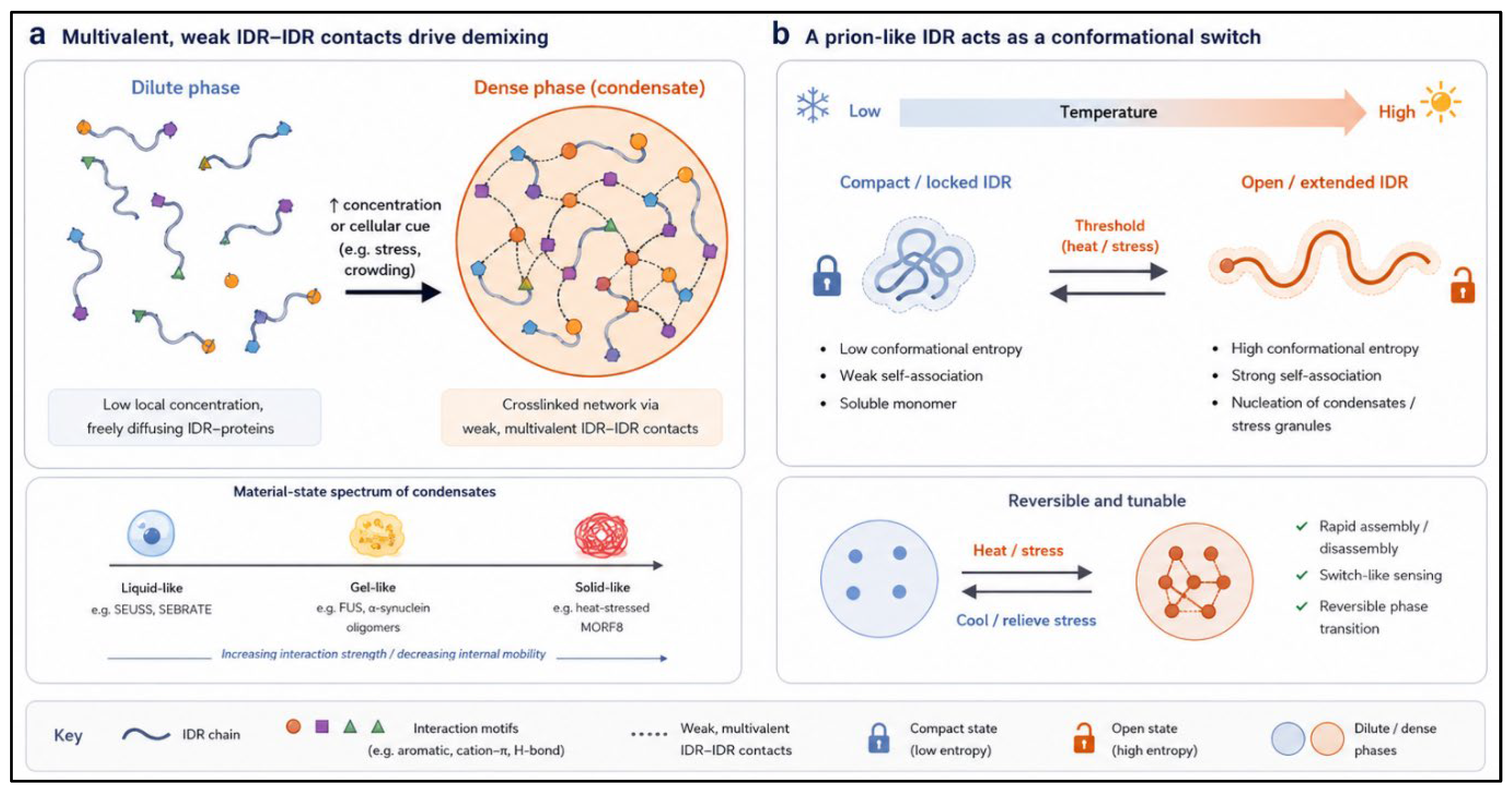
Figure 2.
| Condensates as sensors of temperature, osmolarity and water potential. (a) FUST1 senses heat through its prion-like domain, nucleating stress granules. (b) SEUSS and SAM8 sense osmotic and hydric stress via conformational change or hydration-shell loss, respectively. (c) A shared sensing logic maps a physical cue onto a sensor protein, a molecular switch and a physiological output.
Figure 2.
| Condensates as sensors of temperature, osmolarity and water potential. (a) FUST1 senses heat through its prion-like domain, nucleating stress granules. (b) SEUSS and SAM8 sense osmotic and hydric stress via conformational change or hydration-shell loss, respectively. (c) A shared sensing logic maps a physical cue onto a sensor protein, a molecular switch and a physiological output.
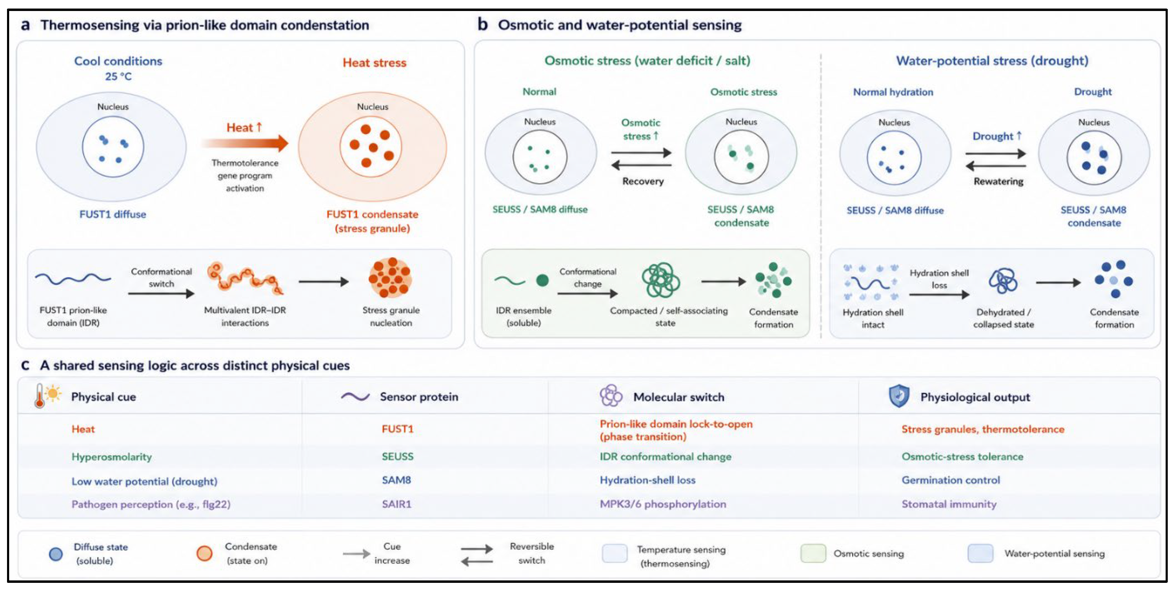
Figure 3.
| Biomolecular condensates as a battleground in plant–pathogen interactions. (a) flg22-triggered, MAPK-dependent condensation of SAIR1 restricts pathogen entry through guard cells. (b) The stripe rust effector Hasp170 binds IDR1 of TaPSTE to dissolve a host immune condensate. (c) Rice stripe virus protein SP hijacks SERRATE phase separation to reprogram host microRNA biogenesis.
Figure 3.
| Biomolecular condensates as a battleground in plant–pathogen interactions. (a) flg22-triggered, MAPK-dependent condensation of SAIR1 restricts pathogen entry through guard cells. (b) The stripe rust effector Hasp170 binds IDR1 of TaPSTE to dissolve a host immune condensate. (c) Rice stripe virus protein SP hijacks SERRATE phase separation to reprogram host microRNA biogenesis.
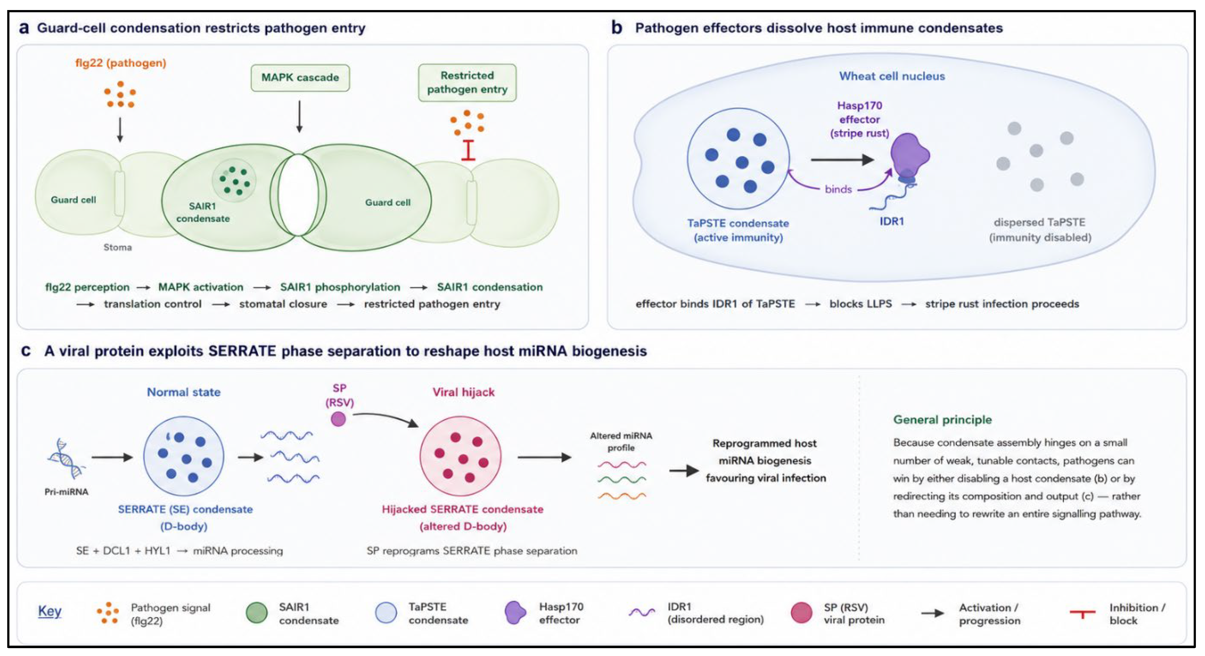
Figure 4.
| Condensates organize nuclear RNA processing and remodel membranes. (a) SERRATE-driven dicing bodies concentrate DCL1, HYL1 and pri-miRNA substrates. (b) An FCA oligomeric core recruits FLL2 to license 3′ RNA processing. (c) FREE1 condensates generate capillary forces that bend and cut endosomal membranes. (d) AtEH1/AtEH2 condensation nucleates clathrin-mediated endocytosis at the plasma membrane.
Figure 4.
| Condensates organize nuclear RNA processing and remodel membranes. (a) SERRATE-driven dicing bodies concentrate DCL1, HYL1 and pri-miRNA substrates. (b) An FCA oligomeric core recruits FLL2 to license 3′ RNA processing. (c) FREE1 condensates generate capillary forces that bend and cut endosomal membranes. (d) AtEH1/AtEH2 condensation nucleates clathrin-mediated endocytosis at the plasma membrane.
Disclaimer/Publisher’s Note: The statements, opinions and data contained in all publications are solely those of the individual author(s) and contributor(s) and not of MDPI and/or the editor(s). MDPI and/or the editor(s) disclaim responsibility for any injury to people or property resulting from any ideas, methods, instructions or products referred to in the content. |
© 2026 by the authors. Licensee MDPI, Basel, Switzerland. This article is an open access article distributed under the terms and conditions of the Creative Commons Attribution (CC BY) license.
Copyright: This open access article is published under a Creative Commons CC BY 4.0 license, which permit the free download, distribution, and reuse, provided that the author and preprint are cited in any reuse.