

Submitted:
07 September 2026
Posted:
08 September 2026
You are already at the latest version
Abstract
The retinal neurovascular unit (NVU) emerges through an ordered but overlapping sequence of endothelial growth, lumen formation, perfusion, selective branch retention and acquisition of inner blood-retinal barrier (iBRB) identity. This narrative review synthesizes primary developmental evidence across mouse, human fetal and non-human-primate retina, with emphasis on the experimental readout supported by each model. Mouse superficial angiogenesis advances postnatally across a broad astrocytic field before deep and intermediate plexuses form. Human central assembly instead includes resident vascular precursors before patent vessels, followed by predominantly angiogenic peripheral and intraretinal expansion; comparative primate anatomy adds a superficial-intermediate-deep laminar sequence and persistent foveal vascular exclusion. Across these systems, RGC-astrocyte-matrix interactions establish the superficial growth environment; dynamic endothelial competition, metabolism, lumenization and flow convert sprouts into a remodelled circulation; layer-specific neuronal, Müller-glial and microglial programmes shape intraretinal invasion; and Norrin/FZD4/β-catenin signalling, transcytosis suppression, mural support and vascular zonation establish barrier competence. The evidence also shows that vessel growth, perfusion and barrier function can be uncoupled, which limits inference from oxygen-induced retinopathy and engineered retinal models. Major gaps remain in the lineage of early human vascular precursors, the gestational timing of functional human iBRB maturation, foveal vascular exclusion and integration of layer-specific neuroglial signals.
Keywords:
retinal neurovascular unit
; retinal vascular development
; vasculogenesis
; angiogenesis
; inner blood-retinal barrier
; astrocytes
; Müller glia
; microglia
; pericytes
; Norrin
1. Building the Retinal Neurovascular Unit
The mature retinal neurovascular unit (NVU) is often introduced as a stable anatomical arrangement: endothelial cells form the inner blood-retinal barrier (iBRB), pericytes cover the vascular wall, astrocytes occupy the nerve-fibre layer, Müller cells span the retina, microglia patrol neural and vascular territories, and neuronal populations lie beside a three-plexus circulation. That description is useful anatomically but does not explain developmental dynamics. None of these relationships appears fully established at the outset. The NVU is built through a changing sequence in which neural geometry, glial state, extracellular matrix, endothelial competence, mural recruitment and blood flow alter one another as the circulation expands through the retina [1,2,3,4].
Selvam, Kumar and Fruttiger provided a broad synthesis review of retinal vascular development in health and disease, including astrocytes, endothelial patterning, pericytes, Norrin signalling and oxygen-induced retinopathy [1]. The present review is complementary: rather than extending that pathway catalogue, it follows reciprocal neurovascular-unit assembly across developmental stages and species and asks how the strength of evidence for each proposed intercellular mechanism should be interpreted.
This developmental view immediately separates outcomes that are often collapsed into the single phrase ‘vascularization’. Endothelial extension does not prove that a continuous lumen has formed; lumenization does not prove that the new connection carries blood; perfusion does not determine whether the connection will be retained during remodelling; and a perfused vessel can still lack mature endothelial transport and barrier properties [5,6,7,8]. The distinction is not semantic. Different perturbations can selectively affect branching, lumenization, flow-dependent pruning, pericyte coverage, transcytosis or barrier identity, and those phenotypes can move in different directions within the same retina [6,9,10,11].
The retinal layers also change the developmental problem. The superficial plexus grows over the nerve-fibre and ganglion-cell layers, where retinal ganglion cell (RGC) axons, astrocytes and their extracellular matrix create the immediate environment for angiogenic expansion. The intermediate and deep plexuses form later within neural tissue dominated by interneurons, synapses and Müller-cell processes. A mechanism established for radial movement across the retinal surface therefore cannot be assumed to explain vertical invasion into the inner nuclear and outer plexiform regions [12,13,14,15]. The endothelial state changes at the same time: leading cells must interpret guidance cues, maintain junctional continuity, lumenize, respond to flow, recruit mural support and progressively acquire a restrictive CNS-barrier phenotype [5,6,10,16].
Reciprocal development does not mean that every cell type contributes equally at every stage. In the superficial retina, neuronal and astrocytic cues are dominant before and during radial endothelial expansion, whereas vascular arrival feeds back by oxygenating astrocyte precursors and promoting their differentiation [17,18]. Microglia alter the cellular and extracellular environment through developmental phagocytosis and defined vascular-patterning pathways [19,20,21,22], while pericytes begin modifying endothelial behaviour before the network is fully mature [10,23,24]. The relevant question is therefore not whether a mature vessel is surrounded by neurons, glia and mural cells, but which interaction is limiting at a particular developmental step and which vascular outcome it changes.
Species add a second axis that is mechanistically important rather than merely chronological. The postnatal mouse retina is exceptionally tractable for inducible genetics, whole-mount topology, tracer studies and defined developmental staging, but the central human fetal circulation is not simply the same programme mentioned earlier. Human histology supports a precursor-associated vasculogenic component during establishment of the central circulation, followed by predominantly angiogenic peripheral and intraretinal expansion; comparative primate anatomy also supports a different order of intraretinal plexus formation from mouse [25,26,27,28,29,30,31,32,33]. The fovea adds a specialization with no mouse equivalent, and the timing of functional human iBRB maturation remains much less resolved than the corresponding mouse sequence [34,35,36,37,38,39,40,41].
The purpose of this review is therefore to follow the circulation as it is built. We first establish the different starting conditions in mouse and human retina. We then move from superficial patterning to endothelial sprouting, lumenization, perfusion and remodelling; from there to intermediate and deep plexus formation; and finally to the molecular conversion of a vascular network into a functional iBRB. Microglia, Müller cells and pericytes are discussed where their actions occur in that sequence rather than as isolated cell-type catalogues. Disease and engineered models are considered after the developmental logic is established, so that rescue of vessel growth, perfusion or barrier function can be interpreted as distinct biological outcomes rather than interchangeable endpoints.
1.1. Scope and Literature Approach
This narrative review was developed through targeted PubMed and primary-publisher searches, supplemented by backward and forward citation tracing of foundational and recent studies. Searches were updated through 1 September 2026 and prioritized primary developmental experiments, native human fetal anatomy, comparative primate evidence, mechanistic mouse studies and functional human retinal models. Evidence was interpreted according to species, developmental age, cell-specific perturbation and measured vascular outcome. This was not a systematic review: no protocol was registered, study selection was not exhaustive, and no formal risk-of-bias or certainty grading was performed.
2. Different Starting Conditions in Mouse and Human Retina
2.1. Mouse: A Postnatal Retinal Angiogenic Programme
Mouse retinal vascular development is predominantly postnatal and angiogenic. The superficial plexus grows centrifugally from the optic nerve head during the first postnatal week and approaches the retinal periphery around P8–P10. Vertical sprouts begin near the end of that first week, the deep plexus develops mainly during approximately P7–P12, and the intermediate plexus becomes evident around P12 and continues to expand and remodel into the later second and third postnatal weeks [12,13]. These intervals overlap rather than forming three isolated construction phases: vertical invasion begins while superficial growth and remodelling are still active, and intermediate-plexus formation starts while the deep network is still maturing [12,13].
The superficial endothelial front enters a retinal surface that has already been paved by neurons and astrocytes. Astrocytes migrate from the optic nerve region and become detectable as differentiated GFAP-positive cells around late embryonic stages; after birth, their network extends well ahead of the superficial vascular front [42,43,44,45]. RGC axons provide a spatial route for astrocytic migration, while neuron-derived PDGF-A acting through PDGFRα controls astrocyte expansion and positioning [44,45]. This is why the phrase ‘astrocytic template’ has real mechanistic content in mouse, although it should not imply a passive stencil: astrocytes proliferate, differentiate, deposit matrix, change VEGF output and are later selectively removed as vessels arrive [17,19,44,45,46,47,48,49].
Microglia are present even earlier than the astrocytic and endothelial waves. Retinal microglia are detectable embryonically in mouse and arise from primitive erythromyeloid/yolk-sac-derived myeloid lineages rather than from the postnatal vascular wave itself [50,51,52,53,54]. Their temporal precedence creates an opportunity to shape the tissue into which astrocytes and vessels later expand, but chronology alone does not establish that microglia initiate astrocyte migration or vascular entry. That causal distinction becomes important later, where complement-dependent astrocyte refinement, myeloid Wnt–Flt1 signalling and microglial TGFβ1 each have separate experimental support [19,20,21,22].
2.2. Human: Retinal Vasculogenesis Followed by Angiogenesis
Human fetal retinal development begins from a different vascular starting point. In a series spanning 14–38 weeks of gestation (WG), Hughes and colleagues described spindle-shaped precursor cells before patent central vessels; by 15 WG, endothelial cords and lumens formed an immature central vascular tree, followed by increasing density and peripheral angiogenic budding [26]. McLeod and colleagues examined 6–23 WG retinas and identified CD39-positive vascular precursor cells in avascular inner retina during the earliest sampled period, with CXCR4 co-expression ahead of the vascular front and changing marker expression as cells entered forming vessels [27]. Hasegawa and colleagues identified an overlapping CXCR4+/c-Kit+ precursor compartment and candidate SDF-1/CXCR4 and SCF/c-Kit guidance systems [28]. These data support a precursor-associated vasculogenic component in early central human retinal assembly without implying a conversion between human WG and mouse postnatal days [26,27,28].
The historical term ‘angioblast’ is useful for understanding the older literature but should not be treated as a modern lineage definition. Human fetal tissue does not permit the genetic fate mapping used in mouse, and CD39, CXCR4 and c-Kit identify overlapping developmental phenotypes rather than a single clonally proven endothelial precursor. The strongest conclusion is therefore not that one marker-defined ‘angioblast’ lineage makes every central endothelial cell, but that resident vascular precursor populations are present before patent vessels and participate in early central assembly [26,27,28]. SDF-1/CXCR4 and SCF/c-Kit are anatomically and temporally plausible participants in this process, but their necessity has not been demonstrated by cell-specific perturbation in human fetal retina [28].
Astrocytes also relate differently to the human vascular front. Chan-Ling and colleagues used Pax2/GFAP/CD34 labelling in retinas at 12, 14, 16 and 20 WG, with complementary vascular-precursor mapping at 12, 16, 17 and 19 WG [29]. ADPase-positive vascular precursor cells extended more than 1 mm into avascular retina, whereas Pax2+/GFAP-negative astrocyte precursor cells occupied only a narrow zone beyond the patent CD34-positive vascular front and differentiated astrocytes concentrated predominantly in vascularized retina [29]. A separate 8–32 WG series showed that Pax2+/vimentin+/GFAP-negative astrocyte precursors were already present at 8 WG, expanded centrifugally and consistently preceded differentiating astrocytes [30]. The earliest human central circulation therefore cannot be reduced to the mouse sequence of endothelial cells invading a broad, pre-existing astrocyte field.
Human microglia also enter the developmental scene early, but the evidence defines a detection window rather than an exact time of origin. Diaz-Araya and colleagues identified retinal microglia in the earliest human fetal material they examined, around 10 weeks of gestation, with subsequent regional and laminar redistribution [55]. Later human studies confirm a molecularly distinct retinal microglial population, but adult or postnatal profiles cannot be used to fill the missing fetal trajectory [56,57]. These data are useful for placing myeloid cells on the human developmental clock; they do not show that the same microglial mechanisms demonstrated genetically in neonatal mouse are conserved unchanged in the fetal human retina.
VEGF biology requires the same species-specific caution. In rodent and feline retina, hypoxia-responsive neuroglial VEGF is a central driver of developmental angiogenesis [58]. In human fetal retina, Hughes and colleagues found substantial central precursor-associated assembly before VEGF was readily detected by their in-situ assay, whereas later angiogenic expansion correlated more closely with VEGF expression and neural differentiation [26]. Provis and colleagues independently localized VEGF around the advancing human vascular region [31], and later studies showed gestational changes in total VEGF-A and splice-form distribution, with VEGF121 prominent during midgestation and pro- and anti-angiogenic VEGF165 variants occupying distinct compartments [32,59]. These observations do not prove that the earliest human vascular phase is VEGF-independent; they show that assay sensitivity and developmental context prevent the broad mouse astrocyte-VEGF initiation model from being imposed unchanged on central human vasculogenesis.
2.3. Primate Lamination and the Foveal Avascular Region
Comparative primate anatomy adds another difference after the central circulation has formed. Gariano and colleagues found that the superficial nerve-fibre/ganglion-cell-layer plexus appears first, followed by a capillary bed at the inner border of the inner nuclear layer and then a deeper bed at the outer border [33]. Mapped onto current three-plexus terminology, this is broadly superficial → intermediate → deep, rather than the mouse superficial → deep → intermediate sequence [33,60]. The exact prenatal timing is resolved much more clearly in macaque than in human tissue, so macaque fetal days should not be translated directly into human gestational weeks. Human intraretinal budding is directly visible by approximately the mid-to-late second trimester, but precise modern intermediate/deep layer timing remains less densely sampled [26,33].
The fovea is the most obvious developmental specialization that mouse cannot reproduce. Human fetal studies show that the central foveal region remains avascular while the perifoveal vascular ring develops; there is no evidence that a transient central retinal vascular plexus first forms and is then pruned away [34]. The avascular territory is present before the mature foveal pit is fully excavated, arguing against a simple model in which pit formation physically displaces pre-existing vessels [34,61,62]. Macaque histology likewise shows vessels and astrocytes defining a perifoveal rim, but the astrocytic boundary and vascular boundary are not identical: astrocytes can extend closer toward the foveal centre than vessels [35].
The molecular environment of that boundary is paradoxical rather than simply antiangiogenic. Before foveal excavation, macaque RGCs in the incipient fovea can express high VEGF despite persistent vascular exclusion [36]. Human fetal macular profiling identified regional enrichment of antiangiogenic and guidance-associated genes, including PEDF-related and axon-guidance programmes, while primate studies identified spatial EphA6/ephrin patterns compatible with a repulsive guidance role [37,38]. These observations support the existence of local restrictive information but do not establish a single causal ‘foveal stop signal’. The important developmental problem is why endothelial cells remain excluded despite local proangiogenic demand, not why the center simply lacks VEGF. Mature neuronal displacement, cone specialization and foveal circuitry develop within this already specialized territory [60,61,62,63,64,65].
Modern human single-cell and spatial atlases now provide essential molecular context for these classical histological observations [39,40,41,66]. They improve cell-state annotation and developmental staging, but they do not retrospectively provide lineage tracing for the early vascular precursor compartment, nor do they establish functional permeability of the fetal iBRB. The current human record is therefore unusually asymmetric: direct histology provides strong information about where cells are and when vessels appear, while experimental causality and barrier function remain much better resolved in mouse. Figure 1 summarizes these different starting conditions without forcing a postnatal-day-to-gestational-week conversion.
3. Constructing the Superficial Vascular Plexus
3.1. RGC Axons Position the Astrocytic Programme
The superficial mouse plexus provides the clearest example of a multicellular developmental relay in the retina. RGC axons are present before astrocytic expansion and provide a physical route through the nerve-fibre layer; when RGC organization is perturbed, astrocyte distribution and subsequent vascular patterning are altered [44,45]. Neuron-derived PDGF-A acts through PDGFRα on astrocyte precursors to support their proliferation and migration [44]. These are separable contributions: axonal geometry helps define where astrocytes can move, while PDGF regulates the size and behaviour of the migrating astrocyte population. Treating both effects as a generic ‘neuronal trophic signal’ loses the logic of the experiment.
The astrocyte network then changes the extracellular environment encountered by endothelial cells. Astrocytic fibronectin supports radial endothelial migration and vessel alignment; laminins containing β2/γ3 chains affect astrocyte migration and angiogenesis; and retinal proteoglycans act as receptors for basement-membrane assembly required for astrocytic movement [46,47,48]. Endothelial filopodia align with this pre-existing astrocyte/matrix landscape, and R-cadherin-associated adhesion contributes to endothelial–astrocyte interactions [49]. The matrix therefore does more than hold cells in place: it determines adhesion, growth-factor localization and the geometry over which a vascular front can migrate [46,47,48,49].
3.2. Astrocytic VEGF: Resolving an Apparent Contradiction
VEGF-A supplies a major local growth input at the superficial vascular front, but identifying its relevant cellular source required more than spatial expression. Classical neuroglial studies placed VEGF ahead of the advancing retinal vasculature and linked its expression to local oxygen availability [58]. Conditional genetic experiments then appeared to disagree. Scott and colleagues found only modest developmental effects after GFAP-Cre-mediated astrocytic Vegfa deletion, supporting the view that astrocyte-derived VEGF might be relatively dispensable [67]. Rattner, Williams and Nathans revisited the question with a GFAP-Cre line showing near-complete recombination in neonatal retinal astrocytes. Under those conditions, the astrocyte network remained largely intact but radial endothelial migration was profoundly inhibited [15]. The discrepancy is mechanistically informative rather than merely technical: incomplete recombination can obscure a local developmental requirement. With efficient deletion, astrocyte-derived VEGF is required for normal superficial endothelial expansion in mouse [15,67], while the spatial distribution and matrix retention of VEGF determine how that growth input is presented to endothelial cells.
The effect of VEGF is spatial rather than simply concentration-dependent. VEGF isoforms differ in extracellular-matrix binding and diffusion; endothelial tip cells sample local VEGF through filopodia; and soluble VEGFR1 can shape the local field by sequestering ligand [68,69]. A broad VEGF-rich environment therefore does not predetermine every branch. It creates growth competence that must be translated into local endothelial decisions. This distinction also helps explain why the developing primate fovea can remain avascular despite local VEGF expression: the endothelial response depends on the permissive/restrictive environment, not VEGF abundance alone [36,37,38].
Vascular arrival then changes the astrocytic state that produced the growth signal. West and colleagues showed reciprocal stabilization between vessels and astrocytes, and later experiments demonstrated that blocking retinal angiogenesis delays astrocytic differentiation whereas oxygen exposure or astrocyte-precursor HIF-2α loss accelerates it [17,18]. The resulting loop is not simply ‘hypoxia makes VEGF’. Immature astrocytes provide a proangiogenic environment ahead of the front; perfusion and oxygenation promote differentiation and reduce that program behind the front. This feedback helps explain how a transient growth-permissive landscape can be converted into a more stable vessel-associated astrocyte network [17,18].
3.3. Endothelial Competition Selects Discrete Sprouts Within a VEGF-Rich Field
Within the endothelial front, Dll4–Notch signaling limits how many neighboring cells simultaneously adopt leading behaviour [70]. The useful modern interpretation is dynamic competition rather than fixed ‘tip’ and ‘stalk’ cell identities. Mosaic and time-lapse experiments showed that endothelial cells exchange the leading position and that relative VEGFR1/VEGFR2 levels influence Dll4–Notch competition [71]. Ubezio and colleagues further showed that the temporal dynamics of Dll4–Notch signalling can switch networks between branching and expansion states [72]. Thus the endothelial front continually re-evaluates local neighbours and ligand exposure as it advances.
Endothelial competition is embedded in the extracellular ligand field. Soluble VEGFR1/Flt1 restricts local VEGF availability around emerging sprouts [68], while tip-cell filopodia sample spatially distributed VEGF cues [69]. The position of a leading cell therefore reflects more than an intrinsic ‘tip-cell programme’: relative VEGFR/Notch state, local ligand availability, junctional plasticity and metabolic competence jointly determine which endothelial cell occupies the front and how a broad angiogenic field is converted into discrete branches [9,68,69,71,72,73].
Tip-state selection is only useful if endothelial cells can execute migration and proliferation. PFKFB3-driven glycolysis supports the high energetic requirements of endothelial migration, filopodia and stalk-cell proliferation, and manipulation of glycolysis changes retinal vascular branching and the consequences of Notch signalling [74]. Endothelial rearrangement also requires junctional plasticity. VE-cadherin-based contacts must remain sufficiently stable to preserve vessel integrity yet sufficiently dynamic to permit cell exchange and collective movement; YAP/TAZ, actin organization and VE-cadherin trafficking all contribute to that balance [8,9,73]. The retinal phenotype of VE-cadherin endocytosis mutants is particularly informative because vascular integrity and common whole-mount morphometric measures can diverge [8].
3.4. Microglial Roles in the Superficial Vascular Environment
Microglia participate in this superficial environment before they become relevant to deeper vascular layers. Their best-established indirect action is removal of excess astrocytes. Astrocyte number falls dramatically during normal postnatal development through a largely non-apoptotic programme in which microglia engulf living astrocytes [19]. Complement C3/C3aR signalling provides a mechanistic link: disrupting this axis changes astrocyte density and organization together with vascular and extracellular-matrix phenotypes [20]. The consequence is not that microglia are simply ‘proangiogenic’ or ‘antiangiogenic’. They reshape the astrocytic landscape that controls where angiogenesis occurs.
Microglia also exert vascular effects that are not reducible to astrocyte removal. Stefater and colleagues showed in developing mouse retina that myeloid Wnt5a/Wnt11 signalling regulates Flt1 and restrains excessive angiogenic branching, particularly in deeper networks [21]. Dudiki and colleagues identified a separate mouse mechanism in which microglial morphology, local tissue stiffness and TGFβ1-dependent paracrine signalling influence vascular architecture [22]. These pathways should remain mechanistically separate. In mouse, juxtavascular microglial contacts and transcriptional states change with developmental age and vascular niche [8,75,76]. Human molecular profiling and primate macular studies describe later or adult spatial specializations [56,77]; they should not be presented as evidence that the same neonatal mouse mechanisms operate unchanged in fetal human retina.
Developmental microglial refinement also extends to newborn retinal ganglion cells. Complement-dependent engulfment and neuronal SIRPα/CD47 signalling contribute to how microglia select neuronal cargo during early retinal development [78,79]. Those experiments establish mechanisms of neuronal removal rather than a direct endothelial signal. They are nevertheless relevant to the vascular environment because changing RGC abundance, axonal geometry or trophic output can alter the substrate on which astrocytes and superficial vessels develop; a vascular consequence should be claimed only when that intermediate is measured directly.
These microglial mechanisms occur against a changing developmental state. Microglia near vessels and the angiogenic front show age- and niche-dependent phenotypes, and juxtavascular contacts evolve as the vascular network matures [75,76,77]. Developmental apoptosis can itself reshape microglial transcriptional state [75], while adult spatial niches described by O’Koren and human/primate profiling should not be retrofitted as neonatal cell states [56,76,77]. The key point is that ‘microglial vascular support’ is not one programme: microglia can remove excess astrocytes, alter VEGF availability through a myeloid Wnt–Flt1 mechanism, and modify vascular architecture through TGFβ1-dependent signalling, with different intermediates and endpoints [19,20,21,22].
The developing superficial plexus is therefore produced by layered decisions rather than one master angiogenic signal. RGCs determine astrocytic routes and expansion; astrocytes construct matrix and provide local VEGF; endothelial VEGFR/Notch dynamics select leaders; metabolism and junctional remodelling permit collective movement; and microglia refine both the astrocytic substrate and vascular branching environment [9,19,20,21,22,44,45,46,47,48,49,70,71,72,73,74]. This is the point at which a vascular front exists. It is not yet the point at which a functional circulation has been completed.
4. From Endothelial Sprouts to a Functional Circulation
4.1. Lumenization Begins Before Sprouting Is Finished
A vascular sprout is not yet a vessel in the physiological sense. Endothelial cells must create an apical luminal surface while continuing to migrate, establish continuous connections with neighbouring sprouts, and then expose the nascent lumen to circulating blood. In P6 mouse retina, apical-membrane and junctional markers reveal both unicellular and multicellular lumen configurations within actively extending sprouts, showing that lumenization overlaps with angiogenic extension [7]. Complementary live work in zebrafish demonstrated that blood pressure can drive lumen expansion through inverse membrane blebbing [7]. The conserved cellular configurations in mouse retina make that mechanism biologically relevant, but the direct haemodynamic perturbation itself should remain explicitly zebrafish evidence.
Anastomosis then converts individual lumenized sprouts into a connected circuit. This transition is easy to miss in fixed whole mounts, because endothelial marker-positive cords may look vessel-like before they support continuous flow. A useful experimental hierarchy is therefore structural rather than rhetorical: endothelial continuity demonstrates a vascular structure; an apical lumen demonstrates lumenization; intravascular tracer or red-cell transit demonstrates perfusion. These readouts answer different questions and should not be substituted for one another, particularly in engineered vascular networks [6,7,80,81].
Endothelial continuity is also a separate property from gross network morphology. Manipulation of VE-cadherin trafficking can produce retinal haemorrhage or vascular-instability phenotypes even when conventional measures such as radial outgrowth, vessel length or branching change little, whereas other perturbations of the same junctional system alter sprouting itself [82]. Morphology, lumen continuity and vascular integrity therefore require different assays. This distinction becomes essential when later engineered systems are described as ‘vascularized’ on the basis of endothelial tubes alone.
4.2. Perfusion Selects Which Connections Are Retained
Once a connection carries blood, haemodynamic differences begin to reshape the network. Franco and colleagues showed in mouse retina that regressing branches are associated with endothelial polarity and rearrangement rather than widespread apoptosis, and haemodynamic modelling linked those behaviours to differences in local flow [5]. Complementary live experiments in zebrafish established that altered flow can redirect endothelial migration during pruning. A later study showed that non-canonical Wnt5a/Wnt11 signalling adjusts the endothelial shear-response threshold, helping immature low-flow connections avoid premature elimination [83]. Thus flow does not merely ‘mature’ a finished plexus; it actively decides which initially formed connections remain.
This distinction separates branch production from branch retention. VEGF/Notch dynamics determine how sprouts emerge and branch, whereas haemodynamic polarity and endothelial rearrangement determine which connections persist after perfusion begins [5,70,71,72,83]. Notch itself also changes role with developmental stage: beyond tip-cell competition, Notch contributes to later venous/perivenous maturation and quiescence [84]. The same signalling pathway can therefore produce different vascular outcomes depending on whether the network is sprouting, remodelling or stabilizing.
4.3. Mural and Glial Feedback Stabilizes the Emerging Circulation
Pericytes enter before remodelling is complete. Endothelial PDGF-B retained in the perivascular extracellular matrix is required for efficient PDGFRβ-dependent mural investment [24]. By P5 in mouse retina, pericytes cover much of the growing endothelial stalk while the leading tip region remains comparatively exposed [10]. Pericytes also regulate growth during assembly: pericyte VEGFR1 restrains VEGF-driven endothelial sprouting, and perturbing pericyte number or Flt1 function alters branch pattern and vessel calibre [23]. Endothelial PDGF-B recruitment and pericyte VEGFR1-mediated ligand control are therefore reciprocal but distinct mechanisms; neither should be collapsed into a single combined signal [23,24,85].
Vascular arrival simultaneously changes the tissue that instructed growth. Perfusion raises local oxygen availability and modifies astrocyte differentiation, proliferation and VEGF expression [17,58]. West and colleagues demonstrated reciprocal feedback in which vascularization contributes to astrocyte maturation and stabilization of the retinal vascular network [17]. The developmental environment is therefore self-erasing in part: the hypoxic, VEGF-rich state that permitted angiogenesis is altered by the circulation it creates.
By the end of this stage the retina contains more than endothelial structures. It contains lumenized, perfused and selectively retained vascular paths with mural investment and changing surrounding glia. Yet even this does not establish a mature iBRB. Perfusion supplies blood and shear; barrier competence requires a separate endothelial transport programme. The next developmental transition—vertical invasion into the neuroretina—makes that distinction particularly clear.
5. Entering the Neuroretina: Building the Intermediate and Deep Plexuses
5.1. Intraretinal Vascularization Uses Layer-Specific Neuroglial Programmes
When endothelial sprouts leave the superficial plane, they enter a different cellular and metabolic environment. Astrocytes are confined to the retinal surface, whereas the inner nuclear and plexiform layers contain interneurons, synapses and Müller-cell processes. The deeper vascular networks therefore cannot be explained as a second pass of the same astrocyte-guided mechanism [14,15]. Their layer specificity reflects a combination of local hypoxia-response programmes, neural activity and endothelial competence to invade the neuroretina.
Usui and colleagues provided direct evidence that retinal interneurons and intraretinal capillaries are developmentally coupled. Perturbing interneuron populations changed the formation of the intermediate/deep vascular networks, and the vascular phenotype involved altered VEGF/HIF signalling [14]. Rattner and colleagues then separated hypoxia-response functions across retinal cell types: HIF-1α-dependent neuronal programmes were required particularly for formation of the intermediate capillary tier, while HIF-2α-associated glial programmes contributed to the outer/deep vascular tier [15]. Independent work in late retinal neuroprogenitors similarly showed that Hif2a loss delays vascular development and leaves a persistent reduction in peripheral deep-plexus density [86]. These experiments give the deeper plexuses a cellular logic that is distinct from the superficial astrocyte/VEGF programme.
Müller cells sit at the centre of that transition. Their radial morphology spans the retinal thickness, positioning them to sense neural activity, metabolic state and vascular arrival across multiple layers. It is nevertheless important not to infer mechanism from anatomy alone. The strongest developmental evidence comes from cell- or lineage-specific manipulations of hypoxia-response and Norrin-related programmes, not simply from Müller processes touching vessels [15,86,87]. The current data support a genuine Müller contribution to deep vascular development while leaving open exactly how Müller state is coordinated with interneuron signals and endothelial invasion.
5.2. Neural Activity Separates Angiogenic from Barriergenic Control
Neural activity adds another layer of instruction before mature visual signalling begins. Spontaneous retinal waves are not uniform background activity; their transmitter systems and spatial patterns change across development [88,89,90]. Weiner and colleagues showed that cholinergic activity generated by starburst amacrine circuits influences layer-specific angiogenesis and iBRB formation [91]. The rescue experiments are especially informative. Exogenous VEGF restored vascular growth after activity blockade but did not restore the barrier defect, whereas endothelial β-catenin activation rescued the barrier phenotype without simply recreating the VEGF growth response [91]. This is one of the strongest direct demonstrations that angiogenesis and barriergenesis can be experimentally uncoupled within the same developmental manipulation.
RGC-derived dopamine provides a second activity-linked instruction. Manipulating dopamine signalling changes layer-specific retinal angiogenesis and alters VEGFR/Notch-associated programmes [92]. The effect is not adequately described as simply proangiogenic or antiangiogenic because both insufficient and abnormal dopaminergic signalling can disrupt patterning. Dopamine is better understood as a developmental modulator that helps match vascular entry to a specific neural state and retinal layer [92].
Glutamatergic activity supplies a third route, with particularly strong connection to Norrin/β-catenin signalling. Biswas and colleagues showed that perturbing glutamatergic neuronal activity changes intraretinal angiogenesis and barrier maturation, and that Norrin/β-catenin pathway manipulation can rescue key aspects of the phenotype [87]. Norrin expression changes in retinal neuronal and Müller-glial populations in this context, so the pathway should not be assigned exclusively to one source cell without source-specific evidence. Nor should this study be concatenated with independent MFSD2A work to create an unsupported glutamate→Norrin→MFSD2A linear pathway. It establishes an activity→Norrin/β-catenin developmental relay; the downstream transcytosis mechanism is supported independently [87,93].
5.3. Microglia and Müller Cells Form an Unresolved Deeper-Layer Relay
Microglia re-enter the story here in a different role from superficial astrocyte refinement. Developmental CSF1R-directed depletion has been associated with reduced Müller maturation markers, altered retinal VEGF isoforms and a marked reduction in deep/intermediate vascular density [94]. Because broad depletion can affect more than one myeloid compartment, this result does not identify the initiating microglial ligand or prove Müller cells are the sole intermediate. What it does establish is that the CSF1R-dependent myeloid compartment contributes to the developmental state in which deeper vascularization occurs. Together with direct Wnt–Flt1 and TGFβ1 microglial mechanisms [21,22], it argues against treating ‘microglial vascular function’ as a single pathway.
The depletion caveat is especially important at the vitreoretinal interface. Hyalocytes are a distinct vitreous-resident macrophage population with yolk-sac-derived ontogeny and long-term tissue residence [95]. A CSF1R inhibitor or broad myeloid driver can therefore perturb more than parenchymal retinal microglia, particularly during development. The Rowthorn-Apel phenotype is nonetheless substantial: reduced Müller maturation markers accompany marked loss of deeper vascular density [94]. The next mechanistic step is not to ask whether myeloid cells matter, but to identify the source cell and ligand that alters Müller state and then test whether Müller-specific rescue restores intermediate/deep vascularization.
The deeper plexuses therefore emerge from convergence rather than repetition. Neuronal HIF-1α/VEGF-associated programmes contribute to intermediate-layer vascularization; late-neuroprogenitor HIF-2α-associated programmes support deeper growth; cholinergic activity independently regulates growth and barrier acquisition; dopamine tunes layer-specific patterning; glutamatergic activity recruits Norrin/β-catenin signalling; and microglial state modifies the neuroglial environment in which these events occur [14,15,21,22,86,87,91,92,94,96]. Müller cells are also implicated in this deeper-layer transition through activity-linked Norrin signalling, but their contribution should be kept distinct from the HIF-2α findings in late neuroprogenitors [87]. The unresolved question is how endothelial cells integrate these inputs at the moment they leave the superficial plane.
6. Converting a Circulation into the Inner Blood-Retinal Barrier
6.1. Barrier Acquisition Begins During Neuroretinal Invasion: A Mouse Chronology
The iBRB is an endothelial phenotype, but it is built in parallel with vascular growth rather than added after a finished network exists. In mouse, specialized diving-tip (D-tip) endothelial cells are detectable in small numbers by P6, are prominent during P8–P9 neuroretinal invasion and are represented in P6 and P10 single-cell datasets [16]. Vertical invasion and deep-plexus construction occur mainly during the second postnatal week. D-tip cells show high TGFβ signalling, and endothelial ALK5/TGFβ signalling is required for normal D-tip identity and deep-plexus formation [16]. Functional barrier development overlaps this process: P1 vessels can retain junctional organization while remaining transcytotically leaky; distal growing vessels remain more permeable at P3–P9, whereas primary and deep networks are largely functionally restrictive by approximately P10 and intermediate-layer sprouts at P12 are already comparatively non-leaky [6]. These ages define a mouse sequence and should not be converted into a human gestational timetable.
6.2. Junctional Restriction Precedes Full Suppression of Transcytosis
The clearest functional chronology comes from mouse tracer and ultrastructural studies. Chow and Gu showed that newly vascularized retinal vessels can already possess restrictive endothelial junctions while circulating macromolecules continue to cross through abundant vesicular transcytosis [6]. Barrier sealing proceeds gradually as transcytosis is suppressed. Mfsd2a loss maintains high vesicular transport and delays functional sealing, whereas loss of caveolin-1 reduces caveolae and accelerates closure [6]. The developmental bottleneck is therefore not simply the appearance of tight-junction proteins. A vessel can look junctionally organized and still remain permeable through the transcellular route.
An independent developmental series reached a compatible conclusion from another angle. Junction-associated proteins are present early, whereas PLVAP—a marker and component of permissive endothelial transport structures—declines as the barrier matures [97]. PLVAP should not be treated as a passive timestamp because its loss can also perturb vascular development, but its developmental downregulation is still informative when interpreted with tracer leakage and ultrastructure [97]. Together, these studies explain why CLDN5, OCLN or TJP1 staining alone cannot establish functional iBRB competence.
6.3. Norrin/FZD4/β-Catenin Couples Vascular Architecture to Endothelial Identity
Norrin/FZD4 signalling couples intraretinal vascular architecture to endothelial identity. Norrin binds the FZD4–LRP5/6 receptor system, with TSPAN12 acting as a retinally important coactivator; endothelial β-catenin signalling controls a transcriptional programme required for intraretinal vascularization and barrier specialization [98,99,100]. TSPAN12 is particularly informative because it potentiates Norrin- but not generic Wnt-induced FZD4/β-catenin signalling [100]. Norrin is therefore an atypical Wnt-family ligand that activates β-catenin through the FZD4–LRP5/6 complex rather than through a separate TSPAN12-to-β-catenin route [98,99,100].
The pathway is also less exclusive than a simple Norrin-only model. Genetic analysis of Norrin and Wnt7a/Wnt7b systems shows partial redundancy and threshold effects in CNS barrier development [101]. This matters when assigning downstream targets such as MFSD2A. Wang and colleagues showed that endothelial Wnt/β-catenin signalling induces MFSD2A and suppresses caveolar transcytosis in retinal barrier models [93]. The most defensible formulation is therefore that retinal Norrin/Wnt inputs activate endothelial β-catenin programmes that promote MFSD2A and suppress caveolar transport, not that every MFSD2A molecule is uniquely Norrin-driven.
Genetic timing experiments make the separation between vascular architecture and endothelial identity especially clear. In Fz4 mosaic retinas, mutant endothelial cells can participate transiently in apparently normal vessel architecture before being selectively lost, showing that an anatomically plausible network can contain molecularly incompetent endothelial cells [98]. Endothelial-specific Tspan12 deletion during development causes intraretinal vascular and barrier defects, whereas late endothelial deletion after the vascular architecture is established can produce profound BRB leakage in a vasculature that remains morphologically intact, muralized and perfused [102]. The same pathway is therefore required for building the network and for maintaining barrier identity, but those roles can be experimentally separated by developmental timing.
Pathway rescue experiments reveal an additional timing dependence. A surrogate that activates the FZD4/LRP5 complex can restore intraretinal vascular and barrier features when given during development in Norrin-pathway-deficient mice, whereas treatment after abnormal architecture is already established can improve barrier properties without recreating the missing vascular layers [11]. Together with the Fz4 mosaic and Tspan12 timing experiments [98,102], this shows that the same endothelial signalling axis has at least two experimentally separable roles: it participates in constructing retinal vascular architecture during a developmental competence window and can regulate barrier identity after that architecture has become relatively fixed.
Apcdd1 provides another example of coupling between remodelling and barrier maturation. As a negative regulator of Wnt/β-catenin signalling expressed in retinal endothelium, APCDD1 coordinates vascular remodelling with barrier-related endothelial state [103]. The broader lesson is that barrier acquisition is not a monotonic ‘more Wnt is always better’ process. Endothelial state depends on ligand context, vascular segment and developmental window.
6.4. Pericytes and Vascular Zonation Complete-But Do Not Define Alone-the Barrier State
Pericytes contribute to this construction but should not be reduced to a generic ANG1 source. Endothelial PDGF-B/PDGFRβ signalling recruits pericytes during neonatal retinal development, and impaired recruitment disrupts normal BRB formation [10,24]. Park and colleagues showed that developmental pericyte deficiency has major consequences for barrier maturation, whereas acute pericyte loss from otherwise stable adult vessels does not immediately dissolve the entire barrier; instead, it increases endothelial susceptibility to VEGF-A and ANG2/FOXO1-related destabilization [10]. Developmental construction and adult maintenance therefore have different dependencies.
Human fetal anatomy does not yet resolve the timing of functional retinal barriergenesis. Studies spanning 6–23 WG included NG2 and smooth-muscle-actin labelling and document changing vascular precursor and mural phenotypes, but they do not provide a gestationally dense functional series for stable retinal pericyte coverage [25,27]. Human choroidal studies demonstrate early mural-cell assembly and later pericyte maturation [104], but choroid is a fenestrated vascular bed and cannot establish the chronology of the retinal iBRB. There is still no native human fetal equivalent of the mouse age series combining tracer permeability, endothelial vesicle density, PLVAP/MFSD2A state and selective transport across gestation [6,25,93,97]. Functional human iBRB maturation should therefore remain explicitly unresolved.
Barrier identity is also spatially heterogeneous. Recent mouse work shows arteriovenous zonation of BRB-related programmes, with Netrin-1/UNC5B and Norrin-related signals contributing differently across vascular segments [105]. A whole-retina barrier average can therefore hide segment-specific vulnerability. Equivalent developmental zonation has not yet been mapped with the same resolution in fetal human retina. Adult brain endothelial cross-species transcriptomic comparisons reinforce the general warning that endothelial identity should not be assumed to transfer unchanged across species and tissues [106].
The mature developmental endpoint is thus more demanding than a continuous endothelial network. A functional iBRB requires restrictive junctions, low transcytosis, selective transport, appropriate non-fenestrated endothelial identity, mural support and compatibility with the neural tissue it perfuses [2,6,10,93,97,105]. Growth, lumenization, perfusion, retention and barrier competence overlap in time, but each can fail independently. Figure 2 places these processes in one developmental sequence without treating them as a single endpoint.
7. What Current Models Reproduce—And What They Still Cannot
7.1. Native Tissue and Animal Models Answer Different Questions
No single model reconstructs the entire human developmental problem. The neonatal mouse retina remains the strongest system for cell-specific causality, whole-mount vascular topology, timed genetic perturbation, flow-associated remodelling and functional tracer studies [6,10,12,13,16]. Its limitations are equally clear: vascularization is postnatal, the central circulation does not arise through the human fetal precursor-associated sequence, and there is no fovea. Mouse experiments should therefore be used to establish mechanisms, not to assign human gestational dates.
Human fetal tissue provides the opposite strength. It directly establishes species-relevant anatomy, cell distribution, precursor phenotypes and prenatal timing [26,27,28,29,30,31,32,34,41]. Yet the tissue is cross-sectional, scarce and not amenable to lineage tracing or controlled perturbation. Spatial transcriptomics and single-cell atlases improve state annotation but do not convert proximity or ligand–receptor co-expression into developmental causality [39,40,41,66]. A human fetal ‘vascular precursor’ signal is valuable precisely because it comes from native tissue; it remains incomplete precisely because the lineage cannot be followed experimentally.
Non-human primate tissue fills selected gaps. Macaque provides foveated anatomy, prenatal retinal activity and much denser sampling of laminar vascular development than human fetal collections [33,35,36,60,90]. It should not be treated as an interchangeable human developmental clock. Its value is comparative: when human anatomy, macaque developmental organization and mouse genetic causality converge, confidence in a general mechanism increases; when they diverge, the divergence itself becomes biological information.
7.2. Engineered Human Systems Are Moving from Endothelial Presence Toward Function
Human retinal organoids solve a different problem: they provide repeated access to developing human neural tissue. Conventional organoids reproduce many neural lineages but lack native perfusion and vascular developmental history [66,107]. Recent vascularized retinal organoid systems have begun to introduce endothelial or vascular-like compartments through inducible endothelial differentiation, fusion and engineered culture formats [80,108,109]. Their advances should be judged by the function actually demonstrated. Endothelial markers establish cell identity only partially; a central cavity demonstrates lumenization; passive dextran access from culture medium is not the same as flow-based perfusion; and CLDN5 expression does not establish functional barrier restriction [6,80,97].
The recent organoid studies occupy different points along this functional continuum. Inagaki and colleagues generated vascularized human retinal organoid structures through engineered organoid assembly [108], while Chen and colleagues combined vascular-like and microglial compartments in a microwell-based system [109]. Sharma and colleagues used inducible ETV2 to create transient endothelial-like networks that reduced organoid hypoxia, supported RGC-associated survival/maturation readouts and improved later electrophysiological outcomes [80]. Those effects are meaningful, but the endothelial networks were transient, dextran access from the culture medium was not equivalent to circulatory perfusion, and the microfluidic functional experiments included primary rat astrocytes [80]. The appropriate conclusion is therefore trophic and structural benefit from engineered vascularization, not reconstruction of a complete human iBRB.
The 2026 human retinal endothelial platform materially changes this landscape. Lin and colleagues derived hPSC retinal endothelial cells using retinal barrier developmental signals, generated perfusable microvascular networks with retinal pericytes and demonstrated retinal vascular integration in vivo [81]. This moves engineered human endothelium beyond generic endothelial-marker expression and provides a manipulable system for human-specific barrier and pericyte mechanisms. It still does not reconstruct the full prenatal history of a native human retinal vessel, and its developmental stage cannot be assigned confidently without a native fetal endothelial reference trajectory.
The most useful comparison is therefore functional. A model may demonstrate endothelial presence, lumen formation, network connectivity, actual perfusion, retinal endothelial identity, barrier transport, mural interaction or neural compatibility; these are progressively stronger but non-equivalent claims [6,7,10,80,81,97]. The appropriate model depends on the question. A two-cell endothelial–pericyte system may be superior to an organoid for a contact-dependent VEGFR2 mechanism [85], while a fetal retinal section is superior for native spatial timing, and a mouse retina is superior for testing whether a cell-specific perturbation changes vessel topology in vivo.
The major missing bridge is now clear. Native human fetal tissue supplies developmental time and spatial context but weak functional vascular resolution; engineered human retinal endothelium supplies perturbability, perfusion and barrier readouts but lacks a definitive native developmental staging reference. A gestationally resolved human retinal endothelial atlas that combines single-cell state, spatial vessel-segment identity, mural association and functional barrier markers would allow engineered systems to be mapped onto the developmental trajectory they are intended to model [41,81,106].
8. Developmental Mechanisms Under Disease Conditions
8.1. ROP and OIR Perturb an Incompletely Built Circulation
Retinopathy of prematurity (ROP) is best understood as disruption of an ongoing developmental programme rather than simply excess angiogenesis. Premature birth exposes an incompletely vascularized human retina to an extrauterine environment in which oxygenation, nutrition, systemic illness and neural maturation differ from the conditions under which peripheral vascularization would normally continue in utero [110]. The vascular phenotype evolves from delayed or arrested physiological growth toward hypoxia-driven pathological neovascularization, while neural and glial development continues. This is why a treatment endpoint such as tuft regression cannot by itself establish restoration of normal retinal development.
The mouse oxygen-induced retinopathy (OIR) model is powerful because it separates vascular states experimentally. Hyperoxic exposure produces central vaso-obliteration; return to room air creates relative retinal hypoxia, reparative regrowth and pathological neovascular tufts [111,112]. OIR is not ‘ROP in a mouse’. It compresses selected oxygen-dependent processes into a defined neonatal paradigm and allows avascular area, physiological revascularization, neovascular tufting and leakage to be measured separately [111,112]. A treatment can reduce tufts while leaving a large avascular retina, or improve revascularization without directly functioning as an anti-neovascular agent.
Disease can also change the role of pathways that are useful during normal construction. VEGF is essential for physiological vascular growth yet can promote pathological permeability and neovascularization when spatially and temporally dysregulated [58,110,111,112]. Microglia that refine astrocytes or regulate normal branching can occupy inflammatory and oxidative states in OIR. The recently described microglial HMOX1–endothelial STAT3 axis is therefore relevant to pathological angiogenesis, but it should not be used as evidence that HMOX1 normally drives developmental retinal vascularization [113]. Likewise, later-life PLX5622-associated branching phenotypes should not be collapsed onto early developmental microglial mechanisms [114].
The importance of separating OIR endpoints is illustrated by Norrin-pathway agonism. In developmental Norrin-pathway deficiency, FZD4/LRP5-directed activation can restore intraretinal vascular and barrier programmes [11,115]. In OIR, however, the same broad signalling axis can reduce pathological neovascularization without producing an equivalent reduction in the residual avascular area [11]. A smaller tuft burden therefore cannot be relabelled as ‘restored vascularization’ unless physiological revascularization and perfusion are measured independently.
8.2. Norrin-Pathway Disease Exposes Developmental Timing and Rescue
Norrin-pathway disease provides a complementary translational example because human genetics and mouse development converge on the same endothelial programme. Pathogenic variants in FZD4, LRP5, NDP and TSPAN12 cause familial exudative vitreoretinopathy or related congenital retinal vascular phenotypes characterized by incomplete vascularization, exudation and variable retinal disorganization [116,117]. These are not simply ‘low angiogenesis’ disorders: experimental loss of Norrin/FZD4 signalling changes intraretinal architecture, endothelial barrier genes and transcytosis [93,98,99,100,101,102].
Rescue experiments expose why developmental timing matters. Norrin/Wnt surrogate agonism during the developmental window can restore intraretinal angiogenesis and barrier features in Tspan12-deficient mice [11]. Direct FZD4/LRP5 agonism can induce a Norrin-like endothelial transcriptional response and improve BRB function [115]. In OIR, however, pathway agonism can reduce pathological neovascularization without equivalently reducing residual avascular area [11]. The same pathway therefore has different observable outputs depending on whether it is building missing developmental architecture, maintaining an existing barrier or operating in an ischemic pathological environment.
The distinction is even clearer when the developmental window has closed. Late endothelial Tspan12 deletion can produce severe BRB leakage in otherwise intact, perfused and muralized vessels [102]. Conversely, activating the pathway in an already abnormal adult architecture can improve barrier function without recreating the missing developmental vascular pattern [11,102,115]. ‘Rescue’ must therefore be named according to what was actually restored: vascular area, laminar architecture, perfusion, endothelial barrier state or visual function.
Recent preclinical AAV-Norrin work extends this logic. AAV-mediated Norrin expression in juvenile Ndp-deficient mice can restore retinal vascular defects and barrier-related outcomes, and the same strategy has been tested in OIR [118]. This is important proof of principle that reintroducing a developmental ligand can restore multiple components of retinal vascular function in mouse models. It remains preclinical evidence and should not be presented as established therapy for Norrie disease or ROP.
8.3. Clinical Control of Pathology Is Not the Same as Developmental Completion
Clinical anti-VEGF trials are relevant here because they separate control of acute proliferative disease from completion of physiological vascularization. BEAT-ROP compared bilateral intravitreal bevacizumab 0.625 mg/0.025 mL with laser in selected stage 3+ ROP [123]. RAINBOW compared bilateral ranibizumab 0.2 mg or 0.1 mg with laser, and its five-year follow-up assessed ocular and non-ocular outcomes [119,120]. FIREFLEYE compared intravitreal aflibercept 0.4 mg with laser; treatment success at 24 weeks was 85.5% versus 82.1%, but the prespecified non-inferiority criterion was not met. Two- and three-year FIREFLEYE next reports describe durable disease control and later ocular, growth and safety outcomes [121,122,124]. These agents inhibit VEGF signalling and can suppress neovascular activity; none of the trials alone demonstrates completion of normal peripheral vascularization, restoration of laminar vascular architecture or normalization of neurodevelopment. Dose, retreatment, recurrence, vascular completion, refraction and long-term systemic and neurodevelopmental follow-up must therefore be interpreted as separate endpoints.
9. What Remains Unresolved
The first unresolved problem lies at the very beginning of the human circulation. Classical fetal studies strongly support precursor-associated central vasculogenesis, but the lineage relationship among early CXCR4+/c-Kit+ cells, CD39-positive vascular precursors and mature endothelial cells remains unresolved [26,27,28]. Modern single-cell and spatial resources now provide the molecular tools to revisit those populations [39,40,41], but human tissue still lacks prospective lineage tracing. One route forward is to integrate native spatial state maps with clone-aware genomic approaches and lineage-informed hPSC models, while being explicit that in vitro lineage relationships are models rather than direct fetal fate maps.
A second gap is the developmental timing of human iBRB function. Human fetal vessels acquire mural association during the second trimester, but there is no gestational series equivalent to the mouse tracer/ultrastructure studies that resolves junctional restriction, transcytosis suppression, MFSD2A/PLVAP state, transporter maturation and vascular-segment zonation together [6,25,93,97]. The field now has sufficiently advanced human retinal endothelial systems to test mechanism [81]; what is missing is the native fetal reference against which those engineered cells can be staged.
A third problem is how the intermediate and deep plexuses integrate multiple neural and glial instructions. Cell-specific HIF programmes, cholinergic activity, dopamine, glutamatergic/Norrin signalling and Müller state all affect intraretinal vascularization [14,15,86,87,91,92]. These pathways have largely been tested one at a time. We do not yet know whether they converge on the same endothelial competence state, act sequentially at different stages of vertical invasion or define partly independent routes to different vascular layers. Factorial perturbations that retain layer-specific vascular and barrier readouts are needed to distinguish those possibilities.
Microglial biology contains a parallel unresolved relay. Direct myeloid Wnt–Flt1 and TGFβ1 mechanisms are established, and microglia refine astrocytes through complement-dependent processes [19,20,21,22]. Developmental depletion also links the myeloid compartment to Müller maturation and deeper vascular density [94]. The missing piece is the specific microglia→Müller signal and whether it is required for intermediate/deep vascularization after controlling for hyalocytes and other CSF1R-dependent ocular macrophages. The spatial specialization of microglia across retinal layers makes a single pan-retinal answer unlikely [8,56,57,75,76,77,95].
The foveal boundary remains an equally concrete human problem. Human development supports vascular exclusion rather than formation-and-pruning, and primate studies show that the endothelial boundary is not identical to the astrocytic boundary [34,35]. VEGF can be high in the incipient fovea while vessels remain excluded [36], and antiangiogenic/guidance programmes are regionally enriched [37,38]. The decisive experiment has not yet been done: perturb a candidate restrictive programme in a system that preserves a fovea-like neural territory and determine whether endothelial cells invade despite continued neural maturation. Patterned human retinal systems may eventually make that question experimentally accessible.
Finally, we still do not understand how local haemodynamics are coupled to acquisition and maintenance of barrier identity. Flow directs endothelial polarity and branch retention [5,83], while barrier programmes are segmentally zonated [105]. Whether local shear contributes directly to developmental MFSD2A/PLVAP states, or instead acts mainly through vascular identity and maturation, remains uncertain. Combining segment-resolved flow, endothelial state and tracer permeability in the same developing retina would link two fields that are usually measured separately.
These gaps are connected by a common methodological need: the field must measure the intermediate state, not only the final morphology. If a neural manipulation changes deep vessels, the intervening glial and endothelial programme should be measured. If a barrier pathway changes leakage, the route of transport and vascular segment should be defined. If an engineered vessel is described as functional, lumen continuity, perfusion and selective permeability should be demonstrated independently. The reward is not stricter terminology for its own sake; it is the ability to explain why the same developmental pathway can produce different outcomes in different retinal layers, species and disease states.
10. Conclusions
The retinal NVU is built through an ordered but overlapping sequence rather than appearing as a mature anatomical unit. Neural and glial cells first establish growth-permissive or growth-restrictive environments; endothelial cells then sprout, lumenize and connect; perfusion and haemodynamic remodelling determine which branches persist; and endothelial transport programmes, mural interactions and segment-specific signalling progressively establish the iBRB. The experimental literature shows that these outcomes can be uncoupled. A vessel may exist without carrying blood, a perfused branch may later regress, and morphologically intact vessels can retain defective barrier identity [5,6,7,82,98,102].
Species change the starting conditions of that sequence. Mouse superficial angiogenesis develops postnatally across a broad astrocytic field before deep and intermediate plexuses form [12,13,14,15,42,43,44,45,50]. Human fetal retinal development includes resident vascular precursor populations before patent central vessels and a substantial precursor-associated vasculogenic component before later angiogenic expansion [26,27,28,29,30,31,32]. Comparative primate anatomy adds a superficial→intermediate→deep sequence and a foveal territory in which vessels remain excluded despite local VEGF and despite nearby astrocytes [33,34,35,36,37,38]. These differences do not diminish the mechanistic power of mouse genetics; they define which questions require direct human or primate evidence.
The same developmental progression also changes which multicellular relay is limiting. RGC axons, PDGF, astrocytic extracellular matrix and astrocyte-derived VEGF organize superficial growth [15,44,45,46,47,48,49,67]; endothelial VEGFR/Notch competition, metabolism, junctional plasticity, lumenization and flow execute and remodel that network [7,9,68,69,70,71,72,73,74,82,83,84]; neuronal HIF programmes, late-neuroprogenitor HIF-2α, Müller-associated Norrin signalling, cholinergic activity, dopamine and glutamatergic/Norrin signalling shape intraretinal vascularization [14,15,86,87,91,92,96]; and Norrin/FZD4/β-catenin, suppression of caveolar transcytosis, pericytes and vascular zonation establish endothelial barrier competence [6,10,11,16,85,93,97,98,99,100,101,102,103,105]. Microglia intervene at several of these stages through distinct astrocyte-refining and vascular-patterning mechanisms rather than one universal ‘vascular’ state [19,20,21,22,94].
Across these examples, pathway function can be better understood as a property of the developmental competence window rather than of pathway identity alone. VEGF supports physiological vascular growth during development but can contribute to pathological neovascularization when spatially or temporally dysregulated; Norrin/FZD4 signalling participates in building vascular architecture during development and in maintaining barrier identity after that architecture is established; and microglial effects change with developmental stage and tissue state [11,15,87,91,102,113,114,115].
The central unresolved problem is coordination across these stages. We still do not know the lineage relationship among early human vascular precursor states, the gestational sequence of functional human iBRB maturation, how multiple neural and glial signals are integrated during intermediate/deep plexus formation, what molecular information keeps the foveal centre avascular despite local angiogenic demand, or how local haemodynamics interact with endothelial barrier zonation. Progress will require native human developmental reference data linked to perturbable human vascular models, while using mouse and primate systems for the causal and anatomical questions they answer best. The endpoint is not simply a retina containing endothelial structures, but a circulation in which the right vessels form in the right layers, carry blood, are retained, acquire the appropriate endothelial identity and support the neural tissue they were built to serve.
Author Contributions
M.T. is the sole author and is responsible for conceptualization, literature synthesis, writing—original draft, writing—review and editing, and figure preparation.
Funding
This work received no external funding.
Institutional Review Board Statement
Not applicable. This narrative review reports no new research involving human participants or animals.
Informed Consent Statement
Not applicable.
Data Availability Statement
No new datasets were generated or analysed. All sources discussed are cited in the reference list.
AI-Assisted Writing and Figure Disclosure
Generative AI tools, including ChatGPT (OpenAI), SciSpace, and LeapSpace, were used for preliminary organization, language drafting, figure design, reference checking, and editorial review. The author reviewed the cited literature, verified the scientific claims and references, substantially revised the manuscript, and takes full responsibility for the final content, interpretations, and conclusions.: Figure 1 and Figure 2 are original schematics created for this review and do not reproduce previously published artwork. No third-party figure permissions are required.
Conflicts of Interest
The author declares no conflicts of interest.
References
- Selvam, S.; Kumar, T.; Fruttiger, M. Retinal vasculature development in health and disease. Prog. Retin Eye Res. 2018, 63, 1–19. [Google Scholar] [CrossRef] [PubMed]
- Hudson, N.; Campbell, M. Tight junctions of the neurovascular unit. Front Mol. Neurosci. 2021, 14, 752781. [Google Scholar] [CrossRef] [PubMed]
- Schnitzer, J. Retinal astrocytes: their restriction to vascularized parts of the mammalian retina. Neurosci. Lett. 1987, 78, 29–34. [Google Scholar] [CrossRef] [PubMed]
- Stone, J.; Dreher, Z. Relationship between astrocytes, ganglion cells and vasculature of the retina. J. Comp. Neurol. 1987, 255, 35–49. [Google Scholar] [CrossRef] [PubMed]
- Franco, C.A.; Jones, M.L.; Bernabeu, M.O.; et al. Dynamic endothelial cell rearrangements drive developmental vessel regression. PLoS Biol. 2015, 13, e1002125. [Google Scholar] [CrossRef] [PubMed]
- Chow, B.W.; Gu, C. Gradual suppression of transcytosis governs functional blood-retinal barrier formation. Neuron 2017, 93, 1325–1333.e3. [Google Scholar] [CrossRef] [PubMed]
- Gebala, V.; Collins, R.; Geudens, I.; Phng, L.K.; Gerhardt, H. Blood flow drives lumen formation by inverse membrane blebbing during angiogenesis in vivo. Nat. Cell Biol. 2016, 18, 443–450. [Google Scholar] [CrossRef] [PubMed]
- Mondo, E.; Becker, S.C.; Kautzman, A.G.; et al. A developmental analysis of juxtavascular microglia dynamics and interactions with the vasculature. J. Neurosci. 2020, 40, 6503–6521. [Google Scholar] [CrossRef] [PubMed]
- Neto, F.; Klaus-Bergmann, A.; Ong, Y.T.; et al. YAP and TAZ regulate adherens junction dynamics and endothelial cell distribution during vascular development. eLife 2018, 7, e31037. [Google Scholar] [CrossRef] [PubMed]
- Park, D.Y.; Lee, J.; Kim, J.; et al. Plastic roles of pericytes in the blood-retinal barrier. Nat. Commun. 2017, 8, 15296. [Google Scholar] [CrossRef] [PubMed]
- Chidiac, R.; Abedin, M.; Macleod, G.; et al. A Norrin/Wnt surrogate antibody stimulates endothelial cell barrier function and rescues retinopathy. EMBO Mol. Med. 2021, 13, e13977. [Google Scholar] [CrossRef] [PubMed]
- Fruttiger, M. Development of the mouse retinal vasculature: angiogenesis versus vasculogenesis. Invest Ophthalmol. Vis. Sci. 2002, 43, 522–527. [Google Scholar] [PubMed]
- Stahl, A.; Connor, K.M.; Sapieha, P.; et al. The mouse retina as an angiogenesis model. Invest Ophthalmol. Vis. Sci. 2010, 51, 2813–2826. [Google Scholar] [CrossRef] [PubMed]
- Usui, Y.; Westenskow, P.D.; Kurihara, T.; et al. Neurovascular crosstalk between interneurons and capillaries is required for vision. J. Clin. Invest. 2015, 125, 2335–2346. [Google Scholar] [CrossRef] [PubMed]
- Rattner, A.; Williams, J.; Nathans, J. Roles of HIFs and VEGF in angiogenesis in the retina and brain. J. Clin. Invest. 2019, 129, 3807–3820. [Google Scholar] [CrossRef] [PubMed]
- Zarkada, G.; Howard, J.P.; Xiao, X.; et al. Specialized endothelial tip cells guide neuroretina vascularization and blood-retina-barrier formation. Dev. Cell 2021, 56, 2237–2251.e6. [Google Scholar] [CrossRef] [PubMed]
- West, H.; Richardson, W.D.; Fruttiger, M. Stabilization of the retinal vascular network by reciprocal feedback between blood vessels and astrocytes. Development 2005, 132, 1855–1862. [Google Scholar] [CrossRef] [PubMed]
- Duan, L.J.; Pan, S.J.; Sato, T.N.; Fong, G.H. Retinal angiogenesis regulates astrocytic differentiation in neonatal mouse retinas by oxygen dependent mechanisms. Sci. Rep. 2017, 7, 17608. [Google Scholar] [CrossRef] [PubMed]
- Puñal, V.M.; Paisley, C.E.; Brecha, F.S.; et al. Large-scale death of retinal astrocytes during normal development is non-apoptotic and implemented by microglia. PLoS Biol. 2019, 17, e3000492. [Google Scholar] [CrossRef] [PubMed]
- Gnanaguru, G.; Bachay, G.; et al. Microglia refine developing retinal astrocytic and vascular networks through the complement C3/C3aR axis. Development 2023, 150, dev201047. [Google Scholar] [CrossRef] [PubMed]
- Stefater, J.A., III; Lewkowich, I.; Rao, S.; et al. Regulation of angiogenesis by a non-canonical Wnt-Flt1 pathway in myeloid cells. Nature 2011, 474, 511–515. [Google Scholar] [CrossRef] [PubMed]
- Dudiki, T.; Meller, J.; Mahajan, G.; et al. Microglia control vascular architecture via a TGFβ1 dependent paracrine mechanism linked to tissue mechanics. Nat. Commun. 2020, 11, 986. [Google Scholar] [CrossRef] [PubMed]
- Eilken, H.M.; Diéguez-Hurtado, R.; Schmidt, I.; et al. Pericytes regulate VEGF-induced endothelial sprouting through VEGFR1. Nat. Commun. 2017, 8, 1574. [Google Scholar] [CrossRef] [PubMed]
- Lindblom, P.; Gerhardt, H.; Liebner, S.; et al. Endothelial PDGF-B retention is required for proper investment of pericytes in the microvessel wall. Genes Dev. 2003, 17, 1835–1840. [Google Scholar] [CrossRef] [PubMed]
- Lutty, G.A.; McLeod, D.S. Development of the hyaloid, choroidal and retinal vasculatures in the fetal human eye. Prog. Retin Eye Res. 2018, 62, 58–76. [Google Scholar] [CrossRef] [PubMed]
- Hughes, S.; Yang, H.; Chan-Ling, T. Vascularization of the human fetal retina: roles of vasculogenesis and angiogenesis. Invest Ophthalmol. Vis. Sci. 2000, 41, 1217–1228. [Google Scholar] [PubMed]
- McLeod, D.S.; Hasegawa, T.; Prow, T.; Merges, C.; Lutty, G. The initial fetal human retinal vasculature develops by vasculogenesis. Dev. Dyn. 2006, 235, 3336–3347. [Google Scholar] [CrossRef] [PubMed]
- Hasegawa, T.; McLeod, D.S.; Prow, T.; Merges, C.; Grebe, R.; Lutty, G.A. Vascular precursors in developing human retina. Invest Ophthalmol. Vis. Sci. 2008, 49, 2178–2192. [Google Scholar] [CrossRef] [PubMed]
- Chan-Ling, T.; McLeod, D.S.; Hughes, S.; et al. Astrocyte-endothelial cell relationships during human retinal vascular development. Invest Ophthalmol. Vis. Sci. 2004, 45, 2020–2032. [Google Scholar] [CrossRef] [PubMed]
- Chu, Y.; Hughes, S.; Chan-Ling, T. Differentiation and migration of astrocyte precursor cells and astrocytes in human fetal retina: relevance to optic nerve coloboma. FASEB J. 2001, 15, 2013–2015. [Google Scholar] [CrossRef] [PubMed]
- Provis, J.M.; Leech, J.; Diaz, C.M.; Penfold, P.L.; Stone, J.; Keshet, E. Development of the human retinal vasculature: cellular relations and VEGF expression. Exp. Eye Res. 1997, 65, 555–568. [Google Scholar] [CrossRef] [PubMed]
- Ma, I.T.; McConaghy, S.; Namachivayam, K.; et al. VEGF mRNA and protein concentrations in the developing human eye. Pediatr. Res. 2015, 77, 500–505. [Google Scholar] [CrossRef] [PubMed]
- Gariano, R.F.; Iruela-Arispe, M.L.; Hendrickson, A.E. Vascular development in primate retina: comparison of laminar plexus formation in monkey and human. Invest Ophthalmol. Vis. Sci. 1994, 35, 3442–3455. [Google Scholar] [PubMed]
- Provis, J.M.; Hendrickson, A.E. The foveal avascular region of developing human retina. Arch. Ophthalmol. 2008, 126, 507–511. [Google Scholar] [CrossRef] [PubMed]
- Provis, J.M.; Sandercoe, T.; Hendrickson, A.E. Astrocytes and blood vessels define the foveal rim during primate retinal development. Invest Ophthalmol. Vis. Sci. 2000, 41, 2827–2836. [Google Scholar] [PubMed]
- Sandercoe, T.M.; Geller, S.F.; Hendrickson, A.E.; Stone, J.; Provis, J.M. VEGF expression by ganglion cells in central retina before formation of the foveal depression in monkey retina: evidence of developmental hypoxia. J. Comp. Neurol. 2003, 462, 42–54. [Google Scholar] [CrossRef] [PubMed]
- Kozulin, P.; Natoli, R.; Madigan, M.C.; O’Brien, K.M.B.; Provis, J.M. Differential expression of anti-angiogenic factors and guidance genes in the developing macula. Mol. Vis. 2009, 15, 45–59. [Google Scholar] [PubMed]
- Kozulin, P.; Natoli, R.; Madigan, M.C.; O’Brien, K.M.B.; Provis, J.M. Gradients of Eph-A6 expression in primate retina suggest roles in both vascular and axon guidance. Mol. Vis. 2009, 15, 2649–2662. [Google Scholar] [PubMed]
- Hoshino, A.; Ratnapriya, R.; Brooks, M.J.; et al. Molecular anatomy of the developing human retina. Dev. Cell 2017, 43, 763–779.e4. [Google Scholar] [CrossRef] [PubMed]
- Lu, Y.; Shiau, F.; Yi, W.; et al. Single-cell analysis of human retina identifies evolutionarily conserved and species-specific mechanisms controlling development. Dev. Cell 2020, 53, 473–491.e9. [Google Scholar] [CrossRef] [PubMed]
- Zhang, J.; Wang, Y.; et al. Spatiotemporally resolved transcriptomics reveals the cellular dynamics of human retinal development. Nat. Commun. 2025, 16, 2307. [Google Scholar] [CrossRef] [PubMed]
- Watanabe, T.; Raff, M.C. Retinal astrocytes are immigrants from the optic nerve. Nature 1988, 332, 834–837. [Google Scholar] [CrossRef] [PubMed]
- Huxlin, K.R.; Sefton, A.J.; Furby, J.H. The origin and development of retinal astrocytes in the mouse. J. Neurocytol. 1992, 21, 530–544. [Google Scholar] [CrossRef] [PubMed]
- Fruttiger, M.; Calver, A.R.; Krüger, W.H.; et al. PDGF mediates a neuron-astrocyte interaction in the developing retina. Neuron 1996, 17, 1117–1131. [Google Scholar] [CrossRef] [PubMed]
- O’Sullivan, M.L.; Punal, V.M.; Kerstein, P.C.; et al. Astrocytes follow ganglion cell axons to establish an angiogenic template during retinal development. Glia 2017, 65, 1697–1716. [Google Scholar] [CrossRef] [PubMed]
- Stenzel, D.; Lundkvist, A.; Sauvaget, D.; et al. Integrin-dependent and -independent functions of astrocytic fibronectin in retinal angiogenesis. Development 2011, 138, 4451–4463. [Google Scholar] [CrossRef] [PubMed]
- Gnanaguru, G.; Bachay, G.; Biswas, S.; et al. Laminins containing the beta2 and gamma3 chains regulate astrocyte migration and angiogenesis in the retina. Development 2013, 140, 2050–2060. [Google Scholar] [CrossRef] [PubMed]
- Tao, C.; Zhang, X. Retinal proteoglycans act as cellular receptors for basement membrane assembly to control astrocyte migration and angiogenesis. Cell Rep. 2016, 17, 1832–1844. [Google Scholar] [CrossRef] [PubMed]
- Dorrell, M.I.; Aguilar, E.; Friedlander, M. Retinal vascular development is mediated by endothelial filopodia, a preexisting astrocytic template and specific R-cadherin adhesion. Invest Ophthalmol. Vis. Sci. 2002, 43, 3500–3510. [Google Scholar] [PubMed]
- Santos, A.M.; Calvente, R.; Tassi, M.; et al. Embryonic and postnatal development of microglial cells in the mouse retina. J. Comp. Neurol. 2008, 506, 224–239. [Google Scholar] [CrossRef] [PubMed]
- Ginhoux, F.; Greter, M.; Leboeuf, M.; et al. Fate mapping analysis reveals that adult microglia derive from primitive macrophages. Science 2010, 330, 841–845. [Google Scholar] [CrossRef] [PubMed]
- Kierdorf, K.; Erny, D.; Goldmann, T.; et al. Microglia emerge from erythromyeloid precursors via Pu.1- and Irf8-dependent pathways. Nat. Neurosci. 2013, 16, 273–280. [Google Scholar] [CrossRef] [PubMed]
- Schulz, C.; Gomez Perdiguero, E.; Chorro, L.; et al. A lineage of myeloid cells independent of Myb and hematopoietic stem cells. Science 2012, 336, 86–90. [Google Scholar] [CrossRef] [PubMed]
- Li, F.; Jiang, D.; Samuel, M.A. Microglia in the developing retina. Neural Dev. 2019, 14, 12. [Google Scholar] [CrossRef] [PubMed]
- Diaz-Araya, C.M.; Provis, J.M.; Penfold, P.L.; Billson, F.A. Development of microglial topography in human retina. J. Comp. Neurol. 1995, 363, 53–68. [Google Scholar] [CrossRef] [PubMed]
- Wolf, J.; Hofmann, N.; Wittig, I.; et al. In-depth molecular profiling specifies human retinal microglia identity. Front Immunol. 2022, 13, 863158. [Google Scholar] [CrossRef] [PubMed]
- Penfold, P.L.; Provis, J.M.; Billson, F.A. Antibodies to human leucocyte antigens indicate subpopulations of microglia in human retina. Vis. Neurosci. 1991, 7, 383–388. [Google Scholar] [CrossRef] [PubMed]
- Stone, J.; Itin, A.; Alon, T.; Pe’er, J.; Gnessin, H.; Keshet, E. Development of retinal vasculature is mediated by hypoxia-induced vascular endothelial growth factor (VEGF) expression by neuroglia. J. Neurosci. 1995, 15, 4738–4747. [Google Scholar] [CrossRef] [PubMed]
- Baba, T.; McLeod, D.S.; Edwards, M.M.; Merges, C.; Sen, T.; Sinha, D.; Lutty, G.A. VEGF165b in the developing vasculatures of the fetal human eye. Dev. Dyn. 2012, 241, 595–607. [Google Scholar] [CrossRef] [PubMed]
- Provis, J.M. Development of the primate retinal vasculature. Prog. Retin Eye Res. 2001, 20, 799–821. [Google Scholar] [CrossRef] [PubMed]
- Sinha, R.; Hoon, M.; Baudin, J.; et al. Cellular and circuit mechanisms shaping the perceptual properties of the primate fovea. Cell 2017, 168, 413–426.e12. [Google Scholar] [CrossRef] [PubMed]
- Peng, Y.R.; Shekhar, K.; Yan, W.; et al. Molecular classification and comparative taxonomics of foveal and peripheral cells in primate retina. Cell 2019, 176, 1222–1237.e22. [Google Scholar] [CrossRef] [PubMed]
- Hendrickson, A.E.; Yuodelis, C. The morphological development of the human fovea. Ophthalmology 1984, 91, 603–612. [Google Scholar] [CrossRef] [PubMed]
- Hendrickson, A.; Possin, D.; Vajzovic, L.; Toth, C.A. Histologic development of the human fovea from midgestation to maturity. Am. J. Ophthalmol. 2012, 154, 767–778.e2. [Google Scholar] [CrossRef] [PubMed]
- Curcio, C.A.; Allen, K.A. Topography of ganglion cells in human retina. J. Comp. Neurol. 1990, 300, 5–25. [Google Scholar] [CrossRef] [PubMed]
- Cowan, C.S.; Renner, M.; De Gennaro, M.; et al. Cell types of the human retina and its organoids at single-cell resolution. Cell 2020, 182, 1623–1640.e34. [Google Scholar] [CrossRef] [PubMed]
- Scott, A.; Powner, M.B.; Gandhi, P.; et al. Astrocyte-derived vascular endothelial growth factor stabilizes vessels in the developing retinal vasculature. PLoS ONE 2010, 5, e11863. [Google Scholar] [CrossRef] [PubMed]
- Chappell, J.C.; Taylor, S.M.; Ferrara, N.; Bautch, V.L. Local guidance of emerging vessel sprouts requires soluble Flt-1. Dev. Cell 2009, 17, 377–386. [Google Scholar] [CrossRef] [PubMed]
- Gerhardt, H.; Golding, M.; Fruttiger, M.; et al. VEGF guides angiogenic sprouting utilizing endothelial tip cell filopodia. J. Cell Biol. 2003, 161, 1163–1177. [Google Scholar] [CrossRef] [PubMed]
- Hellström, M.; Phng, L.K.; Hofmann, J.J.; et al. Dll4 signalling through Notch1 regulates formation of tip cells during angiogenesis. Nature 2007, 445, 776–780. [Google Scholar] [CrossRef] [PubMed]
- Jakobsson, L.; Franco, C.A.; Bentley, K.; et al. Endothelial cells dynamically compete for the tip cell position during angiogenic sprouting. Nat. Cell Biol. 2010, 12, 943–953. [Google Scholar] [CrossRef] [PubMed]
- Ubezio, B.; Blanco, R.A.; Geudens, I.; et al. Synchronization of endothelial Dll4-Notch dynamics switch blood vessels from branching to expansion. eLife 2016, 5, e12167. [Google Scholar] [CrossRef] [PubMed]
- Cao, J.; Ehling, M.; März, S.; et al. Polarized actin and VE-cadherin dynamics regulate junctional remodelling and cell migration during sprouting angiogenesis. Nat. Commun. 2017, 8, 2210. [Google Scholar] [CrossRef] [PubMed]
- De Bock, K.; Georgiadou, M.; Schoors, S.; et al. Role of PFKFB3-driven glycolysis in vessel sprouting. Cell 2013, 154, 651–663. [Google Scholar] [CrossRef] [PubMed]
- Anderson, S.R.; Roberts, J.M.; Ghena, N.; et al. Developmental apoptosis promotes a disease-related gene signature and independence from CSF1R signaling in retinal microglia. Cell Rep. 2019, 27, 2002–2013.e5. [Google Scholar] [CrossRef] [PubMed]
- O’Koren, E.G.; Yu, C.; Klingeborn, M.; et al. Microglial function is distinct in different anatomical locations during retinal homeostasis and degeneration. Immunity 2019, 50, 723–737.e7. [Google Scholar] [CrossRef] [PubMed]
- Singaravelu, J.; Zhao, L.; Fariss, R.N.; Nork, T.M.; Wong, W.T. Microglia in the primate macula: specializations in microglial distribution and morphology with retinal position and with aging. Brain Struct. Funct. 2017, 222, 2759–2771. [Google Scholar] [CrossRef] [PubMed]
- Anderson, S.R.; Roberts, J.M.; Zhang, J.; et al. Complement targets newborn retinal ganglion cells for phagocytic elimination by microglia. J. Neurosci. 2019, 39, 2025–2040. [Google Scholar] [CrossRef] [PubMed]
- Jiang, D.; Burger, C.A.; Akhanov, V.; et al. Neuronal signal-regulatory protein alpha drives microglial phagocytosis by limiting microglial interaction with CD47 in the retina. Immunity 2022, 55, 2318–2335.e7. [Google Scholar] [CrossRef] [PubMed]
- Sharma, K.; Habibey, R.; Ribeiro, M.M.; et al. Retinal ganglion cell survival and functional maturation in transiently vascularized human retinal organoids. Cell Stem Cell 2026, 33, 253–271.e13. [Google Scholar] [CrossRef] [PubMed]
- Lin, Y.-Y.; Esswein, P.; Ramirez, L.; Warren, E.; Nicenboim, J.; Gerecht, S. Derivation of functional retinal endothelial cells from human pluripotent stem cells for therapeutics and modelling. Nat. BioMed Eng. 2026. [Google Scholar] [CrossRef] [PubMed]
- Grimsley-Myers, C.M.; Isaacson, R.H.; Cadwell, C.M.; et al. VE-cadherin endocytosis controls vascular integrity and patterning during development. J. Cell Biol. 2020, 219, e201909081. [Google Scholar] [CrossRef] [PubMed]
- Franco, C.A.; Jones, M.L.; Bernabeu, M.O.; et al. Non-canonical Wnt signalling modulates the endothelial shear stress flow sensor in vascular remodelling. eLife 2016, 5, e07727. [Google Scholar] [CrossRef] [PubMed]
- Ehling, M.; Adams, S.; Benedito, R.; Adams, R.H. Notch controls retinal blood vessel maturation and quiescence. Development 2013, 140, 3051–3061. [Google Scholar] [CrossRef] [PubMed]
- Lin, Y.-Y.; et al. Endothelial-pericyte interactions regulate angiogenesis via VEGFR2 signaling during retinal development and disease. Invest Ophthalmol. Vis. Sci. 2025, 66, 45. [Google Scholar] [CrossRef] [PubMed]
- Cristante, E.; Liyanage, S.E.; Sampson, R.D.; et al. Late neuroprogenitors contribute to normal retinal vascular development in a Hif2a-dependent manner. Development 2018, 145, dev157511. [Google Scholar] [CrossRef] [PubMed]
- Biswas, S.; et al. Glutamatergic neuronal activity regulates angiogenesis and blood-retinal barrier maturation via Norrin/beta-catenin signaling. Neuron 2024, 112, 1978–1996.e6. [Google Scholar] [CrossRef] [PubMed]
- Bansal, A.; Singer, J.H.; Hwang, B.J.; Xu, W.; Beaudet, A.; Feller, M.B. Mice lacking specific nicotinic acetylcholine receptor subunits exhibit dramatically altered spontaneous activity patterns and reveal a limited role for retinal waves in forming ON and OFF circuits in the inner retina. J. Neurosci. 2000, 20, 7672–7681. [Google Scholar] [CrossRef] [PubMed]
- Stacy, R.C.; Demas, J.; Burgess, R.W.; Sanes, J.R.; Wong, R.O. Disruption and recovery of patterned retinal activity in the absence of acetylcholine. J. Neurosci. 2005, 25, 9347–9357. [Google Scholar] [CrossRef] [PubMed]
- Warland, D.K.; Huberman, A.D.; Chalupa, L.M. Dynamics of spontaneous activity in the fetal macaque retina during development of retinogeniculate pathways. J. Neurosci. 2006, 26, 5190–5197. [Google Scholar] [CrossRef] [PubMed]
- Weiner, G.A.; Shah, S.H.; Angelopoulos, C.M.; et al. Cholinergic neural activity directs retinal layer-specific angiogenesis and blood retinal barrier formation. Nat. Commun. 2019, 10, 2477. [Google Scholar] [CrossRef] [PubMed]
- Liang, J.H.; et al. Dopamine signaling from ganglion cells directs layer-specific angiogenesis in the retina. Curr. Biol. 2023, 33, 3821–3834.e5. [Google Scholar] [CrossRef] [PubMed]
- Wang, Z.; Liu, C.H.; Huang, S.; Chen, J. Wnt signaling activates MFSD2A to suppress vascular endothelial transcytosis and maintain blood-retinal barrier. Sci. Adv. 2020, 6, eaba7457. [Google Scholar] [CrossRef] [PubMed]
- Rowthorn-Apel, N.; et al. Microglial depletion decreases Müller cell maturation and inner retinal vascular density. Cell Commun. Signal 2025, 23, 90. [Google Scholar] [CrossRef] [PubMed]
- Rosmus, D.D.; Koch, J.; Hausmann, A.; et al. Redefining the ontogeny of hyalocytes as yolk sac-derived tissue-resident macrophages of the vitreous body. J. Neuroinflammation 2024, 21, 168. [Google Scholar] [CrossRef] [PubMed]
- Caprara, C.; Thiersch, M.; Lange, C.; et al. HIF1A is essential for the development of the intermediate plexus of the retinal vasculature. Invest Ophthalmol. Vis. Sci. 2011, 52, 2109–2117. [Google Scholar] [CrossRef] [PubMed]
- van der Wijk, A.E.; Vogels, I.M.C.; van Veen, H.A.; et al. Expression patterns of endothelial permeability pathways in the development of the blood-retinal barrier in mice. FASEB J. 2019, 33, 5320–5333. [Google Scholar] [CrossRef] [PubMed]
- Wang, Y.; Rattner, A.; Zhou, Y.; Williams, J.; Smallwood, P.M.; Nathans, J. Norrin/Frizzled4 signaling in retinal vascular development and blood brain barrier plasticity. Cell 2012, 151, 1332–1344. [Google Scholar] [CrossRef] [PubMed]
- Ye, X.; Wang, Y.; Cahill, H.; et al. Norrin, Frizzled-4, and Lrp5 signaling in endothelial cells controls a genetic program for retinal vascularization. Cell 2009, 139, 285–298. [Google Scholar] [CrossRef] [PubMed]
- Junge, H.J.; Yang, S.; Burton, J.B.; et al. TSPAN12 regulates retinal vascular development by promoting Norrin- but not Wnt-induced FZD4/beta-catenin signaling. Cell 2009, 139, 299–311. [Google Scholar] [CrossRef] [PubMed]
- Wang, Y.; Cho, C.; Williams, J.; et al. Interplay of the Norrin and Wnt7a/Wnt7b signaling systems in blood-brain barrier and blood-retina barrier development and maintenance. Proc. Natl. Acad. Sci. USA 2018, 115, E11827–E11836. [Google Scholar] [CrossRef] [PubMed]
- Zhang, C.; Lai, M.B.; Pedler, M.G.; et al. Endothelial cell-specific inactivation of TSPAN12 reveals pathological consequences of barrier defects in an otherwise intact vasculature. Arterioscler. Thromb. Vasc. Biol. 2018, 38, 2691–2705. [Google Scholar] [CrossRef] [PubMed]
- Mazzoni, J.; Smith, J.R.; Shahriar, S.; et al. The Wnt inhibitor Apcdd1 coordinates vascular remodeling and barrier maturation of retinal blood vessels. Neuron 2017, 96, 1055–1069.e6. [Google Scholar] [CrossRef] [PubMed]
- Chan-Ling, T.; Hughes, S.; Baxter, L.; et al. Role of CD44+ stem cells in mural cell formation in the human choroid: evidence of vascular instability due to limited pericyte ensheathment. Invest Ophthalmol. Vis. Sci. 2011, 52, 399–410. [Google Scholar] [CrossRef] [PubMed]
- Furtado, J.; et al. Interplay between Netrin-1 and Norrin controls arteriovenous zonation of blood-retina barrier integrity. Proc. Natl. Acad. Sci. USA 2024, 121, e2408674121. [Google Scholar] [CrossRef] [PubMed]
- Miao, Y.; et al. Distinct gene expression profiles in blood-brain barrier capillary endothelial cells between mice and humans. Microvasc. Res. 2026, 166, 104933. [Google Scholar] [CrossRef] [PubMed]
- Capowski, E.E.; Samimi, K.; Mayerl, S.J.; et al. Reproducibility and staging of 3D human retinal organoids across multiple pluripotent stem cell lines. Development 2019, 146, dev171686. [Google Scholar] [CrossRef] [PubMed]
- Inagaki, S.; Nakamura, S.; Kuse, Y.; et al. Establishment of vascularized human retinal organoids from induced pluripotent stem cells. Stem Cells 2025, 43(3), sxae093. [Google Scholar] [CrossRef] [PubMed]
- Chen, H.; Liang, Y.; Sun, X.; et al. Generation of vascularized retinal organoids containing microglia based on a PDMS microwell platform. Sci. Adv. 2025, 11, eady6410. [Google Scholar] [CrossRef] [PubMed]
- Hellström, A.; Smith, L.E.H.; Dammann, O. Retinopathy of prematurity. Lancet 2013, 382, 1445–1457. [Google Scholar] [CrossRef] [PubMed]
- Smith, L.E.H.; Wesolowski, E.; McLellan, A.; et al. Oxygen-induced retinopathy in the mouse. Invest Ophthalmol. Vis. Sci. 1994, 35, 101–111. [Google Scholar] [PubMed]
- Connor, K.M.; Krah, N.M.; Dennison, R.J.; et al. Quantification of oxygen-induced retinopathy in the mouse: a model of vessel loss, vessel regrowth and pathological angiogenesis. Nat. Protoc. 2009, 4, 1565–1573. [Google Scholar] [CrossRef] [PubMed]
- Liu, Y.; et al. Microglial HMOX1 drives retinal angiogenesis via modulation of endothelial STAT3 signaling. Free Radic. Biol. Med. 2026, 243, 29–42. [Google Scholar] [CrossRef] [PubMed]
- Xu, T.; et al. Microglia and retinal vascularity: a deeper dive into branching dynamics. Curr. Eye Res. 2026, 51, 276–284. [Google Scholar] [CrossRef] [PubMed]
- Zhang, L.; et al. A Frizzled4-LRP5 agonist promotes blood-retina barrier function by inducing a Norrin-like transcriptional response. iScience 2023, 26, 107415. [Google Scholar] [CrossRef] [PubMed]
- Robitaille, J.; MacDonald, M.L.E.; Kaykas, A.; et al. Mutant frizzled-4 disrupts retinal angiogenesis in familial exudative vitreoretinopathy. Nat. Genet. 2002, 32, 326–330. [Google Scholar] [CrossRef] [PubMed]
- Poulter, J.A.; Ali, M.; Gilmour, D.F.; et al. Mutations in TSPAN12 cause autosomal-dominant familial exudative vitreoretinopathy. Am. J. Hum. Genet. 2010, 86, 248–253. [Google Scholar] [CrossRef] [PubMed]
- Dillinger, A.E.; Jägle, H.; Fuchs, H.; et al. AAV-norrin gene therapy rescues retinal defects in mice with Norrie disease and oxygen-induced retinopathy. Mol. Ther. Adv. 2026, 34(3), 201795. [Google Scholar] [CrossRef] [PubMed]
- Stahl, A.; Lepore, D.; Fielder, A.; et al. Ranibizumab versus laser therapy for the treatment of very low birthweight infants with retinopathy of prematurity (RAINBOW): an open-label randomised controlled trial. Lancet 2019, 394, 1551–1559. [Google Scholar] [CrossRef] [PubMed]
- Marlow, N.; Reynolds, J.D.; Lepore, D.; et al. Ranibizumab versus laser therapy for retinopathy of prematurity: five-year outcomes of the RAINBOW randomised trial. EClinicalMedicine 2024, 71, 102567. [Google Scholar] [CrossRef] [PubMed]
- Stahl, A.; et al. Effect of intravitreal aflibercept vs laser photocoagulation on treatment success of retinopathy of prematurity: the FIREFLEYE randomized clinical trial. JAMA 2022, 328, 348–359. [Google Scholar] [CrossRef] [PubMed]
- Stahl, A.; et al. Intravitreal aflibercept vs laser therapy for retinopathy of prematurity: two-year efficacy and safety outcomes in FIREFLEYE next. JAMA Netw. Open 2024, 7, e248383. [Google Scholar] [CrossRef] [PubMed]
- Mintz-Hittner, H.A.; Kennedy, K.A.; Chuang, A.Z.; BEAT-ROP Cooperative Group. Efficacy of intravitreal bevacizumab for stage 3+ retinopathy of prematurity. N Engl. J. Med. 2011, 364, 603–615. [Google Scholar] [CrossRef] [PubMed]
- Stahl, A.; Nakanishi, H.; Lepore, D.; et al. Three-year outcomes of intravitreal aflibercept versus laser therapy for retinopathy of prematurity: interim analysis of the efficacy and safety outcomes in the FIREFLEYE next trial. Neonatology 2026, 123, 276–285. [Google Scholar] [CrossRef] [PubMed]
Figure 1.
Species-specific starting conditions for retinal vascular assembly. Mouse superficial angiogenesis advances postnatally across a broad astrocytic field, followed by deep and then intermediate plexus formation. Human central assembly includes resident vascular precursors before patent vessels, followed by peripheral angiogenic expansion; comparative primate anatomy supports superficial–intermediate–deep lamination and persistent foveal vascular exclusion. The two age scales are independent and schematic, not a cross-species conversion. RGC, retinal ganglion cell; P, postnatal day; WG, gestational week; FAZ, foveal avascular zone. Representative sources [12,13,25,26,27,28,29,30,33,34,35,42,43,44,45,50,60]. Original figure created for this review.
Figure 1.
Species-specific starting conditions for retinal vascular assembly. Mouse superficial angiogenesis advances postnatally across a broad astrocytic field, followed by deep and then intermediate plexus formation. Human central assembly includes resident vascular precursors before patent vessels, followed by peripheral angiogenic expansion; comparative primate anatomy supports superficial–intermediate–deep lamination and persistent foveal vascular exclusion. The two age scales are independent and schematic, not a cross-species conversion. RGC, retinal ganglion cell; P, postnatal day; WG, gestational week; FAZ, foveal avascular zone. Representative sources [12,13,25,26,27,28,29,30,33,34,35,42,43,44,45,50,60]. Original figure created for this review.
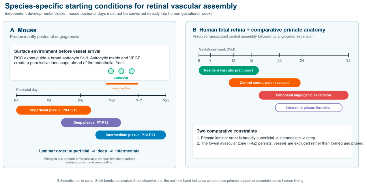
Figure 2.
Overlapping developmental modules from surface patterning to a perfused, barrier-competent retinal circulation. The figure separates tissue conditioning, sprout selection, lumenization/perfusion, vertical invasion and barrier specialization, while emphasizing that growth, lumen continuity, perfusion, branch retention and selective barrier function are non-equivalent readouts. TSPAN12 is shown as a potentiator of Norrin–FZD4–LRP5/6 signalling, and endothelial PDGF-B recruitment of pericytes is separated from pericyte VEGFR1 control of ligand availability. RGC, retinal ganglion cell; PDGF, platelet-derived growth factor; PDGFR, PDGF receptor; ECM, extracellular matrix; VEGF, vascular endothelial growth factor; VEGFR, VEGF receptor; Dll4, delta-like ligand 4; PFKFB3, 6-phosphofructo-2-kinase/fructose-2,6-bisphosphatase 3; D-tip, diving-tip endothelial cell; TGFβ, transforming growth factor beta; ALK5, activin receptor-like kinase 5; HIF, hypoxia-inducible factor; MFSD2A, major facilitator superfamily domain-containing 2A; PLVAP, plasmalemma vesicle-associated protein; iBRB, inner blood-retinal barrier. Representative evidence [5,6,7,10,11,15,16,19,20,21,22,23,24,67,68,69,70,71,72,73,74,82,83,84,85,86,87,88,89,90,91,92,93,94,95,96,97,98,99,100,101,102,103]. Original figure created for this review.
Figure 2.
Overlapping developmental modules from surface patterning to a perfused, barrier-competent retinal circulation. The figure separates tissue conditioning, sprout selection, lumenization/perfusion, vertical invasion and barrier specialization, while emphasizing that growth, lumen continuity, perfusion, branch retention and selective barrier function are non-equivalent readouts. TSPAN12 is shown as a potentiator of Norrin–FZD4–LRP5/6 signalling, and endothelial PDGF-B recruitment of pericytes is separated from pericyte VEGFR1 control of ligand availability. RGC, retinal ganglion cell; PDGF, platelet-derived growth factor; PDGFR, PDGF receptor; ECM, extracellular matrix; VEGF, vascular endothelial growth factor; VEGFR, VEGF receptor; Dll4, delta-like ligand 4; PFKFB3, 6-phosphofructo-2-kinase/fructose-2,6-bisphosphatase 3; D-tip, diving-tip endothelial cell; TGFβ, transforming growth factor beta; ALK5, activin receptor-like kinase 5; HIF, hypoxia-inducible factor; MFSD2A, major facilitator superfamily domain-containing 2A; PLVAP, plasmalemma vesicle-associated protein; iBRB, inner blood-retinal barrier. Representative evidence [5,6,7,10,11,15,16,19,20,21,22,23,24,67,68,69,70,71,72,73,74,82,83,84,85,86,87,88,89,90,91,92,93,94,95,96,97,98,99,100,101,102,103]. Original figure created for this review.
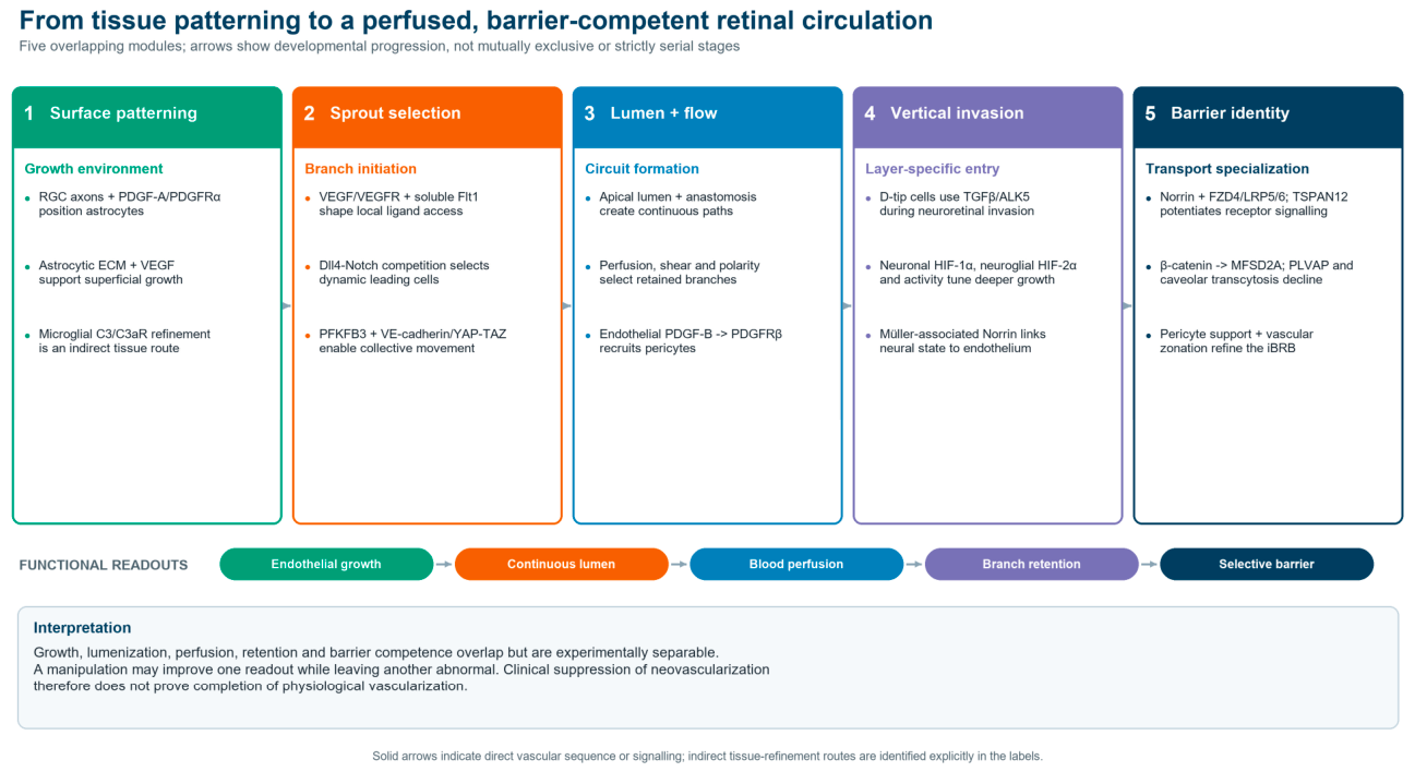
Disclaimer/Publisher’s Note: The statements, opinions and data contained in all publications are solely those of the individual author(s) and contributor(s) and not of MDPI and/or the editor(s). MDPI and/or the editor(s) disclaim responsibility for any injury to people or property resulting from any ideas, methods, instructions or products referred to in the content. |
© 2026 by the authors. Licensee MDPI, Basel, Switzerland. This article is an open access article distributed under the terms and conditions of the Creative Commons Attribution (CC BY) license (http://creativecommons.org/licenses/by/4.0/).
Copyright: This open access article is published under a Creative Commons CC BY 4.0 license, which permit the free download, distribution, and reuse, provided that the author and preprint are cited in any reuse.