

Submitted:
02 August 2026
Posted:
05 August 2026
You are already at the latest version
Abstract
The retinal neurovascular unit (NVU) is not assembled by endothelial cells alone. It emerges through overlapping developmental interactions among neurons, astrocytes, Müller glia, microglia, endothelial cells, pericytes, extracellular matrix and blood flow. These interactions are often inferred from the postnatal mouse retina, yet human retinal vascularization follows a different developmental logic: the early central circulation has substantial histological and immunophenotypic evidence for vasculogenesis from resident vascular precursors, whereas later peripheral and intraretinal expansion proceeds mainly by angiogenesis. Laminar order also differs: the mouse develops superficial, deep and then intermediate plexuses, whereas comparative primate histology supports a superficial, intermediate and then deep sequence, with the clearest fetal timing resolved in macaque and human maturation extending after birth. Here, we integrate species-resolved anatomy with causal developmental studies to explain how vascular growth, perfusion, pruning and inner blood–retinal barrier (iBRB) maturation are coordinated but separable. Retinal ganglion cell axons and astrocytes organize the superficial interface; extracellular matrix, VEGF, Dll4–Notch, endothelial metabolism and haemodynamic forces regulate sprouting and remodelling; microglia refine astrocytic and neuronal populations and influence vascular topology; neural activity and Müller-cell relays direct laminar angiogenesis; and Norrin–FZD4–β-catenin signalling, suppression of caveolar transcytosis, pericyte recruitment and endothelial zonation establish barrier competence. We distinguish causal perturbation from depletion-associated effects, pharmacology, spatial association and hypothesis. We then benchmark fetal tissue, animal models, organoids and stem-cell-derived vascular systems against the functions they claim to reproduce. This framework focuses disease translation on retinopathy of prematurity and inherited Norrin-pathway vasculopathies, in which developmental timing and barrier specialization are central. The resulting synthesis defines five principles: species and compartment specify mechanism; multicellular relays matter; growth, perfusion and barrier acquisition are distinct outputs; refinement is active; and reciprocal stabilization is limited by developmental state and time.
Keywords:
retinal neurovascular unit
; retinal vascular development
; vasculogenesis
; angiogenesis
; inner blood–retinal barrier
; astrocytes
; Müller glia
; microglia
; pericytes
; Norrin
; MFSD2A
; retinal organoids
Key points
- Human retinal vascular development is not a slower version of the mouse astrocyte-template programme. Evidence supports early central vasculogenesis followed by predominantly angiogenic peripheral and intraretinal expansion, with a superficial–intermediate–deep primate sequence rather than the superficial–deep–intermediate mouse sequence.
- Superficial growth, laminar invasion, perfusion, remodelling and iBRB maturation overlap in time but must be measured as distinct outcomes.
- Functional iBRB formation requires more than tight-junction expression: developmental suppression of endothelial transcytosis, MFSD2A induction, PLVAP downregulation, transporter specialization, mural-cell investment and vascular-segment zonation are central.
- Microglia refine the developing retina through several separable actions. Evidence for astrocyte removal and complement-dependent network refinement is causal; other reported microglia–Müller and microglia–endothelial effects are depletion-associated or context-specific
- Neural activity instructs vascular lamination through transmitter- and cell-specific relays, including cholinergic, dopaminergic and glutamatergic pathways coupled to glial or endothelial programmes.
- Engineered vascularization should be benchmarked by stable cell identity, lumenization, flow, quantitative permeability, transcytosis, mural investment, zonation and neural function—not by endothelial markers or network morphology alone.
1. From retinal vascular Development to Reciprocal NVU Assembly
The mature inner retina contains superficial, intermediate and deep vascular plexuses arranged around neuronal and synaptic layers. Endothelial cells form the iBRB, pericytes invest the microvessel wall, astrocytes occupy the nerve-fibre layer, Müller cells traverse the retinal thickness, and microglia patrol layer-specific niches [1,2,3]. This organization is frequently described as a stable anatomical unit. Development instead reveals a moving system in which one population alters the abundance, position, state or competence of another. The relevant object is therefore not a fixed ring of cells around a capillary, but a set of spatially and temporally restricted signalling circuits that eventually produce a perfused, barrier-competent and neuron-supportive vascular network.
The distinction matters because adjacent cells are not necessarily interacting, and expression of a ligand–receptor pair is not proof of a developmental relay. A causal claim should specify the initiating cell, responding cell, developmental window, retinal compartment and functional endpoint. It should also distinguish vascular extension from branch selection, lumenization, perfusion, regression, mural coverage and permeability. These outputs can move in opposite directions: a dense network may remain poorly perfused or leaky, and an endothelial cell may express CLDN5 without suppressing vesicular transport [4,5,6,7].
An influential review by Selvam, Kumar and Fruttiger synthesized retinal vascular development in health and disease, with particular emphasis on astrocytes, endothelial patterning, pericytes, Norrin signalling and oxygen-induced retinopathy [8]. The present review addresses a different, post-2018 question: how is the complete retinal NVU reciprocally assembled, and how should the strength of evidence for each proposed intercellular mechanism be judged? This shift brings neural activity, microglial refinement, Müller-cell relays, transcellular barrier maturation, endothelial zonation, human–mouse divergence and engineered-model validation into one causal framework. It also prevents novelty from resting on a longer catalogue of pathways.
Five analytical rules organize the synthesis. First, species and compartment define what a result can establish. The postnatal mouse retina is a powerful mechanistic system but not a developmental clock for the human fetal retina. Second, intercellular relays should be resolved rather than collapsed into a direct neuron-to-vessel or microglia-to-vessel effect. Third, growth, perfusion and barrier acquisition are related but separable. Fourth, removal of excess cells and low-flow branches is an active part of assembly. Fifth, developmental effects depend on cell state and timing; an intervention that is beneficial during one window may be ineffective or disruptive in another [4,5,9,10].
This framework also sharpens the boundary of the review. The focus is the inner retinal circulation and iBRB. The hyaloid circulation, choroid and retinal pigment epithelium are considered when they change the developmental environment, but they constitute distinct vascular and barrier systems. The optic nerve head is likewise not interchangeable with the intraretinal circulation. It is the entry region for vessels and astrocyte precursors and later becomes a specialized neurovascular compartment. Claims derived from optic stalk, brain barrier or adult retinal disease studies are therefore used only when their anatomical scope is explicit [3,11,12].
2. Species-Resolved Sequence and Developmental Modes
2.1. Mouse: A Postnatal Angiogenic Sequence
In C57BL/6 mice, the superficial plexus grows radially from the optic nerve head after birth and approaches the periphery by approximately postnatal day (P)8. Vertical sprouts begin forming the deep plexus around P7, which expands toward the periphery by approximately P12. The intermediate plexus appears later, around P12, and expands between approximately P12 and P15; remodelling continues through the third postnatal week. Thus, the canonical mouse order is superficial, deep and then intermediate, although exact boundaries vary with strain, retinal region and the marker or functional endpoint used [13,14,15,16]. This accessible schedule permits whole-mount quantification, inducible genetics, pharmacology and live or ex vivo imaging at defined stages.
The superficial sequence is predominantly angiogenic. Astrocyte precursors migrate from the optic nerve, spread ahead of endothelial sprouts and mature in relation to retinal ganglion cell (RGC) axons and vascular contact [17,18,19,20]. Endothelial tip cells extend over this pre-existing cellular and matrix landscape, whereas stalk cells proliferate, lumenize and connect the network. Blood flow then redistributes endothelial cells and stabilizes or removes branches [4]. The mouse retina therefore offers exceptional resolution of angiogenesis, but it does not model de novo central vessel assembly from resident human retinal vascular precursors.
The deeper plexuses have a different cellular environment. Astrocytes are concentrated superficially, whereas Müller-cell radial processes, neuronal somata and synaptic layers dominate the intraretinal path. Cholinergic retinal activity, RGC-derived dopamine and glutamatergic signalling affect layer-specific vascular entry through distinguishable relays [21,22,23]. A mechanism that explains radial movement over astrocytes should not automatically be assigned to vertical invasion.
Extrauterine conditions are another model boundary. Mouse retinal vascularization occurs after birth, during exposure to atmospheric oxygen, postnatal nutrition and maturing visual inputs. Human retinal vessels form mainly in utero. A postnatal-day to gestational-week conversion may align gross morphology, but it cannot reproduce placental physiology, oxygen history, foveal morphogenesis or the prolonged human developmental schedule [9].
2.2. Human: Central Vasculogenesis Followed by Angiogenesis
Human fetal evidence changes the organizing model. Retinal vascularization begins after the hyaloid and choroidal systems and contains at least two developmental modes. In whole-mounted fetal retinas from 14–38 weeks of gestation (WG), Hughes and colleagues observed spindle-shaped mesenchymal precursors entering from the optic disc before 15 WG, endothelial cords and some patent central vessels by 15 WG. The authors interpreted formation of the initial central plexus as vasculogenesis; subsequent increases in density and peripheral and temporal extension occurred through angiogenesis. Buds from existing inner vessels generated what that study termed the outer plexus, beginning around the incipient fovea at approximately 25–26 WG, and angiogenesis also formed the radial peripapillary capillaries [10].
Laminar order is a second, separate species difference. In the comparative monkey–human histological series of Gariano and colleagues, the nerve-fibre/ganglion-cell-layer plexus formed first; the capillary plexus at the inner border of the inner nuclear layer (INL) appeared before the plexus at the outer INL border. Mapped to current three-plexus nomenclature, this is a superficial–intermediate–deep sequence, rather than the mouse superficial–deep–intermediate sequence. The numerical fetal-day landmarks were resolved most clearly in macaque, whereas human tissue was sampled more sparsely and vascular maturation continued after birth. Macaque fetal days should therefore not be converted directly into human gestational weeks. Hughes's 25–26 WG observation establishes angiogenic intraretinal budding in human tissue but, by itself, does not resolve the intermediate and deep plexuses in current terminology [10,11,24].
Studies of earlier tissue strengthened the precursor model. McLeod and colleagues identified scattered CD39-positive cells in the inner retina at 6–8 WG and ahead of formed vessels at later ages. These cells co-expressed CXCR4 during migration and retained CD39 while differentiating into vascular endothelium, with CXCR4 downregulation after incorporation [25]. Hasegawa and colleagues further localized CXCR4- and c-Kit-positive precursor populations and the candidate guidance ligands SDF-1 and stem cell factor in the inner retina from 6–23 WG [26]. Together, these studies provide convergent histological and immunophenotypic evidence for a resident precursor pool participating in central retinal vasculogenesis.
The evidence should not be overstated. Human fetal studies cannot perform lineage tracing or cell-specific perturbation, and marker combinations do not prove clonal origin. Terms such as angioblast, vascular precursor and endothelial progenitor have not always been used identically. The strongest conclusion is therefore that early central human retinal vessel assembly contains a substantial vasculogenic component consistent with differentiation and coalescence of resident precursors; it is not established that one marker-defined lineage accounts for every early endothelial cell [10,11,25,26].
Astrocytes participate in human retinal development but should not be treated as the sole initiating template. Human fetal studies describe relationships among astrocytes, endothelial cells and the growing vascular front, and astrocyte precursors migrate from the optic nerve region [27,28,29]. Yet the presence of CD39/CXCR4-positive vascular precursors before formed vessels and the mixed vasculogenic–angiogenic sequence preclude simple transfer of the mouse template model [25,26]. In humans, astrocytes may organize, stabilize or guide parts of the emerging superficial circulation without being the exclusive source of its first endothelial geometry.
Lutty and McLeod integrated these observations into an ocular sequence: early hyaloid and choroidal vascular systems support the eye, the retinal circulation develops last, and central vasculogenesis gives way to angiogenic expansion and remodelling [11]. They also discussed possible Müller-cell Notch, axonal neuropilin/semaphorin and foveal antiangiogenic cues. These are biologically plausible and spatially supported, but the human evidence is largely histological or associative. They should be framed as candidate guidance or exclusion mechanisms rather than established causal relays. Developing-human-retina atlases add molecular context, but do not replace lineage or perturbation evidence [30,31].
2.3. Fovea and Non-Human Primate Evidence
The foveal centre remains avascular while vessels and astrocytes define a surrounding rim [29,32,33,34,35,36]. This arrangement cannot be reduced to delayed vascular arrival: the mature foveal avascular zone is a persistent specialization coupled to neuronal displacement, extreme cone packing and reliance on choroidal support [37,38,39]. Histology implicates restricted astrocyte distribution, antiangiogenic signals and the organization of axons and Müller cells, but no model reproduces all components causally. Statements about an “active foveal exclusion programme” should therefore be presented as a research problem, not a solved mechanism.
Human and non-human primate evidence must also remain separate. Macaque tissue clarifies prenatal laminar plexus formation, retinal activity and foveal or macular organization under experimentally controlled conditions [24,38,40]. It does not automatically establish the timing or cellular origin of human vessels. Conversely, human fetal histology provides direct species relevance but limited perturbational power [10,25,26]. A strong inference usually requires concordance across human anatomy, primate organization and mouse or engineered-system causality, with each source assigned its proper role.
2.4. Integrated Cellular Chronology of NVU Assembly
Reciprocal assembly can be interpreted only when the relevant populations are placed on the same developmental clock. Tables 1A and 1B therefore distinguish first observation, migration or expansion, anatomical association and functional maturation. These are not interchangeable endpoints: temporal precedence permits an interaction but does not prove one, and the first published detection of a cell is not necessarily its biological time of origin. Mouse and human ages are shown on independent axes because a postnatal mouse day is not equivalent to a human gestational week [9,41].

Table 1.
A. Integrated chronology of mouse retinal NVU assembly.
| Window | Vessels | Astrocytes | Microglia | Pericytes and barrier |
|---|---|---|---|---|
| E8.5–E11.5 | No intraretinal circulation | Retinal colonization has not begun | Primitive yolk-sac myeloid lineages emerge around E8.5–E9.5; retinal microglia are detected by approximately E11.5 | No retinal vascular substrate for mural investment or an iBRB |
| E17–P0 | Superficial angiogenic outgrowth initiates from the optic nerve head around birth | Precursors enter from the optic nerve region from approximately E17 and spread over the retinal nerve-fibre layer | Already resident before the astrocytic and endothelial waves | Recruitment begins with growing endothelium; the evidence does not justify a separate retinal pericyte “birth date” |
| P1–P7/8 | The superficial plexus advances radially and approaches the periphery | The astrocytic network remains ahead of the vascular front; abundance peaks around P5 | Number rises through the first postnatal week to approximately twice adult abundance around P7 | By P5, pericytes cover most endothelial stalks but are absent from leading tip cells; junctions are restrictive while transcytosis remains comparatively high |
| P5–P15 | Deep plexus develops mainly from P7–P12; intermediate plexus begins around P12 and expands through approximately P15 | Total astrocyte abundance falls more than threefold from P5–P14 through microglia-mediated developmental removal | P5 and P10 populations include phagocytic and angiogenic-front-associated states; Hmox1-positive localization at the wavefront is spatial/transcriptomic association, not proof of angiogenic causality | Transcytosis is progressively suppressed and tracer restriction becomes evident around P10, depending on assay; mural coverage stabilizes retained branches |
| P17–P28 | Three-plexus topology remodels toward the mature state | Remaining astrocytes acquire stable vessel-associated organization | Developmental phagocytic programmes decline by P17 and population/topography approach steady state by approximately P28 | Pericytes support established capillaries while transport and segment-specific barrier programmes continue to mature |
Table 1.
B. Integrated chronology of human retinal NVU assembly.
| Window | Vessels | Astrocytes | Microglia | Pericytes and barrier |
|---|---|---|---|---|
| 6–8 WG | CD39-positive, CXCR4-positive vascular precursors occur before patent retinal vessels | Retinal colonization is not securely demonstrated in this interval | No secure retinal observation in the sampled developmental series | No defensible retinal first-detection landmark |
| Approximately 9–12 WG | Precursors are present and begin to organize around the optic disc by approximately 12 WG | Colonization is reported to begin around 9–10 WG; direct fetal series demonstrates astrocyte–vascular-front relationships from 12 WG onward | Retinal microglia are present by approximately 10 WG and show regional and laminar unevenness at 12 WG | First recruitment and functional barrier competence remain unresolved |
| 14–21 WG | Endothelial cords organize by 12–14 WG; patent central vessels are present by approximately 15 WG; angiogenic meshes become prominent at 17–21 WG | Precursors and differentiating astrocytes occur near the expanding front but are not the exclusive initiators of the central circulation | Distribution becomes more even by approximately 20 WG, although regional differences remain | NG2-positive mural/pericyte-like cells are observed on the abluminal vessel surface and around the advancing front at approximately 17–20 WG |
| 25–32 WG | Intraretinal budding is visible around 25–26 WG; superficial vascularization approaches the retinal limits by approximately 32 WG | Astrocytes have nearly reached the periphery by approximately 25 WG but remain excluded from the foveal centre and temporal raphe | Laminar maturation continues, but a mouse-like quantitative postnatal trajectory has not been established | Anatomical coverage increases, but marker expression or apposition does not establish functional fetal iBRB competence |
| Late gestation–postnatal | Comparative primate evidence supports superficial, then inner-INL/intermediate, then outer-INL/deep lamination; human maturation continues after birth | Foveal astrocyte exclusion persists | Postnatal human developmental kinetics remain poorly resolved | Functional human iBRB maturation has not been mapped with the temporal and transport resolution available in mouse |
Figure 1.
Species-resolved chronology of retinal NVU assembly. Mouse and human axes are deliberately independent and are not scaled against one another. Each track separates appearance or expansion from anatomical association and functional maturation; dashed human intervals denote unresolved postnatal kinetics or functional barrier timing. Human intermediate-then-deep order is mapped from comparative primate histology. Mouse chronology is compiled from vascular, astrocytic, microglial, mural-cell and barrier studies [5,6,13,14,18,19,42,43,44,45,46,47]. Human chronology is compiled from fetal vascular, astrocytic, microglial, mural-cell and comparative primate studies [10,11,24,25,26,27,33,36,48,49].
Figure 1.
Species-resolved chronology of retinal NVU assembly. Mouse and human axes are deliberately independent and are not scaled against one another. Each track separates appearance or expansion from anatomical association and functional maturation; dashed human intervals denote unresolved postnatal kinetics or functional barrier timing. Human intermediate-then-deep order is mapped from comparative primate histology. Mouse chronology is compiled from vascular, astrocytic, microglial, mural-cell and barrier studies [5,6,13,14,18,19,42,43,44,45,46,47]. Human chronology is compiled from fetal vascular, astrocytic, microglial, mural-cell and comparative primate studies [10,11,24,25,26,27,33,36,48,49].
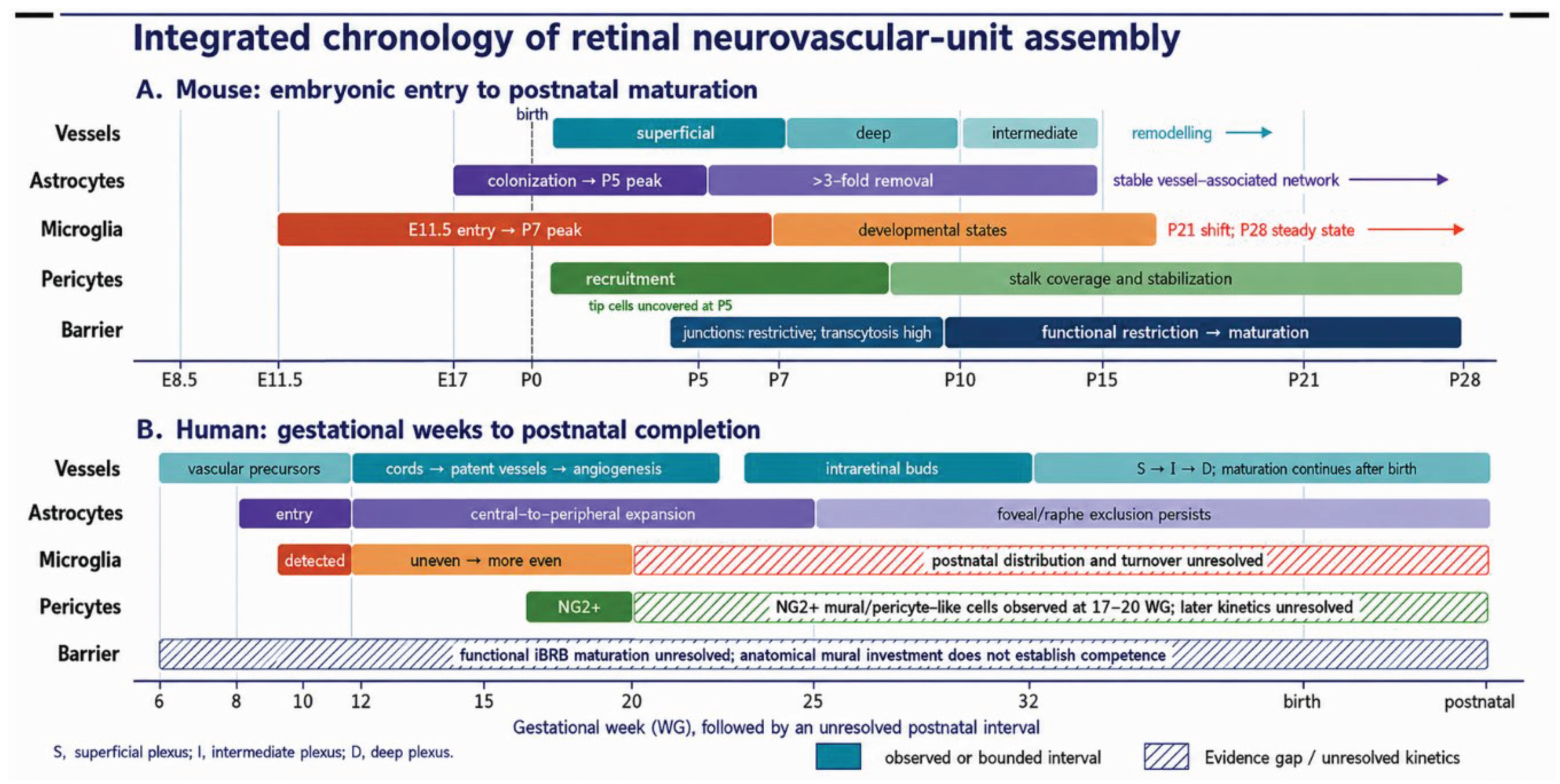
The synchronized chronology changes the causal interpretation. In mouse, microglial entry precedes astrocyte colonization, which precedes the postnatal endothelial wave and accompanying mural recruitment. In human, resident vascular precursors are documented before patent vessels and before the best-resolved astrocyte and microglial observations. Glial presence before perfusion therefore cannot establish that astrocytes exclusively initiate the human superficial circulation. Likewise, mural association precedes proof of barrier competence, and microglial precedence does not prove instruction of astrocyte migration [25,26,41,49].
3. Constructing and Remodelling the Superficial Interface
3.1. RGC Axons, Astrocytes and Extracellular Matrix
Mouse astrocyte precursors enter the retina from the optic nerve and migrate along the inner surface. RGC axons provide geometry, while neuron-derived platelet-derived growth factor A (PDGF-A) acting through astrocytic PDGFRα expands and positions the precursor population [19,50]. The resulting network is not a static stencil. Astrocytes proliferate, differentiate, alter morphology near vessels and are later reduced by developmental cell removal [51,52]. Vascular contact also feeds back on astrocyte maturation and network stabilization [20]. The astrocytic mesh therefore encodes a changing balance among neural input, matrix assembly, oxygen and vascular contact.
The chronology sharpens this mechanism. Mouse retinal colonization begins around E17, astrocyte abundance peaks near P5, and the population falls more than threefold between P5 and P14 through a developmental removal programme in which microglia perform much of the phagocytosis [18,41,42]. In human retina, a cross-species synthesis places the beginning of astrocyte colonization around 9–10 WG, while direct fetal series document astrocyte-precursor relationships with the advancing circulation from approximately 12 WG and near-peripheral coverage by approximately 25 WG, with persistent foveal and temporal-raphe exclusion [10,27,41]. These observations define coexistence windows; they do not establish a human equivalent of every mouse astrocyte-removal mechanism.
Astrocytes and retinal cells organize a permissive extracellular environment. Astrocytic fibronectin supports endothelial adhesion and radial migration; laminin chains and retinal proteoglycans regulate astrocyte migration, basement-membrane assembly and angiogenesis; endothelial filopodia align with pre-existing astrocyte and matrix tracks [53,54,55,56]. These perturbations support a matrix-guidance mechanism more strongly than simple co-localization does. They also show why matrix should be treated as part of the relay: a protein deposited by one cell can retain growth factors or transmit adhesion after the producer has changed state.
Hypoxia-induced VEGF from neuroglial sources supplies a major growth input [57]. VEGF, however, does not specify every branch or layer. Its spatial distribution depends on cellular source, isoform, extracellular-matrix binding and oxygenation. Tip cells interpret local VEGF through filopodia, while Dll4–Notch competition restricts neighbouring cells from adopting the same tip state [58,59]. This lateral inhibition explains how a broad growth field can be converted into selected leaders and ordered branches.
The framework is reciprocal. Endothelial advance changes local oxygen and astrocyte maturation; vascular contact helps stabilize the astrocytic template, while oxygenation suppresses hypoxia-responsive growth signals [20,57]. Vessels therefore read an astrocyte-rich landscape while simultaneously altering it. Evidence for specific endothelial-to-neural trophic factors during normal retinal development remains less direct than evidence for neural- and glial-to-endothelial control. Reciprocal feedback should be claimed only when an endothelial perturbation produces a defined neural or glial response that is not secondary to gross perfusion failure.
3.2. Endothelial state, Metabolism and Mechanical Integration
Tip–stalk selection is only one dimension of endothelial state. Cells at the front migrate and sample guidance cues; stalk cells proliferate and lumenize; perfused endothelial cells polarize with flow; arterial, venous and capillary programmes diverge; and barrier transport properties emerge along a proximal-to-distal and segment-specific axis [4,5,59,60]. Single-cell identities should therefore be interpreted as positions in a changing developmental landscape rather than fixed cell types.
Endothelial sprouting is metabolically active. PFKFB3-driven glycolysis supports tip-cell migration and stalk-cell proliferation in experimental vascular systems that include the neonatal retina [61]. This finding supports a metabolic requirement for sprouting; it does not establish that retinal endothelial metabolism is controlled by a unique neuron-derived cue. Any claim of a retina-specific metabolic relay requires cell-specific manipulation within the developing retina and a matched vascular endpoint.
Mechanical signalling also has direct but bounded retinal support. Endothelial YAP and TAZ regulate adherens-junction dynamics, cell rearrangement and distribution during vascular development, including in the neonatal mouse retina [62]. This establishes a mechanosensitive endothelial programme, not a general causal link between retinal tissue stiffness and plexus depth. Claims about matrix rigidity, intraocular pressure or Müller-cell tension instructing developmental angiogenesis remain hypotheses until those inputs are manipulated in vivo.
3.3. Flow, Branch Selection and Regression
An endothelial cord, a lumenized vessel and a blood-carrying vessel are not equivalent. Once flow begins, shear stress polarizes endothelial cells and redistributes them within the network. In the developing mouse retina and zebrafish, endothelial migration away from low-flow segments and toward higher-flow branches contributes to pruning; regression can therefore occur through rearrangement rather than endothelial apoptosis alone [4]. Flow converts a redundant primitive plexus into a hierarchical circuit.
This mechanism changes how vascular phenotypes should be read. Reduced branch number can reflect failed sprouting, accelerated pruning or loss of perfusion. Increased vascular area can reflect excessive branching, enlarged calibre or delayed regression. Whole-mount studies should pair topology with perfusion and, where possible, flow direction or endothelial polarity. Fixed anatomy without a flow measure cannot establish that the network is functional [4,63].
Flow also intersects with barrier and mural-cell maturation. Shear-dependent endothelial state can change endothelial polarity and arterial–venous identity, while oxygen delivery suppresses local proangiogenic signals [4,57,60]. We therefore distinguish two feedback loops: a tissue loop in which oxygenation modifies glial growth cues, and a vascular loop in which flow stabilizes efficient paths and removes inefficient ones. These loops overlap with, but are conceptually distinct from, the MFSD2A/caveolar programme that suppresses transcytosis [5,64].
4. Microglia as Refiners and Relay Partners
4.1. Identity and Developmental Context
Retinal microglia derive from primitive myeloid lineages and colonize the tissue before completion of the vascular network [43,44,65,66,67]. In mouse, retinal microglia are detected by approximately E11.5, increase from P1 to a peak near P7 at roughly twice adult abundance, and approach a population steady state by approximately P28 [41,44]. Age-resolved profiling at P5, P10 and P17 further shows a transition from heterogeneous developmental and phagocytic programmes toward predominantly homeostatic profiles; Hmox1-positive microglia at the P5 angiogenic wavefront are a spatial and transcriptomic observation, not evidence that HMOX1 causally drives normal retinal angiogenesis [45].
Human microglia are present by approximately 10 WG, show regionally and laminarly uneven distribution at 12 WG, and become more evenly distributed by approximately 20 WG, although regional differences persist [41,48]. A mouse-like quantitative postnatal trajectory has not been established. Morphology, transcriptional state and spatial niche therefore must be assigned to age, retinal layer and region rather than collapsed into one state map [68,69,70]. Adult human and primate studies add molecular and macular specialization but cannot fill the missing fetal-to-postnatal chronology [70,71,72]. A rigorous study must also distinguish parenchymal microglia from hyalocytes in the vitreous, perivascular or border-associated macrophages and recruited monocytes. This is particularly important in depletion studies and oxygen-induced retinopathy, where drugs or Cre drivers may affect more than one myeloid compartment.
Microglial proximity to sprouting vessels was recognized early, and depletion or manipulation can alter vascular density and branching [73]. Proximity alone does not reveal whether microglia promote anastomosis, remove cellular obstacles, alter astrocytes, respond to hypoxia or phagocytose dying cells. The most defensible developmental functions are those in which a target, pathway and downstream network effect have been linked.
4.2. Astrocyte and Neuronal Refinement
During normal mouse development, a large astrocyte population is removed by microglia through a non-apoptotic process [42]. Complement provides one mechanistic bridge: C3/C3aR perturbation changes microglial refinement of astrocytic and vascular networks [74]. These data support an active microglia–astrocyte–vessel relay rather than a generic proangiogenic microglial action. The primary target measured is the astrocyte network, and the vascular phenotype should be interpreted through that intermediate unless a direct endothelial signal is separately demonstrated.
Microglia also remove newborn RGCs and respond to developmental apoptosis. Complement receptor-dependent engulfment and neuronal “do not eat me” signalling through SIRPα/CD47 contribute to target selection, while developmental apoptosis induces a distinct retinal microglial programme [68,75,76]. This neuronal refinement can indirectly change the angiogenic environment by altering cell number, axonal geometry or trophic output. Demonstrating neuronal cargo inside microglia proves uptake, but not necessarily that microglia initiated death. Time-resolved imaging or survival after phagocytic blockade is needed to separate primary killing from clearance.
These functions should not be merged into a single microglial state. Engulfing an astrocyte, clearing an apoptotic RGC, contacting an endothelial junction and supporting a Müller cell may involve different histories and niches [68,69,70]. Transcriptomic clusters can nominate pathways, but cell-state labels are not causal mechanisms unless linked to a temporally defined interaction and functional endpoint.
4.3. Microglia–Müller–Vascular Coupling: What is Established
Gestational and postnatal PLX5622 exposure has been reported to reduce Müller-cell maturation markers and decrease deep or intermediate vascular density at P10–P15 [77]. This is an important depletion-associated result because it connects resident myeloid cells to intraretinal vascularization and Müller state. It does not by itself identify a microglial ligand, establish Müller cells as the sole intermediate, or exclude effects of depletion on hyalocytes and other macrophage populations. The mechanism should therefore be described as a microglia-dependent developmental association with a candidate Müller relay, pending source-cell and rescue experiments.
Xu and colleagues reported altered retinal branching following PLX5622 treatment, assessed at P42 [78]. In oxygen-induced retinopathy, Liu and colleagues described a microglial HMOX1–endothelial STAT3 signalling axis associated with retinal angiogenesis [79]. Together, these studies support context-dependent roles for microglia in retinal vascular remodelling, while their findings should not be generalized directly to microglia–endothelial interactions during normal retinal development.
Table 2.
Evidence levels for representative multicellular mechanisms.
| Evidence class | Representative mechanism | What the evidence supports | What it does not yet establish |
|---|---|---|---|
| Cell-specific or pathway-level causal perturbation | RGC/PDGF–astrocyte expansion; Dll4–Notch tip selection; complement-dependent astrocyte refinement; activity-dependent Norrin relay; endothelial MFSD2A control | A defined pathway changes a specified developmental endpoint in a bounded model | Universal conservation across species or developmental windows |
| Depletion-associated | Microglia depletion with altered Müller maturation and intraretinal vascular density | The depleted compartment is required for the composite phenotype under that regimen | The direct ligand, sole intermediate or exclusivity to parenchymal microglia |
| Pharmacological | Receptor inhibition or broad pathway manipulation in explants or whole animals | Pathway sensitivity under the treatment conditions | Cell-autonomous action or absence of off-target effects |
| Spatial or temporal association | Human vascular precursors with candidate SDF-1/SCF, Müller/axonal or foveal guidance cues | Anatomical plausibility and developmental ordering | Causal source–target signalling |
| Context-specific experimental evidence | PLX5622-associated branching changes at P42; microglial HMOX1–endothelial STAT3 signalling in OIR | Microglia can influence retinal vascular architecture at P42 and pathological angiogenesis in OIR | A shared mechanism or direct generalization to normal developmental angiogenesis |
| Hypothesis | Active foveal exclusion; microglial history controlling later vascular support | A testable integrated model | An established mechanism |
5. Neural Activity and Müller Glia Direct Laminar Angiogenesis
Developmental neurovascular coupling is distinct from adult functional hyperaemia. Patterned retinal waves provide a changing developmental input, and specific cholinergic, dopaminergic and glutamatergic perturbations can alter vascular architecture or barrier state over days [21,22,23,80,81]. The effect is not one generic “activity” signal: transmitter system, firing pattern, developmental window and responding cell determine the outcome.
In the developing mouse retina, cholinergic activity generated by starburst amacrine circuits regulates layer-specific angiogenesis and iBRB formation [21]. RGC-derived dopamine provides another instructive signal for vascular lamination [22]. Glutamatergic activity regulates intraretinal angiogenesis and barrier maturation through Norrin/β-catenin signalling, with genetic and rescue evidence linking neural input to the endothelial programme [23]. Together, these studies establish that neural maturation does more than increase oxygen demand: particular activity-dependent relays alter where vessels grow and which barrier features they acquire.
Müller cells are well positioned to translate these signals. Their radial processes span the retina, contact neurons and vessels, and can contribute VEGF- and Norrin-related outputs in a state-dependent manner [21,23,57]. Yet cellular source must be demonstrated rather than assumed from anatomy. A neural perturbation followed by a whole-retina change in Vegfa or Ndp expression does not identify the producing cell. Strong relay evidence combines source-specific manipulation, target-cell pathway readout, spatial correspondence and rescue.
Norrin is especially important because it connects laminar vascular growth to barrier identity. Endothelial FZD4/LRP5/TSPAN12 signalling activates β-catenin-dependent programmes required for intraretinal vascularization and CNS barrier properties [15,64,82]. Activity-dependent Norrin regulation does not make every Norrin effect activity-dependent, and VEGF and Norrin should not be reduced to interchangeable angiogenic factors. VEGF is a major driver of growth and permeability; Norrin–FZD4 signalling is particularly important for endothelial specialization, intraretinal invasion and suppression of transcytosis.
We propose a coincidence-detector model rather than a one-signal switch. Intraretinal sprouting is predicted to succeed when a competent endothelial cell encounters the appropriate glial signal in a permissive neural layer during the correct activity state; the components are supported individually, but their factorial interaction has not been demonstrated [15,21,22,23,82]. Testing this model requires factorial designs—activity, Müller-cell output and endothelial receptor competence—rather than single perturbations interpreted in isolation.
6. Pericytes and Construction of the Functional iBRB
6.1. Barrier Maturation has Parallel Gates
The iBRB is an endothelial phenotype with at least four coordinated components: restrictive intercellular junctions, low vesicular transcytosis, selective influx and efflux transport, and non-fenestrated vascular identity. Basement membrane, pericytes, glial contacts, Norrin signalling and flow help establish or maintain this state. Barrier maturation cannot be inferred from CLDN5, OCLN or TJP1 staining alone [3,5,6,47,64].
Developmental tracer and ultrastructural studies in mouse retina show why. Chow and Gu found that newly entering retinal vessels already possessed functionally restrictive tight junctions, whereas leakage occurred through abundant transcytotic vesicles. Functional sealing emerged gradually from the optic nerve head toward the vascular front as transcytosis was suppressed. Mfsd2a loss maintained vesicular transport and delayed sealing, whereas Cav1 loss reduced caveolae and produced earlier barrier closure [5]. The limiting developmental gate was therefore transcellular, not a simple absence of junctions.
An independent developmental analysis reached a compatible but not identical conclusion. Tight-junction proteins were present by P5 before complete functional barrier formation, while endothelial PLVAP declined and was absent when the barrier became functionally restrictive [47]. PLVAP is associated with diaphragmed fenestrae and transendothelial channels and is a useful marker of permissive endothelial transport. However, the transient vascular delay in Plvap-deficient mice means that PLVAP should not be described only as a passive immaturity marker; its developmental role may intersect with vascular growth and endothelial differentiation.
Together, these studies support a two-gate developmental model: the paracellular gate becomes restrictive early, while the transcellular gate closes gradually through loss of caveolar transport and acquisition of CNS endothelial lipid and transport programmes [5,47,64]. A third analytical gate—selective transporter maturation—determines which metabolites and solutes can cross even after bulk leakage is suppressed. Barrier function therefore requires both exclusion and controlled exchange [3].
Placed on the cellular timeline, barrier components are deliberately asynchronous. In neonatal mouse retina, pericyte association and junctional-protein expression are already evident before complete suppression of transcytosis and tracer-defined restriction around P10 [5,6,46,47]. Neither mural proximity nor a junctional marker can therefore assign a single “barrier birth date.” In human fetal retina, the available record is even more limited: anatomical mural investment is observable, but an age-resolved functional permeability and transcytosis series comparable to mouse has not been established [11,49].
6.2. Norrin–FZD4, MFSD2A, Caveolae and Endothelial Zonation
Norrin–FZD4–LRP5/TSPAN12 signalling integrates vascular patterning with barrier specialization. In Ndp- and Lrp5-deficient mouse retinas, increased leakage is accompanied by elevated endothelial transcytosis. Wnt/β-catenin signalling directly promotes MFSD2A, and MFSD2A suppresses caveolin-1-positive caveolar transport; restoring MFSD2A can reduce the transcytotic phenotype [64]. The pathway therefore links a retina-enriched ligand system to a fundamental CNS barrier mechanism.
Barrier identity is not uniform along the vascular tree. Arterial, venous and capillary endothelial cells experience different flow, oxygen and ligand environments. Netrin-1/UNC5B and Norrin pathways interact to control arteriovenous zonation of barrier integrity, demonstrating that leakage can be segment-selective rather than a global endothelial state [60]. APCDD1 likewise coordinates vascular remodelling and barrier maturation [83]. These findings argue against reporting one whole-retina “barrier score” without vessel-segment annotation.
Endothelial zonation also changes the meaning of markers. PLVAP re-expression can indicate a permissive or pathological transport state, but its distribution should be assigned to vessel class. MFSD2A is a transcytosis suppressor and lipid transporter, not a complete barrier identity on its own. CLDN5 continuity reports junctional organization, while transporter panels report selective exchange. A mature iBRB phenotype requires concordance across these axes and functional permeability [47,60,64].
6.3. Pericyte Recruitment, Coverage and Developmental Plasticity
Pericytes are recruited to growing retinal vessels through endothelial PDGF-B and mural PDGFRβ. The extracellular-matrix retention motif of PDGF-B is required for concentrated perivascular signalling and proper pericyte investment; disrupting it produces incomplete microvessel coverage and retinal vascular abnormalities [46]. Pericyte number, longitudinal coverage and direct apposition are related but not interchangeable measures. A vessel can be near a PDGFRβ-positive cell without continuous mural contact.
Developmental landmarks should describe recruitment and ensheathment rather than imply an independently established pericyte origin date. In mouse retina at P5, PDGFRβ-positive pericytes cover most of the growing endothelial stalk while leading tip cells remain uncovered [6,46]. In human fetal retina, NG2-positive mural/pericyte-like cells have been observed on the abluminal surface of retinal vessels at 17 WG, adjacent to main vessels and just ahead of the vascular front at 18 WG, and within or ahead of the vascular plexus at 20 WG [49]. These are direct anatomical landmarks, but the study's principal emphasis was choroidal mural-cell formation; the retinal observations do not establish the first onset, lineage origin, completeness of coverage or functional contribution to the fetal iBRB.
Developmental and adult requirements differ. Park and colleagues showed that impaired PDGF-B/PDGFRβ-dependent recruitment disrupts formation and maturation of the mouse BRB. In stable adult retinal vessels, selective pericyte loss did not immediately dissolve the barrier but sensitized endothelial cells to VEGF-A and an ANG2–FOXO1 feedback state [6]. Pericytes are therefore indispensable developmental partners and continuing modulators of endothelial susceptibility, but adult barrier maintenance has partial redundancy over the time window tested.
Pericyte function should be evaluated alongside basement membrane and endothelial transport. Coverage can limit endothelial proliferation, modify VEGFR2 signalling, support matrix organization and stabilize capillaries [6,46,84]. Yet a change in coverage does not establish which of these mechanisms caused leakage. The strongest experiments combine developmental timing, mural-cell contact, endothelial signalling, perfusion, ultrastructure and permeability.
6.4. Flow-Dependent Remodelling and Barrier Competence
Flow and barrier maturation overlap without being identical. Perfusion supplies the shear and oxygen feedback that remodel the network, whereas suppression of transcytosis determines whether perfused vessels retain circulating macromolecules. A vessel can carry blood and still leak; a non-perfused endothelial tube cannot demonstrate a functional barrier at all. Regression of low-flow segments, mural stabilization of retained capillaries and segment-specific Norrin or Netrin signalling must therefore be considered together [4,60].
Developmental permeability assays need explicit design. Tracer molecular size, charge, route, circulation time and fixation affect the result. Endogenous proteins may reveal chronic leakage, whereas intravenously delivered dextrans or biotin report a defined interval. Electron microscopy distinguishes junctional escape from tracer-filled vesicles. In vitro trans-endothelial electrical resistance is useful but does not replace solute-specific permeability, and it is highly sensitive to culture geometry and electrode conditions [5,7,47].
Table 3.
Minimum evidence for claiming functional iBRB maturation.
| Domain | Minimum measurement | Stronger validation | Common over-interpretation |
|---|---|---|---|
| Vascular structure | Continuous endothelial network and lumen | Three-dimensional lumen continuity and basement membrane | A PECAM1-positive cord is a vessel |
| Perfusion | Intravascular tracer or red-cell transit | Quantified flow direction and velocity | Passive medium access is perfusion |
| Paracellular gate | Junctional continuity plus size-defined permeability | Ultrastructural junctional restriction | CLDN5/TJP1 staining proves an intact barrier |
| Transcellular gate | MFSD2A/PLVAP/CAV1 profile | Vesicle quantification and transcytosis assay | Low bulk leakage proves suppressed transcytosis |
| Selective transport | Influx/efflux transporter panel | Substrate-specific flux and inhibition | One transporter defines retinal endothelial identity |
| Mural support | Pericyte identity and quantitative coverage | Direct apposition, contact duration and functional perturbation | PDGFRβ proximity equals mature investment |
| Zonation | Artery, vein and capillary annotation | Segment-resolved permeability and transcriptomics | A whole-retina average represents every vessel |
| Neural compatibility | Viability and layer organization | Electrophysiology, circuit response and metabolic coupling | Larger tissue or more markers equals an assembled NVU |
7. Model Systems: Causal Power and Benchmarked Limitations
7.1. The Model Should Match the Question
No available model reconstructs the entire human retinal NVU. The neonatal mouse retina offers cell-specific genetics, topology, perfusion and barrier assays, but lacks a fovea and uses a postnatal angiogenic sequence [5,14]. Human fetal tissue directly captures prenatal cell relationships and early vasculogenesis, but is scarce, cross-sectional and not experimentally perturbable [10,25,26]. Non-human primate tissue approximates foveated anatomy and prenatal lamination but remains limited in access and manipulation [24,33]. Engineered systems provide controlled human cells and repeated observation while omitting or simplifying haemodynamics, immune history and developmental geometry [7,85].
The claim should therefore be scaled to the model. A two-cell assay can establish contact-dependent signalling, not NVU reconstruction. An organoid can demonstrate trophic effects on neural survival without reproducing a mature iBRB. A vascular network can be lumenized but unperfused, and a perfused channel can lack retinal endothelial identity [5,7,84]. Comparative benchmarking is more informative than asking whether one system is “physiological.”
Species translation also requires tissue matching. Miao and colleagues compared adult human and mouse brain blood–brain barrier capillary endothelial transcriptomes and identified species-associated expression differences [12]. The study supports a general caution against untested cross-species transfer, but species, tissue procurement, cohort and platform are not separable in a retrospective cross-dataset comparison. Because it concerns adult brain rather than developing retina, it cannot establish the human–mouse difference in retinal vascular origin, plexus order or barriergenesis.
7.2. Vascularized Retinal Organoids
Recent studies have introduced endothelial or vascular-like compartments into human retinal organoids through fusion, microwell culture or inducible endothelial differentiation [7,86,87]. These platforms address an important limitation of conventional organoids: diffusion-limited hypoxia and loss of inner retinal neurons. Their value depends on whether the paper's actual endpoint is maintained in the interpretation.
The study by Sharma and colleagues is especially informative because the full article and supplement permit a precise reading [7]. ETV2.2 induction generated endothelial-like cells and transient PECAM1-positive networks. The vascularized organoids were larger, less hypoxic and less apoptotic, retained more RGC-associated markers and later displayed improved electrophysiological outcomes. These findings support transient endothelial trophic and structural benefit.
They do not demonstrate reconstruction of a mature retinal NVU or iBRB. The induced cells were not authenticated as retinal endothelial cells; endothelial structures regressed and “empty sleeves” remained; robust pericyte investment and vascular zonation were not shown; and CLDN5 or TJP1 expression did not establish functional barrier restriction. Dextran entered some lumens after overnight exposure from the culture medium, a passive-access assay rather than flow-based perfusion. Endothelial cells were not recovered as a defined population in the week-18 single-cell/nuclear dataset, so their late identity remains unresolved. The neural data also cannot completely separate increased RGC survival from increased differentiation [7].
The microfluidic functional experiments require another qualification: primary rat astrocytes were included. The system was therefore not entirely human, and improved axonal or electrophysiological behaviour cannot be assigned exclusively to vascularization. Later light-evoked ON, OFF and ON–OFF responses are promising, but additional photoreceptor morphology, pharmacological circuit dissection and stimulus validation are required before attributing them to mature photoreceptor-driven retinal circuits [7]. These limitations do not diminish the study; they define the specific advance accurately.
7.3. Stem-Cell-Derived Retinal Endothelium and Microvessels
Human pluripotent-stem-cell-derived retinal endothelial cells offer a complementary route to barrier modelling and cell therapy [88]. Their evaluation should include retinal and CNS endothelial identity, response to Norrin/FZD4, suppression of PLVAP and caveolar transport, transporter function, stability under flow, interaction with pericytes and glia, and comparison with primary human retinal endothelium [5,6,47,64]. Generic endothelial markers cannot distinguish a retinal barrier cell from an immature vascular cell.
Co-culture can test individual relays. Isogenic endothelial–pericyte systems can quantify contact, VEGFR2 activity, migration and leakage [84]; organoid–vascular fusion can test survival and spatial integration [7,86,87]; microfluidic chips can impose flow and measure solute transport. The strongest designs use the same perturbation across levels—for example, a Norrin-pathway genotype assessed in two-dimensional signalling, perfused microvessels and neural organoids—while retaining cell-line and differentiation-batch replication [85,89].
7.4. A Causal Model Ladder
Table 4.
Model ladder for retinal NVU questions.
| Model | Strongest use | Essential benchmark | Cannot establish alone |
|---|---|---|---|
| Neonatal mouse whole mount | Angiogenic sequence, topology, cell-specific causality, flow and tracer leakage | Age, strain, layer, perfusion and vessel-segment annotation | Human central vasculogenesis or foveal development |
| OIR mouse | Vaso-obliteration, reparative regrowth and pathological tufting | Separate quantification of avascular area, regrowth, tufts and leakage | Normal development or human ROP in full |
| Human fetal tissue | Prenatal timing, cell distribution and precursor phenotypes | Gestational age, region, preservation and multiple lineage markers | Lineage or causal source–target signalling |
| Non-human primate | Foveated anatomy, prenatal lamination and circuit context | Species and developmental-stage matching | Automatic equivalence to human timing or gene regulation |
| Conventional retinal organoid | Human neural differentiation and cell-line comparison | Staging, reproducibility and fetal/adult reference mapping | Perfusion, immune history or a complete iBRB |
| Vascularized organoid | Endothelial trophic support and multicellular spatial interaction | Stable endothelial identity, lumens, flow, mural cells and transport | Mature NVU from marker expression alone |
| Retinal endothelial/pericyte co-culture | Contact-dependent signalling and quantitative permeability | Isogenic controls, barrier transport and flow where relevant | Tissue lamination or circuit function |
| Perfused retinal chip | Haemodynamics, transport and controlled multicellular perturbation | Physiological shear, solute-specific flux and cell identity | Whole-organ developmental history |
7.5. Measurement and inference discipline
Retinal vascular area, radial outgrowth and branch points answer different questions. An astrocyte-guidance study should quantify astrocyte density, state and matrix geometry together with endothelial alignment [20,56]. A microglial-refinement study should identify cargo, distinguish cell death from clearance and measure astrocyte, neuronal and vascular outcomes [42,75]. A pericyte study should quantify coverage and direct apposition rather than report marker-positive cells nearby [6,46]. A barrier study should combine permeability with junctional continuity, transcytosis, transporter state and vascular segment [5,47,60].
Single-cell and spatial methods nominate mechanisms but introduce their own biases. Dissociation can activate glia, lose fragile endothelial populations and erase contact history. Cell abundance reflects recovery as well as biology. Ligand–receptor scores do not measure secretion, receptor localization or pathway activation. Human retinal and organoid atlases are essential references for cell identity and developmental staging, but atlas similarity is not proof of vascular function [85,89]. The correct endpoint is a perturbation that validates a spatially plausible prediction in the relevant developmental window.
Biological replication is equally important. The animal or donor, not the image or vessel segment, is the replicate. Organoid lines and differentiation batches should be modelled explicitly [85,89]. Sex, litter, oxygen exposure, Cre-driver genotype, depletion regimen and vascular region can all modify the phenotype; OIR protocols illustrate why litter, weight and retinal region require explicit handling [63]. High-dimensional measurement cannot rescue an under-replicated design.
8. Developmental Vasculopathies and Therapeutic Logic
8.1. Retinopathy of Prematurity and Oxygen-Induced Retinopathy
Retinopathy of prematurity (ROP) is the clearest disease extension of developmental NVU assembly because premature birth interrupts an incompletely vascularized human retina. Postnatal oxygen exposure, fluctuating oxygenation, nutrition and systemic illness alter a process that normally occurs in utero. The disease contains at least two vascular phases: delayed or arrested physiological vascularization followed by hypoxia-driven pathological neovascularization, while neural and glial development continues [91].
Mouse oxygen-induced retinopathy (OIR) separates hyperoxia-associated central vaso-obliteration from hypoxia-driven regrowth and neovascular tuft formation after return to room air [63,90]. It is a powerful model of defined vascular endpoints, not a complete replica of human ROP. An intervention can reduce tufts by suppressing all growth while leaving a larger avascular retina; reporting tuft area alone would then misclassify the outcome. Reparative revascularization, pathological neovascularization, leakage, mural coverage and neural function should be measured separately.
Developmental pathways can change effect with disease stage. VEGF supports reparative regrowth but also contributes to pathological permeability and tufting when spatially or temporally dysregulated [63,90,91]. Microglia that refine astrocytes during normal development may occupy oxidative or inflammatory states in OIR, and astrocyte and Müller-cell signals are altered by hypoxia and injury. The therapeutic objective is therefore not to restore an embryonic programme wholesale, but to recover orderly perfused growth while closing transcellular and paracellular leak pathways.
8.2. Norrin-Pathway Disorders
Familial exudative vitreoretinopathy (FEVR) and Norrie disease provide human genetic evidence that vascular extension and barrier specialization are coupled through the Norrin pathway. Pathogenic variants affecting FZD4 and TSPAN12 disrupt retinal vascular development, while NDP, LRP5 and downstream pathway defects produce overlapping phenotypes of incomplete peripheral vascularization, exudation or severe retinal disorganization [82,92,93]. The range of severity emphasizes pathway dosage, allelic context and developmental timing.
These disorders are not simply “low angiogenesis.” Experimental loss of Norrin/FZD4 signalling alters intraretinal vascularization, endothelial barrier genes and transcytosis [15,64]. A corrective strategy should therefore be evaluated for peripheral extension, deep-plexus organization, perfusion, mural support and solute transport. Increased endothelial area without barrier rescue would be incomplete.
8.3. Therapeutic Criteria, Not Premature Prescriptions
The developmental synthesis suggests testable treatment sequences but does not justify a universal combination. We propose testing whether controlled reparative growth followed by mural/barrier stabilization, or restoration of Norrin/FZD4 competence alongside limitation of persistent permeability signals, produces a better functional network than simultaneous maximal pathway suppression. These are hypotheses, not treatment recommendations; order, dose, vascular segment and developmental window must be tested against perfusion, leakage and neural endpoints [6,63,64].
Anti-VEGF agents have demonstrated clinical efficacy in selected severe ROP settings, including bevacizumab in BEAT-ROP and ranibizumab in RAINBOW [94,95]. This review does not compare regimens or make a clinical recommendation. Developmental evidence supports a narrower criterion: suppression of pathological growth is not equivalent to reconstruction of a complete, functional NVU. Neonatal interventions should therefore be evaluated for peripheral vascularization, retreatment or recurrence, barrier transport and later ocular and neural outcomes, not only short-term leakage or tuft regression [91].
Microglial intervention requires the same discipline. Broad depletion can remove cells involved in astrocyte refinement, Müller maturation and homeostatic support while also suppressing disease-associated states [42,74,77]. A rational target should be a verified pathway in a defined microglial state and niche, with evidence that beneficial developmental functions are preserved. The microglial HMOX1–endothelial STAT3 axis described in OIR represents a context-specific candidate for therapeutic investigation, but its relevance to normal developmental angiogenesis remains to be established [79].
9. Five Principles and an Experimental Agenda
- Principle 1: species and compartment specify mechanism
Human central vasculogenesis, mouse postnatal superficial angiogenesis, primate foveal specialization and adult barrier maintenance are different biological problems [6,14,25,33]. Every mechanism should state species, developmental age, retinal region, plexus and vessel segment. Cross-species convergence increases confidence, but anatomical similarity alone does not prove conserved causality.
- Principle 2: NVU assembly uses relays
RGC-derived PDGF acts through astrocytes before endothelial growth; neural activity changes glial or endothelial Norrin-related programmes; microglia refine astrocytes and associate with Müller maturation; endothelial PDGF-B recruits pericytes [23,46,50,74,77]. Resolving these intermediate cells explains why a signal can have different outcomes in different layers or developmental windows.
- Principle 3: growth, perfusion and barrier acquisition are distinct
Endothelial extension, lumenization, blood flow, branch pruning, junctional restriction, transcytosis suppression and selective transport are coupled but separable [4,5,7,47]. Each requires a matched assay. This principle is the central benchmark for engineered vascularization and for therapeutic revascularization.
- Principle 4: refinement is active
Microglial removal of astrocytes or newborn neurons and flow-driven regression of inefficient branches are constructive events [4,42,75]. Successful development does not maximize cell or vessel number; it produces an efficient topology matched to neural layers and metabolic demand.
- Principle 5: stabilization is reciprocal and time-limited
Vessels alter oxygenation and astrocyte state; pericytes change endothelial susceptibility; neural activity changes vascular layer entry; and barrier pathways remain partly plastic in disease [6,20,21,64]. These reciprocal effects operate within windows of cellular competence. Acute adult perturbations cannot be assumed to reveal developmental function, and developmental activation cannot be assumed to be safe in injured adult tissue.
-
Experimental priorities
- 1.
- Resolve human vascular precursor lineage. Combine high-quality fetal spatial data with lineage-informed stem-cell models and clone-aware genomic approaches to distinguish resident vasculogenic precursors from sprouting endothelium without treating marker co-expression as lineage proof.
- 2.
- Build an integrated human fetal NVU atlas. Map endothelium, mural cells, astrocytes and myeloid cells against gestational age, central-to-peripheral region, vascular layer and vessel segment, and pair spatial identity with functional barrier measurements. The atlas must preserve donor-level variation and must not substitute marker proximity for developmental interaction [41,49].
- 3.
- Measure human barrier maturation functionally. Establish gestationally staged retinal endothelial references for junctions, transcytosis, transporters and arteriovenous/capillary zonation, then use them to benchmark engineered models.
- 4.
- Test the microglia–Müller relay directly. Pair temporally restricted, compartment-specific manipulation with Müller-cell rescue and deep/intermediate plexus permeability and perfusion endpoints.
- 5.
- Link flow to barrier zonation. Combine segment-resolved haemodynamics, endothelial polarity, MFSD2A/PLVAP state and tracer leakage during normal development and revascularization.
- 6.
- Reconstruct the foveal boundary experimentally. Use primate and human spatial data to test candidate astrocytic, axonal, Müller-cell and antiangiogenic exclusion cues in patterned human systems.
- 7.
- Adopt causal model ladders. Validate a relay in reductionist human culture, a perfused multicellular system and an intact developmental model, with the same direction of perturbation and explicitly different claims at each level.
| Box 1. Minimum evidence for a multicellular developmental mechanism |
These reporting criteria synthesize recurring limitations in causal vascular, barrier, microglial and engineered-model studies [4,5,7,42].
- Define species, age, retinal region, vascular layer and physiological or disease state.
- Identify the initiating cell and responding cell with cell-specific perturbation or rescue.
- Establish temporal order and spatial co-occurrence before claiming a relay.
- Measure each proposed intermediate, not only the final endothelial phenotype.
- Distinguish microglia from hyalocytes, border-associated macrophages and recruited monocytes.
- Measure perfusion and permeability when claiming functional vascularization or barrier formation.
- Separate paracellular leakage, transcytosis and selective transporter function.
- Treat animal, donor or independent stem-cell line as the biological replicate and model litter or differentiation batch.
- State what the experiment cannot establish; this is part of the mechanism, not an editorial afterthought.
10. Conclusions
Retinal vascular development is the reciprocal construction of a layered neurovascular system. The integrated chronology now makes its temporal logic explicit: in mouse, microglial entry precedes astrocyte colonization and postnatal angiogenesis, while mural recruitment and barrier gates mature alongside the growing plexus; in human, vascular precursors precede patent vessels and the best-resolved glial observations, whereas the timing of functional iBRB acquisition remains incompletely mapped [18,25,41,44,46,49]. Mouse studies explain how RGCs, astrocytes, matrix, microglia, neural activity, Müller glia, endothelial cells, pericytes and flow cooperate during angiogenesis and barriergenesis [5,6,20,23,50,74]. Human fetal evidence imposes two further corrections: the early central retinal circulation contains a vasculogenic precursor-associated phase before predominantly angiogenic peripheral and intraretinal expansion, and comparative primate histology supports superficial–intermediate–deep lamination rather than the mouse superficial–deep–intermediate sequence [10,11,24,25,26]. The species therefore illuminate different parts of the problem.
A rigorous synthesis must keep vascular growth, perfusion, pruning and barrier acquisition separate. Tight-junction proteins are not a complete iBRB; suppression of transcytosis, selective transport, mural investment and endothelial zonation are essential [5,6,47,60,64]. Likewise, endothelial-like networks in organoids are not equivalent to stable perfused retinal vessels [7]. When evidence levels are explicit, emerging models become more valuable because their advances can be stated precisely.
The next phase of the field should move beyond pairwise catalogues toward predictive multicellular experiments. A successful model should explain where a vessel forms, whether it carries blood, why it is retained or removed, when it becomes non-leaky, how it changes neighbouring neural and glial cells and whether the resulting circuit functions. Those criteria integrate the functional endpoints exposed by developmental pruning, barrier and engineered-model studies [4,5,7] and provide a common standard for developmental biology, inherited vasculopathy, ROP and regenerative engineering.
Author Contributions
M.T. is the sole author of this work and is responsible for all aspects of it, including conceptualization, literature synthesis, writing—original draft, and writing—review and editing. The author has read and agreed to the submitted version of the manuscript.
Funding
This research received no external funding.
Data Availability Statement
No new data were generated or analysed in this review. All sources discussed are cited in the reference list below.
Acknowledgments
The author declares no acknowledgments.
Conflicts of Interest
The author declares no conflicts of interest.
Declarations
During the preparation of this review, the author used ChatGPT (OpenAI) to support manuscript structuring, wording refinement, Figure 1 development and critical editorial review. The author reviewed and revised the final manuscript and takes full responsibility for its content, citations, interpretations and conclusions.
References
- Schnitzer, J. (1987) Retinal astrocytes: their restriction to vascularized parts of the mammalian retina. Neurosci Lett 78, 29-34. [CrossRef]
- Stone, J. and Dreher, Z. (1987) Relationship between astrocytes, ganglion cells and vasculature of the retina. J Comp Neurol 255, 35-49. [CrossRef]
- Hudson, N. and Campbell, M. (2021) Tight junctions of the neurovascular unit. Front Mol Neurosci 14, 752781. [CrossRef]
- Franco, C.A. et al. (2015) Dynamic endothelial cell rearrangements drive developmental vessel regression. PLoS Biol 13, e1002125. [CrossRef]
- Chow, B.W. and Gu, C. (2017) Gradual suppression of transcytosis governs functional blood-retinal barrier formation. Neuron 93, 1325–1333.e3. [CrossRef]
- Park, D.Y. et al. (2017) Plastic roles of pericytes in the blood–retinal barrier. Nat Commun 8, 15296. [CrossRef]
- Sharma, K. et al. (2026) Retinal ganglion cell survival and functional maturation in transiently vascularized human retinal organoids. Cell Stem Cell 33, 253-271.e13. [CrossRef]
- Selvam, S. et al. (2018) Retinal vasculature development in health and disease. Prog Retin Eye Res 63, 1–19. [CrossRef]
- Van Cruchten, S. et al. (2017) Pre- and Postnatal Development of the Eye: A Species Comparison. Birth Defects Res 109, 1540-1567. [CrossRef]
- Hughes, S. et al. (2000) Vascularization of the human fetal retina: roles of vasculogenesis and angiogenesis. Invest Ophthalmol Vis Sci 41, 1217–1228. PMID: 10752963.
- Lutty, G.A. and McLeod, D.S. (2018) Development of the hyaloid, choroidal and retinal vasculatures in the fetal human eye. Prog Retin Eye Res 62, 58–76. [CrossRef]
- Miao, Y. et al. (2026) Distinct gene expression profiles in blood–brain barrier capillary endothelial cells between mice and humans. Microvasc Res 166, 104933. [CrossRef]
- Fruttiger, M. (2002) Development of the mouse retinal vasculature: angiogenesis versus vasculogenesis. Invest Ophthalmol Vis Sci 43, 522-527.
- Stahl, A. et al. (2010) The mouse retina as an angiogenesis model. Invest Ophthalmol Vis Sci 51, 2813–2826. [CrossRef]
- Wang, Y. et al. (2012) Norrin/Frizzled4 signaling in retinal vascular development and blood brain barrier plasticity. Cell 151, 1332-1344. [CrossRef]
- Biswas, S. et al. (2020) Neuronal and glial regulation of CNS angiogenesis and barriergenesis. Development 147. [CrossRef]
- Watanabe, T. and Raff, M.C. (1988) Retinal astrocytes are immigrants from the optic nerve. Nature 332, 834-837. [CrossRef]
- Huxlin, K.R. et al. (1992) The origin and development of retinal astrocytes in the mouse. J Neurocytol 21, 530-544. [CrossRef]
- O'Sullivan, M.L. et al. (2017) Astrocytes follow ganglion cell axons to establish an angiogenic template during retinal development. Glia 65, 1697-1716. [CrossRef]
- West, H. et al. (2005) Stabilization of the retinal vascular network by reciprocal feedback between blood vessels and astrocytes. Development 132, 1855–1862. [CrossRef]
- Weiner, G.A. et al. (2019) Cholinergic neural activity directs retinal layer-specific angiogenesis and blood retinal barrier formation. Nat Commun 10, 2477. [CrossRef]
- Liang, J.H. et al. (2023) Dopamine signaling from ganglion cells directs layer-specific angiogenesis in the retina. Curr Biol 33, 3821-3834 e3825. [CrossRef]
- Biswas, S. et al. (2024) Glutamatergic neuronal activity regulates angiogenesis and blood-retinal barrier maturation via Norrin/beta-catenin signaling. Neuron 112, 1978-1996.e6. [CrossRef]
- Gariano, R.F. et al. (1994) Vascular development in primate retina: comparison of laminar plexus formation in monkey and human. Invest Ophthalmol Vis Sci 35, 3442-3455. PMID: 8056520.
- McLeod, D.S. et al. (2006) The initial fetal human retinal vasculature develops by vasculogenesis. Dev Dyn 235, 3336–3347. [CrossRef]
- Hasegawa, T. et al. (2008) Vascular precursors in developing human retina. Invest Ophthalmol Vis Sci 49, 2178–2192. [CrossRef]
- Chan-Ling, T. et al. (2004) Astrocyte-endothelial cell relationships during human retinal vascular development. Invest Ophthalmol Vis Sci 45, 2020-2032. [CrossRef]
- Chu, Y. et al. (2001) Differentiation and migration of astrocyte precursor cells and astrocytes in human fetal retina: relevance to optic nerve coloboma. FASEB J 15, 2013-2015. [CrossRef]
- Ogden, T.E. (1978) Nerve fiber layer astrocytes of the primate retina: morphology, distribution, and density. Invest Ophthalmol Vis Sci 17, 499-5doi:10.
- Hoshino, A. et al. (2017) Molecular Anatomy of the Developing Human Retina. Dev Cell 43, 763-779 e764. [CrossRef]
- Lu, Y. et al. (2020) Single-Cell Analysis of Human Retina Identifies Evolutionarily Conserved and Species-Specific Mechanisms Controlling Development. Dev Cell 53, 473-491 e479. [CrossRef]
- Provis, J.M. et al. (2000) Astrocytes and blood vessels define the foveal rim during primate retinal development. Invest Ophthalmol Vis Sci 41, 2827-2836. PMID: 10967034.
- Provis, J.M. (2001) Development of the primate retinal vasculature. Prog Retin Eye Res 20, 799-821. [CrossRef]
- Provis, J.M. et al. (1997) Development of the human retinal vasculature: cellular relations and VEGF expression. Exp Eye Res 65, 555-568. [CrossRef]
- Hendrickson, A.E. and Yuodelis, C. (1984) The morphological development of the human fovea. Ophthalmology 91, 603-612. [CrossRef]
- Hendrickson, A. et al. (2012) Histologic development of the human fovea from midgestation to maturity. Am J Ophthalmol 154, 767-778 e762. [CrossRef]
- Curcio, C.A. and Allen, K.A. (1990) Topography of ganglion cells in human retina. J Comp Neurol 300, 5-25. [CrossRef]
- Sinha, R. et al. (2017) Cellular and Circuit Mechanisms Shaping the Perceptual Properties of the Primate Fovea. Cell 168, 413-426 e412. [CrossRef]
- Peng, Y.R. et al. (2019) Molecular Classification and Comparative Taxonomics of Foveal and Peripheral Cells in Primate Retina. Cell 176, 1222-1237 e1222. [CrossRef]
- Warland, D.K. et al. (2006) Dynamics of spontaneous activity in the fetal macaque retina during development of retinogeniculate pathways. J Neurosci 26, 5190-5197. [CrossRef]
- Huang, K.-C. et al. (2024) Retinal ganglion cell circuits and glial interactions in humans and mice. Trends Neurosci 47, 994–1013. [CrossRef]
- Punal, V.M. et al. (2019) Large-scale death of retinal astrocytes during normal development is non-apoptotic and implemented by microglia. PLoS Biol 17, e3000492. [CrossRef]
- Ginhoux, F. et al. (2010) Fate mapping analysis reveals that adult microglia derive from primitive macrophages. Science 330, 841-845. [CrossRef]
- Santos, A.M. et al. (2008) Embryonic and postnatal development of microglial cells in the mouse retina. J Comp Neurol 506, 224-239. [CrossRef]
- Martineau, S. et al. (2026) Microglia and myeloid cell populations of the developing mouse retina. Glia 74, e70115. [CrossRef]
- Lindblom, P. et al. (2003) Endothelial PDGF-B retention is required for proper investment of pericytes in the microvessel wall. Genes Dev 17, 1835–1840. [CrossRef]
- van der Wijk, A.-E. et al. (2019) Expression patterns of endothelial permeability pathways in the development of the blood-retinal barrier in mice. FASEB J 33, 5320–5333. [CrossRef]
- Diaz-Araya, C.M. et al. (1995) Development of microglial topography in human retina. J Comp Neurol 363, 53-68. [CrossRef]
- Chan-Ling, T. et al. (2011) Role of CD44+ stem cells in mural cell formation in the human choroid: evidence of vascular instability due to limited pericyte ensheathment. Invest Ophthalmol Vis Sci 52, 399–4doi:10. [CrossRef]
- Fruttiger, M. et al. (1996) PDGF mediates a neuron-astrocyte interaction in the developing retina. Neuron 17, 1117-1131. [CrossRef]
- Holden, J.M. et al. (2023) Retinal astrocyte morphology predicts integration of vascular and neuronal architecture. Front Neurosci 17, 1244679. [CrossRef]
- Paisley, C.E. and Kay, J.N. (2021) Seeing stars: Development and function of retinal astrocytes. Dev Biol 478, 144-154. [CrossRef]
- Stenzel, D. et al. (2011) Integrin-dependent and -independent functions of astrocytic fibronectin in retinal angiogenesis. Development 138, 4451-4463. [CrossRef]
- Gnanaguru, G. et al. (2013) Laminins containing the beta2 and gamma3 chains regulate astrocyte migration and angiogenesis in the retina. Development 140, 2050-2060. [CrossRef]
- Tao, C. and Zhang, X. (2016) Retinal Proteoglycans Act as Cellular Receptors for Basement Membrane Assembly to Control Astrocyte Migration and Angiogenesis. Cell Rep 17, 1832-1844. [CrossRef]
- Dorrell, M.I. et al. (2002) Retinal vascular development is mediated by endothelial filopodia, a preexisting astrocytic template and specific R-cadherin adhesion. Invest Ophthalmol Vis Sci 43, 3500-35doi:10. PMID: 12407162.
- Stone, J. et al. (1995) Development of retinal vasculature is mediated by hypoxia-induced vascular endothelial growth factor (VEGF) expression by neuroglia. J Neurosci 15, 4738-4747. [CrossRef]
- Gerhardt, H. et al. (2003) VEGF guides angiogenic sprouting utilizing endothelial tip cell filopodia. J Cell Biol 161, 1163-1177. [CrossRef]
- Hellström, M. et al. (2007) Dll4 signalling through Notch1 regulates formation of tip cells during angiogenesis. Nature 445, 776-780. [CrossRef]
- Furtado, J. et al. (2024) Interplay between Netrin-1 and Norrin controls arteriovenous zonation of blood-retina barrier integrity. Proc Natl Acad Sci U S A 121, e2408674121. [CrossRef]
- De Bock, K. et al. (2013) Role of PFKFB3-driven glycolysis in vessel sprouting. Cell 154, 651–663. [CrossRef]
- Neto, F. et al. (2018) YAP and TAZ regulate adherens junction dynamics and endothelial cell distribution during vascular development. eLife 7, e31037. [CrossRef]
- Connor, K.M. et al. (2009) Quantification of oxygen-induced retinopathy in the mouse: a model of vessel loss, vessel regrowth and pathological angiogenesis. Nat Protoc 4, 1565–1573. [CrossRef]
- Wang, Z. et al. (2020) Wnt signaling activates MFSD2A to suppress vascular endothelial transcytosis and maintain blood-retinal barrier. Sci Adv 6, eaba7457. [CrossRef]
- Kierdorf, K. et al. (2013) Microglia emerge from erythromyeloid precursors via Pu.1- and Irf8-dependent pathways. Nat Neurosci 16, 273-280. [CrossRef]
- Schulz, C. et al. (2012) A lineage of myeloid cells independent of Myb and hematopoietic stem cells. Science 336, 86-90. [CrossRef]
- Li, F. et al. (2019) Microglia in the developing retina. Neural Dev 14, 12. [CrossRef]
- Anderson, S.R. et al. (2019) Developmental Apoptosis Promotes a Disease-Related Gene Signature and Independence from CSF1R Signaling in Retinal Microglia. Cell Rep 27, 2002-2013 e2005. [CrossRef]
- O'Koren, E.G. et al. (2019) Microglial Function Is Distinct in Different Anatomical Locations during Retinal Homeostasis and Degeneration. Immunity 50, 723-737 e727. [CrossRef]
- Wolf, J. et al. (2022) In-Depth Molecular Profiling Specifies Human Retinal Microglia Identity. Front Immunol 13, 863158. [CrossRef]
- Penfold, P.L. et al. (1991) Antibodies to human leucocyte antigens indicate subpopulations of microglia in human retina. Vis Neurosci 7, 383-388. [CrossRef]
- Singaravelu, J. et al. (2017) Microglia in the primate macula: specializations in microglial distribution and morphology with retinal position and with aging. Brain Struct Funct 222, 2759-2771. [CrossRef]
- Checchin, D. et al. (2006) Potential role of microglia in retinal blood vessel formation. Invest Ophthalmol Vis Sci 47, 3595-3602. [CrossRef]
- Gnanaguru, G. et al. (2023) Microglia refine developing retinal astrocytic and vascular networks through the complement C3/C3aR axis. Development 150, dev201047. [CrossRef]
- Anderson, S.R. et al. (2019) Complement Targets Newborn Retinal Ganglion Cells for Phagocytic Elimination by Microglia. J Neurosci 39, 2025-2040. [CrossRef]
- Jiang, D. et al. (2022) Neuronal signal-regulatory protein alpha drives microglial phagocytosis by limiting microglial interaction with CD47 in the retina. Immunity 55, 2318-2335 e2317. [CrossRef]
- Rowthorn-Apel, N. et al. (2025) Microglial depletion decreases Müller cell maturation and inner retinal vascular density. Cell Commun Signal 23, 90. [CrossRef]
- Xu, T. et al. (2026) Microglia and retinal vascularity: a deeper dive into branching dynamics. Curr Eye Res 51, 276-284. [CrossRef]
- Liu, Y. et al. (2026) Microglial HMOX1 drives retinal angiogenesis via modulation of endothelial STAT3 signaling. Free Radic Biol Med 243, 29-42. [CrossRef]
- Bansal, A. et al. (2000) Mice lacking specific nicotinic acetylcholine receptor subunits exhibit dramatically altered spontaneous activity patterns and reveal a limited role for retinal waves in forming ON and OFF circuits in the inner retina. J Neurosci 20, 7672-7681. [CrossRef]
- Stacy, R.C. et al. (2005) Disruption and recovery of patterned retinal activity in the absence of acetylcholine. J Neurosci 25, 9347-9357. [CrossRef]
- Ye, X. et al. (2009) Norrin, Frizzled-4, and Lrp5 signaling in endothelial cells controls a genetic program for retinal vascularization. Cell 139, 285-298. [CrossRef]
- Mazzoni, J. et al. (2017) The Wnt inhibitor Apcdd1 coordinates vascular remodeling and barrier maturation of retinal blood vessels. Neuron 96, 1055-1069.e6. [CrossRef]
- Lin, Y.-Y. et al. (2025) Endothelial-pericyte interactions regulate angiogenesis via VEGFR2 signaling during retinal development and disease. Invest Ophthalmol Vis Sci 66, 45. [CrossRef]
- Capowski, E.E. et al. (2019) Reproducibility and staging of 3D human retinal organoids across multiple pluripotent stem cell lines. Development 146. [CrossRef]
- Inagaki, S. et al. (2025) Establishment of vascularized human retinal organoids from induced pluripotent stem cells. Stem Cells 43, sxae093. [CrossRef]
- Chen, H. et al. (2025) Generation of vascularized retinal organoids containing microglia based on a PDMS microwell platform. Sci Adv 11, eady64doi:10. [CrossRef]
- Lin, Y.-Y. et al. (2026) Derivation of functional retinal endothelial cells from human pluripotent stem cells for therapeutics and modelling. Nat Biomed Eng. [CrossRef]
- Cowan, C.S. et al. (2020) Cell Types of the Human Retina and Its Organoids at Single-Cell Resolution. Cell 182, 1623-1640 e1634. [CrossRef]
- Smith, L.E.H. et al. (1994) Oxygen-induced retinopathy in the mouse. Invest Ophthalmol Vis Sci 35, 101–111. PMID: 7507904.
- Hellström, A. et al. (2013) Retinopathy of prematurity. Lancet 382, 1445–1457. [CrossRef]
- Robitaille, J. et al. (2002) Mutant frizzled-4 disrupts retinal angiogenesis in familial exudative vitreoretinopathy. Nat Genet 32, 326–330. [CrossRef]
- Poulter, J.A. et al. (2010) Mutations in TSPAN12 cause autosomal-dominant familial exudative vitreoretinopathy. Am J Hum Genet 86, 248–253. [CrossRef]
- Mintz-Hittner, H.A. et al. (2011) Efficacy of intravitreal bevacizumab for stage 3+ retinopathy of prematurity. N Engl J Med 364, 603–615. [CrossRef]
- Stahl, A. et al. (2019) Ranibizumab versus laser therapy for the treatment of very low birthweight infants with retinopathy of prematurity (RAINBOW): an open-label randomised controlled trial. Lancet 394, 1551–1559. [CrossRef]
Disclaimer/Publisher’s Note: The statements, opinions and data contained in all publications are solely those of the individual author(s) and contributor(s) and not of MDPI and/or the editor(s). MDPI and/or the editor(s) disclaim responsibility for any injury to people or property resulting from any ideas, methods, instructions or products referred to in the content. |
© 2026 by the author. Licensee MDPI, Basel, Switzerland. This article is an open access article distributed under the terms and conditions of the Creative Commons Attribution (CC BY) license.
Copyright: This open access article is published under a Creative Commons CC BY 4.0 license, which permit the free download, distribution, and reuse, provided that the author and preprint are cited in any reuse.


