
Submitted:
03 August 2026
Posted:
05 August 2026
You are already at the latest version
Abstract
Background/Objectives: Among endocrine malignancies, thyroid cancer is the most prevalent and encompasses a broad clinical spectrum, ranging from indolent differentiated tumors to highly aggressive poorly differentiated and anaplastic thyroid carcinomas. Histopathology and molecular testing incompletely capture the spatial organization of tumor, stromal, and immune compartments that drives invasion, dedifferentiation, and treatment resistance. Spatial transcriptomics preserves tissue architecture while profiling gene expression, mapping molecular programs onto histologic context. We systematically reviewed its platforms, applications, and biological insights in thyroid cancer. Methods: PubMed/MEDLINE, Scopus, and Web of Science Core Collection were searched for English-language studies combining spatial-transcriptomic and thyroid-cancer concepts, following PRISMA 2020 guidance. Primary studies that applied a spatially resolved transcriptomic method to thyroid cancer tissue and reported spatial gene-expression findings were included; data were extracted on subtype, platform, complementary methods, and principal findings. Results: Of 104 records (47 after de-duplication), 21 studies met the inclusion criteria. Most used capture-based whole-transcriptome profiling, particularly 10x Visium, while GeoMx digital spatial profiling supported FFPE-compatible biomarker studies. Recurrent findings included POSTN+ and SERPINE1+ cancer-associated fibroblast programs at invasive fronts, APOE-linked immunosuppressive states, organized immune-evasion niches, and progressive tumor-microenvironment remodeling accompanying dedifferentiation toward ATC. Spatial transcriptomics also clarified invasion, lymph node and distant metastasis, and cell–cell communication. Conclusions: Spatial transcriptomics is opening new avenues for dissecting thyroid cancer progression, but current evidence is limited by small cohorts, platform concentration, and variable validation. Larger, standardized, clinically annotated studies are needed to define reproducible spatial biomarkers.
Keywords:
spatial transcriptomics
; thyroid cancer
; anaplastic thyroid carcinoma
; tumor microenvironment
; cancer-associated fibroblasts
; immune microenvironment
; metastasis
; 10x Visium
; GeoMx digital spatial profiling
1. Introduction
Endocrine malignancies are led in frequency by thyroid cancer, whose diagnosis rates have grown considerably across recent decades [1]. Thyroid neoplasms span a diverse histologic landscape under the 2022 World Health Organization framework. Tumors arising from follicular cells include papillary (PTC), follicular (FTC), oncocytic, poorly differentiated (PDTC), and anaplastic (ATC) carcinomas; these are joined by medullary thyroid carcinoma, a neoplasm traced instead to parafollicular C cells [2]. Indolence characterizes the natural history of most differentiated tumors, but not all: some return after treatment, seed distant organs, cease responding to radioiodine, or shed their differentiated phenotype to become PDTC or ATC. Of these, ATC ranks among the deadliest endocrine cancers, and patients frequently survive only months even after aggressive multimodal treatment [3]. Current risk stratification relies heavily on histopathologic features, anatomic staging, and selected molecular alterations; however, these approaches incompletely capture intratumoral heterogeneity, microenvironmental remodeling, and spatially organized tumor-stroma-immune interactions that contribute to invasion, immune escape, metastatic spread, and therapeutic resistance [4].
Traditional transcriptomic approaches have improved understanding of thyroid cancer biology but remain limited by loss of spatial context. Bulk RNA sequencing identifies tumor-level expression programs but returns one averaged profile per specimen, blending malignant, stromal, endothelial, and immune contributions. Single-cell RNA sequencing recovers per-cell resolution, but the dissociation step that makes it possible destroys the record of which cells were neighbors [21]. Spatial transcriptomics (ST) addresses this limitation differently: transcripts are assayed in situ, so every measurement carries the coordinates at which it was made on the histologic section [5,6,21]. The field was designated Nature Methods’ Method of the Year in 2020, reflecting its importance for linking molecular state to tissue architecture [5]. Tumors make this coordinate information consequential. A transcriptional program confined to the invasive boundary, to a lymphovascular interface, or to a dedifferentiated focus is diluted into the tissue average by any assay that discards position.
Several classes of ST platform are now in routine use, and each balance one capability at the expense of another. For example, subcellular localization is generally purchased with a preselected gene panel, while unbiased transcriptome-wide capture is purchased with spot-level averaging [7]. Capture-based platforms, derived from the original spatial transcriptomics approach described by Ståhl et al., profile RNA across spatially barcoded capture regions and are well suited for discovery-oriented whole-transcriptome studies [6]. Imaging-based platforms, including MERFISH, seqFISH-based methods, CosMx, and Xenium, provide single-cell or subcellular localization of targeted transcripts and are particularly useful for resolving cell states and cell-cell interactions at tumor boundaries [8,9,10]. Region-based platforms, such as GeoMx digital spatial profiling, quantify RNA or protein targets within pathologist-defined regions of interest and are compatible with formalin-fixed paraffin-embedded tissue, making them attractive for archival clinical specimens [11]. These complementary approaches are summarized in Table 1 and provide a framework for interpreting the strengths and limitations of the thyroid cancer ST literature.
Interest in applying ST to thyroid cancer has grown rapidly. Recent studies have used ST to characterize tumor-microenvironment remodeling across thyroid cancer differentiation states, fibroblast programs at invasive fronts, immune-evasion niches, spatial patterns of dedifferentiation, lymph node metastasis, and candidate biomarkers in ATC [12,13,14,15,16,17,18,19,20]. For example, ST studies have identified SERPINE1-positive [14] and POSTN-positive [15] cancer-associated fibroblast programs associated with aggressive behavior, APOE-linked malignant or immunosuppressive tumor states [14,18,20], and immune-suppressive, angiogenic, and extracellular-matrix remodeling during dedifferentiation toward ATC [13,16,17]. Narrative reviews have begun to summarize spatial and single-cell approaches in thyroid oncology [21]; however, a systematic, PRISMA-guided synthesis of the most recent advancements in the primary ST literature of thyroid cancer remains needed. Specifically, the field lacks a consolidated review that catalogs the platforms used, thyroid cancer subtypes studied, sample types analyzed, and recurrent biological findings across independent investigations. We therefore conducted a systematic review to map the current ST platform landscape in thyroid cancer and synthesize the major applications, discoveries, limitations, and future directions of spatially resolved transcriptomics across thyroid cancer subtypes.
2. Materials and Methods
This systematic review was conducted and reported in accordance with the Preferred Reporting Items for Systematic Reviews and Meta-Analyses (PRISMA) 2020 statement [22]. Eligibility criteria, database search strategy, screening approach, and extraction variables were defined before final study selection. The review was not registered in a public registry, and a formal protocol was not separately published; the a priori criteria described here were applied throughout without amendment. Because the objective was to map the available spatial-transcriptomics literature in thyroid cancer rather than estimate a pooled treatment effect, the review was designed as a qualitative evidence synthesis.
A systematic literature search was performed in PubMed/MEDLINE, Scopus, and Web of Science Core Collection with no lower date restriction; all three databases were last searched on July 10, 2026. Searches were limited to English-language records. Boolean search strategies combined terms related to spatial transcriptomics with terms related to thyroid cancer. Exact search criteria for each individual database are included in Supplemental Table 1 for reference. Search results from all databases were exported in RIS and/or CSV format for screening and de-duplication.
Studies were eligible for inclusion if they met all of the following criteria: primary research; application of a spatially resolved transcriptomic method to thyroid cancer tissue or a thyroid cancer dataset; reporting of spatial gene-expression findings; and inclusion of thyroid cancer-specific results. Studies using human thyroid cancer tissue were prioritized. Reanalyses of publicly available thyroid cancer spatial-transcriptomic datasets were included when they generated thyroid cancer-specific spatial findings.
Studies were excluded if they were reviews, editorials, commentaries, conference abstracts without full-text data, non-thyroid studies, pan-cancer studies without thyroid cancer-specific spatial-transcriptomic analysis, benign thyroid disease studies without cancer tissue, spatial-proteomics-only studies without a transcriptomic readout, technical methods papers without thyroid cancer application, studies without retrievable full text, or non-human model studies that did not include human thyroid cancer tissue.
Records were de-duplicated first by DOI and then by title. Unique records were screened by title and abstract against the eligibility criteria by two reviewers working independently, with disagreements resolved by consensus. Reports that appeared potentially eligible were retrieved for full-text review, and full-text articles were then independently assessed for final inclusion, with disagreements resolved by discussion. No automation tools were used during screening or selection.
Data were extracted into a standardized, pre-piloted extraction form by one reviewer and independently verified by a second reviewer, with disagreements resolved by consensus, and study investigators were not contacted for additional data. For each included study, extracted variables included first author, year of publication, journal, thyroid cancer subtype, cohort or sample size, tissue source, sample preparation method, spatial-transcriptomics platform, complementary methods, analytic approach, and principal spatial findings. Thyroid cancer subtypes were recorded as reported by the original studies, including papillary thyroid carcinoma, follicular thyroid carcinoma, poorly differentiated thyroid carcinoma, anaplastic thyroid carcinoma, medullary thyroid carcinoma, and metastatic thyroid cancer when applicable. Spatial platforms were categorized as capture-based whole-transcriptome methods, region-based digital spatial profiling methods, imaging-based targeted transcriptomic methods, or integrated spatial multi-omic approaches.
Complementary methods were also extracted when reported, including single-cell RNA sequencing, bulk RNA sequencing, whole-exome sequencing, immunohistochemistry, multiplex immunofluorescence, spatial proteomics, metabolomics, or computational deconvolution. Key findings were grouped by biological theme, including tumor-microenvironment remodeling, dedifferentiation, cancer-associated fibroblast programs, immune microenvironment organization, cell–cell communication, invasion, lymph node metastasis, distant metastasis, molecular classification, and candidate biomarker discovery.
Because no validated risk-of-bias instrument exists specifically for descriptive spatial-omics studies, included studies were appraised using the Joanna Briggs Institute (JBI) critical-appraisal checklist most closely matching each study design [23,24]. Single-case spatial-transcriptomics studies were assessed using the JBI checklist for case reports; descriptive multi-sample spatial-transcriptomics studies were assessed using the JBI checklist for case series; and comparative or outcome-oriented cohort studies were assessed using the JBI checklist for analytical cross-sectional studies. If studies did not fit existing clinical JBI checklist, they were appraised using a seven-item framework. This framework assessed data provenance, original-platform quality control, analytic pipeline description, batch-effect handling, independent or functional validation, code/data availability, and parameter transparency.
Methodological appraisal was performed by one reviewer and independently checked by a second reviewer, with disagreements resolved by consensus. Each appraisal item was rated as met, unclear/not reported, not met, or not applicable. An overall level of methodological concern was derived from the proportion of applicable items met: low concern, ≥75%; moderate concern, 50–74%; and high concern, <50%. This appraisal approach was adapted for an emerging spatial-omics literature and was intended to characterize reporting quality and methodological transparency rather than to function as a validated quantitative risk-of-bias score.
Because the included studies were heterogeneous in spatial platform, tissue preparation, thyroid cancer subtype, sample size, analytic pipeline, and reported outcomes, meta-analysis was not appropriate. Findings were therefore synthesized qualitatively. Studies were first summarized descriptively by platform, sample type, thyroid cancer subtype, and use of complementary methods. Principal spatial findings were then organized into recurring biological themes. Convergent findings across independent studies were emphasized, particularly when similar tumor, stromal, immune, or metastatic programs were reported across different cohorts or analytic approaches. Platform-specific strengths and limitations were considered when interpreting the evidence base. Because the synthesis was qualitative and did not include meta-analysis, formal statistical assessment of reporting bias across studies was not performed, and a formal grading of certainty of evidence was not applied; instead, confidence in the findings was contextualized qualitatively using the study-level methodological appraisal described above.
3. Results
3.1. Study Selection
The database search identified 104 total records, including 34 from PubMed/MEDLINE, 39 from Scopus, and 31 from Web of Science Core Collection. After removal of 57 duplicates, 47 unique records underwent title and abstract screening. Twenty-four records were excluded at this stage, most commonly because they were not thyroid cancer-specific, were pan-cancer analyses without a thyroid-specific spatial-transcriptomic readout, were reviews or editorials, involved benign thyroid disease without cancer tissue, or lacked a transcriptomic spatial component.
Twenty-three reports were sought for full-text retrieval. One report could not be retrieved, leaving 22 full-text articles for eligibility assessment. One study using a genetically engineered murine model was excluded by consensus to restrict the synthesis to human disease-focused spatial-transcriptomic studies. The study-selection process is summarized in the PRISMA flow diagram in Figure 1. Ultimately, 21 studies were included in the final qualitative synthesis, including 19 primary human spatial-transcriptomics studies and two reanalyses of publicly available thyroid cancer spatial-transcriptomics datasets. The characteristics, spatial-transcriptomics platforms, thyroid cancer subtypes, complementary methods, and principal findings of these studies are summarized in Table 1.
3.2. Spatial Transcriptomics Platform Landscape
As summarized in Table 2, the spatial-transcriptomics platforms relevant to thyroid cancer can be broadly grouped into capture-based whole-transcriptome methods, region-based digital spatial profiling methods, and imaging-based targeted transcriptomic methods. Capture-based whole-transcriptome sequencing, particularly 10x Visium and Visium CytAssist, was the dominant approach across the included literature; study-level platform assignments are detailed in Table 1 [12,13,14,15,16,17,18,19,20,25,26,27,28,29,30,31,32,33,34,35,36]. This likely reflects the compatibility of capture-based methods with discovery-oriented studies of surgical thyroid specimens, where whole-transcriptome profiling can identify tumor, stromal, immune, and invasive-front programs without requiring preselected gene panels. As shown in Table 2, these platforms offer broad transcriptome coverage but generally operate at spot-level rather than true single-cell resolution
The availability of FFPE-compatible workflows also makes Visium CytAssist particularly attractive for thyroid pathology, where archival tumor blocks are commonly available and histologic context is central to diagnosis [15,17,25,29].
Region-based GeoMx digital spatial profiling was used in studies requiring histology-guided profiling of defined regions of interest, particularly in FFPE-compatible biomarker-discovery settings [12,28]. GeoMx was useful for comparing tumor compartments or preselected histologic regions, but its region-level resolution limits direct single-cell interpretation. Other capture-based methods, including Slide-seqV2, Stereo-seq, and Seq-Scope, remain unreported in thyroid cancer but illustrate the continuing increase in spatial resolution [37,38,39]. Imaging-based platforms such as Xenium, CosMx, MERFISH, and seqFISH+ provide targeted transcript detection at single-cell or subcellular resolution [8,9,10,40]. Although these high-resolution platforms were not widely represented among the included thyroid cancer studies, they may be valuable for validating candidate cell states and ligand-receptor interactions at tumor-immune-stromal interfaces.
Overall, the platform landscape shows that thyroid cancer ST remains concentrated around capture-based whole-transcriptome methods, with limited use of region-based profiling and minimal adoption of high-resolution imaging-based transcriptomic platforms. This concentration has enabled broad discovery across tumor subtypes but also creates limitations in cell-level resolution and cross-platform validation.
3.3. Tumor Microenvironment Remodeling and Dedifferentiation
A dominant theme across the included studies was progressive remodeling of the tumor microenvironment during thyroid cancer progression and dedifferentiation. Several studies used ST, often integrated with single-cell RNA sequencing, to map the transition from differentiated thyroid carcinoma toward poorly differentiated thyroid carcinoma and ATC [13,14,16,17]. Liao et al. generated a spatially resolved transcriptomic landscape across para-tumor tissue, PTC, locally advanced PTC, and ATC, identifying three thyrocyte meta-clusters (TG+IYG+, HLA-DR+, and APOE+APOC1+) that marked successive stages of progression [14]. In the same study, SERPINE1+ fibroblasts increased with malignant progression and were associated with prognosis, supporting a role for fibroblast remodeling in aggressive thyroid cancer biology [14].
Additional studies reinforced the concept that dedifferentiation is accompanied by coordinated changes in stromal, immune, angiogenic, and extracellular-matrix programs. Seok et al. used Visium CytAssist to evaluate PTC, FTC, PDTC, and ATC and demonstrated remodeling of the tumor microenvironment across differentiation states, including pathway-level changes identified through computational deconvolution and pathway analysis [17]. Ning et al. showed that ATC regions were enriched for immune suppression, angiogenesis, and extracellular-matrix remodeling, while adjacent differentiated thyroid carcinoma regions shared high mutational burden with ATC, suggesting clonal evolution during dedifferentiation [16]. Ji et al. further linked cancer-associated fibroblasts to dedifferentiation through a ZFP57-PKM2 axis and lactate secretion, identifying a potential therapeutic role for resveratrol in reversing fibroblast-driven dedifferentiation programs [13].
Cancer-associated fibroblast populations were among the most recurrent stromal findings. Loberg et al. described POSTN+ myofibroblast-like CAFs closely associated with invasive tumor cells and linked to poor prognosis and lymph node metastasis [15]. Together, these studies suggest that dedifferentiation is not solely a malignant-cell-intrinsic process but is spatially coupled to fibroblast activation, extracellular-matrix remodeling, immune suppression, and altered metabolic signaling within the tumor microenvironment.
3.4. Cellular Heterogeneity, Cell States, and Molecular Classification
Several studies used ST to resolve spatial and clonal heterogeneity that is difficult to capture with bulk sequencing or dissociated single-cell approaches alone. Zheng et al. integrated single-cell RNA sequencing with spatially resolved transcriptomics to reconstruct the spatial evolution of PTC and improve molecular classification based on spatially organized malignant and microenvironmental features [36]. Yan et al. similarly used ST to identify prognosis-associated cellular heterogeneity within the PTC microenvironment and derived spatial RNA-clinical signature genes that mapped clinically relevant cell-type distributions across tissue sections [33].
Proteogenomic and multi-omic studies further demonstrated how ST can refine molecular classification in advanced thyroid cancer. Zhang et al. incorporated spatial transcriptomics into a proteogenomic analysis of advanced differentiated thyroid cancer and identified clinically relevant subtypes, including canonical, stromal, and immunogenic groups [34]. Xu et al. developed a Molecular Aggression and Prediction score that integrated immune-microenvironment features and improved outcome prediction in aggressive thyroid malignancy [32]. These studies suggest that spatial data may add clinically meaningful information beyond conventional histology and mutation status by identifying where aggressive, immune-rich, stromal-rich, or dedifferentiated programs occur within tumor tissue.
3.5. Immune Microenvironment and Cell–Cell Interactions
Spatial transcriptomics repeatedly revealed organized immune niches and tumor-immune cell communication patterns in thyroid cancer. Wang et al. showed that interactions between LAMP3+ dendritic cells and T-cell subpopulations promoted immune evasion in progressive PTC [19]. Shobab et al. used GeoMx digital spatial profiling and flow cytometry to identify sex-specific differences in the immune microenvironment of differentiated thyroid cancer, including higher dividing natural killer cells and TIGIT+CD8 T cells in male patients [28].
Several studies identified ligand-receptor signaling axes that may mediate tumor-microenvironment crosstalk. Zhang et al. reported that the PROS1-MERTK axis contributed to tumor-microenvironment interactions and progression in papillary thyroid microcarcinoma [35]. Su et al., using public spatial and single-cell data reanalysis, described an APOE/NCF1-associated immunosuppressive niche with prognostic significance in thyroid cancer [18]. Wang et al. identified a CXCL8+ monocyte and SDC1+ tumor-stem-cell interaction network associated with distant metastatic spread [31]. Collectively, these studies support the value of ST for identifying not only which immune populations are present, but also where they are localized and how they communicate with tumor and stromal compartments.
3.6. Invasion, Lymph Node Metastasis, and Distant Metastasis
Because capsular and vascular invasion are central diagnostic features in follicular-patterned thyroid tumors, several studies focused on spatial programs at the invasive front. Condello et al. used whole-transcriptome Visium profiling in FTC and contrasted invasive capsular and vascular fronts with the relatively indolent tumor core, identifying clone-specific dysregulation of extracellular-matrix genes at the invasive boundary [25]. Suzuki et al. applied spatial transcriptomics to follicular tumors and identified invasive-subpopulation biomarkers, including CD74 expression dynamics that helped distinguish invasive FTC from follicular adenoma [29]. These findings highlight the potential utility of ST in resolving biologically important but spatially restricted invasive regions.
Spatial approaches also provided insight into lymph node metastasis. Xiao et al. identified an APOE-negative tumor-cell subpopulation associated with pathological lymph node metastasis in advanced PTC and showed that APOE modulated proliferation and invasion [20]. Jiang et al. used public ST data integrated with single-cell and bulk transcriptomic datasets to develop a lymph node metastasis signature and machine-learning model for N1 versus N0 stratification [26]. Li et al. combined spatial metabolomics with Visium transcriptomics to map metabolic and transcriptional changes associated with PTC tumorigenesis and lymph node metastasis [27]. These studies suggest that metastatic competence may be spatially encoded through coordinated tumor-cell, immune, stromal, and metabolic programs.
Distant metastatic progression was less commonly studied but was addressed in spatial analyses of dedifferentiated and metastatic thyroid carcinoma. Wang et al. profiled PTC, ATC, ATC lymph node metastasis, and ATC gastric metastasis, identifying SFRP4-high tumor-specific myeloid cells associated with dedifferentiation and poor prognosis and localized near CD44+ tissue-resident memory T cells [30]. Although evidence remains limited, these findings suggest that ST can help characterize the architecture of rare metastatic thyroid cancer sites and identify microenvironmental features associated with aggressive dissemination.
3.7. ATC Biomarker Discovery
Several studies specifically addressed ATC or dedifferentiated thyroid carcinoma, reflecting the urgent need for improved biomarkers and therapeutic targets in this highly aggressive disease. In addition to studies mapping dedifferentiation from differentiated thyroid carcinoma to ATC [13,16,17,30], Haq et al. used GeoMx digital spatial profiling to compare ATC with PTC and other non-anaplastic follicular-cell-derived carcinomas [12]. This study identified eight differentially expressed mRNA and protein markers associated with ATC progression and dedifferentiation, supporting the use of region-based spatial profiling for biomarker discovery in FFPE clinical specimens [12]. Across ATC-focused studies, recurrent themes included immune suppression, extracellular-matrix remodeling, angiogenesis, fibroblast activation, metabolic reprogramming, and myeloid-cell-associated tumor progression.
3.8. Summary of Included Studies
The 21 included studies varied substantially in thyroid cancer subtype, spatial platform, sample preparation, complementary methods, and analytic strategy. Most studies focused on PTC or advanced follicular-cell-derived carcinomas, while fewer studies examined FTC, PDTC, ATC, metastatic disease, or medullary thyroid carcinoma. The most common complementary method was single-cell RNA sequencing, which was frequently integrated with ST to improve cell-type annotation and infer cell-cell communication. Other approaches included whole-exome sequencing, bulk RNA sequencing, proteogenomics, multiplex immunofluorescence, flow cytometry, spatial metabolomics, and computational reanalysis of public datasets. The characteristics and principal findings of the included studies are summarized in Table 1.
3.9. Methodological Quality Appraisal
By design, the evidence base comprised 12 case series, four analytical cross-sectional studies, three single-case reports, and two reanalyses of publicly available spatial-transcriptomics datasets. Applying the design-matched JBI checklists and bespoke omics-reanalysis framework, most studies reached moderate methodological quality. Common strengths included histopathological confirmation of thyroid cancer subtype, use of appropriate spatial-transcriptomic analytic pipelines, and, particularly in larger cohorts, orthogonal validation using immunohistochemistry, multiplex immunofluorescence, functional assays, or independent datasets.
Recurrent weaknesses included unclear consecutive or complete participant enrollment, incomplete reporting of cohort demographics and presenting-site information, and limited clinical outcome or follow-up data. The four analytical cross-sectional cohorts, including the MAP-score and proteogenomic studies, scored highest overall. In contrast, the two public-data reanalyses scored lowest, reflecting reliance on secondary data and limited reporting of code, parameter choices, or original platform-level quality-control details. No study was excluded on quality grounds; rather, quality appraisal was used to contextualize confidence in the reported findings. A design-matched summary of methodological concern is provided in Supplementary Table 2, with per-item scoring reported in the accompanying workbook.
4. Discussion
This systematic review identified 21 studies applying spatial transcriptomics to thyroid cancer, reflecting a rapidly expanding but still early evidence base. Across the included studies, spatial transcriptomics was used to investigate thyroid cancer progression, dedifferentiation, immune organization, stromal remodeling, invasion, lymph node metastasis, distant metastatic spread, and ATC biomarker discovery. The principal finding of this review is that spatial transcriptomics adds a layer of biological information that cannot be fully captured by histology, bulk RNA sequencing, or dissociated single-cell approaches alone. By preserving tissue architecture, these platforms allow tumor, stromal, endothelial, and immune programs to be interpreted in relation to invasive fronts, tumor cores, immune niches, vascular interfaces, and dedifferentiated regions.
A major theme across the literature was that thyroid cancer progression is spatially organized. Several studies demonstrated that dedifferentiation from differentiated thyroid carcinoma toward PDTC or ATC is accompanied by coordinated remodeling of the tumor microenvironment, including fibroblast activation, extracellular-matrix remodeling, angiogenesis, immune suppression, and altered metabolic signaling [13,14,15,16,17,30]. These findings support the concept that aggressive thyroid cancer behavior is not solely driven by malignant epithelial cells, but also by the surrounding stromal and immune architecture. In particular, recurrent identification of SERPINE1-positive and POSTN-positive cancer-associated fibroblast programs suggests that fibroblast populations at invasive or dedifferentiated regions may contribute to aggressive tumor behavior and could represent clinically relevant biomarkers or therapeutic targets [14,15].
Spatial transcriptomics also provided insight into tumor immune organization. Studies identified immune-evasion niches, dendritic-cell and T-cell interaction programs, APOE-linked immunosuppressive states, and myeloid-cell-associated patterns in metastatic or dedifferentiated disease [18,19,30,31]. These observations are clinically relevant because immune-checkpoint inhibition has shown activity in selected aggressive thyroid cancers, especially ATC, although response remains variable [3]. Spatial approaches may help clarify why some tumors remain immune-excluded or immunosuppressed despite immune-cell infiltration. In this setting, spatial transcriptomics may be particularly useful for distinguishing inflamed, immune-excluded, and immune-suppressed tumor regions and for identifying local cell-cell communication pathways that could guide rational combination therapy.
The reviewed studies also highlight the value of spatial methods for evaluating invasion and metastasis. In follicular-patterned tumors, where capsular and vascular invasion define malignant behavior, spatial transcriptomics can directly compare invasive fronts with tumor cores or adjacent noninvasive regions [25,29]. In PTC, studies of lymph node metastasis suggest that metastatic potential may be associated with specific tumor-cell states, immune interactions, stromal programs, and metabolic features [20,26,27]. Although distant metastatic thyroid cancer remains underrepresented, early spatial studies of metastatic ATC suggest that spatial profiling can characterize rare aggressive tumor sites and identify myeloid, immune, and dedifferentiation programs associated with poor prognosis [30].
From a technology standpoint, the current thyroid cancer literature remains heavily concentrated around capture-based whole-transcriptome platforms, especially 10x Visium and Visium CytAssist (Table 1 and Table 2). This has enabled broad discovery across thyroid cancer subtypes, but it also creates important limitations. A single 55 µm spot may overlie several adjacent cells, so its expression profile cannot be assigned with confidence to any one compartment; malignant, stromal, and immune signal arrive summed. Included studies handled this in several ways, such as, single-cell RNA sequencing for annotation, computational deconvolution, immunohistochemistry or multiplex immunofluorescence for orthogonal confirmation, and validation in external datasets. However, single-cell or subcellular imaging-based platforms remain underused in thyroid cancer. Future studies using Xenium, CosMx, MERFISH, seqFISH+, or similar approaches may be especially valuable for validating candidate cell states and ligand-receptor interactions at higher spatial resolution [8,9,10,40].
Several translational opportunities emerge from this review. First, spatial transcriptomics may improve biomarker discovery by identifying not only which genes are expressed, but where they are expressed within tumor tissue. This is particularly important in thyroid cancer, where clinically meaningful biology may be concentrated at invasive margins, lymphovascular spaces, immune-excluded niches, or dedifferentiated foci. Second, FFPE-compatible spatial workflows may allow retrospective analysis of archival pathology specimens linked to clinical outcomes. Third, spatial data may help refine molecular classification beyond mutation status by incorporating stromal, immune, and architectural features. Finally, spatial profiling may support therapeutic target discovery in aggressive thyroid cancers, especially ATC, where improved biomarkers and treatment strategies remain urgently needed.
Despite these strengths, the current evidence base has several limitations. Most studies were small, descriptive, and heterogeneous in platform, sample preparation, tumor subtype, analytic pipeline, and validation strategy. PTC was the most frequently studied subtype, while FTC, PDTC, ATC, medullary thyroid carcinoma, and distant metastatic disease were less consistently represented. Many studies focused on discovery rather than prospective validation, and few linked spatial findings to long-term clinical outcomes, treatment response, recurrence, or survival. In addition, differences in tissue preservation, section selection, spatial resolution, normalization, deconvolution, and cell-type annotation limit direct comparison across studies. These limitations precluded quantitative meta-analysis and required qualitative synthesis. The review process itself also had limitations: the search was restricted to three databases (PubMed/MEDLINE, Scopus, and Web of Science) and to English-language reports, without searching grey literature, trial registers, or conference proceedings, and the review was not prospectively registered; relevant studies may therefore have been missed, and language and publication bias cannot be excluded.
The methodological-quality appraisal further contextualizes these findings. Most studies reached moderate methodological quality, with common strengths including histopathologic confirmation, appropriate spatial-omics analytic workflows, and orthogonal validation in larger studies. However, recurrent weaknesses included incomplete reporting of patient selection, unclear consecutive or complete enrollment, limited demographic and presenting-site details, and sparse outcome or follow-up data. The highest-quality evidence came from comparative or outcome-oriented analytical cross-sectional cohorts, whereas public-data reanalyses were more limited by reliance on secondary data and incomplete reporting of code, parameters, and original quality-control details. No study was excluded on quality grounds, but these appraisal findings suggest that current conclusions should be viewed as hypothesis-generating rather than definitive.
Future thyroid cancer spatial-transcriptomics studies should prioritize larger, clinically annotated cohorts; standardized reporting of tissue processing and analytic pipelines; integration with orthogonal validation; and inclusion of clinically meaningful endpoints such as recurrence, metastasis, treatment response, and survival. Particular attention should be given to aggressive and underrepresented subtypes, including PDTC, ATC, medullary thyroid carcinoma, and distant metastatic disease. Combining whole-transcriptome discovery platforms with high-resolution imaging-based validation may provide the strongest framework for translating spatial findings into reproducible biomarkers and therapeutic hypotheses.
5. Conclusions
Spatial transcriptomics is an emerging and powerful approach for studying thyroid cancer biology in its native tissue context. Across the 21 included studies, spatial methods revealed organized programs of tumor–microenvironment remodeling, dedifferentiation, immune evasion, fibroblast activation, invasion, metastasis, and ATC-associated biomarker expression. Current evidence remains limited by small cohorts, concentration on a limited number of platforms, heterogeneous analytic methods, and incomplete clinical outcome data. Nevertheless, spatial transcriptomics provides a promising framework for linking molecular states to histologic architecture and may ultimately improve thyroid cancer classification, prognostication, and therapeutic targeting.
Future research should move beyond spatial discovery toward biological validation and clinical translation. We plan to apply spatial transcriptomic approaches to larger, well-annotated thyroid cancer cohorts to characterize tumors, immune, stromal, and metabolic niches associated with tumor progression, dedifferentiation, treatment resistance, and patient outcomes. Particular emphasis will be placed on identifying spatially organized, therapeutically actionable biomarkers and cell–cell interactions, followed by orthogonal validation using multiplex imaging and targeted spatial assays. Candidate pathways and targets will subsequently be evaluated in functional models, including thyroid cancer cell models and patient-derived organoids, to determine their biological and therapeutic relevance. Integration of spatial transcriptomics with genomic, proteomic, histopathologic, and clinical data may ultimately enable spatially informed biomarkers for patient stratification and precision therapy. Larger, standardized studies with rigorous validation and longitudinal clinical annotation will be essential to translate these spatial discoveries into clinically actionable strategies.
Supplementary Materials
The following supporting information can be downloaded at the website of this paper posted on Preprints.org. Included as separate document for reference.
Author Contributions
Conceptualization, H.C.S. and S.K.; methodology, H.C.S. and J.J.S.; validation, H.C.S., M.M.G. and C.S.G.; formal analysis, H.C.S.; investigation, H.C.S., J.J.S., M.M.G. and C.S.G.; data curation, H.C.S. and M.M.G.; writing—original draft preparation, H.C.S.; writing—review and editing, H.C.S., J.J.S., M.M.G., C.S.G. A.A.S and S.K.; visualization, H.C.S. and C.S.G.; supervision, S.K.; project administration, A.A.S. and S.K. All authors have read and agreed to the published version of the manuscript.
Funding
This research received no external funding.
Institutional Review Board Statement
Not applicable.
Informed Consent Statement
Not applicable.
Data Availability Statement
No new primary data were generated. All data supporting this review are derived from the included published studies and are presented within the article and its Supplementary Materials; the full search strategies, per-study characteristics, and methodological-appraisal results are provided in Supplementary Tables 1–3.
Conflicts of Interest
The authors declare no conflicts of interest.
Acknowledgments
We would like to thank Dr. Alfred A. Simental, Chair, Otolaryngology-Head & Neck Surgery, for continuous support.
Abbreviations
The following abbreviations are used in this manuscript:
| ATC | Anaplastic thyroid carcinoma |
| CAF | Cancer-associated fibroblast |
| CSV | Comma-separated values |
| DEG | Differentially expressed gene |
| DNA | Deoxyribonucleic acid |
| DOI | Digital object identifier |
| DSP | Digital spatial profiling |
| DTC | Differentiated thyroid carcinoma |
| FFPE | Formalin-fixed paraffin-embedded |
| FTC | Follicular thyroid carcinoma |
| GEO | Gene Expression Omnibus |
| GRADE | Grading of Recommendations, Assessment, Development, and Evaluations |
| JBI | Joanna Briggs Institute |
| LN | Lymph node |
| MERFISH | Multiplexed error-robust fluorescence in situ hybridization |
| PDTC | Poorly differentiated thyroid carcinoma |
| PRISMA | Preferred Reporting Items for Systematic Reviews and Meta-Analyses |
| PTC | Papillary thyroid carcinoma |
| PTMC | Papillary thyroid microcarcinoma |
| RIS | Research Information Systems file format |
| RNA | Ribonucleic acid |
| RNA-seq | RNA sequencing |
| ROI | Region of interest |
| scRNA-seq | Single-cell RNA sequencing |
| seqFISH | Sequential fluorescence in situ hybridization |
| SRT | Spatially resolved transcriptomics |
| ST | Spatial transcriptomics |
| TCGA | The Cancer Genome Atlas |
| TME | Tumor microenvironment |
| WDTC | Well-differentiated thyroid carcinoma |
| WES | Whole-exome sequencing |
References
- Lim, H.; Devesa, S.S.; Sosa, J.A.; Check, D.; Kitahara, C.M. Trends in Thyroid Cancer Incidence and Mortality in the United States, 1974-2013. JAMA 2017, 317, 1338–1348. [Google Scholar] [CrossRef] [PubMed]
- Baloch, Z.W.; Asa, S.L.; Barletta, J.A.; Ghossein, R.A.; Juhlin, C.C.; Jung, C.K.; LiVolsi, V.A.; Papotti, M.G.; Sobrinho-Simões, M.; Tallini, G.; et al. Overview of the 2022 WHO Classification of Thyroid Neoplasms. Endocr. Pathol. 2022, 33, 27–63. [Google Scholar] [CrossRef] [PubMed]
- Bible, K.C.; Kebebew, E.; Brierley, J.; Brito, J.P.; Cabanillas, M.E.; Clark, T.J., Jr.; Di Cristofano, A.; Foote, R.; Giordano, T.; Kasperbauer, J.; et al. 2021 American Thyroid Association Guidelines for Management of Patients with Anaplastic Thyroid Cancer. Thyroid 2021, 31, 337–386. [Google Scholar] [CrossRef] [PubMed]
- Haugen, B.R.; Alexander, E.K.; Bible, K.C.; Doherty, G.M.; Mandel, S.J.; Nikiforov, Y.E.; Pacini, F.; Randolph, G.W.; Sawka, A.M.; Schlumberger, M.; et al. 2015 American Thyroid Association Management Guidelines for Adult Patients with Thyroid Nodules and Differentiated Thyroid Cancer: The American Thyroid Association Guidelines Task Force on Thyroid Nodules and Differentiated Thyroid Cancer. Thyroid 2016, 26, 1–133. [Google Scholar] [CrossRef] [PubMed]
- Marx, V. Method of the Year: spatially resolved transcriptomics. Nat. Methods 2021, 18, 9–14. [Google Scholar] [CrossRef] [PubMed]
- Ståhl, P.L.; Salmén, F.; Vickovic, S.; Lundmark, A.; Navarro, J.F.; Magnusson, J.; Giacomello, S.; Asp, M.; Westholm, J.O.; Huss, M.; et al. Visualization and analysis of gene expression in tissue sections by spatial transcriptomics. Science 2016, 353, 78–82. [Google Scholar] [CrossRef] [PubMed]
- Moses, L.; Pachter, L. Museum of spatial transcriptomics. Nat. Methods 2022, 19, 534–546. [Google Scholar] [CrossRef] [PubMed]
- Chen, K.H.; Boettiger, A.N.; Moffitt, J.R.; Wang, S.; Zhuang, X. RNA imaging. Spatially resolved, highly multiplexed RNA profiling in single cells. Science 2015, 348, aaa6090. [Google Scholar] [CrossRef] [PubMed]
- Eng, C.-H.L.; Lawson, M.; Zhu, Q.; Dries, R.; Koulena, N.; Takei, Y.; Yun, J.; Cronin, C.; Karp, C.; Yuan, G.-C.; et al. Transcriptome-scale super-resolved imaging in tissues by RNA seqFISH+. Nature 2019, 568, 235–239. [Google Scholar] [CrossRef] [PubMed]
- He, S.; Bhatt, R.; Brown, C.; Brown, E.A.; Buhr, D.L.; Chantranuvatana, K.; Danaher, P.; Dunaway, D.; Garrison, R.G.; Geiss, G.; et al. High-plex imaging of RNA and proteins at subcellular resolution in fixed tissue by spatial molecular imaging. Nat. Biotechnol. 2022, 40, 1794–1806. [Google Scholar] [CrossRef] [PubMed]
- Merritt, C.R.; Ong, G.T.; Church, S.E.; Barker, K.; Danaher, P.; Geiss, G.; Hoang, M.; Jung, J.; Liang, Y.; McKay-Fleisch, J.; et al. Multiplex digital spatial profiling of proteins and RNA in fixed tissue. Nat. Biotechnol. 2020, 38, 586–599. [Google Scholar] [CrossRef] [PubMed]
- Haq, F.; Bychkov, A.; Mete, O.; Jeon, S.; Jung, C.K. Identification of Specific Biomarkers for Anaplastic Thyroid Carcinoma Through Spatial Transcriptomic and Immunohistochemical Profiling. Endocr. Pathol. 2025, 36, 14. [Google Scholar] [CrossRef] [PubMed]
- Ji, X.; Lv, C.; Wu, X.; Wu, H.; Chang, Y.; Liu, Q.; Dong, W.; Huang, J.; Zhang, D.; Diao, Y.; et al. Mechanism of cancer-associated fibroblast-driven thyroid cancer dedifferentiation via the ZFP57-PKM2 axis-mediated lactate secretion and therapeutic intervention with resveratrol. J. Exp. Clin. Cancer Res. 2026, 45. [Google Scholar] [CrossRef] [PubMed]
- Liao, T.; Zeng, Y.; Xu, W.; Shi, X.; Shen, C.; Du, Y.; Zhang, M.; Zhang, Y.; Li, L.; Ding, P.; et al. A spatially resolved transcriptome landscape during thyroid cancer progression. Cell Rep. Med. 2025, 6, 102043. [Google Scholar] [CrossRef] [PubMed]
- Loberg, M.A.; Xu, G.J.; Chen, S.C.; Chen, H.C.; Wahoski, C.C.; Caroland, K.P.; Tigue, M.L.; Hartmann, H.A.; Gallant, J.N.; Phifer, C.J.; et al. An integrated single-cell and spatial transcriptomic atlas of thyroid cancer progression identifies prognostic fibroblast subpopulations. JCI Insight 2026, 11. [Google Scholar] [CrossRef] [PubMed]
- Ning, K.; Zou, B.; Yu, Y.; Cai, T.; Luo, Z.; Guo, Y.; Wu, Y.; Shen, X.; Li, H.; Fang, M.; et al. Spatial Transcriptomics Reveals Transcriptomic and Immune Microenvironment Reprogramming during Thyroid Carcinoma Dedifferentiation. Adv. Sci. (Weinh) 2025, 12, e06925. [Google Scholar] [CrossRef] [PubMed]
- Seok, J.; Choi, H.; Lee, E.K.; Ryu, C.H.; Lee, D.; Ryu, J.; Park, S.Y.; Jung, Y.-S. Tumor microenvironment remodeling across thyroid cancer differentiation states revealed by spatial transcriptomics. Cancer Immunol. Immunother. 2025, 74, 357. [Google Scholar] [CrossRef] [PubMed]
- Su, H.; Zhang, Z.; Li, N.; Zhang, Z.; Zhang, R.; Li, Z.; Chen, F.; Wang, X.; Zhang, S.; Zhang, C. Spatially resolved single cell analysis suggests an APOE NCF1 associated immunosuppressive niche and its prognostic signature in thyroid cancer. Discov. Oncol. 2026. [Google Scholar] [CrossRef] [PubMed]
- Wang, Z.; Ji, X.; Zhang, Y.; Yang, F.; Su, H.; Zhang, H.; Li, Z.; Zhang, W.; Sun, W. Interactions between LAMP3+ dendritic cells and T-cell subpopulations promote immune evasion in papillary thyroid carcinoma. J. Immunother. Cancer 2024, 12. [Google Scholar] [CrossRef] [PubMed]
- Xiao, G.; Xie, R.; Gu, J.; Huang, Y.; Ding, M.; Shen, D.; Yan, J.; Yuan, J.; Yang, Q.; He, W.; et al. Single-cell RNA-sequencing and spatial transcriptomic analysis reveal a distinct population of APOE(-) cells yielding pathological lymph node metastasis in papillary thyroid cancer. Clin. Transl. Med. 2025, 15, e70172. [Google Scholar] [CrossRef] [PubMed]
- Song, C.; Park, H.-J.; Kim, M.S. Spatial Transcriptomics in Thyroid Cancer: Applications, Limitations, and Future Perspectives. Cells 2025, 14, 936. [Google Scholar] [CrossRef] [PubMed]
- Page, M.J.; McKenzie, J.E.; Bossuyt, P.M.; Boutron, I.; Hoffmann, T.C.; Mulrow, C.D.; Shamseer, L.; Tetzlaff, J.M.; Akl, E.A.; Brennan, S.E.; et al. The PRISMA 2020 statement: an updated guideline for reporting systematic reviews. BMJ 2021, 372, n71. [Google Scholar] [CrossRef] [PubMed]
- Aromataris, E.; Fernandez, R.; Godfrey, C.; Holly, C.; Khalil, H.; Tungpunkom, P. Chapter 9: Umbrella reviews. In JBI Manual for Evidence Synthesis; Aromataris, E., Munn, Z., Eds.; Jbi, 2020. [Google Scholar]
- Aromataris, E.; Munn, Z. JBI Manual for Evidence Synthesis; Jbi, 2020. [Google Scholar]
- Condello, V.; Paulsson, J.O.; Zedenius, J.; Näsman, A.; Juhlin, C.C. Spatial Transcriptomics in a Case of Follicular Thyroid Carcinoma Reveals Clone-Specific Dysregulation of Genes Regulating Extracellular Matrix in the Invading Front. Endocr. Pathol. 2024, 35, 122–133. [Google Scholar] [CrossRef] [PubMed]
- Jiang, H.; Hu, B.; Xu, X.; He, F.; Zhou, Y.; Xie, X.; Zhang, C. Cellular and molecular determinants of lymph node metastasis in papillary thyroid carcinoma: Integrated multi-omics profiling and machine learning models. Comput Biol. Chem. 2026, 121, 108857. [Google Scholar] [CrossRef] [PubMed]
- Li, K.; Pan, Z.; Chang, W.; Gong, Y.; Xu, T.; Lu, B.; He, J.; Ge, M.; Tan, Z.; Huang, P. Integrated spatial metabolomics and transcriptomics reveal the molecular landscape of papillary thyroid cancer and its lymph node metastasis. J. Transl. Med. 2025, 24, 90. [Google Scholar] [CrossRef] [PubMed]
- Shobab, L.; Simpson, J.; McCoy, M.; Zheng, H.; Kumari, S.; Fan, R.; Budd, S.; Lee, W.; Rosen, J.; Vasko, V.; et al. Differentiated Thyroid Cancer Is Associated With Sex-specific Immune Response. J. Endocr. Soc. 2026, 10, bvaf174. [Google Scholar] [CrossRef] [PubMed]
- Suzuki, A.; Nojima, S.; Tahara, S.; Motooka, D.; Kohara, M.; Okuzaki, D.; Hirokawa, M.; Morii, E. Identification of invasive subpopulations using spatial transcriptome analysis in thyroid follicular tumors. J. Pathol. Transl. Med. 2024, 58, 22–28. [Google Scholar] [CrossRef] [PubMed]
- Wang, D.; Lu, R.; Yan, F.; Lin, Y.; Wang, H.; Xiong, H. Analysis of thyroid carcinoma composition and spatial architecture in the progression of dedifferentiation, lymphatic metastasis, and gastric metastasis. J. Transl. Med. 2025, 23, 213. [Google Scholar] [CrossRef] [PubMed]
- Wang, W.; Zhou, J.; Tao, B.; Kong, N.; Shao, J. CXCL8/SDC1 axis mediates tumor stem cell interactions to drive remote transfer in thyroid cancer. J. Pharm. Anal. 2026, 16, 101354. [Google Scholar] [CrossRef] [PubMed]
- Xu, G.J.; Loberg, M.A.; Gallant, J.N.; Sheng, Q.; Chen, S.C.; Lehmann, B.D.; Shaddy, S.M.; Tigue, M.L.; Phifer, C.J.; Wang, L.; et al. Molecular signature incorporating the immune microenvironment enhances thyroid cancer outcome prediction. Cell Genom. 2023, 3, 100409. [Google Scholar] [CrossRef] [PubMed]
- Yan, K.; Liu, Q.Z.; Huang, R.R.; Jiang, Y.H.; Bian, Z.H.; Li, S.J.; Li, L.; Shen, F.; Tsuneyama, K.; Zhang, Q.L.; et al. Spatial transcriptomics reveals prognosis-associated cellular heterogeneity in the papillary thyroid carcinoma microenvironment. Clin. Transl. Med. 2024, 14, e1594. [Google Scholar] [CrossRef] [PubMed]
- Zhang, T.; Cui, W.; Tang, H.; Shen, C.; Zhang, Y.; Sun, Y.; Zhou, Y.; Shen, C.; Xu, H.; Wang, C.; et al. Proteogenomic characterization delineates clinically relevant subtypes of advanced differentiated thyroid cancer. Cell Rep. Med. 2026, 7, 102661. [Google Scholar] [CrossRef] [PubMed]
- Zhang, W.; Zhang, Y.; Liu, Z.; Wang, Z.; Wang, H.; Ji, X.; Su, H.; Yang, F.; Yan, L.; Xu, Y.; et al. PROS1-MERTK Axis Drives Tumor Microenvironment Crosstalk and Progression in Papillary Thyroid Microcarcinoma. Adv. Sci. (Weinh) 2025, 12, e13474. [Google Scholar] [CrossRef] [PubMed]
- Zheng, G.; Chen, S.; Ma, W.; Wang, Q.; Sun, L.; Zhang, C.; Chen, G.; Zhang, S.; Chen, S. Spatial and Single-Cell Transcriptomics Unraveled Spatial Evolution of Papillary Thyroid Cancer. Adv. Sci. (Weinh) 2025, 12, e2404491. [Google Scholar] [CrossRef] [PubMed]
- Chen, A.; Liao, S.; Cheng, M.; Ma, K.; Wu, L.; Lai, Y.; Qiu, X.; Yang, J.; Xu, J.; Hao, S.; et al. Spatiotemporal transcriptomic atlas of mouse organogenesis using DNA nanoball-patterned arrays. Cell 2022, 185, 1777–1792.e1721. [Google Scholar] [CrossRef] [PubMed]
- Cho, C.S.; Xi, J.; Si, Y.; Park, S.R.; Hsu, J.E.; Kim, M.; Jun, G.; Kang, H.M.; Lee, J.H. Microscopic examination of spatial transcriptome using Seq-Scope. Cell 2021, 184, 3559–3572.e3522. [Google Scholar] [CrossRef] [PubMed]
- Stickels, R.R.; Murray, E.; Kumar, P.; Li, J.; Marshall, J.L.; Di Bella, D.J.; Arlotta, P.; Macosko, E.Z.; Chen, F. Highly sensitive spatial transcriptomics at near-cellular resolution with Slide-seqV2. Nat. Biotechnol. 2021, 39, 313–319. [Google Scholar] [CrossRef] [PubMed]
- Janesick, A.; Shelansky, R.; Gottscho, A.D.; Wagner, F.; Williams, S.R.; Rouault, M.; Beliakoff, G.; Morrison, C.A.; Oliveira, M.F.; Sicherman, J.T.; et al. High resolution mapping of the tumor microenvironment using integrated single-cell, spatial and in situ analysis. Nat. Commun. 2023, 14, 8353. [Google Scholar] [CrossRef] [PubMed]
Figure 1.
PRISMA 2020 flow diagram of study identification, screening, and inclusion.
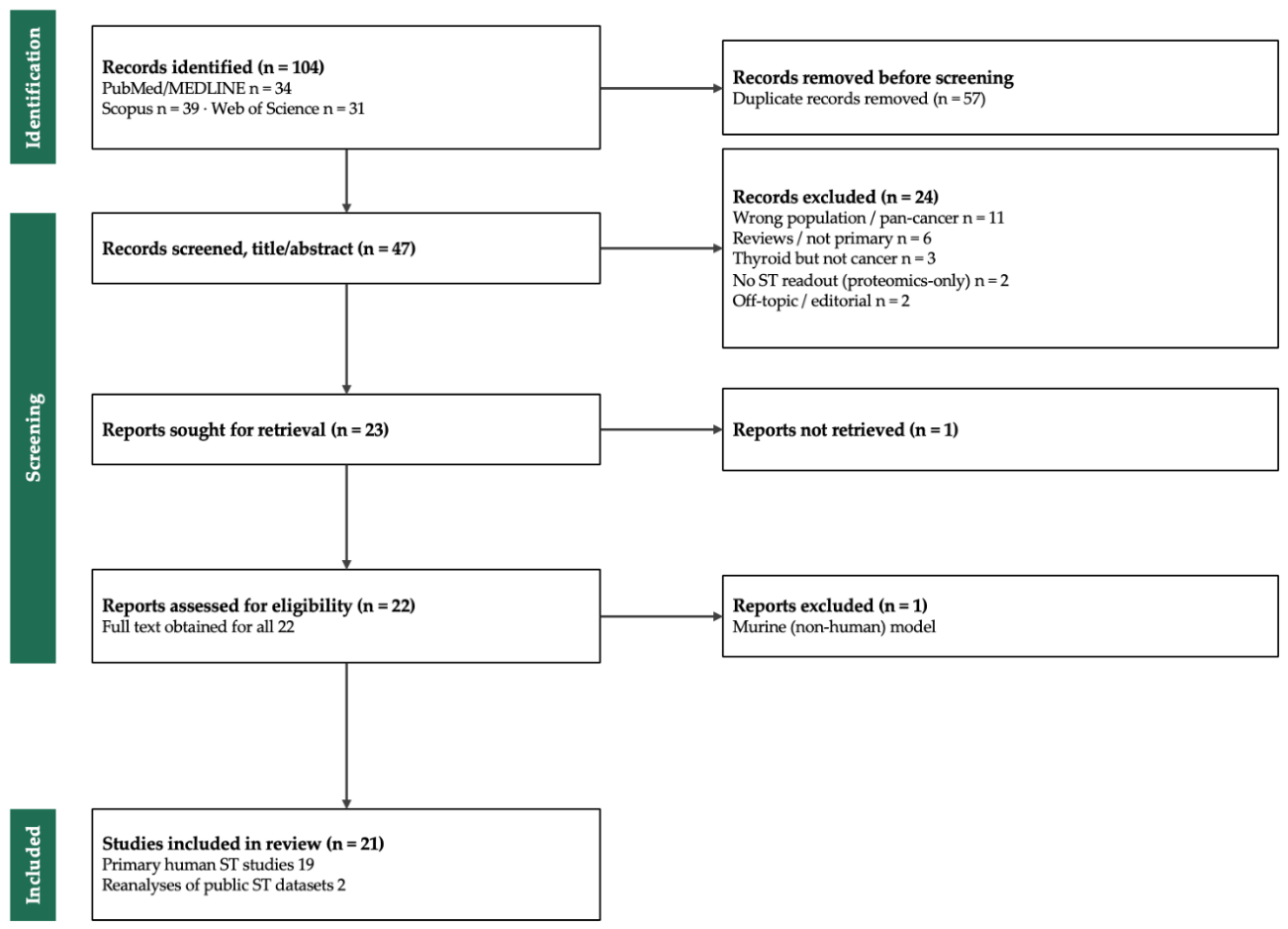

Table 1.
Characteristics and principal findings of the 21 included spatial-transcriptomics studies in thyroid cancer.
Table 1.
Characteristics and principal findings of the 21 included spatial-transcriptomics studies in thyroid cancer.
| Study (Year) | Platform | Subtype(s) | Key Spatial Finding |
|---|---|---|---|
| Liao et al. 2025 | 10x Visium + scRNA-seq | PT, PTC, LPTC, ATC | TME remodeling across progression; 3 thyrocyte meta-clusters (TG+IYG+, HLA-DR+, APOE+APOC1+); SERPINE1+ fibroblasts track malignant progression/prognosis; stage-specific leading-edge remodeling |
| Seok et al. 2025 | Visium CytAssist | PTC, FTC, PDTC, ATC | TME remodeling across differentiation states; CellDART deconvolution; PROGENy/REACTOME pathway shifts with dedifferentiation |
| Ning et al. 2025 | Visium (spRNA-seq) + WES | DTC → PDTC → ATC | ATC regions show immune suppression, angiogenesis, ECM remodeling; adjacent DTC shares ATC-level mutational burden → clonal dedifferentiation continuum |
| Ji et al. 2026 | 10x Visium + scRNA-seq | PTC → PDTC/ATC | CAFs in poorly-differentiated regions upregulate glycolysis; ZFP57–PKM2 lactate axis drives dedifferentiation; resveratrol reverses it |
| Loberg et al. 2026 | Visium (FFPE) + scRNA-seq | WDTC, ATC, composite WDTC/ATC, pediatric DSV | Defines POSTN+ myofibroblast CAFs intimately associated with invasive tumor cells; correlate with poor prognosis and lymph-node metastasis; prognostic fibroblast subpopulations |
| Zheng et al. 2025 | Visium (SRT) + scRNA-seq | PTC | Resolves spatial evolution/heterogeneity of PTC underlying malignancy and metastasis; improves molecular classification |
| Yan et al. 2024 | 10x Visium | PTC | Prognosis-associated cellular heterogeneity; spatial RNA-clinical signature genes map cell-type distributions across sections |
| Zhang et al. 2026 | Proteogenomics + spatial transcriptomics | Advanced DTC | Three DTC subtypes (canonical, stromal, immunogenic); ML classifier; ST maps thyroid-differentiation, immune and stromal scores |
| Xu et al. 2023 | Visium + DNA/RNA-seq + multiplex IF | Aggressive thyroid malignancy | Molecular Aggression and Prediction (MAP) score integrating the immune microenvironment improves prognostication over high-risk mutations alone |
| Wang et al. 2024 | Visium + scRNA-seq | PTC (progressive vs non-progressive) | LAMP3+ dendritic cell–T-cell subpopulation interactions promote immune evasion in PTC |
| Shobab et al. 2025 | GeoMx DSP (NanoString) + flow cytometry | Differentiated TC | Sex-specific immune TME; males show more dividing NK and TIGIT+CD8 T cells; sex differences in immune-gene expression |
| Zhang et al. 2025 | 10x Visium + scRNA-seq | PTC / PTMC | PROS1–MERTK axis mediates tumor-microenvironment crosstalk driving PTMC progression |
| Su et al. 2026 | Public ST (GEO) + scRNA reanalysis | Thyroid cancer | APOE/NCF1-associated immunosuppressive niche and its prognostic signature |
| Wang et al. 2026 | 10x Visium + scRNA-seq | Thyroid cancer (THCA) | CXCL8+ monocyte / SDC1+ tumor-stem-cell interaction network drives remote metastatic transfer |
| Condello et al. 2024 | 10x Visium (CytAssist) | FTC | Clone-specific dysregulation of extracellular-matrix genes at the invasive (capsular/vascular) front vs indolent core |
| Suzuki et al. 2024 | Visium CytAssist | Follicular tumors (FTA vs FTC) | Identifies invasive-subpopulation biomarkers; CD74 expression dynamics help distinguish invasive FTC from adenoma |
| Xiao et al. 2025 | 10x Visium + scRNA-seq | PTC (advanced, LN metastasis) | APOE(–) tumor-cell subpopulation drives pathological lymph-node metastasis; APOE modulates proliferation/invasion |
| Jiang et al. 2026 | Public ST (GSE250521) + scRNA + bulk (TCGA, GSE60542) | PTC (lymph-node metastasis) | LNM signature; random-forest model for preoperative N1/N0 stratification; disseminated clones escape immunity via antigen-presentation downregulation |
| Li et al. 2025 | Spatial metabolomics + Visium | PTC + lymph-node metastasis | Integrated spatial metabolic–transcriptional map of PTC tumorigenesis and LN metastasis; dysregulated metabolic pathways |
| Wang et al. 2025 | Visium + immunofluorescence | PTC, ATC, ATC-LM, ATC-GM | SFRP4-high tumor-specific myeloid cells correlate with dedifferentiation and poor prognosis; localize near CD44+ tissue stem cells |
| Haq et al. 2025 | GeoMx DSP (NanoString) | ATC (vs PTC/non-anaplastic) | Eight DEG mRNA + protein markers for ATC progression/dedifferentiation; distinguish ATC from follicular-cell-derived carcinomas |
Studies are summarized according to the spatial profiling platform used, thyroid cancer subtype evaluated, and principal spatial or tumor microenvironment finding. Abbreviations: ATC, anaplastic thyroid carcinoma; DTC, differentiated thyroid carcinoma; FTC, follicular thyroid carcinoma; PDTC, poorly differentiated thyroid carcinoma; PTC, papillary thyroid carcinoma; scRNA-seq, single-cell RNA sequencing; ST, spatial transcriptomics; TME, tumor microenvironment.
Table 2.
Principal spatial transcriptomics platforms and their reported use in thyroid cancer.
| Platform | Class | Resolution | Coverage | FFPE | Sources | Use in Thyroid Cancer |
|---|---|---|---|---|---|---|
| Visium (10x) | Capture | 55 µm spots | Transcriptome-wide | Yes | [6,7]. | Most-used platform; TME, progression, and invasion |
| Visium CytAssist | Capture | 55 µm spots | Transcriptome-wide | Yes | [15,17,25,29] | FFPE differentiation-state and invasive-front mapping |
| Slide-seqV2 | Capture | ~10 µm beads | Transcriptome-wide | No | [39] | Not yet reported |
| Stereo-seq | Capture | Submicron bins | Transcriptome-wide | Limited | [37] | Not yet reported |
| Seq-Scope | Capture | Submicron pixels | Transcriptome-wide | Limited | [38] | Not yet reported |
| Xenium (10x) | Imaging | Subcellular | Targeted panel | Yes | [40] | Emerging validation platform |
| CosMx (NanoString) | Imaging | Subcellular | Targeted RNA/protein panels | Yes | [10] | Not yet reported in included studies |
| MERFISH/seqFISH+ | Imaging | Subcellular | Hundreds to thousands of targets | Limited | [8,9] | Not yet reported in included studies |
| GeoMx DSP (NanoString) | Region-based | Histology-defined ROI | Whole transcriptome or targeted panels; protein | Yes | [11] | Immune profiling and ATC biomarker discovery |
FFPE, formalin-fixed paraffin-embedded; ROI, region of interest; TME, tumor microenvironment. Resolution and coverage are representative and may vary by assay configuration. ‘Use in thyroid cancer’ refers to the thyroid cancer studies included in this review.
Disclaimer/Publisher’s Note: The statements, opinions and data contained in all publications are solely those of the individual author(s) and contributor(s) and not of MDPI and/or the editor(s). MDPI and/or the editor(s) disclaim responsibility for any injury to people or property resulting from any ideas, methods, instructions or products referred to in the content. |
© 2026 by the authors. Licensee MDPI, Basel, Switzerland. This article is an open access article distributed under the terms and conditions of the Creative Commons Attribution (CC BY) license (http://creativecommons.org/licenses/by/4.0/).
Copyright: This open access article is published under a Creative Commons CC BY 4.0 license, which permit the free download, distribution, and reuse, provided that the author and preprint are cited in any reuse.


