

Submitted:
04 August 2026
Posted:
05 August 2026
You are already at the latest version
Abstract
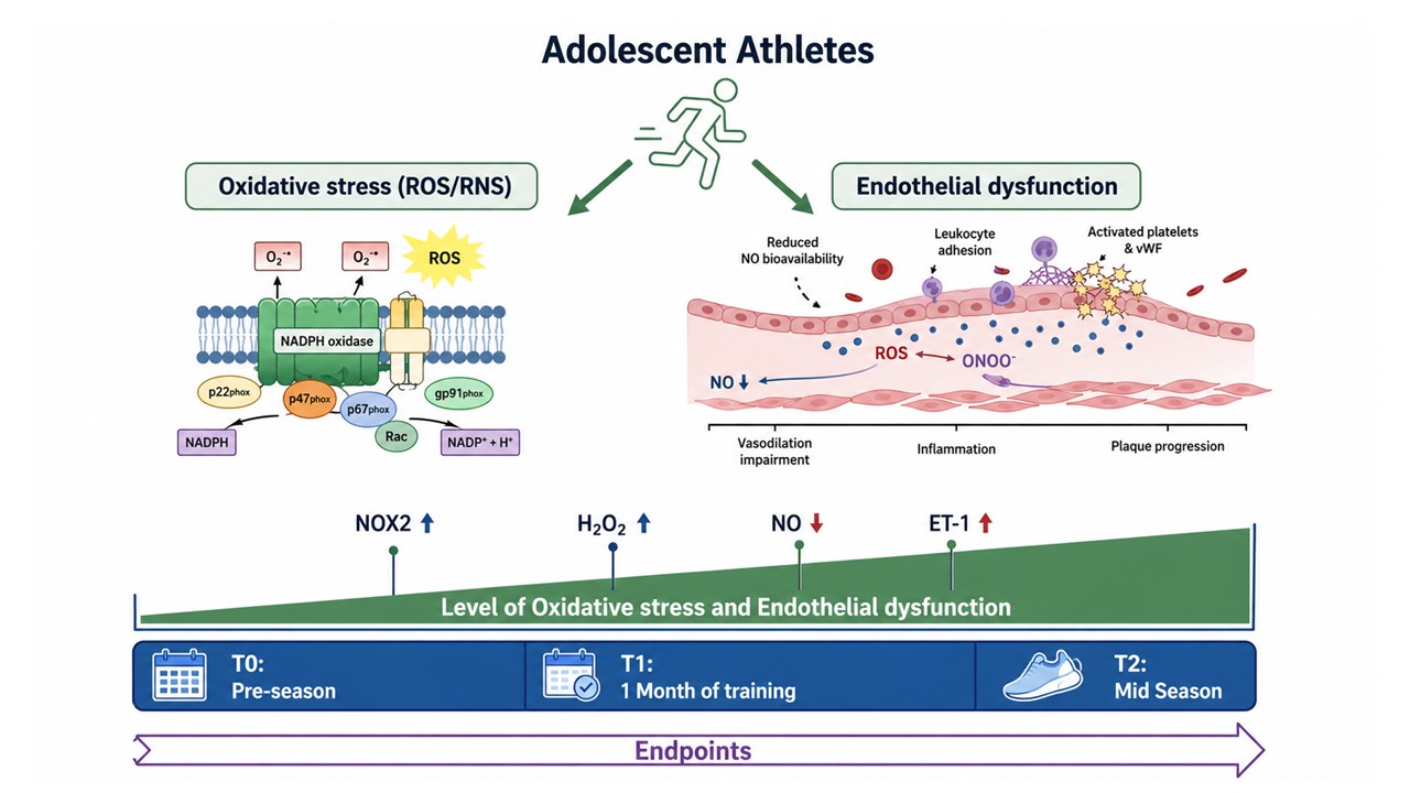
Background: Intense physical exercise induces oxidative stress and inflammation in adult professional athletes, but data on adolescent elite athletes remain limited. Methods: 24 elite adolescent soccer players were recruited and evaluated three times: pre-season (T0), after one month (T1), and after the first half of the season (T2). Saliva samples were col-lected to measure oxidative stress and endothelial function. In vitro studies were per-formed on endothelial cells exposed to H₂O₂ concentrations observed in athletes. Results: Athletes showed a significant increase in oxidative stress markers (NOX2 and H₂O₂) at T1 and T2 versus T0. Endothelial dysfunction was evidenced by reduced total NO concentra-tion and elevated endothelin (ET1) levels. In vitro, endothelial cells exposed to H₂O₂ (8 µM) exhibited greater damage than untreated cells, as evidenced by increased NOX2 activation and ET1 levels. Finally, H₂O₂ (8 µM)-treated cells impaired tube formation, reducing mesh number and area. NOX2ds-tat treatment reduced cell damage and restored angiogenic capacity. Conclusions: Our findings indicate that adolescent elite athletes experience sig-nificant oxidative stress and endothelial impairment, potentially increasing cardiovascu-lar risk. These results underscore the importance of cardiovascular monitoring and the potential benefits of antioxidant strategies, even in young athletes participating in high-intensity sports.
Keywords:
NOX2
; oxidative stress
; endothelial dysfunction
; adolescent elite athletes
‡ These authors are co-senior authors.
1. Introduction
Regular physical activity is widely recognized as a key determinant of cardiovascular health. It is associated with improved endothelial function through both hemodynamic and molecular adaptations [1,2], reducing cardiovascular risk, and enhanced metabolic regulation [3]. When performed regularly, moderate exercise promotes physiological adaptations that improve vascular homeostasis and nitric oxide bioavailability, thereby supporting cardiovascular protection [4]. In contrast, strenuous or exhaustive exercise may induce a transient increase in reactive oxygen species (ROS) production, driven by activation of redox-dependent signaling pathways involved in both exercise adaptation and acute oxidative imbalance [2]. Exercise-induced ROS production originates from multiple cellular sources, including mitochondria and enzymatic sources such as xanthine oxidase and NADPH oxidases. Among these, NADPH oxidase isoform 2 (NOX2) has been extensively investigated in the cardiovascular field and implicated in mechanisms underlying vascular dysfunction [5,6]. Clinical evidence indicates that increased NOX2 activation is associated with arterial dysfunction and elevated systemic oxidative stress in humans [7]. Moreover, NOX2-derived ROS play an important role in redox signaling during physical activity and contribute to the molecular responses induced by acute exercise. Several studies support the role of NOX2 as a major source of ROS generation during exercise. In mice carrying a loss-of-function mutation in the regulatory NOX2 subunit p47phox, ROS production was completely abolished following an acute bout of moderate-intensity treadmill exercise [8]. Furthermore, our group previously reported that elite athletes engaged in highly intensive sports exhibit significantly higher NOX2 levels compared with amateur athletes, accompanied by a marked reduction in antioxidant capacity activation [9]. In addition to oxidative stress, intense physical activity can stimulate a systemic inflammatory response [10]. This temporary response is characterized by increased levels of inflammatory cytokines, particularly interleukin-6 (IL-6) and C-reactive protein (CRP) [10,11]. These processes may amplify ROS production and contribute to tissue damage following strenuous exercise.
Although the inflammatory response and redox modulation in the post-exercise period have been extensively described in healthy adult athletes [12,13], the effects of intense exercise on redox homeostasis in adolescents remain poorly investigated. This population represents a unique physiological context characterized by rapid, ongoing physical and neuromuscular growth, accompanied by hormonal changes and cardiovascular maturation [14]. Interestingly, previous research has shown that NOX2-derived oxidative stress is already detectable during early life and may contribute to oxidative processes in childhood [15]. Furthermore, increased NOX2 activity in pediatric populations has been associated with markers of endothelial dysfunction and vascular impairment [15], suggesting that oxidative stress may influence cardiovascular health even at young ages. The increasing participation of adolescents in competitive sports raises important questions regarding the potential cardiovascular impact of intense training loads during this critical developmental period. Indeed, the impact of training during this critical developmental period is particularly important, as it may not only affect immediate performance but also contribute to long-term motor adaptations. A systematic review has reported the release of cardiac troponin in adolescent swimmers following exertion [16]. Similarly, other studies described the kinetics of high-sensitivity cardiac troponin T (hs-cTnT) after prolonged exercise in adolescents and adults, demonstrating a significantly greater post-exercise increase in adolescents than in adults, followed by subsequent normalization [17,18]. These elevations are not indicative of irreversible myocardial necrosis but may reflect increased cardiac cellular stress and increased transient membrane permeability [17,18]. Among potential mechanisms underlying these responses, endothelial activation and redox imbalance have been proposed as contributors to transient cardiovascular alterations in young athletes. Saliva has emerged as a valuable diagnostic biofluid as the composition of saliva dynamically reflects physiological and pathological changes occurring throughout the body, making it an attractive and non-invasive source of clinically relevant biomarkers. Salivary redox biomarkers have been increasingly investigated for the diagnosis and monitoring of a wide range of disorders, including metabolic diseases such as obesity, insulin resistance, diabetes mellitus, and chronic kidney disease, neurodegenerative disorders, as well as several types of cancer [19]. Non-invasive biomarkers, particularly salivary markers of hydrogen peroxide (H2O2) and nitric oxide (NO) metabolism, have emerged as potential tools for monitoring exercise-induced redox changes [19,20,21,22,23,24,25].
Therefore, the present study aimed to investigate whether sustained high-intensity exercise in adolescent athletes is associated with changes in systemic redox balance and endothelial-related markers, with a particular focus on NOX2-related oxidative stress pathways and salivary biomarkers.
2. Materials and Methods
2.1. Study Design
Twenty-four male adolescent soccer players (mean age of 15 years) were recruited for this observational study after collection of their medical history and eligibility assessment. Salivary samples were obtained non-invasively at three different time points during the season: before the beginning of training activities (T0), after one month of training (T1), and at the end of the first half of the competitive season (T2).
The study was conducted according to the ethical principles outlined in the Declaration of Helsinki and approved by the University Committee for Research of the University of Rome “Foro Italico” (Protocol No. CAR 206/2024).
Participants were instructed to avoid antioxidant supplements for one week before blood sampling; otherwise, no dietary restrictions were imposed, and athletes maintained their usual free diet throughout the study period.
2.2. Saliva Sample Collection
Saliva specimens were collected by the athletes between 8:00 and 9:00 a.m. to minimize the effect of circadian rhythm on secretion. After rinsing the mouth three times with distilled water at room temperature, the patients spat out the saliva accumulated at the bottom of the mouth into a sterile Falcon-type tube until approximately 5 mL. Samples were immediately diluted (1:1 v/v) with sterile phosphate-buffered saline (PBS 1X) to improve handling and pipetting procedures, aliquoted, and stored at −20 °C until analysis. To minimize potential confounding factors, participants were instructed to refrain from consuming food or beverages (except water) and from performing oral hygiene procedures for at least 2 h before saliva collection. Furthermore, no medications were taken within the 8 h preceding sample collection.
Saliva was selected as the biological matrix because it represents a simple and non-invasive sampling approach suitable for repeated monitoring. Furthermore, salivary oxidative stress markers have been reported to reflect both local and systemic redox status, showing correlation with circulating biomarkers [26].
2.3. Salivary NOX2 Levels
Salivary NOX2 concentration was assessed using a commercial ELISA kit (MyBioSource). Standards and samples were incubated in 96-well plates together with HRP-conjugated reagent for 1 h at 37 °C. After addition of chromogenic substrates, absorbance was measured at 450 nm using a microplate reader. Data were expressed as AU/mL. Intra- and inter-assay coefficients of variation (CV) were both below 15%.
2.4. Salivary H2O2 Levels
Hydrogen Peroxide (H2O2) concentrations were evaluated through a colorimetric assay kit (Abcam, ab102500). Quantification was performed using a standard calibration curve (ranging from 0 to 200 µM) H₂O₂.
The assay is based on the Fe³⁺-xylenol orange reaction, in which oxidation of Fe²⁺ by peroxides generates a colored complex proportional to the peroxide concentration. Optical density was recorded at 450 nm, and results were expressed as µM.
2.5. Salivary Nitric Oxide (NO)
Nitric oxide production was indirectly evaluated by measuring nitrite/nitrate (NOx) levels in saliva samples using a commercial colorimetric assay kit (Abcam, Cambridge, UK). Results were expressed as µM; Intra-assay and inter-assay coefficients of variation were both less than 5%.
2.6. Salivary Endothelin-1 Levels
Endothelin-1 (ET-1) concentrations in saliva were quantified using a commercial ELISA kit (Elabscience). Results were reported as pg/mL. Both intra-assay and inter-assay coefficients of variation were lower than 10%.
2.7. Cell Culture and Reagents
Human umbilical vein endothelial cells (HUVECs; Lonza) were maintained in complete EGM-2 medium (Lonza), supplemented with 10% FBS. Cells, between passages 2 and 3, were seeded at a density of 4000 cells/cm2 and cultured for 24 hours in a humidified incubator at 37 °C with 5% CO₂ to sub-confluence level. Before stimulation, cells were pre-treated for 30 min at 37 °C with the NOX2 inhibitor NOX2ds-tat (10 μM; Anaspec). Subsequently, HUVECs were exposed to H₂O₂ (2 or 8 µM) for 3 hours.
Conditioned media were collected for the determination of H2O2, sNOX2dp, endothelin-1 levels, and NO. For protein extraction, cells were lysed in ice-cold RIPA buffer supplemented with protease and phosphatase inhibitors (10 μg/mL; Thermo Fisher Scientific).
Lysates were incubated on ice for 30 min and centrifuged at 10,000 × g for 20 minutes. Supernatants were collected and used for Western blot analysis of p-eNOS/eNOS expression.
2.8. Protein Detection, Electrophoresis, and Western Blot Analysis
Protein concentrations were determined using the DC protein assay (BioRad). Equal amounts of protein (30 µg per lane) were mixed with 4× Laemmli buffer containing 20% β-mercaptoethanol, heated at 95 °C for 4 minutes, and separated by SDS-PAGE on precast polyacrylamide gels.
Proteins were subsequently transferred onto nitrocellulose membranes using the Trans-Blot Turbo system (Bio-Rad).
After blocking nonspecific binding sites with EveryBlot Blocking Buffer (Bio-Rad), membranes were incubated overnight at 4 °C with the following antibodies: anti-phospho-eNOS (Ser1177), anti-eNOS (Cell Signaling), and anti-actin (Santa Cruz Biotechnology). HRP-conjugated secondary antibodies (1:3000 dilution) were incubated for 1 hour at room temperature. Protein bands were visualized by enhanced chemiluminescence (ECL; Bio-Rad) and analyzed densitometrically using Image Lab software version 6.1.0. Data are expressed as arbitrary units (A.U.) and represent the mean of three independent experiments.
2.9. sNOX2dp Evaluation
NOX2 levels were measured in conditioned media cultures through an ELISA test. This assay measures the levels of soluble NOX2-derived peptide (sNOX2-dp) released into the serum following NOX2 activation. Briefly, the procedure included the following steps: (1) coating ELISA 96-well plates overnight at 4 °C with reference standards and serum samples containing 1 µg of protein with known concentrations of sNOX2-dp; washing the wells to remove unbound material; (3) adding horseradish peroxidase (HRP)-conjugated monoclonal anti-sNOX2-dp antibodies directed against the amino acid sequence 224–268 of the extracellular membrane portion of NOX2; and (4) quantifying the bound antibody-enzyme complexes by measuring HRP activity in the presence of the substrate 3,3′,5,5′-tetramethylbenzidine (TMB). Enzymatic activity was determined spectrophotometrically by measuring the increase in absorbance at 450 nm after stopping the reaction with 2 M sulfuric acid. Since absorbance is directly proportional to the concentration of sNOX2-dp, sample values were calculated by interpolation from a standard curve generated using reference standards ranging from 0 to 200 pg/mL in the same assay. Results are expressed as pg/mL. The intra-assay and inter-assay coefficients of variation were 8.95% and 9.01%, respectively.
2.10. Angiogenesis
The tube-forming assay was performed on Matrigel-coated 96-well plates with HUVECs (1,5 × 10⁴ cells/well) treated for 16 h with Growth Factor Reduced Matrigel Matrix Phenol Red Free (356234, BD) in the presence of the EGM2 supplemented with 10 μM NOX2ds-tat in the presence or absence of H2O2. An automated scan of each well was acquired with a 4× objective by a Nikon Eclipse TI inverted microscope with a motorized stage (Nikon Corporation). Mesh number and area were quantified using ImageJ with the Angiogenesis plugin.
2.11. Statistical Analysis
Baseline clinical characteristics were summarized using descriptive statistics. Categorical variables are presented as absolute frequencies. Continuous variables are reported as mean ± standard deviation (SD) for normally distributed data or as median with interquartile range (IQR) for non-normally distributed data, as appropriate. Normality of distribution was assessed using the Shapiro–Wilk test. For normally distributed variables, repeated-measures analysis of variance (ANOVA) was applied, followed by Bonferroni’s post hoc correction for multiple comparisons. For non-normally distributed variables, the Friedman test was used, followed by Dunn’s post hoc test where appropriate.
In vitro experimental data are presented as mean ± standard deviation (SD) and represent at least three independent experiments. Comparisons between multiple groups were performed using one-way or two-way ANOVA. All statistical tests were two-sided, and a p-value < 0.05 was considered statistically significant. Statistical analyses were performed using GraphPad Prism (v. 8.0.2; GraphPad Software, La Jolla, CA, USA).
3. Results
3.1. In Vivo Study
Table 1 summarizes the clinical and anthropometric characteristics of the study participants.
The sample comprised 24 young, healthy, and physically active males, with a mean age of 15 years. Cardiovascular parameters were within normal ranges. Regarding physical activity, training volume was approximately 12 hours per week at the first point (T1) and approximately 9 hours per week at the second time point (T2).
During the pre-season period (T1), elite adolescent soccer players typically followed a structured high-volume training program aimed at developing general and sport-specific fitness. Sessions included aerobic endurance training (e.g., continuous running and small-sided games with reduced recovery), high-intensity interval training, repeated sprint ability drills, neuromuscular and strength training (e.g., resistance exercises targeting major muscle groups), as well as technical–tactical drills focusing on ball control, passing, and positional play. Friendly matches were also included to progressively reintroduce competitive demands.
During the competitive season (T2), training load was adjusted to match schedule demands and primarily focused on performance maintenance and recovery. Weekly micro cycles generally included tactical and technical sessions with higher specificity, one to two high-intensity field sessions, structured strength maintenance sessions, and one official match per week, with additional recovery or low-intensity regenerative activities following matches.
The level of H2O2 in saliva samples was significantly higher in adolescent athletes at T1 compared with T0 (4.7±1.2 µM T1 vs 2.2±0.6 µM T0, p<0.001) and at T2 compared with T0 (5.5±1.3 µM T2 vs 2.2±0.6 µM T0, p<0.0001). Moreover, NOX2 were significantly higher in adolescent athletes at T1 [896.7 (666.0-1233.0) A.U./ml T1 vs 510.1 (159.2-724.0) A.U./ml T0, p<0.01] and at T2 [1050.0 (755.1-1318.0) A.U./ml T2 vs 510.1 (159.2-724.0) A.U./ml T0, p<0.0001] (Figure 1A,B) compared with T0.
Regarding biomarkers of endothelial dysfunction, we found that total NO concentration was significantly reduced at T1 compared to T0 [3.9 (2.7-6.7) µM T1 vs 11.0 (5.3-15.9) µM T0, p<0.01] and at T2 compared to T0 [3.8 (2.2-6.1) µM T2 vs 11.0 (5.3-15.9) µM T0, p<0.0001]. These changes were accompanied by a significant increase in ET-1 levels at T1 [40.7(37.7-43.5) µM T1 vs 30.6 (23.8-33.8) pg/ml T0, p<0.001] and at T2 [43.7 (40.5-45.9) pg/ml T2 vs 30.6 (23.8-33.8) pg/ml T0, p<0.0001] compared with T0 (Figure 1C,D).
These findings suggest that sustained training increased oxidative stress and impaired endothelial function. Interestingly, these alterations remained elevated over time (T2), suggesting persistent oxidative stress and endothelial dysfunction.
3.2. In Vitro Study
To further substantiate the role of NOX2-mediated oxidative stress in endothelial dysfunction, we conducted an in vitro study using human endothelial cells. Human umbilical vein endothelial cells (HUVECs) were incubated with increasing concentrations of H₂O₂ (0, 2, or 8 μM), at levels comparable to those detected in the saliva samples of adolescent athletes. To investigate the mechanisms underlying endothelial damage, oxidative stress, and endothelial function were assessed in the supernatant of treated cells by measuring: (1) the concentration of soluble NOX2-derived peptide (sNOX2-dp), a marker of NOX2 activation, and H₂O₂ production; (2) total NO concentration; and (3) ET-1 concentration. Exposure to 8 μM H₂O₂ induced a significant increase in H₂O₂, sNOX2-dp, and ET-1 levels, along with a marked reduction in total NO concentration, indicating increased oxidative stress and impaired endothelial function (Figure 2A-D). Notably, the addition of a specific NOX2 inhibitor (NOX2ds-tat) significantly attenuated oxidative stress and partially restored endothelial function (Figure 2A-D).
To better elucidate the molecular mechanisms underlying these effects, we examined the phosphorylation status of endothelial nitric oxide synthase (eNOS) at Ser1177, a critical regulator of nitric oxide (NO) production and endothelial homeostasis [27]. Our results showed that treatment with 8 μM H₂O₂ significantly reduced eNOS phosphorylation, whereas pretreatment with NOX2ds-tat restored phosphorylation levels (Figure 2E-F).
Finally, using a Matrigel assay, we assessed the effects of H2O2 on the angiogenic properties of endothelial cells. H2O2-treated cells impaired tube formation, reducing mesh number and mesh area (Figure 3A-C). NOX2ds-tat restored angiogenic capacity, significantly increasing both parameters.
4. Discussion
The present study investigated the impact of intense training on oxidative stress and endothelial function in elite adolescent soccer players. Our findings demonstrate that sustained high-intensity training during the competitive season is associated with a modulation of oxidative stress and endothelial-related markers. We observed increased NOX2 activation and H₂O₂ levels, along with reduced total NO concentration and elevated endothelin-1 concentrations. These findings may reflect the activation of redox-sensitive pathways affecting vascular homeostasis during intensive training.
Regular physical activity is widely recognized for its beneficial effects on cardiovascular health, primarily through improvements in endothelial function and nitric oxide signaling [24]. Moderate exercise promotes physiological adaptations that enhances endothelial nitric oxide synthase (eNOS) activity and increases total NO concentration, thereby supporting structural and functional vascular remodeling and metabolic adaptations in mitochondrial function and glucose/lipid metabolism [25,26]. These vascular adaptations are largely driven by hemodynamic stimulus and by redox-sensitive signaling pathways activated during repeated bouts of exercise. Indeed, exercise alters blood flow, luminal shear stress, and arterial pressure, with the consequence of modulating arterial function, diameter, and wall thickness [26]. However, the relationship between exercise intensity and vascular health follows a complex dose–response pattern. Structured and moderate exercise provides numerous health benefits, improving antioxidant defenses and adaptive redox signaling, thereby mitigating the adverse effects of oxidative stress [27]. In contrast, strenuous or prolonged exercise may lead to excessive production of ROS in critical tissues like blood and skeletal muscles, which are significant sources of ROS during exercise, resulting in oxidative stress [27].
In this context, our data show that oxidative stress progressively increased during the training season, as evidenced by higher salivary NOX2 and H₂O₂ levels at both T1 and T2 compared with pre-season values. Importantly, these changes were accompanied by reduced total NO concentration and increased endothelin-1 levels, suggesting a modulation of endothelial-related signaling pathways. Excessive ROS production has been shown to reduce nitric oxide bioavailability through direct chemical inactivation and by altering endothelial nitric oxide synthase activity, thereby contributing to endothelial dysfunction [28]. These findings support the concept that sustained oxidative stress may interfere with nitric oxide signaling and impair vascular homeostasis during periods of intense training. It should be noted that the biomarkers analyzed in this study, particularly those measured in saliva, may reflect both local oral and systemic processes. Therefore, they should be interpreted cautiously as indicators of redox and endothelial-related overall status rather than direct equivalents of circulating vascular concentrations [29].
Notably, the more pronounced changes observed at T2, despite the shorter training duration compared with T1, may be interpreted in the context of a pre-existing chronic state of low-grade inflammation and oxidative stress in these elite adolescent athletes. Such a condition could potentially amplify the biological response to variations in training load, leading to an exaggerated redox imbalance even after relatively shorter periods of intensified exercise.
Among enzymatic sources of ROS, NADPH oxidases represent key contributors to oxidative stress in the cardiovascular system. The NOX family of enzymes is recognized as one of the main sources of ROS in vascular tissues [30]. In particular, the NOX2 isoform has been extensively implicated in mechanisms of endothelial dysfunction and vascular oxidative stress [31]. Previous experimental and clinical evidence indicates that increased NOX2 activation is associated with vascular impairment and increased systemic oxidative stress [32]. Moreover, recent studies in athletes have reported that intense physical activity may enhance NOX2 activation, suggesting that this enzymatic pathway contributes to exercise-induced oxidative stress [9]. Our findings extend these observations to adolescent athletes, a population in which the effects of sustained training loads on vascular re-dox balance remain poorly characterized.
Similarly, although endothelin-1 is primarily produced by endothelial cells and released in a polarized manner toward vascular smooth muscle, evidence indicates that salivary endothelin-1 levels are not purely independent of the systemic compartment. Previous studies have reported significant correlations between salivary and plasma concentrations of endothelin isoforms, including ET-1, suggesting a partial reflection of circulating endothelin dynamics in saliva [29]. However, this relationship is not based on a direct equilibrium between compartments, and salivary ET-1 likely represents the integrated contribution of systemic endothelial activation and local oral production. Therefore, salivary ET-1 should be interpreted as a marker of endothelial-related signaling rather than a strict quantitative surrogate of plasma levels. The significant associations observed between oxidative stress markers and endothelial dysfunction indicators further support the hypothesis that NOX2 activation may represent a key mediator linking in-tense exercise with vascular alterations. Previous research has demonstrated that oxidative stress and endothelial dysfunction are closely interconnected processes that contribute to early vascular impairment and may represent an early step in the development of cardiovascular disease [33].
To further elucidate the mechanisms underlying these in vivo observations, we conducted in vitro experiments using endothelial cells exposed to H₂O₂ concentrations comparable to those detected in athletes. Exposure to higher H₂O₂ levels resulted in increased NOX2 activation and endothelin-1 release, together with a significant reduction in total NO concentration. These findings indicate that oxidative stress alone is sufficient to reproduce key aspects of the endothelial alterations observed in athletes. Importantly, pharmacological inhibition of NOX2 significantly attenuated these effects, suggesting that NOX2 activation is a mechanistic driver of endothelial dysfunction. A key component of intact endothelial function is a functional eNOS that is in the luminal endothelial cell membrane, producing NO from L-arginine. Phosphorylation at specific sites, primarily S1177, is a post-translational control mechanism activating eNOS and regulating NO production [23]. We found that higher H₂O₂ levels resulted in decreased eNOS phosphorylation, reflected by decreased in vitro NO production and increased ROS production.
In addition to molecular markers of endothelial impairment, our study also demonstrated functional consequences of oxidative stress on endothelial angiogenic capacity. Endothelial cells exposed to elevated H₂O₂ concentrations exhibited impaired tube formation, as evidenced by reductions in both mesh number and mesh area in the Matrigel assay. These alterations suggest that excessive oxidative stress may compromise endothelial repair and angiogenic responses, processes that are essential for maintaining vascular integrity.
Adolescence represents a critical developmental period characterized by rapid growth, hormonal changes, and ongoing maturation of the cardiovascular system [34]. During this phase, exposure to excessive training loads may challenge physiological adaptive mechanisms and induce transient vascular imbalance [35].
Although the oxidative and endothelial changes observed in our study may represent an adaptive response to intense training, their persistence over time raises questions regarding potential long-term cardiovascular implications. Our findings, therefore, highlight the importance of monitoring cardiovascular and redox biomarkers in young athletes engaged in high-intensity sports. Non-invasive approaches, such as salivary biomarker assessment, may be a practical strategy for evaluating oxidative stress and endothelial status during training periods. Furthermore, strategies aimed at optimizing training load, recovery periods, and nutritional antioxidant support may help mitigate excessive oxidative stress while preserving the beneficial effects of exercise.
This study has several limitations that should be acknowledged.
1. the observational design of the study lacked a parallel non-training or low-intensity control group, which limits the ability to completely exclude external confounding temporal factors over the season. However, recruiting non-training controls among elite adolescent athletes is ethically and practically unfeasible without interrupting their athletic development. To minimize inter-individual variance, athletes served as their own internal baseline controls (T0).
2. the sample size was relatively small and limited to male soccer players, which may restrict the generalizability of the findings to other sports or female athletes. Moreover, the absence of formal power analysis may increase the risk of Type II errors in analyses. 3. although salivary biomarkers provide a non-invasive method for monitoring oxidative stress, they may not fully reflect systemic vascular changes and can be influenced by oral metabolism.
4. a limitation of the present pilot study is the absence of a detailed quantification of training load (including both internal and external load variables) at each time point, which will be addressed in future expanded studies involving a larger and more heterogeneous sample and the support of exercise physiology specialists.
Finally, the observational design of the in vivo component does not allow definitive causal relationships to be established between training load and endothelial alterations. In addition, we did not evaluate cardiovascular clinical parameters such as endothelial function assessed by FMD, or electrocardiographic or echocardiographic parameters. Despite these limitations, our study provides novel evidence linking intense training in adolescent athletes to NOX2-mediated oxidative stress and endothelial dysfunction. The combined in vivo and in vitro approaches strengthen the biological plausibility of our findings and support the concept that excessive oxidative stress may represent an early mechanism affecting vascular homeostasis in young athletes.
5. Conclusions
In conclusion, the present findings suggest that sustained high-intensity training during the competitive season is associated with a shift toward a pro-oxidative state and concurrent alterations in endothelial-related biomarkers in elite adolescent soccer players. The consistency between the in vivo observations and the in vitro results supports the biological plausibility of NOX2-dependent oxidative stress as a mechanism involved in the vascular response to intensive exercise. While these changes may reflect physiological adaptations to prolonged training, they also underline the importance of carefully balancing training stimuli and recovery during adolescence, a critical period for cardiovascular development. Further longitudinal studies are warranted to determine whether these alterations are fully reversible or may have implications for long-term vascular health.
Supplementary Materials
The following supporting information can be downloaded at: Preprints.org, Original Images for Blot. Supplementary Figure S1. Original uncropped western blots corresponding to Figure 2.
Author Contributions
“Conceptualization, C.F., F.A., R.C.; methodology, F.A., A.D.A., V.P., V.T; validation, C.F., F.A., C.N. and R.C.; formal analysis, C.N., and R.C.; resources, V.T., S.C., A.B., L.S., C.L.; data curation, F.A., A.D.A and C.N; writing—original draft preparation, M.Z., B.O.P., C.N., R.C.; writing—review and editing, E.C., G.E.C, G.F., A.G., C.F., F.Pi., F.Pe.; supervision, R.C.; project administration, R.C.; All authors have read and agreed to the published version of the manuscript.”.
Funding
This research received no external funding.
Institutional Review Board Statement
“The study was conducted in accordance with the Declaration of Helsinki and approved by the local University Committee for Research of Università Foro Italico, Rome (Protocol No. CAR 206/2024; Approval date: 02/10/2024)”.
Informed Consent Statement
Informed consent was obtained from all subjects involved in the study.
Data Availability Statement
The original contributions presented in this study are included in the article. Further inquiries can be directed at the corresponding author.
Acknowledgments
The authors thank Associazione Sportiva Dilettantistica (A.S.D.) Fondi Calcio, for providing both the adolescent elite athletes and the facilities necessary to conduct this study. SMiLe (Students Medicine Latina) Group: Myriam Chieruzzi, Riccardo D’Antonio, Giulia Leone, Simone Martellucci, Riccardo Vellucci.
Conflicts of Interest
The authors declare no conflicts of interest.
Abbreviations
The following abbreviations are used in this manuscript:
| CRP | C-Reactive Protein |
| eNOS | Endothelial Nitric Oxide Synthase |
| ET-1 | Human Endothelin-1 |
| H2O2 | Hydrogen Peroxide |
| HRP | Horseradish peroxidase |
| hs-cTnT | High-Sensitivity Cardiac Troponin T |
| HUVEC | Human Umbilical Vein Endothelial Cells |
| IL-6 | Interleukin-6 |
| IQR | Interquartile Range |
| NOX2 | NADPH Oxidase Isoform 2 |
| PBS | Phosphate-Buffered Saline |
| ROS | Reactive oxygen species |
| SD | Standard Deviation |
| sNOX2-dp | NOX2-derived peptide |
| TMB | 3,3′,5,5′-Tetramethylbenzidine |
References
- Biernat, K.; Kuciel, N.; Mazurek, J. Hap K: Is It Possible to Train the Endothelium?-A Narrative Literature Review. Life 2024, 14(5). [Google Scholar] [CrossRef] [PubMed]
- Nystoriak, M.A.; Bhatnagar, A. Cardiovascular Effects and Benefits of Exercise. Front Cardiovasc Med. 2018, 5, 135. [Google Scholar] [CrossRef] [PubMed]
- Pinckard, K.; Baskin, K.K.; Stanford. KI: Effects of Exercise to Improve Cardiovascular Health. Front Cardiovasc Med. 2019, 6, 69. [Google Scholar] [CrossRef] [PubMed]
- Jiang, J.; Ni, L.; Zhang, X.; Gokulnath, P.; Vulugundam, G.; Li, G.; Wang, H.; Xiao, J. Moderate-Intensity Exercise Maintains Redox Homeostasis for Cardiovascular Health. Adv. Biol. (Weinh) 2023, 7(4), e2200204. [Google Scholar] [CrossRef] [PubMed]
- Forstermann, U.; Xia, N.; Li, H. Roles of Vascular Oxidative Stress and Nitric Oxide in the Pathogenesis of Atherosclerosis. Circ. Res. 2017, 120(4), 713–735. [Google Scholar] [CrossRef] [PubMed]
- Lassegue, B.; Griendling, K.K. NADPH oxidases: functions and pathologies in the vasculature. Arterioscler. Thromb. Vasc. Biol. 2010, 30(4), 653–661. [Google Scholar] [CrossRef] [PubMed]
- Loffredo, L.; Carnevale, R.; Cangemi, R.; Angelico, F.; Augelletti, T.; Di Santo, S.; Calabrese, C.M.; Della Volpe, L.; Pignatelli, P.; Perri, L.; et al. NOX2 up-regulation is associated with artery dysfunction in patients with peripheral artery disease. Int. J. Cardiol. 2013, 165(1), 184–192. [Google Scholar] [CrossRef] [PubMed]
- Henriquez-Olguin, C.; Knudsen, J.R.; Raun, S.H.; Li, Z.; Dalbram, E.; Treebak, J.T.; Sylow, L.; Holmdahl, R.; Richter, E.A.; Jaimovich, E.; et al. Cytosolic ROS production by NADPH oxidase 2 regulates muscle glucose uptake during exercise. Nat. Commun. 2019, 10(1), 4623. [Google Scholar] [CrossRef] [PubMed]
- D’Amico, A.; Cavarretta, E.; Fossati, C.; Borrione, P.; Pigozzi, F.; Frati, G.; Sciarretta, S.; Costa, V.; De Grandis, F.; Nigro, A.; et al. Platelet Activation Favours NOX2-Mediated Muscle Damage in Elite Athletes: The Role of Cocoa-Derived Polyphenols. Nutrients 2022, 14(8). [Google Scholar] [CrossRef] [PubMed]
- Sohail, M.U.; Al-Mansoori, L.; Al-Jaber, H.; Georgakopoulos, C.; Donati, F.; Botre, F.; Sellami, M.; Elrayess, M.A. Assessment of Serum Cytokines and Oxidative Stress Markers in Elite Athletes Reveals Unique Profiles Associated With Different Sport Disciplines. Front Physiol. 2020, 11, 600888. [Google Scholar] [CrossRef] [PubMed]
- Waskiewicz, Z.; Mukhambet, Z.; Azerbayev, D.; Bondarev, S. Inflammatory Response to Ultramarathon Running: A Review of IL-6, CRP, and TNF-alpha. Int. J. Mol. Sci. 2025, 26(13). [Google Scholar] [CrossRef] [PubMed]
- Gomez-Cabrera, M.C.; Vina, J.; Ji, L.L. Role of Redox Signaling and Inflammation in Skeletal Muscle Adaptations to Training. Antioxidants 2016, 5(4). [Google Scholar] [CrossRef] [PubMed]
- Scheffer, D.D.L.; Latini, A. Exercise-induced immune system response: Anti-inflammatory status on peripheral and central organs. Biochim Biophys. Acta Mol. Basis Dis. 2020, 1866(10), 165823. [Google Scholar] [CrossRef] [PubMed]
- Naughton, G.; Farpour-Lambert, N.J.; Carlson, J.; Bradney, M.; Van Praagh, E. Physiological issues surrounding the performance of adolescent athletes. Sports Med. 2000, 30(5), 309–325. [Google Scholar] [CrossRef] [PubMed]
- Loffredo, L.; Pignatelli, P.; Martino, F.; Carnevale, R.; Bartimoccia, S.; Catasca, E.; Colantoni, C.; Zanoni, C.; Perri, L.; Violi, F. Early increase of NOX2-derived oxidative stress in children: relationship with age. Pediatr. Res. 2013, 73(6), 788–793. [Google Scholar] [CrossRef] [PubMed]
- Cirer-Sastre, R.; Corbi, F.; Lopez-Laval, I.; Carranza-Garcia, L.E.; Reverter-Masia, J. Exercise-Induced Release of Cardiac Troponins in Adolescent vs. Adult Swimmers. Int. J. Env. Res. Public Health 2021, 18(3). [Google Scholar] [CrossRef] [PubMed]
- Conesa-Milian, E.; Cirer-Sastre, R.; Hernandez-Gonzalez, V.; Legaz-Arrese, A.; Corbi, F.; Reverter-Masia, J. Cardiac Troponin Release after Exercise in Healthy Young Athletes: A Systematic Review. Healthcare 2023, 11(16). [Google Scholar] [CrossRef] [PubMed]
- Tian, Y.; Nie, J.; Huang, C.; George, K.P. The kinetics of highly sensitive cardiac troponin T release after prolonged treadmill exercise in adolescent and adult athletes. J. Appl. Physiol. (1985) 2012, 113(3), 418–425. [Google Scholar] [CrossRef] [PubMed]
- Klimiuk, A.; Zalewska, A.; Sawicki, R.; Knapp, M.; Maciejczyk, M. Salivary Oxidative Stress Increases With the Progression of Chronic Heart Failure. J. Clin. Med. 2020, 9(3). [Google Scholar] [CrossRef] [PubMed]
- Lumbikananda, S.; Sriwantana, T.; Rattanawonsakul, K.; Parakaw, T.; Phruksaniyom, C.; Rattanasuwan, K.; Vivithanaporn, P.; Thonabulsombat, C.; Sibmooh, N.; Srihirun, S. Nitrite in paraffin-stimulated saliva correlates with blood nitrite. Nitric Oxide 2021, 116, 1–6. [Google Scholar] [CrossRef] [PubMed]
- Maciejczyk, M.; Zalewska, A.; Ladny, J.R. Salivary Antioxidant Barrier, Redox Status, and Oxidative Damage to Proteins and Lipids in Healthy Children, Adults, and the Elderly. Oxid. Med. Cell Longev. 2019, 2019, 4393460. [Google Scholar] [CrossRef] [PubMed]
- Nishioka, T.; Maki, K.; Kimura, M.; Takahama, U. Determination of salivary peroxidase activity in human mixed whole saliva. Arch. Oral Biol. 2003, 48(5), 397–400. [Google Scholar] [CrossRef] [PubMed]
- Sundar, N.M.; Krishnan, V.; Krishnaraj, S.; Hemalatha, V.T.; Alam, M.N. Comparison of the salivary and the serum nitric oxide levels in chronic and aggressive periodontitis: a biochemical study. J. Clin. Diagn. Res. 2013, 7(6), 1223–1227. [Google Scholar] [CrossRef] [PubMed]
- Thimmegowda, U.; Belagatta, V.; Krishnamurthy, N.H.; Kattimani, S.; Venkatahaumaiah, S. Identification, Quantification and Correlation of Hydrogen Peroxide Present in Saliva to Early Childhood Caries: A Randomized Clinical Trial. Int. J. Clin. Pediatr. Dent. 2022, 15 (Suppl 2), S140–S146. [Google Scholar] [CrossRef] [PubMed]
- Toczewska, J.; Zalewska, A.; Konopka, T.; Maciejczyk, M. Enzymatic antioxidants activity in gingival crevicular fluid and saliva in advanced periodontitis. Oral Dis. 2023, 29(8), 3559–3570. [Google Scholar] [CrossRef] [PubMed]
- Ljubijankic, N.; Popovic-Javoric, R.; Sceta, S.; Sapcanin, A.; Tahirovic, I.; Sofic, E. Daily fluctuation of cortisol in the saliva and serum of healthy persons. Bosn. J. Basic Med. Sci. 2008, 8(2), 110–115. [Google Scholar] [CrossRef] [PubMed]
- Garcia, V.; Sessa, W.C. Endothelial NOS: perspective and recent developments. Br. J. Pharmacol. 2019, 176(2), 189–196. [Google Scholar] [CrossRef] [PubMed]
Figure 1.
Intensive exercise increases oxidative stress and endothelial dysfunction in elite athletes. H2O2 production (A), NOX2 (B), total NO concentration (C), and endothelin-1 levels (D) were measured in salivary samples at three time points: pre-season (T0), after one month of training (T1), and following the first half of the competitive season (T2). For H2O2, data are expressed as mean ± SD. Statistical differences were assessed using repeated-measures analysis of variance (ANOVA). For NOX2, total NO concentration, and endothelin 1 data are expressed as median and interquartile ranges (IQRs). Statistical differences were assessed using the Friedman signed-rank test. *p < 0.05; **p < 0.01; ***p<0.001; ****p<0.0001.
Figure 1.
Intensive exercise increases oxidative stress and endothelial dysfunction in elite athletes. H2O2 production (A), NOX2 (B), total NO concentration (C), and endothelin-1 levels (D) were measured in salivary samples at three time points: pre-season (T0), after one month of training (T1), and following the first half of the competitive season (T2). For H2O2, data are expressed as mean ± SD. Statistical differences were assessed using repeated-measures analysis of variance (ANOVA). For NOX2, total NO concentration, and endothelin 1 data are expressed as median and interquartile ranges (IQRs). Statistical differences were assessed using the Friedman signed-rank test. *p < 0.05; **p < 0.01; ***p<0.001; ****p<0.0001.
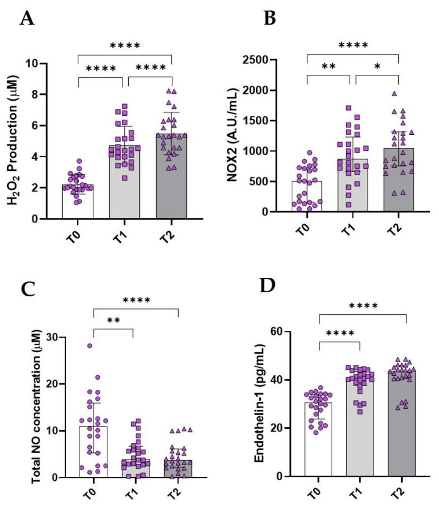
Figure 2.
Inhibition of NOX2 reverses H2O2-induced endothelial dysfunction and oxidative stress in vitro. H2O2 production (A), NOX2 activity (B), total NO concentration (C), endothelin-1 levels (D) in the supernatants of HUVEC treated with H2O2 (2 and 8 μM) in the presence or not of NOX2ds-tat as an inhibitor of NOX2. The ratio of phospho-eNOS to total eNOS (E) and representative Western immunoblots of phospho-eNOS-S1177 and total eNOSand (F) in HUVEC treated with H2O2 (2 and 8 μM) in the presence or not of NOX2ds-tat. Data are represented as mean ± SD of three independent experiments. *p < 0.05; **p<0.01; ***p<0.001. CTR=control; TAT=NOX2ds-tat.
Figure 2.
Inhibition of NOX2 reverses H2O2-induced endothelial dysfunction and oxidative stress in vitro. H2O2 production (A), NOX2 activity (B), total NO concentration (C), endothelin-1 levels (D) in the supernatants of HUVEC treated with H2O2 (2 and 8 μM) in the presence or not of NOX2ds-tat as an inhibitor of NOX2. The ratio of phospho-eNOS to total eNOS (E) and representative Western immunoblots of phospho-eNOS-S1177 and total eNOSand (F) in HUVEC treated with H2O2 (2 and 8 μM) in the presence or not of NOX2ds-tat. Data are represented as mean ± SD of three independent experiments. *p < 0.05; **p<0.01; ***p<0.001. CTR=control; TAT=NOX2ds-tat.
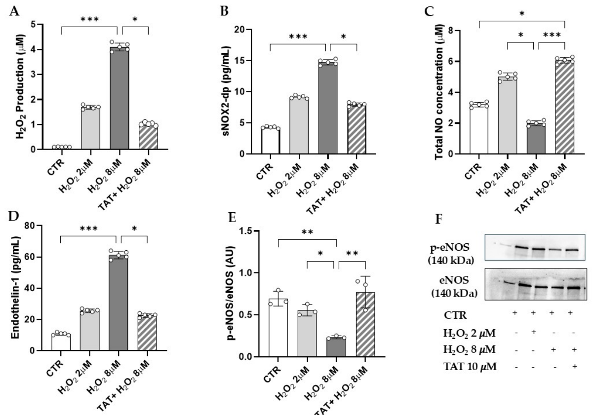
Figure 3.
Inhibition of NOX2 restores angiogenesis impaired by H₂O₂. Representative images of tube formation in HUVECs cultured on Matrigel treated with H2O2 (2 and 8 μM) in the presence or not of NOX2ds-tat as an inhibitor of NOX2 activity (A). Quantification of (B) mesh number and (C) mesh area. Data represent mean ± SD of three independent experiments. *p < 0.05; **p<0.01. CTR=control; TAT=NOX2ds-tat.
Figure 3.
Inhibition of NOX2 restores angiogenesis impaired by H₂O₂. Representative images of tube formation in HUVECs cultured on Matrigel treated with H2O2 (2 and 8 μM) in the presence or not of NOX2ds-tat as an inhibitor of NOX2 activity (A). Quantification of (B) mesh number and (C) mesh area. Data represent mean ± SD of three independent experiments. *p < 0.05; **p<0.01. CTR=control; TAT=NOX2ds-tat.
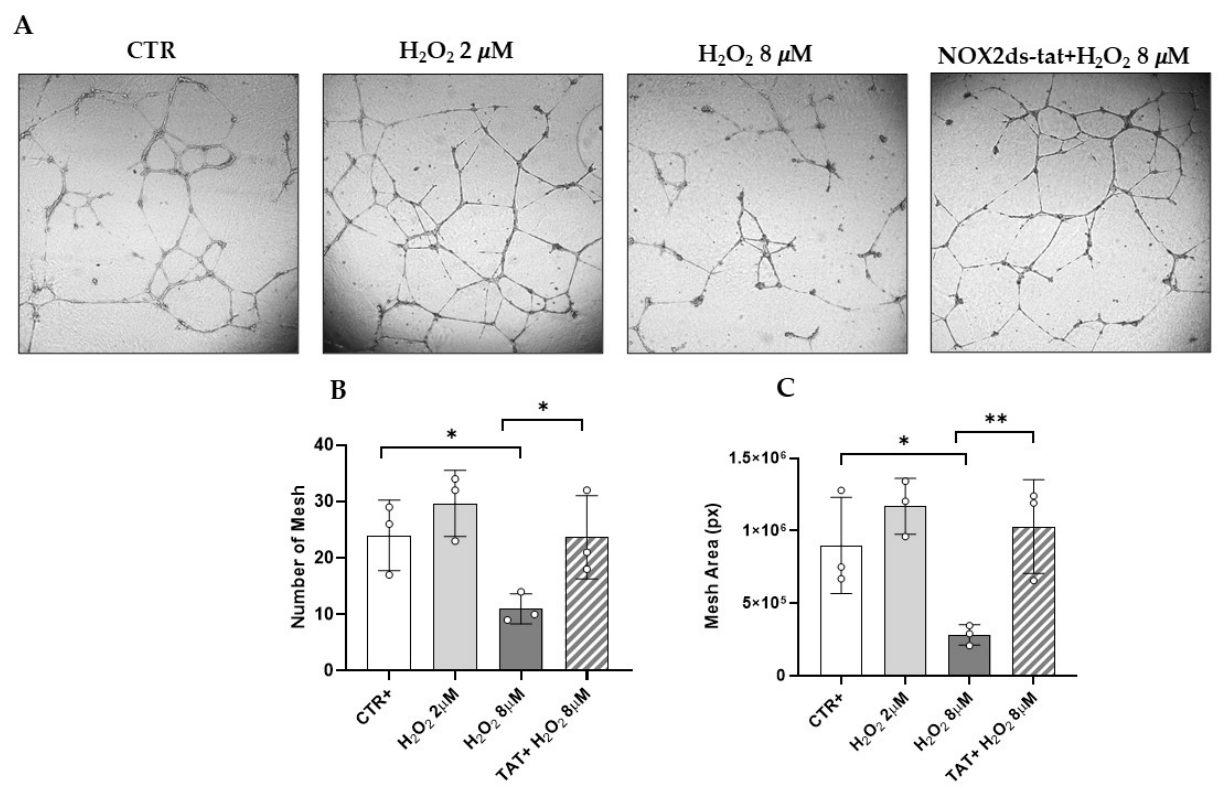

Table 1.
Characteristics of participants.
| Variable | Mean±SD |
| Age (years) | 15±0 |
| Gender (M/F) | 24/0 |
| Height (cm) | 174±7.6 |
| Weight (kg) | 62.6±9.7 |
| BMI | 20.43±2.3 |
| Systolic blood pressure (mmHg) | 116.9±12.5 |
| Diastolic blood pressure (mmHg) | 63.96±4.6 |
Disclaimer/Publisher’s Note: The statements, opinions and data contained in all publications are solely those of the individual author(s) and contributor(s) and not of MDPI and/or the editor(s). MDPI and/or the editor(s) disclaim responsibility for any injury to people or property resulting from any ideas, methods, instructions or products referred to in the content. |
© 2026 by the authors. Licensee MDPI, Basel, Switzerland. This article is an open access article distributed under the terms and conditions of the Creative Commons Attribution (CC BY) license (http://creativecommons.org/licenses/by/4.0/).
Copyright: This open access article is published under a Creative Commons CC BY 4.0 license, which permit the free download, distribution, and reuse, provided that the author and preprint are cited in any reuse.