

Submitted:
04 August 2026
Posted:
05 August 2026
You are already at the latest version
Abstract
Introduction: Müllerianosis is a rare pathological entity characterized by ectopic Müllerian-derived tissues, including endometrium, endocervix, and/or endosalpinx. Most reported cases involve the urinary bladder, while uterine involvement is extremely uncommon. We present a case of müllerianosis presenting as a cystic lesion of the anterior uterine wall, and we provide a review of the available literature. Case Presentation: We report a 37-year-old woman with an asymptomatic anterior uterine wall lesion detected 11 years ago on routine ultrasound. Initially measuring 60 × 35 mm, the lesion was surgically excised and histopathologically confirmed as Müllerianosis, containing endometrial and tubal-type tissue. Eleven years later, she presented with abnormal uterine bleeding. Ultrasound revealed a heterogeneous lesion with cystic areas and uterine asymmetry suggestive of adenomyosis. Conservative management, including curettage, failed, necessitating total abdominal hysterectomy. Histopathology confirmed adenomyosis and endometriosis in addition to prior findings. Discussion: This case is notable for the long follow-up, the uncommon anterior uterine location, and the large lesion size. It supports an embryologic or metaplastic origin of Müllerianosis and suggests potential links with other uterine pathologies. Surgical excision remains the treatment of choice, while imaging, histopathology, and immunohistochemistry are essential for accurate diagnosis. Long-term monitoring is recommended due to recurrence risk and the rare potential for malignant transformation. Conclusion: Our report emphasizes the importance of awareness, careful diagnosis, and extended follow-up in managing rare uterine Müllerian lesions.
Keywords:
Müllerianosis
; adenomyosis
; case report
1. Introduction
Müllerianosis is a rare pathological entity defined by the presence of distinct Müllerian-derived tissues located ectopically, outside their normal anatomical locations. It was first characterized by Clement and Young in 1996[1]. Histologically, the condition comprises endometrial, endocervical, and/or endosalpingeal components. Fewer than several dozen cases have been documented in the literature, most frequently involving the lower urinary tract [2,3]. Because of its variable clinical presentation and its potential to mimic malignant neoplasms, müllerianosis represents a significant diagnostic challenge [4]. We present a rare case of uterine müllerianosis manifesting as a cystic intramyometrial lesion of the anterior uterine wall, one of the rarest reported anatomical locations, with an exceptional 11-year longitudinal follow-up. We also review the available literature to summarize the current understanding of this uncommon disorder.
2. Case Report
We report the case of a 37-year-old female patient [BMI 29.3 kg/m²] who was admitted to our clinic via the emergency department in September 2025 for severe and irregular uterine bleeding, ongoing for the past 20 days. The patient reported using more than 10 sanitary pads per day in recent days. Despite having started treatment with medroxyprogesterone acetate approximately 10 days earlier, the bleeding had only partially decreased in intensity. Laboratory investigations demonstrated secondary anemia with a hemoglobin level of 87 g/L.
The patient was otherwise healthy, with no significant past medical history. In September 2014, a routine transvaginal ultrasound revealed an asymptomatic solid–cystic mass measuring 60 × 35 mm with prominent vascularization on the anterior uterine wall. A laparotomy with resection of the mass was performed. Histopathological analysis revealed a fibrous capsule composed of granulation tissue, containing focal glandular structures lined by cylindrical epithelium of endometrial and tubal type. Immunohistochemically, the stromal component was positive for CD10, SMA, and desmin, while CD117 and HMB-45 were negative. Ki-67 demonstrated proliferative activity within the granulation tissue layer of the capsule. Following the surgical procedure, the patient had three deliveries, each by cesarean section. During the last cesarean, Pomeroy sterilization was performed. In the past several months prior to admission, she developed irregular uterine bleeding and underwent curettage in July 2025, which revealed normal histopathology, including fragments of late-proliferative-phase endometrium.
Upon current admission, transvaginal ultrasound demonstrated a globularly enlarged uterus with asymmetrical walls [anterior wall 65 mm, posterior wall 17 mm], with a total volume of 476 cm³. A heterogeneous, echogenic lesion with cystic areas measuring 75 × 84 × 69 mm was noted in the anterior wall, with a regular posterior acoustic shadow. Color Doppler showed flow through the lesion with a high color score of 4. The endometrium measured 5 mm with an interrupted endometrial-myometrial interface. The uterotomy scar appeared normal, and both adnexa were unremarkable. Beta-hCG on admission was negative. An urgent curettage was performed; however, the patient continued to experience bleeding. An intrauterine Foley catheter was placed for 24 hours to control further hemorrhage. Histopathological examination revealed endometrium with signs of secretory activity and focal metaplastic changes of the epithelium. Due to a further decline in hemoglobin levels, the patient received red blood cell transfusions on two occasions.
Given the failure of medical therapy and curettage, ongoing hemorrhage, and development of severe anemia, a laparotomy with total abdominal hysterectomy was performed. Intraoperatively, the uterus was enlarged, with serosal changes suggestive of endometriotic foci, and the vesicouterine fold adhered densely to the anterior uterine wall. The procedure and postoperative course were uneventful. At a follow-up visit two months after surgery, the patient was reported to be in excellent condition. The final pathological finding describes a whitish submucosal oval mass measuring 8.8 cm in diameter that occupies the entire uterine cavity and is composed of intersecting bundles of smooth muscle cells with occasional islands of well-formed endometrial glands and stroma, with the overlying endometrium thinned and exhibiting a basal-type appearance, consistent with a diagnosis of uterine adenomyoma.
3. Discussion
3.1. Etiology and Pathogenesis
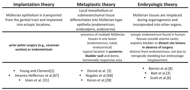
The etiopathogenesis of Müllerianosis has intrigued clinicians since its earliest descriptions. Multiple hypotheses have been proposed, reflecting the complex and multifactorial nature of the disease [Table 1]. In 1996, Young and Clement defined Müllerianosis as the presence of at least two of the three Müllerian tissues—endometrium, endosalpinx, and endocervix and suggested that the condition may result from the implantation of Müllerian tissue, potentially following surgical procedures [1]. Batt et al. later refined this concept by characterizing Müllerianosis as an organoid structure of embryonic origin, a choristoma composed of Müllerian rests [endometrium, endosalpinx, and/or endocervix], occurring either singly or in combination—thereby emphasizing a developmental rather than an acquired etiology [3]. Donne et al. introduced an alternative metaplastic hypothesis, suggesting that Müllerian metaplasia may give rise to tubal, endometrial, or endocervical tissues. The predilection of lesions for the posterior bladder wall and fundus corresponds to areas of peritoneal coverage known to be particularly responsive to female sex hormones [5].
Scott et al. proposed an embryological origin of müllerianosis, suggesting that aberrant activation of developmental signaling pathways (including WNT and HOX genes) during organogenesis may result in ectopic Müllerian tissue [6,7]. Similarly, Barresi et al. argued that displacement of coelomic epithelium and its associated mesenchyme during embryogenesis could explain Müllerian tissue developing at ectopic sites, including the spinal cord [8].
An interesting feature of our case is that the tumor-like lesion developed before the patient underwent any uterine surgical procedures, including three subsequent cesarean sections and the later laparotomy during which the mass was excised. This temporal sequence strongly supports an embryological or metaplastic origin rather than implantation as the underlying etiopathogenic mechanism.
3.2. Clinical Presentation
In the analyzed cohort of 69 Müllerianosis cases [Appendix A], patient age at diagnosis ranged from 11 to 84 years, with a mean of 43.5 years, indicating a predominance in reproductive and perimenopausal age but without strict age limitation. Approximately 40.6% of patients had a history of pelvic or abdominal surgery, most commonly cesarean section. Although this suggests a potential association between prior surgery and the development of Müllerianosis, the relationship remains inconclusive given the considerable number of patients without any surgical history.
Analysis of the reported cases indicates that urinary manifestations constitute the predominant clinical presentation, most commonly dysuria, hematuria, urinary urgency, and pain [2]. Notably, in a substantial proportion of patients, symptoms exhibit a catamenial pattern, occurring exclusively or predominantly during menstruation, thereby supporting the hypothesis of hormonal responsiveness of the ectopic Müllerian tissue [e.g., cyclical hematuria or cyclical dysuria][9,10,11,12,13,14,15,16]. The symptomatology further varies according to lesion location, with gastrointestinal manifestations [such as defecatory difficulty and rectal bleeding] and, more rarely, neurological symptoms—including cyclical neurological deficits—documented in select cases [6,8,17]. A number of cases were entirely asymptomatic, with lesions identified incidentally during evaluation for unrelated conditions.
The most common site of Müllerianosis was the urinary bladder, accounting for more than half of all reported cases. Within the bladder, the posterior wall represented the predominant single location [1,5,9,10,11,12,13,14,15,16,18,19,20,21,22,23,24,25,26,27,28,30,31]. Other intravesical sites included the lateral wall, dome, and trigone. Extrapelvic or extravesical locations were considerably less frequent, with reported lesions in the ureter, uterus, adnexa, appendix, and lymph nodes, as well as exceptionally rare sites such as the liver, lung, retroperitoneum, and spinal cord [4,6,8,32,33,34,35,36,37,38].
In our case report, an asymptomatic lesion of the anterior uterine wall was detected during a routine ultrasound examination. To our knowledge, only three other cases of uterine Müllerianosis have been reported to date: the first involved a cystic lesion of the posterior uterine wall (cystic endosalpingiosis) in a patient with pelvic pain; the second was an asymptomatic cystic endosalpingiosis of the uterine fundus discovered incidentally during pregnancy; and the third presented as a multiloculated cystic mass on the serosal surface of the uterine fundus containing both endometrial and tubal-type epithelium in a patient with pelvic pain [7,39,40]. However, our case is exceptional because the lesion was located entirely within the myometrium of the anterior uterine wall, whereas the previously reported uterine Müllerian lesions were confined to the uterine surface, cervix, or uterine parametrium rather than the myometrium itself. Interestingly, the largest reported lesions have been those involving uterine Müllerianosis, which is consistent with our case, in which the lesion measured 6 cm.
3.3. Diagnostic Evaluation
Across the collected cases, imaging findings demonstrated substantial heterogeneity, reflecting the diverse anatomical locations and morphological patterns of Müllerianosis. Ultrasound was the most frequently used initial modality, typically revealing echogenic, cystic, or polypoid, most often arising from the posterior or posterolateral bladder wall or the bladder dome [Appendix A]. CT and MRI most frequently described soft-tissue or mixed solid–cystic masses involving the bladder, ureter, or adjacent pelvic structures, often with features of heterogeneous enhancement or glandular/cystic morphology. These modalities also helped delineate local extension—such as involvement of the parametrium, abdominal wall, or adnexa—and occasionally identified ureteral obstruction or filling defects suggestive of secondary involvement. Imaging most often demonstrates features that frequently mimic neoplastic pathology [Appendix A]. Cystoscopy, when performed, commonly confirmed polypoid, nodular, mucinous, or cystic intravesical lesions, often consistent with submucosal or posterior wall involvement [Appendix A].
In our case, ultrasound was the initial imaging modality that detected uterine pathology, first 11 years ago with an unusual-appearing mass on the anterior uterine wall measuring 60 × 35 mm, and again recently, when it revealed asymmetry between the anterior and posterior uterine walls with sonomorphologic features suggestive of adenomyosis. Given the patient’s history of Müllerianosis and the location of the first lesion, we interpreted the later ultrasound findings with caution, despite the presence of direct and indirect signs of adenomyosis according to the MUSA criteria [29]. Although heterogeneous and nonspecific, ultrasound remains an accessible and cost-effective method for detecting lesions (whether Müllerianosis or adenomyosis) and for long-term monitoring of uterine structural changes.
3.4. Histological and Immunohistochemical Findings
The recorded lesion sizes demonstrated a broad range, extending from approximately 0.10 cm to 9.0 cm, with a mean diameter of 3.16 cm. This distribution reflects substantial heterogeneity in lesion dimensions, spanning from very small findings to large multiloculated masses. Of the total 69 reported cases, all three tissue types—endometriosis, endosalpingiosis, and endocervicosis—were identified in 22 cases. In over one-third of the cases, two of the three components were observed concurrently. Among the three tissue types, endometrial-type glands were the most frequently reported component, highlighting its predominant occurrence relative to endosalpingiosis and endocervicosis in the studied cohort.
Although Young and Clement define Müllerianosis as requiring the presence of at least two of the three Müllerian-derived tissue types for diagnosis, numerous published reports describe isolated cases of endocervicosis or endosalpingiosis [1]. Batt et al. later refined this broader definition, characterizing a choristoma composed of Müllerian rests (normal endometrium, normal endosalpingeal tissue, and normal endocervical tissue) incorporated singly or in combination into other normal organs during organogenesis. They emphasize that a single-tissue form of Müllerianosis can be diagnosed with a high degree of confidence only when three criteria are satisfied: the absence of pelvic endometriosis, no direct communication with the endocervix, endometrium, or endosalpinx, and no prior surgical intervention involving the reproductive organs [52]. In our case, two of the three Müllerian tissue types—endometrial and tubal—were identified, fulfilling the diagnostic criteria proposed by Young and Clement as well as those outlined by Batt et al.
The immunohistochemical profiles were reported for multiple markers, including ER, PR, CK7, CK20, CD10, PAX8, Ki-67, and other ancillary markers [Appendix A]. A substantial proportion of cases demonstrated positivity for estrogen receptor and progesterone receptor, particularly in the glandular components, reflecting the hormone-responsive nature of Müllerian-derived tissues. Specifically, ER and PR positivity was observed in the majority of reported cases, often in combination with CD10 positivity in the stromal compartment, confirming the presence of endometrial-type stroma. CK7 expression was consistently positive in the Müllerian epithelium, whereas CK20 was predominantly negative, helping to differentiate these lesions from gastrointestinal or urothelial-derived tissues. PAX8 immunoreactivity was frequently observed in glandular elements, supporting the Müllerian origin of the lesions. Ki-67 proliferation indices were generally low, consistent with the benign or low-proliferative nature of these lesions.
3.5. Management and Follow-Up
Most Müllerian lesions are primarily managed with surgical excision, with complete resection typically leading to durable symptom relief. Transurethral resection is the most common treatment, especially for lesions located in the lumen of the posterior bladder wall. Deeper or infiltrative lesions, including those involving the ureter, often required partial cystectomy, ureteral resection, or combined hysterectomy [4]. Hormonal therapy [GnRH agonists or aromatase inhibitors] may reduce lesion size or control symptoms when surgery is not feasible or incomplete [6,8,53,61]. The majority of patients remain symptom-free after adequate treatment.
Most reported cases support the benign nature of this condition. However, one reported case suggests that endocervicosis, although generally considered a benign lesion, may rarely undergo malignant transformation into adenocarcinoma. [23]. Considerably more attention has been directed toward the malignant potential of endosalpingiosis. Chui et al. suggested that endosalpingiosis shows how cancer-related mutations can occur even in cells that appear normal under the microscope. These changes may make such cells more prone to transformation into neoplastic lesions, especially when normal protective mechanisms are lost. They highlighted the role of BRAF and KRAS mutations, which are commonly found in ovarian low-grade serous neoplasms [62]. Moreover, a case of endometrioid adenocarcinoma of the urinary bladder arising in the background of Müllerianosis has been reported, underscoring both the rarity of such transformation and the importance of careful morphological assessment and additional analyses in establishing the correct diagnosis [30].
Recurrences have been reported in four cases, which were subsequently treated with repeat TUR or partial cystectomy. However, follow-up in most cases was either short or not reported, averaging 3–12 months. Our case is unique due to an 11-year follow-up, the longest documented. During this period, the patient experienced three successful pregnancies, resulting in three healthy children. Notably, at the time of hysterectomy, she was found to have developed adenomyosis and endometriosis, raising the question of whether these conditions were etiologically related to prior surgical interventions or whether they share a common pathogenesis with Müllerianosis, potentially arising through the metaplasia theory. The temporal relationship between pregnancy and cesarean delivery suggests that hormonal and mechanical factors may have modulated disease expression. The high-progesterone state of three pregnancies may have temporarily suppressed Müllerian lesion activity, whereas repeated cesarean-related injury and repair responses may have promoted inflammation, fibrosis, and tissue remodeling, contributing to aggressive secondary adenomyosis. Thus, Müllerianosis, endometriosis, and adenomyosis may reflect both shared Müllerian pathogenesis and subsequent modification by hormonal and surgical factors.
4. Conclusions
In conclusion, Müllerianosis is a rare, benign condition of ectopic Müllerian tissues that can mimic malignancy and present variably depending on location. Diagnosis requires careful imaging, histopathology, and immunohistochemistry, while complete surgical excision remains the mainstay of treatment. Our case is notable for the unusually long follow-up, the uncommon anterior uterine location, and the large size of the lesion, as well as the subsequent development of adenomyosis and endometriosis. These findings support an embryologic or metaplastic origin of Müllerianosis and suggest potential links with other uterine pathologies.
Author Contributions
Conceptualization, I.V. and V.B.; formal analysis, D.K and V.B.; writing—original draft preparation, I.V..; writing—review and editing, V.B. and D.K.; supervision,V.B. All authors have read and agreed to the published version of the manuscript.
Funding
This research received no external funding.
Institutional Review Board Statement
Ethical review and approval were waived for this study due to it being a descriptive case report and not a study on humans or animals.
Informed Consent Statement
Informed consent was obtained from all subjects involved in the study.
Data Availability Statement
This is a descriptive case report. No research data was published here, so there are no relevant data to share.
Conflicts of Interest
The authors declare no conflicts of interest.
Appendix A

Table A1.
Müllerianosis Cases: Summary of Clinical and Histopathological Findings.
| First Author, Year | Title | No. of Cases | Patient Age | Previous pelvic operation | Location | Clinical Presentation | E | S | C | IHC | Treatment | Outcome / Follow-up | |
|---|---|---|---|---|---|---|---|---|---|---|---|---|---|
| 1 | Suzuki R., 2025[32] | Subcutaneous Endosalpingiosis of the Buttock Without Prior Surgery or Trauma: A Case Report and Literature Review | 1 | 49 | NO | Subcutaneous left buttock | Palpable, painless subcutaneous mass for >15 years; discomfort when sitting | - | + | - | PAX8 +, WT-1 +; Alcian blue + |
Surgical excision under local anaesthesia | Symptoms resolved |
| 2 | Quillen K. et al., 2025[4] | Polypoid Müllerianosis Mimicking Primary Gynecologic Malignancy | 1 | 57 | NO | Left distal ureter and parametrium | Severe left hydrouretero-nephrosis; mass suspected to be malignant | - | + | + | ND | TAH with partial ureteral resection and re-implantation | postoperative course described as uneventful |
| 3 | Aydın Ö. et al., 2025[41] | Recurrent Müllerianosis in the Bladder: A Rare Case Report That May Mimic an Adenocarcinoma | 1 | 53 | ND | Posterior wall of bladder | Left flank pain; incidental bladder mass on USG | ND | ND | ND | ND | ND | the lesion recurred in two further cystoscopies within 2 years |
| 4 | Ding D. et al., 2025[33] | Müllerian Remnants in Appendix: A Case of Adolescent Appendiceal Endosalpingiosis | 1 | 11 | NO | Appendix | Symptoms suggestive of appendicitis | - | + | - | ND | Appendectomy | ND |
| 5 | Canete- Portillo S. et al., 2025[60] |
Mullerianosis of the Ureter: An Uncommon Intersection of Gynecological and Genitourinary Pathology | 1 | 66 | SC, TAH+BSO [endometriosis] | distal ureter | Hydrouretero-nephrosis | - | + | + | ER+ , PAX8+ | Surgical resection of ureteral lesion | ND |
| 6 | Sánchez Godoy L. et al, 2024[18] |
A 49-year-old woman with long-standing urinary symptoms. Histological and immunohistochemical study of a case of bladder mullerianosis | 1 | 49 | ND | Bladder [posterior wall] | Recurrent UTIs, urinary discomfort, abdominal pain | ND | ND | ND | ND | ND | ND |
| 7 | Panackel C. et al., 2024[34] | A rare case of mullerianosis of the liver and lung mimicking metastatic biliary cystadenocarcinoma | 1 | 41 | ND | Liver and lung | Mimicked metastatic biliary cystadenocarcinoma | ND | ND | ND | ND | Surgical resection of lesions | ND |
| 8 | Liu Z, 2024[17] | Müllerianosis of the anal canal | 1 | 36 | ND | Anal canal [mass between anal canal and vagina] | 2-year history of difficult defecation and intermittent bloody stool; protruding anal mass | + | - | + | PAX8+ | Surgical excision of mass | 6 mo follow-up |
| 9 | Wegrzyn G.H. et al., 2024[19] | Mullerianosis of the urinary bladder: a case report | 1 | 65 | 3 SC | posterior bladder wall /trigonal ridge | Incidental bladder mass; asymptomatic | ND | ND | ND | CK7 +; PAX8 +; CK20 -; GATA3+/- | TURB | ND |
| 10 | Ghosh S. et al., 2021[9] | Müllerianosis of the Urinary Bladder: A Rare Condition Which Mimics Neoplasia | 1 | 31 | NO | posterior wall of the urinary bladder | pain during micturition and increased urinary frequency during menstrual cycles |
+ | + | - | CD10 focal +, ER+, PR+ | TURB | 12 mo follow-up |
| 11 | Bocchialini T. et al., 2021[42] |
Müllerianosis of the urinary bladder may simulate a bladder cancer: a case report | 1 | 61 | appendectomy and SC | left ureteric orifice | hypogastric pain and dysuria |
- | + | - | ND | TURB | 12 mo follow-up |
| 12 | Fakhralddin S.S. et al., 2021[20] |
Mullerianosis of the urinary bladder; A case report | 1 | 38 | 3 SC | posterior wall of the urinary bladder | Hematuria, dysuria, pain | - | + | + | ND | TURB | 3 mo follow-up |
| 13 | Almatrafi M.H. et al., 2020[10] |
Mullerianosis of the urinary bladder | 1 | 32 | ND | posterior wall of the urinary bladder | Periodic hematuria, dysuria | + | + | + | ND | TURB | ND |
| 14 | Mahajan A.D., et al., 2020[43] |
Coexistence of malacoplakia and mullerianosis in the urinary bladder: An uncommon pathology | 1 | 25 | SC | antero-superior part of the urinary bladder, with extension to prevesical space and anterior abdominal wall | dysuria and lower abdominal pain |
+ | + | - | ND | excision of the mass with partial cystectomy | Symptoms resolved |
| 15 | Sancheti S. et al. 2020[11] | Mullerianosis of urinary bladder: The great impersonator | 1 | 37 | NO | left posterior wall of baladder | dysuria, burning micturition 10 days before menstruation | + | - | + | ER+; CD10+ | biopsy of lesion | ND |
| 16 | Rajaian S. et al., 2020[44] | Müllerianosis of the urinary bladder with unilateral complete renal agenesis: a rare coincidental finding causing delayed presentation | 1 | 45 | NO | Urinary bladder [dome & left posterolateral wall] + unilateral left renal agenesis | dysuria and lower abdominal discomfort |
+ | - | + | CA125 +; ER/PR+; GATA3 - [positive in urothelium] |
TURB | 12 mo follow-up |
| 17 | Subbaiah M. et al., 2020[35] | Cystic Uterine Endosalpingiosis in a Patient with Carcinoma Endometrium |
1 | 58 | NO | uterine surface | postmenopausal bleeding |
- | + | - | ND | TAH+BSO | ND |
| 18 | Jhang S., Chen M., Chen L.-C., 2020[21] | Concurrent müllerianosis of the urinary bladder and the umbilicus presenting with umbilical bleeding: a rare case report and review of the literature | 1 | 43 | NO | Urinary bladder [posterior wall] and umbilicus | Intermittent umbilical bleeding; bladder lesion incidentally found | + | + | - | ER +; CD10 + | TURB | 6-month follow-up |
| 19 | Salada R.B., et al., 2019 [12] | Müllerianosis: A Case Report | 1 | 33 | SC | posterior wall of the urinary bladder | Painless hematuria associated with menstruation | + | + | + | ER+; CD10 + | TURB | ND |
| 20 | Yang M, et al. 2019[39] | Uterine endosalpingiosis: Case report and review of the literature | 1 | 31 | 3 vacuum curettage | Uterus [posterior wall] | abdominal mass, AUB, pelvic pain | - | + | - | ND | laparoscopic resection | ND |
| 21 | Gilbert N., et al., 2018[22] | Intravesical salpingiosis: case report and review of the literature | 1 | 40 | TAH without adnexectomy+ Iatrogenic bladder injury; 3 SC | posterior wall of the urinary bladder | Painful gross haematuria | - | + | - | ND | TURB | 3 mo follow-up |
| 22 | Amir R.A.R., et al., 2018[13] | Mullerianosis of the Urinary Bladder: A Case Report | 1 | 40 | NO | posteromedial bladder wall near ureteric orifice | Urgency/urge incontinence during menstruation; gross hematuria | + | + | + | ER+, PR+, CD10 + |
lesion resection | ND |
| 23 | Quirante F.P., et al., 2017[36] | The case of the missing appendix: a case report of appendiceal intussusception at the site of colonic mullerianosis | 1 | 35 | cholecystectomy, hysterectomy and BSO |
Appendix | right iliac fossa pain; initially suspected appendicitis. | + | + | + | ER+, PR+ | ileocecectomy | ND |
| 24 | Iida Y., et al., 2017[59] | Polypoid endometriosis of the ovary and müllerianosis of pelvic lymph nodes mimicking an ovarian carcinoma with lymph node metastasis | 1 | 44 | NO | obturator lymph nodes | swelling in the lower abdomen | + | + | - | ND | TAH+BSO, omentectomy, lymphaden-ectomy | ND |
| 25 | Santos-Lopes S., et al., 2017[45] | Bladder endocervicosis – A rare diagnosis with a silent presentation |
1 | 35 | SC; subtotal hysterectomy, oophorectomy. | Urinary bladder [right lateral wall / dome] | asymptomatic | - | - | + | ER+, PR+ | TURB | 12 mo follow-up |
| 26 | Lee S.H., et al., 2017[46] | Diagnostic dilemma in cervical endocervicosis | 1 | 50 | NO | Cervix | Abnormal vaginal bleeding; cervical mass detected on examination | - | - | + | ER/PR+, CK 7+, CEA-, MUC6-, P53- P16-; low expression Ki 67 |
Surgical excision of cervical lesion | ND |
| 27 | Patel A., et al., 2017[47] | Müllerianosis of the urinary bladder: a rare and problematic bladder tumour | 1 | 59 | NO | Urinary bladder [dome] | Painful macroscopic haematuria, urgency, frequency, incontinence | - | + | + | PAX8+, ER+, PR+ | TURB | yes [part. cystectomy] |
| 28 | Rosell Malchirant M.I., et al., 2016[14] | Mullerianosis of the urinary bladder | 1 | 34 | NO | Urinary bladder [left lateral wall and dome] | 4-month history of worsening dysmenorrhea, dysuria, and intermittent hematuria [cyclical pattern] | + | + | + | ND | TURB; recurrence → part. cystectomy + TAH +resection of endometriotic implants |
Symptom-free for 1 year; later new left ovarian cyst under evaluation |
| 29 | Val-Bernal J-F., et al., 2016[63] | Incidental vaginal müllerianosis | 1 | 59 | hysterectomy | vaginal cuff | Incidental finding during surgery for rectal adenocarci-noma | + | + | + | CK7+, CK8/18+, H-MW CK+, Erα+, AR+; stroma SMA+, h-caldesmon+, CD10+ | Surgical excision of vaginal cuff mass | 3 mo follow-up |
| 30 | Nakaguro M., et al., 2016[23] | Adenocarcinoma arising in urinary bladder endocervicosis | 1 | 58 | TAH and left SO |
posterior wall of the urinary bladder | Asymptomatic hematuria | - | - | + | CK7+, AE1/AE3+, CAM5.2+, HBME1+, CA19-9+; carcinoma: loss of ER expression and higher Ki-67 index | Total cystectomy | ND |
| 31 | Stanimir M., et al., 2016[58] | Müllerianosis of the urinary bladder: a rare case report and review of the literature | 1 | 64 | hysterectomy, SC and appendectomy |
left bladder wall around left ureteric meatus | Left lower abdominal pain, recurrent UTIs, urinary incontinence, hematuria | + | + | + | ND | TURBT → partial cystectomy + left ureter re-implantation | two years later hernia surgery |
| 32 | Morales-Roselló J., et al., 2016[7] | Florid Cystic Endosalpingiosis [Müllerianosis] in Pregnancy | 1 | 30 | NO | the uterine fundus | NO | - | + | - | PAX8+, CK7+, ER+, PR+; CD10-, calretinin-, CK20-; Ki67 < 1%. |
Surgical excision after delivery | ND |
| 33 | Rajarubendra N., et al., 2015[24] | Mullerianosis of the urinary bladder | 1 | 30 | (history of ectopic pregnancy) | posterior wall of the urinary bladder | right iliac fossa pain | - | + | + | ND | partial cystectomy | recurrence after 2mo |
| 34 | Del Carmen S., et al., 2015[57] |
Müllerianosis with Intestinal Metaplasia: A Case Report | 1 | 62 | ND | retroperitoneal tumor adherent to sigmoid serosa | Left inguinal/pelvic pain for 2 months | + | + | + | CK7+, ER+, PR+; CK20-, CEA- | TAH+BSO resection of pelvic mass | 4 mo follow-up |
| 35 | Casasayas- Carles P., et al., 2014[48] | Müllerianosis of the urinary bladder: report of three new cases | 3 | 31–34 | case 1: SC | Bladder | Case 1 and 3: hematuria, pelvic pain; Case 2: recto-vaginal endometriosis surgery | + | + | + | ND | TURBT; partial cystectomy; TAH excision of endometriotic implants | Case 1: asymptomatic at 6 mo post-op; Case 2: asymptomatic; Case 3: asymptomatic after TURBT |
| 36 | Hemalatha A.L., et al., 2014[37] | Cystic endosalpingiosis of uterine parametrium- a scarcely encountered and sparsely documented entity | 1 | 40 | ND | Uterine parametrium | Pelvic pain | - | + | - | ND | Hysterectomy | ND |
| 37 | Jayalakshmy P.S., et al., 2014[40] | Multiloculated cystic Mullerianosis of uterus: A case report | 1 | 48 | NO | Uterus [fundus, serosal surface] | Lower abdominal mass/swelling | + | + | - | CK +; calretinin -low Ki-67 index | TAH+BSO | postoperative course uneventful |
| 38 | Maeda K, et al., 2014[15] | Müllerianosis and endosalpingiosis of the urinary bladder: report of two cases with review of the literature | 2 | 39; 37 | Case 2: previous ovarian cyst resection and rectal endometri-osis surgery | Urinary bladder, posterior wall [both cases] | Case 1: menstrual dysuria; Case 2: incidental finding |
+ | + | + | ER+, PR+ , CD10+ , CK7+, CK20−, low Ki-67 |
TURB | Symptom resolution; no recurrence reported during short follow-up |
| 39 | Scott W.W., et al., 2014[6] | Functional müllerian tissue within the conus medullaris generating cyclical neurological morbidity in an otherwise healthy female |
1 | 29 | spinal surgery in childhood [for presumed ependymoma] | Spinal cord – conus medullaris | Progressive bilateral lower extremity weakness, urinary incontinence; cyclical neurological symptoms | + | - | - | CAM 5.2+, PAX8+, ER+, PR+; SMA+; GFAP in glia; negative for CDX2, TTF1, CEA, inhibin | fenestration, cyst drainage, cysto-subarachnoid shunt leasion resection, postoperative leuprolide for 6 months | Modest recovery of motor function; persistent neurogenic bowel/bladder dysfunction |
| 40 | Rajakumar C., et al., 2014[25] |
Combined transurethral and laparoscopic partial cystectomy and robotically assisted bladder repair for the treatment of bladder endocervicosis: case report and review of the literature | 1 | 32 | NO | Posterior wall of bladder | chronic pelvic pain, dysmenorrhea | - | - | + | CK7+, CK20- | TURB, LPSC Partial Cystectomy; |
12 mo follow-up |
| 41 | Oida et al., 2013[56] | Endocervicosis/endosalpingiosis of the bladder: a case report | 1 | 35 | myomectomy | right posterior wall | NO | - | + | + | ER+ | TURB, recurrence - partial cystectomy | recurrence after 8mo, then 32 mo follow-up |
| 42 | Mishima et al., 2013[55] | A case of endosalpingiosis in submucosa of the urinary bladder | 1 | 39 | NO | Posterior wall of bladder | difficulty in defecation | + | + | - | ER+, PR+ | TURB, then partial cystectomy | 10 mo follow-up |
| 43 | McSorley S., et al., 2013[26] | Grand rounds: müllerianosis of the urinary bladder in a postmenopausal woman | 1 | 61 | 2 SC | Posterior wall of bladder | dysuria, urinary frequency, microscopic hematuria | + | + | - | Epithelium CK7+, ER+, PR+, CD10+ | TURB | ND |
| 44 | Guan H., et al., 2012[54] | Mullerianosis of the urinary bladder: report of a case with diagnosis suggested in urine cytology and review of literature | 1 | 28 | NO | left lateral wall of the bladder | Hematuria; bladder mass; | + | - | + | ND | Surgical resection | ND |
| 45 | Ndokera R., et al., 2012[64] | Müllerianosis: a rare cause of acute renal colic | 1 | 50 | laparoscopic subtotal hysterectomy |
left vesico-ureteric junction | Acute renal colic [left-sided] | + | + | - | CD10 + | TURB | 3-mo follow-up |
| 46 | Kudva R., et al., 2012[61] | Mullerianosis of the urinary bladder | 1 | 32 | NO | right lateral wall of bladder |
dysuria and burning micturition | + | + | + | ND | TURB; GnRH analogue | 24-mo follow-up; lesion was persistent which was smaller in size |
| 47 | Ogah K., et al., 2012[16] | Mullerianosis involving the urinary bladder | 1 | 45 | SC | Posterior wall of bladder | chronic pelvic pain cyclical haematuria, dysmenorrhoea, | + | + | + | ND | TAH, partial cystectomy | 3-mo follow-up |
| 48 | Olivia Vella, et al., 2011[53] | Müllerianosis of the Urinary Bladder | 2 | 70, 43 | Case 1: subtotal hysterectomy + BSO; Case 2: multiple laparotomies for endometriosis |
Urinary bladder [trigone for Case 1; inferior bladder mucosa for Case 2] | Case 1: asymptomatic vaginal bleeding; Case 2: dysuria, urinary frequency, pelvic pain, long-standing endometriosis | + | + | + | CK7 +; CD10 +, ER/PR +; |
Case 1: medical therapy with Arimidex; Case 2: cystoscopic excision |
Case 1: 18-month follow-up, lesions reduced, vaginal bleeding ceased; Case 2: 4-month follow-up, symptom-free |
| 49 | Batt R.E.,et al., 2010[65] | Pathogenesis of Mediastinal Paravertebral Müllerian Cysts of Hattori: Developmental Endosalpingiosis—Müllerianosis | 1 | 41 | NO | Posterior mediastinal paravertebral region [Th6] | Left-sided mid-thoracic pain associated with moderate rotary scoliosis | - | + | - | AE1/3+, CK7+, EMA+, ER+, PR+; CK20-, calretinin- | Surgical excision via left thoracotomy | no recurrence reported during short follow-up |
| 50 | Maniar K.P., et al., 2010[27] | Endosalpingiosis of the urinary bladder: a case of probable implantative origin with characterization of benign Fallopian tube immunohistochemistry | 1 | 54 | Prior pelvic surgery with traumatic bladder injury [~30 years prior] | Posterior wall of bladder | Dysuria, chronic lower urinary tract symptoms worsening over 6 months | - | + | - | CK7+, KER903+, ER+, PR+, patchy p63+; CK20-, CD10-, PSA-, P504S-, prostatic acid phosphatase- | TURB | ND |
| 51 | Abern M.R., et al., 2009[51] | Ureteral mullerianosis | 1 | 84 | hysterectomy | Left proximal ureter | Vague abdominal pain localized to epigastrium; no hematuria | - | + | - | ND | Endoscopic biopsy with fulguration of base of lesion | 36-mo follow-up |
| 52 | Cruz Guerra et al., 2009[49] | Mullerianosis of the urinary bladder: a rare entity | 1 | 30 | History of miscarriage | Urinary bladder [posterolateral wall] | Voiding disturbances coinciding with menstruation, mild hematuria | + | - | + | ND | TURB | 12 mo follow-up |
| 53 | Li W-M., et al., 2007[50] | Müllerianosis of ureter: a rare cause of hydronephrosis | 1 | 49 | 2 SC | Ureter | flank pain, dysuria, or hematuria or fever and chills | + | + | + | ND | Segmental ureteral resection |
3-mo follow-up |
| 54 | Barresi V., et al., 2006[8] | Spinal intradural Müllerianosis: a case report | 1 | 42 | ND | Intradural spinal cord [L2-L3] | Catamenial lumbago and sciatica [3 years], left Achilles tendon areflexia | + | + | + | ER+, PR+, vimentin+, low molecular weight cytokeratins+, CA 19.9+ | Surgical excision [L2-L3 laminectomy], followed by hormonal therapy | Post-op: complete remission of symptoms; 6-month MRI normal |
| 55 | Koren J., et al., 2006[28] | Mullerianosis of the urinary bladder: report of a case with suggested metaplastic origin |
1 | 41 | NO | Posterior wall of bladder | Dysuria, chronic pelvic pain, hematuria [5 years] | + | + | + | ER+, PR+ CK7+, AE1/AE3+; CD10+; vimentin + | TURB | ND |
| 56 | Garavan F, et al., 2004[30] | Endometrioid carcinoma of the urinary bladder complicating vesical Mullerianosis: a case report and review of the literature | 1 | 53 | ND | Posterior wall of bladder | intermittent painful gross haematuria | + | + | + | CD10 +/- | TURBT → TAH + partial cystectomy | ND |
| 57 | Islam S., et al., 2003[31] | Müllerianosis of the urinary bladder | 1 | 37 | 2 SC; left salpingooophore-ctomy for a torted ovarian cyst | Posterior wall of bladder | vaginal discharge and right iliac fossa pain |
+ | + | + | ND | TURB | ND |
| 58 | Lim S., et al., 2003[38] | Mullerianosis of the mesosalpinx: a case report | 1 | 37 | NO | left mesosalpinx | intermittent left–lower-quadrant pain, dysmenorrhea, and leukorrhea. |
+ | + | + | ND | TAH+BSO | ND |
| 59 | Sinkre P., et al., 2002[66] | Mullerianosis of inguinal lymph nodes: report of a case | 1 | 48 | ND | Right inguinal lymph nodes | symptomatic uterine leiomyomas; 2.5-cm, right, firm, groin mass |
- | + | + | CEA+, ER+ | TAH+BSO | 24-MO follow-up |
| 60 | Jiménez-Heffernan J.A., et al., 2000[67] | Endosalpingiosis [müllerianosis] of the bladder: a potential source of error in urinary cytology | 1 | 38 | Laparoscopic hysterectomy | extrinsic bladder lesion | pelvic discomfort and dysuria | + | + | - | ND | lesion excision | 5-mo follow-up |
| 61 | Nogales F.F., et al., 1999[68] | Müllerianosis of the ureter: a metaplastic lesion | 1 | 39 | NO | Right ureter | acute right ureteral colic | + | + | + | ER+, PR+, OC125+, CA19-9+, vimentin+, CAM 5.2 cytokeratins+ | Segmental ureteral resection with ureterovesical reimplantation | 10-year history before resection |
| 62 | Donné C., et al., 1998[5] | Müllerianosis of the urinary bladder: clinical and immunohistochemical findings | 1 | ND | ND | Posterior wall of bladder | Lower urinary tract symptoms [dysuria/hematuria] | + | + | + | ER+, PR+ | TURB | ND |
| 63 | Young R.H., Clement P.B., 1996[1] | Müllerianosis of the urinary bladder | 3 | 37, 44 and 46 | ND | Posterior wall of bladder | Masses up to 4 cm; lower urinary tract symptoms | + | + | + | ND | TURB | ND |
ND = Not described / Not documented; SC = Cesarean section; TAH = Total abdominal hysterectomy; (B)SO = (Bilateral) salpingo-oophorectomy; USG = Ultrasonography; AUB = Abnormal uterine bleeding; UTI = Urinary tract infection; TURB = Transurethral resection of bladder tumor; LPSC = Laparoscopic; E = Endometrial tissue; S = Endosalpingial tissue; C = Endocervical tissue; IHC = Immunohistochemistry; ER = Estrogen receptor; PR = Progesterone receptor; PAX8 = Paired box gene 8; WT-1 = Wilms’ tumor 1 protein; CK7 = Cytokeratin 7; CK20 = Cytokeratin 20; GATA3 = GATA-binding protein 3; CD10 = Cluster of Differentiation 10; CAM5.2 = Low molecular weight cytokeratin; SMA = Smooth muscle actin; GFAP = Glial fibrillary acidic protein; CEA = Carcinoembryonic antigen; MUC6 = Mucin 6; Ki-67 = Proliferation marker; AR = Androgen receptor; CA125 = Cancer antigen 125; CA19-9 = Cancer antigen 19-9; EMA = Epithelial membrane antigen; P504S / AMACR = Alpha-methylacyl-CoA racemase.
References
- Young, R.H.; Clement, P.B. Müllerianosis of the urinary bladder. Mod. Pathol. Off. J. U S Can. Acad. Pathol. Inc. 1996, 9[7], 731–7. [Google Scholar]
- Diamantidis, D.; Tsakaldimis, G.; Lailisidis, S.; Deligeorgiou, E.; Panagiotopoulos, N.; Kritsotaki, N.; et al. Müllerianosis of the urinary bladder in pre- and postmenopausal women: a literature review. Prz. Menopauzalny Menopause Rev. 2024, 23[4], 216–24. [Google Scholar] [CrossRef] [PubMed]
- Batt, R.E.; Smith, R.A.; Buck Louis, G.M.; Martin, D.C.; Chapron, C.; Koninckx, P.R.; et al. Müllerianosis. Histol. Histopathol. 2007, 22[10], 1161–6. [Google Scholar] [PubMed]
- Quillen, K.; Williams, H.J.; Pfaendler, K.S. Polypoid Müllerianosis Mimicking Primary Gynecologic Malignancy. OG Open 2025, 2[4], e103. [Google Scholar] [CrossRef] [PubMed]
- Donné, C.; Vidal, M.; Buttin, X.; Becerra, P.; Carvia, R.; Zuluaga, A.; et al. Müllerianosis of the urinary bladder: clinical and immunohistochemical findings. Histopathology 1998, 33[3], 290–2. [Google Scholar] [PubMed]
- Scott, W.W.; Ray, B.; Rickert, K.L.; Madden, C.J.; Raisanen, J.M.; Mendelsohn, D.; et al. Functional müllerian tissue within the conus medullaris generating cyclical neurological morbidity in an otherwise healthy female. Childs Nerv. Syst. ChNS Off. J. Int. Soc. Pediatr. Neurosurg. 2014, 30[4], 717–21. [Google Scholar]
- Morales-Roselló, J.; Pamplona-Bueno, L.; Montero-Balaguer, B.; Desantes-Real, D.; Perales-Marín, A. Florid Cystic Endosalpingiosis [Müllerianosis] in Pregnancy. Case Rep. Obstet. Gynecol. 2016, 2016, 8621570. [Google Scholar] [CrossRef] [PubMed]
- Barresi, V.; Cerasoli, S.; Vitarelli, E.; Donati, R. Spinal intradural müllerianosis: a case report. Histol. Histopathol. 2006, 21[10], 1111–4. [Google Scholar] [PubMed]
- Ghosh, S.; Onol, F.; Vasquez, J.; Parikh, J. Müllerianosis of the Urinary Bladder: A Rare Condition Which Mimics Neoplasia. Cureus 2021, 13[5], e15147. [Google Scholar] [PubMed]
- Almatrafi, M.H.; Alhazmi, A.M.; Almosaieed, B.N. Mullerianosis of the urinary bladder. Urol. Case Rep. 2020, 33, 101333. [Google Scholar] [CrossRef] [PubMed]
- Sancheti, S.; Somal, P.K.; Chaudhary, D.; Khandelwal, S. Mullerianosis of urinary bladder: The great impersonator. Indian J. Pathol. Microbiol. 2020, 63[4], 627–9. [Google Scholar] [CrossRef] [PubMed]
- Salada, R.B.; Yong, D.; Ho, C.S.B.; Chong, Y.L. Müllerianosis: A Case Report. J. Endourol. Case Rep. 2019, 5[3], 124–7. [Google Scholar] [CrossRef] [PubMed]
- Amir, R.A.R.; Taheini, K.M.; Sheikh, S.S. Mullerianosis of the Urinary Bladder: A Case Report. Case Rep. Oncol. 2018, 11[1], 206–11. [Google Scholar] [CrossRef] [PubMed]
- Rosell Malchirant, M.I.; Fe Pascual, A. [Mullerianosis of the urinary bladder]. Semergen 2016, 42[8], 598. [Google Scholar] [CrossRef] [PubMed]
- Maeda, K.; Kojima, F.; Ishida, M.; Iwai, M.; Kagotani, A.; Kawauchi, A. Müllerianosis and endosalpingiosis of the urinary bladder: report of two cases with review of the literature. Int. J. Clin. Exp. Pathol. 2014, 7[7], 4408–14. [Google Scholar] [PubMed]
- Ogah, K.; Hartis, R.; Hilton, P. Mullerianosis involving the urinary bladder. Int. Urogynecology J. 2012, 23[1], 123–5. [Google Scholar]
- Liu, Z.; Liu, T.; Li, J. Müllerianosis of the anal canal. J. Gastrointest. Surg. Off. J. Soc. Surg. Aliment Tract. 2024, 28[3], 327–8. [Google Scholar] [CrossRef] [PubMed]
- Sánchez Godoy, L.; Oviedo Ramírez, M.I. A 49-year-old woman with long-standing urinary symptoms. Histological and immunohistochemical study of a case of bladder mullerianosis. Rev. Esp. Patol. Publ. Soc. Esp. Anat. Patol. Soc. Esp. Citol. 2024, 57[4], 295–9. [Google Scholar]
- Wegrzyn, G.H.; Kilianek, M.; Iwaz, S.; Vidal, P.; Dobbs, R.W. Mullerianosis of the urinary bladder: a case report. Can. J. Urol. 2024, 31[1], 11809–12. [Google Scholar] [PubMed]
- Fakhralddin, S.S.; Mahmood, S.N.; Qader, D.K.; Ali, A.A.; Kakamad, F.H.; Salih, A.M.; et al. Mullerianosis of the urinary bladder; A case report. Int. J. Surg. Case Rep. 2021, 83, 106040. [Google Scholar] [CrossRef] [PubMed]
- Jhang, S.; Chen, M.; Chen, L.C. Concurrent müllerianosis of the urinary bladder and the umbilicus presenting with umbilical bleeding: a rare case report and review of the literature. BMC Womens Health 2020, 20[1], 66. [Google Scholar] [PubMed]
- Gilbert, N.; Guo, X.; Bauer, J.; Hennig, M.; Kümpers, C.; Merseburger, A.S. Intravesical salpingiosis: case report and review of the literature. Aktuelle Urol. 2018, 49[3], 266–8. [Google Scholar] [PubMed]
- Nakaguro, M.; Tsuzuki, T.; Shimada, S.; Taki, T.; Tsuchiyama, M.; Kitamura, A.; et al. Adenocarcinoma arising in urinary bladder endocervicosis. Pathol. Int. 2016, 66[2], 108–13. [Google Scholar] [CrossRef] [PubMed]
- Rajarubendra, N.; Leang, Y.; Monsour, M. Mullerianosis of the urinary bladder. ANZ J. Surg. 2015, 85[4], 292–3. [Google Scholar] [PubMed]
- Rajakumar, C.; Vilos, G.A.; Vilos, A.G.; Marks, J.L.; Ettler, H.C.; Pautler, S.S. Combined transurethral and laparoscopic partial cystectomy and robotically assisted bladder repair for the treatment of bladder endocervicosis: case report and review of the literature. J. Obstet. Gynaecol. Can. JOGC J. Obstet. Gynecol. Can. JOGC 2014, 36[2], 141–5. [Google Scholar] [CrossRef] [PubMed]
- McSorley, S.; Kochman, A.; DeSouza, J. Grand rounds: müllerianosis of the urinary bladder in a postmenopausal woman. Urology 2013, 81[3], 488–90. [Google Scholar] [CrossRef] [PubMed]
- Maniar, K.P.; Kalir, T.L.; Palese, M.A.; Unger, P.D. Endosalpingiosis of the urinary bladder: a case of probable implantative origin with characterization of benign Fallopian tube immunohistochemistry. Int. J. Surg. Pathol. 2010, 18[5], 381–3. [Google Scholar] [PubMed]
- Koren, J.; Mensikova, J.; Mukensnabl, P.; Zamecnik, M. Mullerianosis of the urinary bladder: report of a case with suggested metaplastic origin. Virchows Arch. Int. J. Pathol. 2006, 449[2], 268–71. [Google Scholar] [CrossRef] [PubMed]
- Harmsen, M.J.; Van den Bosch, T.; de Leeuw, R.A.; Dueholm, M.; Exacoustos, C.; Valentin, L.; et al. Consensus on revised definitions of Morphological Uterus Sonographic Assessment [MUSA] features of adenomyosis: results of modified Delphi procedure. Ultrasound Obstet. Gynecol. Off. J. Int. Soc. Ultrasound Obstet. Gynecol. 2022, 60[1], 118–31. [Google Scholar] [CrossRef] [PubMed]
- Garavan, F.; Grainger, R.; Jeffers, M. Endometrioid carcinoma of the urinary bladder complicating vesical Mullerianosis: a case report and review of the literature. Virchows Arch. Int. J. Pathol. 2004, 444[6], 587–9. [Google Scholar]
- Islam, S.; Tumman, J.J.W.; McMahon, R.F.T.; Payne, S.R. Müllerianosis of the urinary bladder. BJU Int. 2003, 92 Suppl 3, e23. [Google Scholar] [CrossRef] [PubMed]
- Suzuki, R. Subcutaneous Endosalpingiosis of the Buttock Without Prior Surgery or Trauma: A Case Report and Literature Review. Cureus 2025, 17[8], e91316. [Google Scholar] [PubMed]
- Ding, D.; Tian, F.; Zhao, J.; Feng, J.; Ma, Y. Müllerian Remnants in Appendix: A Case of Adolescent Appendiceal Endosalpingiosis. Fetal Pediatr. Pathol. 2025, 44[3], 259–64. [Google Scholar] [CrossRef] [PubMed]
- Panackel, C.; Francis Mathew, J.; John, N.; Jacob, M. A rare case of mullerianosis of the liver and lung mimicking metastatic biliary cystadenocarcinoma. J. R Coll. Physicians Edinb. 2024, 54[2], 149–52. [Google Scholar] [CrossRef] [PubMed]
- Subbaiah, M.; Toi, P.C.; Dorairajan, G.; Stephen, S.N. Cystic Uterine Endosalpingiosis in a Patient with Carcinoma Endometrium. J. -Life Health 2020, 11[3], 178–80. [Google Scholar] [CrossRef] [PubMed]
- Quirante, F.P.; Montorfano, L.M.; Serrot, F.; Billington, M.E.; Da Silva, G.; Lo Menzo, E.; et al. The case of the missing appendix: a case report of appendiceal intussusception at the site of colonic mullerianosis. Gastroenterol. Rep. 2017, 5[4], 309–12. [Google Scholar]
- Hemalatha, A. L.; Ashok, K. P.; Anoosha, K.; Indira, C. S. Cystic endosalpingiosis of uterine parametrium- a scarcely encountered and sparsely documented entity. J. Clin. Diagn. Res. 2014, 8(10), FD06–FD07. [Google Scholar] [CrossRef] [PubMed]
- Lim, S.; Kim, J.Y.; Park, K.; Kim, B.R.; Ahn, G. Mullerianosis of the mesosalpinx: a case report. Int. J. Gynecol. Pathol. Off. J. Int. Soc. Gynecol. Pathol. 2003, 22[2], 209–12. [Google Scholar] [CrossRef] [PubMed]
- Yang, M.; Li, Y.; Chen, M.; Chen, J.; Kung, F.T. Uterine endosalpingiosis: Case report and review of the literature. Taiwan J. Obstet. Gynecol. 2019, 58[3], 324–7. [Google Scholar] [CrossRef] [PubMed]
- Jayalakshmy, P.S.; Velusamy, S.; Augustine, J. Multiloculated cystic Mullerianosis of uterus: A case report. J. Turk. Ger. Gynecol. Assoc. 2014, 15[3], 197–200. [Google Scholar] [CrossRef] [PubMed]
- Aydin, O.; Cinar, I.; Ozdemir, B. Recurrent Müllerianosis in the Bladder: A Rare Case Report That May Mimic an Adenocarcinoma. Urol. Int. 2025, 1–4. [Google Scholar] [CrossRef] [PubMed]
- Bocchialini, T.; Ziglioli, F.; Palmieri, G.; Barbieri, A.; Infranco, A.; Milandri, R.; et al. Müllerianosis of the urinary bladder may simulate a bladder cancer: a case report. Acta Bio-Medica Atenei Parm. 2021, 92[S1], e2021148. [Google Scholar] [CrossRef] [PubMed]
- Mahajan, A.D.; Mahajan, S.A.; Kulkarni, D. Coexistence of malacoplakia and mullerianosis in the urinary bladder: An uncommon pathology. Indian J. Urol. IJU J. Urol. Soc. India 2020, 36[4], 321–3. [Google Scholar] [CrossRef] [PubMed]
- Rajaian, S.; Murugasen, L.; Jain, D.; Chakravarthy Narasimhachar, S. Müllerianosis of the urinary bladder with unilateral complete renal agenesis: a rare coincidental finding causing delayed presentation. BMJ Case Rep. 2020, 13[7], e235217. [Google Scholar] [CrossRef] [PubMed]
- Santos-Lopes, S.; Cebola, A.; Afonso, A.; Ferrito, F. Bladder endocervicosis - A rare diagnosis with a silent presentation. Urol. Ann. 2017, 9[4], 400–2. [Google Scholar] [CrossRef] [PubMed]
- Lee, S.H.; Park, J.W.; Oh, S.R.; Rha, S.H. Diagnostic dilemma in cervical endocervicosis. Obstet. Gynecol. Sci. 2017, 60[4], 396–400. [Google Scholar] [CrossRef] [PubMed]
- Patel, A.; Desai, P.; Malczewski, F.; Stephens, D. Müllerianosis of the urinary bladder: a rare and problematic bladder tumour. BMJ Case Rep. 2017, 2017, bcr2016218772. [Google Scholar] [CrossRef] [PubMed]
- Casasayas-Carles, P.; Fuentes-Marquez, I.; Tarrasa-Sagristá, F.; Gutiérrez Sanz-Gadea, C. Müllerianosis of the urinary bladder: report of three new cases. Arch. Esp. Urol. 2014, 67[9], 771–5. [Google Scholar] [PubMed]
- Cruz Guerra, N.A.; Gómez Raposo, M.D.; Baizán García, M.J.; Prieto Nogal, S.B.; Juan, A.G.; Sierra, M.P. Mullerianosis of the urinary bladder: a rare entity. Arch. Esp. Urol. 2009, 62[2], 150–2. [Google Scholar] [PubMed]
- Li, W.M.; Yang, S.F.; Lin, H.C.; Juan, H.C.; Wu, W.J.; Huang, C.H.; et al. Müllerianosis of ureter: a rare cause of hydronephrosis. Urology 2007, 69[6], 1208.e9–11. [Google Scholar] [CrossRef] [PubMed]
- Abern, M.R.; Benson, J.S.; Hoeksema, J. Ureteral mullerianosis. J. Endourol. 2009, 23[12], 1933–5. [Google Scholar] [CrossRef] [PubMed]
- Batt, R.E. Pathogenesis of a parauterine uterus-like mass: developmentally misplaced müllerian tissue--müllerianosis. Fertil. Steril. 2010, 94[2], e45. [Google Scholar] [CrossRef] [PubMed]
- Olivia Vella, J.E.; Nair, N.; Ferryman, S.R.; Athavale, R.; Latthe, P.; Hirschowitz, L. Müllerianosis of the urinary bladder. Int. J. Surg. Pathol. 2011, 19[4], 548–51. [Google Scholar] [CrossRef] [PubMed]
- Guan, H.; Rosenthal, D.L.; Erozan, Y.S. Mullerianosis of the urinary bladder: report of a case with diagnosis suggested in urine cytology and review of literature. Diagn. Cytopathol. 2012, 40[11], 997–1001. [Google Scholar] [CrossRef] [PubMed]
- Mishima, T.; Harada, J.; Kawa, G.; Okada, T. [A case of endosalpingiosis in submucosa of the urinary bladder]. Hinyokika Kiyo 2013, 59[3], 171–4. [Google Scholar] [PubMed]
- Oida, T.; Otoshi, T.; Kobayashi, K.; Madono, K.; Momohara, C.; Imamura, R.; et al. Endocervicosis/endosalpingiosis of the bladder: a case report. Hinyokika Kiyo 2013, 59[3], 175–7. [Google Scholar] [PubMed]
- Del Carmen, S.; Rodríguez, M.; Gómez, M.A.; Cruz, M.Á.; Núñez, M.A.; Sancho, M. Müllerianosis with Intestinal Metaplasia: A Case Report. Turk. Patoloji Derg. 2015, 31[3], 226–9. [Google Scholar] [PubMed]
- Stanimir, M.; ChiuŢu, L.C.; Wese, S.; Milulescu, A.; Nemeş, R.N.; Bratu, O.G. Müllerianosis of the urinary bladder: a rare case report and review of the literature. Romanian J. Morphol. Embryol. Rev. Roum. Morphol. Embryol. 2016, 57[2 Suppl, 849–52. [Google Scholar]
- Iida, Y.; Tabata, J.; Yorozu, T.; Kitai, S.; Ueda, K.; Saito, M.; et al. Polypoid endometriosis of the ovary and müllerianosis of pelvic lymph nodes mimicking an ovarian carcinoma with lymph node metastasis. Int. Cancer Conf. J. 2017, 6[4], 145–8. [Google Scholar] [CrossRef] [PubMed]
- Canete-Portillo, S.; Coleman, A.D.; Mubeen, A. Mullerianosis of the Ureter: An Uncommon Intersection of Gynecological and Genitourinary Pathology. Int. J. Surg. Pathol. 2025, 33[3], 720–4. [Google Scholar] [CrossRef] [PubMed]
- Kudva, R.; Hegde, P. Mullerianosis of the urinary bladder. Indian J. Urol. IJU J. Urol. Soc. India 2012, 28[2], 206–7. [Google Scholar] [CrossRef] [PubMed]
- Chui, M.H.; Shih, I.M. Oncogenic BRAF and KRAS mutations in endosalpingiosis. J. Pathol. 2020, 250[2], 148–58. [Google Scholar] [CrossRef] [PubMed]
- Val-Bernal, J.F.; Mayorga, M. Incidental vaginal müllerianosis. Pathol. Res. Pract. 2016, 212[6], 568–72. [Google Scholar] [CrossRef] [PubMed]
- Ndokera, R.; Brewster, S.; Dhar, S. Mullerianosis: a rare cause of acute renal colic. BMJ Case Rep. 2012, 2012, bcr2012006704. [Google Scholar] [CrossRef] [PubMed]
- Batt, R.E.; Mhawech-Fauceglia, P.; Odunsi, K.; Yeh, J. Pathogenesis of mediastinal paravertebral müllerian cysts of Hattori: developmental endosalpingiosis-müllerianosis. Int. J. Gynecol. Pathol. Off. J. Int. Soc. Gynecol. Pathol. 2010, 29[6], 546–51. [Google Scholar] [CrossRef] [PubMed]
- Sinkre, P.; Hoang, M.P.; Albores-Saavedra, J. Mullerianosis of inguinal lymph nodes: report of a case. Int. J. Gynecol. Pathol. Off. J. Int. Soc. Gynecol. Pathol. 2002, 21[1], 60–4. [Google Scholar] [CrossRef] [PubMed]
- Jiménez-Heffernan, J.A.; Sánchez-Piedra, D.; Bernaldo de Quiros, L.; Martínez, V. Endosalpingiosis [müllerianosis] of the bladder: a potential source of error in urinary cytology. Cytopathol. Off. J. Br. Soc. Clin. Cytol. 2000, 11[5], 348–53. [Google Scholar] [CrossRef] [PubMed]
- Nogales, F.F.; Zuluaga, A.; Arrabal, M.; Dhakal, H.P.; Aguilar, D. Müllerianosis of the ureter: a metaplastic lesion. J. Urol. 1999, 162[6], 2090–1. [Google Scholar] [CrossRef] [PubMed]
Table 1.
Summary of the principal etiopathogenetic theories proposed for Müllerianosis.
 |
Disclaimer/Publisher’s Note: The statements, opinions and data contained in all publications are solely those of the individual author(s) and contributor(s) and not of MDPI and/or the editor(s). MDPI and/or the editor(s) disclaim responsibility for any injury to people or property resulting from any ideas, methods, instructions or products referred to in the content. |
© 2026 by the authors. Licensee MDPI, Basel, Switzerland. This article is an open access article distributed under the terms and conditions of the Creative Commons Attribution (CC BY) license (http://creativecommons.org/licenses/by/4.0/).
Copyright: This open access article is published under a Creative Commons CC BY 4.0 license, which permit the free download, distribution, and reuse, provided that the author and preprint are cited in any reuse.