
Submitted:
01 August 2026
Posted:
04 August 2026
You are already at the latest version
Abstract
Tryptophan (Trp) metabolism plays a central role in neuroimmune communication, integrating immune, metabolic, endocrine, and neural responses. In addition to serving as a precursor for serotonin and melatonin biosynthesis, Trp is metabolized by both host cells and the gut microbiota, generating bioactive metabolites that influence gut homeostasis, immune regulation, and brain function. Approximately 95% of free Trp is metabolized via the kynurenine pathway through indoleamine 2,3-dioxygenase-1 (IDO-1) and tryptophan 2,3-dioxygenase (TDO), a pathway considered inflammatory, producing neuroactive metabolites such as kynurenine (KYN), quinolinic acid (QUIN), kynurenic acid (KYNA), and 3-hydroxykynurenine (3-HK). In parallel, a portion of intestinal tryptophan is converted by the gut microbiota into indole and indole-derived metabolites, including indole-3-acetic acid (IAA), indole-3-propionic acid (IPA), and indole-3-aldehyde (IAld). These microbial metabolites contribute to intestinal barrier integrity, immune homeostasis, and gut-brain axis signaling, primarily through activation of the aryl hydrocarbon receptor (AhR). Growing evidence indicates that inflammatory signals mediated by Toll-like receptors (TLRs), interferon-gamma (IFN-γ), tumor necrosis factor alpha (TNF-α), and interleukin-1 beta (IL-1β) induce IDO-1 activity, redirecting tryptophan metabolism to the kynurenine pathway and promoting the accumulation of neurotoxic metabolites at the expense of neuroprotective indole-derived compounds. Furthermore, environmental factors such as heavy metals, pesticides, air pollutants, and microplastics can trigger gut dysbiosis and neuroinflammation, thus disrupting tryptophan metabolism and gut-brain communication. This review discusses the mechanisms by which TLR activation, environmental toxins, gut dysbiosis, and dysregulation of the kynurenine and indole pathways contribute to neuroinflammation and neurodegeneration. Additionally, we highlight emerging therapeutic targets, including IDO-1 inhibitors and modulation of kynurenine monooxygenase (KMO), sensitization of AhR, GPR35, and microbiota-derived metabolites, as promising strategies for the prevention and treatment of neurodegenerative disorders.
Keywords:
kynurenine pathway
; neuroinflammation
; toll-like receptors
; astrocytes
; microglia
; environmental pollutants
; gut–brain axis
; Parkinson’s disease
; quinolinic acid
; kynurenic acid
1. Introduction
Tryptophan (Trp) is an essential amino acid involved in multiple physiological processes, including protein synthesis, serotonin production, melatonin biosynthesis, and immune regulation. Both mammals and microorganisms can metabolize Trp, generating a diverse range of bioactive compounds that influence host physiology and cellular homeostasis [1]. The gut microbiota plays a central role in regulating metabolic processes and maintaining host health [2]. Growing evidence indicates that gut dysbiosis disrupts physiological homeostasis and contributes to the development of various metabolic disorders [3]. Environmental exposures can further influence these processes; For example, cooking oil vapors can alter the composition of the gut microbiota and disrupt tryptophan metabolism [2,4]. Among the tryptophan metabolites derived from microorganisms, indole is recognized as an important marker of the intestinal environment [5]. This molecule is produced by bacteria that express the enzyme tryptophanase [6]. However, the biological relevance of indole extends beyond its microbial origin, as both mammals and microorganisms that do not synthesize indole possess receptors that detect and respond to this signaling molecule [5]. Indole signaling helps maintain the integrity of the intestinal barrier, modulates gut-brain communication, and has been associated with healthy aging. In microorganisms, indole suppresses biofilm formation and regulates the virulence of enteric pathogens, among other adaptive responses [5].
In mammals, the main catabolic pathway of Trp metabolism is the kynurenine (KYN) pathway. Approximately 95% of free Trp is metabolized via this pathway by the enzymes indoleamine 2,3-dioxygenase-1 (IDO-1) and tryptophan 2,3-dioxygenase (TDO), generating several biologically active intermediates [7]. In recent decades, the KYN pathway has emerged as a critical regulator of neuroimmune interactions, connecting inflammation, mitochondrial dysfunction, oxidative stress, and neurodegeneration [8,9]. Several KYN metabolites directly influence neuronal excitability and immune homeostasis [10]. Quinolinic acid (QUIN), for example, acts as an agonist of N-methyl-D-aspartate (NMDA) receptors and promotes excitotoxic neuronal injury. In contrast, kynurenic acid (KYNA) exerts neuroprotective effects through antagonism of NMDA receptors and α7 nicotinic acetylcholine receptors [11,12,13,14]. The production and accumulation of metabolites from the KYN pathway are tightly regulated by inflammatory signaling pathways and innate immunity [15]. Toll-like receptors (TLRs), interferon-gamma (IFN-γ), tumor necrosis factor alpha (TNF-α), and interleukin-1 beta (IL-1β) induce the expression of IDO-1, thus increasing Trp degradation and the production of subsequent metabolites [16,17].
In addition to their role in antimicrobial defense, KYN pathway metabolites modulate T cell proliferation, regulatory T cell differentiation, and glial inflammatory responses, underscoring their importance for neuroimmune homeostasis [18].
Recent studies have further demonstrated that environmental toxicants, including lead (Pb), manganese (Mn), cadmium (Cd), arsenic (As), mercury (Hg), pesticides, ozone, and microplastics, can disrupt KYN metabolism through mechanisms involving oxidative stress, mitochondrial dysfunction, intestinal dysbiosis, and TLR-mediated neuroinflammation [18,19]. These alterations have been increasingly linked to the pathogenesis of neurodegenerative disorders, including Parkinson’s disease (PD), Alzheimer’s disease (AD), amyotrophic lateral sclerosis (ALS), and multiple sclerosis (MS) [18,20,21].
In parallel, mounting evidence highlights the contribution of the gut–brain axis to neurodegenerative disease progression. Alterations in gut microbiota composition, enteric glial activation, and suppression of melatonergic pathways appear to influence neuroimmune communication and α-synuclein pathology [21,22]. Collectively, the gut microbiota, innate immune signaling, environmental exposures, and tryptophan metabolism constitute an interconnected regulatory network that shapes neural homeostasis and susceptibility to neurodegenerative disorders. This review discusses how TLR activation, environmental toxicants, glial dysfunction, and disturbances of the gut–brain axis converge on the KYN pathway to promote chronic neuroinflammation and neurodegeneration. Furthermore, we summarize emerging therapeutic approaches targeting immunometabolic signaling pathways associated with kynurenine metabolism.
2. Microbiota and Synthesis of Indole
The human gastrointestinal tract harbors a highly diverse and metabolically active microbial community composed of approximately 100 trillion microorganisms, whose abundance greatly exceeds the total number of human cells. It is estimated that the approximately 500 to 1,000 bacterial species colonizing the intestine collectively encode nearly 100 times more unique genes than the human genome, substantially expanding the host’s metabolic capacity [23]. Among the many functions performed by the gut microbiota, its role in the metabolism of nutrients and essential amino acids, including Trp, is particularly noteworthy. Trp serves as a substrate for the generation of a wide range of bioactive metabolites. In this context, certain intestinal bacteria express the enzyme tryptophanase, which catalyzes the conversion of Trp into indole, an important signaling metabolite involved in host–microbiota communication. Beyond its microbial origin, indole helps maintain intestinal barrier integrity, modulate immune responses, and participate in gut–brain axis signaling, thereby influencing metabolic and neuroimmune processes. Consequently, the gut microbiota plays a fundamental role in physiological homeostasis through its involvement in energy production, food digestion, immune system development, and epithelial regulation [24]. Conversely, alterations in the composition and metabolic activity of this microbial community, a condition known as dysbiosis, may impair the production of tryptophan-derived metabolites, including indole, and have been associated with the development of several pathological conditions, such as diabetes, obesity, cardiovascular diseases, allergies, and inflammatory bowel diseases [23,25,26].
3. Tryptophan Metabolism and the Kynurenine Pathway
The KYN pathway represents the major route of Trp degradation in mammals. IDO-1 and TDO catalyze the initial conversion of Trp into N-formylkynurenine, which is subsequently converted into KYN. KYN can then be metabolized into several neuroactive compounds, including 3-hydroxykynurenine (3-HK), 3-hydroxyanthranilic acid (3-HAA), quinolinic acid (QUIN), and kynurenic acid (KYNA) [27,28,29]. The biological effects of these metabolites are highly heterogeneous. QUIN is considered one of the most neurotoxic KYN metabolites because it selectively activates NMDA receptors, inducing excitotoxicity, calcium overload, oxidative stress, mitochondrial dysfunction, and neuronal apoptosis [26,27] (Figure 1). Additionally, QUIN promotes lipid peroxidation, increases reactive oxygen species (ROS) generation, and disrupts blood–brain barrier integrity. 3-HK and 3-HAA also contribute to oxidative stress through the generation of free radicals and mitochondrial damage [30,31,32]. In contrast, KYNA exhibits neuroprotective properties by antagonizing excitatory glutamatergic signaling through NMDA receptor blockade and inhibition of α7 nicotinic acetylcholine receptors [33,34]. KYNA has also been identified as an endogenous ligand for G protein-coupled receptor 35 (GPR35), contributing to anti-inflammatory signaling [35]. The balance between neuroprotective and neurotoxic metabolites is highly dependent on cellular context. Astrocytes preferentially express kynurenine aminotransferases (KATs), which convert KYN into KYNA, whereas activated microglia preferentially express kynurenine monooxygenase (KMO), favoring the production of 3-HK and QUIN [36,37,38] (Figure 2). Therefore, chronic neuroinflammation shifts KYN metabolism toward a neurotoxic profile. Under physiological conditions, QUIN may be further converted into nicotinamide adenine dinucleotide (NAD+) through quinolinic acid phosphoribosyltransferase (QPRTase), contributing to mitochondrial metabolism [39]. However, persistent inflammatory conditions may overwhelm detoxification mechanisms, resulting in excessive accumulation of neurotoxic metabolites. Approximately 5% of Trp metabolism follows the serotonergic pathway through tryptophan hydroxylase (TPH), generating serotonin (5-HT), which serves as a precursor for melatonin biosynthesis [40,41]. Consequently, excessive activation of the KYN pathway may reduce serotonin and melatonin availability, further contributing to neuropsychiatric and neurodegenerative disorders.
4. Immunological Regulation of the Kynurenine Pathway
The KYN pathway plays a fundamental role in immune regulation and host defense. IDO-1 induction during inflammatory responses suppresses pathogen replication by depleting intracellular Trp, thereby limiting microbial proliferation [42,43]. Simultaneously, Trp depletion and KYN accumulation exert potent immunomodulatory effects on T cells, dendritic cells, macrophages, and natural killer (NK) cells. Activation of TLRs represents a major mechanism linking innate immunity to KYN metabolism. Human peripheral monocytes stimulated with TLR-2, TLR-3, TLR-4, TLR-7/8, and TLR-9 ligands exhibit increased KYN production through IDO-1 activation (The Lancet, 2024). Interestingly, only TLR-3 activation significantly increased downstream metabolites such as KYNA and QUIN, suggesting that distinct TLRs induce specific patterns of Trp degradation [44]. Pro-inflammatory cytokines including IFN-γ, TNF-α, and IL-1β further enhance IDO-1 expression through activation of NF-κB and STAT signaling pathways [45,46,47]. IFN-γ-mediated IDO-1 induction contributes to suppression of T-cell proliferation and promotes apoptotic signaling in activated T lymphocytes [48,49]. KYN itself acts as an endogenous ligand of the aryl hydrocarbon receptor (AhR), a transcription factor involved in immune tolerance and inflammatory regulation [9,50]. Activation of AhR by KYN promotes regulatory T-cell differentiation while suppressing cytotoxic CD8+ T cells and NK cells [48,51]. These mechanisms contribute to immune tolerance during chronic inflammation, cancer progression, and persistent infections. KYNA also participates in immunomodulatory signaling through activation of GPR35, which attenuates inflammatory responses and cytokine production [35]. Conversely, QUIN exhibits strong pro-inflammatory properties. In addition to inducing neuronal excitotoxicity, QUIN stimulates chemokine and cytokine production in astrocytes and promotes apoptosis in T cells, B cells, and NK cells [29,52,53,54]. The studies additionally suggest that IDO-1-mediated activation of general control nonderepressible 2 kinase (GCN2) contributes to macrophage production of IL-10 and transforming growth factor-beta (TGF-β), promoting peripheral immune tolerance [55]. Although these mechanisms may protect against excessive inflammation, chronic activation may also contribute to immunosuppression and disease progression.
5. Neuroinflammation, Glial Cells, and Kynurenine Metabolism
Neuroinflammation is a major pathological feature of neurodegenerative diseases and is strongly associated with dysregulation of KYN metabolism. Astrocytes, microglia, oligodendrocytes, and infiltrating macrophages actively metabolize Trp through the KYN pathway within the central nervous system (CNS) [26]. During inflammatory conditions, cytokines such as IFN-γ, TNF-α, and IL-1β induce IDO-1 expression in glial cells, redirecting Trp metabolism away from serotonin synthesis toward neurotoxic KYN metabolites. Sustained microglial activation increases KMO expression, resulting in elevated production of 3-HK and QUIN [56,57]. These metabolites amplify oxidative stress, mitochondrial dysfunction, and glutamatergic excitotoxicity. Astrocytes play a dual role in neuroinflammation. Under physiological conditions, astrocytes support neurons and detoxify by converting KYN into KYNA through KAT enzymes [7,58,59]. However, reactive astrocytes may also produce inflammatory cytokines, chemokines, and ROS during chronic neuroinflammation [60]. Microglia are particularly important in the progression of neurodegenerative disorders because they represent the main source of QUIN production in the CNS [26]. Activated microglia release inflammatory mediators that further enhance KMO activity, creating a self-perpetuating inflammatory cycle. Excessive QUIN accumulation induces NMDA receptor overactivation, calcium influx, mitochondrial damage, and neuronal death [25,26]. Mitochondrial dysfunction represents another critical component linking KYN metabolism to neurodegeneration. Increased ROS generation, impaired oxidative phosphorylation, and defective mitophagy contribute to progressive neuronal vulnerability [4,13,61,62,63,64]. Moreover, metabolites in the KYN pathway may interfere with NAD+ metabolism, further compromising mitochondrial homeostasis [62]. Importantly, chronic neuroinflammation also alters neuron–glia metabolic interactions. Reduced astrocytic lactate support, impaired antioxidant defenses, and sustained cytokine signaling collectively contribute to synaptic dysfunction and neurodegeneration [12,25].
6. Environmental Stressors and Tryptophan Metabolism Dysregulation
Environmental pollutants have emerged as major contributors to chronic neuroinflammation and neurodegenerative diseases (Table 1). Exposure to heavy metals, pesticides, ozone, endocrine disruptors, and microplastics has been associated with oxidative stress, mitochondrial dysfunction, gut dysbiosis, and activation of TLR-dependent inflammatory pathways [4,18,19].
6.1. Heavy Metals and Immune Activation
6.1.1. Lead and Manganese
Lead (Pb) exposure remains a significant global public health concern. Pb interferes with NMDA receptor signaling, induces oxidative stress, and promotes chronic neuroinflammation. [26,65,66]. Importantly, no safe levels of Pb exposure have been established [20]. A cohort study conducted in Simões Filho, Bahia, Brazil, demonstrated that children with elevated blood lead levels exhibited significant reductions in intelligence quotient (IQ), with manganese (Mn) co-exposure potentiating Pb-associated neurotoxicity [20]. Increased Mn exposure amplified the negative association between Pb and cognitive performance by approximately 28% [20]. Mn preferentially accumulates in basal ganglia structures and has been associated with mitochondrial dysfunction, microglial activation, oxidative stress, and Parkinsonian syndromes [20,35]. Both Pb and Mn may activate TLR-dependent inflammatory pathways, increasing IDO-1 and KMO activity and promoting accumulation of neurotoxic metabolites such as QUIN and 3-HK.
Occupational exposure to heavy metals and trace elements has been associated with alterations in cellular immune response and Trp metabolism. In workers involved in electronic waste recycling, concentrations of metals such as lead and manganese were evaluated, alongside biomarkers of the immune response and the kynurenine pathway. The authors observed that blood lead levels positively correlated with serum concentrations of neopterin, a marker of interferon-γ-mediated cellular immune response activation. The results suggest that simultaneous exposure to multiple metals can influence cellular immune response, neopterin release, and tryptophan metabolism [67].
6.1.2. Cadmium, Arsenic, and Mercury
Cadmium (Cd) exposure induces nephrotoxicity accompanied by systemic oxidative stress, which may secondarily affect tryptophan metabolism [65]. Metabolomic analysis of individuals environmentally exposed to cadmium demonstrated that increasing urinary Cd concentrations were associated with nephrotoxicity and significant alterations in amino acid metabolism, particularly the tryptophan metabolic pathway, identifying L-tryptophan as one of the biomarkers associated with cadmium-induced renal injury [68].
Arsenic (As) exposure has been shown to disrupt gut microbiota composition, impair serotonin biosynthesis, and promote inflammatory responses associated with increased IDO1 activity and KYN production [67]. Experimental studies have demonstrated that As exposure induces gut dysbiosis, disrupts microbial community structure, and alters fecal metabolic pathways involving amino acid metabolism, including Trp metabolism. These alterations are accompanied by intestinal metabolic dysfunction and may contribute to disturbances in host immune and metabolic homeostasis [68].
Mercury (Hg) exposure reduces CNS neurotransmitter levels and impairs neuronal homeostasis [69]. Methylmercury exerts neurotoxic effects through multiple mechanisms, including oxidative stress, mitochondrial dysfunction, synaptic impairment, and neuroinflammation [70]. More recent experimental studies have further demonstrated that methylmercury disrupts central Trp metabolism by reducing 5-HT levels, altering serotonin-derived metabolites, and increasing the KYN/Trp ratio, thereby contributing to neurotransmission deficits and cognitive impairment [71].
6.2. Ozone Exposure and Systemic Inflammation
Ozone (O3) exposure induces systemic inflammatory responses characterized by increased levels of TNF-α, IL-6, NF-κB p50, and Glial Fibrillary Acidic Protein (GFAP) in both pulmonary and brain tissues. These effects suggest that neuroinflammation plays a central role in the neurological and behavioral alterations associated with atmospheric O3 pollution [72]. A study with a murine model demonstrated that chronic exposure to O3, particularly when combined with heat stress, impaired intestinal barrier integrity and epithelial renewal while promoting gut microbial dysbiosis characterized by increased abundance of Alistipes finegoldii. These microbial alterations were accompanied by enhanced production of tryptophan-derived indole metabolites, including indole, indole-3-acetic acid (IAA), and tryptamine, resulting in aberrant activation of the AhR. Activation of this pathway was associated with mitochondrial dysfunction, epithelial injury, and impaired intestinal homeostasis, whereas pharmacological inhibition of AhR attenuated tissue damage. These findings indicate that O3-induced alterations in the gut microbiota can reshape microbial Trp metabolism and activate immune-regulatory signaling pathways, highlighting a potential mechanistic link between environmental ozone exposure, immune dysregulation, and systemic disease [73].
6.3. Organic Pollutants and Microplastics
Endocrine-disrupting compounds including di(2-ethylhexyl) phthalate (DEHP) and bisphenol A (BPA) significantly alter Trp metabolism. DEHP exposure increased NAD+ production and altered hypothalamic Trp metabolism, suggesting excessive KYN pathway activation [74]. BPA exposure reduced serotonin and serotonin metabolite concentrations in both the brain and gastrointestinal tract [74,75]. Nicotine-derived nitrosamine ketone (NNK) has been shown to induce IDO-1 through c-Jun activation, promoting immune evasion and regulatory T-cell expansion [76,77]. Additionally, polystyrene nanoplastics reduce serotonin signaling and induce intestinal inflammation [6]. Because microplastics may adsorb heavy metals and other environmental toxicants, they may amplify TLR activation and chronic inflammatory responses.
Microplastics have recently emerged as environmental contaminants capable of disrupting tryptophan metabolism through activation of the kynurenine pathway. Using human cerebral organoids and an in vivo mouse model, it was demonstrated that microplastics accumulate in brain tissue, induce neuroinflammatory responses, and significantly increase the production of KYN and 3-HK, two neuroactive metabolites derived from Trp metabolism. These alterations were accompanied by DNA fragmentation in the hippocampus and cerebral cortex, suggesting that activation of the KYN pathway contributes to microplastic-induced neurotoxicity. The authors further proposed that the inflammatory milieu generated by microplastic exposure favors the conversion of Trp into neurotoxic metabolites, including the downstream production of quinolinic acid, thereby linking environmental microplastic exposure to impaired brain homeostasis through dysregulation of tryptophan metabolism [78].
7. Gut–Brain Axis, Melatonergic Dysfunction, and Neurodegeneration
The gut–brain axis has become increasingly recognized as an important contributor to neurodegenerative disease progression. Alterations in intestinal microbiota composition, enteric glial activation, intestinal permeability, and immune signaling may influence CNS inflammation and α-synuclein pathology [22,79,80].
Emerging evidence suggests that enteric glial cells and astrocytes regulate neuroimmune homeostasis through melatonergic signaling pathways [81]. Under inflammatory conditions, activation of pSTAT3 and NF-κB signaling suppresses melatonin synthesis in glial cells, impairing mitochondrial function and antioxidant defenses [82].
TLR activation by lipopolysaccharide (LPS) and dysbiosis-associated microbial products induces IDO-1 expression in intestinal dendritic cells and macrophages, increasing KYN production [22,83]. KYN activates AhR, which may further suppress melatonergic pathways through alterations in melatonin metabolism [84].
This proposed signaling cascade links dysbiosis, TLR activation, IDO-1 induction, KYN accumulation, AhR activation, and glial melatonergic suppression to chronic neuroinflammation and α-synuclein pathology. Although additional translational studies are required to validate these mechanisms, emerging evidence suggests that intestinal inflammation may contribute to the initiation and progression of Parkinson’s disease [85,86,87]. Within the CNS, reactive astrocytes and activated microglia amplify inflammatory signaling and oxidative stress. Reduced astrocytic melatonin production may impair neuronal lactate support, decrease sirtuin-3 activity, increase ROS production, and reduce mitophagy efficiency [39]. These mechanisms potentially contribute to neuronal vulnerability and CD8+ T-cell recruitment.
Furthermore, activated microglia produce QUIN through TLR-3 and KMO-dependent mechanisms, reinforcing chronic neuroimmune dysregulation [88,89]. Collectively, these observations support the concept that gut–brain axis dysfunction contributes to KYN pathway activation and progressive neurodegeneration [86,88].
8. Clinical and Translational Implications
The growing recognition of KYN pathway dysregulation in neurodegenerative diseases has stimulated interest in biomarker discovery and targeted immunometabolic therapies. Alterations in circulating KYN/Trp ratios, QUIN levels, and KYNA concentrations have been associated with disease severity, neuroinflammation, cognitive dysfunction, and psychiatric symptoms in multiple neurological disorders [28,43,89,90,91,92,93]. In Parkinson’s disease, elevated neurotoxic metabolites and reduced KYNA levels have been associated with disease progression and mitochondrial dysfunction [86,94,119]. Similar alterations have been observed in Alzheimer’s disease, multiple sclerosis, amyotrophic lateral sclerosis, and psychiatric disorders [35,92,94,95,96,97]. Despite promising findings, several limitations remain. Many available studies are experimental or observational, and substantial heterogeneity exists regarding metabolite quantification, disease stages, and methodological approaches. Additional longitudinal clinical studies are necessary to validate KYN metabolites as reliable biomarkers and therapeutic targets.
9. Therapeutic Perspectives Targeting the Kynurenine Pathway and Neuroinflammation
Given the central role of the KYN pathway at the interface of inflammation, immunity, and neurodegeneration, pharmacological targeting of this metabolic route and its associated signaling mechanisms has emerged as a promising therapeutic strategy for neurodegenerative diseases. Several therapeutic approaches are currently being investigated to restore the balance between neuroprotective and neurotoxic metabolites while simultaneously modulating neuroinflammatory responses. Among these strategies, selective antagonist TLRs have attracted considerable attention due to their ability to reduce the inflammatory induction of key enzymes within the pathway, including indoleamine IDO-1 and KMO. Likewise, IDO-1 and KMO inhibitors have been proposed to limit the formation of neurotoxic metabolites such as QUIN and 3-HK [98,99,100,101]. Another promising approach involves selective AhR modulators, which may promote immune tolerance without exacerbating the neurotoxic effects associated with excessive pathway activation [102]. In addition, agonists of GPR35 have gained increasing interest because of their potential to enhance the anti-inflammatory effects associated with KYNA [35]. Intranasal melatonin administration has also been investigated due to its ability to restore glial mitochondrial function, attenuate oxidative stress, and reinforce endogenous antioxidant defenses [103]. Other therapeutic strategies include inhibiting pSTAT3Ser727 signaling and NLRP3 inflammasome activation, both of which are involved in the maintenance of chronic neuroinflammation, as well as modulating NF-κB dimers to favor immunoregulatory pathways associated with the c-Rel/p50 complex [104,105]. Furthermore, metal-chelating agents and antioxidant compounds may help reduce metal-induced oxidative stress, a mechanism frequently implicated in the progression of neurodegenerative disorders. Within the context of the microbiota–gut–brain axis, microbiota-targeted interventions, including probiotics, short-chain fatty acids, and butyrate supplementation, have demonstrated potential to restore intestinal homeostasis, regulate tryptophan metabolism, and improve neuroimmune communication between the gut and the central nervous system. Experimental studies have demonstrated that TLR may represent important therapeutic targets for regulating aberrant tryptophan metabolism observed in inflammatory and neurodegenerative conditions [17]. Nevertheless, despite the promising results obtained in preclinical models, further translational studies and clinical trials are required to establish the efficacy, safety, and clinical applicability of these therapeutic approaches in human neurodegenerative diseases.
10. Conclusions
The KYN pathway represents a central immunometabolic axis linking innate immune activation, environmental toxicants, gut–brain axis dysfunction, and neurodegeneration. Activation of TLR-dependent inflammatory pathways induces IDO-1 and KMO activity, shifting Trp metabolism toward neurotoxic metabolites such as QUIN and 3-HK while reducing neuroprotective KYNA signaling. Environmental pollutants including Pb, Mn, Cd, As, Hg, pesticides, ozone, and microplastics may exacerbate neuroinflammation through oxidative stress, mitochondrial dysfunction, dysbiosis, and activation of glial inflammatory pathways. In parallel, increasing evidence supports the involvement of enteric glial dysfunction and melatonergic suppression in Parkinson’s disease progression. Although substantial advances have been made in understanding KYN-mediated neuroimmune interactions, important challenges remain regarding mechanistic validation, biomarker standardization, and translational application. Future studies integrating immunology, toxicology, metabolomics, microbiota research, and neuroscience will be essential for developing effective therapeutic strategies targeting KYN pathway dysregulation in neurodegenerative diseases.
Author Contributions
Conceptualization, D.S.A and J.L.G.R.; literature search, writing the original manuscript draft, and illustration preparation, D.S.A.; writing— editing, reviewing, and finalizing the manuscript, D.S.A and J.L.G.R. All authors have read and agreed to the published version of the manuscript.
Conflicts of Interest
The authors declare no conflicts of interest.
Acknowledgments
We would like to thank the Postgraduate Program in Immunology, Federal University of Bahia.
References
- van der Goot, A.T.; Nollen, E.A.A. Tryptophan metabolism: entering the field of aging and age-related pathologies. Trends Mol. Med. 2013, 19, 336–344. [Google Scholar] [CrossRef] [PubMed]
- Riehl, L.; Fürst, J.; Kress, M.; Rykalo, N. The importance of the gut microbiome and its signals for a healthy nervous system and the multifaceted mechanisms of neuropsychiatric disorders. Front. Neurosci. 2024, 17. [Google Scholar] [CrossRef] [PubMed]
- Wang, Y.; Huang, B.; Wei, X.; Guan, Y.; Li, L.; Zheng, Y.; Sun, W. Gut microbiota metabolic reprogramming drives the development of metabolic diseases in the host. Gut Microbes 2026, 18. [Google Scholar] [CrossRef] [PubMed]
- Meng, X.; Song, Q.; Liu, Z.; Liu, X.; Wang, Y.; Liu, J. Neurotoxic β-amyloid oligomers cause mitochondrial dysfunction—the trigger for PANoptosis in neurons. Front. Aging Neurosci. 2024, 16. [Google Scholar] [CrossRef] [PubMed]
- Hoskan, M.A.; Sperandio, V. Indole sensing in host, microbiota, and pathogen interactions. Curr. Opin. Immunol. 2026, 98, 102710. [Google Scholar] [CrossRef] [PubMed]
- Li, T.-T.; Chen, X.; Huo, D.; Arifuzzaman, M.; Qiao, S.; Jin, W.-B.; Shi, H.; Li, X. V.; Iliev, I.D.; Artis, D.; Guo, C.-J. Microbiota metabolism of intestinal amino acids impacts host nutrient homeostasis and physiology. Cell Host Microbe 2024, 32, 661–675.e10. [Google Scholar] [CrossRef] [PubMed]
- Guillemin, G.J.; Kerr, S.J.; Smythe, G.A.; Smith, D.G.; Kapoor, V.; Armati, P.J.; Croitoru, J.; Brew, B.J. Kynurenine pathway metabolism in human astrocytes: a paradox for neuronal protection. J. Neurochem. 2001, 78, 842–853. [Google Scholar] [CrossRef] [PubMed]
- Sharma, M.; Pal, P.; Gupta, S.K.; Potdar, M.B.; Belgamwar, A. V. Microglial-mediated immune mechanisms in autoimmune uveitis: Elucidating pathogenic pathways and targeted therapeutics. J. Neuroimmunol. 2024, 395, 578433. [Google Scholar] [CrossRef] [PubMed]
- Wang, X.; Hu, J.; Xie, S.; Li, W.; Zhang, H.; Huang, L.; Qian, Z.; Zhao, C.; Zhang, L. Hidden role of microglia during neurodegenerative disorders and neurocritical care: A mitochondrial perspective. Int. Immunopharmacol. 2024, 142, 113024. [Google Scholar] [CrossRef] [PubMed]
- Sharma, M.; Pal, P.; Gupta, S.K.; Potdar, M.B.; Belgamwar, A. V. Microglial-mediated immune mechanisms in autoimmune uveitis: Elucidating pathogenic pathways and targeted therapeutics. J. Neuroimmunol. 2024, 395, 578433. [Google Scholar] [CrossRef] [PubMed]
- Verkhratsky, A.; Butt, A.; Li, B.; Illes, P.; Zorec, R.; Semyanov, A.; Tang, Y.; Sofroniew, M. V. Astrocytes in human central nervous system diseases: a frontier for new therapies. Signal Transduct. Target. Ther. 2023, 8, 396. [Google Scholar] [CrossRef] [PubMed]
- Lovelace, M.D.; Varney, B.; Sundaram, G.; Lennon, M.J.; Lim, C.K.; Jacobs, K.; Guillemin, G.J.; Brew, B.J. Recent evidence for an expanded role of the kynurenine pathway of tryptophan metabolism in neurological diseases. Neuropharmacology 2017, 112, 373–388. [Google Scholar] [CrossRef] [PubMed]
- Yang, R.; Yang, B.; Liu, W.; Tan, C.; Chen, H.; Wang, X. Emerging role of non-coding RNAs in neuroinflammation mediated by microglia and astrocytes. J. Neuroinflammation. 2023, 20, 173. [Google Scholar] [CrossRef] [PubMed]
- de Santana, M.R.; dos Santos, Y.B.; Santos, K.S.; Santos Junior, M.C.; Victor, M.M.; Ramos, G. dos S.; do Nascimento, R.P.; Costa, S.L. Differential Interactions of Flavonoids with the Aryl Hydrocarbon Receptor In Silico and Their Impact on Receptor Activity In Vitro. Pharmaceuticals 2024, 17, 980. [Google Scholar] [CrossRef] [PubMed]
- Szelényi, J. Cytokines and the central nervous system. Brain Res. Bull. 2001, 54, 329–338. [Google Scholar] [CrossRef] [PubMed]
- Babcock, T.A.; Carlin, J.M. TRANSCRIPTIONAL ACTIVATION OF INDOLEAMINE DIOXYGENASE BY INTERLEUKIN 1 AND TUMOR NECROSIS FACTOR α IN INTERFERON-TREATED EPITHELIAL CELLS. Cytokine 2000, 12, 588–594. [Google Scholar] [CrossRef] [PubMed]
- Orhan, F.; Bhat, M.; Sandberg, K.; Ståhl, S.; Piehl, F.; Svensson, C.; Erhardt, S.; Schwieler, L. Tryptophan Metabolism Along the Kynurenine Pathway Downstream of Toll-like Receptor Stimulation in Peripheral Monocytes. Scand. J. Immunol. 2016, 84, 262–271. [Google Scholar] [CrossRef] [PubMed]
- Landrigan, P.J.; Fuller, R.; Acosta, N.J.R.; Adeyi, O.; Arnold, R.; Basu, N.; Baldé, Nil); Bertollini, A.B.; Bose-O’Reilly, R.; Boufford, S.; et al. The Lancet Commission on pollution and health. The Lancet 2018, 391, 462–512. [Google Scholar] [CrossRef] [PubMed]
- Hu, H.; Lu, X.; Wu, M.; Bai, Z.; Liu, X. Effects of Environmental Pollutants on Tryptophan Metabolism. Toxics 2025, 13, 311. [Google Scholar] [CrossRef] [PubMed]
- Menezes-Filho, J.A.; Carvalho, C.F.; Rodrigues, J.L.G.; Araújo, C.F.S.; Dos Santos, N.R.; Lima, C.S.; Bandeira, M.J.; Marques, B.L. de S.; Anjos, A.L.S.; Bah, H.A.F.; Abreu, N.; Philibert, A.; Mergler, D. Environmental Co-Exposure to Lead and Manganese and Intellectual Deficit in School-Aged Children. Int. J. Environ. Res. Public Heal. 2018, 15, 2418. [Google Scholar] [CrossRef] [PubMed]
- Anderson, G. Enteric glial cell and astrocyte melatonin regulation in Parkinson disease pathogenesis and pathophysiology. Acad. Biol. 2026, 4. [Google Scholar] [CrossRef]
- Kennedy, P.J.; Cryan, J.F.; Dinan, T.G.; Clarke, G. Kynurenine pathway metabolism and the microbiota-gut-brain axis. Neuropharmacology 2017, 112, 399–412. [Google Scholar] [CrossRef] [PubMed]
- Ley, R.E.; Peterson, D.A.; Gordon, J.I. Ecological and Evolutionary Forces Shaping Microbial Diversity in the Human Intestine. Cell 2006, 124, 837–848. [Google Scholar] [CrossRef] [PubMed]
- Lee, J.-Y.; Tsolis, R.M.; Bäumler, A.J. The microbiome and gut homeostasis. Science 2022, 377. [Google Scholar] [CrossRef] [PubMed]
- Wang, Z.; Klipfell, E.; Bennett, B.J.; Koeth, R.; Levison, B.S.; DuGar, B.; Feldstein, A.E.; Britt, E.B.; Fu, X.; Chung, Y.-M.; Wu, Y.; Schauer, P.; Smith, J.D.; Allayee, H.; Tang, W.H.W.; DiDonato, J.A.; Lusis, A.J.; Hazen, S.L. Gut flora metabolism of phosphatidylcholine promotes cardiovascular disease. Nature 2011, 472, 57–63. [Google Scholar] [CrossRef] [PubMed]
- Guillemin, G.J. Quinolinic acid, the inescapable neurotoxin. FEBS J. 2012, 279, 1356–1365. [Google Scholar] [CrossRef] [PubMed]
- Schwarcz, R.; Stone, T.W. The kynurenine pathway and the brain: Challenges, controversies and promises. Neuropharmacology 2017, 112, 237–247. [Google Scholar] [CrossRef] [PubMed]
- Badawy, A.A.-B. Kynurenine Pathway of Tryptophan Metabolism: Regulatory and Functional Aspects. Int. J. Tryptophan Res. 2017, 10. [Google Scholar] [CrossRef] [PubMed]
- Guillemin, G.J.; Croitoru-Lamoury, J.; Dormont, D.; Armati, P.J.; Brew, B.J. Quinolinic acid upregulates chemokine production and chemokine receptor expression in astrocytes. Glia 2003, 41, 371–381. [Google Scholar] [CrossRef] [PubMed]
- Mor, A.; Tankiewicz-Kwedlo, A.; Krupa, A.; Pawlak, D. Role of Kynurenine Pathway in Oxidative Stress during Neurodegenerative Disorders. Cells 2021, 10, 1603. [Google Scholar] [CrossRef] [PubMed]
- Okuda, S.; Nishiyama, N.; Saito, H.; Katsuki, H. 3-Hydroxykynurenine, an Endogenous Oxidative Stress Generator, Causes Neuronal Cell Death with Apoptotic Features and Region Selectivity. J. Neurochem. 1998, 70, 299–307. [Google Scholar] [CrossRef] [PubMed]
- Colín-González, A.L.; Maldonado, P.D.; Santamaría, A. 3-Hydroxykynurenine: An intriguing molecule exerting dual actions in the Central Nervous System. Neurotoxicology 2013, 34, 189–204. [Google Scholar] [CrossRef] [PubMed]
- Hilmas, C.; Pereira, E.F.R.; Alkondon, M.; Rassoulpour, A.; Schwarcz, R.; Albuquerque, E.X. The Brain Metabolite Kynurenic Acid Inhibits α7 Nicotinic Receptor Activity and Increases Non-α7 Nicotinic Receptor Expression: Physiopathological Implications. J. Neurosci. 2001, 21, 7463–7473. [Google Scholar] [CrossRef] [PubMed]
- Foster, A.C.; Vezzani, A.; French, E.D.; Schwarcz, R. Kynurenic acid blocks neurotoxicity and seizures induced in rats by the related brain metabolite quinolinic acid. Neurosci. Lett. 1984, 48, 273–278. [Google Scholar] [CrossRef] [PubMed]
- Wang, J.; Simonavicius, N.; Wu, X.; Swaminath, G.; Reagan, J.; Tian, H.; Ling, L. Kynurenic Acid as a Ligand for Orphan G Protein-coupled Receptor GPR35. J. Biol. Chem. 2006, 281, 22021–22028. [Google Scholar] [CrossRef] [PubMed]
- Guillemin, G.J.; Kerr, S.J.; Smythe, G.A.; Smith, D.G.; Kapoor, V.; Armati, P.J.; Croitoru, J.; Brew, B.J. Kynurenine pathway metabolism in human astrocytes: a paradox for neuronal protection. J. Neurochem. 2001, 78, 842–853. [Google Scholar] [CrossRef] [PubMed]
- Guillemin, G.J.; Meininger, V.; Brew, B.J. Implications for the Kynurenine Pathway and Quinolinic Acid in Amyotrophic Lateral Sclerosis. Neurodegener. Dis. 2005, 2, 166–176. [Google Scholar] [CrossRef] [PubMed]
- Moroni, F.; Russi, P.; Carlá, V.; Lombardi, G. Kynurenic acid is present in the rat brain and its content increases during development and aging processes. Neurosci. Lett. 1988, 94, 145–150. [Google Scholar] [CrossRef] [PubMed]
- Du, F.; Okuno, E.; Whetsell, W.O.; Köhler, C.; Schwarcz, R. Immunohistochemical localization of quinolinic acid phosphoribosyltransferase in the human neostriatum. Neuroscience 1991, 42, 397–406. [Google Scholar] [CrossRef] [PubMed]
- Dantzer, R.; O’Connor, J.C.; Lawson, M.A.; Kelley, K.W. Inflammation-associated depression: From serotonin to kynurenine. Psychoneuroendocrinology 2011, 36, 426–436. [Google Scholar] [CrossRef] [PubMed]
- Aaldijk, E.; Vermeiren, Y. The role of serotonin within the microbiota-gut-brain axis in the development of Alzheimer’s disease: A narrative review. Aging Res. Rev. 2022, 75, 101556. [Google Scholar] [CrossRef] [PubMed]
- PFEFFERKORN, E.R.; REBHUN, S.; ECKEL, M. Characterization of an Indoleamine 2,3-Dioxygenase Induced by Gamma-Interferon in Cultured Human Fibroblasts. J. Interferon Res. 1986, 6, 267–279. [Google Scholar] [CrossRef] [PubMed]
- Lim, C.K.; Bilgin, A.; Lovejoy, D.B.; Tan, V.; Bustamante, S.; Taylor, B. V.; Bessede, A.; Brew, B.J.; Guillemin, G.J. Kynurenine pathway metabolomics predicts and provides mechanistic insight into multiple sclerosis progression. Sci. Rep. 2017, 7, 41473. [Google Scholar] [CrossRef] [PubMed]
- Lancet What Next Park. Dis.? Lancet 2024, 403, 219. [CrossRef] [PubMed]
- Rossi, F.; Miggiano, R.; Ferraris, D.M.; Rizzi, M. The Synthesis of Kynurenic Acid in Mammals: An Updated Kynurenine Aminotransferase Structural KATalogue. Front. Mol. Biosci. 2019, 6. [Google Scholar] [CrossRef] [PubMed]
- Yu, P.; Li, Z.; Zhang, L.; Tagle, D.A.; Cai, T. Characterization of kynurenine aminotransferase III, a novel member of a phylogenetically conserved KAT family. Gene 2006, 365, 111–118. [Google Scholar] [CrossRef] [PubMed]
- Yu, P.; Di Prospero, N.A.; Sapko, M.T.; Cai, T.; Chen, A.; Melendez-Ferro, M.; Du, F.; Whetsell, W.O.; Guidetti, P.; Schwarcz, R.; Tagle, D.A. Biochemical and Phenotypic Abnormalities in Kynurenine Aminotransferase II-Deficient Mice. Mol. Cell. Biol. 2004, 24, 6919–6930. [Google Scholar] [CrossRef] [PubMed]
- Kumari, S.; Dhapola, R.; Sharma, P.; Nagar, P.; Medhi, B.; HariKrishnaReddy, D. The impact of cytokines in neuroinflammation-mediated stroke. Cytokine Growth Factor Rev. 2024, 78, 105–119. [Google Scholar] [CrossRef] [PubMed]
- Giovannoni, F.; Quintana, F.J. The Role of Astrocytes in CNS Inflammation. Trends Immunol. 2020, 41, 805–819. [Google Scholar] [CrossRef] [PubMed]
- Mondal, H.; Mondal, S. Microglia and Neuroinflammation. Apresentado em. 2023.
- Durán Laforet, V.; Schafer, D.P. Microglia: Activity-dependent regulators of neural circuits. Ann. N. Y. Acad. Sci. 2024, 1533, 38–50. [Google Scholar] [CrossRef] [PubMed]
- Iwaniak, P.; Owe-Larsson, M.; Urbańska, E.M. Microbiota, Tryptophan and Aryl Hydrocarbon Receptors as the Target Triad in Parkinson’s Disease—A Narrative Review. Int. J. Mol. Sci. 2024, 25, 2915. [Google Scholar] [CrossRef] [PubMed]
- Badawy, A.A.-B.; Namboodiri, A.M.A.; Moffett, J.R. The end of the road for the tryptophan depletion concept in pregnancy and infection. Clin. Sci. 2016, 130, 1327–1333. [Google Scholar] [CrossRef] [PubMed]
- Ting, K.K.; Brew, B.J.; Guillemin, G.J. Effect of quinolinic acid on human astrocytes morphology and functions: implications in Alzheimer’s disease. J. Neuroinflammation. 2009, 6, 36. [Google Scholar] [CrossRef] [PubMed]
- Fiore, A.; Murray, P.J. Tryptophan and indole metabolism in immune regulation. Curr. Opin. Immunol. 2021, 70, 7–14. [Google Scholar] [CrossRef] [PubMed]
- Zwilling, D.; Huang, S.-Y.; Sathyasaikumar, K.V.; Notarangelo, F.M.; Guidetti, P.; Wu, H.-Q.; Lee, J.; Truong, J.; Andrews-Zwilling, Y.; Hsieh, E.W.; Louie, J.Y.; Wu, T.; Scearce-Levie, K.; Patrick, C.; Adame, A.; Giorgini, F.; Moussaoui, S.; Laue, G.; Rassoulpour, A.; Flik, G.; Huang, Y.; Muchowski, J.M.; Masliah, E.; Schwarcz, R.; Muchowski, P.J. Kynurenine 3-Monooxygenase Inhibition in Blood Ameliorates Neurodegeneration. Cell 2011, 145, 863–874. [Google Scholar] [CrossRef] [PubMed]
- Fujigaki, H.; Yamamoto, Y.; Saito, K. L-Tryptophan-kynurenine pathway enzymes are therapeutic targets for neuropsychiatric diseases: Focus on cell type differences. Neuropharmacology 2017, 112, 264–274. [Google Scholar] [CrossRef] [PubMed]
- Jones, S.P.; Guillemin, G.J.; Brew, B.J. The Kynurenine Pathway in Stem Cell Biology. Int. J. Tryptophan Res. 2013, 6, IJTR.S12626. [Google Scholar] [CrossRef] [PubMed]
- Lovelace, M.D.; Varney, B.; Sundaram, G.; Lennon, M.J.; Lim, C.K.; Jacobs, K.; Guillemin, G.J.; Brew, B.J. Recent evidence for an expanded role of the kynurenine pathway of tryptophan metabolism in neurological diseases. Neuropharmacology 2017, 112, 373–388. [Google Scholar] [CrossRef] [PubMed]
- Lawrence, J.M.; Schardien, K.; Wigdahl, B.; Nonnemacher, M.R. Roles of neuropathology-associated reactive astrocytes: a systematic review. Acta Neuropathol. Commun. 2023, 11, 42. [Google Scholar] [CrossRef] [PubMed]
- Rosenkranz, S.C.; Shaposhnykov, A.A.; Träger, S.; Engler, J.B.; Witte, M.E.; Roth, V.; Vieira, V.; Paauw, N.; Bauer, S.; Schwencke-Westphal, C.; Schubert, C.; Bal, L.C.; Schattling, B.; Pless, O.; van Horssen, J.; Freichel, M.; Friese, M.A. Enhancing mitochondrial activity in neurons protects against neurodegeneration in a mouse model of multiple sclerosis. Elife 2021, 10. [Google Scholar] [CrossRef] [PubMed]
- Castro-Portuguez, R.; Sutphin, G.L. Kynurenine pathway, NAD+ synthesis, and mitochondrial function: Targeting tryptophan metabolism to promote longevity and healthspan. Exp. Gerontol. 2020, 132, 110841. [Google Scholar] [CrossRef] [PubMed]
- Zhang, C.-C.; Yin, X.; Cao, C.-Y.; Wei, J.; Zhang, Q.; Gao, J.-M. Chemical constituents from Hericium erinaceus and their ability to stimulate NGF-mediated neurite outgrowth on PC12 cells. Bioorg. Med. Chem. Lett. 2015, 25, 5078–5082. [Google Scholar] [CrossRef] [PubMed]
- CHEN, W.-W.; ZHANG, X.; HUANG, W.-J. Role of neuroinflammation in neurodegenerative diseases (Review). Mol. Med. Rep. 2016, 13, 3391–3396. [Google Scholar] [CrossRef] [PubMed]
- Eugenin, E.A.; D’Aversa, T.G.; Lopez, L.; Calderon, T.M.; Berman, J.W. MCP-1 (CCL2) protects human neurons and astrocytes from NMDA or HIV-tat-induced apoptosis. J. Neurochem. 2003, 85, 1299–1311. [Google Scholar] [CrossRef] [PubMed]
- Birch, P.J.; Grossman, C.J.; Hayes, A.G. Kynurenic acid antagonizes responses to NMDA via an action at the strychnine-insensitive glycine receptor. Eur. J. Pharmacol. 1988, 154, 85–87. [Google Scholar] [CrossRef] [PubMed]
- Muhammetli, S.; Demir, C.; Sanajou, S.; Girgin, G.; Baydar, T. Relationships among tryptophan, kynurenine, and neopterin levels and exposure to heavy metals and trace elements in e-waste recycling workers. Pteridines 2025, 36. [Google Scholar] [CrossRef]
- Zeng, T.; Liang, Y.; Chen, J.; Cao, G.; Yang, Z.; Zhao, X.; Tian, J.; Xin, X.; Lei, B.; Cai, Z. Urinary metabolic characterization with nephrotoxicity for residents under cadmium exposure. Environ. Int. 2021, 154, 106646. [Google Scholar] [CrossRef] [PubMed]
- Yan, L.-J.; Allen, D.C. Cadmium-Induced Kidney Injury: Oxidative Damage as a Unifying Mechanism. Biomolecules 2021, 11, 1575. [Google Scholar] [CrossRef] [PubMed]
- Chen, L.; Li, C.; Zhong, X.; Lai, C.; Zhang, B.; Luo, Y.; Guo, H.; Liang, K.; Fang, J.; Zhu, X.; Zhang, J.; Guo, L. The gut microbiome promotes arsenic metabolism and alleviates the metabolic disorder in its mammal host under arsenic exposure. Environ. Int. 2023, 171, 107660. [Google Scholar] [CrossRef] [PubMed]
- Kang, B.; Wang, J.; Guo, S.; Yang, L. Mercury-induced toxicity: Mechanisms, molecular pathways, and gene regulation. Sci. Total Environ. 2024, 943, 173577. [Google Scholar] [CrossRef] [PubMed]
- Ramsson, E.S.; Covey, D.P.; Daberkow, D.P.; Litherland, M.T.; Juliano, S.A.; Garris, P.A. Amphetamine augments action potential-dependent dopaminergic signaling in the striatum in vivo. J. Neurochem. 2011, 117, 937–948. [Google Scholar] [CrossRef] [PubMed]
- Wang, W.; Chen, F.; Zhang, L.; Wen, F.; Yu, Q.; Li, P.; Zhang, A. Neurotransmitter disturbances caused by methylmercury exposure: Microbiota-gut-brain interaction. Sci. Total Environ. 2023, 873, 162358. [Google Scholar] [CrossRef] [PubMed]
- González-Guevara, E.; Martínez-Lazcano, J.C.; Custodio, V.; Hernández-Cerón, M.; Rubio, C.; Paz, C. Exposure to ozone induces a systemic inflammatory response: possible source of the neurological alterations induced by this gas. Inhal. Toxicol. 2014, 26, 485–491. [Google Scholar] [CrossRef] [PubMed]
- Zhang, J.; Li, M.; Guo, Y.; Jiang, X.; Li, W.; Huang, J.; Yang, Y.; Ou, Z.; Cai, Y.; Kong, Y.; Li, L.; Deng, B.; Ye, L.; Chen, L.; Wang, Q.; Li, D.; Chen, W.; Chen, S. Combined effects of ozone and heat on intestinal barrier integrity and epithelial renewal: mediating roles of gut microbiota and tryptophan metabolism. Environ. Int. 2026, 208, 110128. [Google Scholar] [CrossRef] [PubMed]
- Yu, Z.; Lin, Y.; Wu, L.; Wang, L.; Fan, Y.; Xu, L.; Zhang, L.; Wu, W.; Tao, J.; Huan, F.; Liu, W.; Wang, J.; Gao, R. Bisphenol F exposure induces depression-like changes: Roles of the kynurenine metabolic pathway along the “liver-brain” axis. Environ. Pollut. 2024, 346, 123356. [Google Scholar] [CrossRef] [PubMed]
- Gao, J.; Xu, K.; Liu, H.; Liu, G.; Bai, M.; Peng, C.; Li, T.; Yin, Y. Impact of the Gut Microbiota on Intestinal Immunity Mediated by Tryptophan Metabolism. Front. Cell. Infect. Microbiol. 2018, 8. [Google Scholar] [CrossRef] [PubMed]
- Liang, F.; Wang, G.-Z.; Wang, Y.; Yang, Y.-N.; Wen, Z.-S.; Chen, D.-N.; Fang, W.-F.; Zhang, B.; Yang, L.; Zhang, C.; Han, S.-C.; Yang, F.-Y.; Wang, D.; Liang, L.-J.; Wang, Z.; Zhao, Y.; Wang, C.-L.; Zhang, L.; Zhou, G.-B. Tobacco carcinogen induces tryptophan metabolism and immune suppression via induction of indoleamine 2,3-dioxygenase 1. Signal Transduct. Target. Ther. 2022, 7, 311. [Google Scholar] [CrossRef] [PubMed]
- Wawrzak-Pienkowska, K.; Golonko, A.; Ignatiuk, D.; Swidnicka-Siergiejko, A.; Pienkowski, T.; Kurek, K. Kynurenine pathway impact on immune evasion and inflammation in acute myeloid leukemia. Life Sci. 2026, 400, 124474. [Google Scholar] [CrossRef] [PubMed]
- Park, S.B.; Jo, J.H.; Kim, S.S.; Jung, W.H.; Bae, M.A.; Koh, B.; Kim, K.Y. Microplastics Accumulation Induces Kynurenine-Derived Neurotoxicity in Cerebral Organoids and Mouse Brain. Biomol. Ther. . 2025, 33, 447–457. [Google Scholar] [CrossRef] [PubMed]
- Heyes, M.P.; Saito, K.; Markey, S.P. Human macrophages convert l -tryptophan into the neurotoxin quinolinic acid. Biochem. Journal. 1992, 283, 633–635. [Google Scholar] [CrossRef] [PubMed]
- Cao, S.; Cai, R.; Xi, S.; Wang, Y. Subchronic arsenic exposure induced intestinal microbiota dysbiosis and intestinal inflammation via activating the NF-κB signaling pathway. Food Chem. Toxicol. 2026, 211, 115991. [Google Scholar] [CrossRef] [PubMed]
- Won, W.; Bhalla, M.; Lee, J.-H.; Lee, C.J. Astrocytes as Key Regulators of Neural Signaling in Health and Disease. Annu. Rev. Neurosci. 2025, 48, 251–276. [Google Scholar] [CrossRef] [PubMed]
- Córdoba-Moreno, M.O.; Santos, G.C.; Muxel, S.M.; dos Santos-Silva, D.; Quiles, C.L.; Sousa, K.D.S.; Markus, R.P. Fernandes, P.A.C.M.: IL-10-induced STAT3/NF-κB crosstalk modulates pineal and extra-pineal melatonin synthesis. J. Pineal Res. 2024, 76. [Google Scholar] [CrossRef] [PubMed]
- Goldman, J.G.; Postuma, R. Premotor and nonmotor features of Parkinson’s disease. Curr. Opin. Neurol. 2014, 27, 434–441. [Google Scholar] [CrossRef] [PubMed]
- Seok, S.-H.; Ma, Z.-X.; Feltenberger, J.B.; Chen, H.; Chen, H.; Scarlett, C.; Lin, Z.; Satyshur, K.A.; Cortopassi, M.; Jefcoate, C.R.; Ge, Y.; Tang, W.; Bradfield, C.A.; Xing, Y. Trace derivatives of kynurenine potently activate the aryl hydrocarbon receptor (AHR). J. Biol. Chem. 2018, 293, 1994–2005. [Google Scholar] [CrossRef] [PubMed]
- Coretti, L.; Buommino, E.; Lembo, F. The aryl hydrocarbon receptor pathway: a linking bridge between the gut microbiome and neurodegenerative diseases. Front. Cell. Neurosci. 2024, 18. [Google Scholar] [CrossRef] [PubMed]
- Ratajczyk, K.; Kaczorowska, E.; Wyka, K.; Tarasiuk-Zawadzka, A.; Fichna, J.; Gajos, A. Gut–Brain Signaling in Parkinson’s Disease: A Narrative Review. Int. J. Mol. Sci. 2026, 27, 3531. [Google Scholar] [CrossRef] [PubMed]
- Gonzales, J.; Gulbransen, B.D. The Physiology of Enteric Glia. Annu. Rev. Physiol. 2025, 87, 353–380. [Google Scholar] [CrossRef] [PubMed]
- Parrott, J.M.; O’Connor, J.C. Kynurenine 3-Monooxygenase: An Influential Mediator of Neuropathology. Front. Psychiatry. 2015, 6. [Google Scholar] [CrossRef] [PubMed]
- Sorgdrager, F.J.H.; Vermeiren, Y.; Van Faassen, M.; van der Ley, C.; Nollen, E.A.A.; Kema, I.P.; De Deyn, P.P. Age- and disease-specific changes of the kynurenine pathway in Parkinson’s and Alzheimer’s disease. J. Neurochem. 2019, 151, 656–668. [Google Scholar] [CrossRef] [PubMed]
- Badawy, A.A.-B.; Guillemin, G. The Plasma [Kynurenine]/[Tryptophan] Ratio and Indoleamine 2,3-Dioxygenase: Time for Appraisal. Int. J. Tryptophan Res. 2019, 12. [Google Scholar] [CrossRef] [PubMed]
- Ludolph, A.C.; Riepe, M.; Ullrich, K. Excitotoxicity, Energy Metabolism and Neurodegeneration; 1993. [Google Scholar]
- Heylen, A.; Vermeiren, Y.; Kema, I.P.; van Faassen, M.; van der Ley, C.; Van Dam, D.; De Deyn, P. P.: Brain Kynurenine Pathway Metabolite Levels May Reflect the Extent of Neuroinflammation in ALS, FTD and Early-Onset AD. Pharmaceuticals 2023, 16, 615. [Google Scholar] [CrossRef] [PubMed]
- Fathi, M.; Vakili, K.; Yaghoobpoor, S.; Tavasol, A.; Jazi, K.; Mohamadkhani, A.; Klegeris, A.; McElhinney, A.; Mafi, Z.; Hajiesmaeili, M.; Sayehmiri, F. Dynamic changes in kynurenine pathway metabolites in multiple sclerosis: A systematic review. Front. Immunol. 2022, 13. [Google Scholar] [CrossRef] [PubMed]
- Tanaka, M.; Toldi, J.; Vécsei, L. Exploring the Etiological Links behind Neurodegenerative Diseases: Inflammatory Cytokines and Bioactive Kynurenines. Int. J. Mol. Sci. 2020, 21, 2431. [Google Scholar] [CrossRef] [PubMed]
- Wang, Q.; Liu, C.-Z.; Li, B.-T.; Yu, X.-Q.; Zhang, J.-Y.; Wang, Z.-T.; Liao, L.-J.; Liu, X.-D. Ozone controls the metabolism of tryptophan protecting against sepsis-induced intestinal damage by activating aryl hydrocarbon receptor. World J. Gastroenterol. 2025, 31. [Google Scholar] [CrossRef] [PubMed]
- Wang, Q.; Lu, M.; Zhu, X.; Gu, X.; Zhang, T.; Xia, C.; Yang, L.; Xu, Y.; Zhou, M. The role of microglia immunometabolism in neurodegeneration: Focus on molecular determinants and metabolic intermediates of metabolic reprogramming. Biomed. Pharmacother. 2022, 153, 113412. [Google Scholar] [CrossRef] [PubMed]
- Skorobogatov, K.; Autier, V.; Foiselle, M.; Richard, J.-R.; Boukouaci, W.; Wu, C.-L.; Raynal, S.; Carbonne, C.; Laukens, K.; Meysman, P.; Coppens, V.; le Corvoisier, P.; Barau, C.; De Picker, L.; Morrens, M.; Tamouza, R.; Leboyer, M. Kynurenine pathway abnormalities are state-specific but not diagnosis-specific in schizophrenia and bipolar disorder. Brain Behav. Immun. Heal. 2023, 27, 100584. [Google Scholar] [CrossRef] [PubMed]
- Tsuji, A.; Ikeda, Y.; Yoshikawa, S.; Taniguchi, K.; Sawamura, H.; Morikawa, S.; Nakashima, M.; Asai, T.; Matsuda, S. The Tryptophan and Kynurenine Pathway Involved in the Development of Immune-Related Diseases. Int. J. Mol. Sci. 2023, 24, 5742. [Google Scholar] [CrossRef] [PubMed]
- Badawy, A.A.-B. Kynurenine Pathway of Tryptophan Metabolism: Regulatory and Functional Aspects. Int. J. Tryptophan Res. 2017, 10. [Google Scholar] [CrossRef] [PubMed]
- Schilling, S.; Chausse, B.; Dikmen, H.O.; Almouhanna, F.; Hollnagel, J.-O.; Lewen, A.; Kann, O. TLR2- and TLR3-activated microglia induce different levels of neuronal network dysfunction in a context-dependent manner. Brain Behav. Immun. 2021, 96, 80–91. [Google Scholar] [CrossRef] [PubMed]
- Argolo, D.S.; de Oliveira, L.M.G.; Guillemin, G.J.; Barreto, G.E.; Butt, A.M.; Costa, S.L.; Costa, M. de F.D.: Tryptophan Metabolism Through the Kynurenine Pathway in Glial Cells. Neuroglia 2025, 6, 14. [Google Scholar] [CrossRef]
- Sahm, F.; Oezen, I.; Opitz, C.A.; Radlwimmer, B.; von Deimling, A.; Ahrendt, T.; Adams, S.; Bode, H.B.; Guillemin, G.J.; Wick, W.; Platten, M. The Endogenous Tryptophan Metabolite and NAD+ Precursor Quinolinic Acid Confers Resistance of Gliomas to Oxidative Stress. Cancer Res. 2013, 73, 3225–3234. [Google Scholar] [CrossRef] [PubMed]
- Li, J.; Li, Y.; Du, M.; Zang, D.; Men, Q.; Su, P.; Guo, S. Exogenous melatonin improves drought stress tolerance via regulating tryptophan metabolism and flavonoid biosynthesis pathways in wheat. Physiol. Plant. 2024, 176. [Google Scholar] [CrossRef] [PubMed]
- dos Santos, B.L.; dos Santos, C.C.; Soares, J.R.P.; da Silva, K.C.; de Oliveira, J.V.R.; Pereira, G.S.; de Araújo, F.M.; Costa, M. de F.D.; David, J.M.; da Silva, V.D.A.; Butt, A.M.; Costa, S.L. The Flavonoid Agathisflavone Directs Brain Microglia/Macrophages to a Neuroprotective Anti-Inflammatory and Antioxidant State via Regulation of NLRP3 Inflammasome. Pharmaceutics 2023, 15, 1410. [Google Scholar] [CrossRef] [PubMed]
- dos Santos, B.L.; dos Santos, C.C.; Soares, J.R.P.; da Silva, K.C.; de Oliveira, J.V.R.; Pereira, G.S.; de Araújo, F.M.; Costa, M. de F.D.; David, J.M.; da Silva, V.D.A.; Butt, A.M.; Costa, S.L. The Flavonoid Agathisflavone Directs Brain Microglia/Macrophages to a Neuroprotective Anti-Inflammatory and Antioxidant State via Regulation of NLRP3 Inflammasome. Pharmaceutics 2023, 15, 1410. [Google Scholar] [CrossRef] [PubMed]
Figure 1.
Schematic representation of the kynurenine pathway showing the conversion of tryptophan into kynurenine and downstream neuroactive metabolites, including QUIN, KYNA, and 3-HK, highlighting the roles of IDO-1, TDO, KMO, and KAT enzymes.
Figure 1.
Schematic representation of the kynurenine pathway showing the conversion of tryptophan into kynurenine and downstream neuroactive metabolites, including QUIN, KYNA, and 3-HK, highlighting the roles of IDO-1, TDO, KMO, and KAT enzymes.
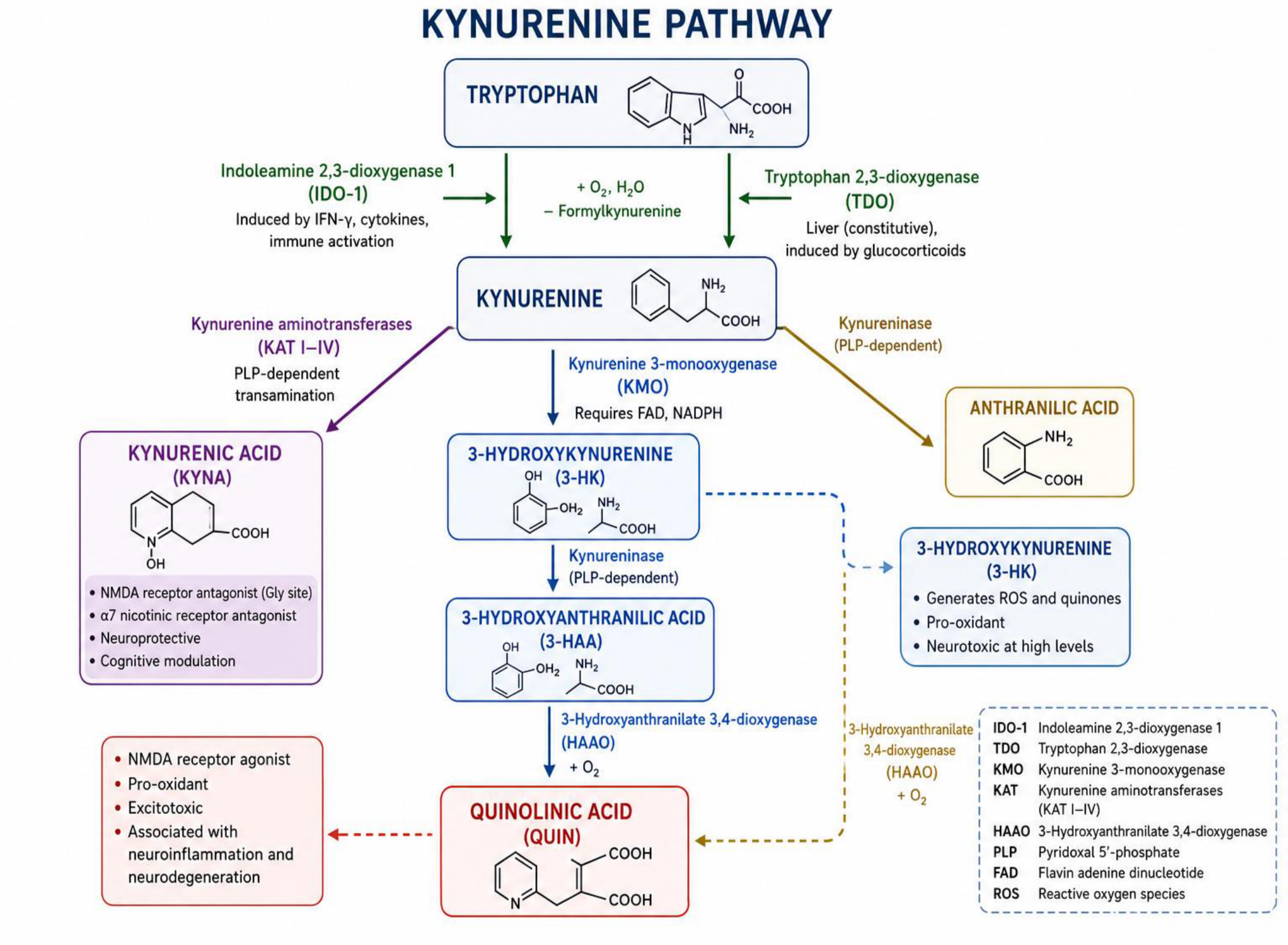
Figure 2.
Cell-specific kynurenine metabolism in astrocytes and microglia illustrating neuroprotective versus neurotoxic pathways.
Figure 2.
Cell-specific kynurenine metabolism in astrocytes and microglia illustrating neuroprotective versus neurotoxic pathways.
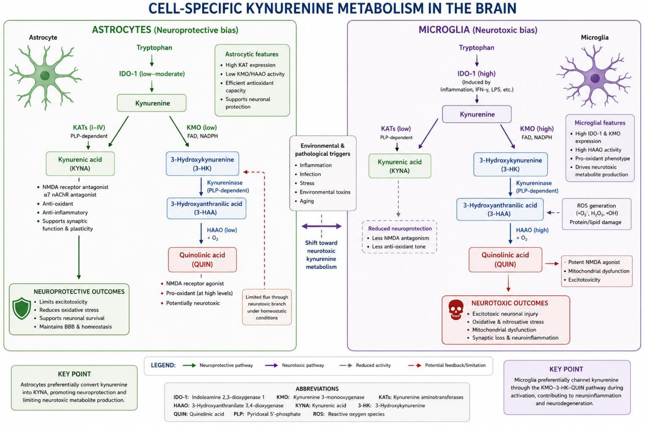

Table 1.
Summary of major environmental pollutants, their proposed mechanisms of action, effects on the kynurenine pathway, and potential neurobiological consequences.
Table 1.
Summary of major environmental pollutants, their proposed mechanisms of action, effects on the kynurenine pathway, and potential neurobiological consequences.
| Environmental Pollutant | Proposed Mechanism | KYN Pathway Effect | Neurobiological Consequence |
| Lead (Pb) [17,19,20,30] |
Oxidative stress and TLR activation | Increased IDO-1/KMO activity | Cognitive impairment |
| Manganese (Mn) [20,26,32,83,106] |
Mitochondrial dysfunction | Increased QUIN and 3-HK | Parkinsonism |
| Cadmium (Cd) [107,108] |
Renal metabolic dysregulation | Altered Trp metabolism | Oxidative stress |
| Arsenic (As) [19,22,68,80] |
Gut dysbiosis and inflammation | Increased KYN accumulation | Cognitive decline |
| Mercury (Hg) [33,34,69,71] |
Neurotransmitter depletion | Reduced KYNA | Neurotoxicity |
| Microplastics [17,75,78,100] |
Intestinal inflammation | TLR activation | Chronic neuroinflammation |
Disclaimer/Publisher’s Note: The statements, opinions and data contained in all publications are solely those of the individual author(s) and contributor(s) and not of MDPI and/or the editor(s). MDPI and/or the editor(s) disclaim responsibility for any injury to people or property resulting from any ideas, methods, instructions or products referred to in the content. |
© 2026 by the authors. Licensee MDPI, Basel, Switzerland. This article is an open access article distributed under the terms and conditions of the Creative Commons Attribution (CC BY) license.
Copyright: This open access article is published under a Creative Commons CC BY 4.0 license, which permit the free download, distribution, and reuse, provided that the author and preprint are cited in any reuse.


