

Submitted:
03 August 2026
Posted:
04 August 2026
You are already at the latest version
Abstract
Microglia encounter combinations of soluble, membrane-bound and mechanical signals whose meaning changes with anatomical niche, developmental or disease stage, receptor state, metabolism, sex and prior experience. Yet most mechanistic studies isolate one ligand–receptor pair, and the term signal integration is often applied to evidence that demonstrates only co-expression or context dependence. Here, we define direct integration as a functional interaction between two or more experimentally manipulated inputs and distinguish it from sequential or multicellular relays, context dependence, coexistence and hypothesis. We then organize the literature around microglial decisions: where to position a process or cell; whether a contacted structure is a target; whether to internalize and degrade it; and how an executed function changes later state and output. The clearest direct example is noradrenergic antagonism of ATP–P2Y12-directed movement. In adolescent mesofrontal plasticity, dopamine-receptor signalling and P2Y12 are both required for enhanced microglial surveillance and bouton formation, although their direct interaction remains unresolved. Strong directional and reciprocal relays include astrocyte-derived IL-33 control of developmental engulfment, activity-gated microglia–astrocyte Wnt signalling, an injury-calibration circuit in which astrocytic ATP recruits tunable microglial IL-1β feedback, perivascular SPP1 control of a complement-linked phagocytic state, and MerTK-triggered TGF-β1 autocrine feedback. By contrast, dose-dependent IL-34 effects, sex-dependent TREM2 phenotypes and circuit-restricted complement requirements establish context dependence rather than cue–cue integration. We also identify major blind spots: two-signal inflammasome logic, GABA-receptive microglia, mechanical sensing, human-state validation and the frequent inability of static microscopy to distinguish engulfment from trogocytosis or scavenging. An evidence-graded framework exposes how few microglial cue combinations have been tested factorially and provides standards for converting spatial or single-cell predictions into causal, functionally interpretable mechanisms.
Keywords:
microglia
; signal integration
; context dependence
; phagocytosis
; purinergic signalling
; TREM2
; inflammasome
; neurotransmitters
; neurovascular unit
Review approach and evidence appraisal
This is a critical narrative review rather than a systematic review. Primary studies were prioritized when they permitted cell-specific, temporal, factorial or rescue-based inference; reviews were used for background and citation discovery. Evidence was classified using the Tier A–E framework according to the experimental design supporting each claim rather than journal prestige or method complexity. Multicellular relays were included when microglia were a causally resolved sender or receiver, even when signal convergence occurred in another cell type. The review does not claim exhaustive coverage of every reported microglial receptor or disease model.
1. From Receptor Pathways to Cellular Decisions
Microglia are long-lived CNS macrophages whose processes continually survey neural tissue and whose functions include injury sensing, circuit remodelling, target clearance, trophic support and coordination of multicellular responses. In vivo imaging displaced the idea of a morphologically “resting” but inactive cell, and developmental studies established that microglia can alter synapse number and circuit maturation. [1,2,3,4,5]
The field nevertheless remains organized largely around receptor-centred modules: P2Y12 for chemotaxis, complement receptors for synaptic elimination, CSF1R for survival, cytokine receptors for inflammatory output and neurotransmitter receptors for neuronal control. These modules are experimentally tractable, but they are not the environment experienced by a microglial cell. A plaque-associated, capillary-associated, developmental or injury-border microglial cell is exposed to nucleotide metabolites, neuromodulators, trophic factors, cytokines, lipids, matrix mechanics, target-bound opsonins and inhibitory checkpoints over different spatial and temporal scales. [6,7,8,9,10]
This review asks a narrower question than “what signals regulate microglia?” It asks what evidence shows that multiple inputs are combined to change a defined microglial decision. The scope includes extracellular chemical and mechanical cues that alter positioning, target selection, internalization, cargo processing, identity or inflammatory and trophic output. It does not attempt to catalogue every reported receptor. Instead, it distinguishes receptor competence from causal contribution, a state marker from a function, and contextual variation from a measured interaction.
This distinction matters because apparently sophisticated data can remain mechanistically weak. Co-expression of a ligand and receptor does not establish access or signalling. Spatial proximity does not establish directionality. A transcriptional state does not identify the inputs that caused it or the functions it retains. A global knockout or systemic drug can demonstrate organismal relevance without locating the responsible receptor to microglia. Even a rescue by receptor inhibition does not prove that two pathways interact unless each input and their combination were tested in the same causal design.
The paper is organized around five linked decisions: (1) process or cell positioning; (2) target recognition; (3) execution of uptake; (4) cargo degradation and recovery; and (5) subsequent state and output. This sequence is more informative than the M1–M2 framework, which cannot represent simultaneous phagocytic, trophic, inflammatory, metabolic and reparative programmes within the same population. [6,11]
Attribution to parenchymal microglia also requires discipline. Perivascular, meningeal and choroid-plexus macrophages share receptors and can be captured by broad myeloid markers or Cx3cr1-based tools. Vascular and CSF1R studies should therefore combine anatomical location, lineage, marker panels and experimental timing rather than rely on IBA1 or CX3CR1 alone. [9,12,13,14]
2. What Counts as Signal Integration?
We use five evidence tiers (Table 1). Only Tier A demonstrates a direct interaction between cues. Tier B establishes a directional relay or feedback loop. Tier C demonstrates that the output of a pathway changes with a defined biological variable, but it does not by itself establish that two extracellular inputs interact. Tier D nominates exposure or coexistence. Tier E is a testable hypothesis. These categories describe the strength and type of causal inference, not the prestige of the method or journal.
2.1. Context Filters Act at Different Stages
Five variables recur across the literature. First, source and geometry determine whether a cue is diffuse, synaptic, somatic, vascular, matrix-bound or displayed on a target membrane. Second, dose, duration and sequence distinguish a transient physiological signal from chronic exposure, receptor desensitization or priming. Third, receptor and identity state determine surface availability, adaptor use and transcriptional interpretation. Fourth, metabolic and degradative capacity constrain movement, uptake and processing. Fifth, history—including prior inflammation, ageing and previously engulfed cargo—changes subsequent responsiveness. [7,8,11,15,16]
Spatial presentation can change the decision without changing ligand identity. An ATP microgradient provides direction, whereas uniformly elevated nucleotide can erase directional information. Target-bound C1q or exposed phosphatidylserine identifies a local structure in a way that a bath-applied ligand cannot reproduce. Amyloid fibril stiffness can itself become a signal through PIEZO1. [10,17,18,19]
Time is equally important. Transient ATP recruits processes, sustained hyperactivity perturbs nucleotide microgradients, a neonatal 5-HT2B signal can influence adult inflammatory responsiveness, and a brief ischaemic episode can produce an acute ATP burst followed by subacute microglial and circuit changes. [20,21,22]
Subcellular localization can overturn receptor-based assumptions. RNA detection does not establish a functional surface receptor. Recent α7 nicotinic-receptor localization data—in which immunoreactivity and bioenergetic responses in mouse microglia are predominantly mitochondrial rather than consistent with a canonical surface ion channel—illustrates why localization and coupling must be measured. [23]
Identity is not merely another output. TGF-β–SMAD signalling maintains Sall1, while SALL1 redirects SMAD4 binding toward microglial regulatory elements and away from inappropriate macrophage programmes. Thus, the same SMAD machinery can interpret a ligand differently depending on a cell-type-specific transcription factor. [16,24]
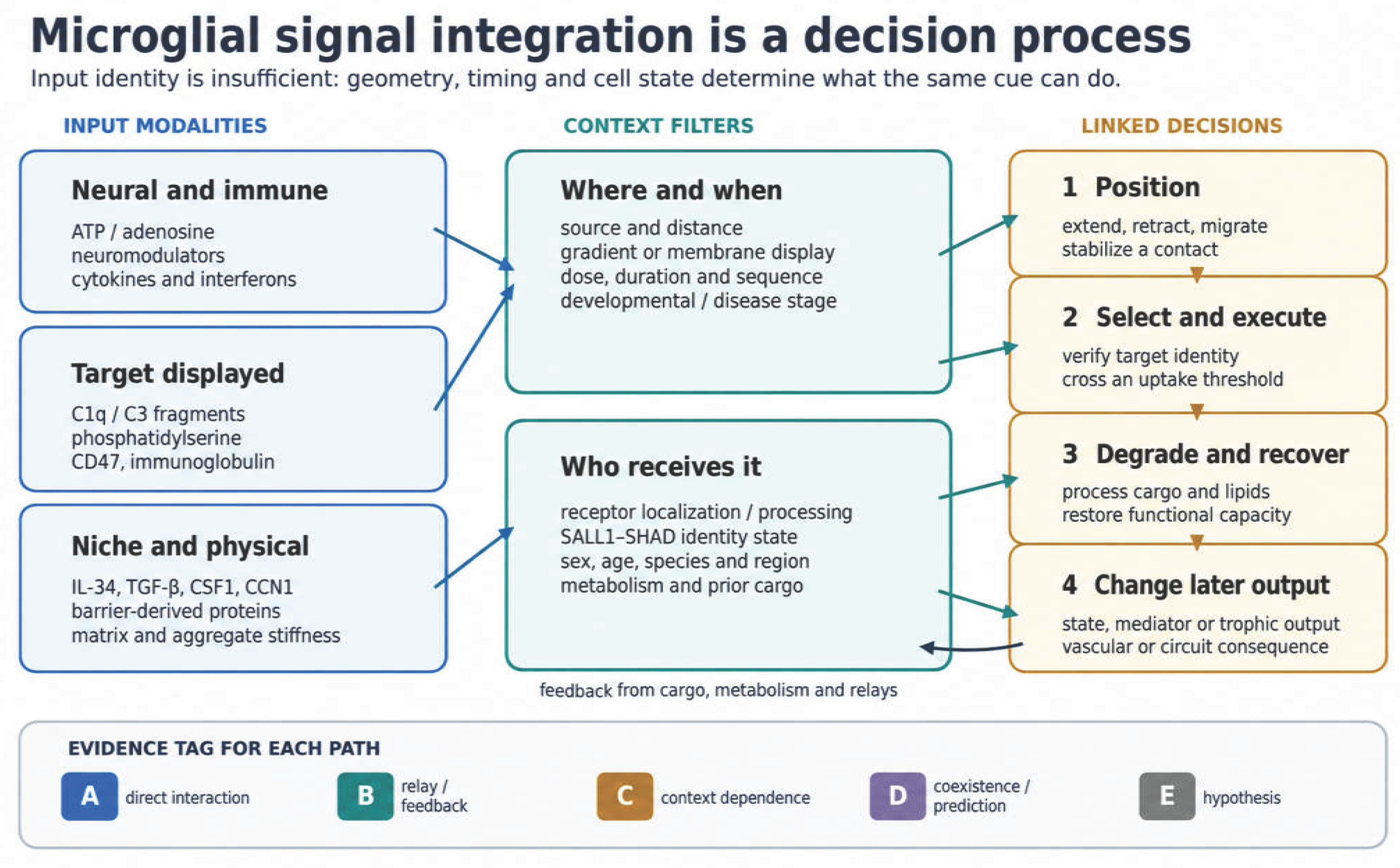
Figure 1.
Decision-and-evidence model of microglial signal integration. Chemical, target-bound and physical inputs are filtered by spatial, temporal and cell-intrinsic context before producing linked decisions. Executed functions feed back through cargo, metabolism and multicellular signals. The diagram is a conceptual architecture, not evidence that every displayed path is experimentally demonstrated; each proposed path must be assigned an evidence tier.
Figure 1.
Decision-and-evidence model of microglial signal integration. Chemical, target-bound and physical inputs are filtered by spatial, temporal and cell-intrinsic context before producing linked decisions. Executed functions feed back through cargo, metabolism and multicellular signals. The diagram is a conceptual architecture, not evidence that every displayed path is experimentally demonstrated; each proposed path must be assigned an evidence tier.
2.2. Representative Exemplars
This classification reveals an important asymmetry: the literature contains abundant context dependence and several strong relays, but relatively few factorial demonstrations of direct cue–cue interaction. That is a substantive conclusion, not a deficiency to conceal.

Table 2.
Representative motifs classified by evidence type.
| Motif | Pathways | Decision and conclusion | Tier | Critical boundary |
| Direct antagonism | Norepinephrine–β2AR versus ATP–P2Y12 | Noradrenergic signalling retracts processes and suppresses ATP-directed extension | A | Acute slices/cultures establish interaction; disease importance needs separate in vivo testing [25,26] |
| Context-dependent co-requirement | Dopamine receptors plus P2Y12 | Both systems are required for enhanced surveillance and dopaminergic bouton formation during adolescent mesofrontal plasticity | C | Developmental and circuit-specific; the direct relationship between dopamine-receptor and P2Y12 signalling was not established [27] |
| Reciprocal injury-feedback relay | Astrocytic Panx1–ATP to microglial IL-1β to astrocytic IL-1R1–Ca2+–calcineurin | Injury-scaled ATP recruits tunable microglial feedback that restrains further astrocytic ATP release and stabilizes the early tissue response | B | Acute focal cortical injury in mice; chronic disease and human conservation remain unresolved [130] |
| Sequential innate-immune logic | Priming plus ATP/P2X7–NLRP3 activation | Transcriptional priming and a second activating signal control IL-1 family cytokine maturation | A/B | Reductionist mouse-microglia experiments support the sequential input logic; priming, licensing, assembly and pyroptosis are separable, and disease studies are not all microglia-exclusive [28,29,131] |
| Directional glial relay | Astrocyte IL-33 to microglia | Astrocyte-derived IL-33 promotes developmental synapse engulfment and circuit maturation | B | Developmental spinal cord and thalamus; not a universal pro-phagocytic rule [30] |
| Activity-gated glial relay | Neuronal CX3CL1 to microglial CX3CR1 to Wnt-responsive astrocytes | Sensory perturbation recruits a microglia-to-astrocyte signal that permits synapse remodelling | B | Postnatal barrel cortex after whisker lesioning; not a generic adult pruning programme [31] |
| Sequential feedback | MerTK to PU.1/IRF8 to microglial TGF-β1 | Phagocytic signalling induces a later autocrine state programme | B | Does not establish that cargo identity alone is the instructive input [15] |
| Reciprocal repair relay | Astrocyte CSF1 and microglial IFN-β | Each glial population supports the other after spinal injury | B | Injury-specific and anatomically restricted [32] |
| Non-monotonic context | IL-34 abundance and developmental timing | Too little and too much IL-34 disrupt different aspects of maturation and engulfment | C | Dose response is not direct multi-cue integration [33] |
| Receptor/state context | TREM2-T96K, sex and amyloid environment | Biochemical ligand hyper-responsiveness does not predict plaque-associated microglial function | C | Endpoint- and sex-dependent; several in vivo outcomes were unchanged [34] |
| Perivascular niche relay | SPP1, amyloid context and complement-linked phagocytic state | Perivascular SPP1 promotes microglial C1q-associated synaptic engulfment in mouse Alzheimer models | B | Not evidence that SPP1 alone specifies every plaque-associated state [9] |
| Mechanical context | Amyloid-fibril stiffness and PIEZO1 | Physical properties of a deposit alter clustering, compaction and uptake | C | Disease-model-specific; chemical ligands and mechanics were not factorially separated in every endpoint [10] |
3. Positioning: Purinergic Direction Under Neuromodulatory and Vascular Control
3.1. A Nucleotide Field, Not a Single Ligand
ATP and ADP provide some of the strongest evidence for directional microglial signalling. Focal injury releases nucleotides that recruit microglial processes through P2Y12, and neuronal NMDA-receptor activation can evoke ATP-dependent process outgrowth in acute slices. [17,18,35]
“Purinergic signalling” is not a unitary input. P2Y12 is Gi-coupled and strongly linked to chemotaxis and physiological contacts; P2Y6 has been associated with phagocytic responses; P2Y4 can support uptake of soluble amyloid-β in culture; and P2X4 and P2X7 are ionotropic receptors that couple nucleotide exposure to calcium, ionic flux and inflammatory outputs. Ectonucleotidases continuously convert ATP through ADP and AMP to adenosine, whose A2A and A3 receptors can favour process retraction or extension in defined preparations. [36,37,38,39]
The relevant input is therefore a changing metabolite field whose composition depends on release, diffusion and enzymatic conversion. A study that applies a fixed ATP concentration cannot reproduce a focal gradient or establish which metabolite and receptor dominate in vivo. Source is also preparation-dependent: stressed neurons, astrocytes, damaged cells and vascular or blood elements can contribute nucleotides. Temporal precedence of neuronal activity before microglial movement does not prove direct glutamate sensing when ATP is the intermediary. [35,40,41]
Astrocytes can also generate structured ATP signals rather than merely contribute diffuse nucleotides after damage. In focal cortical injury, astrocytic Panx1-dependent ATP events scaled with lesion severity. Microglia sensed the changing ATP environment and adjusted feedback strength through IL-1β, linking nucleotide magnitude to a later reciprocal response. This mechanism reinforces the need to distinguish actively organized ATP release from passive nucleotide leakage after cellular injury. [130]
P2Y12-dependent functions also diverge by endpoint. During ocular-dominance plasticity, receptor loss or blockade alters microglial process behaviour, contacts, engulfment-related readouts and the cortical response to monocular deprivation. In the developing retina, P2Y12-dependent behaviour is coupled temporally to the availability of apoptotic targets. At vascular interfaces, PANX1–P2Y12 signalling contributes to capillary regulation, while P2Y12-dependent process convergence can help close a focal blood–brain barrier lesion. [42,43,44,45]
These results do not support a generic statement that P2Y12 is beneficial or harmful. Process speed, target contact, uptake, neuronal feedback, capillary diameter and barrier closure occur on different time scales and can dissociate. The receptor can be present while downstream actin, potassium-channel, calcium or metabolic machinery is inadequate for a specific function. [7,8,20]
3.2. Duration Converts Surveillance into Dysfunction
Transient and sustained nucleotide exposure can have different consequences. Neuronal hyperactivity disrupts ATP microgradients, reduces microglial motility, decreases selected phagocytic-receptor expression and uncouples apoptosis from clearance in the experimental system used by Abiega et al. Microglial conversion of ATP to adenosine can, in another setting, suppress neuronal activity through neuronal adenosine receptors. [20,40]
The 2026 TIA study provides a useful sequential example. A five-minute middle-cerebral-artery occlusion without detected infarction produced focal extracellular ATP events, prolonged microglial transcriptional and morphological changes, loss of synaptic readouts, network disconnection and cognitive deficits. Intracisternal P2Y12 inhibition normalized several microglia–synapse, synaptic and behavioural endpoints without altering systemic platelet aggregation in the reported assays. [22]
This evidence is stronger than a cross-sectional association but should be described precisely. The critical ATP source was unresolved; the engulfment metric used static containment/colocalization of synaptic markers within P2Y12-labelled microglial volumes; and the authors reported no prospective power calculation. The study therefore supports an ATP–P2Y12-dependent sequence connecting brief ischaemia to prolonged microglia–synapse interactions and functional deficits, but it does not provide direct live proof that microglia removed intact synapses.
3.3. Direct Antagonism and Context-Dependent Co-Requirement
Norepinephrine provides a direct filter on purinergic movement. In acute slices and cultured microglia, β2-adrenergic signalling causes process retraction and suppresses ATP-directed extension. Inflammatory stimulation changes adrenergic-receptor expression and can shift the receptor contribution. The first observation is Tier A antagonism; receptor switching is Tier C context dependence. [25,26]
Dopamine provides a rarer in vivo example of context-dependent co-requirement. During adolescent mesofrontal plasticity, rewarding experience or optogenetic activation of dopaminergic axons increases microglial surveillance of parenchyma and dopaminergic boutons. Dopamine-receptor signalling and P2Y12 are both required for the reported increase in surveillance and new bouton formation, but the study does not establish a direct interaction between the two pathways. The result should not be generalized beyond the developmental window and frontal circuit, although it exceeds transmitter-receptor expression in culture. [27]
3.4. From Process Dynamics to Circuit Computation
P2Y12-dependent contacts can influence neuronal and network endpoints, but receptor-specific and whole-microglia perturbations need not be equivalent. In the 2026 barrel-cortex study, pharmacological depletion and germline P2Y12 loss both increased baseline firing in putative narrow-spiking neurons and altered oscillatory or thalamocortical measures, while several stimulus-evoked firing measures were preserved. [46]
The study extends P2Y12 biology from morphology to circuit computation, but it remains Tier C rather than direct signal integration. Only male mice were used; spike width identifies putative rather than molecularly confirmed interneuron classes; germline knockout permits developmental compensation; CSF1R-inhibitor depletion perturbs the cellular ecosystem; and functional ultrasound reports haemodynamic signals that can be influenced by P2Y12-dependent neurovascular effects. The strongest conclusion is that microglial presence and P2Y12 function constrain selected baseline and network properties in this circuit, not that a single P2Y12 mechanism explains every readout.
4. Target Selection: From Contact to Degradation
Engulfment is a sequence, not a binary state. A microglial process must locate a candidate, maintain contact, interpret target-bound and soluble cues, cross an execution threshold, reorganize the cytoskeleton, internalize material, degrade it and recover. ATP can contribute to finding; complement fragments and exposed phosphatidylserine can contribute to target recognition; CD47–SIRPα can restrain execution; and cargo burden can alter later state. The relative importance of each step depends on target, circuit, age and disease. [47,48]
4.1. Complement Labels Targets but Does Not Define a Universal Pruning Programme
In the developing visual system, C1q and C3 contribute to activity-dependent elimination of retinal inputs involving CR3-expressing microglia. Complement–microglia signalling also contributes to early synapse loss in amyloid models and to synaptic pathology in selected inflammatory and neurodegenerative settings. [4,49,50,51]
The cascade separates target labelling from uptake. C1q can accumulate on selected structures, C3 cleavage products provide opsonins, and CR3 participates in recognition and cytoskeletal responses. C3aR and C5aR1 influence migration and inflammatory signalling independently of opsonic uptake. A statement that “complement activates microglia” obscures these branches. [52,53]
Cellular source is component- and condition-specific. Astrocytes can produce C3 in defined amyloid-associated models, stressed neurons can express complement components, and microglia produce receptors and selected components. Source claims should therefore name the component, tissue and state. [54,55]
The visual-system literature encouraged the assumption that complement-dependent pruning is a general CNS mechanism. The evidence instead establishes circuit and disease restriction. C3 deficiency protects synapses in some plaque-rich models; CR3-dependent synapse loss precedes neuronal loss in a systemic-inflammation Parkinson model; other circuits and target types use alternative routes. [51,56,57]
Postoperative injury provides another disease-restricted complement context. In a mouse model of postoperative neurocognitive disorder, tibial-fracture surgery increased hippocampal classical-complement activity and C1q-associated excitatory and inhibitory synaptic material within microglial lysosomal compartments. Intraventricular C1q neutralization and CA1 microglia-targeted C1q knockdown preserved synaptic proteins and dendritic spines and improved postoperative memory measures. NF-κB inhibition also reduced surgery-associated C1q expression and cognitive impairment. These findings support a causal requirement for microglial C1q in postoperative synapse loss, but they do not demonstrate direct integration between C1q and another extracellular target-selection cue. The upstream postoperative signal, the complete NF-κB–C1q sequence and the distinction between removal of intact synapses and clearance of synaptic debris remain incompletely resolved [137]
This is Tier C context dependence unless complement and an alternative target cue are manipulated together. Likewise, pioglitazone-induced reduction of synaptic C1q, microglial engulfment-related readouts and spine loss in APP/PS1 mice demonstrates that the target environment is pharmacologically modifiable, but it does not prove direct, microglia-exclusive PPAR-γ action. [58]
Complement output is also gated intracellularly. During early postnatal hippocampal development, conditional loss of microglial INPP5D/SHIP1 increases complement abundance, synaptic material in microglial phagolysosomes and synapse loss; neuronal CD55 expression prevents the synapse-loss phenotype. Early, but not adult, deletion produces later cognitive defects, and SHIP1-null human iPSC-derived microglia show increased synaptosome uptake. [59] This is strong evidence that developmental state and an intracellular phosphatase set complement-dependent execution, but it is not a second extracellular cue.
4.2. Phosphatidylserine Is a Target State, Not a Verdict
Phosphatidylserine is normally enriched on the inner plasma-membrane leaflet but can become exposed during apoptosis, cellular stress and local membrane remodelling. Microglia recognize externalized phosphatidylserine through receptor and bridging systems that include TAM receptors with Gas6 or Protein S, MFG-E8 with αv integrins, GPR56 and TREM2 in defined CNS contexts. Peripheral-macrophage evidence for other receptors should not be presented as equivalent adult microglial proof. [19,60,61,62]
Exposure does not necessarily mean irreversible cell death. Local externalization can mark restricted synaptic or stressed-neuronal domains, creating the possibility of phagoptosis when an otherwise viable structure crosses an execution threshold. MFG-E8-dependent removal of stressed neurons during neuroinflammation illustrates how a bridging molecule can convert phosphatidylserine exposure into αv-integrin-dependent uptake without requiring the same mechanism to operate for every cargo. [63,64,65]
Receptor use depends on target and niche. GPR56 participates in phosphatidylserine-dependent synaptic refinement, whereas MerTK is required for removal of persistently phosphatidylserine-exposed inhibitory postsynapses after neuronal Cdc50a loss. These are not redundant pathways simply because they recognize a common lipid feature. [61,66]
Rueda-Carrasco et al. add disease-relevant evidence that externalized phosphatidylserine and TREM2 contribute to selective removal of hyperactive synapses in Alzheimer models and can reduce neuronal hyperactivity. [67] This result shows that PtdSer–TREM2 recognition is not merely speculative. It does not, however, establish that TREM2 is the sole phosphatidylserine receptor or that synapse removal is uniformly harmful; in this model the functional consequence was protective.
4.3. TREM2 Output Depends on Ligand, Processing, Sex and Endpoint
TREM2 binds several lipid- and protein-associated ligands and signals through TYROBP/DAP12 and downstream kinases to influence survival, metabolism, clustering, phagocytosis and state transitions. A “TREM2 function” is therefore not one scalar quantity. Ligand binding, surface delivery, proteolytic shedding, soluble TREM2, adaptor coupling and disease substrate can diverge. [11,34]
The TREM2-T96K study makes this point unusually clearly. T96K had previously been classified as gain of function by ligand-dependent activation assays, yet the knock-in variant was associated with Alzheimer risk and reduced selected plaque-associated microglial responses, soluble TREM2 and disease-associated state transitions, especially in female 5xFAD mice. Aβ uptake was impaired in engineered microglial-cell assays, whereas in vivo methoxy-X04 uptake and several neuronal, synaptic and plaque-burden endpoints were not uniformly changed. Cognitive function was not tested. [34]
The appropriate conclusion is not that “too much TREM2 is harmful” or that gain and loss of function are equivalent. The study shows that an assay-defined receptor property is insufficient to predict processing, sex-dependent plaque behaviour or organismal outcome. This is Tier C context dependence and a warning for agonist development.
4.4. Inhibitory Checkpoints Set Execution Thresholds
CD47 on neuronal or synaptic membranes engages microglial SIRPα and recruits inhibitory phosphatases that restrain cytoskeletal activation. During development, CD47 protects synapses from excess pruning; in neurodegeneration models, microglial SIRPα loss can increase engulfment and worsen synaptic or cognitive outcomes. [68,69,70,71]
These studies establish an inhibitory axis, but they do not by themselves demonstrate direct competition between CD47–SIRPα and complement or phosphatidylserine. The common claim that pro-engulfment and “do-not-eat-me” cues are integrated at a target threshold is a strong hypothesis supported by separately causal pathways; it becomes Tier A only when the positive and negative inputs are manipulated factorially on the same target.
The axis is also not a universal one-ligand/one-receptor brake. Neuronal SIRPα can limit access of neuronal CD47 to microglial SIRPα during retinal development, producing a circuit-specific configuration that promotes rather than restrains phagocytosis. Global CD47 manipulation in EAE affects peripheral and CNS immune compartments and changes with intervention timing. [72,73,74]
Age introduces additional inhibitory receptors. CD22 rises on aged microglia, and blockade in aged mice restores selected homeostatic and phagocytic measures and improves cognitive performance. [75] This is a compelling Tier C ageing context, not evidence that CD22 blockade will improve clearance of every target or remain safe in younger or diseased tissue.
Fcγ receptors provide a separate disease-associated route. In Parkinson tissue and experimental models, low-affinity Fcγ receptor expression is associated with phagocytic microglia, and receptor blockade reduces elimination of dopaminergic cells. [76] The result should remain restricted to immunoglobulin-rich or inflammatory contexts and should not be used as a general citation for all Parkinson-related microglial cues.
4.5. “Inside Microglia” Does Not Resolve What Was Removed
Static three-dimensional containment of a synaptic marker within a microglial volume is stronger than two-dimensional overlap but still does not establish the history of the material. It can represent whole-structure engulfment, presynaptic trogocytosis, uptake of shed fragments, clearance after neuronal elimination, or segmentation error. In a developmental hippocampal preparation, Weinhard et al. observed presynaptic trogocytosis and microglia-induced spine-head filopodia rather than wholesale postsynaptic engulfment. [77]
The postoperative C1q study illustrates the same limitation: lysosomal colocalization of Homer1 or gephyrin with microglial markers supports internalization of synaptic material, but fixed-tissue imaging cannot conclusively distinguish removal of intact synapses from uptake of shed or degenerating material [137]
Strong claims should combine several orthogonal criteria: lysosomal localization, live or correlative imaging, evidence of target-number loss, cargo degradation, temporal order, receptor-specific loss and rescue, and exclusion of shed material. The biological claim should match the assay: “microglia-associated synaptic material,” “internalized synaptic protein,” “trogocytosis,” “clearance of shed material” and “elimination of an intact synapse” are not synonyms.
4.6. Cargo Processing Feeds Back on Later Competence
Uptake is not the terminal step. Human iPSC-derived microglia exposed to synaptosomes, myelin, apoptotic neurons or amyloid fibrils adopt cargo-associated transcriptional programmes involving APOE, GPNMB, lipid handling and lysosomes. [11] Because these experiments use an engineered in vitro environment, they establish cargo responsiveness rather than a persistent in vivo memory.
The MerTK-triggered pathway provides a stronger molecular bridge from execution to later state. In optic-nerve injury and 5xFAD models, MerTK-dependent phospholipase-C signalling increases PU.1 and IRF8, induces microglial TGF-β1 and establishes an autocrine programme required for selected neurodegenerative responses. [15] Astrocyte-derived CCN1 acting through microglial SDC4 after spinal injury adds a complementary lesson: more intracellular myelin signal can reflect impaired digestion rather than more effective clearance, and lipid buffering can determine whether uptake remains productive. [78]
A bidirectional lysosomal relay makes the same point from the opposite direction. Microglia constitutively secrete β-hexosaminidase that can enter neuronal lysosomes and support GM2 degradation. In Hexb-deficient mice, accumulated neuronal GM2 engages microglial MGL2 and drives inflammatory output; replacement with enzyme-competent myeloid cells interrupts the cycle. [79] This is a strong disease-specific Tier B loop linking microglial secretory capacity, neuronal cargo metabolism and a later microglial response, not a general model of lipid sensing.
The term “engulfment memory” should therefore be used cautiously. Current data support cargo-induced and receptor-induced state changes whose duration, reversibility and dependence on the surrounding niche remain incompletely defined.
5. Identity and State Determine How a Cue Is Interpreted
5.1. CSF1R Ligands Support Different Niches
CSF1R signalling is essential for microglial development, survival and maintenance, but the receptor is shared with other CNS macrophages. CSF1 and IL-34 activate the same receptor while differing in cellular source, spatial distribution, molecular presentation and developmental timing. Defined-medium and genetic studies establish a requirement for CSF1R-dependent niche support, whereas global receptor disruption and pharmacological depletion have systemic and ecosystem effects. [80,81,82,83]
CSF1R inhibition is therefore not a microglia-specific subtraction. Dose and duration can affect border-associated macrophages, and the surviving or repopulating cells encounter tissue altered by cell loss, accumulated cargo, cytokines, matrix and neuronal activity. Depletion–repopulation studies should be interpreted as niche perturbations. Sex-dependent responses to CSF1R inhibition further argue against a universal depletion model. [14,81]
IL-34 provides a strong Tier C example. In the developing anterior cingulate cortex, excitatory-neuron-derived IL-34 supports microglial abundance and maturation and restrains aberrant engulfment of thalamocortical synapses. Partial blockade increases inappropriate phagocytic readouts, whereas overexpression suppresses physiologically appropriate engulfment. [33] This non-monotonic response links ligand abundance to maturation and target selection, but it is not direct evidence that IL-34 interacts with phosphatidylserine, complement or CD47.
The result also cautions against equating survival-factor action with cell number. Altered microglial maturation and engulfment among surviving cells imply that CSF1R ligand context changes competence as well as abundance.
5.2. TGF-β Is Filtered Through Microglial Identity
TGF-β signalling maintains microglial identity, but ligand source and activation vary by context. In adult mice, microglial TGF-β1 is required cell-autonomously for homeostatic markers and normal cognitive function; deletion from astrocytes or forebrain neurons does not reproduce the phenotype. During neurodegeneration, MerTK signalling induces an additional microglial TGF-β1 autocrine programme. [15,24]
Production is only one layer. TGF-β is secreted in a latent form and requires extracellular activation before receptor engagement. The response is then interpreted through the cell’s regulatory state. SMAD4 supports Sall1 expression, and SALL1 in turn promotes microglia-specific SMAD4 binding while suppressing inappropriate binding at alternative macrophage enhancers. [16]
This reciprocal SALL1–SMAD mechanism is a paradigmatic context filter: neither receptor abundance nor SMAD phosphorylation alone identifies the transcriptional output. Loss of identity can change the meaning of the same TGF-β signal.
Astrocytic TGF-β remains relevant in defined inflammatory settings. IL-10-redirected astrocytes can produce TGF-β that suppresses microglial inflammatory activation. [84] A source- and state-specific synthesis is therefore warranted rather than a search for one universal ligand-producing cell.
5.3. Sex, Age, Region and Species Are Mechanistic Variables
Adult male and female mouse microglia show differences in transcriptome, translation, density and selected functions. Age, sex and genotype jointly alter plaque-associated microglial responses in Alzheimer models. [85,86] The TREM2-T96K phenotype reinforces this principle because prominent effects on plaque clustering, soluble TREM2 and state transitions occurred in female 5xFAD mice. [34]
Sex should therefore be treated as a potential interaction variable, not only a demographic line in methods. A male-only result is evidence in males unless independent data support generalization. Studies powered for an overall treatment effect are not automatically powered for a treatment-by-sex interaction, and an apparent sex specificity requires formal comparison rather than significance in one sex and non-significance in the other.
Regional identity is equally important. White-matter ageing, developmental stage, local neuronal subtype and vascular proximity generate distinct microglial environments. The same receptor can be required for a developmental retinal contact, an adult cortical network property or vascular repair without the downstream mechanisms being interchangeable. [43,44,78]
Human microglia also differ from mouse microglia, and removal from the brain rapidly remodels their transcriptional and enhancer landscape. Gosselin et al. showed that the human CNS environment is required to maintain a substantial part of the ex vivo identity programme. [87] Human iPSC-derived microglia, organoids and xenografts are valuable because they permit perturbation, but each reconstructs a different subset of adult human context.
Human midbrain–striatal assembloids now provide a causal model-level bridge. Regionally patterned microglia show calcium responses to circuit activation involving GABA_B receptors; in assembloids carrying an SCN2A loss-of-function variant, increased microglial calcium activity and synaptic pruning are reduced by pharmacological GABA_B-receptor inhibition or microglial GABBR1 deletion. [88] This is stronger than receptor RNA or postmortem association, but it remains a developing in vitro circuit model rather than evidence from adult human brain.
Recent human datasets add needed state resolution. Ex vivo profiling of microglia from 189 postmortem brains identified Alzheimer-associated changes in genes, isoforms, co-expression and molecular subtypes. [89] Spatial and single-nucleus profiling across octogenarian and centenarian brains in 2026 identified early and late plaque-induced programmes across an Aβ–tau inflection and different patterns in cognitively resilient individuals. [90] These studies provide strong human Tier D/C evidence for stage- and niche-associated states; they do not identify the extracellular cue combinations that caused them.
6. Sequential Immune Logic: Priming, Activation and Feed-Forward Pathology
A signal-integration analysis must distinguish sequential control of pathway competence from execution. The NLRP3 inflammasome provides the clearest example.
6.1. Priming and Activation Are Separable Decisions
Canonical NLRP3 activation is often described as a two-signal process. A priming input—frequently through pattern-recognition receptors and NF-κB—raises NLRP3 and pro-IL-1β abundance and can license the pathway. A subsequent activating input, including extracellular ATP acting through P2X7 in appropriate contexts, promotes ionic changes, inflammasome assembly, caspase-1 activation and maturation of IL-1β and IL-18; gasdermin-D cleavage can lead to pyroptotic membrane permeabilization. These steps are regulated and should not be collapsed into “microglial activation.”
This architecture can meet Tier A when priming and activating inputs are manipulated factorially with an interaction or dependency measured. Reductionist studies in mouse microglia show functional NLRP3 responses after priming followed by classical activating inputs including ATP. [131] Disease-model genetics then establish context and consequence rather than replacing the two-input experiment. In APP/PS1 mice, NLRP3 or caspase-1 deficiency alters amyloid-associated pathology and microglial responses. In tau models, NLRP3 loss reduces tau hyperphosphorylation and aggregation. [28,29]
The pathway can also create a relay beyond cytokine release. Microglia-derived ASC specks bind amyloid-β and promote aggregation and spreading in experimental systems, providing a feed-forward link between inflammasome execution and extracellular pathology. [91]
Several boundaries are essential. High extracellular ATP does not prove NLRP3 activation; P2X7 has other outputs. Increased NLRP3, ASC or pro-IL-1β expression does not prove assembled inflammasomes or cytokine processing. A whole-animal Nlrp3 knockout does not by itself locate the decisive pathway to microglia. Finally, reduced pathology after inhibition does not establish that physiological inflammasome signalling is uniformly harmful.
6.2. Cytosolic DNA Sensing Couples Intracellular Damage to Intercellular Output
cGAS–STING adds a different form of integration. Cytosolic DNA generated by mitochondrial or nuclear stress is detected intracellularly, but the pathway converts that history into type-I-interferon and multicellular outputs. In aged mice, mitochondrial DNA release engages cGAS–STING and drives reactive microglial states, neurodegeneration and cognitive decline. [92]
In a tauopathy model, pathogenic tau activates microglial cGAS and type-I-interferon responses partly through mitochondrial DNA leakage; Cgas ablation preserves synaptic and cognitive measures without reducing tau load and alters a neuronal MEF2C-linked resilience programme. [93] In an amyloid/tau knock-in model, pharmacological STING blockade reduces inflammatory, NLRP3, synaptic and pathological readouts, although neuronal as well as microglial STING can contribute. [94]
These studies show how one cell’s internal damage sensor can become another cell’s extracellular context. Signal integration should therefore be understood as a path from extracellular or cargo-derived perturbation through intracellular sensing to intercellular output, rather than as a process restricted to plasma-membrane receptors
7. Multicellular Relays and Physical Niches
7.1. Astrocytes Can Specify the Target and the Phase of Response
Astrocytes do not provide a single pro- or anti-inflammatory signal. Their secretome changes with region, injury and time, and microglia in turn alter astrocyte state.
During development, astrocyte-derived IL-33 signals primarily to microglia in the studied spinal-cord and thalamic contexts, promotes synapse engulfment and is required for normal circuit maturation. [30] This is stronger and more specific than a general statement that IL-33 “increases phagocytosis.” It identifies source, receiver, target class and functional consequence while remaining developmentally restricted.
Astrocytes can also signal through a processed extracellular matrix protein. A proteolytically generated C-terminal fragment of astrocytic Hevin engages microglial TLR4, induces a TLR2-high phagolysosomal state and supports elimination of thalamocortical synapses during early postnatal refinement. [95] This Tier B relay is anatomically and developmentally delimited; it should not be used to infer that TLR4 activation is generically pro-pruning.
The direction reverses after sensory perturbation. In postnatal barrel cortex, neuronal CX3CL1–microglial CX3CR1 signalling promotes microglial Wnt release; canonical Wnt signalling in astrocytes reduces perisynaptic astrocyte contacts and is required for subsequent microglial engulfment and synapse loss after whisker lesioning. [31] This is an unusually complete Tier B sequence because neuronal input, microglial intermediate, astrocytic receiver and structural outcome are causally linked. It remains a lesion- and circuit-specific relay rather than direct integration of two extracellular inputs by one microglial cell.
After inflammatory stimulation, microglial IL-1α, TNF and C1q jointly induce a neurotoxic astrocyte programme in the original culture and disease models. [96] The combination is important, but the historical “A1” label should not be generalized to every reactive astrocyte or used as a functional diagnosis without direct testing.
In experimental autoimmune encephalomyelitis, microglial TGF-α and VEGF-B exert opposing effects on astrocyte pathogenicity, and microbial tryptophan metabolites act upstream through microglial aryl-hydrocarbon-receptor signalling. [97] This is a multi-level relay in which a systemic metabolic input changes the balance of two microglial outputs received by astrocytes.
After spinal-cord injury, lesion-border astrocytic CSF1 supports perilesional microglial proliferation and repair, while microglial IFN-β supports astrocyte survival and border formation. Cell-specific deletion and rescue make this a strong reciprocal Tier B relay. [32]
A reciprocal cortical injury circuit provides another strong Tier B example. Following focal injury, astrocytic Panx1-dependent ATP events scale with lesion severity. Microglia adjust IL-1β output according to extracellular ATP and cellular state, and microglial IL-1β is received by astrocytic IL-1R1, engaging Ca²⁺– calcineurin signalling to suppress further Panx1-dependent ATP release. Cell-specific perturbations and rescue support the direction and functional consequence of this feedback. The mechanism demonstrates that IL-1β can function as a rapid restraining signal within a defined reciprocal circuit rather than carrying an invariant pro-inflammatory meaning. Its present evidentiary boundary is acute focal cortical injury in mice. [130]
7.2. Niche Signals Couple Phagocytosis to Metabolism
Perivascular and white-matter studies show why ligand source cannot be inferred from microglial expression alone. In mouse Alzheimer models, perivascular macrophages and, to a lesser extent, fibroblasts produce SPP1. SPP1 is required for microglia to upregulate a C1q-associated phagocytic programme and engulf synaptic material in the presence of amyloid-β challenge; Spp1 loss preserves synapses in the reported models. [9]
The study links a spatial niche signal to complement and target removal, but the integration claim should remain precise. SPP1 is an extrinsic modulator required in that amyloid context; it is not sufficient evidence that all SPP1-positive microglia share one function or that complement and SPP1 interact identically in other regions.
In degenerating white matter after spinal injury, lesion-remote astrocytes produce CCN1, which binds microglial SDC4 and supports lipid-droplet buffering and intracellular digestion of myelin-rich debris. Astrocyte-specific Ccn1 loss produces more numerous but dysfunctional debris-laden microglial nodules and impaired recovery. [78] This result separates uptake from successful clearance and gives metabolic capacity a defined extracellular regulator.
These mechanisms are more informative than broad labels such as “reactive astrocyte” or “phagocytic microglia.” They specify the sender, receptor, anatomical niche, cargo and functional bottleneck.
7.3. Vascular Leakage Changes Both Ligand Access and Cell Attribution
Barrier disruption introduces plasma proteins into a compartment from which they are normally excluded. Fibrinogen therefore reports vascular damage and provides an integrin ligand. Interfering selectively with the fibrin–CD11b interaction can suppress inflammatory pathology in experimental disease without globally blocking coagulation. [98]
Endothelial MMP-3 after spinal injury can activate microglia and contribute to oligodendrocyte damage, whereas VEGF can attract microglia toward amyloid-associated tissue or reduce scavenger-receptor expression in other ischaemic settings. [99,100,101] These findings establish context-dependent vascular inputs, not one sign for “vascular activation.”
Attribution is particularly difficult near vessels because parenchymal microglia, perivascular macrophages and infiltrating myeloid cells share markers. Longitudinal imaging and lineage-resolved tools are needed to determine whether vascular dysfunction recruits microglia, microglial responses stabilize the lesion, or a later inflammatory phase worsens it. [14,44,45,102]
7.4. Mechanics Is a Signal Modality
The extracellular matrix and protein deposits present force, stiffness and topology as well as chemical ligands. Amyloid-associated tissue is mechanically altered, and microglial PIEZO1 senses fibril stiffness in an Alzheimer model, increasing calcium entry, plaque clustering, compaction and phagocytic responses. [10]
This study extends the integration problem from “which ligands coexist?” to “how is a ligand physically presented?” A stiff fibril and a soluble peptide can share sequence while imposing different mechanical and receptor contexts. Future designs should vary chemical identity and stiffness independently; without that factorial separation, PIEZO1 evidence remains strong context dependence rather than proof of integration with a specified chemical receptor.
8. Neuromodulators: Circuit Information, Not Fixed Immune Instructions
Neuromodulators report activity, arousal, reward and circuit identity. Their concentration at a microglial membrane is rarely equivalent to concentration in a synaptic cleft: microglia often sample extrasynaptic spillover, volume transmission or injury-associated release. Innervation density, transporters, degradation, behavioural state and distance from the source determine effective exposure. [103,104]
Co-release and indirect signalling complicate attribution. Neuronal activity can release ATP, and dopamine, acetylcholine or glutamate can change the activity pattern that generates nucleotide release. A transmitter-associated microglial response can therefore contain a direct receptor component and an indirect purinergic component. [27,35,41]
Table 3.
Neuromodulator evidence and boundaries.
| Transmitter | Best-supported microglial decision | Evidence class | Boundary |
| Norepinephrine | Process retraction and antagonism of ATP-directed extension; state-dependent receptor switching | Tier A for acute antagonism; Tier C for disease/state effects | Locus-coeruleus lesions also alter neuronal activity, sleep and vessels [25,26,105] |
| Dopamine | Dopamine-receptor- and P2Y12-dependent surveillance and bouton formation during adolescent frontal plasticity | Tier C co-requirement | Developmental and circuit-specific; the direct dopamine-receptor/P2Y12 interaction was not established, and culture or Parkinson models do not establish the same mechanism [27,106,107] |
| GABA | Selective interaction with and remodelling of inhibitory cortical synapses during a postnatal window | Tier C/B | Developmental mouse cortex; not evidence for a universal adult GABA response [108] |
| Glutamate | Direct receptor competence in selected preparations and indirect ATP-dependent process outgrowth | Mostly Tier C/D | Expression is preparation-sensitive; NMDA-driven microglial movement can be purinergic [35,109,110,111] |
| Acetylcholine | Muscarinic M3-dependent recruitment/repair after stroke; α7-linked inflammatory or metabolic effects in culture | Tier C | α7 localization may be mitochondrial in mouse microglia; agonists act on many cell types [23,112,113] |
| Histamine | H3-dependent suppression of ATP-evoked calcium, chemotaxis and phagocytosis in primary cultures | Tier A/C in vitro | Receptor pharmacology, neuronal autoreceptors and indirect glial effects limit in vivo attribution [114,115,116] |
| Serotonin | Motility/phagocytosis in postnatal preparations; 5-HT2B-dependent developmental programming; HMGB1-dependent modulation of 5-HT7R/cAMP signalling in stroke-related models | Tier C for most in vivo effects; direct receptor-level modulation in reductionist systems | Developmental timing, receptor subtype and disease model differ; global Htr7 deletion, systemic HMGB1 inhibition and male-only tMCAO limit microglia-specific attribution [21,117,118,132] |
8.1. Norepinephrine and Dopamine Illustrate Direct Antagonism and Co-Requirement
Noradrenergic tone changes microglial process behaviour across sleep–wake and inflammatory states. β2-adrenergic signalling dominates several homeostatic motility responses, whereas inflammation changes receptor expression and can increase α2A contribution. [25,26,119] Locus-coeruleus degeneration, however, removes more than a microglial ligand: it alters network activity, vascular tone, arousal and possibly sleep organization. Lesion studies establish physiological relevance but cannot assign every downstream effect to direct microglial receptors. [120,121,122]
In 5xFAD mice, plaque-associated microglia downregulate β2 receptors; microglial receptor loss worsens selected plaque and neuritic outcomes, while stimulation attenuates pathology in the reported model. [105] This is a therapeutic context result layered onto the direct norepinephrine–ATP antagonism, not proof that β2 activation is universally protective.
Dopamine-receptor pharmacology in cultured microglia alters adhesion, motility, phagocytosis and inflammatory mediators in concentration- and state-dependent ways. [106,107,123] Parkinson models simultaneously change dopamine, neuronal survival, ATP, lipid cargo, immunoglobulins and vascular or astrocytic signals. The adolescent frontal-cortex study remains the cleanest evidence for dopamine–P2Y12 co-requirement, but not direct cooperation; it should not be merged mechanistically with dopaminergic-neuron loss.
8.2. GABA Demonstrates Circuit-Matched Microglial Specialization
Favuzzi et al. identified GABA-receptive microglia that preferentially interact with inhibitory cortical synapses during a critical postnatal period. Microglial GABA-receptor ablation disrupted the associated transcriptional remodelling programme, inhibitory connectivity and behavioural outcomes without producing an equivalent effect on excitatory synapses. [108]
This study is central because it connects a transmitter, a specialized microglial state, a target class and a developmental window. It should nevertheless be classified conservatively. It demonstrates circuit-matched context and a directional transmitter-to-microglia programme, not that GABA and a second extracellular cue were integrated factorially.
8.3. Glutamate and Acetylcholine Expose Receptor-Attribution Problems
Glutamatergic activity influences microglia directly in some preparations and indirectly through activity-dependent ATP release. Reported ionotropic and metabotropic receptor profiles vary between neonatal cultures, cell lines, acute slices and freshly isolated adult cells. Functional electrophysiology, protein localization or receptor-specific genetics should accompany transcript or antibody evidence. [35,104,110]
Metabotropic receptor studies report divergent inflammatory and trophic outputs, including BDNF induction by group-II agonism and context-dependent mGluR5 effects. [109,111,124] These data establish receptor competence in specific preparations, not a general rule that glutamate is pro- or anti-inflammatory.
Cholinergic studies require the same restraint. Muscarinic M3 signalling influences recruitment, phagocytosis and injury evolution after stroke in mouse models. [112] α7 nicotinic agonists reduce inflammatory outputs in fetal microglia, cell lines or conditioned-medium systems, but cell specificity and receptor mechanism are often uncertain. [113,125,126]
The 2025 localization study found little of the chaperone machinery expected for conventional surface α7 receptors in isolated mouse microglia, predominantly mitochondrial α7 immunoreactivity, no canonical choline-evoked calcium response and increased ATP production after agonism. [23] Earlier pharmacological effects may be real, but a plasma-membrane ion-channel mechanism cannot be assumed.
8.4. Histamine and Serotonin Are Evidence-Limited Modifiers
H3-receptor activation suppresses ATP-evoked calcium responses, chemotaxis, phagocytosis and cytokine secretion in primary mouse microglia. [114] Other histamine receptors have been linked to cAMP, oxidative or inflammatory effects, but systemic agonists, neuronal autoreceptors and culture-dependent expression complicate in vivo interpretation. [115,116,127,128]
Serotonin can enhance injury-directed motility while reducing phagocytosis in postnatal or cultured preparations, showing that movement and uptake need not share a sign. [117] Microglia-specific 5-HT2B deletion during the neonatal period produces more persistent adult inflammatory consequences than deletion in adulthood, and 5-HT2B supports microglial integrity in an ALS model. [21,118] These are developmental and disease-context results, not a fixed acute serotonin programme.
A post-stroke depression study extends serotonergic context beyond 5-HT2B by reporting that HMGB1 associates with 5-HT7R and suppresses receptor-linked cAMP signalling. BRET in HEK293 cells and interaction assays in primary microglia support direct receptor-level modulation. The in vivo attribution is less specific: 5-HT7R deletion was global, HMGB1 inhibition was systemic, only male mice were studied, and receptor expression also changed in astrocytes and neurons. Moreover, the reported “M2 microglial ferroptosis” combines marker-defined polarization with ferroptosis-associated biochemical and ultrastructural readouts rather than establishing a distinct M2 lineage undergoing ferroptosis. The study therefore provides strong reductionist evidence for HMGB1–5-HT7R modulation but context-dependent evidence for a microglia-specific mechanism in post-stroke depression. [132]
8.5. A Proposed Glutamate–GABA–Dopamine Relay Remains a Hypothesis
In male 6-OHDA rats, golexanolone treatment was associated with changes in IBA1-defined microglial morphology and glutaminase, tissue glutamate, astrocytic GABA-related measures, dopaminergic markers and behaviour. [129] The authors propose a microglia→glutamate→astrocyte GAT3/GABA→neuron TH/dopamine relay.
That chain has not been causally established in the model. The drug acts systemically and modulates neurosteroid potentiation of GABA_A receptors; microglial glutamate flux or release was not measured directly; GAT3 was not interrupted; and no cell-specific manipulation placed microglia upstream of the astrocytic and neuronal changes. The study is therefore Tier E hypothesis-generating evidence. A decisive design would combine microglia-specific glutaminase manipulation, extracellular glutamate measurement, astrocytic GAT3 interruption and rescue while testing golexanolone against each disrupted step.
9. Recurrent Rules and Unresolved Combinations
Several rules recur across otherwise different pathways.
First, receptor identity is insufficient without receptor and cell state. Adrenergic receptor switching, SALL1-dependent SMAD interpretation, α7 localization and TREM2 processing all show that a receptor transcript does not predict the downstream decision. [16,23,25,34]
Second, spatial and physical presentation carry information. An ATP gradient, target-bound complement, localized phosphatidylserine, membrane CD47, perivascular SPP1 and fibril stiffness are not reproduced by uniform soluble ligand. [9,10,17,19,68]
Third, sequence creates new competence. NLRP3 priming precedes activation; uptake can initiate MerTK–TGF-β1 feedback; brief ischaemia can generate an ATP pulse followed by subacute microglial and circuit changes. [15,22,28]
Fourth, target selection and cargo processing are separable. More intracellular cargo can mean efficient clearance or failed digestion. CCN1–SDC4-dependent lipid buffering after white-matter injury and cargo-associated iPSC-microglial states illustrate the distinction. [11,78]
Fifth, the sign of a pathway depends on the endpoint. Complement can support developmental refinement or pathological synapse loss; TREM2-linked removal of hyperactive synapses can be protective while other phagocytic programmes are harmful; norepinephrine can suppress acute process extension yet support selected plaque-associated functions. [4,50,67,105] The sign of IL-1β likewise depends on circuit and timing: in the focal-injury feedback loop, microglial IL-1β restrains astrocytic ATP amplification. [130]
Sixth, methods define the apparent mechanism. Culture changes identity, germline knockout permits developmental adaptation, broad pharmacology affects several cell types, and static microscopy can misclassify adjacency or fragments as whole-target engulfment. [77,87]
Many attractive combinations remain Tier E. It is plausible that sleep-dependent norepinephrine changes complement-dependent target selection, that IL-34 sets a threshold for phosphatidylserine-tagged synapses, or that myelin uptake alters later P2Y12 or SIRPα responsiveness. No such interaction should be written as established until the relevant inputs are manipulated together.
Box 1 High-priority interaction tests
1. Test norepinephrine and complement factorially across sleep–wake states while measuring process contact, target labelling and actual synapse loss separately.
2. Combine IL-34 dose manipulation with phosphatidylserine, complement or CD47 perturbation during developmental engulfment.
3. Present identical phosphatidylserine targets under controlled CD47 and bridging-protein abundance to test a true execution threshold.
4. Vary amyloid chemistry and matrix stiffness independently while perturbing PIEZO1 and TREM2.
5. Track defined cargo longitudinally, then later test P2Y12, MerTK, TREM2 and SIRPα functions in the same cells.
6. Reconstruct perivascular SPP1, amyloid and complement combinations with source-specific rescue.
10. Therapeutic Implications: Target Configurations, Not Receptor Reputations
A signal-integration framework explains why microglia-directed interventions can show stage-dependent or apparently contradictory effects. CSF1R inhibitors change cell number, surviving populations and the repopulation niche. Complement inhibition can preserve synapses in a complement-dependent phase but may be irrelevant in another circuit. P2Y12 inhibition can reduce prolonged microglia–synapse interactions while impairing physiological surveillance, capillary regulation or barrier repair. [22,44,45,57]
The therapeutic unit should therefore be a verified configuration: target cell, anatomical compartment, disease stage, receptor state, interacting cues and functional endpoint. “Reducing activation” is not an adequate objective. Cytokine reduction can coexist with harmful target removal, and increased phagocytosis is not beneficial if the target is a viable synapse or neuron.
The reciprocal ATP–IL-1β circuit also cautions against assigning a fixed therapeutic sign to cytokine inhibition. Blocking IL-1R1 during the early focal-injury response increased astrocytic ATP events, indicating that intervention timing and receiver identity can reverse the expected effect of an ostensibly inflammatory mediator. [130]
Examples illustrate different intervention points. Pioglitazone changes synaptic C1q deposition and engulfment-related outcomes in an amyloid model, modifying the target environment rather than proving direct microglial action. [58] β2-adrenergic stimulation restores a pathway downregulated in plaque-associated microglia in 5xFAD mice. [105] Fcγ-receptor blockade interrupts an immunoglobulin-associated route in a Parkinson model. [76] The TREM2-T96K study warns that enhancing a receptor’s assay-defined ligand response may not improve processing or in vivo function. [34]
Combination therapy is rational only when the interaction is measured. If one drug changes target labelling and another changes execution, each agent and the combination should be tested with an interaction analysis and delayed-treatment windows. Otherwise, an apparent additive benefit can conceal loss of a homeostatic function or an effect in a non-microglial compartment.
Human translation requires a hierarchy. First, localize ligand and receptor protein in well-characterized tissue with spatial and pathological annotation. Second, test coupling in acutely isolated or carefully benchmarked iPSC-derived cells. Third, restore multicellular context with organoid or xenotransplant systems. Fourth, ask whether the corresponding human niche or state exists across donors. [11,87,89,90]
Among the human studies reviewed here, none establishes configuration-based microglial therapy as a class [11,87,89,90]. Human transcriptomic and imaging data can define stage-associated states and candidate niches, but causal ligand combinations still require perturbation. This gap should be presented explicitly rather than bridged with receptor-RNA detection.
11. Experimental and Computational Standards
11.1. Designs That Can Detect Interaction
To determine whether two cues interact, experiments should include four matched conditions: control, cue A alone, cue B alone and the A–B combination. Evidence for integration requires an A × B statistical interaction or a predefined mechanistic dependency, rather than significance of the combined treatment relative to control alone. Experiments should test a range of doses when receptor saturation, desensitization, threshold effects or U-shaped and inverted-U responses are plausible
Sequential designs should vary order, interval and washout. Priming, adaptation and receptor internalization make A→B biologically different from B→A. A relay should be interrupted at each step and rescued with the predicted intermediate where feasible.
The primary endpoint should be functional and decision-specific: process displacement, stable target contact, internalization, degradation, survival, mediator release, vessel function or circuit consequence. Marker panels and transcriptional states are secondary unless the claim is explicitly about state. [133]
11.2. Preserve Spatial Presentation and Biological Replication
Soluble bath application is inadequate for membrane-bound checkpoints, opsonized targets, short-range gradients or mechanical signals. Micropatterned ligands, controlled target membranes, local uncaging, organotypic preparations and stiffness-matched materials can preserve relevant geometry[10,17,19,68] .
11.3. Engulfment Needs Orthogonal Validation
Static microscopy should be complemented by lysosomal localization, live or correlative imaging, target counts, cargo degradation and receptor-specific rescue. Segmentation parameters and three-dimensional containment thresholds should be reported. Where whole-target removal is claimed, the study should distinguish it from trogocytosis and scavenging of shed material [19,77].
11.4. Computational Predictions Are Tier D Until Perturbed
Single-cell and spatial datasets can identify niches in which candidate senders and receivers coexist. Ligand–receptor algorithms prioritize possible communication events, but their outputs depend on the interaction resource and computational method and do not by themselves demonstrate ligand access, receptor protein, signalling direction or functional consequence[136]
Spatial scale must match biology. Expression in the same brain region is not evidence that a ligand reaches a receptor at a synapse, capillary or plaque boundary. Imaging, spatial transcriptomics or proximity methods should establish plausible geometry, followed by source-specific and receiver-specific perturbation.
Table 4.
Minimum reporting requirements for multi-cue microglial studies.
| Domain | Minimum information |
| Biological context | Species, age, sex, CNS region, developmental or disease stage; relevant genotype |
| Input definition | Cellular source, molecular form, concentration, duration, sequence and spatial/mechanical presentation |
| Receptor state | Protein localization, abundance, co-receptors, processing and state-dependent coupling |
| Causal design | Each cue alone and together; order controls for sequential studies; cell-specific loss and rescue |
| Functional output | Decision-specific endpoint rather than an undifferentiated activation marker |
| Engulfment evidence | Three-dimensional containment plus orthogonal evidence for uptake, degradation and target loss |
| Statistics | Interaction term; donor/animal as replicate; nested structure; exclusions and multiplicity |
| Evidence language | Explicit Tier A–E classification and separation of association from causation |
11.5. A Practical Validation Loop
The most informative workflow is iterative:
1. Identify candidate cue combinations and spatial niches in single-cell or spatial datasets.
2. Validate sender identity, ligand form, receptor protein and spatial access.
3. Reconstruct the configuration with each cue alone and together.
4. Perturb sender and receiver independently and rescue the predicted intermediate.
5. Measure a functional decision and a downstream tissue or circuit consequence.
6. Return to human tissue to test whether the niche, receptor state and stage relationship are conserved.
This workflow converts an atlas of plausible exposure into evidence for signal integration without pretending that any single model reproduces the adult human brain.
12. Conclusions
Microglial behaviour emerges from local cues filtered through space, time, identity, metabolism, sex, age and history. The literature contains convincing direct antagonism, several strong sequential and multicellular relays, and many examples of dose, stage and circuit dependence. It contains far fewer direct tests of the cue combinations commonly invoked to explain target selection or disease-associated states.
That evidence gap should define the field’s next phase. Complement, phosphatidylserine and inhibitory checkpoints should be tested on the same target; transmitter and purinergic inputs should be varied together in the relevant circuit; mechanical and chemical presentation should be separated; and cargo uptake should be followed through degradation and later competence. Human spatial states should nominate configurations, not substitute for perturbation.
The therapeutic objective is therefore neither to activate nor suppress microglia globally. It is to alter a causally verified signalling configuration at the correct place and time while preserving functions required for surveillance, barrier support, circuit stability and repair. An evidence-graded language makes that objective testable—and prevents biological coexistence from being mistaken for integration.
Author Contributions
M.T. is the sole author and is responsible for conceptualization, literature review, evidence appraisal, writing—original draft, and writing—review and editing. The author has read and agreed to the submitted version of the manuscript.
Funding
This work received no external funding.
Institutional Review Board Statement and Informed Consent Statement
Not applicable. This work is a narrative review of previously published literature and contains no new human- or animal-subject research.
Data Availability Statement
No new datasets were generated or analysed for this narrative review. All cited sources are listed in the reference section.
Conflicts of Interest
The author declares no conflicts of interest.
Ethics
Ethical approval and informed consent were not applicable because this work is a review of previously published literature and contains no new human- or animal-subject research.
Use of Generative AI
During the preparation of this review, the author used ChatGPT (OpenAI) to support manuscript structuring, wording refinement, Figure 1 development and critical editorial review. The author reviewed and revised the final manuscript and takes full responsibility for its content, citations, interpretations and conclusions.
References
- Sierra, A.; et al. The “Big Bang” for modern glial biology: translation and comments on Pío del Río-Hortega’s 1919 series of papers on microglia. Glia 2016, 64(11), 1801–1840. [Google Scholar] [CrossRef] [PubMed]
- Nimmerjahn, A.; Kirchhoff, F.; Helmchen, F. Resting microglial cells are highly dynamic surveillants of brain parenchyma in vivo. Science 2005, 308, 1314–1318. [Google Scholar] [CrossRef] [PubMed]
- Paolicelli, R.C.; et al. Synaptic pruning by microglia is necessary for normal brain development. Science 2011, 333(6048), 1456–1458. [Google Scholar] [CrossRef] [PubMed]
- Schafer, D.P.; et al. Microglia sculpt postnatal neural circuits in an activity and complement-dependent manner. Neuron 2012, 74(4), 691–705. [Google Scholar] [CrossRef] [PubMed]
- Wake, H.; et al. Resting microglia directly monitor the functional state of synapses in vivo and determine the fate of ischemic terminals. J. Neurosci. 2009, 29(13), 3974–3980. [Google Scholar] [CrossRef] [PubMed]
- Li, Q.; Barres, B.A. Microglia and macrophages in brain homeostasis and disease. Nat. Rev. Immunol. 2018, 18(4), 225–242. [Google Scholar] [PubMed]
- Cserép, C.; et al. Microglia monitor and protect neuronal function through specialized somatic purinergic junctions. Science 2020, 367(6477), 528–537. [Google Scholar] [CrossRef] [PubMed]
- Whitelaw, B.S.; Stoessel, M.B.; Majewska, A.K. Movers and shakers: microglial dynamics and modulation of neural networks. Glia 2023, 71(7), 1575–1591. [Google Scholar] [PubMed]
- De Schepper, S.; Ge, J.Z.; Sierksma, A.; et al. Perivascular cells induce microglial phagocytic states and synaptic engulfment via SPP1 in mouse models of Alzheimer’s disease. Nat. Neurosci. 2023, 26, 406–415. [Google Scholar] [CrossRef] [PubMed]
- Hu, J.; Chen, Q.; Zhu, H.; et al. Microglial Piezo1 senses Aβ fibril stiffness to restrict Alzheimer’s disease. Neuron 2023, 111(1), 15–29.e8. [Google Scholar] [CrossRef] [PubMed]
- Dolan, M.J.; et al. Exposure of iPSC-derived human microglia to brain substrates enables the generation and manipulation of diverse transcriptional states in vitro. Nat. Immunol. 2023, 24(8), 1382–1390. [Google Scholar] [CrossRef] [PubMed]
- Mondo, E.; et al. A developmental analysis of juxtavascular microglia dynamics and interactions with the vasculature. J. Neurosci. 2020, 40(34), 6503–6521. [Google Scholar] [CrossRef] [PubMed]
- Mayer, M.G.; Fischer, T. Microglia at the blood brain barrier in health and disease. Front. Cell. Neurosci. 2024, 18, 1360195. [Google Scholar] [CrossRef] [PubMed]
- Bijnen, M.; et al. Brain macrophages in vascular health and dysfunction. Trends Immunol. 2025, 46(1), 46–60. [Google Scholar] [CrossRef] [PubMed]
- Huang, Y.; Deng, Z.; Zhou, Z.; et al. MerTK-triggered TGF-β1 autocrine signal regulates microglial response to neurodegeneration. Nat. Commun. 2026, 17, 2312. [Google Scholar] [CrossRef] [PubMed]
- Fixsen, B.R.; Han, C.Z.; Zhou, Y.; et al. SALL1 enforces microglia-specific DNA binding and function of SMADs to establish microglia identity. Nat. Immunol. 2023, 24(7), 1188–1199. [Google Scholar] [CrossRef] [PubMed]
- Davalos, D.; et al. ATP mediates rapid microglial response to local brain injury in vivo. Nat. Neurosci. 2005, 8(6), 752–758. [Google Scholar] [CrossRef] [PubMed]
- Haynes, S.E.; et al. The P2Y12 receptor regulates microglial activation by extracellular nucleotides. Nat. Neurosci. 2006, 9(12), 1512–1519. [Google Scholar] [PubMed]
- Scott-Hewitt, N.; Perrucci, F.; Morini, R.; et al. Local externalization of phosphatidylserine mediates developmental synaptic pruning by microglia. EMBO J. 2020, 39, e105380. [Google Scholar] [CrossRef] [PubMed]
- Abiega, O.; et al. Neuronal hyperactivity disturbs ATP microgradients, impairs microglial motility, and reduces phagocytic receptor expression triggering apoptosis/microglial phagocytosis uncoupling. PLoS Biol. 2016, 14(5), e1002466. [Google Scholar] [CrossRef] [PubMed]
- Béchade, C.; et al. The serotonin 2B receptor is required in neonatal microglia to limit neuroinflammation and sickness behavior in adulthood. Glia 2021, 69(3), 638–654. [Google Scholar] [PubMed]
- Llovera, G.; Heindl, S.; Varga, D.P.; et al. Blocking microglial reactivity via purinergic receptors prevents subacute cognitive deficits after TIA. EMBO Mol. Med. 2026, 18, 1150–1173. [Google Scholar] [CrossRef] [PubMed]
- Nakamura, Y.; Matsuda, R.; Kuribayashi, S.; Takemura, M.; Hisaoka-Nakashima, K.; Morioka, N. Microglial α7-nicotinic acetylcholine receptors are expressed in mitochondria rather than on the plasma membrane: roles in mitochondrial function. J. Neurochem. 2025, 169, e70139. [Google Scholar] [CrossRef] [PubMed]
- Bedolla, A.; Wegman, E.; Weed, M.; et al. Adult microglial TGF-β1 is required for microglia homeostasis via an autocrine mechanism to maintain cognitive function in mice. Nat. Commun. 2024, 15, 5306. [Google Scholar] [CrossRef] [PubMed]
- Gyoneva, S.; Traynelis, S.F. Norepinephrine modulates the motility of resting and activated microglia via different adrenergic receptors. J. Biol. Chem. 2013, 288(21), 15291–15302. [Google Scholar] [CrossRef] [PubMed]
- Schneble, N.; et al. Phosphoinositide 3-kinase γ ties chemoattractant-and adrenergic control of microglial motility. Mol. Cell. Neurosci. 2017, 78, 1–8. [Google Scholar] [CrossRef] [PubMed]
- Stowell, R.; Wang, K.H. Dopaminergic signaling regulates microglial surveillance and adolescent plasticity in the mouse frontal cortex. Nat. Commun. 2025, 16, 7974. [Google Scholar] [CrossRef] [PubMed]
- Heneka, M.T.; Kummer, M.P.; Stutz, A.; et al. NLRP3 is activated in Alzheimer’s disease and contributes to pathology in APP/PS1 mice. Nature 2013, 493(7434), 674–678. [Google Scholar] [CrossRef] [PubMed]
- Ising, C.; Venegas, C.; Zhang, S.; et al. NLRP3 inflammasome activation drives tau pathology. Nature 2019, 575(7784), 669–673. [Google Scholar] [CrossRef] [PubMed]
- Vainchtein, I.D.; Chin, G.; Cho, F.S.; et al. Astrocyte-derived interleukin-33 promotes microglial synapse engulfment and neural circuit development. Science 2018, 359(6381), 1269–1273. [Google Scholar] [CrossRef] [PubMed]
- Faust, T.E.; Lee, Y.H.; O’Connor, C.D.; et al. Microglia-astrocyte crosstalk regulates synapse remodeling via Wnt signaling. Cell 2025, 188(19), 5212–5230.e21. [Google Scholar] [CrossRef] [PubMed]
- Cao, T.; Hemati-Gourabi, M.; Liu, Y.; et al. Astrocyte-microglia crosstalk via CSF1 and IFN-β promotes central nervous system repair. Cell Rep. 2026, 45, 117418. [Google Scholar] [CrossRef] [PubMed]
- Devlin, B.A.; Nguyen, D.M.; Ribeiro, D.; et al. Excitatory-neuron-derived interleukin-34 supports cortical developmental microglia function. Immunity 2025, 58, 1948–1965.e6. [Google Scholar] [CrossRef] [PubMed]
- Pilat, D.J.; Le, H.; Prokopenko, D.; et al. The gain-of-function TREM2-T96K mutation increases risk for Alzheimer’s disease by impairing microglial function. Neuron 2026, 114(1), 46–66.e13. [Google Scholar] [CrossRef] [PubMed]
- Dissing-Olesen, L.; et al. Activation of neuronal NMDA receptors triggers transient ATP-mediated microglial process outgrowth. J. Neurosci. 2014, 34(32), 10511–10527. [Google Scholar] [CrossRef] [PubMed]
- Illes, P.; et al. Regulation of microglial functions by purinergic mechanisms in the healthy and diseased CNS. Cells 2020, 9(5), 1108. [Google Scholar] [CrossRef] [PubMed]
- Zarrinmayeh, H.; Territo, P.R. Purinergic receptors of the central nervous system: biology, PET ligands, and their applications. Mol. Imaging 2020, 19, 1536012120927609. [Google Scholar] [CrossRef] [PubMed]
- Orr, A.G.; et al. Adenosine A2A receptor mediates microglial process retraction. Nat. Neurosci. 2009, 12(7), 872–878. [Google Scholar] [CrossRef] [PubMed]
- Ohsawa, K.; et al. Adenosine A3 receptor is involved in ADP-induced microglial process extension and migration. J. Neurochem. 2012, 121(2), 217–227. [Google Scholar] [CrossRef] [PubMed]
- Badimon, A.; et al. Negative feedback control of neuronal activity by microglia. Nature 2020, 586(7829), 417–423. [Google Scholar] [CrossRef] [PubMed]
- Fields, R.D.; Burnstock, G. Purinergic signalling in neuron–glia interactions. Nat. Rev. Neurosci. 2006, 7(6), 423–436. [Google Scholar] [CrossRef] [PubMed]
- Sipe, G.O.; Lowery, R.L.; Tremblay, M.-È.; Kelly, E.A.; Lamantia, C.E.; Majewska, A.K. Microglial P2Y12 is necessary for synaptic plasticity in mouse visual cortex. Nat. Commun. 2016, 7, 10905. [Google Scholar] [CrossRef] [PubMed]
- Blume, Z.I.; et al. Microglia in the developing retina couple phagocytosis with the progression of apoptosis via P2RY12 signaling. Dev. Dyn. 2020, 249(6), 723–740. [Google Scholar] [CrossRef] [PubMed]
- Bisht, K.; et al. Capillary-associated microglia regulate vascular structure and function through PANX1-P2RY12 coupling in mice. Nat. Commun. 2021, 12(1), 5289. [Google Scholar] [CrossRef] [PubMed]
- Lou, N.; et al. Purinergic receptor P2RY12-dependent microglial closure of the injured blood–brain barrier. Proc. Natl. Acad. Sci. 2016, 113(4), 1074–1079. [Google Scholar] [CrossRef] [PubMed]
- Király, B.; Császár, E.; Balázsfi, D.; et al. Microglia modulate information processing in the mouse barrel cortex. J. Neurosci. 2026, 46(12), e0941252026. [Google Scholar] [CrossRef] [PubMed]
- Butler, C.A.; et al. Microglial phagocytosis of neurons in neurodegeneration, and its regulation. J. Neurochem. 2021, 158(3), 621–639. [Google Scholar] [CrossRef] [PubMed]
- Lemke, G. How macrophages deal with death. Nat. Rev. Immunol. 2019, 19(9), 539–549. [Google Scholar] [CrossRef] [PubMed]
- Stevens, B.; et al. The classical complement cascade mediates CNS synapse elimination. Cell 2007, 131(6), 1164–1178. [Google Scholar] [CrossRef] [PubMed]
- Hong, S.; et al. Complement and microglia mediate early synapse loss in Alzheimer mouse models. Science 2016, 352(6286), 712–716. [Google Scholar] [CrossRef] [PubMed]
- Cai, L.; Zhang, Y.; Li, J.; et al. Complement receptor 3-dependent microglial synapse elimination drives Parkinson's disease pathogenesis in systemic inflammation. Cell Death Dis. 2026, 17, 319. [Google Scholar] [CrossRef] [PubMed]
- Coulthard, L.G.; Hawksworth, O.A.; Woodruff, T.M. Complement: the emerging architect of the developing brain. Trends Neurosci. 2018, 41(6), 373–384. [Google Scholar] [CrossRef] [PubMed]
- Tenner, A.J.; Stevens, B.; Woodruff, T.M. New tricks for an ancient system: Physiological and pathological roles of complement in the CNS. Mol. Immunol. 2018, 102, 3–13. [Google Scholar] [CrossRef] [PubMed]
- Lian, H.; et al. NFκB-activated astroglial release of complement C3 compromises neuronal morphology and function associated with Alzheimer’s disease. Neuron 2015, 85(1), 101–115. [Google Scholar] [CrossRef] [PubMed]
- Lian, H.; et al. Astrocyte-microglia cross talk through complement activation modulates amyloid pathology in mouse models of Alzheimer's disease. J. Neurosci. 2016, 36(2), 577–589. [Google Scholar] [CrossRef] [PubMed]
- Shi, Q.; et al. Complement C3 deficiency protects against neurodegeneration in aged plaque-rich APP/PS1 mice. Sci. Transl. Med. 2017, 9(392), eaaf6295. [Google Scholar] [CrossRef] [PubMed]
- Schartz, N.D.; Tenner, A.J. The good, the bad, and the opportunities of the complement system in neurodegenerative disease. J. Neuroinflammation 2020, 17(1), 354. [Google Scholar] [CrossRef] [PubMed]
- Zu, J.; Li, C.; Cui, M.; et al. Pioglitazone attenuates complement-mediated microglial synaptic engulfment in an Alzheimer's disease model. Brain 2026, 149, 668–679. [Google Scholar] [CrossRef] [PubMed]
- Matera, A.; Compagnion, A.-C.; Pedicone, C.; et al. Microglial lipid phosphatase SHIP1 limits complement-mediated synaptic pruning in the healthy developing hippocampus. Immunity 2025, 58(1), 197–217.e13. [Google Scholar] [CrossRef] [PubMed]
- Fourgeaud, L.; et al. TAM receptors regulate multiple features of microglial physiology. Nature 2016, 532(7598), 240–244. [Google Scholar] [CrossRef] [PubMed]
- Li, T.; et al. A splicing isoform of GPR56 mediates microglial synaptic refinement via phosphatidylserine binding. EMBO Journal. 2020, 39(16), e104136. [Google Scholar] [CrossRef] [PubMed]
- Lemke, G.; Rothlin, C.V. Immunobiology of the TAM receptors. Nat. Rev. Immunol. 2008, 8(5), 327–336. [Google Scholar] [CrossRef] [PubMed]
- Fricker, M.; et al. MFG-E8 mediates primary phagocytosis of viable neurons during neuroinflammation. J. Neurosci. 2012, 32(8), 2657–2666. [Google Scholar] [CrossRef] [PubMed]
- Akakura, S.; et al. The opsonin MFG-E8 is a ligand for the αvβ5 integrin and triggers DOCK180-dependent Rac1 activation for the phagocytosis of apoptotic cells. Exp. Cell Res. 2004, 292(2), 403–416. [Google Scholar] [CrossRef] [PubMed]
- Albert, M.L.; Kim, J.I.; Birge, R.B. αvβ5 integrin recruits the CrkII–Dock180–Rac1 complex for phagocytosis of apoptotic cells. Nat. Cell Biol. 2000, 2(12), 899–905. [Google Scholar] [CrossRef] [PubMed]
- Park, J.; et al. Microglial MERTK eliminates phosphatidylserine-displaying inhibitory post-synapses. EMBO Journal. 2021, 40(15), e107121. [Google Scholar] [CrossRef] [PubMed]
- Rueda-Carrasco, J.; Sokolova, D.; Lee, S.-E.; et al. Microglia-synapse engulfment via PtdSer-TREM2 ameliorates neuronal hyperactivity in Alzheimer’s disease models. EMBO J. 2023, 42, e113246. [Google Scholar] [CrossRef] [PubMed]
- Lehrman, E.K.; et al. CD47 protects synapses from excess microglia-mediated pruning during development. Neuron 2018, 100(1), 120–134. [Google Scholar] [CrossRef] [PubMed]
- Ding, X.; et al. Loss of microglial SIRPα promotes synaptic pruning in preclinical models of neurodegeneration. Nat. Commun. 2021, 12(1), 2030. [Google Scholar] [CrossRef] [PubMed]
- Barclay, A.N.; Van den Berg, T.K. The interaction between signal regulatory protein α (SIRPα) and CD47: structure, function, and therapeutic target. Annu. Rev. Immunol. 2014, 32(1), 25–50. [Google Scholar] [CrossRef] [PubMed]
- Logtenberg, M.E.; Scheeren, F.A.; Schumacher, T.N. The CD47-SIRPα immune checkpoint. Immunity 2020, 52(5), 742–752. [Google Scholar] [CrossRef] [PubMed]
- Jiang, D.; et al. Neuronal signal-regulatory protein α drives microglial phagocytosis by limiting microglial interaction with CD47 in the retina. Immunity 2022, 55(12), 2318–2335. [Google Scholar] [CrossRef] [PubMed]
- Han, M.H.; et al. Janus-like opposing roles of CD47 in autoimmune brain inflammation in humans and mice. J. Exp. Med. 2012, 209(7), 1325–1334. [Google Scholar] [CrossRef] [PubMed]
- Gheibihayat, S.M.; et al. CD47 in the brain and neurodegeneration: an update on the role in neuroinflammatory pathways. Molecules 2021, 26(13), 3943. [Google Scholar] [CrossRef] [PubMed]
- Pluvinage, J.V.; Haney, M.S.; Smith, B.A.H.; et al. CD22 blockade restores homeostatic microglial phagocytosis in ageing brains. Nature 2019, 568(7751), 187–192. [Google Scholar] [CrossRef] [PubMed]
- Casanova, P.V.; Freitag-Berenguel, I.; Saavedra-Lopez, E.; et al. Microglial low-affinity FcγR mediates the phagocytic elimination of dopaminergic neurons in Parkinson's disease degeneration. npj Park. Dis. 2026, 12, 35. [Google Scholar] [CrossRef] [PubMed]
- Weinhard, L.; di Bartolomei, G.; Bolasco, G.; et al. Microglia remodel synapses by presynaptic trogocytosis and spine head filopodia induction. Nat. Commun. 2018, 9, 1228. [Google Scholar] [CrossRef] [PubMed]
- McCallum, S.; Suresh, K.B.; Islam, T.S.; et al. Lesion-remote astrocytes govern microglia-mediated white matter repair. Nature 2026, 649, 959–970. [Google Scholar] [CrossRef] [PubMed]
- Frosch, M.; Shimizu, T.; Wogram, E.; et al. Microglia–neuron crosstalk through Hex–GM2–MGL2 maintains brain homeostasis. Nature 2025, 646(8086), 913–924. [Google Scholar] [CrossRef] [PubMed]
- Lin, H.; et al. Discovery of a cytokine and its receptor by functional screening of the extracellular proteome. Science 2008, 320(5877), 807–811. [Google Scholar] [CrossRef] [PubMed]
- Bohlen, C.J.; et al. Diverse requirements for microglial survival, specification, and function revealed by defined-medium cultures. Neuron 2017, 94(4), 759–773. [Google Scholar] [CrossRef] [PubMed]
- Wei, S.; et al. Functional overlap but differential expression of CSF-1 and IL-34 in their CSF-1 receptor-mediated regulation of myeloid cells. J. Leukoc. Biol. 2010, 88(3), 495–505. [Google Scholar] [CrossRef] [PubMed]
- Muñoz-Garcia, J.; et al. The twin cytokines interleukin-34 and CSF-1: masterful conductors of macrophage homeostasis. Theranostics 2021, 11(4), 1568–1593. [Google Scholar] [CrossRef]
- Norden, D.M.; et al. TGFβ produced by IL-10 redirected astrocytes attenuates microglial activation. Glia 2014, 62(6), 881–895. [Google Scholar] [CrossRef] [PubMed]
- Villa, A.; Gelosa, P.; Castiglioni, L.; et al. Sex-specific features of microglia from adult mice. Cell Rep. 2018, 23(12), 3501–3511. [Google Scholar] [CrossRef] [PubMed]
- Sala Frigerio, C.; Wolfs, L.; Fattorelli, N.; et al. The major risk factors for Alzheimer’s disease: age, sex, and genes modulate the microglia response to Aβ plaques. Cell Rep. 2019, 27(4), 1293–1306.e6. [Google Scholar] [CrossRef] [PubMed]
- Gosselin, D.; Skola, D.; Coufal, N.G.; et al. An environment-dependent transcriptional network specifies human microglia identity. Science 2017, 356(6344), eaal3222. [Google Scholar] [CrossRef] [PubMed]
- Wu, J.; Chen, X.; Zhang, J.; et al. Human microglia in brain assembloids display region-specific diversity and respond to hyperexcitable neurons carrying SCN2A mutation. Sci. Adv. 2026, 12(8), eady2977. [Google Scholar] [CrossRef] [PubMed]
- Kosoy, R.; Fullard, J.F.; Bendl, J.; et al. Alzheimer’s disease transcriptional landscape in ex vivo human microglia. Nat. Neurosci. 2025, 28, 1830–1843. [Google Scholar] [CrossRef] [PubMed]
- Lu, A.; Chen, W.-T.; Dalby, M.; et al. Human microglial transitions at the Aβ–tau inflection point associate with divergent pathways to dementia and resilience. Nat. Med. 2026, 32, 2047–2059. [Google Scholar] [CrossRef] [PubMed]
- Venegas, C.; Kumar, S.; Franklin, B.S.; et al. Microglia-derived ASC specks cross-seed amyloid-β in Alzheimer’s disease. Nature 2017, 552(7685), 355–361. [Google Scholar] [CrossRef] [PubMed]
- Gulen, M.F.; Samson, N.; Keller, A.; et al. cGAS–STING drives ageing-related inflammation and neurodegeneration. Nature 2023, 620, 374–380. [Google Scholar] [CrossRef] [PubMed]
- Udeochu, J.C.; Amin, S.; Huang, Y.; et al. Tau activation of microglial cGAS–IFN reduces MEF2C-mediated cognitive resilience. Nat. Neurosci. 2023, 26(5), 737–750. [Google Scholar] [CrossRef] [PubMed]
- Chung, S.; Jeong, J.-H.; Park, J.-C.; et al. Blockade of STING activation alleviates microglial dysfunction and a broad spectrum of Alzheimer’s disease pathologies. Exp. Mol. Med. 2024, 56, 1936–1951. [Google Scholar] [CrossRef] [PubMed]
- Ramirez, J.J.; Hardin, E.J.; Sakers, K.; et al. Astrocyte-microglia crosstalk through Hevin and Toll-like receptor signaling controls developmental thalamocortical synapse refinement. Neuron 2026, 114(8), 1433–1453.e11. [Google Scholar] [CrossRef] [PubMed]
- Liddelow, S.A.; Guttenplan, K.A.; Clarke, L.E.; et al. Neurotoxic reactive astrocytes are induced by activated microglia. Nature 2017, 541(7638), 481–487. [Google Scholar] [CrossRef] [PubMed]
- Rothhammer, V.; Borucki, D.M.; Tjon, E.C.; et al. Microglial control of astrocytes in response to microbial metabolites. Nature 2018, 557(7707), 724–728. [Google Scholar] [CrossRef] [PubMed]
- Adams, R.A.; et al. The fibrin-derived γ377-395 peptide inhibits microglia activation and suppresses relapsing paralysis in central nervous system autoimmune disease. J. Exp. Med. 2007, 204(3), 571–582. [Google Scholar] [CrossRef] [PubMed]
- Lee, J.Y.; Choi, H.Y.; Yune, T.Y. MMP-3 secreted from endothelial cells of blood vessels after spinal cord injury activates microglia, leading to oligodendrocyte cell death. Neurobiol. Dis. 2015, 82, 141–151. [Google Scholar] [CrossRef] [PubMed]
- Ryu, J.K.; Cho, T.; Choi, H.B.; Wang, Y.T.; McLarnon, J.G. Microglial VEGF receptor response is an integral chemotactic component in Alzheimer's disease pathology. J. Neurosci. 2009, 29(1), 3–13. [Google Scholar] [CrossRef] [PubMed]
- Xu, Z.; et al. Vascular endothelial growth factor is neuroprotective against ischemic brain injury by inhibiting scavenger receptor A expression on microglia. J. Neurochem. 2017, 142(5), 700–709. [Google Scholar] [CrossRef] [PubMed]
- Jolivel, V.; et al. Perivascular microglia promote blood vessel disintegration in the ischemic penumbra. Acta Neuropathol. 2015, 129, 279–295. [Google Scholar] [CrossRef] [PubMed]
- Albertini, G.; Etienne, F.; Roumier, A. Regulation of microglia by neuromodulators: Modulations in major and minor modes. Neurosci. Lett. 2020, 733, 135000. [Google Scholar] [CrossRef] [PubMed]
- Pocock, J.M.; Kettenmann, H. Neurotransmitter receptors on microglia. Trends Neurosci. 2007, 30(10), 527–535. [Google Scholar] [CrossRef] [PubMed]
- Le, L.H.D.; Feidler, A.M.; Calcines Rodriguez, L.; et al. Noradrenergic signaling controls Alzheimer's disease pathology via activation of microglial β2 adrenergic receptors. Brain Behav. Immun. 2025, 128, 307–322. [Google Scholar] [CrossRef] [PubMed]
- Färber, K.; Pannasch, U.; Kettenmann, H. Dopamine and noradrenaline control distinct functions in rodent microglial cells. Mol. Cell. Neurosci. 2005, 29(1), 128–138. [Google Scholar] [CrossRef] [PubMed]
- Fan, Y.; et al. Differential regulation of adhesion and phagocytosis of resting and activated microglia by dopamine. Front. Cell. Neurosci. 2018, 12, 309. [Google Scholar] [CrossRef] [PubMed]
- Favuzzi, E.; Huang, S.; Saldi, G.; et al. GABA-receptive microglia selectively sculpt developing inhibitory circuits. Cell 2021, 184(15), 4048–4063.e32. [Google Scholar] [CrossRef] [PubMed]
- Venero, J.L.; et al. DCG-IV but not other group-II metabotropic receptor agonists induces microglial BDNF mRNA expression in the rat striatum. Correlation with neuronal injury. Neuroscience 2002, 113(4), 857–869. [Google Scholar] [CrossRef] [PubMed]
- Geurts, J.J.G.; et al. Altered expression patterns of group I and II metabotropic glutamate receptors in multiple sclerosis. Brain 2003, 126(8), 1755–1766. [Google Scholar] [CrossRef] [PubMed]
- Zhang, Y.N.; et al. Metabotropic glutamate receptor 5 inhibits α-synuclein-induced microglia inflammation to protect from neurotoxicity in Parkinson’s disease. J. Neuroinflammation 2021, 18, 23. [Google Scholar] [CrossRef] [PubMed]
- Costa, A.; et al. Deletion of muscarinic acetylcholine receptor 3 in microglia impacts brain ischemic injury. Brain Behav. Immun. 2021, 91, 89–104. [Google Scholar] [CrossRef] [PubMed]
- Cortes, M.; et al. α7 nicotinic acetylcholine receptor signaling modulates the inflammatory phenotype of fetal brain microglia: first evidence of interference by iron homeostasis. Sci. Rep. 2017, 7(1), 10645. [Google Scholar] [CrossRef] [PubMed]
- Iida, T.; et al. Histamine H3 receptor in primary mouse microglia inhibits chemotaxis, phagocytosis, and cytokine secretion. Glia 2015, 63(7), 1213–1225. [Google Scholar] [CrossRef] [PubMed]
- Dong, H.; et al. Histamine induces upregulated expression of histamine receptors and increases release of inflammatory mediators from microglia. Mol. Neurobiol. 2014, 49, 1487–1500. [Google Scholar] [CrossRef] [PubMed]
- Pannell, M.; et al. The subpopulation of microglia sensitive to neurotransmitters/neurohormones is modulated by stimulation with LPS, interferon-γ, and IL-4. Glia 2014, 62(5), 667–679. [Google Scholar] [CrossRef] [PubMed]
- Krabbe, G.; et al. Activation of serotonin receptors promotes microglial injury-induced motility but attenuates phagocytic activity. Brain Behav. Immun. 2012, 26(3), 419–428. [Google Scholar] [CrossRef] [PubMed]
- El Oussini, H.; et al. Serotonin 2B receptor slows disease progression and prevents degeneration of spinal cord mononuclear phagocytes in amyotrophic lateral sclerosis. Acta Neuropathol. 2016, 131, 465–480. [Google Scholar] [CrossRef] [PubMed]
- Ma, C.; et al. Microglia regulate sleep through calcium-dependent modulation of norepinephrine transmission. Nat. Neurosci. 2024, 27(2), 249–258. [Google Scholar] [CrossRef] [PubMed]
- Bharani, K.L.; et al. A noradrenergic lesion aggravates the effects of systemic inflammation on the hippocampus of aged rats. PLoS ONE 2017, 12(12), e0189821. [Google Scholar] [CrossRef] [PubMed]
- Yao, N.; et al. Lesion of the locus coeruleus aggravates dopaminergic neuron degeneration by modulating microglial function in mouse models of Parkinson's disease. Brain Res. 2015, 1625, 255–274. [Google Scholar] [CrossRef] [PubMed]
- Heneka, M.T.; et al. Locus ceruleus controls Alzheimer's disease pathology by modulating microglial functions through norepinephrine. Proc. Natl. Acad. Sci. 2010, 107(13), 6058–6063. [Google Scholar] [CrossRef] [PubMed]
- Huck, J.H.; et al. De novo expression of dopamine D2 receptors on microglia after stroke. J. Cereb. Blood Flow Metab. 2015, 35(11), 1804–1811. [Google Scholar] [CrossRef] [PubMed]
- Haas, L.T.; et al. Silent allosteric modulation of mGluR5 maintains glutamate signaling while rescuing Alzheimer’s mouse phenotypes. Cell Rep. 2017, 20(1), 76–88. [Google Scholar] [CrossRef] [PubMed]
- Amaral, C.L.D.; et al. Activation of the α7 nicotinic acetylcholine receptor prevents against microglial-induced inflammation and insulin resistance in hypothalamic neuronal cells. Cells 2022, 11(14), 2195. [Google Scholar] [CrossRef] [PubMed]
- Mencel, M.; Nash, M.; Jacobson, C. Neuregulin upregulates microglial α7 nicotinic acetylcholine receptor expression in immortalized cell lines: implications for regulating neuroinflammation. PLoS ONE 2013, 8(7), e70338. [Google Scholar] [CrossRef] [PubMed]
- Rocha, S.M.; et al. Histamine induces microglia activation and dopaminergic neuronal toxicity via H1 receptor activation. J. Neuroinflammation 2016, 13, 137. [Google Scholar] [CrossRef] [PubMed]
- Ferreira, R.; et al. Histamine modulates microglia function. J. Neuroinflammation 2012, 9, 90. [Google Scholar] [CrossRef] [PubMed]
- Pedrosa, M.A.; Mincheva, G.; Martinez-Garcia, M.; et al. Golexanolone affords sustained improvement of Parkinson’s symptoms in rats by reducing microglia activation that restores the glutamate-GABA-dopamine pathway. Neuropharmacology 2026, 283, 110759. [Google Scholar] [CrossRef] [PubMed]
- Luan, P.; Jia, J.; Chen, Y.; et al. A reciprocal glial circuit calibrates injury information and governs the tissue response balance; Advance online publication; Neuron, 2026. [Google Scholar] [CrossRef] [PubMed]
- Gustin, A.; Kirchmeyer, M.; Koncina, E.; et al. NLRP3 inflammasome is expressed and functional in mouse brain microglia but not in astrocytes. PLoS ONE 2015, 10(6), e0130624. [Google Scholar] [CrossRef] [PubMed]
- Du, O.; Wu, C.; Yang, Y.-X.; et al. High mobility group box 1, a novel serotonin receptor-7 negative modulator, contributes to M2 microglial ferroptosis and neuroinflammation in post-stroke depression. Free Radic. Biol. Med. 2025, 237, 666–683. [Google Scholar] [CrossRef] [PubMed]
- Nieuwenhuis, Sander; Forstmann, Birte U.; Wagenmakers, Eric-Jan. Erroneous analyses of interactions in neuroscience: a problem of significance. Nat. Neurosci. 2011, 14.9, 1105–1107. [Google Scholar] [CrossRef] [PubMed]
- Lazic, Stanley E. The problem of pseudoreplication in neuroscientific studies: is it affecting your analysis? BMC Neurosci. 2010, 11.1, 5. [Google Scholar] [CrossRef] [PubMed]
- Aarts, Emmeke; et al. A solution to dependency: using multilevel analysis to accommodate nested data. Nat. Neurosci. 2014, 17.4, 491–496. [Google Scholar] [CrossRef] [PubMed]
- Dimitrov, Daniel; et al. Comparison of methods and resources for cell-cell communication inference from single-cell RNA-Seq data. Nat. Commun. 2022, 13.1, 3224. [Google Scholar] [CrossRef] [PubMed]
- Lv, Jingjing; et al. C1q-mediated synapse loss by microglial phagocytosis is associated with postoperative neurocognitive disorder in mice. Br. J. Anaesth. 2026. [Google Scholar] [CrossRef] [PubMed]
Table 1.
Evidence ladder for microglial signal integration.
| Tier | Evidence class | Minimum design | Permitted conclusion | Common overstatement |
| A | Direct interaction | Each input alone and together; cell-relevant functional endpoint; interaction term or mechanistic dependency | Antagonism, cooperation, synergy or gating between specified inputs | Calling two required pathways “cooperative” when they were tested in separate experiments |
| B | Sequential or multicellular relay | Source and receiver identified; order or direction perturbed; rescue or interruption across steps | A defined feed-forward, feedback or reciprocal relay | Inferring direction from ligand–receptor scores alone |
| C | Context dependence | One pathway tested across dose, time, sex, age, region, disease stage, receptor state or physical presentation | The pathway’s effect is conditional | Calling non-monotonicity or regional divergence multi-cue integration |
| D | Coexistence or prediction | Co-expression, proximity, spatial transcriptomics or computational communication score | Candidate exposure or interaction | Treating transcript coexistence as signalling |
| E | Hypothesis | Plausible mechanism without joint causal testing | A prioritized experiment | Writing a proposed relay as an established pathway |
Disclaimer/Publisher’s Note: The statements, opinions and data contained in all publications are solely those of the individual author(s) and contributor(s) and not of MDPI and/or the editor(s). MDPI and/or the editor(s) disclaim responsibility for any injury to people or property resulting from any ideas, methods, instructions or products referred to in the content. |
© 2026 by the authors. Licensee MDPI, Basel, Switzerland. This article is an open access article distributed under the terms and conditions of the Creative Commons Attribution (CC BY) license (http://creativecommons.org/licenses/by/4.0/).
Copyright: This open access article is published under a Creative Commons CC BY 4.0 license, which permit the free download, distribution, and reuse, provided that the author and preprint are cited in any reuse.


