

Submitted:
03 August 2026
Posted:
04 August 2026
You are already at the latest version
Abstract
Canine parvovirosis is a fatal disease traditionally caused by canine parvovirus (CPV-2), which evolved from feline panleukopenia virus (FPV). While FPV was historically restricted to felids, FPV-like strains were recently reported causing clinical disease in dogs. This study aims to characterize an FPV-like strain detected in an eight-week-old unvaccinated female dog in Spain exhibiting hemorrhagic diarrhea and severe leukopenia. After fecal DNA isolation, a 1745 base pair fragment of the VP2 gene was sequenced for phylogenetic and molecular analysis. Our results revealed that this strain shared high homology with local feline FPV strains; the sequence was clustered near to other FPV strains detected in dogs elsewhere. Molecular analysis identified an isoleucine-to-threonine substitution at position 101 (I101T) of the VP2 protein. This mutation, common in contemporary FPV and CPV variants, has been described as a structural modification that optimizes interaction with the canine transferrin receptor, suggesting its essential role for canine host adaptation. In conclusion, this study represents the first report in Spain of FPV associated with clinical gastroenteritis in dogs. These findings underscore the necessity of continuous genomic surveillance of these highly evolving viruses for monitoring host-switching events and assessing the ongoing efficacy of current immunization strategies.
Keywords:
feline panleukopenia virus
; dogs
; host range
; VP2
; sequencing
1. Introduction
Canine parvovirosis is a serious disease characterized by acute haemorrhagic enteritis that is frequently fatal, especially in naïve or poorly immunised hosts [1]. The aetiological agent of this disease is the canine parvovirus type 2 (CPV-2), a small, non-enveloped, single-stranded DNA virus first described in 1978 currently classified as Carnivore protoparvovirus 1 species; since its emergence, the original strain of CPV-2 has rapidly evolved into different genetic and antigenic variants distributed worldwide, which have completely replaced the ancestral one [2,3]. Five types of CPV-2 are currently described based on the amino acid residues located at positions 426 and 297 of the VP2 capsid protein: CPV-2a, CPV-2b, CPV-2c, new-CPV-2a and new-CPV-2b [4,5,6]. This VP2 protein is the major component of viral capsid and is highly related to host affinity through the interaction with the transferrin receptor (TfR) type-1 in host cells, acting as a molecular “key” that must perfectly fit into this receptor to initiate infection [7,8,9].
Despite being DNA viruses, the members of the Carnivore protoparvovirus 1 species are characterized by a high substitution rate, estimated at approximately 1.09 x 10⁻⁴ - 1.79 x 10⁻⁴ nucleotide substitutions per site and year, a frequency remarkably similar to that of many RNA viruses [9]. Although there is a lack of direct evidence, it has been suggested that CPV-2 evolved as a host variant of feline panleukopenia virus (FPV), possibly through transmission dynamics involving wild fauna [10]. In fact, this hypothesis is mainly supported by the detection of intermediate FPV-like variants in wild carnivore populations, the 98-99% nucleotide similarity between FPV and CPV-2 and the a priori host range restriction of FPV [3,9]. In this regard, investigations analysing the host-range of both viruses revealed that all CPV-2 variants have been identified in both healthy and clinical cats since they can infect different feline cells [11,12,13,14,15]. In contrast, FPV or FPV-like infections in dogs are occasional and limited to a small number of cases [11,16,17,18,19,20,21,22], given that the virus has been demonstrated to replicate only in canine thymus and bone marrow cells [23]. Thus, current research is focused on the characterization of the VP2 sequence in CPV-2 and FPV variants from both cats and dogs, in order to identify different mutations potentially associated with host range and pathogenicity [18,19,21,22]. These findings are essential for unravelling the evolutionary routes of these viruses and for assessing the role of FPV as a primary causative pathogen for dogs.
Related to this, current immunization strategies face significant challenges due to the rapid evolution of Carnivore Protoparvovirus 1. In canine medicine, most commercial vaccines utilize modified live virus (MLV) formulations based on either the original CPV-2 strain or the CPV-2b variant. While these formulations typically provide robust cross-protection against all circulating variants (2a, 2b, and 2c), reports of “vaccination failures” in adult dogs have become a subject of intense research [14,15]. The situation regarding feline populations and FPV circulation presents similar difficulties. Feline vaccines utilizing the classic Cu-4 strain; however, a recent molecular survey have revealed a significant genetic gap between this vaccine strain and contemporary field isolates exhibit a significant genetic gap relative to contemporary field isolates, which could potentially reduce effectiveness [22]. Moreover, the frequent infection of cats with CPV-2 variants adds complexity, raising concerns about whether FPV-based vaccines provide adequate cross-immunity in felines [14].
The epidemiological landscape is further influenced by the increasing density of pet populations and the close cohabitation of dogs and cats in urban environments and shelters. These settings act as “biological melting pots” where high viral loads and frequent interspecies contact favor the emergence of novel variants [15]. In cats, the occurrence of mixed infections—where an individual is simultaneously infected with FPV and CPV-2—provides opportunities for superinfection and increases the genetic heterogeneity of the viral quasispecies. This diversity acts as a reservoir of variants, some of which may possess the necessary traits to explore new host species or tissue tropisms [22].
Considering this background, the present study aims to characterize the complete VP2 sequence of an FPV-like strain detected in a dog with hemorrhagic diarrhea in Spain, analyzing the differences in its amino acid sequence compared with reference and local FPV and CPV-2 strains.
2. Materials and Methods
2.1. Case Presentation
In June 2021, an eight-week-old female dog with no prior vaccination history was presented to a veterinary clinic in Lugo (NW Spain) due to severe gastrointestinal signs. The animal was lethargic and exhibited vomiting and yellowish, mucoid diarrhea. Physical examination revealed fever (40.4 °C), moderate dehydration (5–8%), and abdominal pain on palpation. Hematological analysis showed mild regenerative anemia (4.26 × 10⁶ erythrocytes/µL; hematocrit 31%), accompanied by leukopenia (2,940 cells/µL) and neutropenia (763 cells/µL). Serum biochemistry revealed hypoalbuminemia, mildly decreased electrolyte concentrations (N+⁺ and −l⁻), and a slight increase in blood urea nitrogen (BUN) and creatinine. As the diarrhea became hemorrhagic shortly after admission, a rapid immunochromatographic test for CPV-2, CCoV and Giardia duodenalis (PARVO-CORONA-GIARDIA, Petia Vet Health, O Porriño, Spain) was performed, yielding a positive result for CPV-2. A fecal sample was subsequently submitted to the Infectious Diseases Unit of the Veterinary Faculty in Lugo for molecular confirmation.
Based on these findings, the patient was hospitalized and treatment was initiated; briefly, the dog was managed with intensive supportive care, including fluid therapy, antiemetic treatment, analgesia, antimicrobial therapy, and early enteral nutrition. Intravenous Ringer’s lactate solution (Hartmann Braun, Melsungen, Germany) was administered according to the degree of dehydration, with the volume prescribed during the first 24 hours calculated to cover maintenance requirements, estimated fluid deficit, and ongoing gastrointestinal losses. Maropitant (1 mg/kg, SC, SID; Cerenia® 10 mg/ml, Zoetis, Louvain-La-Neuve, Belgium) was used for control of vomiting, while omeprazole (1 mg/kg, IV, SID; Omeprazol Normon 40 mg EFG, Laboratorios Normon, Madrid, Spain) was administered for the prevention of gastroduodenal ulceration. Analgesia was provided with buprenorphine (0.01 mg/kg, IV, TID; Bupaq® 0.3 mg/ml, VetViva Richter GmbH, Wels, Austria). In addition, metamizole (25 mg/kg, TID; Buscapina Compositum® 500 mg/ml + 4 mg/ml, Boehringer Ingelheim Vetmedica GmbH, Ingelheim am Rhein, Germany) was administered for control of pyrexia, monitoring rectal temperature every two hours to avoid subsequent hypothermia until thermal stability was maintained for at least 12–24 hours; in addition, this product also contains butylscopolamine, which is useful for the management of abdominal pain. Antimicrobial therapy consisted of amoxicillin–clavulanic acid (12.5–25 mg/kg, SC, SID; Synulox® 140 mg/ml + 35 mg/ml, Zoetis, Louvain-La-Neuve, Belgium). Early enteral nutrition with a commercial gastrointestinal liquid diet was initiated 12 hours after resolution of vomiting (Royal Canin GI High Energy Liquid, Royal Canin, Aimargues, France); feeding was initiated at 25% of the daily energy requirement and progressively increased by approximately 30% per day until full caloric intake was reached. Recombinant feline interferon omega (2.5 MU/kg, IV, SID for 3 consecutive days; Virbagen® 10 MU, Virbac, Carros, France) was administered as an adjunctive treatment, as its use has been associated with reduced mortality and clinical severity in dogs naturally infected with canine parvovirus [24]. As hypoalbuminemia was not severe, frozen canine plasma transfusion was not considered. Clinical and laboratory monitoring was performed every 24 hours and included body weight, blood glucose, total protein and albumin concentrations, complete blood count, and serum electrolytes, in order to stablish therapeutic adjustments.
2.2. PCR Analysis and Sequencing
Firstly, DNA was isolated and purified using a commercial kit specifically optimized to remove stool-derived PCR inhibitors, which combines mechanical and chemical lysis with silica membrane-based purification (HigherPurity™ Stool DNA Isolation Kit, Canvax, Valladolid, Spain); the extraction was performed using 200 mg of fecal sample, following the manufacturer’s instructions at every step. Subsequently, a conventional PCR assay targeting a 1745 bp fragment of the VP2 gene of FPV/CPV-2 was performed using primers previously described [25]. PCR reactions were performed in a final volume of 25 μl containing 2.5 U of NZYTaq II DNA Polymerase, 1x Reaction Buffer, 1x Optimizer Solution, 2 mM of MgCl2, 0.2 mM of dNTP’s (all reagents supplied by NZYTech Lda., Lisbon, Portugal), 0.5 μM of each primer (Invitrogen Custom Primers, Thermo Fisher Scientific, Carlsbad, CA, United States), and 5 μl of template DNA. Amplification was carried out in a Bio-Rad T100 thermocycler (Bio-Rad Laboratories Inc., Hercules, CA, United States) under the following thermal set up: initial hot start at 95 °C for 60 s; 10 touch-down cycles (95 °C for 20 s, 55 °C for 30 s, and 68 °C for 60 s), decreasing the annealing temperature by 0.5 °C per cycle; 25 cycles (95 °C for 20 s, 50 °C for 30 s, and 6 8 °C for 60 s); and a final extension at 68 °C for 3 min. Two positive controls (previously confirmed FPV and CPV-2 samples) and a negative control (HyPure Molecular Biology Grade Water, HyClone Laboratories, South Logan, UT, United States) were included. PCR products were visualized by electrophoresis on a 1% agarose gel in 1x TAE buffer stained with RedSafe™ (iNtRON Biotechnology Inc., Boston, MA, USA). Finally, PCR products were purified and sequenced by Sanger sequencing at an external service provider (StabVida, Lisbon, Portugal). The chromatograms obtained from both forward and reverse strands were assembled and manually proofread using ChromasPro 2.1.4 (Technelysium, Brisbane, Australia). The consensus sequence obtained was initially compared with those deposited in the GenBank NCBI database using the BLAST tool (https://blast.ncbi.nlm.nih.gov/Blast.cgi?PROGRAM=blastn&BLAST_SPEC=GeoBlast&PAGE_TYPE=BlastSearch) available at the National Center for Biotechnology Information (NCBI).
2.3. Phylogenetic Analysis
Prior to phylogenetic tree construction, the obtained sequence was aligned with selected sequences using the ClustalW multiple alignment algorithm implemented in MEGA X [26]; briefly, previously reported FPV sequences detected in dogs worldwide, as well as both reference and representative regional FPV and CPV-2 sequences previously obtained from the study area, were included in the analysis (Supplementary File 1). In addition, a nucleotide homology matrix with all the employed sequences was generated using BioEdit 7.2.5 [27]. A mink enteritis virus sequence (GenBank accession no. M23999) was included in the alignment as an outgroup to infer ancestral relationships. Subsequently, the best fitting nucleotide substitution model was selected by using the Akaike Information Criterion (AIC) implemented in jModelTest v.2.1.10 [28,29]. The phylogenetic tree was constructed using MrBayes 3.2.7 [30], applying a Bayesian approach with Markov Chain Monte Carlo sampling (20,000,000 generations with sampling every 1,000 steps). The resulting tree was visualized and edited using FigTree 1.4.3 (http://tree.bio.ed.ac.uk/software/figtree/). Finally, the nucleotide alignment was also translated into amino acid sequences to identify amino acid changes between the VP2 sequence obtained in this study and previously reported FPV sequences detected in dogs elsewhere.
3. Results
The sample from the clinical case (CP50FPV) tested positive by conventional PCR, confirming the presence of viral DNA. After comparing our sequence with those deposited in the GenBank, the highest nucleotide homology (99.89%) was observed in a FPV strain identified in a domestic cat from Spain (GenBank accession no. PQ436979). The novel sequence obtained in the present study was deposited in GenBank under accession number PZ279925.
The phylogenetic tree is shown in Figure 1. According to the Akaike Information Criterion (AIC), the best-fitting nucleotide substitution model was the HKY model with a proportion of invariable sites. As it can be observed, the separation between FPV and CPV-2 sequences is clearly defined, although one previously described FPV sequence of Chinese origin (OQ815873) is positioned between both clades. Regarding the sequence obtained in the present study, it was clearly included within a well-defined clade with other FPV strains obtained from cats from the study region. Similarly, this group of sequences also clusters with most FPV sequences previously described in dogs elsewhere. These observations are further supported by the sequence analysis results. Thus, the CP50FPV isolate showed nucleotide identity values ranging from 99.6% to 100% when compared to FPV strains from the same study region, whereas homology was lower (98.1-98.6%) compared with CPV-2 strains from the same area. Similarly, the nucleotide identity with other FPV strains previously reported in dogs elsewhere ranged from 99.4% to 99.8%.
Table 1 summarizes the amino acid changes observed among the analyzed sequences. As shown, our CP50FPV sequence exhibits a substitution of isoleucine to threonine at position 101 (I101T) relative to the FPV reference strain M38246, while the rest of the amino acid sequence is identical. This mutation I101T is also present in all FPV strains previously reported in dogs from other parts of the world; however, these strains also show at least an additional glycine-to-alanine substitution at position 568 (G568A) which is not present in the CP50FPV sequence.
4. Discussion
The biological boundary that traditionally excluded FPV from infecting dogs has become increasingly blurred in last years. Historically, FPV was considered a virus showing restricted tropism because its replication in dogs was mainly limited to primary lymphoid tissues without resulting in significant viral shedding via the fecal route [9,21,23]. However, the emergence of CPV-2 in the 1970s—derived from FPV or very closely related ancestors in wild carnivores—demonstrated the potential of this viral family to colonize new niches through minimal genetic changes [2,9,10]. At present, a number of recent studies from various countries, including Vietnam, Thailand, China, Japan, Pakistan, Egypt, and Italy, have identified FPV strains in dogs associated with clinical gastroenteritis, suggesting an active evolutionary process in which FPV is acquiring the capacity to overcome the species barrier [11,16,17,18,19,20,21,22]. To the authors’ knowledge, the present study represents the first detection of a FPV strain in a dog with clinical signs compatible with parvovirosis in Spain, and the second in Europe.
Most studies indicate that the VP2 gene plays a key/major role in host range specificity since it encodes the major protein of the viral capsid and, consequently, defines the external architecture of the virus [32]. In addition, this protein also has a decisive role interacting with the host’s transferrin receptor type 1 (TfR), a critical step for cellular entry and for determining the range of susceptible species [33,34]. Regarding this, available data suggests that a complete genomic overhaul is not required for FPV adaptation to dogs but rather point mutations in strategic amino acid residues improving affinity for this canine TfR receptor [21,22].
Available literature has proposed a number of molecular changes associated with FPV host specificity, including the substitution of isoleucine for threonine at position 101 (I101T) in VP2 which has been described as a critical event [35]. While prototypical or older FPV strains retained isoleucine, contemporary variants have almost universally adopted threonine at this site such as the strain CP50FPV identified in this study [18,19,20,21,22,36]. This mutation is not simply random genetic drift, since residue 101 lies at the core of the receptor-binding region and overlaps with an epitope recognized by neutralizing antibodies. Bioinformatic analyses have shown that the substitution to threonine at this position facilitates a polar interaction with residue Asp99, modifying the electrostatic charge and topography of a canyon-like structure on the capsid surface; thus, it was suggested that this structural adjustment optimize interaction with TfR, enabling the virus to more efficiently explore new hosts as canine species [18].
The frequency of the I101T mutation is remarkably high in current isolates. For example, all FPV strains detected in dogs from Vietnam presented this variant. It is worth noting that this same mutation (Thr101) was consistently identified in CPV-2a, 2b and 2c variants when compared to original CPV-2 strains, reinforcing the idea that threonine at position 101 is nearly indispensable for viral persistence in the canine population [18,21]. The ubiquity of this mutation in modern FPV populations—even in cats, as it was also found among the feline FPV sequences of this study—suggests that FPV already possesses one of the molecular changes necessary for host switching.
However, the I101T mutation appears to be only one piece of the puzzle. Recent research has identified other substitutions that could act as powerful determinants of host tropism. A particularly impactful finding has been the identification of the A300P mutation in FPV strains isolated directly from cats [22]. Residue 300 is located at the “shoulder” of the capsid’s threefold spikes, a region that directly contacts the canine TfR. Structurally, the introduction of proline at this position expands the spike area, altering capsid conformation and, consequently, its recognition by canine cells. Thus, FPV isolates harboring this specific mutation have demonstrated an unprecedented ability to replicate stably and at high titers in canine cell lines such as MDCK and A72, differently from conventional FPV strains. Moreover, pathogenicity studies have confirmed that these variants not only replicate in vitro but are also capable of infecting dogs via the oral route, causing intestinal lesions, mesenteric lymphadenitis, and viral shedding in feces, and thereby mimicking the clinical behavior of CPV-2. This provides compelling evidence that FPV can transform into a functional canine pathogen through specific amino acid changes. Nevertheless, it is important to note that this mutation has thus far been identified in only one study; considering the substantial number of reported cases involving FPV strains infecting dogs, this mutation does not appear to be essential for interspecies transmission. Similar to this, another mutation of interest in this evolutionary process is the lysine-to-asparagine substitution (K93N) in residue 93, considered one of the key changes that enabled the original FPV to evolve into CPV-2 and recently detected in FPV strains isolated from dogs with diarrhea in Thailand [19]. It has been suggested that the presence of K93N in an FPV “classical” strain might be related to an evolutionary convergence; thus, feline parvovirus would be reaching the same mutational milestones that gave rise to canine parvovirus five decades ago [19]. Nevertheless, most FPV strains detected in dogs worldwide, as the one found in the present study, lack this specific amino acid substitution, suggesting that it is unlikely that K93N mutation plays a critical role for adaptation to canine hosts.
Regarding the emergence of these FPV canine-adapted variants, it has been proposed that feline susceptibility to both FPV and CPV-2 can facilitate natural coinfections in this species, creating a permissive environment for genomic recombination [14,15]. This phenomenon may generate intermediate FPV-like variants with chimeric capsid properties enabling their entry and efficient replication in dog cells [16]. Consequently, cats can represent a crucial reservoir for the emergence of recombinant protoparvoviruses with the potential to overcome species barriers. A thorough analysis of available molecular epidemiological data, including the results of the present investigation, indicates that FPV is not a static agent; in this regard, the phylogenetic inference demonstrates that current circulating FPV strains isolated from cats are genetically distant from those classical FPV strains in which current vaccines are based, agreeing with previous reports [22]. This genetic gap, combined with a high selection pressure and close contact between dogs and cats, represents an ideal scenario for the emergence of FPV variants with expanded host tropism. Thus, detection of FPV in dogs is not a diagnostic error but a biological reality supported by the identification of key VP2 mutations. In relation to the present study, the described case involves an animal from a rural area where interaction between domestic dogs and cats is frequent, thereby facilitating exposure to species-specific viruses. Furthermore, it is noteworthy that the patient was an eight-week-old puppy, an age that represents a critical window of susceptibility to parvovirus infection. This period coincides with the decline of maternally derived antibodies (MDA), creating an ‘immunity gap’ that leaves the individual vulnerable to field strains. This susceptibility was further compounded by the fact that the animal had not yet received its primary vaccination series against core diseases, including canine parvovirosis. Consequently, it is plausible that the inherent host-related factors of this patient predisposed the individual to the development of overt clinical disease following FPV infection. Nevertheless, continuous surveillance of these clinical cases is imperative to determine whether this host-switching phenomenon occurs with a greater frequency than previously anticipated.
5. Conclusions
This report constitutes the first documented case of an FPV-like strain causing disease in dogs in Spain. The variability of the VP2 gene is the driving force that allows FPV to challenge its natural barriers. The presence of the I101T mutation in reported canine FPV strains such as that detected in the present investigation appears to be a prerequisite for viral adaptation to canine hosts. Thus, from a virology point of view, FPV should be considered not only a feline pathogen but a dynamic agent with a demonstrated ability to cause systemic disease in dogs. In this context, monitoring molecular changes, especially in the VP2 gene, is essential to anticipate the consolidation of these “host-switching” strains, as well as the assessment of current immunization strategies to evaluate their effectiveness in this scenario.
Author Contributions
Conceptualization, A.P., P.D. and G.F.; methodology, S.V., D.G.D. and R.B.; formal analysis, S.V., J.M.D. and G.L.; investigation, S.V., D.G.D. and G.L.; writing—original draft preparation, A.P., S.V., G.L. and R.B.; writing—review and editing, A.P., J.M.D. and P.D.; supervision, A.P. and G.F.; funding acquisition, G.F. All authors have read and agreed to the published version of the manuscript.
Funding
This research was funded by the Programme for Consolidating and Structuring Competitive Research Groups (grant number ED431C2023/16; Xunta de Galicia, Spain).
Institutional Review Board Statement
Ethical review and approval were waived for this study, as no additional invasive procedures were performed on the included animals. All samples analyzed were submitted directly by the attending veterinarian and had been collected as part of routine clinical practice.
Data Availability Statement
The original data presented in the study are openly available in GenBank Database at https://www.ncbi.nlm.nih.gov/genbank/. Local sequences analyzed in this study were deposited under accession numbers PZ279922-PZ279932.
Acknowledgments
The authors would like to thank the clinical veterinarians who participated in the collection of the samples used in this study for their voluntary and selfless contribution.
Conflicts of Interest
The authors declare no conflicts of interest.
References
- Mazzaferro, E.M. Update on Canine Parvoviral Enteritis. Vet. Clin. North Am. Small Anim. Pract. 2020, 50(6), 1307–1325. [Google Scholar] [CrossRef] [PubMed]
- Goddard, A.; Leisewitz, A. L. Canine parvovirus. Vet. Clin. North Am. Small Anim. Pract. 2010, 40(6), 1041–1053. [Google Scholar] [PubMed]
- Decaro, N.; Buonavoglia, C. Canine parvovirus—A review of epidemiological and diagnostic aspects, with emphasis on type 2c. Vet. Microbiol. 2012, 155(1), 1–12. [Google Scholar] [PubMed]
- Pu, J.; Zhang, Y.; Zhong, D.; Chen, Q. Detection and genetic characterization of circulating canine parvovirus from stray dogs in Shanghai, China. Virology 2024, 595, 110041. [Google Scholar] [CrossRef] [PubMed]
- Raj, J. M.; Mukhopadhyay, H. K.; Thanislass, J.; Antony, P. X.; Pillai, R. M. Isolation, molecular characterization and phylogenetic analysis of canine parvovirus. Infect. Genet Evol. 2010, 10(8), 1237–1241. [Google Scholar] [CrossRef] [PubMed]
- Geng, Y.; Guo, D.; Li, C.; Wang, E.; Wei, S.; Wang, Z.; Yao, S.; Zhao, X.; Su, M.; Wang, X.; Wang, J.; Wu, R.; Feng, L.; Sun, D. Co-circulation of the rare CPV-2c with unique Gln370Arg substitution, new CPV-2b with unique Thr440Ala substitution, and new CPV-2a with high prevalence and variation in Heilongjiang Province, Northeast China. PLoS ONE 10(9), e0137288. [CrossRef] [PubMed]
- Shackelton, L. A.; Parrish, C. R.; Truyen, U.; Holmes, E. C. High rate of viral evolution associated with the emergence of carnivore parvovirus. Proc. Natl. Acad. Sci. USA 2005, 102(2), 379–384. [Google Scholar] [CrossRef]
- Goodman, L. B.; Lyi, S. M.; Johnson, N. C.; Cifuente, J. O.; Hafenstein, S. L.; Parrish, C. R. Binding site on the transferrin receptor for the parvovirus capsid and effects of altered affinity on cell uptake and infection. J. Virol. 2010, 84(10), 4969–4978. [Google Scholar] [CrossRef] [PubMed]
- Allison, A. B.; Kohler, D. J.; Fox, K. A.; Brown, J. D.; Gerhold, R. W.; Shearn-Bochsler, V. I.; Dubovi, E.J.; Parrish, C.R.; Holmes, E. C. Frequent cross-species transmission of parvoviruses among diverse carnivore hosts. J. Virol. 2013, 87(4), 2342–2347. [Google Scholar] [CrossRef] [PubMed]
- Truyen, U. Evolution of canine parvovirus – A need for new vacines? Vet. Microbiol. 2006, 117(1), 9–13. [Google Scholar] [CrossRef] [PubMed]
- Mochizuki, M.; Harasawa, R.; Nakatani, H. Antigenic and genomic variabilities among recently prevalent parvoviruses of canine and feline origin in Japan. Vet. Microbiol. 1993, 38, 1–10. [Google Scholar] [CrossRef] [PubMed]
- Nakamura, K.; Sakamoto, M.; Ikeda, Y.; Sato, E.; Kawakami, K.; Miyazawa, T.; Tohya, Y.; Takahashi, E.; Mikami, T.; Mochizuki, M. Pathogenic potential of canine parvovirus types 2a and 2c in domestic cats. Clin. Diagn. Lab Immunol. 2001, 8(3), 663–668. [Google Scholar] [CrossRef] [PubMed]
- Decaro, N.; Buonavoglia, D.; Desario, C.; Amorisco, F.; Colaianni, M. L.; Parisi, A.; Terio, V.; Elia, G.; Lucente, M.S.; Cavalli, A.; Martella, V.; Buonavoglia, C. Characterisation of canine parvovirus strains isolated from cats with feline panleukopenia. Res. Vet. Sci. 2010, 89(2), 275–278. [Google Scholar] [CrossRef] [PubMed]
- Battilani, M.; Balboni, A.; Ustulin, M.; Giunti, M.; Scagliarini, A.; Prosperi, S. Genetic complexity and multiple infections with more Parvovirus species in naturally infected cats. Vet. Res. 2011, 42(1), 43. [Google Scholar] [CrossRef] [PubMed]
- Capozza, P.; Buonavoglia, A.; Pratelli, A.; Martella, V.; Decaro, N. Old and Novel Enteric Parvoviruses of Dogs. Pathogens 2023, 12(5), 722. [Google Scholar] [CrossRef] [PubMed]
- Ahmed, N.; Riaz, A.; Zubair, Z.; Saqib, M.; Ijaz, S.; Nawaz-Ul-Rehman, M. S.; Al-Qahtani, A.; Mubin, M. Molecular analysis of partial VP-2 gene amplified from rectal swab samples of diarrheic dogs in Pakistan confirms the circulation of canine parvovirus genetic variant CPV-2a and detects sequences of feline panleukopenia virus (FPV). Virol. J. 2018, 15(1), 45. [Google Scholar] [CrossRef] [PubMed]
- Charoenkul, K.; Tangwangvivat, R.; Janetanakit, T.; Boonyapisitsopa, S.; Bunpapong, N.; Chaiyawong, S.; Amonsin, A. Emergence of canine parvovirus type 2c in domestic dogs and cats from Thailand. Transbound. Emerg. Dis. 2019, 66(4), 1518–1528. [Google Scholar] [CrossRef] [PubMed]
- Hoang, M.; Wu, C.N.; Lin, C.F.; Nguyen, H.T.T.; Le, V.P.; Chiou, M.T.; Lin, C.N. Genetic characterization of feline panleukopenia virus from dogs in Vietnam reveals a unique Thr101 mutation in VP2. PeerJ 2020, 8, e9752. [Google Scholar] [CrossRef] [PubMed]
- Inthong, N.; Kaewmongkol, S.; Meekhanon, N.; Sirinarumitr, K.; Sirinarumitr, T. Dynamic evolution of canine parvovirus in Thailand. Vet. World 2020, 13(2), 245. [Google Scholar] [CrossRef] [PubMed]
- Chen, B.; Zhang, X.; Zhu, J.; Liao, L.; Bao, E. Molecular epidemiological survey of canine parvovirus circulating in China from 2014 to 2019. Pathogens 2021, 10(5), 588. [Google Scholar] [CrossRef] [PubMed]
- Diakoudi, G.; Desario, C.; Lanave, G.; Salucci, S.; Ndiana, L. A.; Zarea, A. A. K.M.; Fouad, E.A.; Lorusso, A.; Alfano, F.; Cavalli, A.; Buonavoglia, C.; Martella, V.M.; Decaro, N. Feline panleukopenia virus in dogs from Italy and Egypt. Emerg. Infect. Dis. 2022, 28(9), 1933. [Google Scholar] [CrossRef] [PubMed]
- Li, J.; Peng, J.; Zeng, Y.; Wang, Y.; Li, L.; Cao, Y.; Cao, L.; Chen, Q.; Ye, Z.; Zhou, D.; Cao, S.; Li, Q. Isolation of a feline-derived feline panleukopenia virus with an A300P substitution in the VP2 protein and confirmation of its pathogenicity in dogs. Anim. Dis. 2024, 4(1), 5. [Google Scholar] [CrossRef]
- Truyen, U.; Parrish, C.R. Canine and feline host ranges of canine parvovirus and feline panleukopenia virus: Distinct host cell tropisms of each virus in vitro and in vivo. J. Virol. 1992, 66, 5399–5408. [Google Scholar] [CrossRef] [PubMed]
- de Mari, K.; Maynard, L.; Eun, H. M.; Lebreux, B. Treatment of canine parvoviral enteritis with interferon-omega in a placebo-controlled field trial. Vet. Rec. 2003, 152(4), 105–108. [Google Scholar] [CrossRef] [PubMed]
- Battilani, M.; Scagliarini, A.; Ciulli, S.; Morganti, L.; Prosperi, S. High genetic diversity of the VP2 gene of a canine parvovirus strain detected in a domestic cat. Virology 2006, 352(1), 22–26. [Google Scholar] [CrossRef] [PubMed]
- Kumar, S.; Stecher, G.; Li, M.; Knyaz, C.; Tamura, K. MEGA X: molecular evolutionary genetics analysis across computing platforms. Mol. Biol. Evol. 2018, 35(6), 1547–1549. [Google Scholar] [CrossRef] [PubMed]
- Hall, T. A. BioEdit: a user-friendly biological sequence alignment editor and analysis program for Windows 95/98/NT. Nucleic Acids Symp. Ser. 1999, 41, 95–98. [Google Scholar]
- Guindon, S.; Gascuel, O. A Simple, Fast, and Accurate Algorithm to Estimate Large Phylogenies by Maximum Likelihood. Syst. Biol. 2003, 52, 696–704. [Google Scholar] [CrossRef] [PubMed]
- Darriba, D.; Taboada, G.L.; Doallo, R.; Posada, D. JModelTest 2: More models, new heuristics and parallel computing. Nat. Methods 2012, 9, 772. [Google Scholar] [CrossRef] [PubMed]
- Ronquist, F.; Teslenko, M.; van der Mark, P.; Ayres, D.L.; Darling, A.; Höhna, S.; Larget, B.; Liu, L.; Suchard, M.A.; Huelsenbeck, J.P. MrBayes 3.2: Efficient Bayesian Phylogenetic Inference and Model Choice Across a Large Model Space. Syst. Biol. 2012, 61, 539–542. [Google Scholar] [CrossRef] [PubMed]
- Parrish, C. R. Mapping specific functions in the capsid structure of canine parvovirus and feline panleukopenia virus using infectious plasmid clones. Virology 1991, 183(1), 195–205. [Google Scholar] [CrossRef] [PubMed]
- Callaway, H. M.; Welsch, K.; Weichert, W.; Allison, A. B.; Hafenstein, S. L.; Huang, K.; Iketani, S.; Parrish, C. R. Complex and dynamic interactions between parvovirus capsids, transferrin receptors, and antibodies control cell infection and host range. J. Virol. 2018, 92(13), e00460-18. [Google Scholar] [CrossRef] [PubMed]
- Hueffer, K.; Govindasamy, L.; Agbandje-McKenna, M.; Parrish, C.R. Combinations of two capsid regions controlling canine host range determine canine transferrin receptor binding by canine and feline parvoviruses. J. Virol. 2003, 77, 10099–10105. [Google Scholar] [CrossRef] [PubMed]
- Palermo, L.M.; Hafenstein, S.L.; Parrish, C.R. Purified feline and canine transferrin receptors reveal complex interactions with the capsids of canine and feline parvoviruses that correspond to their host ranges. J. Virol. 2006, 80, 8482–8492. [Google Scholar] [CrossRef] [PubMed]
- Miranda, C.; Thompson, G. Canine parvovirus: the worldwide occurrence of antigenic variants. J. Gen. Virol. 2016, 97(9), 2043–2057. [Google Scholar] [CrossRef] [PubMed]
- Hoelzer, K.; Shackelton, L. A.; Holmes, E. C.; Parrish, C. R. Within-host genetic diversity of endemic and emerging parvoviruses of dogs and cats. J. Virol. 2008, 82(22), 11096–11105. [Google Scholar] [CrossRef] [PubMed]
Figure 1.
Bayesian phylogenetic tree showing the relation among FPV and CPV-2 reference sequences and those obtained from the study area. Green clade: FPV sequences obtained from dogs elsewhere; red clade: FPV sequences from cats of the study area including the novel PFV strain (CP50FPV) detected in a dog the present study; blue clade: CPV-2 sequences from dogs of the study area.
Figure 1.
Bayesian phylogenetic tree showing the relation among FPV and CPV-2 reference sequences and those obtained from the study area. Green clade: FPV sequences obtained from dogs elsewhere; red clade: FPV sequences from cats of the study area including the novel PFV strain (CP50FPV) detected in a dog the present study; blue clade: CPV-2 sequences from dogs of the study area.
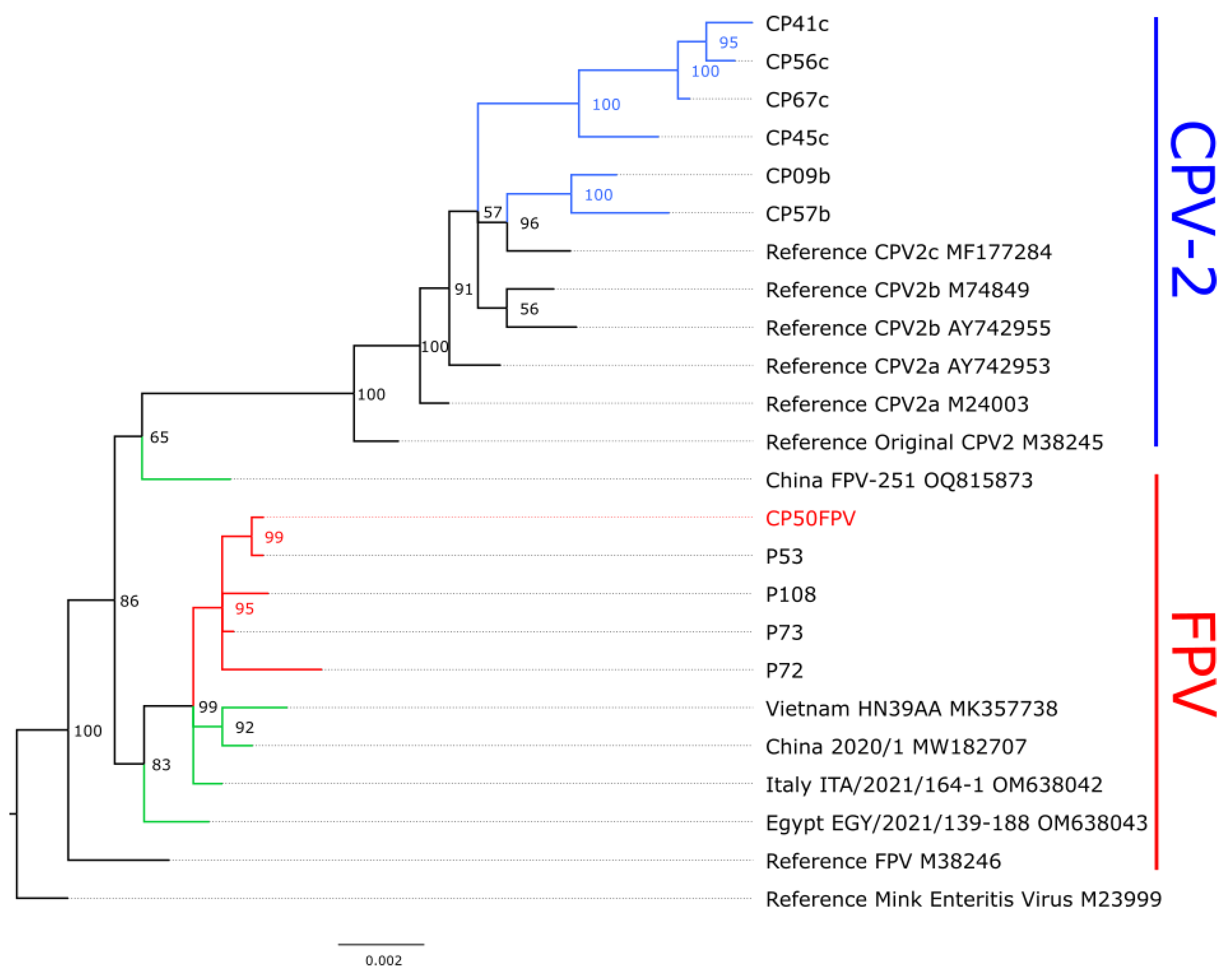

Table 1.
VP2 amino acid substitutions at relevant positions in the FPV strain obtained in this study regarding references sequences and other FPV strains detected in dogs elsewhere.
Table 1.
VP2 amino acid substitutions at relevant positions in the FPV strain obtained in this study regarding references sequences and other FPV strains detected in dogs elsewhere.
| Sequence name | Amino acid position | Country |
Accession number |
Reference | |||||||||||
| 80 | 93 | 101 | 103 | 232 | 300 | 323 | 375 | 411 | 562 | 564 | 568 | ||||
| Reference FPV | K | K | I | V | V | A | D | D | E | V | N | G | M38246 | [31] | |
| CP50FPV | - | - | T | - | - | - | - | - | - | - | - | - | Spain | PZ279925 | This study |
| HN39AA | - | - | T | - | - | - | - | - | - | - | - | A | Vietnam | MK357738 | [18] |
| FPV-251 | - | - | T | - | I | P | - | - | A | L | - | A | China | OQ815873 | [22] |
| ITA/2021/164-1 | - | - | T | - | - | - | - | - | - | - | - | A | Italy | OM638042 | [21] |
| EGY/2021/139-188 | - | - | T | - | - | - | - | - | - | - | - | A | Egypt | OM638043 | [21] |
| FPV-VT2020 | - | N | T | - | - | - | - | - | - | - | - | A | Thailand | MN270937 | [19] |
| 2020/1 | - | - | T | - | - | - | - | - | - | - | - | A | China | MW182707 | [20] |
| Reference CPV-2 | R | N | I | A | I | - | N | N | - | - | S | - | M38245 | [31] | |
Disclaimer/Publisher’s Note: The statements, opinions and data contained in all publications are solely those of the individual author(s) and contributor(s) and not of MDPI and/or the editor(s). MDPI and/or the editor(s) disclaim responsibility for any injury to people or property resulting from any ideas, methods, instructions or products referred to in the content. |
© 2026 by the authors. Licensee MDPI, Basel, Switzerland. This article is an open access article distributed under the terms and conditions of the Creative Commons Attribution (CC BY) license (http://creativecommons.org/licenses/by/4.0/).
Copyright: This open access article is published under a Creative Commons CC BY 4.0 license, which permit the free download, distribution, and reuse, provided that the author and preprint are cited in any reuse.