
Submitted:
31 July 2026
Posted:
03 August 2026
You are already at the latest version
Abstract
The genus Renanthera is valued for its fiery-red floral coloration and distinctive spider-like floral architecture; however, the molecular mechanisms underlying floral organ specialization and perianth color differentiation remain poorly understood. In this study, using integrated morphological, transcriptomic, and metabolomic analyses of R. coccinea, we found that the cell types of the dorsal and lateral perianth organs were broadly similar but clearly distinct from those of the lip and the column. Developmentally, the organ-specific expression patterns of AGAMOUS-LIKE6-1/2 (AGL6-1/2), APETALA3-1/2 (AP3-1/2), and AGAMOUS (AG) were consistent with the conserved orchid floral organ identity model. Furthermore, lip-enriched expression of brassinosteroid-responsive RING H2 (BRH1) and flavin-containing monooxygenase YUCCA10 (YUC10) suggests that brassinosteroid- and auxin-associated pathways may contribute to lip specialization, especially spur development. Regarding pigmentation, high expression of flavonoid 3'-hydroxylase (F3'H) was associated with a cyanidin-dominated anthocyanin profile. The color divergence among the dorsal sepal, petals, and lateral sepals was associated with differential expression of UDP-glucose: flavonoid 3-O-glucosyltransferase (UFGT), which was co-expressed with plant U-box protein 9 (PUB9) and carbon catabolite repressor 4 (CCR4), resulting in distinct patterns of pigment modifications. These findings provide a molecular framework for understanding floral organ development and color patterning in R. coccinea and candidate genes for future validation and molecular breeding in Renanthera.
Keywords:
Renanthera
; floral organ identity
; lip specialization
; flower color patterning
1. Introduction
Renanthera Lour. (Flame Orchid), a genus within the subtribe Aeridinae (Vandeae, Orchidaceae), comprises approximately 20 species of perennial epiphytic or lithophytic herbs distributed mainly from the eastern Himalayas to Southeast Asia [1]. In China, three species have been recorded: R. citrina, R. coccinea, and R. imschootiana, the latter two of which are listed as Endangered (EN) and Critically Endangered (CR), respectively, in the Red List of China’s Higher Plants [2,3,4]. The zygomorphic flowers of Renanthera consist of one dorsal sepal, two lateral sepals, two petals, a spurred lip (labellum), and a column (gynostemium) [1,5]. Known for their spectacular branched inflorescences and long-lasting blooms, these orchids are characterized by a signature vermilion red color, although genetic studies suggest that yellow is the genuine base hue [1]. As a core genetic source for red hybrids in the Southeast Asian cut-flower industry, Renanthera holds an irreplaceable position in landscape horticulture and molecular breeding [1,6]. Therefore, deciphering the molecular mechanisms underlying floral development and pigment biosynthesis will deepen our understanding of floral organ identity and morphological diversification from an Evolutionary Developmental Biology (Evo-Devo) perspective and provide a theoretical basis for the precision breeding of floral shape and color in Renanthera.
The ABCE and floral symmetry models, derived largely from mutant studies in model species such as Arabidopsis thaliana and snapdragon (Antirrhinum majus), provide a fundamental framework for understanding the developmental basis of floral diversity [7,8,9]. According to these models, sepal identity is specified by the combination of A- and E-function genes, while petals, stamens, and carpels are determined by A+B+E, B+C+E, and C+E gene complexes, respectively [10]. Notably, except for AP2, an A-function gene belonging to the AP2/ERF family [11], nearly all well-characterized floral organ identity genes encode MADS-box transcription factors [12,13]. The determination of bilateral symmetry (zygomorphy) is primarily orchestrated by the CYCLOIDEA (CYC) and DICHOTOMA (DICH) genes within the TCP family, alongside the MYB family genes RADIALIS (RAD) and DIVARICATA (DIV). Specifically, CYC/DICH and RAD direct dorsal identity, whereas DIV governs lateral differentiation [14,15,16,17]. Building on these models, comparative studies across angiosperm lineages have shown that changes in the spatiotemporal expression of floral identity and symmetry genes contribute substantially to the diversification of floral architecture [18,19,20,21,22]. In Orchidaceae, the diversification of floral structures is specifically interpreted through the “Orchid Code”, “Homeotic Transformation (HOT)”, and “Perianth code (P-code)” models, which are rooted in the duplication and functional divergence of the AP3 and AGL6 lineages [23,24,25]. Recent advances have further identified specific regulators of lip specialization: Phalaenopsis aphrodite WUSCHEL-related homeobox 3 (PaWOX3) and AGL6-2 function redundantly in orchestrating specialized lip structures [26], whereas the P. equestris Auxin-Associated F-box protein (PeAAF) gene facilitates labellum expansion by amplifying auxin responsiveness [27]. Additionally, P. equestris NAC domain-containing protein 67 (PeNAC67) and P. equestris KANADI 2 (PeKAN2) have been implicated in promoting labellum traits [28], further highlighting the complex and potentially lineage-specific mechanisms governing lip differentiation. Moreover, the regulatory programs dictating bilateral symmetry appear to be evolutionarily divergent among orchid genera. While the dorsal expression of three CYC genes mediates zygomorphy in Phalaenopsis [29], symmetry in Cymbidium sinense is predominantly governed by Class B and Class G genes [30]. Such discrepancies suggest that the developmental pathways for bilateral symmetry have undergone lineage-specific evolution within the Orchidaceae family.
The mechanisms of floral pigmentation are primarily centered on four major classes of pigments: chlorophylls, carotenoids, anthocyanins, and betalains [31,32]. Chlorophylls confer green color to cells; carotenoids produce yellow, orange, or red hues; while anthocyanins and betalains provide a broad spectrum ranging from purple to blue and orange to red, respectively [32,33,34]. Except for betalains, which are largely restricted to Caryophyllales and are mutually exclusive with anthocyanins, these pigment classes are widely distributed across higher plants. The key structural genes regulating the biosynthesis of these pigments are highly conserved throughout evolution [31,32,35]. Despite this genetic conservation, the mechanisms responsible for diverse color patterns are complex and varied. For instance, in the blue/purple Ipomoea purpurea, pink and red flowers have emerged; the former results from a mutation in the coding region of the structural gene flavonoid 3’-hydroxylase (F3’H) [36,37], while the latter is caused by the downregulation of F3’H expression [36,38,39]. Notably, to date, the formation of irregular patches on floral organs has been attributed to the insertion of transposons into structural genes. For example, variegated phenotypes in I. purpurea and Torenia fournieri are driven by transposon insertions within structural genes and subsequent partial reversion mutations [40,41]. The mechanisms underlying other specific types of irregular patterning remain largely unknown.
In the present study, we used R. coccinea as a representative species and integrated morphological, anatomical, transcriptomic, and metabolomic approaches to investigate floral organ development and pigmentation. Our objectives were to: (1) characterize the micromorphological variations of epidermal cells among different floral organs; (2) elucidate the developmental programs of the flower, specifically the mechanisms governing lip specialization; (3) identify the key pigment constituents responsible for the irregular spotting on dorsal sepal and petals versus the monochromatic coloration of lateral sepals; and (4) explore the regulatory networks underlying the divergent pigmentation patterns observed between dorsal and lateral floral organs.
2. Results
2.1. Morphological, Anatomical, and Micromorphological Floral Characteristics of R. coccinea
To gain insights into the floral morphology and structural organization of the Flame Orchid, we investigated R. coccinea, a representative wild species distributed on Hainan Island. The flower is zygomorphic, with a single oblong-narrowly elliptic dorsal sepal that is red with red-blotched yellow. The lateral sepals are ovate, widening from a linear base; their margins are undulate, bright red, spreading vertically downwards, and slightly divergent or overlapping. The petals are linear and similar to the dorsal sepal. The lip is small and shallowly three-lobed. The side lobes feature red stripes, while the mid-lobe is yellowish-white with red stripes and a red surface, ending in an acute apex. Both the column and anther cap are red, with red bands at the column’s base and a distinct yellow band across the middle of the anther cap (Figure 1a). Clearly, sepal, petal, and lip differ markedly in morphology, structure, and color.
To clarify the differences in epidermal cell morphology among these floral organs, we examined their epidermal cells using scanning electron microscopy (SEM). We found that the adaxial epidermal cells of the sepals and petals were uniformly conical, whereas their abaxial epidermal cells were consistently pavement-like, with stomata occasionally scattered across both structures (Figure 1b, a-e). The lip exhibited conical epidermal cells with surface modifications on the adaxial side and striated epidermal cells with surface modifications on the abaxial side (Figure 1B, f-f’). By contrast, the spur of the lip possessed sub-rectangular interlocked epidermal cells on its adaxial surface and flattened pavement cells on its abaxial surface (Figure 1B, g-g’). Furthermore, the column displayed elongated rod-shaped cells at the apex and elongated interlocked epidermal cells at the base (Figure 1b, h-i). These observations suggest that the identity specification programs of distinct floral organs, including the dorsal sepal, petals, lateral sepals, the lip, and the gynostemium, may differ substantially.
2.2. Evolution and Expression of Floral Organ Identity Genes in R. coccinea
To gain more insights into the molecular mechanisms underlying the development and evolution of Flame Orchids, we first retrieved putative orthologs of floral organ identity genes from the de novo assembled reference transcriptome of R. coccinea, generated from pooled RNAs of floral buds, leaves, stems, and roots at different developmental stages. This reference transcriptome contained 39,390 predicted protein-coding genes. Phylogenetic analyses of floral organ identity genes and floral symmetry genes revealed that, while AGL6-1, AGL6-2, AP3-1, AP3-2, and PI lineage genes are almost always single-copy genes, AG, SEP, DIV, and RAD lineage genes may have experienced recent duplication events. For the AG and SEP lineages, the transcriptome-based phylogenetic results suggested possible lineage-specific duplication, giving rise to the AG1, AG2, and AG3 clades and the SEP1, SEP2, and SEP3 clades (Supplemental Figure S1; Supplemental Figure S2; Supplemental Data Set S1). Among the floral symmetry genes, the two previously identified Orchis CYC lineages underwent duplication, resulting in the CYC1 and CYC2 clades. In some genera, a subsequent duplication of CYC2 gave rise to CYC3. This evolutionary trend is mirrored in Renanthera, which features three copies, namely CYC1, CYC2, and CYC3. Similar to the evolutionary history of CYC genes, the DIV gene also produced three copies: DIV1, DIV2, and DIV3 in Renanthera (Supplemental Figure S3; Supplemental Data Set S1).
To investigate the expression patterns of the aforementioned genes, transcriptome profiling was performed among dorsal sepal/petals, lateral sepals, the lip, and the column from R. coccinea at both the small (marking the completion of organ differentiation and the onset of coloration)and middle (representing the full maturation of floral pigmentation)stages (Figure 2a). Principal Component Analysis (PCA) revealed significant stage-specific clustering, with PC1 and PC2 accounting for 23% and 16.56% of the total variance, respectively (Figure 2b; Supplemental Data Set S2). These results indicate that the developmental stage is the primary driver of transcriptomic variation, despite the inclusion of various organ types, while also indicating clear biological structure and overall reproducibility among the RNA-seq samples. Digital gene expression profiles revealed clear expression divergence of AP3 and AGL6 paralogs across floral organs (Figure 2c; Supplemental Data Set S3). AP3-1 was predominantly expressed in the dorsal sepal/petals and lateral sepals, whereas AP3-2 showed a preference for the petals and lip. Similarly, AGL6-1 exhibited high expression levels in both dorsal and lateral perianth organs, displaying a progressively increasing trend along the floral axis of symmetry. In contrast, AGL6-2 was primarily restricted to the lip, with its expression intensity showing a decreasing gradient along the same axis. These results are consistent with findings in Oncidium species [25], at least at the small and middle floral developmental stages. AG-1, AG-2, and AG-3 were specifically expressed in the column, with AG-1 and AG-2 showing significantly higher expression levels in this structure. While SEP-1 and SEP-3 exhibited higher expression levels among the three SEP genes, these genes were found to be expressed across all types of floral organs. Interestingly, although DIV and RAD were also widely expressed across various floral organs, their transcript levels were markedly lower than those of floral organ identity genes (Figure 2c; Supplemental Data Set S3). Furthermore, no detectable expression of CYC genes was observed at these two developmental stages (Supplemental Data Set S3). Taken together, these findings suggest that duplicated and diversified AP3- and AGL6-lineage genes represent strong candidates underlying the origin of the complex floral morphology in Flame Orchids, further supporting that the P-code hypothesis is applicable to perianth formation in this group (Figure 2d).
2.3. Genes Specifically and Preferentially Expressed in the Lip of R. coccinea
To understand the uniqueness of the lip, we comprehensively compared the gene expression profiles across five distinct floral organs (column, petal, dorsal sepal, lateral sepal, and lip) at both the small (Figure 3a; Supplemental Data Set S4) and middle (Figure 3b; Supplemental Data Set S4) floral bud stages. Of all the genes expressed, 1,264 and 2,168 were identified as lip-specific (defined as FPKM ≥ 1.0 in the lip and < 1.0 in all other floral organs) at the small and middle stages, respectively. For comparison, the numbers of organ-specific genes at the small and middle stages were 848 and 1,037 in the petal, 1,221 and 2,107 in the dorsal sepal, 884 and 2087 in the lateral sepal and 3,091 and 253 in the column, respectively. Given that the orchid lip is typically a highly specialized organ, our study further focused on the lip tissue to identify genes related to its development and morphological elaboration. By screening for genes that maintained lip-specific expression across both developmental stages (FPKM ≥ 1.0), we identified a core set of 37 genes (Supplemental Data Set S5). Among these, two genes brassinosteroid-responsive RING H2 (BRH1) and flavin-containing monooxygenase YUCCA10 (YUC10) are closely associated with cell elongation and hormone responses (Figure 3c).
Quantitative gene expression analysis (FPKM) supported the lip-enriched expression patterns of these two candidate genes (Figure 3d, e). The results showed that BRH1 and YUC10 were transcribed at high abundance almost exclusively in the lip, remaining virtually silent in the other floral organs. Notably, BRH1 exhibited a highly significant stage-dependent expression pattern (Figure 3d). Its expression was dramatically upregulated in the middle lip, far exceeding the levels observed at the small stage. This pattern suggests that BRH1 may participate in the later stages of labellum specialization, particularly in spur elongation and morphogenesis. Concurrently, YUC10, which encodes a key enzyme involved in local auxin biosynthesis, maintained high and specific expression in the lip at both developmental stages (Figure 3e). This expression pattern suggests that localized auxin biosynthesis may contribute to lip formation and development in R. coccinea, although functional validation is required to confirm this role.
2.4. Spatio-Temporal Accumulation of Cyanidin Derivatives Drives Floral Color Divergence Between Dorsal and Lateral Sepals in R. coccinea
To elucidate the differences in the main chromogenic substances between the red-blotched yellow dorsal sepals and petals and the pure red lateral sepals of R. coccinea (Figure 4a), we performed widely targeted metabolomic profiling on both tissue types at the small and middle developmental stages (Supplemental Data Set S6). Principal component analysis (PCA) revealed a distinct and significant separation between the two stages along the PC1 axis (33.06%; Figure 4b), indicating a profound spatiotemporal evolution of metabolic profiles during floral maturation. Furthermore, samples from the two tissue types (dorsal sepal vs. lateral sepals) formed distinct clusters along the PC2 axis (16.33%; Figure 4b), suggesting that tissue-specific metabolic differentiation also contributes substantially to the overall variance. Together, these results demonstrate that developmental stage and tissue type are two major determinants of metabolic variation, providing a solid basis for further identifying the specific metabolites responsible for the distinct color phenotypes.
A total of 207 compounds were identified in the dorsal sepal across the two developmental stages. Analysis of differentially accumulated metabolites (DAMs) revealed 26 up-regulated and 181 down-regulated compounds (Supplemental Data Set S7). Among the 26 up-regulated metabolites, three exhibited significant increases, notably cyanidin chloride, a key pigment most closely associated with floral coloration (Figure 4c; Supplemental Figure S4; Supplemental Figure S5). Research indicates that cyanidin chloride is inherently unstable under physiological conditions, typically requiring glycosylation for structural stability [42]. In R. coccinea, this inherent instability likely drives localized pigment degradation, explaining the specific pigmentation patterns observed. Since yellow serves as the ground coloration, the breakdown of red cyanidin pigments unmasks the underlying yellow background. Consequently, the red-blotched phenotype emerges from this spatially uneven loss of red pigmentation, where areas of cyanidin depletion reveal the yellow base while the persisting pigments form the characteristic red patches. In contrast, we identified a total of 146 compounds in the lateral sepals across the two developmental stages and found that the lateral sepals showed marked accumulation of cyanidin 3-O-glucoside during development, with its fold-change significantly surpassing other metabolites (Figure 4d; Supplemental Figure S4; Supplemental Figure S5). By integrating our findings within the framework of the known anthocyanin biosynthetic pathway (Figure 4e), these results suggest that quantitative and tissue-specific accumulation of cyanidin and its derivatives contributes to the distinct coloration patterns observed between the dorsal and lateral sepals of R. coccinea.
2.5. Characterization of the Anthocyanin Biosynthetic Pathway and Its Transcriptional Regulatory Network in R. coccinea
To elucidate the molecular mechanisms underlying red pigmentation (cyanidin), we identified candidate genes involved in anthocyanin biosynthesis and examined their expression patterns. A total of 26 candidate structural genes were identified in the anthocyanin biosynthetic network (Figure 5a, b; Supplemental Data Set S8). Except for chalcone synthase (CHS-2) and flavonoid 3’,5’-hydroxylase (F3’5’H), which showed no detectable or low expression, all other genes exhibited high expression signals at two stages. As expected, consistent with the metabolomic data, the high expression of F3’H directs the anthocyanin biosynthetic flux primarily toward the cyanidin pathway (Figure 5a, b). In contrast, the extremely low expression of F3’5’H hinders delphinidin synthesis, while the preferential involvement of dihydroflavonol 4-reductase (DFR) in the cyanidin branch may also restrict pelargonidin production. Collectively, these molecular mechanisms account for why cyanidin is the predominant pigment responsible for the coloration of Flame Orchid. Additionally, a significant down-regulation of F3’H, DFR, and anthocyanidin synthase (ANS) was observed in the lip and column as development progressed toward the middle stage. This reduction likely limits the intensity of red pigmentation, providing a molecular basis for why these tissues do not achieve a completely deep red hue.
To elucidate the molecular basis of the striking dorsal/lateral floral color divergence observed in the Flame Orchid, a pattern uniquely characterized by the accumulation of cyanidin chloride in dorsal tissues versus cyanidin 3-O-glucoside in lateral sepals, we performed comprehensive transcriptomic and DGE profiling. An initial analysis of the anthocyanin biosynthetic pathway highlighted UDP-glucose: flavonoid 3-O-glucosyltransferase (UFGT) as the primary enzyme mediating the crucial biochemical conversion of cyanidin into its stable glucoside form. Given that the expression of the vast majority of anthocyanin-related structural genes peaks during the ripening stage to facilitate rapid pigment deposition, we first conducted a temporal comparative analysis of lateral sepals across the two stages (Supplemental Data Set S9). This investigation revealed that UFGT was significantly upregulated as the flowers transitioned from the small to the middle stage (Figure 5c). To ascertain whether UFGT is also the specific factor driving spatial color patterning, we compared transcript levels between dorsal and lateral sepals during the middle stage (Supplemental Data Set S10). The results showed that UFGT was expressed at significantly higher levels in lateral sepals than in dorsal sepals, perfectly mirroring the localized metabolic profiles of the pigments (Figure 5d). Collectively, these findings identify UFGT as a strong candidate associated with dorsal/lateral pigmentation divergence, providing new insights into the regulatory complexity of floral patterning in orchids.
2.6. Identification of Candidate Transcription Factors Underlying Dorsal/Lateral Color Divergence via Co-Expression Network Analysis
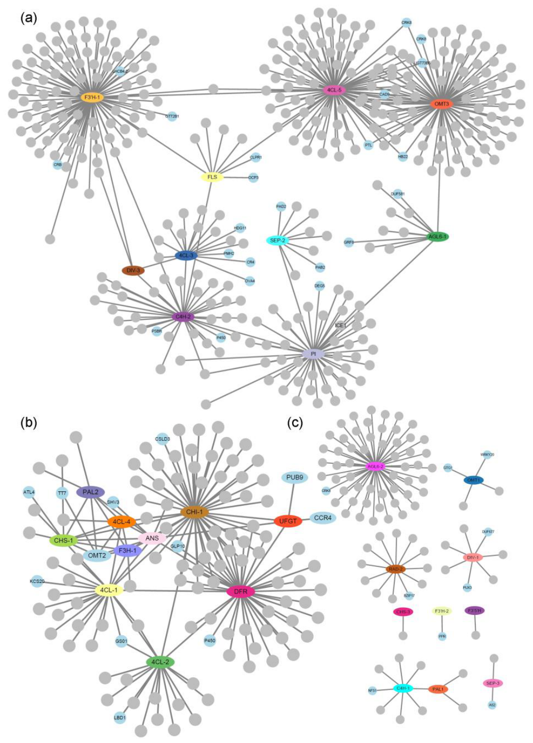
Building on the identification of AGL6-2 and UFGT as key differentially expressed genes (DEGs) between lateral and dorsal sepal, we sought to further identify co-expression modules involving core anthocyanin structural and organ identity genes. Using these genes as anchors, we identified tightly co-expressed candidate genes within the resulting co-expression modules. Using stringent criteria, including PCC > 0.85 among anchor genes and PCC > 0.97 between each anchor gene and its associated co-expression genes, we identified co-expression partners for 33 genes (Supplemental Data Set S11). These genes were partitioned into distinct co-expression groups. One network connected anthocyanin structural genes flavonol synthase (FLS), flavonoid 3’-hydroxylase (F3’H-1), cinnamate-4-hydroxylase (C4H-2), 4-coumarate:CoA ligase (4CL-3/5) and o-methyltransferase (OMT3) with floral developmental genes (AGL6-1, SEP4, PI, and DIV-3), suggesting that these organ identity genes not only specify organ identity but may also coordinate floral pigmentation through interactions with anthocyanin biosynthetic genes (Figure 6a). A second network contained several anthocyanin structural genes, including phenylalanine ammonia-lyase (PAL-2), 4CL-1/2/4, CHS-1,chalcone isomerase (CHI-1), flavanone 3-hydroxylase (F3H-1), DFR, ANS, UFGT, and OMT2, underscoring that UFGT, which was identified in our differential expression analysis of dorsal and lateral sepals, is indeed a core component of the anthocyanin biosynthetic regulatory network. Co-expression analysis revealed that transparent testa 7 (TT7), F3’H and one P450 family gene exhibited strong co-expression patterns with CHS-1 and DFR, respectively. These enzymatic components may contribute to anthocyanin biosynthetic flux in the perianth of the Flame Orchid (Figure 6b). Additionally, pairwise co-expression was observed for PAL-1/C4H-1 and SEP2/3 (Figure 6c), whereas the remaining genes failed to incorporate into any co-expression modules; this suggests that these organ identity genes may not play a primary role in the regulation of floral pigmentation in this context. These results further demonstrate that UFGT is indeed the key gene driving the differentiation of coloration patterns between the dorsal and lateral perianths in R. coccinea. Intriguingly, while traditional transcription factors (TFs) were absent from the UFGT-containing module, two non-canonical regulatory genes, plant U-box protein 9 (PUB9, an E3 ubiquitin ligase) and carbon catabolite repressor 4 (CCR4, a key component of the deadenylase complex), were found to be tightly co-expressed with UFGT. This suggests that the distinct coloration of lateral sepals may be governed by post-translational or post-transcriptional mechanisms rather than direct transcriptional activation alone.
3. Discussion
3.1. Conservation of Floral Organ Identity Mechanisms and Regulatory Model Construction in Flame Orchid
In this study, we conducted the first systematic identification of floral organ identity and zygomorphy-related genes in the representative species of R. coccinea, and characterized their expression profiles during key developmental stages (small and middle buds). Among the 11 identified organ identity genes, all except AG-3 exhibited high expression abundance, underscoring their pivotal roles in the floral ontogeny of Flame Orchid. Our results revealed that AGL6-1, AP3-1, and AP3-2 were predominantly expressed in the dorsal floral organs, whereas AGL6-2 and AP3-2 were specifically and significantly upregulated in the lip. Class C AG genes were strictly confined to the column. This spatial expression pattern is highly congruent with those observed in typical orchids such as Oncidium and Phalaenopsis [25,43]. Based on the conservation of these expression patterns and functional insights from closely related species, such as the non-functional expression of AP3-2 in the petals of Oncidium [44] we propose that Flame Orchid follows an Orchid Code-like regulatory pattern. Specifically, dorsal organ identity is primarily defined by AGL6-1 and AP3-1, while lip identity is co-determined by AGL6-2 and AP3-2, and column identity is maintained by AG genes. Accordingly, we have constructed a genetic regulatory model for floral development in Flame Orchid. These findings not only refine the molecular framework of the genus Renanthera but also further substantiate the high degree of conservation in floral organ identity mechanisms across the Orchidaceae at the family level.
3.2. AP3-like and AGL6-like Genes May Contribute to Floral Zygomorphy in Flame Orchid
In this study, eight genes associated with floral zygomorphy were identified. Notably, the CYC-like genes, which serve as the traditional core regulators of symmetry, were nearly undetectable across the two sampled developmental stages. This absence may be attributed to highly restricted spatio-temporal expression, potentially initiating at an extremely early stage, or it may suggest that CYC genes are not the primary factors maintaining zygomorphy in Flame Orchid. Furthermore, other classes of symmetry-related genes (i.e., DIV and RAD) generally exhibited lower expression levels compared to MADS-box genes and showed broad, non-tissue-specific expression patterns. These observations are congruent with findings in other orchid species, suggesting that the unique zygomorphic structures of Orchidaceae, particularly lip specialization, may not be predominantly governed by the traditional CYC pathway. Instead, this process appears to be driven by the ectopic expression or functional divergence of organ identity genes (MADS-box). Evolutionary evidence further supports this hypothesis: in the basal orchid genus Apostasia, the absence of specific AP3 subfamilies fails lip specialization, leading to actinomorphic (radially symmetrical) features similar to those of the petals [45]. Moreover, in Cymbidium sinense, the development of floral symmetry has been linked to the expression patterns of Cymbidium sinense APETALA3-2 (CsAP3-2) and Cymbidium sinense AGAMOUS-LIKE 6-2 (CsAGL6-2) [30]. Integrating our data, we propose that the pivotal role of AP3-class and AGL6-class genes in lip specialization not only defines organ identity but also serves as the core genetic driver for the evolution of floral zygomorphy in Flame Orchid.
3.3. Molecular Mechanisms Underlying Lip Development in Flame Orchid
Beyond the core floral organ identity genes, tissue-specific expression analysis identified two hormone-associated, lip-enriched genes, YUC10, encoding an auxin biosynthesis-related flavin-containing monooxygenase, and BRH1, encoding a brassinosteroid-responsive RING-H2 protein. Both genes showed strong lip-enriched expression in Flame Orchid. Given that AP3-class genes have been shown to contribute to lip identity in orchids [45]. We propose that these genes may act as downstream components of hormone-associated pathways involved in lip morphogenesis. The spur represents the most conspicuous specialized feature of the Flame Orchid lip.
The spur is a critical morphological trait for pollinator adaptation in Orchidaceae, and its development is governed by a complex hormonal landscape. In the model plant Aquilegia, studies have shown that the auxin response factors auxin response factors (ARF6/8) maintain high expression levels during spur development, determining spur length by regulating longitudinal cell elongation rather than cell division; conversely, inhibition of their function results in significantly shortened spurs due to restricted cell elongation [46]. In addition to auxin, the brassinosteroid (BR) signaling pathway is equally pivotal. Exogenous BR application significantly increases spur length in Aquilegia, while the suppression of BR signaling components BES1/BZR1-Homologs (BEH1/BEH3) reduces cell-growth anisotropy, leading to spur stunting [47]. Furthermore, in A. brevistyla, the bHLH transcription factor increased Leaf Inclination Binding bHLH1-Like (IBL1) has been shown to control the curved growth of the spur by fine-tuning hormonal balance [48].
This hormone-driven mechanism of spur development exhibits functional convergence across diverse species. For instance, in Impatiens, genes encoding extensin proteins involved in cell-wall synthesis are specifically expressed in the spur, where they determine morphology by modulating anisotropic cell growth. Similarly, the auxin-binding protein ABP precisely regulates spur curvature by influencing differential cell division rates between the proximal and distal regions [49]. Integrating these findings with our results, the lip-specific high expression of the auxin biosynthesis gene YUC10 and the BR-responsive gene BRH1 suggests a similar regulatory logic: following the establishment of lip identity by MADS-box genes, the activation of hormonal signaling pathways is likely mediated by factors such as YUC10 and BRH1, thereby inducing localized polar elongation or differential cell division. Such coordinated hormonal and cellular processes may contribute to the morphogenesis of complex lip structures, including the spur, in Flame Orchid.
3.4. Differential Expression of Structural Genes Is Associated with Dorsal/Lateral Floral Color Variation in Flame Orchid
Cyanidin has been reported as a major contributor to flower coloration in orchid plants [50,51]. In this study, we identified cyanidin aglycones and cyanidin-3-O-glucoside as the major pigments in the dorsal sepal and lateral sepals of Flame Orchid, respectively. Through developmental stage-specific analysis, we found that UFGT was differentially expressed between these organs (highly upregulated in the lateral sepals). This indicates that the two pigmentation patterns are associated with differential modification of anthocyanins, particularly through variation in UFGT expression. Such transcriptional divergence serves as the molecular foundation for the color variation between the dorsal and lateral floral organs of Flame Orchid. Although anthocyanin biosynthesis is regulated by the MYB-bHLH-WDR (MBW) trimeric complex, recent studies have highlighted the critical roles of epigenetic and post-translational modifications (PTMs) in fine-tuning this pathway during plant development and environmental adaptation. Intriguingly, our analysis identified PUB9 and CCR4 as candidate genes strongly associated with UFGT. PUB9 is a member of the E3 ubiquitin ligase family, and CCR4 serves as the core catalytic component of the CCR4-NOT complex involved in mRNA degradation in eukaryotes. Research has demonstrated that both the E3-mediated degradation of transcription factors and the inhibition of MBW complex formation by R3-MYB factors are essential for maintaining anthocyanin homeostasis in plants [52,53]. This suggests that the spatial regulation of UFGT in Flame Orchid may be primarily governed by post-translational or post-transcriptional mechanisms. Specifically, the positive co-expression of PUB9 with UFGT raises the possibility that ubiquitin-mediated protein turnover may influence anthocyanin modification, the patterns observed in Flame Orchid point toward a mode of action involving the targeted degradation of transcriptional repressors by PUB9. Furthermore, the co-expression of CCR4 with UFGT highlights the significance of mRNA stability in floral pigmentation. The CCR4-NOT complex may also influence transcript stability of UFGT or its upstream regulators in the lateral sepals, thereby facilitating rapid and robust pigment accumulation.
4. Materials and Methods
4.1. Plant Materials and Growth Conditions
R. coccinea specimens are preserved at the Orchid Germplasm Resource Nursery of the Hainan Academy of Agricultural Sciences in Haikou, China. Cultivation was conducted under a typical tropical monsoon climate (25~32℃; ~80% humidity) and natural light. The plants were grown either in a substrate mixture of crushed stone, charcoal, and pine bark or epiphytically on tree trunks and rocks. Typically, this species flowers from March to May, with the blooming period lasting approximately three months.
4.2. Morphological and Micromorphological Observations
For stereomicroscopic photography, various floral organs of R. coccinea were dissected and photographed using a Nikon Model CDSS230 stereomicroscope equipped with a Nikon DXM1200F digital camera (Nikon Instech Co. Ltd., Kawasaki, Japan). For scanning electron microscopy (SEM), floral organs from mature flowers were fixed in fresh FAA (3.7% formaldehyde, 5% acetic acid, and 50% ethanol), followed by dehydration in a graded ethanol series and drying with a CO2 critical-point dryer. After being sputter-coated with gold, the samples were examined using a Hitachi S-4800 scanning electron microscope.
4.3. Reference Transcriptome Assembly and DGE Profiling
To generate a comprehensive reference transcriptome for R. coccinea, total RNA was extracted from various developmental stage tissues (buds, leaves, stems, and roots) and pooled for hybrid sequencing using both PacBio Sequel II and Illumina HiSeq 2000 platforms. Illumina-derived transcripts, assembled via Trinity [54], were integrated with PacBio full-length transcripts, followed by redundancy removal using CD-HIT [55]. Transcripts representing different isoforms were clustered into unigenes through all-by-all BLASTNs [56]. ANGEL was employed for CDS prediction, filtering for protein sequences >100 amino acids. The resulting unigenes were functionally annotated via BLASTX (E-value < 1e-5) against the TAIR Arabidopsis thaliana CDS database.
For transcriptomic profiling, floral tissues were collected at both the small (S, marking the completion of organ differentiation and the onset of coloration) and middle (M, representing the full maturation of floral pigmentation) stages. Five distinct floral organs were sampled at each stage: the petal (pe), dorsal sepal (ds), lateral sepal (ls), lip (lp), and column (co), resulting in ten sample groups. Total RNA was extracted using the SV Total RNA Isolation System (Promega) following the manufacturer’s protocol. In total, 30 independent libraries were constructed for 150-bp paired-end sequencing on the Illumina HiSeq platform (Novogene). The clean reads were mapped to the aforementioned reference transcriptome using Bowtie2 [57]. Gene expression levels for each floral organ were quantified as Fragments Per Kilobase of transcript per Million mapped reads (FPKM) and raw counts using the RSEM software package [58] with the “rsem-calculate-expression --bowtie2” parameters. To evaluate the overall similarities between expression profiles, Principal Component Analysis (PCA) was performed using the prcomp function in R (version 4.3.1), with FPKM values used as input data.
4.4. Generation of the Metabolomic Datasets and DAM Profiles
Metabolomic profiling was performed on dorsal sepals (ds) and lateral sepals (ls) collected at the same developmental stages (S and M) as the transcriptomic samples, with three biological replicates per group. Powdered samples were extracted as described by Zhang et al. (2020) and subsequently analyzed using an ultra-performance liquid chromatography-electrospray ionization-tandem mass spectrometry (UHPLC-ESI-MS/MS) system. Target detection was conducted on a QTrap system (QTRAP 6500+; AB Sciex). A widely targeted compound database was constructed using ion information extracted from raw QTOF data via AB Sciex MS Converter software. Raw mass spectrometry data were processed using MultiQuant 3.0.3 software, where peak areas were integrated with the MQ4 algorithm to obtain quantitative data. Principal Component Analysis (PCA) and screening of differentially accumulated metabolites (DAMs) were performed in R. The criteria for DAMs were set as VIP ≥ 1.0, |log₂FC| ≥ 1, and Welch’s t-test P-value < 0.05. Volcano plots were generated using the ggplot2 package to visualize metabolite distribution [59].
4.5. Gene Co-Expression Network Analysis
Gene co-expression networks were constructed based on transcript abundance data log2(FPKM + 1) derived from RNA-seq analysis. Pairwise Pearson correlation coefficients (PCCs) were calculated across all expression profiles using the cor function in R. Genes exhibiting strong co-expression relationships (PCC > 0.85) with at least one anthocyanin structural or regulatory gene were defined as core co-expressed genes, while those with extremely high correlations (PCC > 0.97) with key structural genes were classified as highly associated genes. Only gene pairs with PCC > 0.97 were retained for final network construction. Network visualization was performed in Cytoscape v3.10.0 [60] using an edge-weighted force-directed layout. In this representation, edge lengths reflect correlation strength, with shorter edges denoting more robust co-expression relationships.
Supplementary Materials
The following supporting information can be downloaded at the website of this paper posted on Preprints.org, Supplemental Figure S1. Phylogenetic trees of other floral organ identity genes. Supplemental Figure S2. Phylogenetic trees of SEP-like genes. Supplemental Figure S3. Phylogenetic trees of floral symmetry genes. Supplemental Figure S4. Heatmap of 17 anthocyanin compounds in R. coccinea. Supplemental Figure S5. Bar chart of 17 anthocyanin compounds in R. coccinea. Supplemental Data Set S1. Gene information used for phylogenetic analyses. Supplemental Data Set S2. Mean FPKM values of the 39,390 Genes across three replicates for each organ in R. coccinea. Supplemental Data Set S3. Mean FPKM values of floral organ identity and floral symmetry genes across three replicates for each organ in R. coccinea. Supplemental Data Set S4. Statistical summary of organ-specific and preferential gene expression in R. coccinea. Supplemental Data Set S5. Mean FPKM values of 37 genes specifically or preferentially expressed in the lip across three biological replicates at small and middle stages of R. coccinea. Supplemental Data Set S6. Mean metabolite abundance of 1158 compounds across three replicates for two organs in R. coccinea. Supplemental Data Set S7.Identification of differentially accumulated metabolites (DAMs) between small and middle stages in ds and ls samples. Supplemental Data Set S8. Mean FPKM values of anthocyanin biosynthetic pathway genes across three replicates for each organ in R. coccinea. Supplemental Data Set S9. Comparative expression profiling between ls samples between small and middle stages of R. coccinea by DESeq2. Supplemental Data Set S10. Comparative expression profiling between ds and ls samples at the middle stages of R. coccinea by DESeq2. Supplemental Data Set S11. Calculation of Pearson Correlation Coefficients (PCC) between the target gene and 39,390 other genes, emphasizing length classes and layout weights.
Author Contributions
X. Y., X W., X. L., J. R., and H. Z. designed the research. L. Z., S. H., and J. W. were responsible for the cultivation and maintenance of R. coccinea materials. X. Y. and X. L. generated the transcriptomes and analyzed the expression profile data with the help of L. S., M. S., and X. C.; X. Y., X W., and H. Z. performed phylogenetic analyses; H. Z. performed SEM with the help of J. Y., M. S., and C. W.; X. Y. generation of the metabolomic datasets and DAM profiles with the help of D. L., M. S., and H. Z.; X. Y. gene co-expression network analysis with the help of X W., and X. L.; H. Z. wrote the manuscript with the help of X. Y., X W., X. L., J. R.
Funding
This work was supported by grants from the Key R & D Project of Hainan Province (ZDYF2022XDNY254), the National Natural Science Foundation of China (32300209 and 32560057), the Internal Research Project of the Hainan Academy of Agricultural Sciences (HAAS2023RCQD12, HAAS2025KJCX14 and HAAS2025KJCX005) and the Natural Science Foundation of Hainan Province (325QN485).
Data Availability Statement
The RNA-seq datasets generated in this study have been deposited in the NCBI Sequence Read Archive (SRA) under accession number PRJNA1457784 and in the Gene Expression Omnibus (GEO) under accession number GSE330382. All other data supporting the findings of this study are included in this published article and its supplementary information files or are available from the corresponding author upon reasonable request.
Acknowledgments
We thank Yi Yuan for the guidance on co-expression network analysis, comments and helpful discussions. We also thank Fanrui Meng the guidance for reference transcriptome assembly and DGE profiling.
Conflicts of Interest
The authors declare that there are no conflicts of interest regarding the publication of this paper.
References
- Teoh, E.S. Orchid Species from Himalaya and Southeast Asia Vol. 3 (R – Z).pp.1–24, 2021.
- Qin, H.; Yang, Y.; Dong, S.; He, Q.; Jia, Y.; Zhao, L.; Yu, S.; Liu, H.; Liu, B.; Yan, Y.; et al. Threatened Species List of China’s Higher Plants. Biodivers Sci 2017, 25, 696–744. [CrossRef]
- Chen, X.; Liu, Z.; Zhu, G.; Lang, K.; Ji, Z.; Luo, Y.; Jin, X.; Cribb, P.J.; Wood, J.J.; Gale, S.W.; et al. Orchidaceae; Wu, Z.R., Peter H.; Hong, Deyuan, Ed.; pp.8–466, 2009.
- Xiao, T.; He, L.; Yue, L.; Zhang, Y.; Lee, S.Y. Comparative phylogenetic analysis of complete plastid genomes of Renanthera (Orchidaceae). Front Genet 2022, 13, 998575. [CrossRef]
- Ang, W.F.; Lok, A.F.S.L.; Yeo, C.K.; Angkasa, A.; Ng, P.X.; Ng, B.Y.Q.; Tan, H.T.W. Rediscovery of Renanthera elongata (Blume) Lindl. (Orchidaceae) in Singapore. Nat Singap 2011, 4, 297–301.
- Yang, F.; Gao, J.; Li, J.; Wei, Y.; Xie, Q.; Jin, J.; Lu, C.; Zhu, W.; Wong, S.-M.; Zhu, G. The China orchid industry: past and future perspectives. Orna Plant Res 2024, 4, eoo2. [CrossRef]
- Carpenter, R.; Coen, E.S. Floral homeotic mutations produced by transposon-mutagenesis in Antirrhinum majus. Genes Dev 1990, 4, 1483–1493. [CrossRef]
- Coen, E.S.; Meyerowitz, E.M. The war of the whorls: genetic interactions controlling flower development. Nature 1991, 353, 31–37. [CrossRef]
- Theißen, G. Development of floral organ identity: stories from the MADS house. Current Opinion in Plant Biol 2001, 4, 75–85. [CrossRef]
- Pelaz, S.; Ditta, G.S.; Baumann, E.; Wisman, E.; Yanofsky, M.F. B and C floral organ identity functions require SEPALLATA MADS-box genes. Nature 2000,405,200-203.
- Riechmann, J.L.; Meyerowitz, E.M. The AP2/EREBP family of plant transcription factors. Biol Chem 1998, 379, 633–646. [CrossRef]
- Theissen, G.; Melzer, R.; Rumpler, F. MADS-domain transcription factors and the floral quartet model of flower development: linking plant development and evolution. Development 2016, 143, 3259–3271. [CrossRef]
- Shore, P.; Sharrocks, A.D. The MADS-box family of transcription factors. Eur J Biochem 1995, 229, 1–13. [CrossRef]
- Hileman, L.C. Bilateral flower symmetry--how, when and why? Curr Opin Plant Biol 2014, 17, 146–152. [CrossRef]
- Busch, A.; Zachgo, S. Flower symmetry evolution: towards understanding the abominable mystery of angiosperm radiation. Bioessays 2009, 31, 1181–1190. [CrossRef]
- Preston, J.C.; Hileman, L.C. Developmental genetics of floral symmetry evolution. Trends Plant Sci 2009, 14, 147–154. [CrossRef]
- Kalisz, S.; Ree, R.H.; Sargent, R.D. Linking floral symmetry genes to breeding system evolution. Trends Plant Sci 2006, 11, 568–573. [CrossRef]
- Soltis, D.E.; Ma, H.; Frohlich, M.W.; Soltis, P.S.; Albert, V.A.; Oppenheimer, D.G.; Altman, N.S.; dePamphilis, C.; Leebens-Mack, J. The floral genome: an evolutionary history of gene duplication and shifting patterns of gene expression. Trends Plant Sci 2007, 12, 358–367. [CrossRef]
- Theissen, G.; Melzer, R. Molecular mechanisms underlying origin and diversification of the angiosperm flower. Ann Bot 2007, 100, 603–619. [CrossRef]
- Citerne, H.; Jabbour, F.; Nadot, S.; Damerval, C. The Evolution of Floral Symmetry. 2010, pp. 85–137.
- Zhao, H.; Liao, H.; Li, S.; Zhang, R.; Dai, J.; Ma, P.; Wang, T.; Wang, M.; Yuan, Y.; Fu, X.; et al. Delphinieae flowers originated from the rewiring of interactions between duplicated and diversified floral organ identity and symmetry genes. Plant Cell 2023, 35, 994–1012. [CrossRef]
- Reyes, E.; Sauquet, H.; Nadot, S. Perianth symmetry changed at least 199 times in angiosperm evolution. Taxon 2016, 65, 945–964. [CrossRef]
- Mondragon-Palomino, M.; Theissen, G. MADS about the evolution of orchid flowers. Trends Plant Sci 2008, 13, 51–59. [CrossRef]
- Pan, Z.J.; Cheng, C.C.; Tsai, W.C.; Chung, M.C.; Chen, W.H.; Hu, J.M.; Chen, H.H. The duplicated B-class MADS-box genes display dualistic characters in orchid floral organ identity and growth. Plant Cell Physiol 2011, 52, 1515–1531. [CrossRef]
- Hsu, H.F.; Hsu, W.H.; Lee, Y.I.; Mao, W.T.; Yang, J.Y.; Li, J.Y.; Yang, C.H. Model for perianth formation in orchids. Nat Plants 2015, 1,1-8. [CrossRef]
- Hsu, H.F.; Li, Y.C.; Shen, Y.H.; Yang, C.H. PaWOX3 and PaWOX3B Regulate Flower Number and the Lip Symmetry of Phalaenopsis. Plant Cell Physiol 2024, 65, 1572–1585. [CrossRef]
- Chen, W.H.; Hsu, W.H.; Hsu, H.F.; Yang, C.H. A tetraspanin gene regulating auxin response and affecting orchid perianth size and various plant developmental processes. Plant Direct 2019, 3, e00157. [CrossRef]
- Xu, Q.; Yang, Z.; Jia, Y.; Wang, R.; Zhang, Q.; Gai, R.; Wu, Y.; Yang, Q.; He, G.; Wu, J.H.; et al. PeNAC67-PeKAN2-PeSCL23 and B-class MADS-box transcription factors synergistically regulate the specialization process from petal to lip in Phalaenopsis equestris. Mol Hortic 2024, 4, 15. [CrossRef]
- Lin, Y.F.; Chen, Y.Y.; Hsiao, Y.Y.; Shen, C.Y.; Hsu, J.L.; Yeh, C.M.; Mitsuda, N.; Ohme-Takagi, M.; Liu, Z.J.; Tsai, W.C. Genome-wide identification and characterization of TCP genes involved in ovule development of Phalaenopsis equestris. J Exp Bot 2016, 67, 5051–5066. [CrossRef]
- Su, S.; Shao, X.; Zhu, C.; Xu, J.; Lu, H.; Tang, Y.; Jiao, K.; Guo, W.; Xiao, W.; Liu, Z.; et al. Transcriptome-Wide Analysis Reveals the Origin of Peloria in Chinese Cymbidium (Cymbidium sinense). Plant Cell Physiol 2018, 59, 2064–2074. [CrossRef]
- Tanaka, R.; Tanaka, A. Tetrapyrrole biosynthesis in higher plants. Annu Rev Plant Biol 2007, 58, 321–346. [CrossRef]
- Tanaka, Y.; Sasaki, N.; Ohmiya, A. Biosynthesis of plant pigments: anthocyanins, betalains and carotenoids. Plant J 2008, 54, 733–749. [CrossRef]
- Stange; Claudia. Carotenoids in Nature: Biosynthesis, Regulation and Function; Claudia, Ed.; 2016; pp.3-35.
- Sadiq, E.A.H.Z.; Haq, M.Z.U. Betalains: Biomolecular Aspects. 2018,pp.1-56.
- Timoneda, A.; Feng, T.; Sheehan, H.; Walker-Hale, N.; Pucker, B.; Lopez-Nieves, S.; Guo, R.; Brockington, S. The evolution of betalain biosynthesis in Caryophyllales. New Phytol 2019, 224, 71–85. [CrossRef]
- Zufall, R.A.; Rausher, M.D. The Genetic Basis of a Flower Color Polymorphism in the Common Morning Glory (Ipomoea purpurea). J Hered 2003, 94, 442–448. [CrossRef]
- Hoshino, A.; Morita, Y.; Choi, J.D.; Saito, N.; Toki, K.; Tanaka, Y.; Iida, S. Spontaneous Mutations of the Flavonoid 3’-hydroxylase Gene Conferring Reddish Flowers in the Three Morning Glory Species. Plant Cell Physiol 2003, 44, 990–1001.
- Streisfeld, M.A.; Rausher, M.D. Genetic changes contributing to the parallel evolution of red floral pigmentation among Ipomoea species. New Phytol 2009, 183, 751–763. [CrossRef]
- Des Marais, D.L.; Rausher, M.D. Parallel evolution at multiple levels in the origin of hummingbird pollinated flowers in Ipomoea. Evolution 2010, 64, 2044–2054. [CrossRef]
- Nishijima, T.; Morita, Y.; Sasaki, K.; Nakayama, M.; Yamaguchi, H.; Ohtsubo, N.; Niki, T.; Niki, T. A Torenia (Torenia fournieri Lind. ex Fourn.) Novel Mutant ‘Flecked’ Produces Variegated Flowers by Insertion of a DNA Transposon into an R2R3-MYB Gene. J Japan Soc Hor Sci 2013, 82, 39–50. [CrossRef]
- Habu, Y.; Hisatomi, Y.; Iida, S. Molecular characterization of the mutable flaked allele for flower variegation in the common morning glory. Plant J 1998, 16, 371–376. [CrossRef]
- Otto, S.; Krasowska, M.; MacWilliams, S.; Beattie, D.; Blencowe, A. The solid-state stability of anthocyanins under various conditions and the implications for storage and shelf-life. Dyes Pigm 2024, 231. [CrossRef]
- Wang, F.; Zuo, X.; Sze, A.W.; Li, Z.; Xie, T.; Shan, H.; Zhang, R.; Jia, R.; Kong, H.; Wang, P. Molecular mechanisms underlying floral trait formation in Phalaenopsis orchids. Hortic Res 2026, 13, uhaf340. [CrossRef]
- Hsu, H.F.; Hsu, W.H.; Lee, Y.I.; Mao, W.T.; Yang, J.Y.; Li, J.Y.; Yang, C.H. Model for perianth formation in orchids. Nat Plants 2015, 1,1-8. [CrossRef]
- Zhang, G.Q.; Liu, K.W.; Li, Z.; Lohaus, R.; Hsiao, Y.Y.; Niu, S.C.; Wang, J.Y.; Lin, Y.C.; Xu, Q.; Chen, L.J.; et al. The Apostasia genome and the evolution of orchids. Nature 2017, 549, 379–383. [CrossRef]
- Conway, S.J.; Walcher-Chevillet, C.L.; Barbour, K.S.; Kramer, E.M. Brassinosteroids regulate petal spur length in Aquilegia by controlling cell elongation. Ann Bot 2021, 128, 931–942. [CrossRef]
- Zhang, R.; Min, Y.; Holappa, L.D.; Walcher Chevillet, C.L.; Duan, X.; Donaldson, E.; Kong, H.; Kramer, E.M. A role for the Auxin Response Factors ARF6 and ARF8 homologs in petal spur elongation and nectary maturation in Aquilegia. New Phytol 2020, 227, 1392–1405. [CrossRef]
- Edwards, M.B. Exploring the development and diversification of the unique Aquilegia nectar spur. 2022,pp.91-149.
- Li, X.Y.; Li, Z.F.; Zhang, X.L.; Yang, M.Q.; Wu, P.Q.; Huang, M.J.; Huang, H.Q. Adaptation mechanism of three Impatiens species to different habitats based on stem morphology, lignin and MYB4 gene. BMC Plant Biol 2024, 24, 453. [CrossRef]
- Liang, C.Y.; Rengasamy, K.P.; Huang, L.M.; Hsu, C.C.; Jeng, M.F.; Chen, W.H.; Chen, H.H. Assessment of violet-blue color formation in Phalaenopsis orchids. BMC Plant Biol 2020, 20, 212. [CrossRef]
- Li, Y.; Zhang, S.; Zhang, B.; Yin, Y.; Yu, H. Single-cell and haplotype-resolved transcriptomics reveal molecular signatures of orchid floral structures. Mol Plant 2026. [CrossRef]
- Maier, A.; Schrader, A.; Kokkelink, L.; Falke, C.; Welter, B.; Iniesto, E.; Rubio, V.; Uhrig, J.F.; Hulskamp, M.; Hoecker, U. Light and the E3 ubiquitin ligase COP1/SPA control the protein stability of the MYB transcription factors PAP1 and PAP2 involved in anthocyanin accumulation in Arabidopsis. Plant J 2013, 74, 638–651. [CrossRef]
- Xu, W.; Dubos, C.; Lepiniec, L. Transcriptional control of flavonoid biosynthesis by MYB-bHLH-WDR complexes. Trends Plant Sci 2015, 20, 176–185. [CrossRef]
- Grabherr, M.G.; Haas, B.J.; Yassour, M.; Levin, J.Z.; Thompson, D.A.; Amit, I.; Adiconis, X.; Fan, L.; Raychowdhury, R.; Zeng, Q.; et al. Full-length transcriptome assembly from RNA-Seq data without a reference genome. Nat Biotechnol 2011, 29, 644–652. [CrossRef]
- Fu, L.; Niu, B.; Zhu, Z.; Wu, S.; Li, W. CD-HIT: accelerated for clustering the next-generation sequencing data. Bioinformatics 2012, 28, 3150–3152. [CrossRef]
- Quesneville, H.; Bergman, C.M.; Andrieu, O.; Autard, D.; Nouaud, D.; Ashburner, M.; Anxolabehere, D. Combined evidence annotation of transposable elements in genome sequences. PLoS Comput Biol 2005, 1, 166–175. [CrossRef]
- Langmead, B.; Salzberg, S.L. Fast gapped-read alignment with Bowtie 2. Nat Methods 2012, 9, 357–359. [CrossRef]
- Li, B.; Dewey, C.N. RSEM: accurate transcript quantification from RNA-Seq data with or without a reference genome. BMC Bioinformatics 2011, 12,323. [CrossRef]
- Zhang, N.; Meng, Y.; Li, X.; Zhou, Y.; Ma, L.; Fu, L.; Schwarzlander, M.; Liu, H.; Xiong, Y. Metabolite-mediated TOR signaling regulates the circadian clock in Arabidopsis. Proc Natl Acad Sci U S A 2019, 116, 25395–25397. [CrossRef]
- Shannon, P.; Markiel, A.; Ozier, O.; Baliga, N.S.; Wang, J.T.; Ramage, D.; Amin, N.; Schwikowski, B.; Ideker, T. Cytoscape: a software environment for integrated models of biomolecular interaction networks. Genome Res 2003, 13, 2498–2504. [CrossRef]
Figure 1.
Floral morphology and epidermal cell specialization in R. coccinea. (a) Top and lateral views of mature flowers; (b) Dissected mature floral organs and representative scanning electron microscopy images showing epidermal cell morphology across floral tissues; a-c, Conical epidermal on the adaxial surfaces of dorsal sepal, lateral sepal and petal, respectively; a’-c’, Pavement epidermal cells on the abaxial surfaces of dorsal sepal, lateral sepal and petal, respectively; d-e, Representative stomata distributed on the adaxial and abaxial surfaces of sepals and petals; f, Conical epidermal cell with surface ornamentation on the adaxial surface of lip; f’, Striated epidermal cells with surface ornamentation on the abaxial surface of lip; g, Sub-rectangular interlocking epidermal cells on the adaxial surface of the spur; g’, Flattened pavement cells on the abaxial surface of the spur; h, Elongated/rod-shaped cells at the distal region of column; i, Elongated interlocking epidermal cells at the basal region of column. Yellow arrow indicates the spur of the lip. Scale bars: flower image (a-b) 1cm; SEM images (a-i) 50µm.
Figure 1.
Floral morphology and epidermal cell specialization in R. coccinea. (a) Top and lateral views of mature flowers; (b) Dissected mature floral organs and representative scanning electron microscopy images showing epidermal cell morphology across floral tissues; a-c, Conical epidermal on the adaxial surfaces of dorsal sepal, lateral sepal and petal, respectively; a’-c’, Pavement epidermal cells on the abaxial surfaces of dorsal sepal, lateral sepal and petal, respectively; d-e, Representative stomata distributed on the adaxial and abaxial surfaces of sepals and petals; f, Conical epidermal cell with surface ornamentation on the adaxial surface of lip; f’, Striated epidermal cells with surface ornamentation on the abaxial surface of lip; g, Sub-rectangular interlocking epidermal cells on the adaxial surface of the spur; g’, Flattened pavement cells on the abaxial surface of the spur; h, Elongated/rod-shaped cells at the distal region of column; i, Elongated interlocking epidermal cells at the basal region of column. Yellow arrow indicates the spur of the lip. Scale bars: flower image (a-b) 1cm; SEM images (a-i) 50µm.
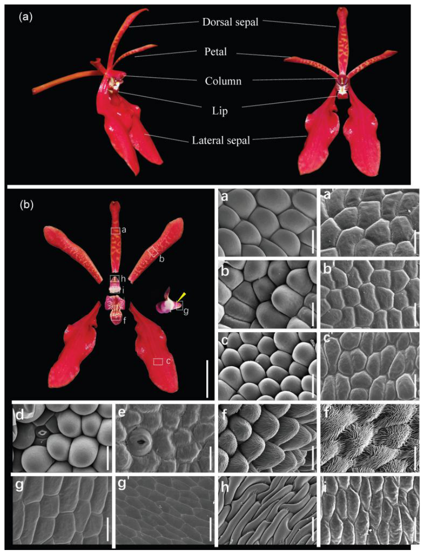
Figure 2.
Expression landscape of floral organ identity and symmetry-related genes reveals the perianth development model of R. coccinea. (a) Morphology of dissected floral organs from small (left) and middle (right) stage floral buds. (b) Principal component analysis of the 10 floral organ transcriptomes from small and middle stage floral buds. (c) Spatiotemporal expression profiles of floral organ identity genes and floral symmetry-related genes across floral tissues and developmental stages. The left heatmap represents log2(FPKM+1)-transformed expression values, whereas the right heatmap represents Z-score-normalized expression values. Genes showing prominent expression differences in the perianth are highlighted in bold. (d) Proposed regulatory model for perianth identity specification in R. coccinea. PI may provide a basal perianth identity program, whereas AP3-1/AGL6-1 are associated with sepal/petal identity, and AP3-2/AGL6-2 are associated with lip identity. Sco, column of small floral bud; Spe, petal of small floral bud; Sds, dorsal sepal of small floral bud; Sls, lateral sepal of small floral bud; Slp, lip of small floral bud; Mco, column of middle floral bud; Mpe, petal of middle floral bud; Mds, dorsal sepal of middle floral bud; Mls, lateral sepal of middle floral bud; Mlp, lip of middle floral bud.
Figure 2.
Expression landscape of floral organ identity and symmetry-related genes reveals the perianth development model of R. coccinea. (a) Morphology of dissected floral organs from small (left) and middle (right) stage floral buds. (b) Principal component analysis of the 10 floral organ transcriptomes from small and middle stage floral buds. (c) Spatiotemporal expression profiles of floral organ identity genes and floral symmetry-related genes across floral tissues and developmental stages. The left heatmap represents log2(FPKM+1)-transformed expression values, whereas the right heatmap represents Z-score-normalized expression values. Genes showing prominent expression differences in the perianth are highlighted in bold. (d) Proposed regulatory model for perianth identity specification in R. coccinea. PI may provide a basal perianth identity program, whereas AP3-1/AGL6-1 are associated with sepal/petal identity, and AP3-2/AGL6-2 are associated with lip identity. Sco, column of small floral bud; Spe, petal of small floral bud; Sds, dorsal sepal of small floral bud; Sls, lateral sepal of small floral bud; Slp, lip of small floral bud; Mco, column of middle floral bud; Mpe, petal of middle floral bud; Mds, dorsal sepal of middle floral bud; Mls, lateral sepal of middle floral bud; Mlp, lip of middle floral bud.
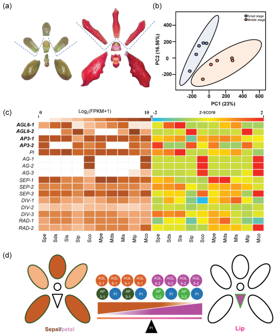
Figure 3.
Identification of genes preferentially expressed in various floral organs, specifically highly expressed in the lip, and associated with specialization of the lip, particularly the development of the spur in R. coccinea. (a) and (b) Venn diagrams showing the numbers of genes preferentially expressed in different organs in small stage (a) and middle stage (b) floral buds. (c) Heatmap showing the top 37 genes specifically and highly expressed in the lip of R. coccinea. (d) and (e) Expression profile of two candidate spur development-associated genes, BRH1 (d) and YUC10 (e), across floral organs.
Figure 3.
Identification of genes preferentially expressed in various floral organs, specifically highly expressed in the lip, and associated with specialization of the lip, particularly the development of the spur in R. coccinea. (a) and (b) Venn diagrams showing the numbers of genes preferentially expressed in different organs in small stage (a) and middle stage (b) floral buds. (c) Heatmap showing the top 37 genes specifically and highly expressed in the lip of R. coccinea. (d) and (e) Expression profile of two candidate spur development-associated genes, BRH1 (d) and YUC10 (e), across floral organs.
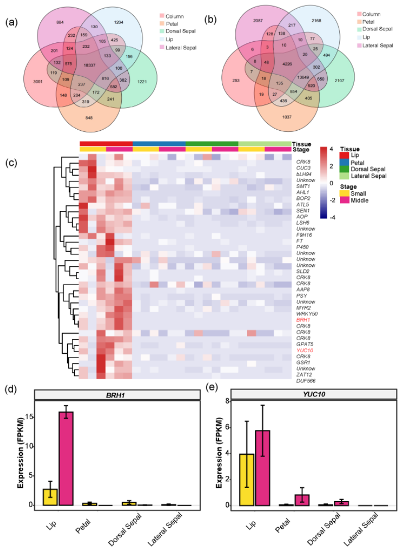
Figure 4.
Identification of the floral color pigments and metabolic network in R. coccinea. (a) Floral morphology and pigmentation patterns of mature floral tissues. Boxed regions highlight the pigmentation differences between the dorsal and lateral sepal. (b) Principal component analysis of the 12 metabolome profiles from dorsal and lateral sepal at small and middle developmental stages. (c), (d), Volcano plots showing differentially accumulated metabolites between small and middle stages in the dorsal sepal (c) and lateral sepal (d). (e) Cyanidin 3-O-glucoside biosynthesis pathway in R. coccinea.
Figure 4.
Identification of the floral color pigments and metabolic network in R. coccinea. (a) Floral morphology and pigmentation patterns of mature floral tissues. Boxed regions highlight the pigmentation differences between the dorsal and lateral sepal. (b) Principal component analysis of the 12 metabolome profiles from dorsal and lateral sepal at small and middle developmental stages. (c), (d), Volcano plots showing differentially accumulated metabolites between small and middle stages in the dorsal sepal (c) and lateral sepal (d). (e) Cyanidin 3-O-glucoside biosynthesis pathway in R. coccinea.
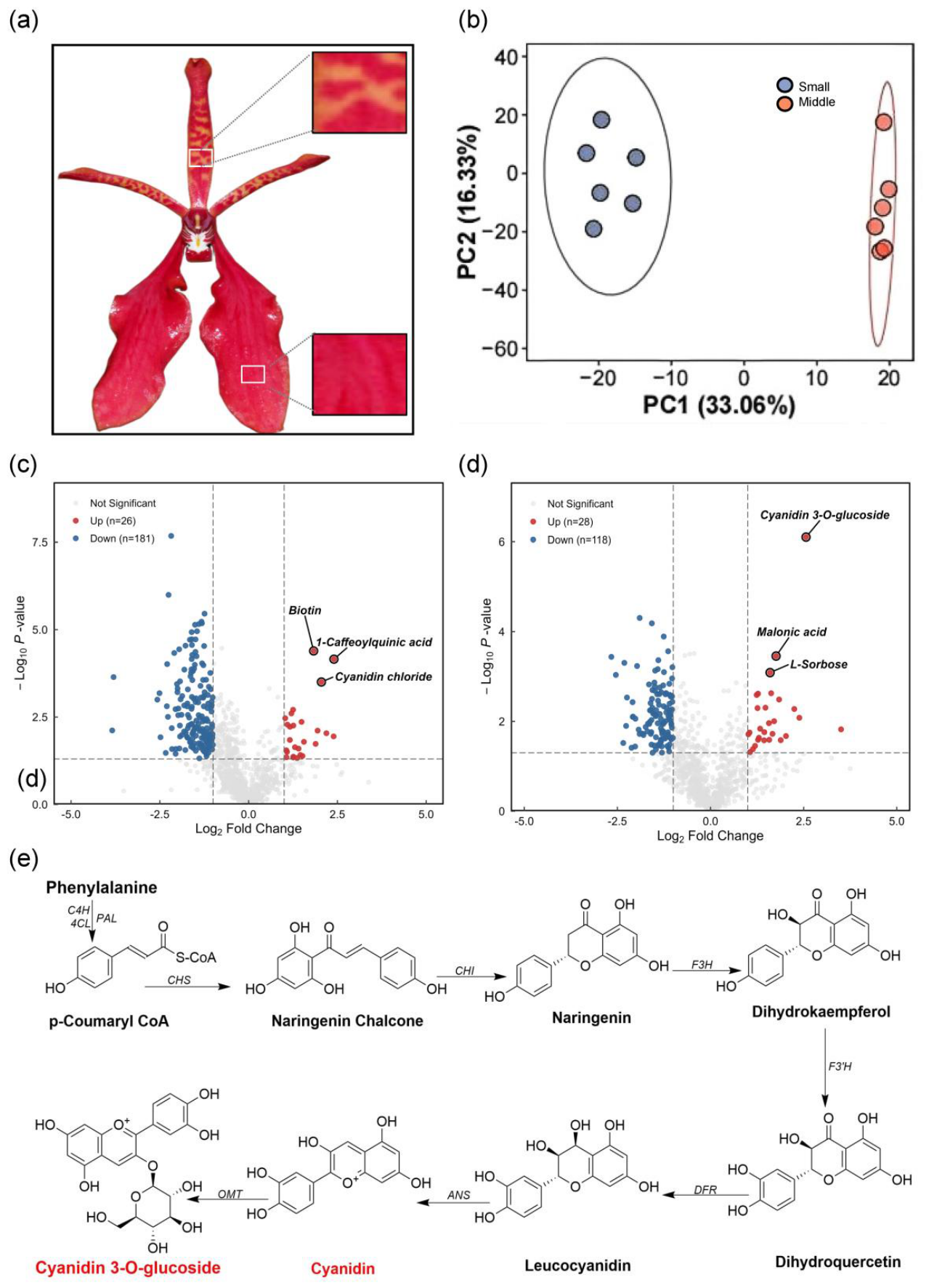
Figure 5.
Transcriptomic analysis of the anthocyanin biosynthesis pathway and key differentially expressed genes associated with floral pigmentation in R. coccinea. (a) Schematic representation of the anthocyanin biosynthesis pathway and major structural enzymes involved in pigment formation. (b) Spatiotemporal expression profiles of anthocyanin biosynthesis-related genes across developmental stages (S, small stage; M, middle stage) and floral tissues (left panel: log2(FPKM+1) heatmap; right panel: Z-score normalized heatmap). (c) Volcano plot showing differentially expressed genes (DEGs) between small and middle stage lateral sepals. (d) Volcano plot showing differentially expressed genes between dorsal sepals and lateral sepals at the middle stage. The structural gene UFGT and transcription factor AGL6-2 are highlighted in the volcano plots, providing molecular insights into anthocyanin accumulation and tissue-specific floral coloration in R. coccinea.
Figure 5.
Transcriptomic analysis of the anthocyanin biosynthesis pathway and key differentially expressed genes associated with floral pigmentation in R. coccinea. (a) Schematic representation of the anthocyanin biosynthesis pathway and major structural enzymes involved in pigment formation. (b) Spatiotemporal expression profiles of anthocyanin biosynthesis-related genes across developmental stages (S, small stage; M, middle stage) and floral tissues (left panel: log2(FPKM+1) heatmap; right panel: Z-score normalized heatmap). (c) Volcano plot showing differentially expressed genes (DEGs) between small and middle stage lateral sepals. (d) Volcano plot showing differentially expressed genes between dorsal sepals and lateral sepals at the middle stage. The structural gene UFGT and transcription factor AGL6-2 are highlighted in the volcano plots, providing molecular insights into anthocyanin accumulation and tissue-specific floral coloration in R. coccinea.
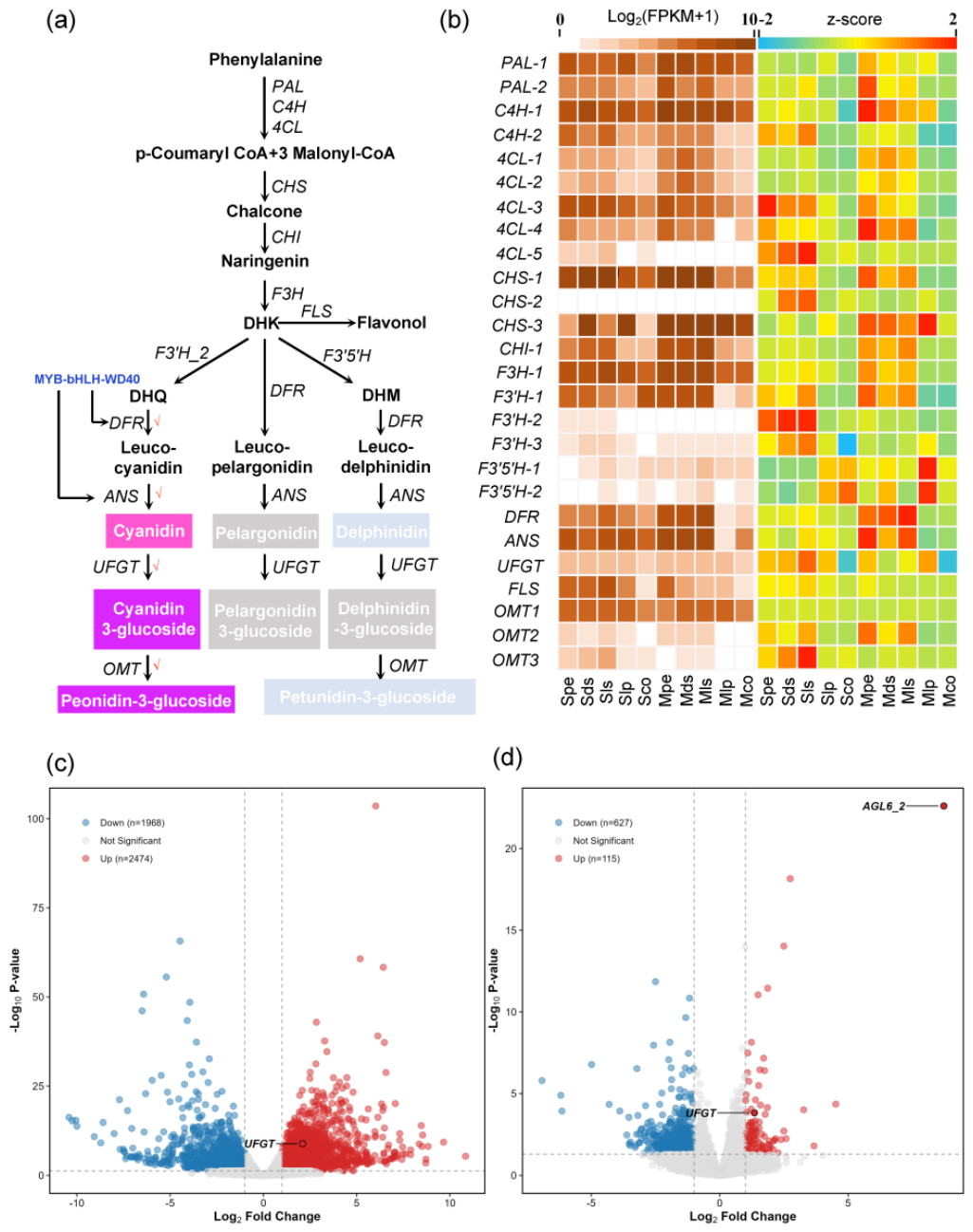
Figure 6.
Co-expression network analysis of floral development and anthocyanin biosynthesis pathway genes. (a) Regulatory network of hub genes involved in both floral development and anthocyanin biosynthesis pathway genes. (b) Regulatory network consisting exclusively of hub genes from the anthocyanin biosynthesis pathway genes. (c) Regulatory networks/modules comprising only one or two hub genes from either floral development or anthocyanin biosynthesis pathway genes. Nodes represent genes, and edges indicate co-expression correlations. Key functional hub genes are color-coded. These networks further elucidate the complex interaction mechanisms between structural genes and co-expression genes in determining the specific floral coloration of R. coccinea.
Figure 6.
Co-expression network analysis of floral development and anthocyanin biosynthesis pathway genes. (a) Regulatory network of hub genes involved in both floral development and anthocyanin biosynthesis pathway genes. (b) Regulatory network consisting exclusively of hub genes from the anthocyanin biosynthesis pathway genes. (c) Regulatory networks/modules comprising only one or two hub genes from either floral development or anthocyanin biosynthesis pathway genes. Nodes represent genes, and edges indicate co-expression correlations. Key functional hub genes are color-coded. These networks further elucidate the complex interaction mechanisms between structural genes and co-expression genes in determining the specific floral coloration of R. coccinea.
Disclaimer/Publisher’s Note: The statements, opinions and data contained in all publications are solely those of the individual author(s) and contributor(s) and not of MDPI and/or the editor(s). MDPI and/or the editor(s) disclaim responsibility for any injury to people or property resulting from any ideas, methods, instructions or products referred to in the content. |
© 2026 by the authors. Licensee MDPI, Basel, Switzerland. This article is an open access article distributed under the terms and conditions of the Creative Commons Attribution (CC BY) license (http://creativecommons.org/licenses/by/4.0/).
Copyright: This open access article is published under a Creative Commons CC BY 4.0 license, which permit the free download, distribution, and reuse, provided that the author and preprint are cited in any reuse.