
Submitted:
30 July 2026
Posted:
31 July 2026
You are already at the latest version
Abstract
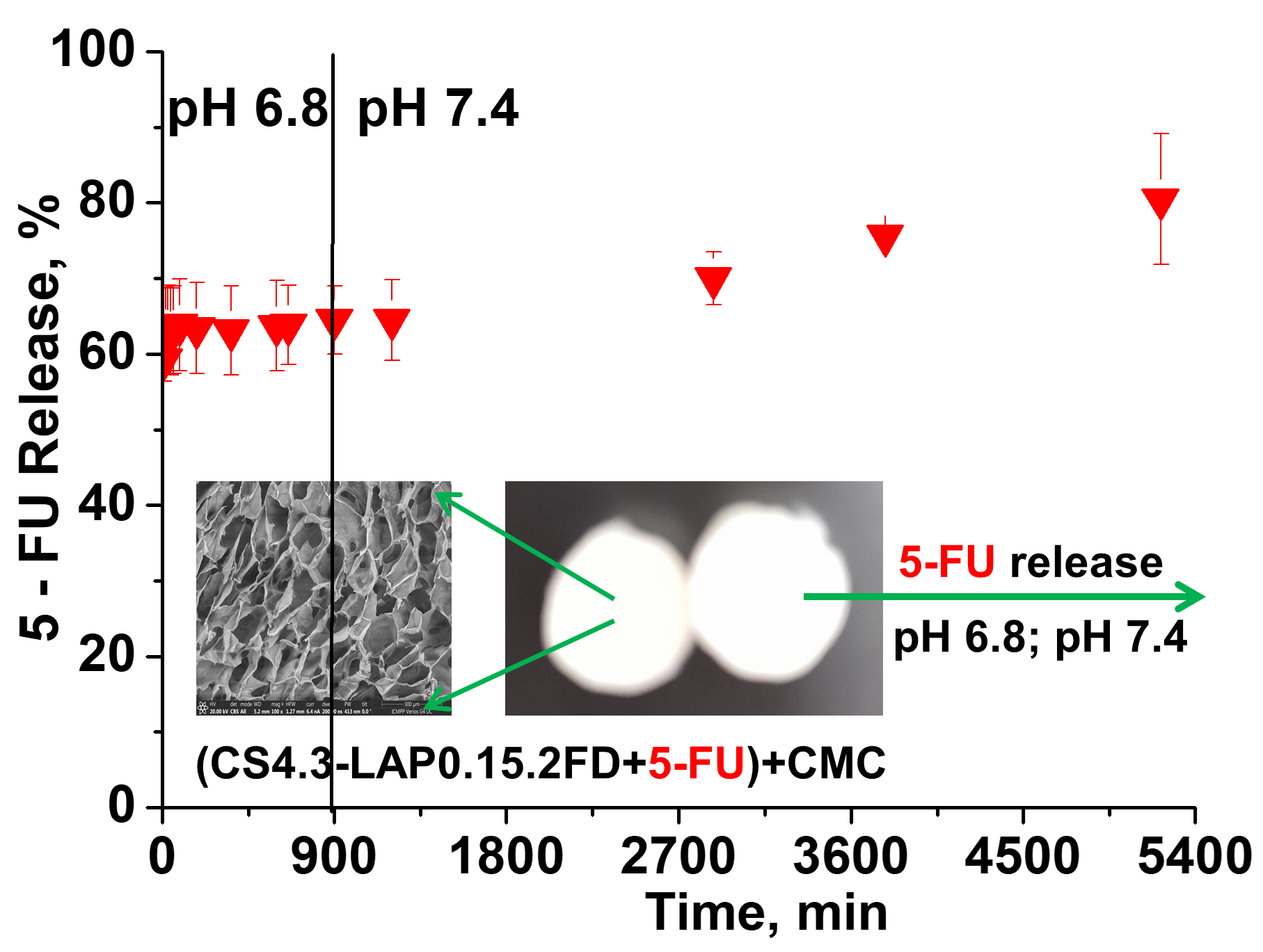
The impact of chitosan (CS) characteristics (deacetilation degree and molar mass), and concentration as well as of the strategy employed to fabricate porous nanocomposites CS-laponite (LAP), on the physicochemical properties of CS-LAP sponges was investigated in the paper. Information about the incorporation of LAP within the CS-LAP nanocomposites were obtained by EDX and FTIR spectroscopy. The influence of the fabrication conditions on the physical cross-linking was evidenced first by the swelling of the composite sponges at equilibrium in distilled water, with the values of the swelling ratio of about 6-18, for the CS-LAP composites prepared with one freeze drying (1FD) step, and of about 22-40, for the composites prepared with 2FD steps. The average pore size and pore size distribution were evaluated using software assisted analysis of SEM micrographs. The elastic modulus ranged between 6 - 21 kPa for CS-LAP composites prepared with 1FD, and between 2 - 10 for the nanocomposites prepared with 2FD steps. The physicochemical properties of CS-LAP nanocomposite sponges were correlated with their behavior in the loading and in vitro release of 5-fluorouracil (5-FU) as a function of pH. It was found that the CS-LAP composites with the highest swelling ratio exhibited the fastest release of 5-FU in simulated gastric fluid (pH 2.0), with the cumulative release in the range of 85 – 100%, in about 60 min, at 37 oC. The CS-LAP composites with the lowest swelling ratio, prepared by 1FD strategy, displayed the slowest release rate of drug. The drug release from the double component composites was accompanied by the carrier dissolution/disintegration. To retard the release of 5-FU in the gastric environment, the drug was sealed in the porous CS-LAP nanocomposites by simple complexation of the free positive charges of CS with carboxymethylcellulose as polyanion. These systems kept their integrity in pH 2.0, pH 6.8 and phosphate buffer with pH 7.4, and released 5-FU in a controlled manner making them suitable systems for delivery of 5-FU in both the stomach and small intestine.
Keywords:
chitosan
; laponite RD
; cryogels
; 5-fluorouracil
; drug delivery
1. Introduction
The fast evolution of controlled drug delivery systems (DDSs) is motivated by the stringent requirement to enhance the drug selectivity and efficiency, minimizing the high toxicity of anticancer drugs while increasing the clinical efficiency, the drug stability and the patient compliance [1,2,3,4,5,6,7]. Numerous DDSs are based on hydrogels, able to absorb and retain large volumes of water without dissolving owing to the physical or chemical cross-links [1,3,5]. Intelligent hydrogels (smart responsive) change their volume in response to a slight variation of the external/internal stimuli, such as pH, temperature, ionic strength, solvent composition, light, electric or magnetic field. These fascinating properties made them especially attractive for biomedical applications such as DDSs, tissue regeneration, wound management, artificial organs, cartilages, actuators [1,4,5]. Hydrophilic synthetic polymers [8,9,10] and polymers with origin in renewable resources such as polysaccharides [4,11,12,13,14,15,16,17,18,19,20] represent main elements in the construction of biocompatible and biodegradable hydrogels as drug carriers. Among polysaccharides, chitosan (CS) by its biocompatibility, nontoxicity, and intrinsic antibacterial properties has attracted a huge interest for fabrication of novel drug carriers of various medicines provided with delayed drug release [12,15,21,22,23,24,25].
The main weak points of hydrogels, consisting of the poor mechanical strength and slow response at stimuli, have been alleviated by diverse strategies including interpenetrating polymer networks [2,8,9], introducing hydrophobic substituents, or engineering hydrogel-silicate nanocomposites [11,13,14,16]. Laponite is a representative silicate efficient as physical cross-linker in the fabrication of nanocomposite hydrogels for biomedical purposes [13,14,16,17,18,19] including cell adhesion [13], bone regeneration [16], drug delivery [14,17,18]. Various CS-LAP nanocomposites have been designed [26,27,28,29,30,31,32,33,34,35,36,37,38] either as injectable hydrogels [30,33], carriers for anticancer drugs [31,32,34,35], antibiotics [27,36], or cell proliferation for wound healing [38].
5-Fluorouracil (5-FU) is one of the most recommended chemotherapeutic drugs in the treatment of tumors including gastric, colorectal, breast, liver, skin and lung but it suffers of low bioavailability, short half-time in the blood circuit system and high toxicity [39,40,41,42]. Moreover, the lack of drug specificity damages the healthy tissues beside the targeted cancer cells [39]. Therefore, there is a stringent need to find novel carriers endowed with as smart as possible control on the release of 5-FU, with high cell selectivity, enhanced stability and bioavailability of encapsulated drug. In this context, pH-responsive carbohydrate polymers have gained significant attention as promising candidates for next-generation of oral DDSs. The gastrointestinal tract (GIT), with its distinct pH environments, constitutes a suitable platform for the investigation of performances of pH controlled DDSs [2,4,41]. Various biodegradable composite hydrogels based on CS as chemically cross-linked films [40], ionically cross-linked nanoparticles [41], m-PEG-CS nanogels [42], alginate-CS/montmorillonite nanocomposites [43], CS microspheres [44,45], electrospun nanofibrous composite mats based on CS and polyvinylpyrrolidone containing graphene oxide [46], suitable for oral delivery of 5-FU with the main purpose of colorectal cancer therapy, have been lately designed. In another approach, anticancer 5-FU drug was loaded into CS/polyacrylic acid/Fe3O4 magnetic nanocomposite hydrogel through immersing the nanocomposite in the drug solution [47]. It was demonstrated that the porous CS-based nanocomposites can hold large amounts of drugs within their network, with controlled and sustained release of drug while ensure an enhanced stability of the payload [39]. Coating the drug carrier with synthetic or natural polymers bearing carboxylic groups can give the opportunity to deliver drugs directly to large bowel because these polymers can prevent the drug release and degradation in the gastric environment of stomach [42,48,49,50]. Thus, 5-FU has been encapsulated into the porous Zn-based metal-organic framework (MOF-5) and carboxymethylcellulose (CMC) has been used to cover 5-FU@MOF-5 hybrid and to protect the drug through the digestive system [48]. However, it was found that the CS-LAP nanocomposite sponges have been not evaluated as regards the potential carrier and pH controlled release of 5-FU.
Therefore, the main objective of the present study was to investigate first the impact of the swelling rate and morphology (the average pore size, and pore size distribution) of the CS-LAP composite sponges, tailored by the CS characteristics, the concentration of CS, as well as the strategy employed to generate porous nanocomposite materials, on the delivery profile of 5-FU controlled by pH. It was also postulated that by the free positive charges of CS, which can be complexed with a polyanion such as CMC, after the drug loading, these nanocomposites will offer a plentiful platform for a systematic investigation of the opportunities to release 5-FU in response to the specific pH of the three compartments of GIT.
2. Results and Discussion
In this work, series of 3D and porous nanocomposite cryogels were developed from CS and LAP to be evaluated for their potential in controlled delivery of 5-FU. The fabrication of CS-LAP nanocomposite sponges and their performances in the wound healing have been recently reported by the authors [38]. A constant concentration (4 wt.%) of CS with molar mass of 207 kDa and DD of 94%, with LAP concentrations varied in the range 0.2 - 2 wt.% have been previously used. To optimize the properties of CS-LAP macroporous nanocomposites, making them suitable as DDSs, the molar mass and the DD of CS, and the concentration of CS and LAP in the initial mixture of components were systematically varied in this work, as presented in Table 1.
Our interest in the tight control of the nanocomposites morphology conducted to the fabrication of CS-LAP sponges through two approaches, as presented in Scheme 1: (1) cryostructuration in one step, when the homogeneous dispersion of LAP nanodiscs in aqueous solution of protonated CS was first frozen and then, the stabilization of the nanocomposite cryogel morphology was achieved by the deprotonation of CS in NaOH 3M, followed by washing at neutral pH and then freeze drying, coded with 1FD; (2) cryostructuration of the CS-LAP nanocomposites in two steps, achieved by freeze-drying first of the frozen dispersion of LAP in aqueous solution of protonated CS, followed by the deprotonation of CS in NaOH 3M, washing at neutral pH, and again freeze dried to reach stable macroporous nanocomposites, coded with 2FD.
As can be seen in Scheme 1, after the exfoliation of LAP in H2Od, the CS particles were spread between the LAP nanodiscs with the final formation of a colloidal dispersion of CS and LAP. By the addition of acetic acid, the formed protonated CS chains (CS-NH3+) come in contact with the negatively charged surface of LAP discs. We are expecting as the LAP discs distribution to be influenced by the molar mass and DD of CS, CS concentration and the ratio between CS and LAP in the initial mixture. Both series of composite sponges underwent comprehensive characterization to decide the optimum conditions for the fabrication of CS-LAP nanocomposites as potential DDS for 5-FU.
2.1. Characterization of CS-LAP Nanocomposites
The influence of the synthesis conditions on the formation yield of the CS-LAP sponges was not so dramatic, as the values of yield in Table 1 show. As a general trend, at concentration of CS of 3 wt.%, the yield values were comparable, irrespective of the synthesis strategy (1FD or 2FD) and CS characteristics, i.e., around 87 - 89%, However, the highest values of the formation yield were found in the case of the composites prepared with CS4 (the highest DD, 94%), with 91.86% and 89.13%, for 1FD and 2FD, respectively, suggesting that the cross-linking efficiency was the most effective when the CS with the highest content of cationic centers was involved in the sponge formation. The yield in nanocomposite sponges was the lowest when the concentration of CS in the initial mixture of CS and LAP was only 1 wt.% (81.56%, for the sample CS3.1-LAP0.1.2FD). The above results support the opportunity to modulate the CS-LAP nanocomposite properties by the CS characteristics, CS concentration and CS : LAP ratio.
2.1.1. Internal Morphology and Pore Size Distribution
It was expected as the intrinsic characteristics of CS (molar mass and DD), the CS concentration, the CS:LAP ratio as well as the engineering of the composite porosity to behave as instruments to tailor the composite morphology, as shown for other porous materials [51,52,53]. Figure 1 presents the SEM micrographs of nanocomposites at the same magnification (100x).
At the first sight, the composites prepared by the 2FD strategy display much more regular pores, for all samples prepared with CS concentration of 3 wt.% (CS1.3-LAP0.15.2FD, CS2.3-LAP0.15.2FD, CS3.3-LAP0.15.2FD and CS4.3-LAP0.15.2FD) compared with those prepared by the 1FD strategy (CS1.3-LAP0.15.1FD, CS2.3-LAP0.15.1FD, CS3.3-LAP0.15.1FD and CS4.3-LAP0.15.1FD).
Decreasing the concentration of CS from 3 wt.% to 2 wt.% (CS3.2-LAP0.2.1FD, CS3.2-LAP0.2.2FD, CS3.2-LAP0.1.1FD and CS3.2-LAP0.1.2FD) conducted to the diminish of the differences in sponges morphology.
The pore size distribution presented in Figure 2 indicates a relative narrow polydispersity for all composites prepared at CS concentration of 3 wt.%, with a relative frequency of ~ 90% for pores with sizes in the range 60-100 µm. However, the pore size distribution is even narrower in the case of the nanocomposites prepared by the strategy 2FD (CS1.3-LAP0.15.2FD, CS2.3-LAP0.15.2FD, CS3.3-LAP0.15.2FD), excepting the nanocomposite prepared with CS4, where the pore size distribution is narrower in the case of the composite prepared by the strategy 1FD. The decrease of the CS concentration at 2 wt.% conducted to the increase of the pore size polydispersity, more in the case of the lower content of LAP (CS3.2-LAP0.1.1FD and CS3.2-LAP0.1.2FD), with the largest pores found in the case of these composite sponges. These results clearly support the possible correlations between the CS characteristics, CS concentration and CS:LAP ratio in the forecast of the nanocomposites morphology and pore size distribution. The narrow distribution of pore sizes found in the case of the nanocomposite sponges mentioned above would be helpful in predicting the sustained drug release profiles.
To show the presence and the distribution of LAP nanodiscs in the CS-LAP sponges, SEM micrographs at a magnitude of 20000x were included in Supporting Information (Figure S1). As can be seen, the LAP nanodiscs are almost evenly distributed into the nanocomposite walls. The presence of a higher amount of LAP nanodiscs is evident in the SEM micrograph of the CS3.2-LAP0.2 nanocomposites, where the CS:LAP ratio was 10:1, compared with CS3.2-LAP0.1, with CS:LAP ratio of 20:1.
The EDX spectra of the CS-LAP nanocomposite sponges are presented in Figure S2 and the elements content as Atomic % (C, N, Mg and Si), as a function of the synthesis conditions of nanocomposites, can be seen in Figure 3. The EDX data unambiguously demonstrate the incorporation of LAP nanoparticles within the nanocomposite sponges at a level well correlated with the synthesis conditions. Thus, at a CS concentration of 3 wt.% and a ratio CS : LAP of 20:1, the content in atomic C was in the range 49.59-52.65%, and that of atomic N was in the range 9.49-10.55% (Figure 3A), while the values of Mg and Si were in the range 0.43 – 1.03%, and 0.475 - 0.96% (Figure 3B), respectively, irrespective of the synthesis strategy. Increasing the content of LAP in the initial mixture of CS and LAP (CS : LAP ratio of 10:1) conducted to the decrease of the atomic C at 49.52 and 49.50% for CS3.2-LAP0.2.1FD and CS3.2-LAP0.2.2FD, respectively (Figure 3A) and to the increase of atomic Mg at 1.0275 ± 0.08% for CS3.2-LAP0.2.1FD, and 0.8425 ± 0.0192%, for CS3.2-LAP0.2.2FD, and of Si at 0.96 ± 0.152% for CS3.2-LAP0.2.1FD and 0.817 ± 0.0746%, for CS3.2-LAP0.2.2FD (Figure 3B) compared with about half of these values found for CS3.2-LAP0.1.1FD and CS3.2-LAP0.1.2FD composites, different only by the CS:LAP ratio (20:1).
The EDX percentages of the atomic O and Na, for all CS-LAP composites, were presented in Figure S3.
2.1.2. Swelling Kinetics of CS-LAP Nanocomposite Sponges
The values of the swelling ratio (SR) plotted in Figure 4 as a function of time show that a common characteristic of the CS-LAP composites is their very fast swelling (with the equilibrium of swelling reached in 5-10 sec), behavior which supports the strong connectivity of their pores [8,12,53,54].
The influence of CS molar mass and DD on the swelling kinetics, tightly correlated with the level of physical interactions in these nanocomposites, can be seen in Figure 4A.
As can be observed, the SR values of the composites CS1.3-LAP0.15 and CS2.3-LAP0.15, constructed with CS characterized by the same DD but different molar masses (Table 1), are comparable in the case of composites with CS deprotonation just after freezing (SR = 7.79±0.87 and 6.84±0.05, for CS1.3-LAP0.15.1FD and CS2.3-LAP0.15.1FD, respectively). The values of SR were much higher in the case of composites prepared with 2FD steps. This shows that the deprotonation of CS before freeze drying conducted to a more compact morphology compared with that resulted when the freezing was followed by freeze-drying, with the CS deprotonation after that. A higher value of SR was observed for the composite prepared with CS1 (SR = 30.45±1.16) than that of the composite prepared with CS2 (SR = 23.88±1.07). The SR value of the composite CS1.3-LAP0.15 higher than that of the composite CS2.3-LAP0.15 can be attributed to a higher cross-linking density (a more compact structure) in the case of the sample prepared with the highest molar mass (Mv = 305 kDa) than that of the composite prepared with CS1 (Mv = 147 kDa). Therefore, to conclude, the influence of CS molar mass could be revealed only by the comparison of the samples obtained with 2FD steps, with a higher molar mass favorable for a greater number of physical cross-links.
Information about the influence of DD of CS on the swelling kinetics could be found by the comparison of the SR values obtained for CS1.3-LAP0.15 (Mv = 147 kDa and DD = 82%) with those obtained for the composite CS4.3-LAP0.15 (CS with Mv = 207 kDa and DD = 94%, Figure 4A). Thus, the SR values of the composites CS1.3-LAP0.15.1FD and CS4.3-LAP0.15.1FD were 7.79±0.87 and 17.95±0.1, respectively, while those of the composites CS1.3-LAP0.15.2FD and CS4.3-LAP0.15.2FD were 30.34±1.16 and 23.89±0.57, respectively. As can be seen, the influence of DD on the efficiency of physical cross-linking was much stronger than that of the CS molar mass. It is evident that the composite CS4.3-LAP0.15.1FD was less compact than the composite CS1.3-LAP0.15.1FD, while in the case of composites prepared with 2FD steps the order was reversed (CS4.3-LAP0.15.2FD more compact than CS1.3-LAP0.15.2FD). The values of SR for the nanocomposites CS4.3-LAP0.15.1FD and CS4.3-LAP0.15.2FD are in agreement with the similar pore size distribution found for these samples (Figure 2).
CS concentration was another parameter of interest for the optimization of the CS-LAP nanocomposite properties. The SR values vs. time for the composites CS3.3-LAP0.15 and CS3.2-LAP0.1, both 1FD and 2FD, different by the concentration of CS (Table 1), with the ratio CS : LAP of 20:1 in all cases, were plotted in Figure 4B. The values of SR were: 14.59±0.56 and 29.17±0.58 for CS3.3-LAP0.15.1FD and CS3.3-LAP0.15.2FD, respectively, and 23.36±2.4 and 39±0.7 for CS3.2-LAP0.1.1FD and CS3.2-LAP0.1.2FD, respectively. The SR values were much higher for the composites prepared with 2%, w/v CS compared with those prepared with 3%, w/v CS, for both 1FD and 2FD, and this indicates the decrease of the composite compactness with the decrease of CS concentration.
The influence of CS:LAP ratio was evidenced first in Figure 4B by the comparison of composites CS3.2-LAP0.2.1FD and CS3.2-LAP0.2.2FD, where the CS:LAP was 10:1, with the composites CS3.2-LAP0.1.1FD and CS3.2-LAP0.1.2FD, where the CS:LAP was 20:1. The SR values of composites CS3.2-LAP0.2.1FD and CS3.2-LAP0.2.2FD were 22.78±1.7 and 37.35±1.4, respectively. The influence of CS:LAP ratio is more consistent for the composites with 2FD, with SR values lower for CS:LAP = 10:1 (37.35±1.4 for CS3.2-LAP0.2.2FD) than for 20:1 (39±0.7 for CS3.2-LAP0.1.2FD). This indicates that a higher density of cross-links was attained at a lower CS:LAP ratio. The SR values for the nanocomposite sponges prepared with CS3 at a concentration of 1 wt% and CS:LAP ratio of 10:1 are displayed in Figure 4C. The SR values found at equilibrium were 21.25±1.07 for CS3.1-LAP0.1.1FD and 32.4±2.3 for CS3.1-LAP0.1.2FD, values which are lower than those found for CS concentration of 2 wt.%, and the same CS:LAP ratio (Figure 4B).
2.1.3. Structural Characterization by FTIR Spectroscopy
The chemical structures of all CS-LAP nanocomposites were evidenced by the FTIR spectroscopy (the FTIR spectra presented in Figure 5 and Figure S4 in the Supplementary Information), with the FTIR spectra of CS1 and LAP included in Figure S4 for comparison. The characteristic bands of CS, visible in Figure S4A at: 1651 cm-1, 1599 cm-1, 1425 cm-1, 1381 cm-1, 1321 cm-1, 1261 cm-1, were assigned to: the C=O stretching vibration in acetamide groups (amide I), NH in primary amino groups, C–H and –CH2 stretching [55,56,57], the symmetrical deformation mode of CH3 [58], C-N in amide III, and the C-N stretching modes, respectively. The band characteristic to NH vibration in amide (amide II), normally located at 1550 cm-1, is missing possibly overlapped by other bands [56].
The fingerprint bands of CS located at 1157 cm-1, ~1070 cm-1, and ~1030 cm-1, are assigned to the stretching vibrations of the C-O-C bonds in polysaccharides, while the small peak at ~895 cm-1 corresponds to the wagging of the saccharide structure of CS.
The bands of CS located at 1157, 1070 and 1030 cm-1, which represent the fingerprint of CS, were assigned to the stretching vibration of C-O bonds in C-O-C bridge in polysaccharides, while the small peak at 895 cm-1 corresponds to the wagging of the saccharide structure of CS. The bands present in the FTIR spectrum of LAP (Figure S3B) are situated at 1007 and 465 cm-1. As can be seen in the spectra of CS-LAP nanocomposite sponges and in Table S1, the main bands are located at around: 2916, 2878, 1659, ~1590, 1420, 1379, 1321, 1261, 1153, 1084, 1034 cm-1. It is obvious that no consistent changes took place in the shape, location and intensity of the main FTIR bands and this supports that only physical interactions occurred between CS and LAP.
The incorporation of LAP within the nanocomposite is supported by the bands situated at around 660 cm-1, associated with Mg-O-Mg bending vibration in LAP; the characteristic band of LAP, located at 465 cm-1 in the spectrum of LAP (Figure S4B), and assigned to Si-O-Mg stretching vibration bonds [59], is shifted at around 444 cm-1 in all FTIR spectra of nanocomposites and conform the incorporation of LAP within all nanocomposite sponges and the strong electrostatic interaction between CS and LAP.
2.1.4. Elasticity and Shape Memory of CS-LAP Nanocomposites
The mechanical performance of the CS–LAP nanocomposites (series “1” and “2”) is governed by the combined effects of CS molar mass, degree of deacetylation, CS concentration, and LAP content, which together determine network organization and reinforcement efficiency (Figure 6 and Figure 7, Tables S2 and S3).
As Figure 6 shows, all samples exhibit a typical nonlinear stress–strain behavior characteristic of polymer–clay nanocomposite hydrogels, with an initial low-stiffness region followed by a sharp stress increase at higher strain.
This strain-hardening behavior is generally associated with progressive network densification and increasing resistance to deformation. Although the stress–strain profiles differ among formulations, all nanocomposite hydrogels maintain high compressibility, with strains ranging from approximately 76 to 94% (Figure 6A, 6B).
The mechanical parameters obtained from compression testing are summarized in Figure 7 and Tables S2 and S3. For the “1” series, increasing the CS molar mass from 147 to 305 kDa (CS1.3-LAP0.15.1FD and CS2.3-LAP0.15.1FD) results in a clear enhancement of mechanical properties, with the compressive elastic modulus rising from 10.82 to 18.68 kPa and the compressive stress from 658.25 to 717.37 kPa (Figure 7, Table S2). This reflects improved chain entanglement and more efficient stress transfer. However, the dependence is not strictly linear, as CS3.3-LAP0.15.1FD (256 kDa) exhibits intermediate modulus values (16.36 kPa) and a lower compressive stress (627.00 kPa), indicating that network organization and intermolecular interactions also contribute to the mechanical response.
The DD has a pronounced effect, with CS4.3-LAP0.15.1FD (94% DD) exhibits the highest modulus (20.98 kPa), along with high compressive stress (633.07 kPa) and the largest strain (94.40%) (Figure 7, Table S2), demonstrating that increased amino group content enhances hydrogen bonding and polymer–laponite interactions, leading to a stronger yet more deformable network. Reducing the CS concentration from 3 wt.% to 2 wt.% (CS3.2-LAP0.2.1FD, CS3.2-LAP0.1.1FD) leads to a significant decrease in stiffness (6.77 and 5.96 kPa, respectively), while compressive stress remains relatively similar (643.6 and 631.47 kPa) (Figure 7, Table S2), suggesting that LAP still contributes to load-bearing even in less dense networks. A slightly higher modulus for CS3.2-LAP0.2.1FD compared to CS3.2-LAP0.1.1FD indicates a modest reinforcing effect at higher LAP content.
A similar but less pronounced behavior is observed for the “2” series, as shown in Figure 6B and summarized in Figure 7 and Table S3. The increase in molar mass from 147 to 305 kDa (CS1.3-LAP0.15.2FD and CS2.3-LAP0.15.2FD) results in only a minor change in stiffness (4.35 to 4.57 kPa) and a moderate increase in compressive stress (472.45 to 503.43 kPa), suggesting a limited contribution from chain entanglement alone. A more pronounced improvement is observed for CS3.3-LAP0.15.2FD and CS4.3-LAP0.15.2FD, where modulus increases to 7.53 kPa and 10.45 kPa (Figure 7 and Table S3), respectively, accompanied by higher stresses (510.03 and 529.9 kPa), highlighting the combined role of molecular weight and DD. As in the “1” series, CS4.3-LAP0.15.2FD (CS with 94% DD) exhibits the best mechanical performance within this group.
The influence of polymer concentration can be assessed by comparing samples containing CS with identical molecular characteristics (256 kDa, DD = 86%). Increasing CS concentration from 1 wt.% (CS3.1-LAP0.1.2FD) to 2 wt.% (CS3.2-LAP0.1.2FD) and 3 wt.% (CS3.3-LAP0.15.2FD) results in a marked increase in compressive modulus from 1.57 to 2.82 and 7.53 kPa, respectively. The higher polymer content likely increases chain entanglement density and the number of polymer–clay interactions, producing a denser network structure and greater resistance to deformation. Interestingly, CS3.1-LAP0.1.2FD exhibits the highest compressive stress (833.7 kPa) despite having the lowest modulus. This behavior indicates that the more flexible network can undergo substantial deformation before failure, allowing efficient dissipation of mechanical energy under compression.
The role of inorganic content can be evaluated by comparing CS3.2-LAP0.2.1FD (10:1) and CS3.2-LAP0.1.1FD (20:1), which have identical CS characteristics and concentration (2 wt.%). Increasing the CS:LAP ratio from 10:1 to 20:1 leads to an increase in compressive stress from 492.9 to 532.6 kPa.
Overall, both series demonstrate that the DD and polymer concentration strongly influence compressive behavior. Higher DD generally increase stiffness, while reductions in polymer concentration primarily decrease the elastic modulus. Although the “1” samples generally exhibit higher compressive modulus values (6–21 kPa vs. 2–10 kPa), compressive stress (627–717 kPa vs. 472–833 kPa) does not follow the same trend across all formulations, as evidenced by the exceptionally high stress sustained by CS3.1-LAP0.1.2FD hydrogels. These results highlight the complex interplay between molecular characteristics, polymer concentration, and filler content in determining the mechanical performance of CS–LAP nanocomposite hydrogels.
2.2. Loading and Release of 5-FU in/from CS-LAP Nanocomposite Sponges
The pH-responsiveness of CS-based composite porous materials makes them suitable for oral administration of the drug release systems controlled through a pH-dependent mechanism [12,60,61]. In this work, the loading and release of 5-FU in/from CS-LAP nanocomposite sponges were associated with the preparation conditions. Such CS-LAP nanocomposite sponges have never been evaluated as potential delivery systems for controlled release of 5-FU. The percentages of the drug loaded into some double components CS-LAP composites are presented in Table 2.
To demonstrate the presence of 5-FU molecules into the CS-LAP composites after the drug loading, FTIR spectra presented in Figure S5 were examined comparative with the FTIR spectrum of 5-FU. Comparing the FTIR spectrum of the 5-FU-loaded CS=LAP nanocomposites with that of the pristine CS-LAP, some additional absorption bands located at 752 cm-1, 810 cm-1 and 1250 cm-1 can be observed, attributed to the out-of-plane bending vibration of -CF-CH- groups and to C-F stretch band associated with the presence of 5-FU [45,48]. These bands can be seen in the FTIR spectrum of 5-FU at 752, 810 and 1246 cm-1 and confirm the successful loading of 5-FU within the selected CS-LAP nanocomposite sponges.
To gain information on the influence of DD of CS on the drug release, two composite samples different by the DD of CS used in their construction (CS1.3-LAP0.15.2FD and CS4.3-LAP0.15.2FD, Table 1) were tested for their performances in the release of 5-FU. Information about the influence of the strategy used to fabricate the CS-LAP sponges on the drug release kinetics were received from the comparison between two composite different only by the synthesis strategy (CS1.3-LAP0.15.1FD and CS1.3-LAP0.15.2FD). The release kinetics of 5-FU from the CS-LAP nanocomposites presented in Table 2, at pH 2.0, were depicted in Figure 8A. As can be observed, there is a clear influence of the DD of CS on the release kinetics of 5-FU, as the fastest release occurred from the composite constructed with CS4 (DD = 94%, Table 1). Almost the same profile was identified in the case of the composite constructed with the CS3 at a concentration of 2 wt.% CS. The release rate of 5-FU from the four nanocomposite sponges can be ordered as follows: CS1.3-LAP0.15.1FD < CS1.3-LAP0.15.2FD < CS3.2-LAP0.2.2FD ≤ CS4.3-LAP0.15.2FD. For these composites must be specified that a common feature was they completely disintegrated [62,63] in about three hours. This order of drug release would indicate that: (i) the slower release of the drug from the CS1.3-LAP0.15.1FD could be associated with the lower swelling rate of this composite sponge; (ii) the fast drug release observed in the case of CS3.2-LAP0.2.2FD can be attributed to the very high swelling rate (~36, Figure 4B), associated with the CS concentration of 2 wt.%, while the fast release of 5-FU from CS4.3-LAP0.15.2FD is probably related to a lower efficiency of cross-linking at the same CS:LAP ratio. Such composite systems could be recommended for the therapy of gastric cancer, as the drug release of 5-FU in stomach is lately of strong interest [17,50,64].
Completely different release profiles were observed in pH 6.8, when the composites with the fastest release rate of 5-FU, in pH 2.0, were compared (Figure 8B). In this environment, the payload was completely released in about 20 min, without the disintegration of the carrier. To retard the release rate of 5-FU from CS-LAP composite sponges and to better protect the drug stability along the GIT, the drug was sealed inside the porous composites by the complexation of free positive charges of CS with CMC as polyanion, after the drug loading.
The release kinetics of 5-FU at pH 2.0 from (CS1.3-LAP0.15.2FD+5-FU)+CMC, (CS4.3-LAP0.15.2FD+5-FU)+CMC and CS3.2-LAP0.2.2FD+5-FU)+CMC composites are presented in Figure 8C. It is obvious that the presence of CMC has a positive effect on the drug release, with the slowest release rate found in the case of the composite synthesized with CS4, i.e., with the highest DD [(CS4.3-LAP0.15.2FD+5-FU)+CMC], and the fastest from the composite fabricated with CS1, i.e., with the lowest DD [(CS1.3-LAP0.15.2FD+5-FU)+CMC}, with CS3.2-LAP0.2.2FD+5-FU)+CMC located in between. The explanation is connected with the efficiency of the complex formation between CS and CMC, a stronger interaction and a higher stability being expected in the case of CS4 than in the case of CS1. This assumption is supported by the optical images presented in Figure 8D, which show that, during the drug release, the composite (CS1.3-LAP0.15.2FD)+CMC swelled at a much higher level that the other two composites. The release of 5-FU was extended at pH 6.8, in the case of (CS4.3-LAP0.15.2FD+5-FU)+CMC with the release of ~ 92% of 5-FU during 180 min.
To gain further knowledge about the potential of the CS-LAP sponges as carriers of 5-FU in the GIT environments, two of the above tested composites [(CS4.3-LAP0.15.2FD+5-FU)+CMC and (CS3.2-LAP0.2.2FD+5-FU)+CMC, Figure 8C)] were employed in the drug delivery first at pH 6.8 and then at pH 7.4 (Figure 8E). It is obvious that both composites display a burst release in pH 6.8 followed by a steady release up to 900 min, at a value of CR of about 63 ± 5% for [(CS4.3-LAP0.15.2FD+5-FU)+CMC] and 85.5 ± 2% for [(CS3.2-LAP0.2.2FD+5-FU)+CMC]. After the replace of release medium with pH 7.4, a different profile of the drug release was found, consisting in the steady release of drug from the composite [(CS4.3-LAP0.15.2FD+5-FU)+CMC, and a fast release from the composite [(CS3.2-LAP0.2.2FD+5-FU)+CMC] up to 100%. These results support the potential of the [(CS4.3-LAP0.15.2FD+5-FU)+CMC composite to perform as a drug carrier for 5-FU along the GIT environments.
3. Conclusions
CS-LAP nanocomposite sponges with morphology and swelling properties modulated by the DD of CS, CS concentration and the strategy employed to fabricate porous nanocomposites, with potential for oral administration of 5-FU, were successfully achieved in this work. As a function of the swelling capacity, the CS-LAP nanocomposite sponges proved to be suitable for delivery of 5-FU, either in the stomach (pH 2.0) by the release of drug simultaneous with dissolution/disintegration of the composite carrier, or into the small intestine (pH 6.8) and colon (pH 7.4) after the temporary sealing of drug into the CS-LAP by complexation of CS with CMC. Release profiles of 5-FU from CS-LAP nanocomposites investigated in the paper clearly revealed the influence of synthesis conditions and pH dependence, making these nanocomposites potential candidates for use in tumor localized drug delivery. Using CS-LAP sponges as carriers for 5-FU brings some advantages over conventional drug therapies, additionally expecting to overcome side effects regarding to dose and toxicity. Nevertheless, further optimization studies of these promising systems must be performed, both in vitro and in vivo.
4. Materials and Methods
4.1. Materials
Four CS samples different by the molar mass and DD were purchased from Sigma Aldrich and used as received. The intrinsic viscosity of each sample, dissolved in a 1:1 (v/v) mixture of 0.3 M CH3COOH and 0.2 M CH3COONa, was determined with an Ubbelohde viscometer at 25 ± 0.1 ◦C. The viscometric average molar masses (Mv), evaluated according to [65], are presented in Table 1. The DD was determined from the liquid state proton spectrum, according to the procedure described in literature [66]. Laponite RD, with 1000 kg/m3 bulk density, surface area of 370 m2/g, a chemical composition of SiO2 59.5%, MgO 27.5%, Li2O 0.8%, Na2O 2.8%, and loss on ignition 8.2%, was obtained from Rockwood Additive Ltd. (BYK-Chemie GmbH, Wesel, Germany). Glacial acetic acid, NaOH p.a. (≥ 98%), and ethanol p.a., purchased from Chemical Company, were all used as received.
4.2. Methods
4.2.1. Preparation of the CS-LAP Porous Nanocomposites
In this work, the as prepared nanocomposite sponges are denoted as: CSx.y-LAPz (Table 1), where CS and LAP indicate chitosan and Laponite RD, respectively, x represents the code of CS sample (1, 2, 3, 4, Table 1), and y and z represent the corresponding concentrations as CS/100 mL H2Od (w/v), and LAP/100 mL H2Od (w/v), respectively. For example, CS1.3-LAP0.15 (Table 1) means the nanocomposite based on CS1 (Mv = 147 kDa and DD = 82%) containing 3% w/v CS and 0.15% w/v Laponite RD. After the mild dispersion of the LAP clay in H2Od to reach the concentrations specified in Table 1, followed by ~ 2 h of magnetic stirring at room temperature (RT), the CS powder was dispersed under continuously magnetic mixing and kept under stirring 30 min, and then glacial acetic acid corresponding to a concentration of 2% (v/v) was added, the stirring being kept until a homogeneous mixture was obtained (about 5 h). The homogeneous dispersion of LAP in the CS solution was kept overnight at RT for removal of air bubbles. The CS-LAP mixtures were loaded in syringes of 5 mL, and then frozen overnight in a cryostat at -20 oC. The frozen monoliths were carefully pushed out from syringes after ~1 min at RT, cut in fragments of about 10 mm, and divided in two portions: the first one were immediately transferred into NaOH 3 M aqueous solution for 24 h, time sufficient for deprotonation of CS, and the other one into a Freeze Dryer Biobase BK-FD10S device, for freeze drying (48 h, at -57 oC and 0.045 mbar). The dried cryostructured CS-LAP sponges were transferred in 3 M NaOH aqueous solution for 24 h, to remove the acetic acid and to stabilize their morphology. Thereafter, the deprotonated CS-LAP sponges were washed with copious amounts of water to neutral pH, frozen overnight at -20 oC, and again lyophilized (2FD) to obtain resilient physically cross-linked CS-LAP nanocomposite sponges. For the fabrication of CS-LAP nanocomposites in this work, the CS4.3-LAP0.15 is given as an example. A weighed amount of 0.045 g LAP was mildly dispersed in 30 mL H2Od (~ 2 h) at RT, and after that 0.9 g of CS4 was dispersed and stirred about 30 min. After that, 0.45 mL acetic acid were dropped and the mixture was kept under stirring for about 5 h. The homogeneous mixture remained overnight at RT and then transferred into six syringes of 5 mL, which were placed into the cryostat at -20 oC, overnight. The syringes were taken out from cryostat one-by-one, the monoliths were carefully ejected, cut into ~10 mm fragments, the content of three syringes was put in NaOH 3M and the other fragments were returned into the cryostat for the next step, which was freeze drying.
4.2.2. Characterization Methods
To evaluate the yield in nanocomposite as a function of fabrication conditions, the samples have been further dried under vacuum in the presence of P2O5, until the constant weight was reached.
The yield was evaluated by Eq. (1):
where: Wd – weight of dried composite; Wm – total weight of initial components.
Y = (Wd/Wm) x 100 (%)
For the characterization by FTIR spectroscopy, the samples were frozen in liquid nitrogen (LN) and grounded into fine particles using a mortar. FTIR spectra were recorded using a Bruker Vertex FT-IR spectrometer, with a resolution of 2 cm-1 over the range of 4000–400 cm-1, through the KBr pellet technique, with about 5 mg of sample in each pellet.
The evaluation of surface morphology for the obtained hydrogels was performed with the help of a Verios G4 UC Scanning electron microscope (Thermo Scientific, Brno, Czech Republic). The samples were fixed on aluminum stubs with double-adhesive carbon tape and afterwards coated with a 7 nm platinum layer using a Leica EM ACE200 Sputter coater prior examination in order to provide electrical conductivity and prevent charge buildup which can occur during exposure to the electron beam. SEM investigations were carried out in High Vacuum mode using a concentric backscatter electron detector (CBS) at 20 kV accelerating voltage and 6.4 nA spot size, the magnification being indicated on the micrographs. The average diameter of pores for all the investigated hydrogels was evaluated from the SEM images by ImageJ 1.52a software, measuring approximately 30 pores on three images [38,51]. The identification of the chemical elements of the studied materials was performed by using an energy dispersive X-ray spectroscopy analyzer (EDAX Octane Elite Energy Dispersive X-ray Spectroscopy analyzer, Ametek, Berwyn, PA, USA) coupled to Verios G4 UC scanning electron microscope. The EDAX spectra were recorded in different areas of the samples to confirm the presence of LAP in hydrogels and also to obtain the map distribution of the elements within the obtained systems.
Swelling kinetics (swelling ratio, SR, as a function of time) were evaluated in distilled water by Eq. (2).
where: Wt represents the mass of the swollen gel at time t; Wd is the mass of gel in dry state.
SR = Wt/Wd
The mean of SR measurements on three independent samples has been plotted as a function of time, in the case of the swelling kinetics evaluation.
The mechanical behavior of the swollen sponges was characterized at room temperature using a Shimadzu EZ-SX universal testing machine (Kyoto, Japan). Cylindrical samples, measuring approximately 9–11 mm in diameter and 8–10 mm in height, were compressed under uniaxial loading with a 50 N load cell at a deformation rate of 1 mm min⁻¹. To ensure reproducible contact conditions, an initial preload of 0.1 N was applied before each measurement. Compressive stress, strain, and elastic modulus were determined according to previously established procedures for porous composites [12,38].
Loading/Release of 5-FU
The selected CS-LAP sponge samples were loaded with 5-FU by the sorption-solvent evaporation technique [12,23,47,49]. Solutions of 5-FU in distilled water with a concentration of 5 mg/mL were first prepared and added to certain amounts of composites as carriers up to their maximum sorption capacity was reached (indicated by their equilibrium swelling ratio presented in Figure 4). The samples were kept 24 hours in closed bottles, at +4 oC, in the dark, for the drug sorption at equilibrium. After that, monolith fragments loaded with 5-FU were quickly washed with distilled water, to remove the drug on the sponge surface, and transferred into clean bottles, frozen and freeze-dried into a Freeze Dryer Biobase BK-FD10S device (48 h, at -57 oC and 0.045 mbar). All solutions containing drug were collected and the drug amount was evaluated by UV-Vis. The loading of CS-LAP sponges with 5-FU (DL, %) was evaluated with Eq. 3, by weighing the dried samples loaded with 5-FU. The real amount of drug sorbed into the composites was evaluated by subtraction the nonsadsorbed drug amount from the initial amount of drug:
5-Fluorouracil (FU) was entrapped in the hydrogels, as a model therapeutic agent, and the in vitro release profiles of the drug were established at 37 oC in pH 2.0, 6.8 and 7.4.
Thus, the in vitro release of 5-FU at pH 2.0 and 6.8 was performed by immersing the sample loaded with 5-FU in 10 mL release medium. At predetermined time intervals, 1 mL of supernatant was withdrawn and analyzed for the concentration of 5-FU at λmax of 266 nm using the UV-Vis calibration curve [67] previously recorded with Spectrophotometer (SPECORD 200 Analytik Jena). The removed solution was replaced with an identical volume of fresh releasing solution. When two environment with different pH were explored (pH 2.0 followed by pH 6.8, or pH 6.8 followed by pH 7.4), the first release medium was replaced with 10 mL of the second medium, for a certain release duration. The cumulative release (CR) of 5-FU was calculated with Eq. 4:
where: Cn and Cn-1 are the concentrations of 5-FU (mg L-1) in the releasing medium after n and (n - 1) withdrawing steps; n is the number of withdrawing steps of the release medium; V is the volume of sample (1 mL); mo is the amount of drug loaded in the sample.
Supplementary Materials
The following supporting information can be downloaded at the website of this paper posted on Preprints.org, Figure S1. SEM micrographs of CS-LAP sponges: Scale bar is 1 µm and mag 20000x in all micrographs; Figure S2. EDX spectra of CS-LAP cryogels as a function of the CS : LAP sets (according to Table 1); Figure S3. EDX data of O and Na for the nanocomposite sponges CS-LAP: (1) CS1.3-LAP0.15.1FD; (2) CS1.3-LAP0.15.2FD; (3) CS2.3-LAP0.15.1FD; (4) CS2.3-LAP0.15.2FD; (5) CS3.3-LAP0.15.1FD; (6) CS3.3-LAP0.15.2FD; (7) CS4.3-LAP0.15.1FD; (8) CS4.3-LAP0.15.2FD; (9) CS3.2-LAP0.2.1FD; (10) CS3.2-LAP0.2.2FD; (11) CS3.2-LAP0.1.1FD; (12) CS3.2-LAP0.1.2FD; Figure S4. FTIR spectra of chitosan (A), Laponite RD (B), CS-LAP composites prepared with CS2 (C) and CS-LAP composites prepared with CS3 (D); Figure S5. (A) FTIR spectra of 5-FU and CS1.3-LAP0.15 loaded with 5-FU; (B) FTIR spectra of CS4.3-LAP0.15 and CS3.2-LAP0.2 loaded with 5-FU; Figure S6. Calibration curve of 5FU in PBS, pH 7.4. Table S1. Main absorption bands in the FTIR spectra of the CS-LAP nanocomposite sponges and their assignment. Table S2. Values of compressive elastic modulus, compressive nominal stress, and strain of CS-LAP nanocomposite sponges obtained by one freeze drying step (1FD). Table S3. Values of compressive elastic modulus, compressive nominal stress, and strain of CS-LAP nanocomposite sponges obtained by two freeze drying steps (2FD).
Author Contributions
Conceptualization, E.S.D.; validation, E.S.D. and M.V.D.; formal analysis, E.S.D. and M.V.D.; investigation, E.S.D., M.V.D. M.M.L. and D.R.; resources, E.S.D. and M.V.D.; writing—original draft preparation, E.S.D., M.V.D. and D.R.; writing—review and editing, E.S.D., M.V.D., M.M.L.; visualization, E.S.D., M.V.D. and D.R.; supervision, ESD.; project administration, M.V.D.; funding acquisition, M.V.D. All authors have read and agreed to the published version of the manuscript.
Funding
M.V. Dinu and M.M. Lazar gratefully thank for the financial support from the Executive Agency for Higher Education, Research, Development and Innovation Funding (UEFISCDI) through the project PN-IV-P1-PCE-2023-1968 (4PCE/08.01.2025).
Institutional Review Board Statement
Not applicable.
Informed Consent Statement
Not applicable.
Data Availability Statement
We encourage all authors of articles published in MDPI journals to share their research data. In this section, please provide details regarding where data supporting reported results can be found, including links to publicly archived datasets analyzed or generated during the study. Where no new data were created, or where data is unavailable due to privacy or ethical restrictions, a statement is still required. Suggested Data Availability Statements are available in section “MDPI Research Data Policies” at https://www.mdpi.com/ethics.
Acknowledgments
The authors thank to Dr. Alina Nicolescu for the evaluation of DD of CS by 1H-NMR spectroscopy. .
Conflicts of Interest
The authors declare no conflicts of interest.
References
- Liu, B.; Chen, K. Advances in hydrogel-based drug delivery systems. Gels 2024, 10, 262. [CrossRef]
- Shahi, F.; Zarei, S.; Othman, R.S.; Afshar, H.; Kamran, P.; Taromi, A.A.; Khonakdar, H.A. Interpenetrating polymer networks in biomedical fields: Recent advanced and applications. Polym. Adv. Technol. 2025, 36, e70099. [CrossRef]
- Yoon, M.S.; Lee, J.M.; Jo, M.J.; Kang, S.J.; Yoo, M.K.; Park, S.Y.; Bong, S.; Park, C.S.; Park, C.W.; Kim, J.S.; Han, S.B.; Lee, H.J.; Shin, D.H. Dual-drug delivery systems using hydrogel-nanoparticle composites: Recent advances and key applications. Gels 2025, 11, 520. [CrossRef]
- Boldrini, D.E. Carbohydrate polymer derivatives as gastrointestinal pH-responsive oral drug delivery systems: A review. Innov. Med. Omics 2026, 3, 1-14. [CrossRef]
- Taokaew, S.; Kaewkong, W.; Kriangkrai, W. Recent development of functional chitosan-based hydrogels for pharmaceutical and biomedical applications. Gels 2023, 9, 277. [CrossRef]
- Murgia, D.; Estevao, B.M.; Portioli, C.; Palomba, R.; Decuzzi, P. Optimizing the fabrication of shape-defined microparticles for sustained drug delivery. ACS Appl. Eng. Mater. 2024, 2, 2535-2548. [CrossRef]
- Jiang, L.; Wang, X.; Jiang, H. Versatile chitosan-based hydrogel dressings for multi-scenario wound management. Small 2026, 22, e14813. [CrossRef]
- Dragan, E.S.; Cocarta, A.I. Smart macroporous IPN hydrogels responsive to pH, temperature, and ionic strength: Synthesis, characterization, and evaluation of controlled release of drugs. ACS Appl. Mater. Interfaces 2016, 8, 12018-12030. [CrossRef]
- Apopei Loghin, D.F.; Biliuta, G.; Coseri, S.; Dragan, E.S. Preparation and characterization of oxidizedstarch/poly(N,N-dimethylaminoethyl methacrylate) semi-IPN cryogels and in vitro controlled release evaluation of indomethacin. Int. J. Biolog. Macromol. 2017, 96, 589-599. [CrossRef]
- Dragan, E.S.; Dinu, M.V. Nitrogen based synthetic polycations as ubiquitous items of composites with environmental and biomedical applications. React. Funct. Polym. 2026, 220, 106608. [CrossRef]
- Caharwar, A.K.; Schexnailder, P.J.; Kline, B,P.; Schmidt, G. Assessment of using LaponiteR cross-linked poly(ethylene oxide) for controlled cell adhesion and mineralization. Acta Biomater. 2011, 7, 568-577. [CrossRef]
- Dragan, E.S.; Dinu, M.V.; Ghiorghita, C.A. Chitosan based polyelectrolyte complex cryogels with elasticity, toughness and delivery of curcumin engineered by polyions pair and cryostructuration steps. Gels 2022, 8, 240. [CrossRef]
- Katti, K.S.; Jasuja, H.; Jaswandkar, S.V.; Mohanty, S.; Katti, D.R. Nanoclays in medicine: a new frontier of an ancient medical practice. Mater. Adv. 2022, 3, 7484-7500. [CrossRef]
- Stealey, S.T.; Gaharwar, A.K.; Zustiak, S.P. Laponite-based nanocomposite hydrogels for drug delivery applications. Pharmaceuticals, 2023, 16, 821.
- Ullah, S.; Azad, A.K. Nawaz, A.; Shah, K.U.; Iqbal, M.; Albadrani, G.M.; Al-Joufi, F.A.; Sayed, A.A.; Abdel-Daim, M.M. 5-Fluorouracil-loaded folic-acid-fabricated chitosan nanoparticles for site-targeted drug delivery cargo. Polymers 2022, 14, 2010. [CrossRef]
- Magalhães, L.S.S.M.; Andrade, D.B.; Bezerra, R.D.S.; Morais, A.I.S.; Oliveira, F.C.; Rizzo, M.S.; Silva-Filho, E.C.; Lobo, A.O. Nanocomposite hydrogel produced from PEGDA and Laponite for bone regeneration. J. Funct. Biomater. 2022, 13, 53. [CrossRef]
- Luo, J.; Ma, Z.; Yang, F.; Wu, T.; Wen, S.; Zhang, J.; Huang, L.; Deng, S.; Tan, S. Fabrication of Laponite-reinforced dextran-based hydrogels for NIR-responsive controlled drug release. ACS Biomater. Sci. Eng. 2022, 8, 1554-1565. [CrossRef]
- Kiaee, G.; Dimitrakakis, N.; Sharifzadeh, S.; Kim, H.J.; Avery, J.K.; Moghaddam, K.M.; Hoghniaz, R.; Yalcitas, E.P.; de Barros, N.R.; Karamikamkar, S.; Libanori, A.; Khademhosseini, A, Khoshakhlagh, P. Laponite-based nanomaterials for drug delivery. Adv. Healthcare Mater. 2022, 11, 2102054. [CrossRef]
- Orakdogen, N.; Boyaci, T. pH-stimulus on-off switching behavior and improved response rate of slightly charged poly(N,N-dimethylaminoethyl methacrylate) nano-sized composites with incorporated Laponite as cross-linker. React. Funct. Polym. 2016, 102, 82-92. [CrossRef]
- Dragan, E.S.; Dinu, M.V. Polysaccharides constructed hydrogels as vehicles for proteins and peptides. A review. Carbohydr. Polym. 2019, 225, 115210. [CrossRef]
- Yang, H.; Hua, S.; Wang, W.; Wang, A. Composite hydrogel beads based on chitosan and laponite: Preparation, swelling, and drug release behaviour. Iran. Polym. J. 2011, 20, 479-490.
- Ghiorghita, C.A.; Dinu, M.V.; Dragan, E.S. Burst-free and sustained release of diclofenac sodium from mesoporous silica/PEI microspheres coated with carboxymethyl cellulose/chitosan layer-by-layer films. Cellulose 2022, 29, 395-412. [CrossRef]
- Dragan, E.S.; Ghiorghita, C.A.; Dinu, M.V.; Duceac, I.A.; Coseri, S. Fabrication of self-antibacterial chitosan/oxidized starch polyelectrolyte complex sponges for controlled delivery of curcumin. Food Hydrocoll. 2023, 135, 108147. [CrossRef]
- Valderruten, N.E.; Garcia, J. Interpenetrating polymer networks of poly(2-hydroxyethyl methacrylate-co-itaconic acid) and chitosan as a controlled release matrix. React. Funct. Polym. 2025, 210, 106192. [CrossRef]
- Shakouripour, F.; Olad, A.; Bayramoglu, G. Preparation of interpenetrating networks from chitosan and poly(hydroxypropyl methacrylate) or p(hydroxyethyl methacrylate) for controlled release of doxorubicin and curcumin: Investigation of potential use in wound dressing. Int. J. Biolog. Macromol. 2025, 301, 140929. [CrossRef]
- Oliveira, M.J.A.; Silva, E.O.; Braz, L.M.A.; Maia, R.; Amato, V.S.; Lugao, A.B.; Parra, D.F. Influence of chitosan/clay in drug delivery of glucantime from PVP membranes. Rad. Phys. Chem. 2014, 94, 194-198. [CrossRef]
- Ordikhani, F.; Dehghani, M.; Simchi, A. Antibiotic-loaded chitosan-laponite films for local drug delivery by titanium implants: cell proliferation and drug release studies. J. Mater. Sci. Mater. Med. 2015, 26, 269. [CrossRef]
- Raut, S.Y.; Gahane, A.; Joshi, M.B.; Kalthur, G.; Mutalik, S. J. Nanocomposite-clay polymer microbeads for oral controlled drug delivery: Development and, in vitro and in vivo evaluations. Drug Deliv. Sci. Technol. 2019, 51, 234-243. [CrossRef]
- Heragh, B.K.; Javanshir, S.; Mahdavinia, G.R.; Jamal, M.R.N. Hydroxyapatite grafted chitosan/laponite RD hydrogel: Evaluation of the encapsulation capacity, pH-responsivity, and controlled release behavior. Int. J. Biolog. Macromol. 2021, 190, 351-359. [CrossRef]
- Guo, H.; Shen, H.; Ma, J.; Wang, P.; Yao, Z.; Zhang, W.; Tan, X.; Chi, B. Versatile injectable carboxymethyl chitosan hydrogel for immediate hemostasis, robust tissue adhesion barrier, and antibacterial applications. ACS Appl. Mater. Interfaces 2023, 15, 52290-52304. [CrossRef]
- Heragh, B.K.; Taherinezhad, H.; Mahdavinia, G.R.; Javanshir, S.; Labib, P.; Ghasemsolb, S. pH-responsive co-delivery of doxorubicin and saffron via cross-linked chitosan/laponite RD nanoparticles for enhanced-chemotherapy. Mater. Today Commun. 2023, 34, 104956. [CrossRef]
- Mahdavinia, G.R.; Hoseinzadeh, H.; Labib, P.; Jabbari, P.; Mohebbi, A.; Barzeger, S.; Jahari, H. (Magnetic laponite/κ-carrageenan)/chitosan core-shell carrier for pH-sensitive release of doxorubicin. Polym. Bull. 2023, 80, 12923-12943. [CrossRef]
- Ranjbardamghani, F.; Eslahi, N.; Jahanmardi, R. An injectable chitosan/laponite hydrogel synthesized via hybrid cross-linked system: A smart platform for cartilage. Polym. Adv. Tecnhol. 2023, 34, 2298-2311. [CrossRef]
- Jafari, H.; Namazzi, H. pH-sensitive biosystem based on laponite RD/chitosan/polyvinyl alcohol hydrogels for controlled delivery of curcumin to breast cancer cells. Colloid Surf. B: Biointerfaces 2023, 231, 113585. [CrossRef]
- Jafari, H.; Namazzi, H. Chitosan/laponite clay bio-nanocomposite as an efficient cytocompatible nanocarrier for pH-sensitive controlled release of curcumin. Inorg. Chem. Commun. 2024, 168, 112940. [CrossRef]
- Wei, W.; Wang, M.; Liu, Z.; Zheng, W.; Tremblay, P.L.; Zhang, T. An antibacterial nanoclay-and chitosan-based quad composite with controlled drug release for infected skin wound healing. Carbohydr. Polym. 2024, 324, 121507. [CrossRef]
- Papagiannopoulos, A.; Nikolakis, S.-P.; Pamvouxoglou, A.; Koutsopoulou, E. Physicochemical properties of electrostatically crosslinked carrageenan/chitosan hydrogels and carrageenan/chitosan/Laponite. Int. J. Biolog. Macromol. 2023, 225, 565-573. [CrossRef]
- Dragan, E.S.; Lazar, M.M.; Dinu, M.V.; Dascalu, I.A.; Nacu, I., Verestiuc, L. Superelastic chitosan/Laponite nanocomposite sponges with tunable functional properties as promising biomaterials for wound management. ACS Appl. Bio Mater. 2025, 8, 7699-7714. [CrossRef]
- Entezar-Almahdi, E.; Mohammadi-Samadi, S.; Tayebi, L.; Farjadian, F. Recent advances in designing 5-Fluorouracil delivery systems: A stepping stone in the safe treatment of colorectal cancer. Int. J. Nanomed. 2020, 15, 5445-5458. [CrossRef]
- Abdelaal, M.Y.; Abdel-Razik, E.A.; Abdel-Bary, E.M.; El-Sherbiny, I.M. Chitosan-based interpolymeric pH-responsive hydrogels for in vitro drug release. J. Appl. Polym. Sci. 2007, 103, 2864-2874. [CrossRef]
- Aydin, R.S.T.; Pulat, M. 5-Fluorouracil encapsulated chitosan nanoparticles for pH-stimulated drug delivery: Evaluation of controlled release kinetics. J. Nanomater. 2012, Art. ID 13961. [CrossRef]
- Nguyen, D.H. Potential 5-Fluorouracil encapsulated mPEG-chitosan nanogels for controlling drug release. J. Adv. Med. Pharm. Sci. 2017, 12, 1-7. [CrossRef]
- Azhar, F.F.; Olad, A. A study on sustained release formulations for oral delivery of 5-fluorouracil based on alginate-chitosan/montmorrillonite nanocomposite systems. Appl. Clay Sci. 2014, 101, 288-296. [CrossRef]
- Wan, T.; Zhang, Q.; Jin, G.; Xu, S. Controlled delivery of 5-fluorouracil from monodisperse chitosan microspheres prepared by emulsion cross-linking. RSC Advances 2024, 14, 11311-11321. [CrossRef]
- Șahin, M.O.; Șanli, O. In vitro 5-fluorouracil release properties investigation from pH sensitive sodium alginate coated and uncoated methyl cellulose/ chitosan microspheres. Int. J. Biolog. Macromol. 2024, 258, 128895. [CrossRef]
- Grant, J.J.; Pillai, S.C.; Perova, T.S.; Brennan, B.; Hinder, S.J.; McAfee, M.; Sarah Hehir, S.; Breen, A. Enhancement of 5-Fluorouracil drug delivery in a graphene oxide containing electrospun chitosan/polyvinylpyrrolidone construct. Materials 2024, 17, 5300. [CrossRef]
- Amini-Fazl, M.S.; Mohammadi, R.; Kheiri, K. 5-Fluorouracil loaded chitosan/polyacrylic acid/Fe3O4 magnetic nanocomposite hydrogel as a potential anticancer drug delivery system. Int. J. Biolog. Macromol. 2019, 132, 506-513.
- Javanbakht, S.; Hemmati, A.; Namazi, H.; Heydari, A. Carboxymethylcellulose-coated 5-fluorouracil@MOF-5 nano-hybrid as a bio-nanocomposite carrier for the anticancer oral delivery. Int. J. Biolog. Macromol. 2020, 155, 876-882. [CrossRef]
- Zolfagharian, S.; Zahedi, P.; Ardestani, M.S.; Khatibi, A.; Jafarkhani, S. Sodium alginate/xanthan-based nanocomposite hydrogels containing 5-fluorouracil: Characterization and cancer cell death studies in presence of halloysite nanotube. J. Ind. Eng. Chem. 2023, 120, 374-386. [CrossRef]
- Bereyhi, M.; Zare-Dorabei, R.; Safarifard, V. A biocompatible NMOF-based drug delivery system designed for the sustained and controlled release via pH-responsive poorly soluble drug 5-fluorouracil. Langmuir 2025, 41, 6544-6552. [CrossRef]
- Venkataraman, R.; Das, G.; Singh, S.R.; Pathak, L.C.; Ghosh, R.N.; Venkataraman, B.; Krishnamurthy, R. Study on influence of porosity, pore size, spatial and topological distribution of pores on microhardness of plasma sprayed ceramic coatings. Mater. Sci. Eng. A 2007, 445-446, 269-274.
- Kaczmarek, B.; Sionkowska, A.; Osyczka, A.M. The comparison of physic-chemical properties of chitosan/collagen/hyaluronic acid composites with nano-hydroxyapatite cross-linked by dialdehyde starch and tannic acid. Polym. Testing 2017, 62, 171-176. [CrossRef]
- Lazar, M.M.; Ghiorghita, C.A.; Rusu, D.; Dinu, M.V. Nanocomposite cryogels based on chitosan for efficient removal of a triphenylmethan dye from aqueous systems. Gels 2025, 11, 729.
- Jain, E.; Zhang, K.; Tiwari, R.M. Properties and characterization of cryogels: Structural, mechanical and functional insights. ACS Omega 2025, 10, 36771-36787.
- Yuan, Q.; Shah, J.; Hein, S. Misra, R.D.K. Controlled and extended drug release behavior of chitosan-based nanoparticle carrier. Acta biomaterialia 2010, 6, 1140-1148.
- Queiroz, M.F.; Melo, K.R.T.; Sabry, D.A.; Sassaki, G.L.; Rocha. H.A.O. Does the Use of Chitosan Contribute to Oxalate Kidney Stone Formation? Mar. Drugs 2015, 13, 141-158.
- Ailincai; D.; Bercea, M.; Rosca, I.; Sandu, I.A.; Marin, M. Antimicrobial chitosan-based hydrogels: A novel approach to obtain sanitizers. Carbohydr. Polym. 2025, 354, 123288.
- Li, Y.; Sun, S.; Gao, P.; Zhang, M.; Fan, C.; Lu, Q.; Li, C.; Chen, C.; Lin, B.; Jiang, Y. A Tough Chitosan-Alginate Porous Hydrogel Prepared by Simple Foaming Method. J. Solid State Chem. 2021, 294, 121797.
- Xiong, Z.Q.; Li, X.D.; Fu, F.; Li, Y.N. Performance evaluation of laponite as a mud-making materialnfor drilling fluids. Petroleum Sci. 2019, 16, 890-900.
- Takeshita, S., Zhao, S., Malfait, W.J., Koebel, M.M. Chemistry of chitosan aerogels: Three-dimensional pore control for tailored applications. Angew. Chem. Int. Ed., 2021, 60, 9828-9851.
- Gorshkova, N.; Brovko, O.; Palamarchuk, I.; Bogolitsyn, K.; Ivakhnov, A. Preparation of bioactive aerogel material based on sodium alginate and chitosan for controlled release of levomycetin. Polym. Adv. Technol. 2021, 32, 3474-3482.
- Narasihman, B.; Peppas, N.A. Molecular analysis of drug delivery systems controlled by dissolution of the polymer carrier. J. Pharm. Sci. 1997, 86, 287-304.
- Liw, J.J.; Teoh, X.Y.; Teoh, A.X.Y.; Chan, S.Y. The effect of carrier-drug ratios on dissolution performances of poorly soluble drug in crystalline solid dispersion system. J. Pharm. Sci. 2022, 111, 95-101.
- Reis, B.; Frenzel, R.; Gerlach, N.; Muller, M.; Schultz, J.; Tutwa, S.; Weatherby, J.; Dasog, M.; Schwarz, S. Gold nanorods decorated by conjugated microporous polymers for infrared responsive cytostatic drug delivery. Langmuir 2025, 41, 2471-2479.
- Gamzazade, A.I.; Slimak, V.M.; Skljar, A.M.; Stykova, E.V.; Pavlova, S.S.A.; Rogozin, S.V. Investigation of the hydrodynamic properties of chitosan solutions. Acta Polym. 1985, 36, 420–424.
- Lavertu, M.; Xia, Z.; Serreqi, A.N.; Berrada, M.; Rodrigues, A.; Wang, D.; Buschmann, M.D.; Gupta, A. A validated 1H NMR method for the determination of the degree of deacetylation of chitosan. J. Pharm. Biomed. Anal. 2003, 32, 1149-1158.
- Cojocaru, I.C.; Ochiuz, L.; Spac, A.; Popa, G.; Palade, L.; Popovici, I. The validation of the UV spectrophotometric method for the assay of 5 fluorouracil. Farmacia 2012, 60, 379-385.
Scheme 1.
Main steps involved in the preparation of CS-LAP sponges according to the two strategies developed in this work.
Scheme 1.
Main steps involved in the preparation of CS-LAP sponges according to the two strategies developed in this work.

Figure 1.
SEM micrographs of the CS-LAP nanocomposites as a function of synthesis conditions. Scale bar was 300 µm and mag 100x in all micrographs. Sample Code is that presented in Table 1.
Figure 1.
SEM micrographs of the CS-LAP nanocomposites as a function of synthesis conditions. Scale bar was 300 µm and mag 100x in all micrographs. Sample Code is that presented in Table 1.

Figure 2.
Histograms of the pore size distribution of the CS-LAP nanocomposite sponges based on different CS samples, CS concentration and CS:LAP ratio and synthesis strategy.
Figure 2.
Histograms of the pore size distribution of the CS-LAP nanocomposite sponges based on different CS samples, CS concentration and CS:LAP ratio and synthesis strategy.

Figure 3.
EDX data of C and N (A), Mg and Si (B) for the nanocomposite sponges CS-LAP: (1) CS1.3-LAP0.15.1FD; (2) CS1.3-LAP0.15.2FD; (3) CS2.3-LAP0.15.1FD; (4) CS2.3-LAP0.15.2FD; (5) CS3.3-LAP0.15.1FD; (6) CS3.3-LAP0.15.2FD; (7) CS4.3-LAP0.15.1FD; (8) CS4.3-LAP0.15.2FD; (9) CS3.2-LAP0.2.1FD; (10) CS3.2-LAP0.2.2FD; (11) CS3.2-LAP0.1.1FD; (12) CS3.2-LAP0.1.2FD.
Figure 3.
EDX data of C and N (A), Mg and Si (B) for the nanocomposite sponges CS-LAP: (1) CS1.3-LAP0.15.1FD; (2) CS1.3-LAP0.15.2FD; (3) CS2.3-LAP0.15.1FD; (4) CS2.3-LAP0.15.2FD; (5) CS3.3-LAP0.15.1FD; (6) CS3.3-LAP0.15.2FD; (7) CS4.3-LAP0.15.1FD; (8) CS4.3-LAP0.15.2FD; (9) CS3.2-LAP0.2.1FD; (10) CS3.2-LAP0.2.2FD; (11) CS3.2-LAP0.1.1FD; (12) CS3.2-LAP0.1.2FD.

Figure 4.
Swelling kinetics of the CS-LAP composites as a function of CS molar mass and DD (A), CS concentration (B), and CS : LAP ratio (C).
Figure 4.
Swelling kinetics of the CS-LAP composites as a function of CS molar mass and DD (A), CS concentration (B), and CS : LAP ratio (C).

Figure 5.
FTIR spectra of CS-LAP nanocomposite sponges prepared with CS1 (A), CS4 (B) and CS3 at CS concentration of 2 wt.% (C and D) (the sample code is that of sets presented in Table 1).
Figure 5.
FTIR spectra of CS-LAP nanocomposite sponges prepared with CS1 (A), CS4 (B) and CS3 at CS concentration of 2 wt.% (C and D) (the sample code is that of sets presented in Table 1).

Figure 6.
Compressive stress–strain behavior of CS–LAP nanocomposites for (left) series “1FD” and (right) series “2FD”. The curves correspond to samples with varying CS molar mass, DD, CS concentration, and CS:LAP ratio.
Figure 6.
Compressive stress–strain behavior of CS–LAP nanocomposites for (left) series “1FD” and (right) series “2FD”. The curves correspond to samples with varying CS molar mass, DD, CS concentration, and CS:LAP ratio.

Figure 7.
Mechanical properties of CS–LAP nanocomposites for samples “1FD” and “2FD”. (A, B) strain at maximum compression (%), (C, D) compressive nominal stress (kPa), and (E, F) compressive elastic modulus (kPa) for the investigated samples. Error bars represent standard deviations (SD) obtained from three replicate measurements.
Figure 7.
Mechanical properties of CS–LAP nanocomposites for samples “1FD” and “2FD”. (A, B) strain at maximum compression (%), (C, D) compressive nominal stress (kPa), and (E, F) compressive elastic modulus (kPa) for the investigated samples. Error bars represent standard deviations (SD) obtained from three replicate measurements.

Figure 8.
(A, B, C and D) Release kinetics of 5-FU from CS-LAP composite sponges controlled by pH; optical images of the CS-LAP/CMC composites after the drug release at pH 2.0 (E).
Figure 8.
(A, B, C and D) Release kinetics of 5-FU from CS-LAP composite sponges controlled by pH; optical images of the CS-LAP/CMC composites after the drug release at pH 2.0 (E).

Table 1.
Sample codes, compositions and yields.
| Sample Code* | Chitosan | Laponite RD, %, w/v |
CS:LAP ratio | Yield, % | |||
| Mv, kDa | DD, % | %, w/v | 1FD | 2FD | |||
| CS1.3-LAP0.15 | 147 | 82 | 3 | 0.15 | 20:1 | 84.75 | 87.89 |
| CS2.3-LAP0.15 | 305 | 82 | 3 | 0.15 | 20:1 | 86.83 | 86.57 |
| CS3.3-LAP0.15 | 256 | 86 | 3 | 0.15 | 20:1 | 89.19 | 88.84 |
| CS4.3-LAP0.15 | 207 | 94 | 3 | 0.15 | 20:1 | 91.86 | 89.13 |
| CS3.2-LAP0.1 | 256 | 86 | 2 | 0.1 | 20:1 | 89.25 | 87.02 |
| CS3.2-LAP0.2 | 256 | 86 | 2 | 0.2 | 10:1 | 88.3 | 87.81 |
| CS3.1-LAP0.1 | 256 | 86 | 1 | 0.1 | 10:1 | 82.1 | 81.56 |
*The codes of CS were: CS1, CS2, CS3 and CS4 having the molar mass of 147 kDa, 305 kDa, 256 kDa and 207 kDa.
Table 2.
Drug loading of CS-LAP composites tested for drug release as a function of pH.
| Sample Code* | CS1.3-LAP0.15.1FD | CS1.3-LAP0.15.2FD | CS4.3-LAP0.15.2FD | CS3.2-LAP0.2.2FD |
| Drug Loading, % | 7.482 | 11.02 | 9.252 | 10.76 |
* The sample code is identical with that in Table 1.
Disclaimer/Publisher’s Note: The statements, opinions and data contained in all publications are solely those of the individual author(s) and contributor(s) and not of MDPI and/or the editor(s). MDPI and/or the editor(s) disclaim responsibility for any injury to people or property resulting from any ideas, methods, instructions or products referred to in the content. |
© 2026 by the authors. Licensee MDPI, Basel, Switzerland. This article is an open access article distributed under the terms and conditions of the Creative Commons Attribution (CC BY) license (http://creativecommons.org/licenses/by/4.0/).
Copyright: This open access article is published under a Creative Commons CC BY 4.0 license, which permit the free download, distribution, and reuse, provided that the author and preprint are cited in any reuse.