

Submitted:
29 July 2026
Posted:
30 July 2026
You are already at the latest version
Abstract
Spongiform degeneration, or status spongiosis, is characterized by vacuoles within the central nervous system. It appears in numerous neurological diseases, including trans-missible spongiform encephalopathies, mitochondrial disorders, and lysosomal storage diseases. Traditionally considered secondary to neurodegeneration, vacuolar changes frequently involve white matter and form within the myelin sheath. This review ex-amines the evidence from various diseases and genetic models that exhibit this pathology to support the hypothesis that white matter vacuolation represents a myelin defect, and explores potential causative mechanisms. Our findings suggest that spongiform change in white matter represents a common endpoint of pathway disruptions that lead to metabolic or ionic dyshomeostasis, causing an osmotic imbalance and vacuole formation within myelin. We advocate for further research into myelin-preserving pathways as potential therapeutic avenues to treat conditions exhibiting this pathology.
Keywords:
spongiform encephalopathy
; CNS vacuolation
; status spongiosis
; myelination
; myelinopathy
; leukodystrophies
1. Introduction
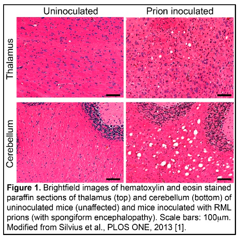
Spongiform, or vacuolar, degeneration is a distinctive histopathological feature (Figure 1) [1] associated with numerous neurological conditions. Traditionally, its significance has been unclear and vacuolation is often dismissed as a byproduct of neuronal or glial injury. However, the frequent localization of such changes in white matter-rich regions of the central nervous system (CNS) suggests the possibility of a direct link to abnormalities in myelin biology.
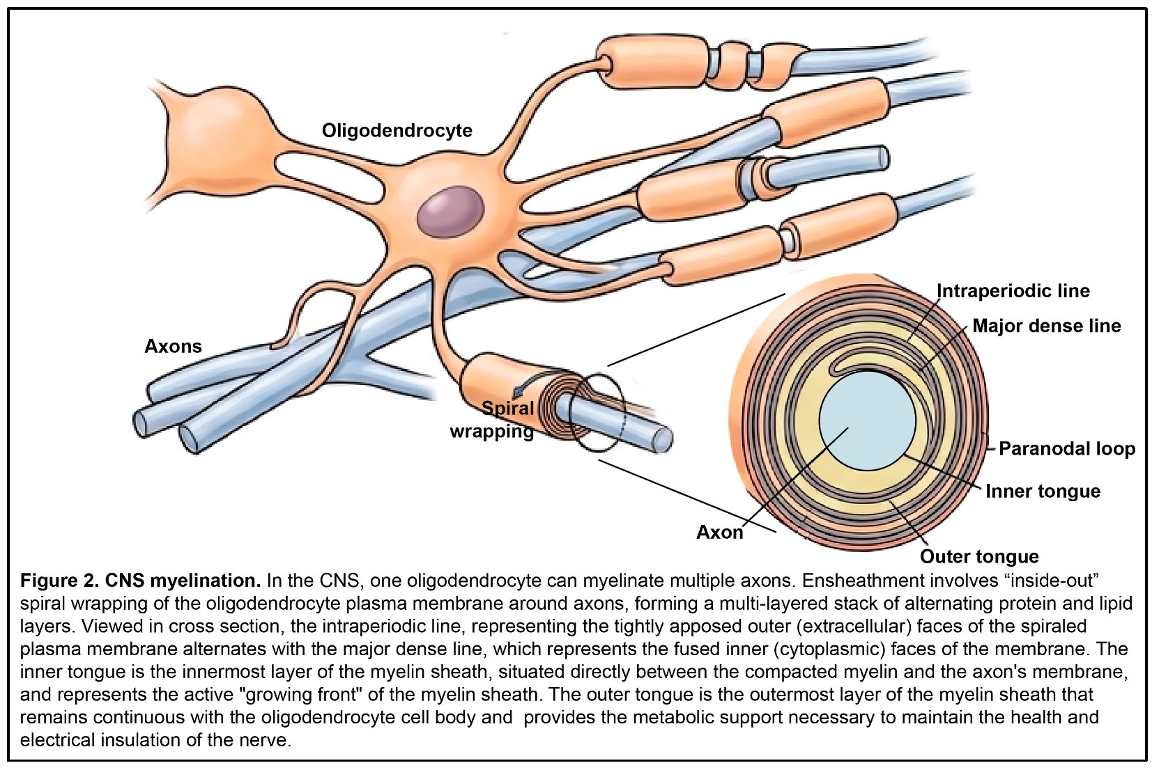
Myelination is the process by which neuronal axons are wrapped in a lipid-rich membrane (the myelin sheath), which insulates axons to facilitate rapid action potential propagation and provides metabolic support (Figure 2) [2]. Myelin is produced by mature oligodendrocytes in the CNS and by Schwann cells in the peripheral nervous system (PNS). While each Schwann cell myelinates a single axon, individual oligodendrocytes in the CNS can ensheathe one or more axons. Myelination involves “inside-out” spiral wrapping of the oligodendrocyte plasma membrane around axons, forming a multi-layered stack of alternating protein and lipid layers (Figure 2) [3]. It is a tightly regulated process that involves bidirectional communication between axons and oligodendroglia and is typically dependent on axonal activity. As discussed in detail below, oligodendrocytes in the CNS generate and maintain myelin through intricate intercellular communication, lipid synthesis, and precise regulation of metal ions like iron. Disruptions in any of these processes can destabilize myelin and impair remyelination.
Here, we put forth the hypothesis that spongiform change in CNS white matter reflects intrinsic defects in myelin rather than solely being a downstream effect of neuronal degeneration. This review presents supporting evidence from various disorders, including prion, mitochondrial and lysosomal storage diseases, and identifies converging pathways that contribute to vacuolar pathology in the CNS.
2. Spongiform Degeneration in Transmissible Spongiform Encephalopathies (TSEs)
Transmissible spongiform encephalopathies (TSEs) such as Creutzfeldt-Jakob disease (CJD) in humans, scrapie in sheep, and bovine spongiform encephalopathy (BSE, or “mad cow disease”) in cattle, are a group of fatal neurodegenerative disorders caused by the accumulation of the misfolded, pathogenic form (scrapie, PrPSc) of the prion protein [4,5]. A hallmark feature of TSE pathology is spongiform degeneration, in which microscopic vacuoles appear within neural tissues, giving the brain a sponge-like appearance [6,7]. Historically, vacuoles were considered a secondary phenomenon, simply reflecting neuronal loss and the presence of toxic PrPSc aggregates. However, growing evidence indicates that white matter and myelin structures are also primary targets in TSEs, implicating myelin pathology as a possible underlying driver of spongiform change.
2.1. White Matter Involvement in TSEs
While gray matter vacuolation, particularly in the neuropil, is a prominent feature of TSEs, numerous studies have also documented significant changes in white matter [8,9,10]. Vacuoles are often found within the myelin sheath itself, with electron microscopy revealing splitting of the myelin lamellae and the formation of intramyelinic vacuoles in affected regions [7,11,12]. White matter pathology in TSEs also encompasses disruption of myelin architecture, accumulation of abnormal protein aggregates within myelin, and degenerative changes in oligodendrocytes [13,14]. Collectively, these observations suggest that myelin damage may be a central feature of TSE pathology rather than a downstream consequence of neuronal death.
2.2. Roles of Normal Cellular Prion Protein (PrPC) in Myelination
Under physiological conditions, normal cellular prion protein (PrPC) is predominantly expressed in neurons, but it is also found in glial cells, including oligodendrocytes [15,16]. Several lines of evidence indicate that PrPC plays an important role in myelin health and maintenance. Prnp knockout (Prnp–/–) mice, which lack the gene encoding PrPC, initially developed normal peripheral and central myelin, but as these animals aged, they exhibited progressive white matter vacuolation, reactive astrogliosis, and deficits in motor coordination and balance [17]. Peripheral myelin pathology in Prnp–/– mice could be rescued by neuron-specific expression of PrPC, suggesting that neuronal PrPC provides trophic support necessary for Schwann cell-dependent myelination [18]. In contrast, expressing PrPC exclusively in Schwann cells failed to rescue the peripheral myelin phenotype, underscoring the critical role of neuronal PrPC in intercellular signaling. Although transgenic goats [19] and cattle [20] lacking PrPC did not display gross phenotypes indicative of PNS myelination defects, closer examination of teased nerve-fiber preparations from PrPC-deficient goats revealed histopathological changes characteristic of demyelinating neuropathy similar to that seen in Prnp–/– mice [21,22]. Together, these findings suggest that PrPC plays a direct role in PNS myelin maintenance.
One proposed role for PrPC in myelin biology involves signaling through the G-protein coupled receptor, adhesion G-protein coupled receptor 6 (ADGRG6, also known as GPR126). In the PNS, PrPC interacts with ADGRG6, which is expressed on Schwann cells, to activate the cyclic AMP (cAMP)–protein kinase A (PKA) signaling pathway [23,24]. This pathway promotes Schwann cell proliferation and differentiation and activates expression of Egr2 (also known as Krox20), which in turn activates transcription of myelin genes, including those encoding myelin basic protein (MBP), myelin protein zero (MPZ) and peripheral myelin protein 22 (PMP22) [23,25]. The N-terminal fragment of PrPC, which is generated through proteolytic cleavage, contains the binding motif for ADGRG6 and may act as the functional ligand for this pathway [23]. Whether a similar receptor–ligand mechanism operates in the CNS remains unknown, but the fact that ADGRG6 expression has not been detected in oligodendrocytes suggests that alternative receptors or signaling pathways mediate any effects of PrPC on CNS myelination.
Another potential link between PrPC and myelination is through metal homeostasis. The octapeptide repeat region of PrPC binds copper ions and may facilitate redox reactions necessary for cellular metabolism [26,27]. In addition, PrPC appears to exhibit ferrireductase activity, converting ferric iron (Fe3+) to its bioavailable ferrous form (Fe2+) [28]. This function is noteworthy because oligodendrocytes contain some of the highest levels of iron in the brain, and iron is essential for synthesizing myelin lipids, including cholesterol and sphingolipids [29,30]. Iron and copper deficiency and dysregulation have been reported in the brains of Prnp–/– mice and patients with prion diseases such as CJD, and could result in impaired myelin synthesis or destabilization of existing myelin, thereby contributing to vacuolation and white matter degeneration [31,32].
2.3. Integrated Perspective
Taken together, the data presented above suggest that vacuolation in TSEs may reflect both direct toxicity from the presence of misfolded prion protein and the loss of its normal physiological functions as PrPC is converted into PrPSc. In particular, PrPC appears necessary for maintaining myelin integrity through glial signaling pathways, supporting oligodendrocyte metabolism via metal homeostasis, and potentially participating in neuron–glia interactions that regulate myelin maintenance. These insights challenge the traditional view of spongiform degeneration as a purely neuronal phenomenon and instead support the hypothesis that white matter vacuolation in TSEs represents a myelination defect.
3. Lipid Homeostasis and Lysosomal Storage Diseases
While the composition of most cellular membranes is approximately 50% protein and 50% fat, mammalian myelin in the CNS is approximately 30% protein and 70% lipid [33,34]. Myelin lipids include cholesterol, phospholipids, and sphingolipids, and are essential for forming and maintaining the multilayered architecture of myelin sheaths. As a result, normal synthesis, transport and degradation of lipids by oligodendrocytes is critical to myelin health and integrity. While it is more intuitive that disrupted lipid metabolism in oligodendrocytes would affect myelin synthesis through cell autonomous mechanisms, lipid homeostasis in neurons is also important since neuronal accumulation of lipids is toxic, and axonal integrity is involved in maintaining the myelin sheath [35,36]. Within cells, lipids enter the lysosomal pathway via endocytosis or autophagy [37,38]. Lysosomal hydrolases, such as lysosomal acid lipase, break down complex lipids into free fatty acids and cholesterol, which are then trafficked to cellular membranes or the endoplasmic reticulum (ER) [39]. It is perhaps not surprising, given the critical role of lysosomes in lipid homeostasis, that lysosomal storage disorders (LSDs) are frequently associated with profound changes in myelin.
3.1. Myelin Lipids
The galactolipid galactocerebroside (GalC) and its derivative, sulfatide, make up 20-30% of the total dry weight of the myelin bilayer. UDP-galactose:ceramide galactosyltransferase (encoded by the UGT8 gene) catalyzes the transfer of galactose to ceramide in the final step of GalC synthesis. Ugt8 knockout mice formed compact myelin but it contained glucocerebroside, which is not normally found in myelin, developed a tremor, showed multiple abnormalities in the CNS including hypo- and dys-myelination, intramyelin vacuolation of spinal cord fibers, and disrupted paranodal axo-glial interactions, and they died prematurely [40,41,42,43]. Exposure of spinal cords from young Ugt8 null mice to 4-aminopyridine (4-AP), a drug that blocks potassium channels located in the paranodal region, partially rescued their action potential conduction defects [41]. Normally, these channels are occluded by the myelin sheath and do not participate in the conduction process, which suggests that abnormalities in the myelin produced in the absence of UGT8 resulted in access of the drug to these potassium channels, perhaps due to paranodal abnormalities [41,43]. This ties defects in myelin lipids directly to potassium buffering, which is discussed in Section 5 of this review. Similar defects, including myelin vacuolation and paranodal junction abnormalities, were observed in mice deficient for galactose-3-O-sulfotransferase 1 (GAL3ST1, also known as cerebroside sulfotransferase, CST), which transfers the sulfate group of the sulfoglycolipids from 3′-phosphoadenosine 5′-phosphosulfate in the GalC biosynthesis pathway [44].
In addition to being a component of myelin that provides structural stability to the compacted myelin sheath, very long acyl (C22-C24) chain sphingolipids also play a role in the correct sorting and transport of MBP [45]. Loss of these sphingolipids in mice lacking ceramide synthase 2 (Cers2–/–), the enzyme that catalyzes their synthesis, resulted in myelination degeneration and detachment from axons, perhaps due to the incorporation of structurally inferior shorter-chain ceramides into the myelin sheath, as well as bilateral and symmetrical vacuolization and reactive gliosis [46].
3.2. Canavan Disease and N-Acetylaspartate Metabolism
Canavan disease is a well-characterized example of an LSD that links metabolic dysfunction and lipid homeostasis to white matter spongiform degeneration. It is an autosomal recessive disorder that arises from mutations in the aspartoacylase (ASPA) gene, which encodes an enzyme predominantly expressed in oligodendrocytes [47,48,49,50]. Patients develop spongiform degeneration of cerebral white matter, astrocytic swelling, and extensive demyelination [51]. Similarly, mice lacking ASPA exhibited early-onset spongiform degeneration, hypomyelination, and white matter vacuolation [52,53]. ASPA catalyzes the hydrolysis of N-acetyl-L-aspartate (NAA) into aspartate and acetate. NAA is synthesized in neurons and transferred to oligodendrocytes, where the resulting acetate serves as a key precursor for the synthesis of fatty acids and cholesterol required for myelin production [54,55]. In Canavan disease, deficiency of ASPA leads to markedly elevated brain levels of NAA and a corresponding reduction in acetate availability [56,57,58].
Several mechanisms have been proposed to explain how loss of ASPA function leads to white matter vacuolation [59]. Excessive accumulation of NAA may cause osmotic imbalance and lead to intracellular swelling and vacuole formation within oligodendrocytes and the myelin sheath. Deficiency of lipid precursors due to lack of acetate may impair the synthesis of essential lipids, which would disrupt myelin formation and maintenance. Elevated NAA levels might also increase oxidative stress, damaging oligodendrocytes and destabilizing myelin membranes [60]. In addition, acetate deficiency can impair histone acetylation, potentially leading to epigenetic dysregulation that alters gene expression necessary for oligodendrocyte differentiation and myelin maintenance [61]. These processes could act independently or in combination to contribute to vacuole formation.
3.3. PNPLA6 and Phospholipid Homeostasis
PNPLA6 (Patatin-like phospholipase domain-containing protein 6), also known as neuropathy target esterase (NTE), is a phospholipase that regulates phosphatidylcholine turnover and is essential for maintaining neuronal and myelin membrane integrity [62]. It plays a key role is deacylating lysophosphatidylcholine (LPC) and lysophosphatidylethanolamine (LPE) into non-toxic components [63,64,65], and loss of PNPLA6 leads to accumulation of LPC, which intercalates into and destabilizes lipid bilayers [66]. In mice, neuron-specific knockout of NTE led to vacuolation, neurodegeneration, and ER stress [67,68,69]. ER stress disrupts the trafficking of lipids required to maintain axons, which in turn would disrupt signaling between neurons and oligodendrocytes. Since membrane lipids directly regulate inward rectifying potassium (Kir) channels, lipid imbalance in neurons can significantly disrupt the electrochemical environment of the periaxonal space by altering potassium (K+) homeostasis, which is discussed in detail in Section 5 of this review. Alternatively, or in addition, toxic lipids may be extruded into the periaxonal space, where their accumulation could disrupt the tight association between the axon and the myelin sheath to create vacuoles and lamellar splitting.
The consequences of glial-specific deletion of Pnpla6 in mice has not been assessed, but loss of the Drosophila PNPLA6 ortholog, Swiss-Cheese (sws), from neurons and glia or glia alone resulted in glial wrapping defects, myelin abnormalities and axon damage, as well as the formation of multilamellar bodies believed to be secondary lysosomes in the process of digesting endosomal cargo, similar to what is observed in LSDs. This brain pathology in flies was associated with locomotor defects, mitochondrial abnormalities, endoplasmic reticulum stress, and accumulation of reactive oxygen species (ROS), lipid droplets and lysosomes [67,70,71,72,73,74]. These results indicate an essential role for PNPLA6 in neurons and glia, with loss from either cell type disrupting axon-glial interactions and resulting in myelination defects and severe CNS vacuolization. In humans, mutations in PNPLA6 cause neurodegenerative disorders including spastic paraplegia (SPG39) and syndromes with cerebellar ataxia, underscoring the critical role of this protein and neuronal lipid homeostasis in brain health [62].
3.4. FIG4, VAC14, and PI(3,5)P2 Signaling Link Lipid Homeostasis to Lysosomal Storage Defects
A key link between lipid dyshomeostasis, lysosomal dysfunction, and spongiform degeneration has emerged from studies of mouse genetic models with disrupted phosphoinositide signaling, particularly mutations in Factor-Induced Gene 4 (Fig4), which encodes a phosphoinositide phosphatase, or Vac14, which encodes the VAC14 component of Phosphoinositide Kinase, FYVE-finger containing (PIKFYVE) complex. FIG4 complexes with PIKfyve (also known as FAB1) and VAC14 to cleave 5-phosphate from phosphatidylinositol 3,5-bisphosphate [PI(3,5)P2], a lipid essential for endo-lysosomal membrane dynamics (including lysosomal fission), vesicle trafficking, and autophagy [75,76,77,78,79]. In mice, homozygosity for loss-of-function mutations in Fig4 or Vac14 disrupts these processes, leading to accumulation of enlarged lysosomes and cytoplasmic vacuolation in neurons and glial cells, hypomyelination, white matter degeneration, and progressive neurodegeneration [76,80]. Although transgenic rescue studies demonstrated that restoring Fig4 expression exclusively in neurons prevented CNS vacuolation and hypomyelination [81], subsequent studies using a Fig4 conditional knockout mouse indicated that loss of FIG4 from neurons impaired oligodendrocyte maturation, while deleting it in oligodendrocytes resulted in a more severe de/dys-myelination phenotype [79].
Oligodendrocyte-specific deletion of Pikfyve resulted in a severe, early-onset phenotype, with mice exhibiting significant tremor and death at 2-weeks-of-age, and histopathological analysis of their brains revealed profound hypomyelination of CNS white matter and a virtual absence of mature oligodendrocytes; it is not clear, however, whether the brains of these mice exhibited spongiform change [79]. In the same study, Fig4–/– and wildtype primary oligodendrocyte precursor cells were shown to have mostly normal-sized lysosomes, but differentiated Fig4–/– oligodendrocytes showed an increase in size and number of perinuclear vesicles that expressed LAMP1 and RAB7, consistent with a late endosome/lysosome identity, and these vesicles prominently labeled with an antibody against myelin associated glycoprotein (MAG). Myelin components including MAG and proteolipid protein 1 (PLP1) have been shown to transiently accumulate on the plasma membrane of oligodendrocyte cell bodies prior to being endocytosed, followed by trafficking through the endo-lysosomal pathway to reach the myelin sheet [82,83,84]. Live imaging of oligodendrocytes in culture demonstrated that MAG was trafficked from the plasma membrane into the endo-lysosomal pathway in wildtype and Fig4–/– cells, but in the latter, it accumulated in enlarged LAMP1-positive vesicles, consistent with impaired intracellular trafficking of myelin components that use this pathway to reach the myelin sheet [79]. The endo-lysosomal defects observed in FIG4-deficient cells has led to the reclassification of FIG4-related disorders in humans, including Charcot-Marie-Tooth type 4J (CMT4J) and Yunis-Varón Syndrome, as LSDs.
3.5. PI(3,5)P2 and Mucolipidosis Type IV
Mucolipidosis type IV (MLIV), is another LSD associated with hypomyelinating leukodystrophy and myelin vacuolization, and it has important connections to FIG4 and PI(3,5)P2. MLIV is caused by mutations in the mucolipin-1 gene (MCOLN1), which encodes transient receptor potential mucolipin 1 (TRPML1), a non-selective cation channel that localizes to late endosomal and lysosomal membranes and regulates lysosomal pH and homeostasis through the transport of calcium, iron, zinc, and other cations from the lysosome to the cytosol [85,86,87,88,89,90,91,92]. Because TRPML1 interacts with and is activated by PI(3,5)P2, and FIG4 plays a key role in maintaining PI(3,5)P2 levels on lysosomes, TRPML1 function is normally considered dependent on FIG4 activity [93]. When TRPML1 was over-expressed, the vacuolization phenotypes seen in Fig4 and Vac14 deficient cells was partially rescued, presumably because enough TRPML1 was able to localize to lysosomes to restore calcium efflux and activate lysosomal fission [94,95,96]. TRPML1 has also been shown to regulate cellular trafficking and heavy metal homeostasis, and to function as a ROS sensor, all of which affect key pathways implicated in spongiform encephalopathy discussed in this review [89,90,92].
3.6. Broader Spectrum of Lysosomal and Autophagic Disorders
Beyond Canavan disease and FIG4-related conditions, several other LSDs and disorders with disrupted autophagy exhibit spongiform changes and white matter pathology. In lysosomal lipid metabolism disorders such as Krabbe disease, the accumulation of lipids within lysosomes disrupts the lipid balance necessary for myelin compaction and stability, predisposing white matter to vacuolation [97]. Autophagy regulators like TMEM106B have also been implicated in neurodegenerative diseases characterized by vacuolar changes, where impaired autophagy leads to the accumulation of damaged organelles and abnormal lipid storage, ultimately compromising oligodendrocyte function and myelin integrity [98]. In mice, deletion of the endosomal sorting complex required for transport (ESCRT)-I component Tumor Susceptibility Gene 101 (Tsg101) from oligodendroglia led to rapid onset vacuolation of white matter and de/dys-myelination, associated with a significant tremor [99]. As TSG101 is required for normal trafficking of ubiquitinated receptors from early endosomes into multivesicular bodies [100], it plays critical roles in endo-lysosomal trafficking and autophagy, both of which were impaired in the oligodendrocyte-specific Tsg101 knockout mice. Disturbances in iron metabolism may also intersect with lysosomal dysfunction, since mutations in the genes encoding ferritin autophagy receptors, such as nuclear receptor coactivator 4 (NCOA4), result in iron sequestration in ferritin clusters, potentially impairing oligodendrocyte metabolism and contributing to white matter pathology [101].
3.7. Mechanistic Insights
Impaired lipid homeostasis or lysosomal function have the potential to contribute to spongiform degeneration through several convergent pathways. Lipid synthesis and recycling are important for normal myelin composition and compaction. Disruptions in these processes could lead to the formation of intramyelinic vacuoles directly due to structural instability of myelin or secondary effects of paranodal junction abnormalities on potassium buffering (which is discussed in detail in Section 5). Endo-lysosomal trafficking is also important for the formation of structurally normal myelin as myelin lipids are recycled via this pathway and it is also involved in the trafficking of some myelin proteins, such as MAG and PLP1, to the myelin sheath. Impaired vesicular trafficking, such as that caused by altered phosphoinositide signaling, disrupts vesicular transport and results in enlarged endo-lysosomal compartments, cytoplasmic vacuolation, and reduced myelin biogenesis. Osmotic stress may arise from the buildup of metabolites such as NAA, causing cellular swelling and vacuole formation. In addition, impaired lysosomal clearance of damaged organelles increases oxidative stress through elevated reactive oxygen species, further contributing to myelin degradation. Collectively, these findings emphasize that lysosomal health is fundamental for maintaining myelin integrity. Disrupted lysosomal and autophagic pathways are increasingly recognized as primary drivers of spongiform degeneration, linking diverse LSD to white matter pathology.
4. Mitochondrial Dysfunction and Spongiform Change
4.1. Mitochondrial Disorders
Mitochondrial disorders frequently feature CNS vacuolation, particularly in myelin rich regions. Patients with Leigh syndrome exhibit white matter spongiform degeneration with relative preservation of neurons [102]. This syndrome can be caused by mutations in over 35 different genes that encode proteins involved in mitochondrial respiration [103]. In a mouse model for Leigh syndrome involving deletion of the gene encoding NADH:ubiquinone oxidoreductase subunit S4 (NDUFS4), an accessory subunit of mitochondrial complex I, spongiform degeneration of the CNS was accompanied by elevated reactive oxygen species (ROS) and myelin defects [104,105]. Vacuolar degeneration of the white matter in Toxic Spongiform Leukoencephalopathy (TLE) caused by “chasing the dragon” (inhaling heated opioid vapors) is thought to result from oligodendrocyte toxicity of compounds created when opioids such as heroin or fentanyl are heated [106,107]. TLE is associated with severe mitochondrial dysfunction in oligodendrocytes, leading to impaired energy metabolism, demyelination, and vacuole formation [108].
4.2. Mouse Mutants with Mitochondrial Dysfunction
Spongiform degeneration is observed in mice homozygous for loss-of-function mutations in genes that play important roles in regulating oxidative stress and redox homeostasis, including superoxide dismutase 2 (Sod2) and the transcription factor nuclear factor, erythroid derived 2, like 2 (Nfe2l2, also known as Nrf2) [109,110]. SOD2 localizes specifically to mitochondria and converts toxic superoxide radicals into hydrogen peroxide [109,111]. NFE2L2 activates the antioxidant response element pathway, which acts as a defense against mitochondrial reactive oxygen species. It also regulates the expression of mitochondrial biogenesis factors, including peroxisome proliferator-activated receptor gamma coactivator 1-alpha (PGC-1α) and nuclear respiratory factor 1 (NRF1) [112]. Deletion of either of these genes resulted in severe mitochondrial dysfunction characterized by oxidative damage to electron transport chain complexes (I, II, and V) and decreased ATP production.
Mice lacking the E3 ubiquitin ligase, Mahogunin Ring Finger-1 (MGRN1) or its interacting partner, Attractin (ATRN), exhibit mitochondrial dysfunction, elevated ROS, and dysmyelination in the CNS, and both develop widespread CNS vacuolation [113,114,115,116,117]. While MGRN1 and ATRN have been shown to form a complex that ubiquitinates and regulates melanocortin receptors [118], their role in mitochondrial homeostasis remains unclear. MGRN1 has been reported to ubiquitinate and stabilize mitofusin-1 oligomers [119], but it is not known whether loss of this activity underlies the mitochondrial dysfunction observed in Mgrn1 knockout mice, and no mitochondrial role has emerged for ATRN to date.
4.3. Mechanistic Insights
Mitochondrial dysfunction is associated with ATP deficiency, elevated ROS, myelin damage and vacuolar pathology. Mitochondria are key sites for iron-sulfur biogenesis and heme synthesis, and mitochondrial dysfunction leads to reduced iron-sulfur cluster synthesis [120,121]. Cells sense this as an iron deficiency, to which they respond by inducing iron influx through the transferrin receptor and the divalent metal transporter 1 (DMT1, also known as SLC11A2) [122]. Iron accumulation leads to oxidative stress and ROS generation. ROS can directly damage lipids, which could affect myelin components directly or cause osmotic stress in oligodendrocytes and the myelin sheath by disrupting K+ homeostasis (discussed in Section 3 and Section 5). Evidence from Ndufs4 knockout mice supports this proposed mechanism, suggesting that impaired iron-sulfur cluster biogenesis within mitochondria is a primary driver of their iron dys-homeostasis and subsequent myelin instability [104,123].
5. Potassium Homeostasis
White matter vacuolation also arises in conditions that specifically disrupt the coordinated ionic and metabolic interactions between neurons, oligodendrocytes, and astrocytes. Glial connexins and the potassium channel Kir4.1, expressed on oligodendrocytes, act together to maintain potassium (K+) homeostasis during neural activity. Disruption of this spatial buffering process leads to vacuoles in oligodendrocytes and/or the myelin sheath.
5.1. Potassium Buffering and Kir4.1 Channels
During neuronal firing, large quantities of potassium ions (K+) are released into the extracellular space. Excess extracellular K+ can disrupt neuronal excitability and axonal conduction unless it is rapidly cleared. Astrocytes and oligodendrocytes take up excess K+ and redistribute it, a process called spatial buffering. They do so through the action of inwardly rectifying potassium channels, particularly Kir4.1. This channel is abundantly expressed in glial cells surrounding nodes of Ranvier and within the myelin sheath, where it helps maintain the ionic microenvironment necessary for normal axonal function [124,125,126,127,128]. In humans, mutations in the gene encoding Kir4.1 (KCNJ10) are associated with EAST/SeSAME syndrome, a condition characterized by epilepsy, ataxia, sensorineural deafness, and tubulopathy [129,130]. Kcnj10 (Kir4.1) knockout mice exhibited motor defects, tremor, and hypo/dys-myelination (especially adjacent to gray matter) and severe spongiform vacuolation of spinal cord and brain stem white matter [131].
5.2. The Role of Glial Gap Junctions in Potassium Homeostasis
Connexins (Cx) are integral membrane proteins that can oligomerize to form gap junctions. Gap junctions between glial cells allow direct cytoplasmic continuity for the transfer of ions, metabolites, and signaling molecules [132]. In the CNS, oligodendrocytes express connexins such as Cx32 and Cx47, while astrocytes predominantly express Cx30 and Cx43 [133]. The gap junctions they form result in an extensive panglial network that is critical for spatial buffering of ions (including K+) and providing metabolic support [134,135]. Cx32 forms reflexive gap junctions that create a "radial shortcut" across the layers of the myelin from the innermost layer (adjacent to the axon) to the oligodendrocyte cell body. These gap junctions allow K+ to quickly move radially, outward to the cell body instead of through the spiral path of the myelin cytoplasm. Heterotypic coupling between oligodendrocytes and astrocytes typically involves Cx47 and Cx43 and allows for K+ to pass from oligodendrocytes to astrocytes, which have a higher capacity to clear K+ [133,135,136].
In humans, autosomal recessive mutations in the gap junction protein, gamma 2 gene (GJC2), which encodes Cx47, cause Pelizaeus-Merzbacher-like disease 1 (PMLD1) [137,138]. This disorder is characterized by CNS hypomyelination, nystagmus (rapid, uncontrollable eye movements in one or both eyes), developmental delay, and spasticity [139,140,141,142]. Mutations in gap junction protein, beta 1 (GJB1), which encodes Cx32, cause Charcot-Marie-Tooth disease type 1X (CMT1X), an X-linked progressive peripheral neuropathy associated with progressive distal muscle atrophy, weakness, pes cavus (high arches), bilateral foot drop, reduced reflexes, and sometimes sensory abnormalities [143,144]. The phenotype of mice lacking Cx47 (Gjc2–/–) appears to be influenced by genetic background, as one knockout showed no CNS phenotype [145] while another developed vacuoles within CNS myelin [146]. Cx32-deficient (Gjb1 knockout) mice developed an adult-onset demyelinating peripheral neuropathy, while CNS involvement included diminished myelinated fiber and myelin volume density, but no vacuolation [147,148,149,150]. Mice lacking both Cx32 and Cx47 exhibited profound CNS de/dys-myelination, white matter vacuolation, and splitting of the myelin sheath, associated with gross tremors and tonic seizures that caused death by the time the mice were 6-weeks-old [135]. The severity of vacuolation increased under conditions of elevated neuronal activity [151], consistent with a role for impaired K+ buffering. Genetic interaction studies provided further evidence that Cx32, Cx47 and Kir4.1 act in a shared pathway: Cx32 (Gjb1–/Y) knockout mice that were haploinsufficient for either Cx47 (Gjc2+/–) or Kir4.1 (Kcnj10+/–) displayed no phenotype, but Cx32 null mice haploinsufficent for both Cx47 and Kir4.1 developed significant white matter vacuolation [151]. Vacuoles appeared to be associated with the outer aspect of myelin sheaths, making them more similar in appearance to those seen in Kir4.1 deficient mice than Cx32/Cx47 double knockouts.
Other mouse models provide further evidence that glial uncoupling can lead to the formation of vacuoles within the oligodendrocyte cell body, within myelin lamellae, or both. Cx32/Cx43 double knockout mice exhibit white matter vacuoles with no ultrastructural changes in myelin [152], while Cx47/Cx30 double knockout mice develop widespread white matter vacuolation and thin myelin sheaths [153]. Interestingly, mice with astrocyte-targeted deletion of Cx43 and global loss of Cx30 (both of which are predominantly expressed on astrocytes) also developed widespread pathology of white matter tracts, with vacuolated oligodendrocytes and intramyelinic edema [154].
5.3. Mechanistic Insights
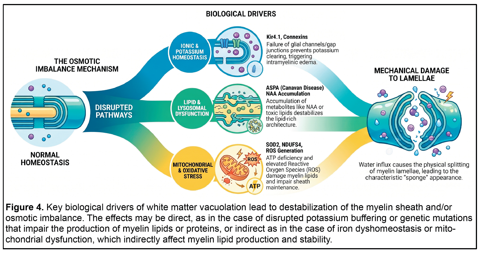
Within the CNS, K+ homeostasis influences the electrical excitability of neurons and osmotic stability of glial cells [155]. Loss of Kir4.1 function leads to the accumulation of K+ in the periaxonal space, creating an osmotic gradient that leads to the influx of water, vacuole formation, and splitting of lamellae of the myelin sheath. When gap junction communications are impaired, potassium buffering is also disrupted, and K+ ions entering oligodendrocytes during neuronal activity cannot be redistributed to the astrocyte network or moved radially out of the myelin layers. As a result, K+ and metabolic waste normally transferred to astrocytes accumulate within the cell body of oligodendrocytes as well as the myelin lamellae, which disrupts the osmotic balance and eventually impairs K+ uptake from the periaxonal space, leading to vacuole formation and splitting of the myelin sheath. The accumulation of K+ also impairs neuronal membrane repolarization and can induce tonic firing and trigger seizures. Loss of Cx47 also disrupts the transfer of nutrients essential for the production of myelin, such as lactate, from astrocytes to oligodendrocytes, further destabilizing myelin. The more severe phenotype observed in Cx32 deficient mice haploinsufficient for Cx47 and Kir4.1 most likely reflects impaired K+ handling at multiple steps.
6. Direct Connections Between Spongiosis and Myelin Proteins
The two major protein constituents of mammalian CNS myelin are proteolipid protein 1 (PLP1) and MBP, with other proteins, including 2′:3′-cyclic nucleotide-3′-phosphodiesterase (CNP), and glycoproteins such as myelin associated glycoprotein (MAG) and myelin-oligodendrocyte glycoprotein (MOG) present at lower concentrations [156]. These proteins are necessary for proper compaction and structural integrity of the myelin sheath and if they are absent or reduced, vacuoles may form as intramyelinic fluid-filled spaces that develop when myelin layers separate.
6.1. Proteolipid Protein 1 (PLP1)
Mutations in the gene encoding PLP1 cause X-linked myelin disorders ranging from severe Pelizaeus-Merzbacher disease (PMD) to milder Spastic Paraplegia Type 2 (SPG2) [157,158,159]. PMD is a severe, often fatal, infantile-onset leukodystrophy characterized by nystagmus, hypotonia (low muscle tone), and severe motor and cognitive impairment due to extensive hypomyelination. At the histopathological level, PMD-associated myelin defects include myelin sheath decompaction, vacuolation, and splitting [160]. PLP1 is the main protein component of CNS myelin and is expressed as two different isoforms, PLP1 and DM20, which use alternative splice donor sites in exon 3, joining the same acceptor site for exon 4 [161,162]. DM20 is more abundant prior to the onset of myelination, at which time the PLP1 isoform becomes the predominant form. The protein encoded by the PLP1 transcript is 35 amino acids longer than the one encoded by DM20, and the relative abundance of DM20 and PLP1 is important for myelin stability. Interestingly, given the possible mechanisms contributing to CNS vacuolation discussed elsewhere in this review, PLP1 is not only a primary component of the myelin sheath, but the extra 35 amino acids in PLP1 (absent from DM20) encode an intracellular loop that is essential for translocation of PLP1 into mitochondria, which results in increased mitochondrial mass and reduced oxidative phosphorylation [163,164,165]. Approximately 70% of PMD-associated PLP1 mutations involve duplication of the entire PLP1 gene, and the resulting over-expression of PLP1 leads to its misfolding and accumulation. This overloads the ER machinery and causes ER stress and activates the unfolded protein response, disrupting myelin formation and promoting oligodendrocyte apoptosis. ER stress also increases the interaction between the ER and mitochondria, which (also) leads to mitochondrial dysfunction [166,167,168,169,170,171].
6.2. Transcriptional Regulation of Myelination
Rare mutations in the gene encoding SRY-box transcription factor 10 (SOX10) result in central and peripheral de/dys-myelinating disease in humans (PCWH) [172,173,174,175], but it is not clear that the CNS phenotype is associated with vacuolation. In mice, however, there is evidence that disruption of SOX10 leads to spongiform encephalopathy. Specifically, mice homozygous for the Sox10gt (gray tremor) mutation exhibit hypo/dys-myelination of the CNS and severe vacuolation of white matter by 3 weeks of age (Figure 3), and the onset of vacuolation correlates with the initiation of CNS myelination [176,177].
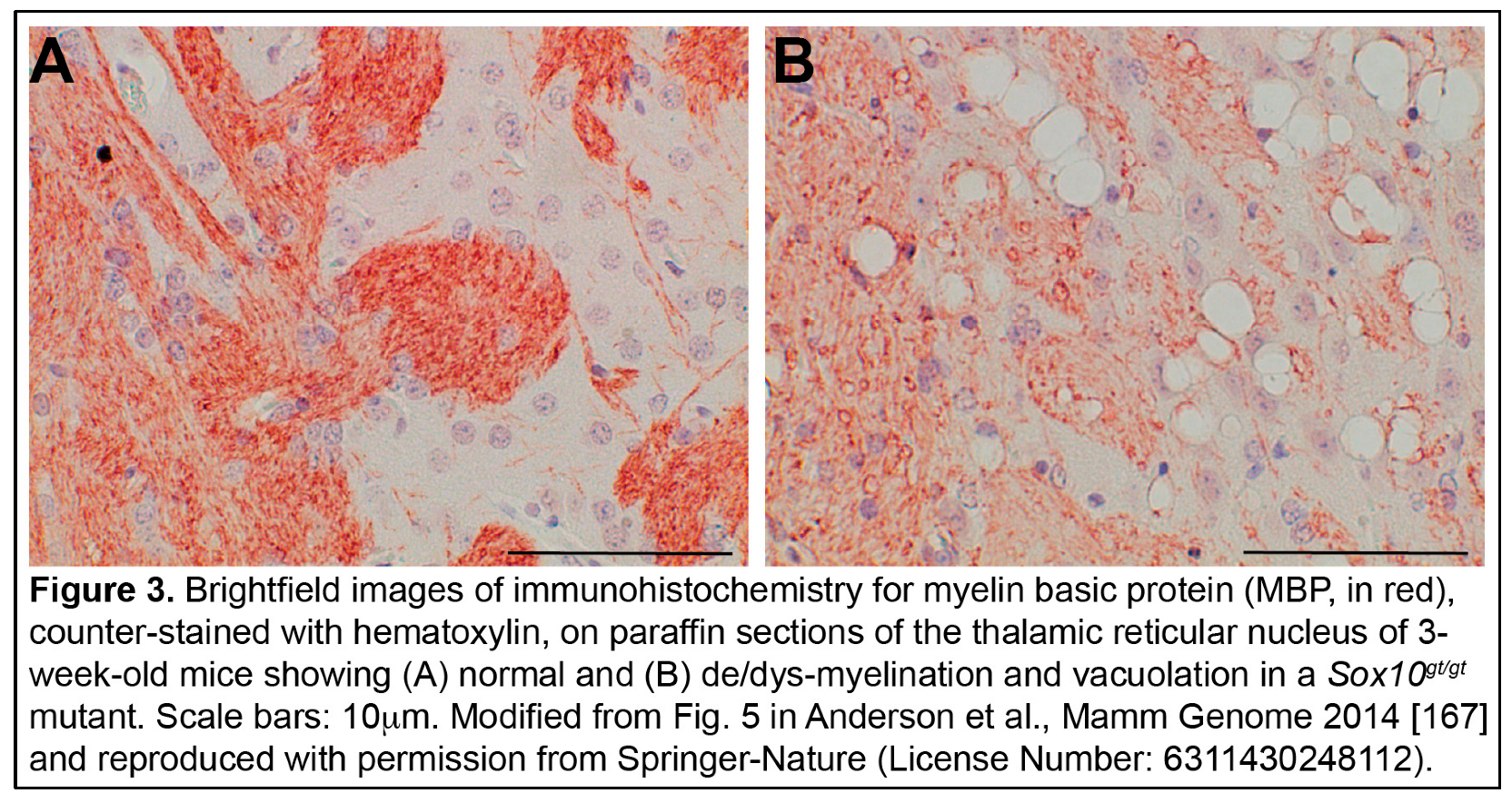
The Sox10gt mutation is a point mutation that changes a highly conserved glutamic acid in the dimerization domain to glycine [176]. The dimerization domain is unique to members of the SOXE family (SOX8, 9 and 10), and promotes stable binding of SOXE hetero- or homo-dimers to DNA to activate or repress target gene expression [178,179]. It has been shown that a single intact dimerization within a SOXE dimer pair is sufficient to stabilize dimeric binding to target sequences [180], which likely explains the recessive nature of the Sox10gt mutation and suggests that SOX10 homodimers, specifically, play a critical role in OL maturation and myelination. Genes with putative SOX10 homodimer binding sites in their promoters include PLP1, MBP, GJC2/CX47, Ugt8, claudin 11 (CLDN11), CNP, and myelin regulatory factor (MYRF). PLP1, MBP and CNP encode essential myelin components, and it is worth noting that a mutation in a SOX10 binding site in intron 3 of PLP1 disrupts long-range regulatory interactions and affects alternative splicing to alter the balance of DM20:PLP1, leading to mild PMD [181,182,183]. GJC2 encodes the Cx47 gap junction protein, connecting SOX10 to pathways discussed in Section 5 of this review (Potassium Homeostasis). CLDN11 encodes a critical tight junction protein that forms the radial component of myelin and acts as a diffusion barrier; when CLDN11 is absent, the myelin laminar structure is more readily disrupted by water [184,185,186]. CLDN11 deficiency causes hypomyelinating leukodystrophy (HLD22) but is not associated with vacuolation [187]. MYRF is a transcription factor that interacts with SOX10 to regulate expression of many essential myelin genes, thus playing a key role in oligodendrocyte maturation and myelin maintenance [188,189,190,191,192]. In humans, rare autosomal dominant mutations in MYRF cause MYRF- related mild encephalopathy with reversible myelin vacuolization (MMERV), which is associated with transient CNS myelin vacuolation [193,194]. Similarly, Myrf deletion in adult mouse oligodendrocytes caused severe CNS demyelination and myelin vacuolation [192].
6.3. Mechanistic Insights
Mutations in SOX10 or MYRF disrupt the transcriptional regulation of proteins and channels, including PLP1, that are essential for myelin structure and ion homeostasis. As discussed in section 5, mutations that compromise the “tight seal” of the myelin lamellae are predicted to disrupt spatial buffering of K+, creating an osmotic gradient that draws water into the myelin sheath. This leads to physical separation of the lamellae, leading to intramyelinic edema or vacuoles.
7. Conclusions
Spongiform degeneration of white matter is a pathological feature observed in numerous CNS disorders. While traditionally regarded as a downstream consequence of neuronal injury, accumulating evidence suggests that white matter vacuolation reflects a structural failure in myelin. This review has presented evidence to support this hypothesis, using representative examples spanning a broad spectrum of neurological conditions, including prion diseases, lysosomal storage disorders, and mitochondrial encephalopathies, as well as mouse genetic models that exhibit spongiform encephalopathy. Taken together, these examples demonstrate that vacuolation often originates within the myelin sheath and can be driven by intrinsic oligodendrocyte defects or disrupted axon-glial communication. The diverse genetic and environmental triggers discussed here converge on a shared pathological endpoint of metabolic and ionic dyshomeostasis, which leads to osmotic swelling within the myelin lamellae to form vacuoles (Figure 4). Vacuoles, in turn, cause mechanical damage to myelin that can lead to de/dys-myelination as well as impaired axonal conduction.
Clinically, the shift in perspective proposed in this review reframes status spongiosis from an irreversible marker of neuronal loss to a dynamic target for therapeutic intervention, and the reversible nature of MMERV suggests that this pathology may be transient or potentially reversible if the underlying metabolic or ionic balance can be restored. Diagnostic strategies should increasingly view white matter vacuolation as a signature of disrupted myelin formation or maintenance. Further research is needed, and this review identifies several areas of potential focus. These include: dissecting the potential role of PrPC in CNS myelination; elucidating the full regulatory scope of the SOX10 and MYRF network(s) to identify downstream targets; and defining how mitochondrial ROS and impaired iron-sulfur cluster biogenesis intersect with the high iron requirements of oligodendrocytes to destabilize myelin. By prioritizing the preservation of oligodendrocyte health and stabilization of the periaxonal environment, we envision moving toward therapies that directly address the structural and functional integrity underlying structural failures of the CNS.
Author Contributions
Both authors contributed to researching and writing the original draft, review, and editing; conceptualization, T.M.G.; visualization, T.M.G. All authors have read and agreed to the published version of the manuscript.
Funding
T.M.G. was supported in part by the National Institute of General Medical Sciences of the National Institutes of Health under Award Number P20GM152335. The APC was funded by Touro College of Osteopathic Medicine - Montana.
Institutional Review Board Statement
Not applicable.
Informed Consent Statement
Not applicable.
Data Availability Statement
Not applicable.
Acknowledgments
During the preparation of this manuscript, the authors used images generated by Google Gemini (June 2026) and NotebookLM (June 2026) to help generate Figures 2 and 4, respectively. The authors have reviewed and edited the output and take full responsibility for the content of this publication.
Conflicts of Interest
The authors declare no conflicts of interest.
Abbreviations
The following abbreviations are used in this manuscript:
| CNS | Central nervous system |
| PNS | Peripheral nervous system |
| TSE | Transmissible spongiform encephalopathy |
| CJD | Creutzfeldt-Jakob disease |
| PrPSc | Prion protein, scrapie form |
| PrPC | Prion protein, normal cellular form |
| Prnp | Mouse prion protein gene |
| ADGRG6 | Adhesion G-protein coupled receptor 6 |
| cAMP | Cyclic adenosine monophosphate |
| PKA | Protein kinase A |
| Egr2 | Early growth response 2 gene |
| MBP | Myelin basic protein |
| MPZ | Myelin protein zero |
| PMP22 | Peripheral myelin protein 22 |
| Fe3+ | Ferric iron |
| Fe2+ | Ferrous iron |
| ER | Endoplasmic reticulum |
| LSD | Lysosomal storage disease |
| GalC | Galactolipid galactocerbroside |
| UGT8 | UDP-galactose:ceramide galactosyltransferase |
| UDP | Uridine diphosphate |
| 4-AP | 4-aminopyridine |
| GAL3ST1 | Galactose-3-O-sulfotransferase 1 |
| CST | Cerebroside sulfotransferase |
| Cers2 | Ceramide synthase 2 gene |
| ASPA | Aspartoacylase gene |
| ASPA | Aspartoacylase (protein) |
| NAA | N-acetyl-L-aspartate |
| PNPLA6 | Patatin-like phospholipase domain-containing protein |
| NTE | Neuropathy target esterase |
| LPC | Lysophosphatidylcholine |
| LPE | Lysophosphatidylethanolamine |
| Kir | Inward rectifying potassium channels |
| K+ | Potassium ion(s) |
| sws | Swiss-Cheese gene (Drosophila) |
| ROS | Reactive oxygen species |
| SPG39 | Hereditary spastic paraplegia type 39 |
| Fig4/FIG4 | Factor-induced gene 4 (mouse/human) |
| FIG4 | Factor-induced gene 4 protein |
| VAC14 | Vac14 homolog (S. cerevisiae) protein |
| PIKFYVE | Phosphoinositide kinase, FYVE-type zinc finger containing |
| FAB1 | PIKFYVE in mammals |
| PI(3,5)P2 | Phosphatidylinositol 3,5-bisphosphate |
| LAMP1 | Lysosome-associated membrane protein 1 |
| RAB7 | RAB7, member RAS oncogene family |
| MAG | Myelin associated glycoprotein |
| PLP1 | Proteolipid protein |
| CMT4J | Charcot-Marie-Tooth disease, type 4J |
| MLIV | Mucolipidosis type IV |
| MCOLN1 | Mucolipin-1 gene |
| TRPML1 | Transient receptor potential mucolipin 1 |
| TMEM106B | Transmembrane protein 106B |
| ESCRT | Endosomal sorting complex required for transport |
| Tsg101 | Tumor susceptibility gene 101 (mouse) |
| TSG101 | Tumor susceptibility gene 101 protein |
| NCOA4 | Nuclear receptor coactivator 4 |
| NDUFS4 | NADH:ubiquinone oxidoreductase subunit S4 |
| NADH | Nicotinamide adenine dinucleotide |
| TLE | Toxic spongiform leukoencephalopathy |
| Sod2 | Superoxide dismutase 2 gene (mouse) |
| SOD2 | Superoxide dismutase 2 protein |
| Nfe2l2 | Nuclear factor, erythroid derived 2, like 2 gene (mouse) |
| Nrf2 | Alternate name for Nfe2l2 |
| PGC-1a | Peroxisome proliferator-activated receptor gamma coactivator 1-alpha |
| NRF1 | Nuclear respiratory factor 1 |
| ATP | Adenosine triphosphate |
| MGRN1 | Mahogunin, ring finger 1 |
| ATRN | Attractin |
| Mgrn1 | Mahogunin, ring finger 1 gene (mouse) |
| DMT1 | Divalent metal transporter 1 |
| SLC11A2 | solute carrier family 11 (proton-coupled divalent metal ion transporters), member 2 |
| Kir4.1 | inwardly rectifying potassium channel encoded by the KCNJ10 gene |
| KCNJ10 | potassium inwardly-rectifying channel, subfamily J, member 10 gene (human) |
| Kcnj10 | potassium inwardly-rectifying channel, subfamily J, member 10 gene (mouse) |
| EAST | Epilepsy, Ataxia, Sensorineural deafness, and Tubulopathy |
| SeSAME | Seizures, Sensorineural deafness, Ataxia, Mental retardation, and Electrolyte imbalance (alternate name for EAST syndrome) |
| Cx | Connexin |
| GJC2 | Gap junction protein, gamma 2 gene (human), encodes Cx47 |
| PMLD1 | Pelizaeus-Merzbacher-like disease 1 |
| GJB1 | Gap junction protein, beta 1 gene (human), encodes Cx32 |
| CMT1X | Charcot-Marie-Tooth disease, type 1X |
| Gjc2 | Gap junction protein, gamma 2 gene (mouse) |
| Gjb1 | Gap junction protein, beta 1 gene (mouse) |
| CNP | 2':3'-cyclic nucleotide-3'-phosphodiesterase |
| PMD | Pelizaeus-Merzbacher disease |
| SPG2 | Spastic paraplegia type 2 |
| DM20 | Diphasic myelin transcript (alternatively spliced isoform of PLP1) |
| SOX10 | SRY (sex determining region Y)-box 10 |
| PCWH | Peripheral demyelinating neuropathy, Central dysmyelinating leukodystrophy, Waardenburg syndrome, and Hirschsprung disease |
| gt | gray tremor (mouse Sox10 mutant allele) |
| SOXE | SRY (sex determining region Y)-box family E |
| Cldn11 | Claudin-11 gene (mouse) |
| Myrf | Myelin regulatory factor gene (mouse) |
| MYRF | Myelin regulatory factor protein |
| HLD22 | Hypomyelinating leukodystrophy |
| MMERV | MYRF-related mild encephalopathy with reversible myelin vacuolization |
References
- Silvius, D.; Pitstick, R.; Ahn, M.; Meishery, D.; Oehler, A.; Barsh, G.S.; DeArmond, S.J.; Carlson, G.A.; Gunn, T.M. Levels of the Mahogunin Ring Finger 1 E3 Ubiquitin Ligase Do Not Influence Prion Disease. PLOS ONE 2013, 8, e55575. [CrossRef]
- Simons, M.; Nave, K.-A. Oligodendrocytes: Myelination and Axonal Support. Cold Spring Harb. Perspect. Biol. 2015, 8, a020479. [CrossRef]
- Rasband, M.N.; Macklin, W.B. Chapter 10 - Myelin Structure and Biochemistry. In Basic Neurochemistry (Eighth Edition); Brady, S.T., Siegel, G.J., Albers, R.W., Price, D.L., Eds.; Academic Press: New York, 2012; pp. 180–199 ISBN 978-0-12-374947-5.
- Moore, R.A.; Taubner, L.M.; Priola, S.A. Prion Protein Misfolding and Disease. Curr. Opin. Struct. Biol. 2009, 19, 14–22. [CrossRef]
- Prusiner, S.B. Novel Proteinaceous Infectious Particles Cause Scrapie. Science 1982, 216, 136–144. [CrossRef]
- Hadlow, W.J. Reflections on the Transmissible Spongiform Encephalopathies. Vet. Pathol. 1999, 36, 523–529. [CrossRef]
- Liberski, P.P. Spongiform Change--an Electron Microscopic View. Folia Neuropathol. 2004, 42 Suppl B, 59–70.
- Caverzasi, E.; Mandelli, M.L.; DeArmond, S.J.; Hess, C.P.; Vitali, P.; Papinutto, N.; Oehler, A.; Miller, B.L.; Lobach, I.V.; Bastianello, S.; et al. White Matter Involvement in Sporadic Creutzfeldt-Jakob Disease. Brain 2014, 137, 3339–3354. [CrossRef]
- Matsusue, E.; Kinoshita, T.; Sugihara, S.; Fujii, S.; Ogawa, T.; Ohama, E. White Matter Lesions in Panencephalopathic Type of Creutzfeldt-Jakob Disease: MR Imaging and Pathologic Correlations. AJNR Am. J. Neuroradiol. 2004, 25, 910–918.
- Reiniger, L.; Mirabile, I.; Lukic, A.; Wadsworth, J.D.; Linehan, J.M.; Groves, M.; Lowe, J.; Druyeh, R.; Rudge, P.; Collinge, J.; et al. Filamentous White Matter Prion Protein Deposition Is a Distinctive Feature of Multiple Inherited Prion Diseases. Acta Neuropathol. Commun. 2013, 1, 8. [CrossRef]
- Sasaki, S.; Mizoi, S.; Akashima, A.; Shinagawa, M.; Goto, H. Spongiform Encephalopathy in Sheep Scrapie : Electron Microscopic Observations. Jpn. J. Vet. Sci. 1986, 48, 791–796. [CrossRef]
- Liberski, P.P.; Yanagihara, R.; Wells, G.A.H.; Gibbs, C.J.; Gajdusek, D.C. Ultrastructural Pathology of Axons and Myelin in Experimental Scrapie in Hamsters and Bovine Spongiform Encephalopathy in Cattle and a Comparison with the Panencephalopathic Type of Creutzfeldt-Jakob Disease. J. Comp. Pathol. 1992, 106, 383–398. [CrossRef]
- Liu, X.-L.; Feng, X.-L.; Wang, G.-M.; Gong, B.-B.; Ahmad, W.; Liu, N.-N.; Zhang, Y.-Y.; Yang, L.; Ren, H.-L.; Cui, S.-S. Exploration of the Main Sites for the Transformation of Normal Prion Protein (PrPC) into Pathogenic Prion Protein (PrPsc). J. Vet. Res. 2017, 61, 11–22. [CrossRef]
- Reiniger, L.; Mirabile, I.; Lukic, A.; Wadsworth, J.D.; Linehan, J.M.; Groves, M.; Lowe, J.; Druyeh, R.; Rudge, P.; Collinge, J.; et al. Filamentous White Matter Prion Protein Deposition Is a Distinctive Feature of Multiple Inherited Prion Diseases. Acta Neuropathol. Commun. 2013, 1, 8. [CrossRef]
- Prinz, M.; Montrasio, F.; Furukawa, H.; Haar, M.E. van der; Schwarz, P.; Rülicke, T.; Giger, O.T.; Häusler, K.-G.; Perez, D.; Glatzel, M.; et al. Intrinsic Resistance of Oligodendrocytes to Prion Infection. J. Neurosci. 2004, 24, 5974–5981. [CrossRef]
- Moser, M.; Colello, R.J.; Pott, U.; Oesch, B. Developmental Expression of the Prion Protein Gene in Glial Cells. Neuron 1995, 14, 509–517. [CrossRef]
- Nazor, K.E.; Seward, T.; Telling, G.C. Motor Behavioral and Neuropathological Deficits in Mice Deficient for Normal Prion Protein Expression. Biochim. Biophys. Acta BBA - Mol. Basis Dis. 2007, 1772, 645–653. [CrossRef]
- Bremer, J.; Baumann, F.; Tiberi, C.; Wessig, C.; Fischer, H.; Schwarz, P.; Steele, A.D.; Toyka, K.V.; Nave, K.-A.; Weis, J.; et al. Axonal Prion Protein Is Required for Peripheral Myelin Maintenance. Nat. Neurosci. 2010, 13, 310–318. [CrossRef]
- Zhu, C.; Li, B.; Yu, G.; Chen, J.; Yu, H.; Chen, J.; Xu, X.; Wu, Y.; Zhang, A.; Cheng, G. Production of Prnp-/- Goats by Gene Targeting in Adult Fibroblasts. Transgenic Res. 2009, 18, 163–171. [CrossRef]
- Richt, J.A.; Kasinathan, P.; Hamir, A.N.; Castilla, J.; Sathiyaseelan, T.; Vargas, F.; Sathiyaseelan, J.; Wu, H.; Matsushita, H.; Koster, J.; et al. Production of Cattle Lacking Prion Protein. Nat. Biotechnol. 2007, 25, 132–138. [CrossRef]
- Skedsmo, F.S.; Malachin, G.; Våge, D.I.; Hammervold, M.M.; Salvesen, Ø.; Ersdal, C.; Ranheim, B.; Stafsnes, M.H.; Bartosova, Z.; Bruheim, P.; et al. Demyelinating Polyneuropathy in Goats Lacking Prion Protein. FASEB J. Off. Publ. Fed. Am. Soc. Exp. Biol. 2020, 34, 2359–2375. [CrossRef]
- Nuvolone, M.; Hermann, M.; Sorce, S.; Russo, G.; Tiberi, C.; Schwarz, P.; Minikel, E.; Sanoudou, D.; Pelczar, P.; Aguzzi, A. Strictly Co-Isogenic C57BL/6J-Prnp−/− Mice: A Rigorous Resource for Prion Science. J. Exp. Med. 2016, 213, 313–327. [CrossRef]
- Küffer, A.; Lakkaraju, A.K.K.; Mogha, A.; Petersen, S.C.; Airich, K.; Doucerain, C.; Marpakwar, R.; Bakirci, P.; Senatore, A.; Monnard, A.; et al. The Prion Protein Is an Agonistic Ligand of the G Protein-Coupled Receptor Adgrg6. Nature 2016, 536, 464–468. [CrossRef]
- Mogha, A.; Benesh, A.E.; Patra, C.; Engel, F.B.; Schöneberg, T.; Liebscher, I.; Monk, K.R. Gpr126 Functions in Schwann Cells to Control Differentiation and Myelination via G-Protein Activation. J. Neurosci. 2013, 33, 17976–17985. [CrossRef]
- Glenn, T.D.; Talbot, W.S. Analysis of Gpr126 Function Defines Distinct Mechanisms Controlling the Initiation and Maturation of Myelin. Development 2013, 140, 3167–3175. [CrossRef]
- Brown, D.R.; Qin, K.; Herms, J.W.; Madlung, A.; Manson, J.; Strome, R.; Fraser, P.E.; Kruck, T.; von Bohlen, A.; Schulz-Schaeffer, W.; et al. The Cellular Prion Protein Binds Copper in Vivo. Nature 1997, 390, 684–687. [CrossRef]
- MILLHAUSER, G.L. Copper Binding in the Prion Protein. Acc. Chem. Res. 2004, 37, 79–85. [CrossRef]
- Singh, A.; Haldar, S.; Horback, K.; Tom, C.; Zhou, L.; Meyerson, H.; Singh, N. Prion Protein Regulates Iron Transport by Functioning as a Ferrireductase. J. Alzheimers Dis. JAD 2013, 35, 541–552. [CrossRef]
- Connor, J.R.; Menzies, S.L. Relationship of Iron to Oligodendrocytes and Myelination. Glia 1996, 17, 83–93. [CrossRef]
- Cheli, V.T.; Correale, J.; Paez, P.M.; Pasquini, J.M. Iron Metabolism in Oligodendrocytes and Astrocytes, Implications for Myelination and Remyelination. ASN Neuro 2020, 12, 1759091420962681. [CrossRef]
- Singh, A.; Mohan, M.L.; Isaac, A.O.; Luo, X.; Petrak, J.; Vyoral, D.; Singh, N. Prion Protein Modulates Cellular Iron Uptake: A Novel Function with Implications for Prion Disease Pathogenesis. PloS One 2009, 4, e4468. [CrossRef]
- Gasperini, L.; Meneghetti, E.; Legname, G.; Benetti, F. In Absence of the Cellular Prion Protein, Alterations in Copper Metabolism and Copper-Dependent Oxidase Activity Affect Iron Distribution. Front. Neurosci. 2016, 10. [CrossRef]
- Yang, D.; Wang, X.; Zhang, L.; Fang, Y.; Zheng, Q.; Liu, X.; Yu, W.; Chen, S.; Ying, J.; Hua, F. Lipid Metabolism and Storage in Neuroglia: Role in Brain Development and Neurodegenerative Diseases. Cell Biosci. 2022, 12, 106. [CrossRef]
- Lipids as Emerging Biomarkers in Neurodegenerative Diseases Available online: https://www.mdpi.com/1422-0067/25/1/131 (accessed on 19 June 2026).
- Duncan, G.J.; Simkins, T.J.; Emery, B. Neuron-Oligodendrocyte Interactions in the Structure and Integrity of Axons. Front. Cell Dev. Biol. 2021, 9. [CrossRef]
- Marshall-Phelps, K.L.H.; Almeida, R.G. Axonal Neurotransmitter Release in the Regulation of Myelination. Biosci. Rep. 2024, 44, BSR20231616. [CrossRef]
- Brown, M.S.; Goldstein, J.L. Familial Hypercholesterolemia: Defective Binding of Lipoproteins to Cultured Fibroblasts Associated with Impaired Regulation of 3-Hydroxy-3-Methylglutaryl Coenzyme A Reductase Activity. Proc. Natl. Acad. Sci. U. S. A. 1974, 71, 788–792. [CrossRef]
- Singh, R.; Kaushik, S.; Wang, Y.; Xiang, Y.; Novak, I.; Komatsu, M.; Tanaka, K.; Cuervo, A.M.; Czaja, M.J. Autophagy Regulates Lipid Metabolism. Nature 2009, 458, 1131–1135. [CrossRef]
- Ebner, M.; Fröhlich, F.; Haucke, V. Mechanisms and Functions of Lysosomal Lipid Homeostasis. Cell Chem. Biol. 2025, 32, 392–407. [CrossRef]
- Bosio, A.; Büssow, H.; Adam, J.; Stoffel, W. Galactosphingolipids and Axono-Glial Interaction in Myelin of the Central Nervous System. Cell Tissue Res. 1998, 292, 199–210. [CrossRef]
- Coetzee, T.; Fujita, N.; Dupree, J.; Shi, R.; Blight, A.; Suzuki, K.; Suzuki, K.; Popko, B. Myelination in the Absence of Galactocerebroside and Sulfatide: Normal Structure with Abnormal Function and Regional Instability. Cell 1996, 86, 209–219. [CrossRef]
- Marcus, J.; Dupree, J.L.; Popko, B. Myelin-Associated Glycoprotein and Myelin Galactolipids Stabilize Developing Axo-Glial Interactions. J. Cell Biol. 2002, 156, 567–577. [CrossRef]
- Dupree, J.L.; Coetzee, T.; Blight, A.; Suzuki, K.; Popko, B. Myelin Galactolipids Are Essential for Proper Node of Ranvier Formation in the CNS. J. Neurosci. 1998, 18, 1642–1649. [CrossRef]
- Honke, K.; Hirahara, Y.; Dupree, J.; Suzuki, K.; Popko, B.; Fukushima, K.; Fukushima, J.; Nagasawa, T.; Yoshida, N.; Wada, Y.; et al. Paranodal Junction Formation and Spermatogenesis Require Sulfoglycolipids. Proc. Natl. Acad. Sci. 2002, 99, 4227–4232. [CrossRef]
- Sun, H.; Mo, L.; Cao, M.; Tian, Y.; Mo, L.; Ni, Z.; Zhang, S.; Huang, X.; Wang, Y.; Lam, S.M.; et al. Very Long Chain Sphingolipids Govern Brain Myelination by Regulating Oligodendrocyte Differentiation and Membrane Microdomain Integrity. J. Transl. Med. 2026, 24, 550. [CrossRef]
- Ben-David, O.; Pewzner-Jung, Y.; Brenner, O.; Laviad, E.L.; Kogot-Levin, A.; Weissberg, I.; Biton, I.E.; Pienik, R.; Wang, E.; Kelly, S.; et al. Encephalopathy Caused by Ablation of Very Long Acyl Chain Ceramide Synthesis May Be Largely Due to Reduced Galactosylceramide Levels. J. Biol. Chem. 2011, 286, 30022–30033. [CrossRef]
- Baslow, M.H. Canavan’s Spongiform Leukodystrophy: A Clinical Anatomy of a Genetic Metabolic CNS Disease. J. Mol. Neurosci. MN 2000, 15, 61–69. [CrossRef]
- Kirmani, B.F.; Jacobowitz, D.M.; Namboodiri, M. a. A. Developmental Increase of Aspartoacylase in Oligodendrocytes Parallels CNS Myelination. Brain Res. Dev. Brain Res. 2003, 140, 105–115. [CrossRef]
- Baslow, M.H.; Suckow, R.F.; Sapirstein, V.; Hungund, B.L. Expression of Aspartoacylase Activity in Cultured Rat Macroglial Cells Is Limited to Oligodendrocytes. J. Mol. Neurosci. MN 1999, 13, 47–53. [CrossRef]
- Madhavarao, C.N.; Moffett, J.R.; Moore, R.A.; Viola, R.E.; Namboodiri, M.A.A.; Jacobowitz, D.M. Immunohistochemical Localization of Aspartoacylase in the Rat Central Nervous System. J. Comp. Neurol. 2004, 472, 318–329. [CrossRef]
- Matalon, R.; Michals, K.; Sebesta, D.; Deanching, M.; Gashkoff, P.; Casanova, J. Aspartoacylase Deficiency and N-Acetylaspartic Aciduria in Patients with Canavan Disease. Am. J. Med. Genet. 1988, 29, 463–471. [CrossRef]
- Surendran, S.; Campbell, G.A.; Tyring, S.K.; Matalon, R. Aspartoacylase Gene Knockout Results in Severe Vacuolation in the White Matter and Gray Matter of the Spinal Cord in the Mouse. Neurobiol. Dis. 2005, 18, 385–389. [CrossRef]
- Traka, M.; Wollmann, R.L.; Cerda, S.R.; Dugas, J.; Barres, B.A.; Popko, B. Nur7 Is a Nonsense Mutation in the Mouse Aspartoacylase Gene That Causes Spongy Degeneration of the CNS. J. Neurosci. Off. J. Soc. Neurosci. 2008, 28, 11537–11549. [CrossRef]
- Chakraborty, G.; Mekala, P.; Yahya, D.; Wu, G.; Ledeen, R.W. Intraneuronal N-Acetylaspartate Supplies Acetyl Groups for Myelin Lipid Synthesis: Evidence for Myelin-Associated Aspartoacylase. J. Neurochem. 2001, 78, 736–745. [CrossRef]
- Mehta, V.; Namboodiri, M.A. N-Acetylaspartate as an Acetyl Source in the Nervous System. Brain Res. Mol. Brain Res. 1995, 31, 151–157. [CrossRef]
- Baslow, M.H.; Guilfoyle, D.N. Canavan Disease, a Rare Early-Onset Human Spongiform Leukodystrophy: Insights into Its Genesis and Possible Clinical Interventions. Biochimie 2013, 95, 946–956. [CrossRef]
- Hoshino, H.; Kubota, M. Canavan Disease: Clinical Features and Recent Advances in Research. Pediatr. Int. Off. J. Jpn. Pediatr. Soc. 2014, 56, 477–483. [CrossRef]
- Madhavarao, C.N.; Arun, P.; Moffett, J.R.; Szucs, S.; Surendran, S.; Matalon, R.; Garbern, J.; Hristova, D.; Johnson, A.; Jiang, W.; et al. Defective N-Acetylaspartate Catabolism Reduces Brain Acetate Levels and Myelin Lipid Synthesis in Canavan’s Disease. Proc. Natl. Acad. Sci. 2005, 102, 5221–5226. [CrossRef]
- Wei, H.; Moffett, J.R.; Amanat, M.; Fatemi, A.; Tsukamoto, T.; Namboodiri, A.M.; Slusher, B.S. The Pathogenesis of, and Pharmacological Treatment for, Canavan Disease. Drug Discov. Today 2022, 27, 2467–2483. [CrossRef]
- Francis, J.S.; Strande, L.; Markov, V.; Leone, P. Aspartoacylase Supports Oxidative Energy Metabolism during Myelination. J. Cereb. Blood Flow Metab. Off. J. Int. Soc. Cereb. Blood Flow Metab. 2012, 32, 1725–1736. [CrossRef]
- Singhal, N.K.; Huang, H.; Li, S.; Clements, R.; Gadd, J.; Daniels, A.; Kooijman, E.E.; Bannerman, P.; Burns, T.; Guo, F.; et al. The Neuronal Metabolite NAA Regulates Histone H3 Methylation in Oligodendrocytes and Myelin Lipid Composition. Exp. Brain Res. 2017, 235, 279–292. [CrossRef]
- Kretzschmar, D. PNPLA6/NTE, an Evolutionary Conserved Phospholipase Linked to a Group of Complex Human Diseases. Metabolites 2022, 12, 284. [CrossRef]
- Li, Y.; Dinsdale, D.; Glynn, P. Protein Domains, Catalytic Activity, and Subcellular Distribution of Neuropathy Target Esterase in Mammalian Cells. J. Biol. Chem. 2003, 278, 8820–8825. [CrossRef]
- Quistad, G.B.; Barlow, C.; Winrow, C.J.; Sparks, S.E.; Casida, J.E. Evidence That Mouse Brain Neuropathy Target Esterase Is a Lysophospholipase. Proc. Natl. Acad. Sci. U. S. A. 2003, 100, 7983–7987. [CrossRef]
- Zaccheo, O.; Dinsdale, D.; Meacock, P.A.; Glynn, P. Neuropathy Target Esterase and Its Yeast Homologue Degrade Phosphatidylcholine to Glycerophosphocholine in Living Cells. J. Biol. Chem. 2004, 279, 24024–24033. [CrossRef]
- Plemel, J.R.; Michaels, N.J.; Weishaupt, N.; Caprariello, A.V.; Keough, M.B.; Rogers, J.A.; Yukseloglu, A.; Lim, J.; Patel, V.V.; Rawji, K.S.; et al. Mechanisms of Lysophosphatidylcholine-Induced Demyelination: A Primary Lipid Disrupting Myelinopathy. Glia 2018, 66, 327–347. [CrossRef]
- Akassoglou, K.; Malester, B.; Xu, J.; Tessarollo, L.; Rosenbluth, J.; Chao, M.V. Brain-Specific Deletion of Neuropathy Target Esterase/Swisscheese Results in Neurodegeneration. Proc. Natl. Acad. Sci. U. S. A. 2004, 101, 5075–5080. [CrossRef]
- Rosenbluth, J.; Schiff, R.; Lam, P.; Nuriel, T.; Chao, M.V. Spongiform Pathology in Mouse CNS Lacking ‘Neuropathy Target Esterase’ and Cellular Prion Protein. Neurobiol. Dis. 2009, 35, 433–437. [CrossRef]
- Read, D.J.; Li, Y.; Chao, M.V.; Cavanagh, J.B.; Glynn, P. Neuropathy Target Esterase Is Required for Adult Vertebrate Axon Maintenance. J. Neurosci. 2009, 29, 11594–11600. [CrossRef]
- Ryabova, E.V.; Melentev, P.A.; Komissarov, A.E.; Surina, N.V.; Ivanova, E.A.; Matiytsiv, N.; Shcherbata, H.R.; Sarantseva, S.V. Morpho-Functional Consequences of Swiss Cheese Knockdown in Glia of Drosophila Melanogaster. Cells 2021, 10, 529. [CrossRef]
- Melentev, P.A.; Ryabova, E.V.; Surina, N.V.; Zhmujdina, D.R.; Komissarov, A.E.; Ivanova, E.A.; Boltneva, N.P.; Makhaeva, G.F.; Sliusarenko, M.I.; Yatsenko, A.S.; et al. Loss of Swiss Cheese in Neurons Contributes to Neurodegeneration with Mitochondria Abnormalities, Reactive Oxygen Species Acceleration and Accumulation of Lipid Droplets in Drosophila Brain. Int. J. Mol. Sci. 2021, 22, 8275. [CrossRef]
- Dutta, S.; Rieche, F.; Eckl, N.; Duch, C.; Kretzschmar, D. Glial Expression of Swiss Cheese (SWS), the Drosophila Orthologue of Neuropathy Target Esterase (NTE), Is Required for Neuronal Ensheathment and Function. Dis. Model. Mech. 2016, 9, 283–294. [CrossRef]
- Tsap, M.I.; Yatsenko, A.S.; Hegermann, J.; Beckmann, B.; Tsikas, D.; Shcherbata, H.R. Unraveling the Link between Neuropathy Target Esterase NTE/SWS, Lysosomal Storage Diseases, Inflammation, Abnormal Fatty Acid Metabolism, and Leaky Brain Barrier. eLife 2024, 13, e98020. [CrossRef]
- Mühlig-Versen, M.; da Cruz, A.B.; Tschäpe, J.-A.; Moser, M.; Büttner, R.; Athenstaedt, K.; Glynn, P.; Kretzschmar, D. Loss of Swiss Cheese/Neuropathy Target Esterase Activity Causes Disruption of Phosphatidylcholine Homeostasis and Neuronal and Glial Death in Adult Drosophila. J. Neurosci. Off. J. Soc. Neurosci. 2005, 25, 2865–2873. [CrossRef]
- Botelho, R.J.; Efe, J.A.; Teis, D.; Emr, S.D. Assembly of a Fab1 Phosphoinositide Kinase Signaling Complex Requires the Fig4 Phosphoinositide Phosphatase. Mol. Biol. Cell 2008, 19, 4273–4286. [CrossRef]
- Jin, N.; Chow, C.Y.; Liu, L.; Zolov, S.N.; Bronson, R.; Davisson, M.; Petersen, J.L.; Zhang, Y.; Park, S.; Duex, J.E.; et al. VAC14 Nucleates a Protein Complex Essential for the Acute Interconversion of PI3P and PI(3,5)P(2) in Yeast and Mouse. EMBO J. 2008, 27, 3221–3234. [CrossRef]
- Zolov, S.N.; Bridges, D.; Zhang, Y.; Lee, W.-W.; Riehle, E.; Verma, R.; Lenk, G.M.; Converso-Baran, K.; Weide, T.; Albin, R.L.; et al. In Vivo, Pikfyve Generates PI(3,5)P2, Which Serves as Both a Signaling Lipid and the Major Precursor for PI5P. Proc. Natl. Acad. Sci. U. S. A. 2012, 109, 17472–17477. [CrossRef]
- Lenk, G.M.; Meisler, M.H. Mouse Models of PI(3,5)P2 Deficiency with Impaired Lysosome Function. Methods Enzymol. 2014, 534, 245–260. [CrossRef]
- Mironova, Y.A.; Lenk, G.M.; Lin, J.-P.; Lee, S.J.; Twiss, J.L.; Vaccari, I.; Bolino, A.; Havton, L.A.; Min, S.H.; Abrams, C.S.; et al. PI(3,5)P2 Biosynthesis Regulates Oligodendrocyte Differentiation by Intrinsic and Extrinsic Mechanisms. eLife 2016, 5, e13023. [CrossRef]
- Chow, C.Y.; Zhang, Y.; Dowling, J.J.; Jin, N.; Adamska, M.; Shiga, K.; Szigeti, K.; Shy, M.E.; Li, J.; Zhang, X.; et al. Mutation of FIG4 Causes Neurodegeneration in the Pale Tremor Mouse and Patients with CMT4J. Nature 2007, 448, 68–72. [CrossRef]
- Ferguson, C.J.; Lenk, G.M.; Jones, J.M.; Grant, A.E.; Winters, J.J.; Dowling, J.J.; Giger, R.J.; Meisler, M.H. Neuronal Expression of Fig4 Is Both Necessary and Sufficient to Prevent Spongiform Neurodegeneration. Hum. Mol. Genet. 2012, 21, 3525–3534. [CrossRef]
- Simons, M.; Trajkovic, K. Neuron-Glia Communication in the Control of Oligodendrocyte Function and Myelin Biogenesis. J. Cell Sci. 2006, 119, 4381–4389. [CrossRef]
- Simons, M.; Trajkovic, K. Neuron-Glia Communication in the Control of Oligodendrocyte Function and Myelin Biogenesis. J. Cell Sci. 2006, 119, 4381–4389. [CrossRef]
- Winterstein, C.; Trotter, J.; Krämer-Albers, E.-M. Distinct Endocytic Recycling of Myelin Proteins Promotes Oligodendroglial Membrane Remodeling. J. Cell Sci. 2008, 121, 834–842. [CrossRef]
- Bassi, M.T.; Manzoni, M.; Monti, E.; Pizzo, M.T.; Ballabio, A.; Borsani, G. Cloning of the Gene Encoding a Novel Integral Membrane Protein, Mucolipidin-and Identification of the Two Major Founder Mutations Causing Mucolipidosis Type IV. Am. J. Hum. Genet. 2000, 67, 1110–1120. [CrossRef]
- Bargal, R.; Avidan, N.; Ben-Asher, E.; Olender, Z.; Zeigler, M.; Frumkin, A.; Raas-Rothschild, A.; Glusman, G.; Lancet, D.; Bach, G. Identification of the Gene Causing Mucolipidosis Type IV. Nat. Genet. 2000, 26, 118–123. [CrossRef]
- Raychowdhury, M.K.; González-Perrett, S.; Montalbetti, N.; Timpanaro, G.A.; Chasan, B.; Goldmann, W.H.; Stahl, S.; Cooney, A.; Goldin, E.; Cantiello, H.F. Molecular Pathophysiology of Mucolipidosis Type IV: pH Dysregulation of the Mucolipin-1 Cation Channel. Hum. Mol. Genet. 2004, 13, 617–627. [CrossRef]
- Rue, B.E.; Dischler, A.M.; Salvagio, L.A.; Zhu, M.; Xu, G.; Flores, P.C.; Donovan, C.L.; Liu, X.; Minckley, T.F.; Agulnek, B.; et al. Differential Ion Selectivity and Disease-Associated Dysfunction of TRPML Channels Revealed by Patient and Engineered Mutants. J. Biol. Chem. 2026, 302, 110953. [CrossRef]
- Colletti, G.A.; Kiselyov, K. TRPML1. Adv. Exp. Med. Biol. 2011, 704, 209–219. [CrossRef]
- Di Paola, S.; Scotto-Rosato, A.; Medina, D.L. TRPML1: The Ca(2+)Retaker of the Lysosome. Cell Calcium 2018, 69, 112–121. [CrossRef]
- Dong, X.-P.; Cheng, X.; Mills, E.; Delling, M.; Wang, F.; Kurz, T.; Xu, H. The Type IV Mucolipidosis-Associated Protein TRPML1 Is an Endolysosomal Iron Release Channel. Nature 2008, 455, 992–996. [CrossRef]
- Wang, W.; Zhang, X.; Gao, Q.; Xu, H. TRPML1: An Ion Channel in the Lysosome. Handb. Exp. Pharmacol. 2014, 222, 631–645. [CrossRef]
- Dong, X.; Shen, D.; Wang, X.; Dawson, T.; Li, X.; Zhang, Q.; Cheng, X.; Zhang, Y.; Weisman, L.S.; Delling, M.; et al. PI(3,5)P(2) Controls Membrane Trafficking by Direct Activation of Mucolipin Ca(2+) Release Channels in the Endolysosome. Nat. Commun. 2010, 1, 38. [CrossRef]
- Li, X.; Wang, X.; Zhang, X.; Zhao, M.; Tsang, W.L.; Zhang, Y.; Yau, R.G.W.; Weisman, L.S.; Xu, H. Genetically Encoded Fluorescent Probe to Visualize Intracellular Phosphatidylinositol 3,5-Bisphosphate Localization and Dynamics. Proc. Natl. Acad. Sci. 2013, 110, 21165–21170. [CrossRef]
- Dong, X.-P.; Wang, X.; Xu, H. TRP Channels of Intracellular Membranes. J. Neurochem. 2010, 113, 313–328. [CrossRef]
- Cao, Q.; Yang, Y.; Zhong, X.Z.; Dong, X.-P. The Lysosomal Ca2+ Release Channel TRPML1 Regulates Lysosome Size by Activating Calmodulin. J. Biol. Chem. 2017, 292, 8424–8435. [CrossRef]
- Wenger, D.A.; Rafi, M.A.; Luzi, P.; Datto, J.; Costantino-Ceccarini, E. Krabbe Disease: Genetic Aspects and Progress toward Therapy. Mol. Genet. Metab. 2000, 70, 1–9. [CrossRef]
- Feng, T.; Sheng, R.R.; Solé-Domènech, S.; Ullah, M.; Zhou, X.; Mendoza, C.S.; Enriquez, L.C.M.; Katz, I.I.; Paushter, D.H.; Sullivan, P.M.; et al. A Role of the Frontotemporal Lobar Degeneration Risk Factor TMEM106B in Myelination. Brain 2020, 143, 2255–2271. [CrossRef]
- Walker, W.P.; Oehler, A.; Edinger, A.L.; Wagner, K.-U.; Gunn, T.M. Oligodendroglial Deletion of ESCRT-I Component TSG101 Causes Spongiform Encephalopathy. Biol. Cell 2016, 108, 324–337. [CrossRef]
- Bache, K.G.; Brech, A.; Mehlum, A.; Stenmark, H. Hrs Regulates Multivesicular Body Formation via ESCRT Recruitment to Endosomes. J. Cell Biol. 2003, 162, 435–442. [CrossRef]
- Kaur, J.; Debnath, J. Autophagy at the Crossroads of Catabolism and Anabolism. Nat. Rev. Mol. Cell Biol. 2015, 16, 461–472. [CrossRef]
- Ruhoy, I.S.; Saneto, R.P. The Genetics of Leigh Syndrome and Its Implications for Clinical Practice and Risk Management. Appl. Clin. Genet. 2014, 7, 221–234. [CrossRef]
- Magro, G.; Laterza, V.; Tosto, F. Leigh Syndrome: A Comprehensive Review of the Disease and Present and Future Treatments. Biomedicines 2025, 13, 733. [CrossRef]
- van de Wal, M.A.E.; Adjobo-Hermans, M.J.W.; Keijer, J.; Schirris, T.J.J.; Homberg, J.R.; Wieckowski, M.R.; Grefte, S.; van Schothorst, E.M.; van Karnebeek, C.; Quintana, A.; et al. Ndufs4 Knockout Mouse Models of Leigh Syndrome: Pathophysiology and Intervention. Brain J. Neurol. 2022, 145, 45–63. [CrossRef]
- Quintana, A.; Kruse, S.E.; Kapur, R.P.; Sanz, E.; Palmiter, R.D. Complex I Deficiency Due to Loss of Ndufs4 in the Brain Results in Progressive Encephalopathy Resembling Leigh Syndrome. Proc. Natl. Acad. Sci. U. S. A. 2010, 107, 10996–11001. [CrossRef]
- Maisenbacher, M.; Arcot Jayagopal, L. Chasing the New Dragon. Neurology 2026, 106, e214517. [CrossRef]
- Wolters, E.C.; van Wijngaarden, G.K.; Stam, F.C.; Rengelink, H.; Lousberg, R.J.; Schipper, M.E.; Verbeeten, B. Leucoencephalopathy after Inhaling “Heroin” Pyrolysate. Lancet 1982, 2, 1233–1237. [CrossRef]
- Alshamam, M.S.; Sumbly, V.; Nso, N.; Saliaj, M.; Gurung, D.O. Heroin-Induced Leukoencephalopathy. Cureus 13, e13093. [CrossRef]
- Melov, S.; Schneider, J.A.; Day, B.J.; Hinerfeld, D.; Coskun, P.; Mirra, S.S.; Crapo, J.D.; Wallace, D.C. A Novel Neurological Phenotype in Mice Lacking Mitochondrial Manganese Superoxide Dismutase. Nat. Genet. 1998, 18, 159–163. [CrossRef]
- Hubbs, A.F.; Benkovic, S.A.; Miller, D.B.; O’Callaghan, J.P.; Battelli, L.; Schwegler-Berry, D.; Ma, Q. Vacuolar Leukoencephalopathy with Widespread Astrogliosis in Mice Lacking Transcription Factor Nrf2. Am. J. Pathol. 2007, 170, 2068–2076. [CrossRef]
- Weisiger, R.A.; Fridovich, I. Superoxide Dismutase. Organelle Specificity. J. Biol. Chem. 1973, 248, 3582–3592.
- He, F.; Ru, X.; Wen, T. NRF2, a Transcription Factor for Stress Response and Beyond. Int. J. Mol. Sci. 2020, 21, 4777. [CrossRef]
- Bronson, R.T.; Donahue, L.R.; Samples, R.; Kim, J.H.; Naggert, J.K. Mice with Mutations in the Mahogany Gene Atrn Have Cerebral Spongiform Changes. J. Neuropathol. Exp. Neurol. 2001, 60, 724–730. [CrossRef]
- He, L.; Lu, X.-Y.; Jolly, A.F.; Eldridge, A.G.; Watson, S.J.; Jackson, P.K.; Barsh, G.S.; Gunn, T.M. Spongiform Degeneration in Mahoganoid Mutant Mice. Science 2003, 299, 710–712. [CrossRef]
- He, L.; Gunn, T.M.; Bouley, D.M.; Lu, X.Y.; Watson, S.J.; Schlossman, S.F.; Duke-Cohan, J.S.; Barsh, G.S. A Biochemical Function for Attractin in Agouti-Induced Pigmentation and Obesity. Nat. Genet. 2001, 27, 40–47. [CrossRef]
- Gunn, T.M.; Silvius, D.; Lester, A.; Gibbs, B. Chronic and Age-Dependent Effects of the Spongiform Neurodegeneration-Associated MGRN1 E3 Ubiquitin Ligase on Mitochondrial Homeostasis. Mamm. Genome Off. J. Int. Mamm. Genome Soc. 2019, 30, 151–165. [CrossRef]
- Sun, K.; Johnson, B.S.; Gunn, T.M. Mitochondrial Dysfunction Precedes Neurodegeneration in Mahogunin (Mgrn1) Mutant Mice. Neurobiol. Aging 2007, 28, 1840–1852. [CrossRef]
- Parashara, P.; Gao, L.; Riglos, A.; Lartey, D.; Sidhu, S.B.; Marks, T.; Williams, C.; Siauw, G.; Lee, K.-J.; Ostrem, A.I.L.; et al. The E3 Ubiquitin Ligase MGRN1 Targets Melanocortin Receptors MC1R and MC4R via Interactions with Transmembrane Adapters. J. Cell Sci. 2025, 138, jcs264084. [CrossRef]
- Mukherjee, R.; Chakrabarti, O. Regulation of Mitofusin1 by Mahogunin Ring Finger-1 and the Proteasome Modulates Mitochondrial Fusion. Biochim. Biophys. Acta 2016, 1863, 3065–3083. [CrossRef]
- Ben Zichri- David, S.; Shkuri, L.; Ast, T. Pulling Back the Mitochondria’s Iron Curtain. Npj Metab. Health Dis. 2025, 3, 6. [CrossRef]
- Stehling, O.; Wilbrecht, C.; Lill, R. Mitochondrial Iron-Sulfur Protein Biogenesis and Human Disease. Biochimie 2014, 100, 61–77. [CrossRef]
- Koleini, N.; Shapiro, J.S.; Geier, J.; Ardehali, H. Ironing out Mechanisms of Iron Homeostasis and Disorders of Iron Deficiency. J. Clin. Invest. 131, e148671. [CrossRef]
- Valsecchi, F.; Grefte, S.; Roestenberg, P.; Joosten-Wagenaars, J.; Smeitink, J.A.M.; Willems, P.H.G.M.; Koopman, W.J.H. Primary Fibroblasts of NDUFS4(-/-) Mice Display Increased ROS Levels and Aberrant Mitochondrial Morphology. Mitochondrion 2013, 13, 436–443. [CrossRef]
- Kalsi, A.S.; Greenwood, K.; Wilkin, G.; Butt, A.M. Kir4.1 Expression by Astrocytes and Oligodendrocytes in CNS White Matter: A Developmental Study in the Rat Optic Nerve. J. Anat. 2004, 204, 475–485. [CrossRef]
- Schirmer, L.; Möbius, W.; Zhao, C.; Cruz-Herranz, A.; Ben Haim, L.; Cordano, C.; Shiow, L.R.; Kelley, K.W.; Sadowski, B.; Timmons, G.; et al. Oligodendrocyte-Encoded Kir4.1 Function Is Required for Axonal Integrity. eLife 2018, 7, e36428. [CrossRef]
- Looser, Z.J.; Faik, Z.; Ravotto, L.; Zanker, H.S.; Jung, R.B.; Werner, H.B.; Ruhwedel, T.; Möbius, W.; Bergles, D.E.; Barros, L.F.; et al. Oligodendrocyte-Axon Metabolic Coupling Is Mediated by Extracellular K+ and Maintains Axonal Health. Nat. Neurosci. 2024, 27, 433–448. [CrossRef]
- Brasko, C.; Hawkins, V.; De La Rocha, I.C.; Butt, A.M. Expression of Kir4.1 and Kir5.1 Inwardly Rectifying Potassium Channels in Oligodendrocytes, the Myelinating Cells of the CNS. Brain Struct. Funct. 2017, 222, 41–59. [CrossRef]
- Zhang, Y.; Chen, K.; Sloan, S.A.; Bennett, M.L.; Scholze, A.R.; O’Keeffe, S.; Phatnani, H.P.; Guarnieri, P.; Caneda, C.; Ruderisch, N.; et al. An RNA-Sequencing Transcriptome and Splicing Database of Glia, Neurons, and Vascular Cells of the Cerebral Cortex. J. Neurosci. Off. J. Soc. Neurosci. 2014, 34, 11929–11947. [CrossRef]
- Bockenhauer, D.; Feather, S.; Stanescu, H.C.; Bandulik, S.; Zdebik, A.A.; Reichold, M.; Tobin, J.; Lieberer, E.; Sterner, C.; Landoure, G.; et al. Epilepsy, Ataxia, Sensorineural Deafness, Tubulopathy, and KCNJ10 Mutations. N. Engl. J. Med. 2009, 360, 1960–1970. [CrossRef]
- Scholl, U.I.; Choi, M.; Liu, T.; Ramaekers, V.T.; Häusler, M.G.; Grimmer, J.; Tobe, S.W.; Farhi, A.; Nelson-Williams, C.; Lifton, R.P. Seizures, Sensorineural Deafness, Ataxia, Mental Retardation, and Electrolyte Imbalance (SeSAME Syndrome) Caused by Mutations in KCNJ10. Proc. Natl. Acad. Sci. U. S. A. 2009, 106, 5842–5847. [CrossRef]
- Neusch, C.; Rozengurt, N.; Jacobs, R.E.; Lester, H.A.; Kofuji, P. Kir4.1 Potassium Channel Subunit Is Crucial for Oligodendrocyte Development and in Vivo Myelination. J. Neurosci. Off. J. Soc. Neurosci. 2001, 21, 5429–5438. [CrossRef]
- Goodenough, D.A.; Paul, D.L. Gap Junctions. Cold Spring Harb. Perspect. Biol. 2009, 1, a002576. [CrossRef]
- Orthmann-Murphy, J.L.; Freidin, M.; Fischer, E.; Scherer, S.S.; Abrams, C.K. Two Distinct Heterotypic Channels Mediate Gap Junction Coupling between Astrocyte and Oligodendrocyte Connexins. J. Neurosci. Off. J. Soc. Neurosci. 2007, 27, 13949–13957. [CrossRef]
- Nagy, J.I.; Rash, J.E. Connexins and Gap Junctions of Astrocytes and Oligodendrocytes in the CNS. Brain Res. Brain Res. Rev. 2000, 32, 29–44. [CrossRef]
- Kamasawa, N.; Sik, A.; Morita, M.; Yasumura, T.; Davidson, K.G.V.; Nagy, J.I.; Rash, J.E. Connexin-47 and Connexin-32 in Gap Junctions of Oligodendrocyte Somata, Myelin Sheaths, Paranodal Loops and Schmidt-Lanterman Incisures: Implications for Ionic Homeostasis and Potassium Siphoning. Neuroscience 2005, 136, 65–86. [CrossRef]
- Magnotti, L.M.; Goodenough, D.A.; Paul, D.L. Functional Heterotypic Interactions between Astrocyte and Oligodendrocyte Connexins. Glia 2011, 59, 26–34. [CrossRef]
- Abrams, C.K.; Scherer, S.S. Gap Junctions in Inherited Human Disorders of the Central Nervous System. Biochim. Biophys. Acta 2012, 1818, 2030–2047. [CrossRef]
- Abrams, C.K. Mechanisms of Diseases Associated with Mutation in GJC2/Connexin 47. Biomolecules 2023, 13, 712. [CrossRef]
- Kim, M.S.; Gloor, G.B.; Bai, D. The Distribution and Functional Properties of Pelizaeus-Merzbacher-like Disease-Linked Cx47 Mutations on Cx47/Cx47 Homotypic and Cx47/Cx43 Heterotypic Gap Junctions. Biochem. J. 2013, 452, 249–258. [CrossRef]
- Uhlenberg, B.; Schuelke, M.; Rüschendorf, F.; Ruf, N.; Kaindl, A.M.; Henneke, M.; Thiele, H.; Stoltenburg-Didinger, G.; Aksu, F.; Topaloğlu, H.; et al. Mutations in the Gene Encoding Gap Junction Protein Alpha 12 (Connexin 46.6) Cause Pelizaeus-Merzbacher-like Disease. Am. J. Hum. Genet. 2004, 75, 251–260. [CrossRef]
- Bugiani, M.; Al Shahwan, S.; Lamantea, E.; Bizzi, A.; Bakhsh, E.; Moroni, I.; Balestrini, M.R.; Uziel, G.; Zeviani, M. GJA12 Mutations in Children with Recessive Hypomyelinating Leukoencephalopathy. Neurology 2006, 67, 273–279. [CrossRef]
- Orthmann-Murphy, J.L.; Enriquez, A.D.; Abrams, C.K.; Scherer, S.S. Loss-of-Function GJA12/Connexin47 Mutations Cause Pelizaeus-Merzbacher-like Disease. Mol. Cell. Neurosci. 2007, 34, 629–641. [CrossRef]
- Bergoffen, J.; Trofatter, J.; Pericak-Vance, M.A.; Haines, J.L.; Chance, P.F.; Fischbeck, K.H. Linkage Localization of X-Linked Charcot-Marie-Tooth Disease. Am. J. Hum. Genet. 1993, 52, 312–318.
- Scherer, S.S.; Kleopa, K.A. X-Linked Charcot-Marie-Tooth Disease. J. Peripher. Nerv. Syst. JPNS 2012, 17 Suppl 3, 9–13. [CrossRef]
- Menichella, D.M.; Goodenough, D.A.; Sirkowski, E.; Scherer, S.S.; Paul, D.L. Connexins Are Critical for Normal Myelination in the CNS. J. Neurosci. Off. J. Soc. Neurosci. 2003, 23, 5963–5973. [CrossRef]
- Odermatt, B.; Wellershaus, K.; Wallraff, A.; Seifert, G.; Degen, J.; Euwens, C.; Fuss, B.; Büssow, H.; Schilling, K.; Steinhäuser, C.; et al. Connexin 47 (Cx47)-Deficient Mice with Enhanced Green Fluorescent Protein Reporter Gene Reveal Predominant Oligodendrocytic Expression of Cx47 and Display Vacuolized Myelin in the CNS. J. Neurosci. Off. J. Soc. Neurosci. 2003, 23, 4549–4559. [CrossRef]
- Sutor, B.; Schmolke, C.; Teubner, B.; Schirmer, C.; Willecke, K. Myelination Defects and Neuronal Hyperexcitability in the Neocortex of Connexin 32-Deficient Mice. Cereb. Cortex 2000, 10, 684–697. [CrossRef]
- Sargiannidou, I.; Vavlitou, N.; Aristodemou, S.; Hadjisavvas, A.; Kyriacou, K.; Scherer, S.S.; Kleopa, K.A. Connexin32 Mutations Cause Loss of Function in Schwann Cells and Oligodendrocytes Leading to PNS and CNS Myelination Defects. J. Neurosci. Off. J. Soc. Neurosci. 2009, 29, 4736–4749. [CrossRef]
- Scherer, S.S.; Xu, Y.T.; Nelles, E.; Fischbeck, K.; Willecke, K.; Bone, L.J. Connexin32-Null Mice Develop Demyelinating Peripheral Neuropathy. Glia 1998, 24, 8–20. [CrossRef]
- Anzini, P.; Neuberg, D.H.; Schachner, M.; Nelles, E.; Willecke, K.; Zielasek, J.; Toyka, K.V.; Suter, U.; Martini, R. Structural Abnormalities and Deficient Maintenance of Peripheral Nerve Myelin in Mice Lacking the Gap Junction Protein Connexin 32. J. Neurosci. Off. J. Soc. Neurosci. 1997, 17, 4545–4551. [CrossRef]
- Menichella, D.M.; Majdan, M.; Awatramani, R.; Goodenough, D.A.; Sirkowski, E.; Scherer, S.S.; Paul, D.L. Genetic and Physiological Evidence That Oligodendrocyte Gap Junctions Contribute to Spatial Buffering of Potassium Released during Neuronal Activity. J. Neurosci. 2006, 26, 10984–10991. [CrossRef]
- Magnotti, L.M.; Goodenough, D.A.; Paul, D.L. Deletion of Oligodendrocyte Cx32 and Astrocyte Cx43 Causes White Matter Vacuolation, Astrocyte Loss and Early Mortality. Glia 2011, 59, 1064–1074. [CrossRef]
- Tress, O.; Maglione, M.; May, D.; Pivneva, T.; Richter, N.; Seyfarth, J.; Binder, S.; Zlomuzica, A.; Seifert, G.; Theis, M.; et al. Panglial Gap Junctional Communication Is Essential for Maintenance of Myelin in the CNS. J. Neurosci. Off. J. Soc. Neurosci. 2012, 32, 7499–7518. [CrossRef]
- Lutz, S.E.; Zhao, Y.; Gulinello, M.; Lee, S.C.; Raine, C.S.; Brosnan, C.F. Deletion of Astrocyte Connexins 43 and 30 Leads to a Dysmyelinating Phenotype and Hippocampal CA1 Vacuolation. J. Neurosci. Off. J. Soc. Neurosci. 2009, 29, 7743–7752. [CrossRef]
- Rash, J.E. Molecular Disruptions of the Panglial Syncytium Block Potassium Siphoning and Axonal Saltatory Conduction: Pertinence to Neuromyelitis Optica and Other Demyelinating Diseases of the Central Nervous System. Neuroscience 2010, 168, 982–1008. [CrossRef]
- Morell, P.; Quarles, R.H. Characteristic Composition of Myelin. In Basic Neurochemistry: Molecular, Cellular and Medical Aspects. 6th edition; Lippincott-Raven, 1999.
- Khalaf, G.; Mattern, C.; Begou, M.; Boespflug-Tanguy, O.; Massaad, C.; Massaad-Massade, L. Mutation of Proteolipid Protein 1 Gene: From Severe Hypomyelinating Leukodystrophy to Inherited Spastic Paraplegia. Biomedicines 2022, 10, 1709. [CrossRef]
- Gencic, S.; Abuelo, D.; Ambler, M.; Hudson, L.D. Pelizaeus-Merzbacher Disease: An X-Linked Neurologic Disorder of Myelin Metabolism with a Novel Mutation in the Gene Encoding Proteolipid Protein. Am. J. Hum. Genet. 1989, 45, 435–442.
- Hudson, L.D.; Puckett, C.; Berndt, J.; Chan, J.; Gencic, S. Mutation of the Proteolipid Protein Gene PLP in a Human X Chromosome-Linked Myelin Disorder. Proc. Natl. Acad. Sci. U. S. A. 1989, 86, 8128–8131. [CrossRef]
- Laukka, J.J.; Kamholz, J.; Bessert, D.; Skoff, R.P. Novel Pathologic Findings in Patients with Pelizaeus-Merzbacher Disease. Neurosci. Lett. 2016, 627, 222–232. [CrossRef]
- Greer, J.M.; Lees, M.B. Myelin Proteolipid Protein--the First 50 Years. Int. J. Biochem. Cell Biol. 2002, 34, 211–215. [CrossRef]
- Nave, K.A.; Lai, C.; Bloom, F.E.; Milner, R.J. Splice Site Selection in the Proteolipid Protein (PLP) Gene Transcript and Primary Structure of the DM-20 Protein of Central Nervous System Myelin. Proc. Natl. Acad. Sci. U. S. A. 1987, 84, 5665–5669. [CrossRef]
- Appikatla, S.; Bessert, D.; Lee, I.; Hüttemann, M.; Mullins, C.; Somayajulu-Nitu, M.; Yao, F.; Skoff, R.P. Insertion of Proteolipid Protein into Oligodendrocyte Mitochondria Regulates Extracellular pH and Adenosine Triphosphate. Glia 2014, 62, 356–373. [CrossRef]
- Hüttemann, M.; Zhang, Z.; Mullins, C.; Bessert, D.; Lee, I.; Nave, K.-A.; Appikatla, S.; Skoff, R.P. Different Proteolipid Protein Mutants Exhibit Unique Metabolic Defects. ASN Neuro 2009, 1, e00014. [CrossRef]
- Somayajulu, M.; Bessert, D.A.; Hüttemann, M.; Sohi, J.; Kamholz, J.; Skoff, R.P. Insertion of Proteolipid Protein into Mitochondria but Not DM20 Regulates Metabolism of Cells. Neurosci. Lett. 2018, 678, 90–98. [CrossRef]
- Duan, R.; Li, L.; Yan, H.; He, M.; Gao, K.; Xing, S.; Ji, H.; Wang, J.; Cao, B.; Li, D.; et al. Novel Insight into the Potential Pathogenicity of Mitochondrial Dysfunction Resulting from PLP1 Duplication Mutations in Patients with Pelizaeus–Merzbacher Disease. Neuroscience 2021, 476, 60–71. [CrossRef]
- Numata, Y.; Morimura, T.; Nakamura, S.; Hirano, E.; Kure, S.; Goto, Y.-I.; Inoue, K. Depletion of Molecular Chaperones from the Endoplasmic Reticulum and Fragmentation of the Golgi Apparatus Associated with Pathogenesis in Pelizaeus-Merzbacher Disease. J. Biol. Chem. 2013, 288, 7451–7466. [CrossRef]
- Ruiz, M.; Bégou, M.; Launay, N.; Ranea-Robles, P.; Bianchi, P.; López-Erauskin, J.; Morató, L.; Guilera, C.; Petit, B.; Vaurs-Barriere, C.; et al. Oxidative Stress and Mitochondrial Dynamics Malfunction Are Linked in Pelizaeus-Merzbacher Disease. Brain Pathol. 2018, 28, 611–630. [CrossRef]
- Numasawa-Kuroiwa, Y.; Okada, Y.; Shibata, S.; Kishi, N.; Akamatsu, W.; Shoji, M.; Nakanishi, A.; Oyama, M.; Osaka, H.; Inoue, K.; et al. Involvement of ER Stress in Dysmyelination of Pelizaeus-Merzbacher Disease with PLP1 Missense Mutations Shown by iPSC-Derived Oligodendrocytes. Stem Cell Rep. 2014, 2, 648–661. [CrossRef]
- Southwood, C.M.; Garbern, J.; Jiang, W.; Gow, A. The Unfolded Protein Response Modulates Disease Severity in Pelizaeus-Merzbacher Disease. Neuron 2002, 36, 585–596. [CrossRef]
- Gow, A.; Southwood, C.M.; Lazzarini, R.A. Disrupted Proteolipid Protein Trafficking Results in Oligodendrocyte Apoptosis in an Animal Model of Pelizaeus-Merzbacher Disease. J. Cell Biol. 1998, 140, 925–934. [CrossRef]
- Inoue, K.; Khajavi, M.; Ohyama, T.; Hirabayashi, S.; Wilson, J.; Reggin, J.D.; Mancias, P.; Butler, I.J.; Wilkinson, M.F.; Wegner, M.; et al. Molecular Mechanism for Distinct Neurological Phenotypes Conveyed by Allelic Truncating Mutations. Nat. Genet. 2004, 36, 361–369. [CrossRef]
- Verheij, J.B.G.M.; Sival, D.A.; van der Hoeven, J.H.; Vos, Y.J.; Meiners, L.C.; Brouwer, O.F.; van Essen, A.J. Shah-Waardenburg Syndrome and PCWH Associated with SOX10 Mutations: A Case Report and Review of the Literature. Eur. J. Paediatr. Neurol. EJPN Off. J. Eur. Paediatr. Neurol. Soc. 2006, 10, 11–17. [CrossRef]
- Akutsu, Y.; Shirai, K.; Takei, A.; Goto, Y.; Aoyama, T.; Watanabe, A.; Imamura, M.; Enokizono, T.; Ohto, T.; Hori, T.; et al. A Patient with Peripheral Demyelinating Neuropathy, Central Dysmyelinating Leukodystrophy, Waardenburg Syndrome, and Severe Hypoganglionosis Associated with a Novel SOX10 Mutation. Am. J. Med. Genet. A. 2018, 176, 1195–1199. [CrossRef]
- Inoue, K.; Tanabe, Y.; Lupski, J.R. Myelin Deficiencies in Both the Central and the Peripheral Nervous Systems Associated with a SOX10 Mutation. Ann. Neurol. 1999, 46, 313–318. [CrossRef]
- Anderson, S.R.; Lee, I.; Ebeling, C.; Stephenson, D.A.; Schweitzer, K.M.; Baxter, D.; Moon, T.M.; LaPierre, S.; Jaques, B.; Silvius, D.; et al. Disrupted SOX10 Function Causes Spongiform Neurodegeneration in Gray Tremor Mice. Mamm. Genome Off. J. Int. Mamm. Genome Soc. 2015, 26, 80–93. [CrossRef]
- Carlson, G.A.; Banks, S.; Lund, D.; Reichert, C.; Groth, D.; Torchia, M.; Dearmond, S.J.; Prusiner, S.B. Failure to Transmit Disease from Gray Tremor Mutant Mice. J. Virol. 1997, 71, 2342–2345. [CrossRef]
- Peirano, R.I.; Wegner, M. The Glial Transcription Factor Sox10 Binds to DNA Both as Monomer and Dimer with Different Functional Consequences. Nucleic Acids Res. 2000, 28, 3047–3055. [CrossRef]
- Schlierf, B.; Ludwig, A.; Klenovsek, K.; Wegner, M. Cooperative Binding of Sox10 to DNA: Requirements and Consequences. Nucleic Acids Res. 2002, 30, 5509–5516. [CrossRef]
- Huang, Y.-H.; Jankowski, A.; Cheah, K.S.E.; Prabhakar, S.; Jauch, R. SOXE Transcription Factors Form Selective Dimers on Non-Compact DNA Motifs through Multifaceted Interactions between Dimerization and High-Mobility Group Domains. Sci. Rep. 2015, 5, 10398. [CrossRef]
- Gotoh, L.; Inoue, K.; Helman, G.; Mora, S.; Maski, K.; Soul, J.S.; Bloom, M.; Evans, S.H.; Goto, Y.-I.; Caldovic, L.; et al. GJC2 Promoter Mutations Causing Pelizaeus-Merzbacher-like Disease. Mol. Genet. Metab. 2014, 111, 393–398. [CrossRef]
- Meyer, E.; Kurian, M.A.; Morgan, N.V.; McNeill, A.; Pasha, S.; Tee, L.; Younis, R.; Norman, A.; van der Knaap, M.S.; Wassmer, E.; et al. Promoter Mutation Is a Common Variant in GJC2-Associated Pelizaeus-Merzbacher-like Disease. Mol. Genet. Metab. 2011, 104, 637–643. [CrossRef]
- Osaka, H.; Hamanoue, H.; Yamamoto, R.; Nezu, A.; Sasaki, M.; Saitsu, H.; Kurosawa, K.; Shimbo, H.; Matsumoto, N.; Inoue, K. Disrupted SOX10 Regulation of GJC2 Transcription Causes Pelizaeus-Merzbacher-like Disease. Ann. Neurol. 2010, 68, 250–254. [CrossRef]
- Devaux, J.; Gow, A. Tight Junctions Potentiate the Insulative Properties of Small CNS Myelinated Axons. J. Cell Biol. 2008, 183, 909–921. [CrossRef]
- Gow, A.; Southwood, C.M.; Li, J.S.; Pariali, M.; Riordan, G.P.; Brodie, S.E.; Danias, J.; Bronstein, J.M.; Kachar, B.; Lazzarini, R.A. CNS Myelin and Sertoli Cell Tight Junction Strands Are Absent in Osp/Claudin-11 Null Mice. Cell 1999, 99, 649–659. [CrossRef]
- Denninger, A.R.; Breglio, A.; Maheras, K.J.; LeDuc, G.; Cristiglio, V.; Demé, B.; Gow, A.; Kirschner, D.A. Claudin-11 Tight Junctions in Myelin Are a Barrier to Diffusion and Lack Strong Adhesive Properties. Biophys. J. 2015, 109, 1387–1397. [CrossRef]
- Riedhammer, K.M.; Stockler, S.; Ploski, R.; Wenzel, M.; Adis-Dutschmann, B.; Ahting, U.; Alhaddad, B.; Blaschek, A.; Haack, T.B.; Kopajtich, R.; et al. De Novo Stop-Loss Variants in CLDN11 Cause Hypomyelinating Leukodystrophy. Brain J. Neurol. 2021, 144, 411–419. [CrossRef]
- Bujalka, H.; Koenning, M.; Jackson, S.; Perreau, V.M.; Pope, B.; Hay, C.M.; Mitew, S.; Hill, A.F.; Lu, Q.R.; Wegner, M.; et al. MYRF Is a Membrane-Associated Transcription Factor That Autoproteolytically Cleaves to Directly Activate Myelin Genes. PLoS Biol. 2013, 11, e1001625. [CrossRef]
- Hornig, J.; Fröb, F.; Vogl, M.R.; Hermans-Borgmeyer, I.; Tamm, E.R.; Wegner, M. The Transcription Factors Sox10 and Myrf Define an Essential Regulatory Network Module in Differentiating Oligodendrocytes. PLoS Genet. 2013, 9, e1003907. [CrossRef]
- Aprato, J.; Sock, E.; Weider, M.; Elsesser, O.; Fröb, F.; Wegner, M. Myrf Guides Target Gene Selection of Transcription Factor Sox10 during Oligodendroglial Development. Nucleic Acids Res. 2020, 48, 1254–1270. [CrossRef]
- Emery, B.; Agalliu, D.; Cahoy, J.D.; Watkins, T.A.; Dugas, J.C.; Mulinyawe, S.B.; Ibrahim, A.; Ligon, K.L.; Rowitch, D.H.; Barres, B.A. Myelin Gene Regulatory Factor Is a Critical Transcriptional Regulator Required for CNS Myelination. Cell 2009, 138, 172–185. [CrossRef]
- Koenning, M.; Jackson, S.; Hay, C.M.; Faux, C.; Kilpatrick, T.J.; Willingham, M.; Emery, B. Myelin Gene Regulatory Factor Is Required for Maintenance of Myelin and Mature Oligodendrocyte Identity in the Adult CNS. J. Neurosci. Off. J. Soc. Neurosci. 2012, 32, 12528–12542. [CrossRef]
- Kurahashi, H.; Azuma, Y.; Masuda, A.; Okuno, T.; Nakahara, E.; Imamura, T.; Saitoh, M.; Mizuguchi, M.; Shimizu, T.; Ohno, K.; et al. MYRF Is Associated with Encephalopathy with Reversible Myelin Vacuolization. Ann. Neurol. 2018, 83, 98–106. [CrossRef]
- Yao, S.; Mo, X.; Luo, C.; Qu, C. MYRF-Related Mild Encephalopathy with Reversible Myelin Vacuolization: A Case Report and Literature Review. Front. Genet. 2023, 14, 1284060. [CrossRef]
Disclaimer/Publisher’s Note: The statements, opinions and data contained in all publications are solely those of the individual author(s) and contributor(s) and not of MDPI and/or the editor(s). MDPI and/or the editor(s) disclaim responsibility for any injury to people or property resulting from any ideas, methods, instructions or products referred to in the content. |
© 2026 by the authors. Licensee MDPI, Basel, Switzerland. This article is an open access article distributed under the terms and conditions of the Creative Commons Attribution (CC BY) license (http://creativecommons.org/licenses/by/4.0/).
Copyright: This open access article is published under a Creative Commons CC BY 4.0 license, which permit the free download, distribution, and reuse, provided that the author and preprint are cited in any reuse.


