Preprint
Article

This version is not peer-reviewed.

Marine Macroalgae-Derived Selenium Nanoparticles as Potent Antimycobacterial Agents Against Drug-Resistant Mycobacterium tuberculosis

Submitted:

28 July 2026

Posted:

29 July 2026

You are already at the latest version

Abstract

The global rise of multidrug-resistant tuberculosis has intensified the need for novel and sustainable therapeutics that can overcome the limitations of conventional antibiotics, including toxicity and declining efficacy. Selenium nanoparticles (Se-NPs) are a biocompatible and multifunctional alternative, yet most studies rely on chemically synthesised forms. This study investigated the potential of extracts from marine macroalgae (Palmaria palmata, Ulva intestinalis, Fucus vesiculosus, F. serratus, and Laminaria digitata), with and without ascorbic acid supplementation, to function as reducing and stabilising agents. Se-NPs were successfully synthesised without chemical reductants, maintaining comparable size, morphology, and stability to their ascorbic acid-aided counterparts and were systematically characterised using UV-Vis spectroscopy, FTIR, XRD, DLS, zeta potential, TEM, SEM, and EDX techniques. Biological efficacy was assessed against five Mycobacterium tuberculosis strains with varying resistance phenotypes. Crude macroalgae extracts displayed no intrinsic antimycobacterial activity, whereas Se-NPs exhibited robust and broad-spectrum inhibition, with minimum inhibitory concentrations (MIC50 and MIC90) in several cases comparable to reference antibiotics. Se-NPs derived from P. palmata and U. intestinalis were the most effective, including against resistant strains. This is, to the best of our knowledge, the first study to evaluate Se-NPs synthesised from macroalgae against M. tuberculosis. The findings highlight their potential as sustainable, low-cost, and biocompatible nanotherapeutics for combating wild-type and multidrug-resistant tuberculosis.
Keywords: 
;  ;  ;  

1. Introduction

Tuberculosis (TB), caused by Mycobacterium tuberculosis, is the leading cause of death from a single bacterial pathogen. M. tuberculosis is characterised by a uniquely complex and lipid- rich cell wall, containing mycolyl-arabinogalactan-peptidoglycan (mAGP), which confers resistance to desiccation and chemical damage, enables evasion of host immune responses, and is intrinsic to the development of antimicrobial resistance [1]. An estimated 390,000 cases of the 1.23 million estimated worldwide deaths in 2024 were classified as multidrug-resistant (MDR-TB) [1]. Drug-sensitive TB infection treatment involves one of the most complex and prolonged antimicrobial regimens, reliant on a combination of first-line antibiotics administered over at least six months [2,3]. The global rise of MDR-TB presents a formidable challenge, requiring second-line drugs that are more toxic, have severe side effects, and require a longer treatment period of 18 to 24 months [4,5]. Therefore, novel innovative, non-antibiotic-based interventions that can bypass existing resistance mechanisms, reduce treatment burden, and improve therapeutic accessibility are needed.
Nanoparticle-based therapies have demonstrated potential to enhance therapeutic efficacy and reduce adverse effects [2,5,6,7]. They offer several key advantages within integrated treatment plans, including some that possess inherent antimicrobial activity that bypasses classical drug resistance mechanisms through membrane disruption, oxidative stress, or enzyme inhibition [5]; selenium is one such example [8]. However, toxicity limits its direct therapeutic application. Converting selenium into nanoparticulate form reduces toxicity, improves bioactivity, and confers novel properties relative to its bulk or ionic forms, making it a promising candidate for biomedical applications [9,10].
Zero-charged selenium (Se0) nanoparticles can be synthesised using physical or chemical routes; however, these are energy-intensive and involve the use of toxic reagents, thereby limiting the scope of their sustainability and biocompatibility [11,12]. Chemical methods involve the reduction of selenium salts, e.g., sodium selenite and selenium dioxide, to promote nucleation and growth processes typically using ascorbic acid, hydrazine, citrate, or sodium borohydride. A surfactant (a capping agent) is normally incorporated with the reductants to inhibit nanoparticle aggregation [11]. Physical synthesis reduces the bulk size of the salt source [13], yielding relatively pure nanoparticles without the need for reducing agents; however, they are energy-intensive and require specialised equipment.
Green synthesis is emerging as a safer and less environmentally taxing alternative to physical and chemical methods [13,14]. These approaches utilise biological resources such as plants, microbes, and algae as reducing and stabilising agents, which lessens the need for harsh chemicals [15,16,17,18,19,20]. Marine macroalgae (seaweeds) are emerging as promising candidates for nanoparticle biosynthesis [21,22]. Macroalgae are reservoirs of structurally diverse biomolecules that can act in nano-based domains as electron donors and capping agents, enabling the reduction of metal or metalloid salts into stable nanoparticulate forms without the addition of synthetic reductants. When such compounds participate in nanoparticle formation, they can impart additional biofunctional coatings to the nanoparticle surface, potentially enhancing biological compatibility and therapeutic performance [23].
Comparative studies on producing nanoparticles using macroalgae are lacking, with the majority of synthesis studies focused on metallic nanoparticles such as gold [24,25], silver [26,27,28,29], or zinc oxide [30,31,32]. The production of selenium nanoparticles (Se-NPs) remains poorly characterised, particularly relating to the capability to independently generate stable Se-NPs without co-reductants.
Although chemically synthesised and stabilised Se-NPs have demonstrated broad- spectrum antimicrobial potential, evidence regarding their efficacy against M. tuberculosis is currently limited [33]. Hence, establishing macroalgae as a sole reducing and stabilising system would represent an important advance, combining ecological sustainability with clinical relevance. This study addresses two interlinked sustainability and clinical challenges: demonstration that marine macroalgae can serve as a feedstock for Se-NP production – including whether a supplementary capping agent (i.e., ascorbic acid) is needed; and whether the produced Se-NPs exhibit potent antimycobacterial activity, including against drug-resistant M. tuberculosis strains.

2. Results and Discussion

2.1. UV-Vis Spectroscopy

Aqueous macroalgal extracts were investigated to determine if they can act as reducing and capping agents during Se-NP synthesis. Successful synthesis was indicated by a colour change to an intense reddish-orange as evidenced in the ascorbic acid-supplemented groups (+) and PC1 and PC2. The negative controls demonstrated no colour change due to the absence of the reducing agent in NC1 and the absence of a Se donor in NC2. Se-NPs generally exhibit absorption peaks within the 200 to 400 nm range corresponding to surface plasmon resonance (SPR), with maximum absorbance values directly correlated with the relative concentration (Figure 1) [39,48,49]. The spectral data reveal variations of peak intensity across the algae species, demonstrating that the biogenic reducing agents present in U. intestinalis and F. serratus were significantly more efficient at mediating selenium nucleation than those in L. digitata (One-way ANOVA: DF= 18; F= 5.66; P= <0.0001). Post-hoc analysis revealed that NC1 was significantly lower than all other groups due to the absence of Se-NP formation. Also, Post-Hoc test showed a difference significance between PC2 and PC3 and that could be attributed to the different materials used in the in case of PC3 production which was unknown, including both the technique and the stabilising agents that have been used for the commercial Se-NPs. No significant differences were observed between the algae-treated groups compared with PC2, indicating that macroalgae feedstocks were comparable to chemical approaches for Se-NP synthesis under similar operational conditions.
Most Se-NPs from the macroalgae displayed a singular λmax within the anticipated absorption range. This contrasts with the reduction behaviour of other elements, such as manganese, nickel, and germanium, which often display multiple peaks. The P. palmata spectrum was an exception wherein both groups (+ & -) exhibited two distinct peaks, likely due to variation in nanoparticle sizes or the intrinsic characteristics of selenium as a nonmetal, which possesses several isotopic forms [50]. Similar observations have been observed where monodispersed Au-NPs showed a single SPR band, while polydispersity or structural variation often introduced multiple peaks in the spectrum [51]. A λmax at 376 nm was recorded for plant- mediated Se-NPs [52], while a λmax at 264 nm was found for lysozyme-stabilised Se-NPs [53]. Minor variations in the data are attributable to differences in nanoparticle size and/or morphology. Ascorbic-free groups (-) showed equal or higher lambda max values than their ascorbic acid-supplemented (+) counterparts. For instance, F. vesiculosus (-) and F. serratus (-) showed red-shifted peaks (longer wavelength) relative to their (+) forms, which may reflect increases in nanoparticle size or differences in surface chemistry; red shifts are typically associated with larger particle sizes, while blue shifts are indicative of smaller nanoparticles [54]. The spectral profiles between ascorbic acid-supplemented (+) and ascorbic-free (-) treatments support the hypothesis that algal extracts can function as standalone reducing agents.

2.2. Fourier Transform Infrared Spectroscopy

The FTIR spectra of the macroalgal-derived Se-NPs exhibited several distinct yet consistent peaks (Supplementary Figure S1; full data are provided in Supplementary Table S1). Prominent peaks were in the 3200–3550 cm−1 and 1600–1660 cm−1 wavelength ranges. The broad peaks in the first range indicate OH-stretching vibrations or N-H groups, typically associated with alcohol groups and phenolic rings from polyphenols and polysaccharides [52,55]. The sharp bands in the second range can be attributed to the stretching vibrations of the carbonyl group (C=O) that exist in flavonoid and terpenoid compounds known to cap the nanoparticles. The presence of these bands may also reflect the contribution of amide-I and amide-II bonds from peptides as observed when gold nanoparticles were synthesised from red macroalgae extracts [24]. This suggests that red algae extracts act as complex capping matrices, utilising both phytochemicals (such as flavonoids and terpenoids) and proteins to stabilise the Se-NPs. The consistency of the spectra across the algae suggests stable biomolecular capping layers that play a crucial role in stabilising and forming Se-NPs as well as preventing aggregation. However, noticeable peak shifts were observed in these bands, which are interpreted as a binding or surface interaction with Se-NPs.
Peaks at 1321, 1450, 1029, 695 and 544 cm−1 may correspond to (C-N) amine groups in the proteins [56], (C-H) methyl bending with alkanes [49], (C-O) stretching vibration glycoside association, and (C=C) alkene and (C-Br) halo compounds, respectively [57,58]. These functional groups are associated with reducing and stabilising phases.
Absorption bands in the 500–400 cm−1 range that were found in all treatment groups indicate the presence of (Se–Se) stretching, which confirms the formation of Se bonds between Se atoms and provides strong evidence for the successful synthesis of Se-NPs. The P. palmata and F. serratus spectra showed almost identical functional group profiles, indicating that the surface chemistry remained consistent, regardless of the presence of ascorbic acid. Interestingly, some bands in the L. digitata spectra disappeared or had low intensity in the ascorbic acid-free group (-) compared with the ascorbic acid-aided (+), including peaks at 2980, 2162, and 560 cm−1. The appearance or disappearance of some functional groups may be associated with their participation in the reducing and stabilising phases of the bioreduction scheme [35]. FTIR analysis affirmed the successful interaction between the macroalgal extracts and the precursor donating element in producing biofunctionalised Se-NPs.

2.3. Powder X-Ray Diffraction

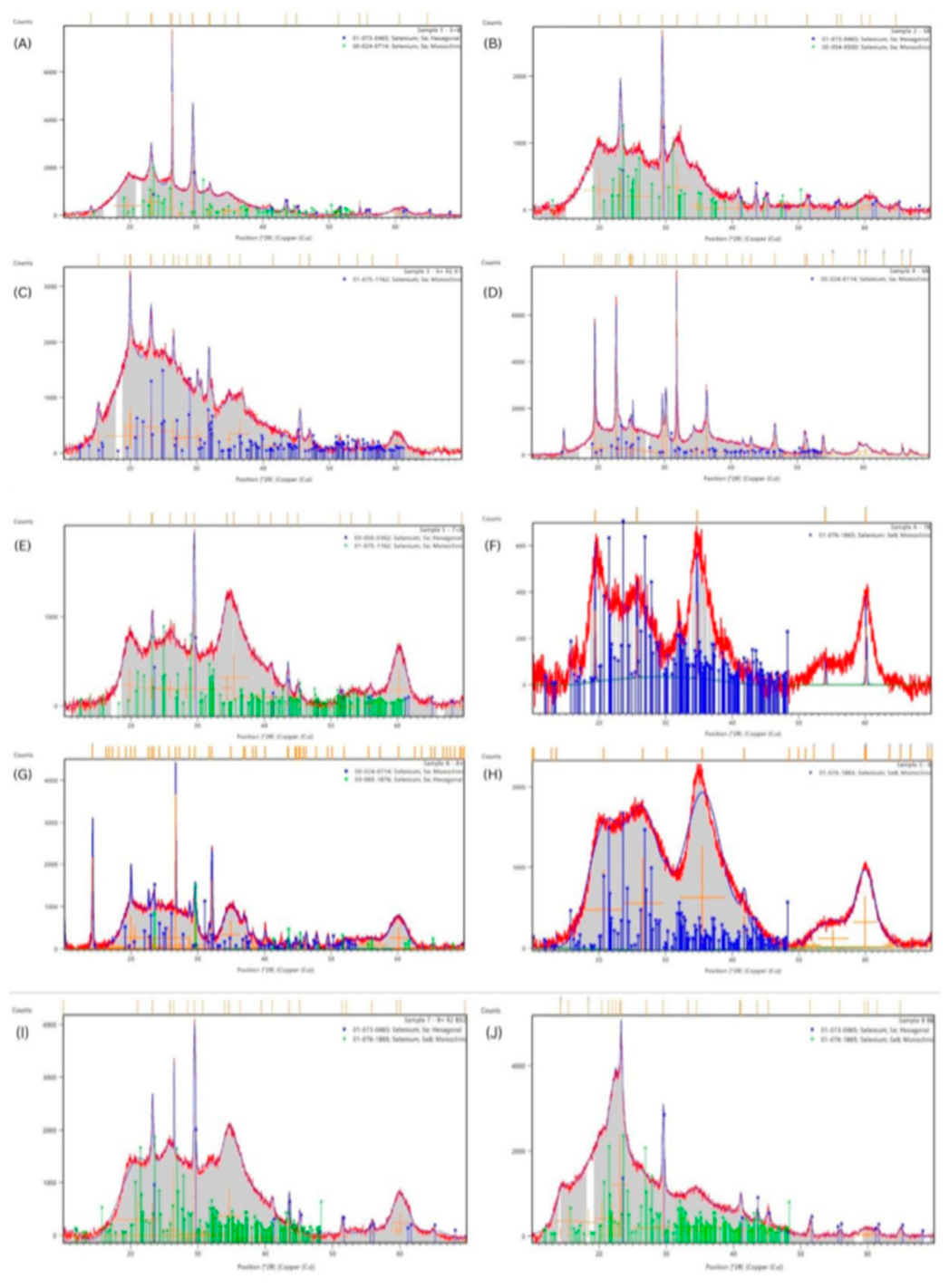
The diffractograms provide insights into particle size and phase composition. There were noticeable variations in crystallinities (Figure 2); in most cases, broad diffraction bands of relatively low intensity were observed, consistent with nanoscale crystallites. Table 1 summarises the corresponding International Centre for Diffraction Data (ICDD) matches, which confirmed the presence of selenium phases in both hexagonal and monoclinic forms across different treatments.
Both treated groups (-, +) of P. palmata, F. vesiculosus, F. serratus, and L. digitata exhibited a mixture of hexagonal Se (h-Se) and monoclinic Se (β-Se) phases, confirming successful synthesis of crystalline Se-NPs. In contrast, Se-NPs from U. intestinalis predominantly displayed diffraction peaks associated with the β-Se phase, which refers to a metastable monoclinic crystalline form of elemental Se. Se-NPs from F. vesiculosus exhibited overlapping peaks that could not be distinctly matched to a single phase; however, these patterns suggest contributions from the β-Se with varying crystallite sizes. The coexistence of both h-Se and β-Se across several species suggests the formation of nanocrystalline Se with anisotropic nanocrystallite domains, which is commonly observed in nanoparticle synthesis. Such polymorphic variation is likely driven by variations in biochemical constituents of the macroalgal extracts and their interaction with the selenium precursor during nucleation and growth [59,60].
Peak sharpness and intensity serve as indicators of nanoparticle crystallinity. Across most treatments, the diffractograms displayed a broad hump between 20° and 30°, which is characteristic of a partially amorphous phase (a-Se) or poorly crystalline zero-charged Se. An exception was observed in the U. intestinalis Se-NPs which displayed sharper and more well- defined peaks, suggesting relatively higher crystallinity. Nevertheless, the generally detected low-intensity and broadened diffraction peaks, when compared to the standard reference patterns, reflect the low crystallinity. These findings are typical for nanocrystalline size and low
sample quantities. Nanocrystalline Se-NPs exhibit disorder, causing peak broadening and reduced intensity due to small crystallite size and potential amorphous content.
These findings demonstrate that crude macroalgae extracts support the formation of crystalline Se-NPs, predominantly in the hexagonal phase, with additional monoclinic contributions depending on the extract content. The variation in composition suggests that macroalgae metabolites may influence the structural outcome, which could in turn affect stability and biological properties. A comparison of the h-Se phases, as shown in Table 1, revealed no distinguishable variation in XRD patterns across samples derived from the same algae species, regardless of whether ascorbic acid was included in the synthesis. This suggests that the structural characteristics of the h-Se phase remained consistent under both treatments. In contrast, slight differences were observed in the β-Se phase, which may reflect its inherently less stable, metastable nature compared to the thermodynamically favoured hexagonal form.

2.4. Transmission Electron Microscopy (TEM)

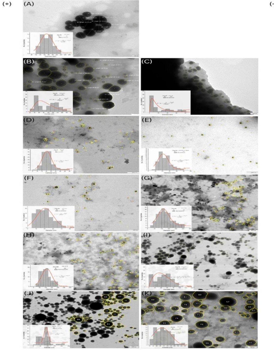
TEM analysis was conducted to characterise particle size and morphology in comparison with PC3 (since PC1 and PC2 failed to produce Se-NPs). PC3 displayed distinct spherical nanoparticles, though at low density relative to the other treatments that yielded over 100 nanoparticles per image field, suggesting batch variability or stabilisation differences.
All macroalgae-derived Se-NPs showed successful formation of well-defined, predominantly spherical Se-NPs irrespective of the presence of ascorbic acid (Figure 3). The nanoparticles were coated by a thin and uniform film (lighter tone contrasting with the darker tones of the particle core). Products arising in the absence of ascorbic acid should be composed of algal biocomponents. Cruz et al. (2018) discovered comparable layers coating their bio- fabricated nanoparticles [15]. The role of surface-bound compounds in altering colloidal features, such as size and stability, is documented in [61,62], with the capping layers persisting even after washing and precipitation, reflecting the strong interactions between Se-NPs and the phytochemical constituents. Marginal differences in particle size were noticeable, with treatments without ascorbic acid occasionally displaying larger diameters. The absence of ascorbic acid may decelerate Se-NP nucleation kinetics (without the donation of 2e- from ascorbic acid), allowing algal molecules to rapidly encapsulate the initial creation of nano- seeds. Ascorbic acid was more competitive in interacting with Se atoms than the bioorganic compounds, since it is a small molecule with high mobility compared to polysaccharides or proteins, which was obvious in the agglomeration patterns in most ascorbic acid-free treatments instead of separated monodispersed particles. This may occur when many available functional groups bind with insufficient quantities of selenious acid ions, as has been observed in Se-NPs fabricated using Withania somnifera extract, where aggregation was linked to fewer nucleation events [63].
Particle sizes were within the expected nanometric range, and the size distribution ranged from Gaussian in highly uniform Se-NPs, especially in the case of U. intestinalis (+), to log-normal in others (e.g., L. digitata (-)), which likely reflects the diversity in phytochemical profiles across the macroalgae. Most distribution patterns exhibited positive skews, which are common for nanoparticles. The goodness-of-fit test (α = 0.05) confirmed that Se-NP data from PC3, U. intestinalis, F. vesiculosus and L. digitata had a near-normal unimodal distribution indicating better control during synthesis. Lognormal, in the case of P. palmata (+), and Weibull distributions for F. serratus (-) provided more appropriate fits. Some nanoparticles, e.g., P. palmata (-), L. digitata (+) exhibited bimodal distributions.

2.5. Scanning Electron Microscopy-Energy-Dispersive X-Ray Spectroscopy (SEM-EDX)

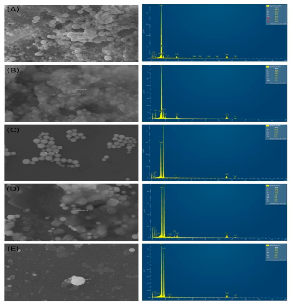
SEM images (Figure 4) evidenced morphologically distinct Se-NPs, predominantly spherical but with some irregular forms. The degree of particle dispersion and surface uniformity differed by species. P. palmata and F. vesiculosus had well-dispersed, uniform particles, indicating the presence of effective capping agents, likely related to their higher concentrations of bioactive polysaccharides that resulted in efficient nucleation and stabilisation processes, as mentioned in the critical review of extracted bioactive compounds from P. palmata, which highlighted that 20-25% of its dry weight was phycobiliproteins (water-soluble and well-known antioxidants) [64]. In contrast, U. intestinalis presented greater morphological diversity, with both spherical and occasional elongated or clustered particles, potentially reflecting more complex or uneven nucleation dynamics. This feature complements the broader size range observed in its TEM histogram (Figure 3). F. serratus and L. digitata exhibited slightly rougher particle outlines and less densely packed structures. This was consistent with their heterogeneous particle dispersion, as seen in their TEM images (Figure 3).
All spectra displayed distinct selenium peaks at approximately 1.37 KeV (Se Lα) and 11.22 KeV (Se Kα). The atomic percentage (At%) of Se differed among species, although P. palmaria was the only species to have significantly elevated levels of up to 31.2 At%, indicating either enhanced loading efficiency or greater Se incorporation within the nanoparticle matrix. Extracts of brown algae species resulted in moderate-to-low atomic percentages with F. vesiculosus and F. serratus around 11 At%, and the lowest percentage was for L. digitata (7.4 At%), where silicon and carbon elements were dominant. All spectra showed prominent peaks of oxygen (O) and carbon (C), which are typically associated with organic capping agents [65]. The frequent detection of sodium (Na), chlorine (Cl), potassium (K), and trace elements such as phosphorus (P) and silicon (Si) signifies the biochemical intricacy of macroalgal extracts, which inherently comprise salts, proteins, and polysaccharides [66].

2.6. Dynamic Light Scattering (DLS) and Zeta-Potential

Mean particle sizes ranged from 53 ± 0.5 nm to 426.6 ± 8.7 nm, with polydispersity index (PDI) values ranging from 0.14 ± 0.14 (U. intestinalis (-)) to 0.36 ± 0.09 (L. digitata (-)) (Table 2). This indicates varying degrees of polydispersity across the two main treatment groups and moderate dispersity in the colloidal form compared with the reference control PC3, which had a lower PDI value of 0.09 ± 0.03, meaning mono- and narrow dispersity. The closer the PDI value is to zero, the more uniform the particle shape or size distribution is in an aqueous solution. To clarify: particles with values from 0 to 0.08 are monodispersed, values from 0.08 to 0.7 have a medium distribution, and 0.7–1 generally have polydispersity [67].
Se-NPs from P. palmata with ascorbic acid exhibited the smallest and most uniform nanoparticles (53 ± 0.5 nm, PDI 0.24 ± 0.01), followed by its corresponding untreated group (-) (83.2 ± 0.8 nm, PDI 0.28 ± 0.007). Conversely, the largest particles were produced from the brown seaweeds, in particular, the ones that originated from the untreated group (-), with F. serratus exhibiting a mean size of 426.6 ± 8.7 nm with PDI 0.24 ± 0.04. Also, Se-NPs from both L. digitata-treatment groups had a mean size of 330 ± 7.2 nm with a PDI of 0.27 ± 0.5 in (+), while they had a mean size of 302 ± 22 nm with a PDI of 0.36 ± 0.09 in the ascorbic acid- free group. It is important to note that the mean sizes indicated in Table 3 and Supplementary Figure S2 do not quantify the precise diameter of the core particle; rather, they represent the entire hydrodynamic halo of the nanoparticle, encompassing the particle core along with the absorbed functional group identified through FTIR analysis (the organic shell) of the stabiliser, as well as any existing aggregation [52]. The hydrodynamic diameters of macroalgae-based Se- NPs treated without ascorbic acid were marginally larger than those treated with ascorbic acid, with the exception of those derived from L. digitata; this may result from the influence of ascorbic acid and the modification of other competing phytochemicals in the medium, reducing the shell thickness [61].
The zeta potential analysis revealed that all synthesised Se-NPs exhibited a negatively charged surface, likely attributable to deprotonated hydroxyl, carboxyl, or sulphate groups derived from the macroalgae extract. Particles with surface charges beyond ± 25 mV are considered electrostatically stable; thus, Se-NPs from P. palmata (-), U. intestinalis (+, -), F. vesiculosus (+, -), and L. digitata (+) demonstrated colloidal stability comparable to PC3, which was -27.2 ± 0.72 mV. It is worth noting that several measurements were conducted on diluted suspensions, as undiluted ones failed to generate graphs due to limitations in signal strength. Hence, dilution reduces ionic strength and electrophoretic mobility, leading to an underestimation of the surface charge values. For instance, the actual surface charges in undiluted Se-NP colloids were higher, such as colloids of P. palmata (+), U. intestinalis (+, -), F. vesiculosus (+), F. serratus (-), and L. digitata (+).

2.7. Antimycobacterial Activity

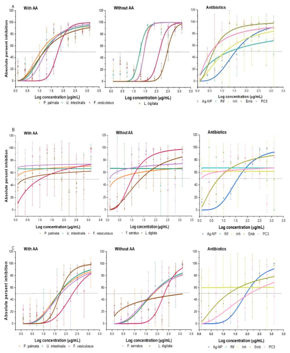
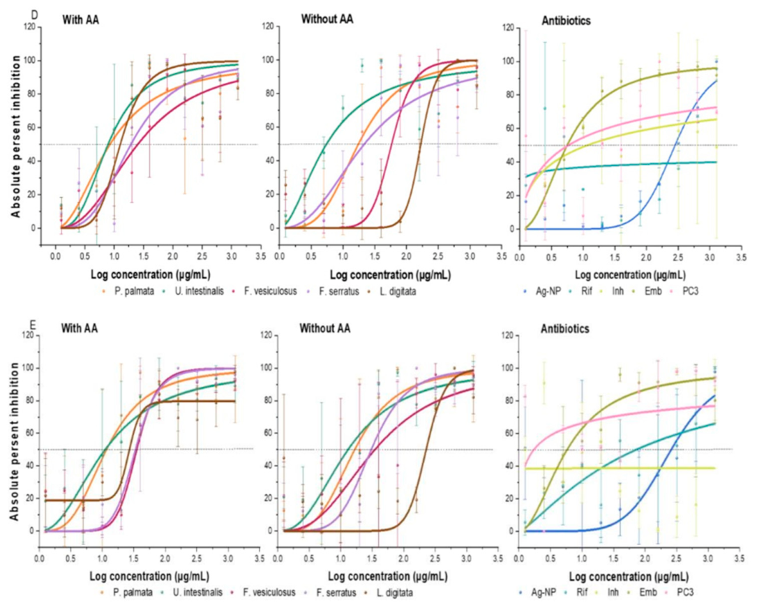
The antimycobacterial efficacy (MIC50, MIC90) of macroalgae-derived Se-NPs and commercially synthesised Se- and Ag-NPs were compared with three first-line antibiotics (rifampicin, isoniazid, and ethambutol) (Figure 5 and Table 3). The primary endpoints for all treatment groups were the MIC50 and MIC90, determined by non-linear regression analysis of dose-response curves. Therefore, MIC50 indicates the overall potency of a treatment that reflects the concentration required to inhibit 50 % of the bacterial population, while MIC90 represents the treatment effectiveness when the exact dosage can inhibit 90 % of the population. The crude macroalgal extracts (NC2) had no inhibitory activity; therefore, any antimycobacterial activity can be attributable to the nanoparticles. Spavieri et al. (2010) assayed crude extracts from 21 brown algae, including F. serratus, F. vesiculosus, and L. digitata, with no activity against M. tuberculosis (strain H37Rv) apart from Bifurrcaria bifurrcata (MIC50 = 64.0 μg/mL) [68]. Akbari et al. (2018) also found that crude extracts of Sargassum boveanum were inactive [69].
There was a positive significant association between treatments in terms of the MIC50 values, based on Kruskal-Wallis analysis for the wild-type strain mc27902 (H = 27.61, df = 12, p = 0.006), and multidrug-resistant strains mc28250 (H = 29.44, df = 12, p = 0.003), and mc28258 (H = 26.76, df = 12, p = 0.008). The single-drug-resistant mc28245 and mc28247 showed no significant differences. The lack of significance in single-drug-resistant strains may reflect lower susceptibility, the requirement of higher concentrations or different uptake (e.g., synergistic agents). Unlike for the MIC50, the effectiveness of the biologically sourced Se-NPs (MIC90) were not significantly different across sourced treatment groups for any of the testedstrains, reflecting that macroalgae-mediated Se-NPs were more effective at partial inhibition (MIC50) than full suppression (MIC90).
The wild-type M. tuberculosis (mc27902) strain was inhibited, with substantial variation in potency depending on the algal source and the presence of ascorbic acid during synthesis. Ascorbic acid-supplemented treatments (+) consistently exhibited antimycobacterial activities with MIC values in the low to moderate μg/mL range (Table 3). The most potent was observed for P. palmata (MIC50 = 14.64 ± 1.35 μg/mL; MIC90 = 846.14 ± 3.72 μg/mL), followed by U. intestinalis (MIC50 = 15.06 ± 1.27 μg/mL; MIC90 = 304.26 ± 2.58 μg/mL) and F. serratus (MIC50 = 19 ± 1.2 μg/mL; MIC90 = 192.1 ± 2.1 μg/mL). F. vesiculosus showed moderate activity, while L. digitata was least potent. These values highlight that a relatively low concentration was needed for an initial effect, but a much higher concentration was required to achieve a near-complete growth inhibition (90 % bacterial growth inhibition).
The ascorbic acid-free synthesis activity profiles (-) were, in some cases, characterised by higher MIC50 values (ranging from 18.34 to 294.3 μg/mL), indicating lower intrinsic activity at reduced concentrations. However, these particles proved more effective (lower MIC90 values) overall, (ranging from 40.21 to 673.4 μg/mL), which suggests a superior capacity for near- complete bacterial inhibition compared to the (+) groups (less potent (MIC50) but more effective (with lower MIC90 values compared to their relative (+) groups)). The most pronounced reduction in potency was observed for L. digitata (-), whose MIC50 was 294.3 μg/mL and MIC90 was 673.4 μg/mL.
When benchmarked against conventional anti-TB drugs, all Se-NPs were less potent: rifampicin (MIC50 = 0.07 μg/mL; MIC90 = 0.61 μg/mL), isoniazid (MIC50 = 3.62 μg/mL; MIC90 = 13.23 μg/mL), and ethambutol (MIC50 = 6.88 μg/mL; MIC90 = 12.54 μg/mL), but their efficacies (MIC90) were more comparable to isoniazid, which required a higher concentration than the tested range. Notably, all synthesised Se-NPs were more effective in reaching 90 % inhibited bacterial growth (MIC90) than silver nanoparticles (Ag-NPs) (MIC90 = 1076 ± 1.77 μg/mL) with a comparable potency (MIC50 = 35.6 ± 1.15 μg/mL). These higher MICs were expected findings for a novel compound, which needs further optimisation. The observed improvement with ascorbic acid supplementation (Figure 5) suggests it may play a significant role beyond reducing activity, potentially influencing the final properties of these nanoparticles and enhancing interaction with the mycobacterial cell wall. Potential bioactive compounds adhering to the nanoparticles’ surface may compete differently in the presence of ascorbic acid, leading to different capsulating biomolecules.
The isoniazid-resistant M. tuberculosis (mc28245) strain had generally lower MIC50 and MIC90 values compared to the wild-type strain (Table 3). Surprisingly, Se-NPs derived from U. intestinalis both with and without ascorbic acid failed to display activity. Also, Se-NPs produced from P. palmata and F. serratus with ascorbic acid showed no activity. Both F. vesiculosus and L. digitata were active with the lowest MIC50 values compared to the previous strain (10.5 ± 2 and 2.51 ± 3.61 μg/mL, respectively). In terms of reaching 90 % bacterial growth inhibition, inhibitory efficacy consistently exceeded the highest limiting concentration observed in the active groups. In the ascorbic acid-free synthesised groups, F. serratus-Se-NPs registered the most potent green nanoparticles with a minimal MIC50 value registered of 1.22 ± 1.5 μg/mL, compared to the other active nanoparticles. Following that, Se-NPs derived from P. palmata had an MIC50 value of 2.73 ± 2.6 μg/mL.
Ethambutol and rifampicin maintained activity at MIC50 = 8.47 and MIC90 12.20 and at MIC50 0.07, MIC90 0.58 μg/mL, respectively, indicating that the resistance in this strain is drug- specific rather than broad-spectrum, while isoniazid, as expected, had a markedly reduced effect. Ag-NPs (MIC50 = 39.3 ± 1.2 μg/mL; MIC90 = 697 ± 2.3 μg/mL) were less active than all Se-NPs in terms of their potency (MIC50), further highlighting the relative advantage of selenium over silver in targeting drug-resistant M. tuberculosis. Ascorbic acid-aided Se-NPs produced steeper inhibition profiles than ascorbic acid-free treatments. Nonetheless, the leftward shift of these curves (for the active Se-NPs) to lower concentrations, relative to mc27902, indicates that INH resistance confers enhanced susceptibility to Se-NPs in active cases. This phenomenon is perhaps linked to the specific genetic mutations, which are associated with large deletions in the katG gene. This gene is essential for managing oxidative stress within the bacterium; its loss results in a compromised or altered redox homeostasis, making the mycobacterium highly sensitive to the oxidative stress of Se-NPs and potentially altered cell wall permeability. The rifampicin-resistant (mc28247) strain exhibited a reduced susceptibility spectrum compared to the wild-type and INH-resistant strains. There were no significant differences between treatments. The most pronounced increase in potency was observed for P. palmata and U. intestinalis for both ascorbic acid-added (+) and -free (-) groups (MIC50 = 48.3 ± 1.31, 66.7 ± 1.5; 48.5 ± 1.3, 60.8 ± 1.3 μg/mL, respectively), followed by L. digitata and F. serratus (Table 3). The effectiveness (MIC90) of these extracts exceeded the highest examined dosage (1280 μg/mL), except for the ascorbic acid-free L. digitata extract which was inactive. F. vesiculosus had MIC50 values of 198.9 ± 1.3, 203.3 ± 1.1 μg/mL and MIC90 values of > 1280, 995 ± 1.6 μg/mL.
Rifampicin was ineffective. Ethambutol and isoniazid were active with MIC50 14.34, 5.19 and MIC90 24.91, 8.97 μg/ mL, respectively. Moderate activity was observed for Ag-NP with MIC50 = 88.2 ± 1.2 and MIC90 = 1043 ± 1.9 μg/mL. Rifampicin-resistance mechanisms had a less pronounced impact on macroalgae-mediated Se-NP efficacy than isoniazid- resistance. These differences can be explained by the distinct genetic mechanisms underlying each resistance phenotype. Herein, the rif-resistant strain is driven by a point of mutation in the rpoB gene (specifically the H445Y), which alters the drug-binding site of the RNA polymerase. This mutation relies on different mechanisms (affecting RNA production) without defending systems’ alteration or cell wall permeability. Consequently, the physiological barriers governing Se-NPs' penetration, resulting in a less pronounced shift in susceptibility.
The multidrug-resistant M. tuberculosis (mc28250) strains, resistant to both isoniazid and rifampicin, presented the most challenging inhibitory profile. Nevertheless, several Se-NP formulations demonstrated activity, particularly those synthesised with ascorbic acid, e.g., U. intestinalis: MIC50 = 7.85 ± 1.2 μg/mL; MIC90 = 75.2 ± 2 μg/mL and P. palmata: MIC50 = 8.45 ± 1.32 μg/mL; MIC90 = 516 ± 4.3 μg/mL (Table 3). The inhibitory effect was reduced without ascorbic acid for F. vesiculosus and L. digitata, with MIC50 values rising from 25.1 ± 1.3 to 58.4 ± 1.1 μg/mL and from 12.6 ± 1.1 to 168 ± 1.1 μg/mL, respectively. However, F. vesiculosus-Se-NP showed improved efficacy with a substantial reduction in MIC90 value from > 1280 to 148.4 ± 1.3 μg/mL. The P. palmata-Se-NP MIC90 was also reduced from 516 ± 4.3 to 180.7 ± 2.1 μg/mL. Rifampicin and isoniazid were completely ineffective, while ethambutol remained active (MIC50 = 8.79; MIC90 = 20.80 μg/mL). Ag-NPs had a limited effect (MIC50 = 282.4 ± 1.11 μg/mL; MIC90 = 320.2 ± 1.3 μg/mL), substantially lower potency than Se-NP formulations, suggesting Se-NPs' superior utility in overcoming 50 % of the mycobacterial growth of combined INH and RIF resistance but with an almost similar effective level in overcoming 90 % of the growth (MIC90 = 320.2 ± 1.3 μg/mL) compared with the green synthesised Se-NPs.
The M. tuberculosis mc28258 strain (rifampicin- and isoniazid-resistant) shares the dual resistance profile of mc28250 and represents a genetically modified strain with potentially greater phenotypic variability and resilience. This variability in resilience is likely governed by the specific genetic mutations defining each strain resistance profile. While both are resistant to isoniazid and rifampicin, the mc28258 strain has specific nutrient auxotrophies such as deletions in the amino acid or pantothenate biosynthetic pathway, e.g., Delta panCD, Delta leuCD, or Delta argB. These targeted gene deletions impose different metabolic mechanisms and distinct alterations to cell wall assembly compared to the mutation profile of mc28250. Consequently, this specific genetic background for this strain preserves a more robust cell envelope or more effective metabolic pathway, rendering it more resilient to membrane- disrupting and pro-oxidant mechanisms of the biogenic Se-NPs. The most active ascorbic acid- aided formulations were from P. palmata (MIC50 and MIC90 = 11.6 ± 1.2 μg/mL and 119.7 ± 2.4 μg/mL) and U. intestinalis (MIC50 and MIC90 = 11 ± 1.3 μg/mL and 728.6 ± 4.1 μg/mL, respectively; Table 3). L. digitata, F. serratus, and F. vesiculosus extracts displayed lower activity but were still considered strongly active (MIC50 = 26.6 ± 1.2, 33.5 ± 1.1, and 36 ± 1.1 μg/mL; MIC90 = 43.4 ± 1.4, 93.6 ± 1.4, and 88 ± 1.3 μg/mL, respectively). No significant drop was noticed in the inhibitory potency of treatments without ascorbic acid, except for L. digitata (MIC50 = 227.3 ± 1.18 μg/mL). This may be influenced by the actual ascorbic acid content of the seaweed, which does vary [70]. The lower intrinsic ascorbic acid content of L. digitata may have led to a more pronounced reliance on ascorbic acid supplementation.
Ethambutol retained the highest potency (MIC50 = 14.33 μg/mL) with a moderate efficacy (MIC90 24.84 μg/mL). Isoniazid had activity in terms of its potency with high MIC50 value of 336.02 μg/mL, while it was inactive in terms of its efficacy (MIC90). Rifampicin was inactive. Ag-NPs exhibited moderate activity (MIC50 = 258.7 ± 1.4 μg/mL; MIC90 > 1280 μg/mL). For most macroalgal sources, there was no significant deviation in antimycobacterial activity between ascorbic acid-supplemented and -free treatments (MIC50 values) (Table 4). However, a significant difference (p < 0.001) was observed when L. digitata extract was used for Se-NP production. The ascorbic acid-free treatment consistently exhibited higher inhibition values. No significant differences were observed in all MIC90 values for all macroalgae-derived Se-NPs between those two different treated groups.

2.8. Comparative Benchmarking

Across all M. tuberculosis phenotypes, Se-NPs derived from P. palmata and U. intestinalis extracts were the most potent, with median MIC50 values of approximately 13.1 and 14 μg/mL, respectively (ascorbic acid assisted). This may be a consequence of their smaller size, as confirmed by TEM and DLS. Their corresponding MIC90 values (681 and 516 μg/mL) indicated great efficiency, as these biogenic Se-NPs achieved near-complete bacterial inhibition, although requiring higher concentrations for full inhibition. F. vesiculosus and F. serratus were more variable. F. vesiculosus (+) produced MIC50 and MIC90 medians of 35 and 26 μg/mL, while F. serratus displayed a broader range with a median MIC90 of 258 μg/mL. L. digitata- Se-NPs were generally less potent than the other species, with median MIC50 and MIC90 values of 26 and 237 μg/mL (ascorbic acid assisted), respectively, and markedly weaker activity without ascorbic acid supplementation.
In comparison with conventional TB medications, ethambutol maintained activity in all strains; as expected, rifampicin and isoniazid were inactive in dual-resistant isolates. Compared to ethambutol, Se-NPs were less potent on a per mass basis but offered efficacy against strains resistant to first-line drugs. When benchmarked against Ag-NPs, Se-NPs approached, or in some cases surpassed their potency, which offers a potential advantage in reduced cytotoxicity [33,38]. The efficacy spectrum of ascorbic acid-aided Se-NPs fell within a relatively contained range from approximately 3-230 μg/mL. This wide distribution indicates how strain-specific genetic backgrounds and varying resistance phenotypes dictate the initial susceptibility threshold to these nanoparticles. Such stability, even in highly multi-resistant strains, is an important factor for potential therapeutic agents. This is in line with a review [71] that reported significant inhibitory effects of Se-NPs on M. tuberculosis growth synthesised using various approaches. [33] also reported a significant inhibitory effect of chemically synthesised Se-NPs stabilised with different reagents, with MIC values of 0.40 μg/mL against Mycobacterium smegmatis, a fast-growth strain, and 0.195 μg/mL against the slow-growth M. tuberculosis (H37Rv strain).
Commercial Se-NPs (PC3) generally exhibited higher potency (lower MIC50 values) in several instances; however, algae-mediated Se-NPs demonstrated superior performance against specific resistant strains (mc28245 (INH-R) and mc28247 (RIF-R)). Ascorbic acid-free Se-NPs derived from P. palmata and F. serratus were significantly more potent than PC3, yielding MIC50 values of 2.73, 2.6, and 1.22 1.5 μg/mL, respectively, compared to 2.9 μg/mL for the commercial particles. The inclusion of ascorbic acid during synthesis enhanced potency in one notable case (L. digitata-derived Se-NPs), which then competed effectively with the commercial standard. Nearly all biogenic Se-NPs against mc28247, regardless of ascorbic acid supplementation, outperformed the commercial control (PC3) in potency, with the sole exception of those derived from F. vesiculosus. In terms of efficacy (MIC90), PC3 required substantially higher concentrations than the biogenic Se-NPs to achieve 90 % mycobacterial inhibition, which was most pronounced against the multidrug-resistant (MDR) strains (mc28259). PC3 initially appeared more potent (MIC50) against mc28258 strain; however, they failed to reach MIC90 within the examined concentration range. Consequently, while commercial Se-NPs may exhibit lower inhibitory thresholds (MIC50) in certain scenarios, macroalgae-mediated Se-NPs offer an advantage in efficacy, achieving near-complete pathogen suppression at significantly lower concentrations.

2.9. Se-NPs Mode of Action

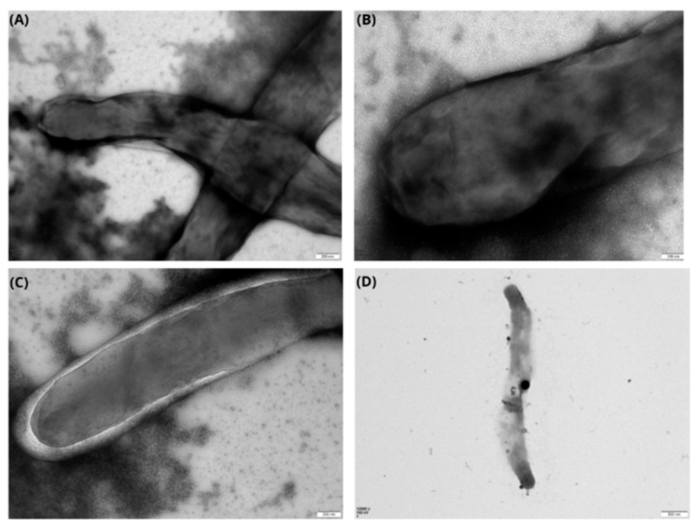
Figure 6 represents M. tuberculosis cells before and after exposure to algae-mediated Se-NPs. Sub-Figure 6A and B represent untreated bacteria (controls), confirming the thick and dense cell envelope, to compare them with their counterpart-exposed bacteria. The negative-stained sub-Figure 6C (treated with the biogenic Se-NPs) displayed a shrinking cytoplasmic membrane relative to untreated cells. A most optimal view was in sub-Figure 6D, showing a superficial association between biogenic Se-NPs and the bacterial cell wall with light penetration that induced the disruption of the cell wall and the cytoplasmic material leakage. Although, there are few studies that have evaluated the antibacterial activity of Se-NPs against M. tuberculosis, they have shown promise as antibacterial tools, primarily through mechanisms such as cell envelope disruption, oxidative stress induction, and metabolic interference. Chitosan-stabilised Se-NPs can damage the integrity of the bacterial cell envelope, leading to increased permeability and eventual cell death [33]. Pi and co-workers modified Se-NPs to include mannose to target macrophages containing M. tuberculosis infectious bacteria [72]. These nanoparticles were incorporated into isoniazid. This combination demonstrated multiple synergistic anti-M. tuberculosis mechanisms, including phagolysosomal fusion, which enhances bacterial degradation within macrophages; ROS generation, leading to oxidative stress-induced cell damage; and autophagy activation, prompting intracellular pathogen clearance and immune modulation [72]. Se-NPs are effective against a wide range of pathogens, including gram-positive and gram-negative bacteria; opportunistic fungi such as Candida albicans, and even against multidrug-resistant clinically isolated bacteria [73]. Unlike conventional antibiotics, Se-NPs demonstrate low toxicity to mammalian cells [74]. Compared to more toxic nanoparticles, such as silver and copper, Se-NPs offer a safer antimicrobial profile, especially when stabilised with natural biomolecules [11]. Se-NPs can be engineered to accumulate within infected macrophages, the primary host cells for M. tuberculosis, which assists in drug concentration at the site of infection and then minimises systemic toxicity [72]. Functionalised nanoparticles can synergise with existing antibiotics to increase their effectiveness against MDR-TB [75].

3. Materials and Methods

3.1. Sample Collection and Preparation

Macroalgae (Ulva lactuca, Chondrus crispus, Palmaria palmata, Fucus vesiculosus, F. serratus, and Laminaria digitata) were collected in plastic zipper bags from Cullercoats Bay, Tyne & Wear, UK (lat: 55° 02’ N, long: 1° 26' W) in May of 2021 to 2023, stored on ice and transported immediately to the laboratory. All samples were washed several times under running tap water to remove sand and impurities, soaked for 24 hours in reverse osmosis water to reduce salt content, then were gently air-dried in darkness for approximately three weeks. The dried samples were ground to a fine powder using an electric stainless-steel blender. Extractions were done by maceration [34] in which 20 g of powdered sample was mixed with 200 mL of Milli-Q water (1:10 w/v total ratio), covered and left to infuse for 24 hours at room temperature. Following infusion, the broth was heated at 70 °C for 20 min and filtered using 200 and 70 μm meshes. The filtrate was centrifuged at 7573 × g for 15 min to remove bacterial contaminants. The supernatant was stored at -18 °C until use [35,36].

3.2. Biosynthesis of Selenium Nanoparticles

Three positive controls (PC) were established: PC1 was prepared by mixing 2 mL of a 2% bovine serum albumin (BSA) stock solution (CAS number 9048-46-8, Sigma Aldrich UK) with 10 mL of 30 mM selenious acid H2SeO3 (SeA) (CAS number 7783-00-8, Sigma Aldrich Germany). This was subsequently reacted with 200 μL of 40 mM ascorbic acid C6H8O6 (CAS number 50-81-7, Sigma Aldrich UK), resulting in a reaction mixture with a final concentration of approximately 0.33 % BSA, 73 mM SeA, and 0.66 mM ascorbic acid; PC2 contained 10 mL of 30 mM selenious acid and 200 μL of 40 mM ascorbic acid; PC3 was 0.15 wt.% of commercially synthesised Se-NPs (CAS number 7782-49-2, Sigma Aldrich UK). Negative controls (NC) were 30 mM selenious acid precursor solution only (NC1) and 10 mL of the 10 % algal extract (m/v %) (NC2).
Treatment group one – ascorbic acid supplemented (denoted by the [+] symbol) was 2 mL of 10 % (w/v) stock of each macroalgal extract added to 10 mL of 89 mM SeA. Where applicable, 200 μL of 40 mM ascorbic acid was introduced to initiate reduction. This maintained a consistent environment across all trials, with final concentrations of 16.4 % (v/v) algal extract, 72.95 mM SeA, and ascorbic acid (1:60 w/v). Group two had no ascorbic acid added (denoted by the [-] symbol). The precursor stock solution was prepared in advance for all groups, whereas the ascorbic acid solution was freshly prepared for each experiment to prevent photooxidation. Milli-Q water was used for all preparations. Stirring was done on an electromagnetic stirrer for 5 min [37]. All groups were adjusted to pH 7 by dropwise addition of sodium hydroxide (NaOH). All mixtures were incubated in a shaking incubator at 250 rpm for an indefinite duration in the dark at 60 °C. All experiments were run in triplicate. The endpoint was determined by a colour change to reddish-orange [38].
Unreacted selenium ions were removed by three successive washes with Milli-Q water: wash 1, 1:1 v/v with Milli-Q water and centrifuged for 10 mins at 7754 × g to remove larger particles and unattached biological impurities; washes 2 and 3, the supernatant was resuspended with 40 mL of Milli-Q water and centrifuged at 69,670 × g for 30 mins [39]. Finally, the pellet was oven-dried at 50 °C for 8 hours to determine dry weight and thereby the exact concentration that would be used in subsequent biological applications.

3.3. Nanoparticle Characterisation: UV-Vis Spectroscopy

The Materials and Methods should be described with sufficient detail to allow others to replicate and build on the published results. Please note that the publication of your manuscript implies that you must make all materials, data, computer code, and protocols associated with the publication available to readers. Please disclose at the submission stage any restrictions on the availability of materials or information. New methods and protocols should be described in detail while well-established methods can be briefly described and appropriately cited.
Surface plasmon resonance (SPR) absorbance was conducted in triplicate. The bioreduction of the Se-NPs was monitored periodically throughout. Before measurements, the colloidal Se-NPs were diluted 1:4 (v/v) with Milli-Q water to reduce scattering [35]. Scanning was at a resolution rate of 1 nm/s across a range of 200-800 nm. Absorbance was assessed every 24 hours at ambient temperature.

3.4. Fourier Transform Infrared Spectroscopy

Interactions between Se-NPs and the capping agent were examined using a PerkinElmer Spectrum 2 Fourier transform infrared (FTIR) spectroscope, with samples placed on a high reflective index crystal. FTIR spectra were acquired in attenuated total reflectance mode with a resolution of 4 cm−1 over the spectral range of 4000 to 400 cm−1. Peak identification was conducted by comparing the acquired spectra with prior literature and established reference spectra found in FTIR databases such as the FTIR Functional Table in the LibreTexts libraries website and the website of the Search [40,41].

3.5. X-Ray Diffraction Spectroscopy

Analysis of nanoparticle purity and crystalline phase was performed with powder X-ray diffraction (P-XRD) analysis (Bruker D2 PHASER XRD3 diffractometer) equipped with a LYNXEYE line detector that uses Cu Kα radiation (λ = 1.5406 Å) at a voltage of 30 kV and a current of 10 mA. Scanning was performed in the 2 theta (2θ) range (10° to 70°) with a step size of 0.03° and a time per step of 2 seconds. The XRD diffraction patterns were analysed using the HighScore+ software for phase identification. The International Centre for Diffraction Data (ICDD PDF-2) and Crystallography Open Database (COD), using a filter for Se, were used to match the experimental diffraction peaks with standard reference patterns [42].

3.6. Dynamic Light Scattering and Zeta Potential

A Malvern Zeta-sizer Nano (ZS) (Malvern Instruments Ltd, UK) was used to confirm nanoparticle size, size distribution, and surface charge. 1 mL dispersed Se-NPs in Milli-Q water was pipetted into a disposable polystyrene cuvette post-sonication for 10 mins, following filtration through a 0.22 μm syringe filter to remove large aggregates, and then measured in technical triplicates at 25 °C, refractive index of 1.330, and viscosity (cP) at 0.887. Data were obtained via the Zeta-Sizer NanoRange software, while zeta potential data were acquired using DTS1060C disposable clear zeta cells.

3.7. Transmission Electron Microscopy

Nanoparticle morphology was visualised by transmission electron microscopy (Hitachi HT7800 at an accelerating voltage of 100 kV). A 10 μL sample was loaded onto a carbon- coated copper grid of 400 mesh. Images were obtained using a EMSIS CMOS Xarosa camera and analysed using ImageJ software (Version 2024), OriginLab Corporation, Northampton, MA, USA, measuring up to 100 randomly selected particles.

3.8. Scanning Electron Microscopy and Energy-Dispersive X-Ray Spectroscopy

Polished silicon wafers (5 × 5 mm) were decontaminated by immersion in ethanol 70 %, followed by rinsing with Milli-Q water and air drying. 10 mL of Se-NP suspension were drop- cast onto the silicon chip surface and air-dried at room temperature. The topographic surface of the nanoparticles was imaged using a Jeol JSM-5610LV scanning electron microscope. An Oxford Instruments ‘Aztec’ EDX system with an ‘X-act’ thin-window detector with 129 eV resolution was used at 5.9 keV to validate the presence of atomic selenium and provide information about the surface composition.

3.9. Antibacterial Efficacy

Minimum inhibitory concentrations (MIC50, 90) for the Se-NPs were determined against five M. tuberculosis strains, including four drug-resistant strains (Table 5) conferring distinct growth requirements and antimicrobial resistance profiles. Efficacy was benchmarked against standard first-line anti-TB drugs, namely rifampicin, isoniazid, and ethambutol, alongside commercially synthesised Se- and Ag-NPs.
M. tuberculosis strains were cultivated in either Middlebrook 7H9 broth or 7H11 agar medium with 0.4 % (v/v) (for 7H9)/ 0.5 % (v/v) (for 7H11) glycerol supplemented with oleic acid–albumin–dextrose–catalase (OADC) enrichments purchased from BD Biosciences, USA, 24 μg/mL pantothenate, 200 μg/mL L-arginine, 50 μg/mL L-leucine, 1 μg/mL penicillin G, and 10 μg/mL cycloheximide, (7H9OPALPen1Cyc10) [43]. Cultures of M. tuberculosis strains containing the pMV261 vector were supplemented with kanamycin 25 μg/mL. All other reagents were from Sigma-Aldrich, UK.
For antimycobacterial susceptibility testing, bacterial cultures were grown in a broth of 7H9OPALPen1Cyc10(kan25) containing 0.05 % (v/v) Tween80 at 37 °C, 5 % CO2 for 7-10 days to reach the mid-log phase. The turbidity of the bacterial suspension was adjusted and resuspended in fresh 7H9OPALPen1Cyc10(kan25) media to a McFarland 0.5 standard (approximately 1 × 10⁷ CFU/mL) at OD600 nm. This stock suspension was further diluted 1:25 in fresh 7H9OPALPen1Cyc10(kan25) medium to achieve an estimated final inoculum concentration of approximately 4 × 10⁵ CFU/mL. All M. tuberculosis and inoculum preparation procedures were performed in accordance with established laboratory protocols, as previously detailed by [44].

3.10. Resazurin Microtiter Assay

Minimum inhibition concentrations (MIC50/ MIC90) of Se-NPs, conventional anti-TB drugs, and silver nanoparticles (CAS: 7440-22-4, Sigma-Aldrich, UK) (Ag-NPs) were determined using the Resazurin Microtiter Assay (REMA) [44]. Stock dispersions were prepared at 100 mg/mL for synthesised Se-NP in phosphate-buffered saline, 1 mg/mL in sterile dimethyl sulfoxide (DMSO) for rifampicin (CAS: 13292-46-1, Sigma-Aldrich, UK) and 10 mg/mL in DMSO for ethambutol (CAS: 1070-11-7, Sigma-Aldrich, UK). A 2-fold serial dilution of each Se-NP (5.12 μg/mL) dispersion was then performed in 100 μL of Middlebrook 7H9OPALPen1Cyc10(kan25) broth across a final concentration range of 1.25 to 1280 μg/mL in a 96-well microplate. The anti-tuberculosis agents were added (2.56 μL) and serially diluted to achieve test concentrations ranging from 0.0625 to 64 μg/mL. Ag-NPs were serially diluted to a range of 1.25 to 1280 μg/mL. 100 μL of standardised M. tuberculosis inoculum was added to each well, giving a total volume of 200 μL per well. The last column of the 96-well microplate was used for bacterial growth controls, as the first four (top wells) were occupied by a positive control (only bacterial inoculum and 7H9OPALPen1Cyc10(kan25) broth without any tested compounds) to monitor uninhibited bacterial growth. The four bottom wells were for the negative control, containing only Middlebrook 7H9OPALPen1Cyc10(kan25) broth without bacteria or tested compounds, to ensure no contamination. Positive controls for bacterial growth were employed to verify viability and indicate 0% inhibition, whereas negative controls for sterility were utilised to address background fluorescence and signify 100% inhibition. The biological replicates were performed on different days to mitigate positional biases and were incubated under identical conditions to reduce variation in humidity or evaporation. Microplates were sealed with a breathable adhesive film and incubated at 37 °C for 5-7 days in a humidified incubator containing 5% CO2. After monitoring bacterial growth in the positive control, 10.5 μL of 0.1 % (w/v) sterilised resazurin solution (prepared freshly by dissolving resazurin powder (CAS: 62758-13-8, Thermo Fisher Scientific, UK) in sterile PBS was added. Plates were re-sealed and incubated for an additional 48 hours at 37 °C. A colourimetric transition of resazurin from blue to pink (resorufin form) signified bacterial metabolic activity and proliferation, whereas the persistence of the blue hue indicated inhibition. For quantitative evaluation, fluorescence was measured using a microplate reader (FLUOstar Optima, BMG Labtech) at excitation/emission wavelengths of 530/ 590 nm. Each treatment was tested in three independent biological replicates. To provide a comprehensive profile of Se-NPs' performance, both MIC50 and MIC90 values were determined. The MIC50 was utilised as a measure of potency, identifying the lower concentration threshold required to significantly inhibit bacterial metabolism. In contrast, the MIC90 was used to evaluate efficacy (efficiency), defined as the mean concentration causing ≥ 90 % growth inhibition relative to the positive control, representing the capacity of the Se-NPs to achieve near-complete suppression of the pathogen.

3.11. Se-NPs with M. tuberculosis Interaction

To visualise the direct interaction between algae-mediated Se-NPs and M. tuberculosis inside targeted cells, transmission electron microscopy (TEM) was used. In this part, the wild-type M. tuberculosis strain was selected as the sole representative model. The nanoparticle-exposed bacterial cultures (cultivated bacteria with Se-NPs) were centrifuged at 4.8 × 10³ g for 10 minutes. The resulting pellet was immediately mixed with an equal volume (500 μL) of the primary fixative (2% glutaraldehyde in 0.1 M sodium cacodylate buffer, pH 7). The samples were fixed overnight at ambient temperature. The fixed samples were subjected to a rinsing step using 0.1 M phosphate buffer (pH 7.2- 7.4) to ensure complete removal of the residual glutaraldehyde. This wash step was repeated two or three times. Samples were post-fixed with 1% osmium tetroxide (OsO4) prepared in 0.1 M phosphate buffer for 2 hours at room temperature to preserve and enhance the contrast of the cellular membrane lipids (secondary fixation) [45]. Final rinsing of the samples was with phosphate buffer, as described in the initial rinsing step, to remove the excess OsO4. A drop of the fixed cultivated mycobacteria was uploaded directly to the TEM Cu grid for a negative staining technique. For the sake of comparison, M. tuberculosis without Se-NPs was used to identify any alterations.

3.12. Statistical Analysis

The Materials and Methods should be described with sufficient detail to allow others to replicate and build on the published results. Please note that the publication of your manuscript implies that you must make all materials, data, computer code, and protocols associated with the publication available to readers. Please disclose at the submission stage any restrictions on the availability of materials or information. New methods and protocols should be described in detail while well-established methods can be briefly described and appropriately cited.
Shapiro-Wilk and Levene's tests were used to determine normality and homogeneity of variance. Where test assumptions were met, one-way analysis of variance (ANOVA) was used to identify any significant differences between treatment UV-Vis λmax values. Inter-group pairwise comparisons were performed using Tukey’s Honest Significant Difference (HSD) test. The significance level (α) was set at p < 0.05.
Dose-response data were analysed using a four-parameter logistic (4PL, Hill 1, sigmoidal model) with log-transformed concentrations, after normalising the raw dose-response data to a scale of 0-100 % inhibition, following recommended practices for accuracy and reproducibility [46]. This sigmoidal model was defined by the following Hill 1 function:
Y = START + ((END – START) * xn / (kn + xn))
where:
  • y is the observed response (percentage inhibition);
  • x is the concentration of inhibitor;
  • START is the baseline response (bottom asymptote);  END is the maximum response (top asymptote);
  • k represents the IC50 value (the concentration at half-maximal effect);
  • n is the Hill slope, indicating the steepness of the curve.
For each Se-NP formulation, replicate data were combined, and curves were fitted to the mean response with the corresponding standard error of the mean. Both MIC50 and MIC90 are expressed as mean ± standard error.
Initial examination for distributional assumptions was conducted using the Shapiro- Wilk test for normality. Some treatment groups deviated from normal distribution. Consequently, comparisons of MIC50 and MIC90 values were conducted using the Kruskal- Wallis test with Dunn’s Multiple Comparison post-hoc test [47]. To assess the influence of ascorbic acid supplementation, the MICs were analysed using the Mann–Whitney U test. All curve fitting and statistical analyses were performed using OriginPro v.9 software with significance set at a p-value of < 0.05.

4. Conclusion

This research successfully demonstrated the first systematic investigation, to the best of our knowledge, that macroalgae are capable of reducing selenious acid to zero-charged Se-NPs, with and without ascorbic acid supplementation, yielding nanoparticles with comparable size distributions, morphologies, and with activity against M. tuberculosis. While well-structured Se-NPs could be produced without ascorbic acid, the presence of ascorbic acid consistently yielded particles with more optimised properties, aligning with a smaller size distribution. The influence of ascorbic acid supplementation on the ultimate antimycobacterial efficacy (MIC90) was not statistically significant for most of the macroalgae-derived Se-NPs (Table 4). Se-NPs derived from P. palmata and U. intestinalis consistently exhibited smaller mean diameters and were more uniformly spherical compared to those from L. digitata and F. serratus. This translated into improved potency, presenting an increased surface area for enhanced molecular interactions and catalytic processes. The most critical discovery was the stable efficacy of the biogenic Se-NPs across almost all tested strains, even against dual-resistant strains, suggesting that the mode of action of green-synthesised Se-NPs is fundamentally different from that of conventional drugs. The consistent efficacy of Se-NPs likely involves the simultaneous induction of oxidative stress, disruption of proteins or DNA production, and physical damage to the cell membrane. This multi-pronged attack makes it more difficult for bacteria to evolve resistance.
The scientific significance of this research extends beyond the successful synthesis and characterisation of biogenic nanoparticles; it lies in the demonstration of a robust and versatile therapeutic candidate derived from sustainable sources. By demonstrating that marine macroalgae can serve as effective green feedstocks for Se-NP synthesis and that the resulting nanoparticles exhibit potent antimycobacterial activity, this study highlights a dual contribution (advancing biomedical innovation while promoting environmentally responsible nanotechnology). Unlike chemical synthesis methods that often require toxic reducing and stabilising agents, this approach leverages the inherent biochemical composition of these marine organisms, aligning perfectly with the principles of green chemistry. This bio-inspired method not only reduces environmental toxicity but also provides a scalable and cost-effective pathway for synthesising therapeutic nanoparticles, a critical factor for low-resource settings where the TB burden is highest. Nonetheless, translations for therapeutic applications will require further optimisation, mechanistic validation, and in vivo studies.

Supplementary Materials

The following supporting information can be downloaded at the website of this paper posted on Preprints.org.

Author Contributions

Conceptualization, S. Alsaif; methodology, S. Alsaif and A. Brown; validation, S. Alsaif and A. Brown; formal analysis, S. Alsaif and A. Brown; investigation, S. Alsaif; resources, S. Alsaif and A. Brown; data curation, S. Alsaif; writing—original draft preparation, S. Alsaif; writing—review and editing, S. Alsaif, G. Caldwell and A. Brown; visualization, S. Alsaif; supervision, G. Caldwell and A. Brown; project administration, G. Caldwell; funding acquisition, S. Alsaif. All authors have read and agreed to the published version of the manuscript.

Conflicts of Interest

The authors declare that they have no known competing financial interests or personal relationships that could have appeared to influence the work reported in this paper.

Acknowledgments

This project was supported by King Saud University, Deanship of Scientific Research, College of Science Research Center. Equipment for transmission electron microscope at Newcastle University was founded by BBSRC grant reference BB/R013942/1.

Abbreviations

The following abbreviations are used in this manuscript:
(+) Targeted group 1 (with ascorbic acid)
(–) Targeted group 2 (without AA)
FTIR Fourier transform infrared spectroscopy
MDR Multi-drug-resistance pathogens
MDR-TB Multidrug-resistant TB
OADC Oleic acid, albumin, dextrose, and catalase
PBS Phosphate-buffered saline
PC1 Positive control (2% bovine serum albumin + SeA + A
PC2 (Chemical approach) 30 mM SeA (donating) + 40 mM AA (reducing agent)
PC3 Commercially synthesised selenium nanoparticles

References

  1. WHO. Global tuberculosis report 2025. 2025.
  2. Nair, A.; Greeny, A.; Nandan, A.; Sah, R.K.; Jose, A.; Dyawanapelly, S.; Junnuthula, V.; Sadanandan, P. Advanced drug delivery and therapeutic strategies for tuberculosis treatment. J. Nanobiotech. 2023, 21, 414. [Google Scholar] [CrossRef] [PubMed]
  3. Velayati, A.A.; Masjedi, M.R.; Farnia, P.; Tabarsi, P.; Ghanavi, J.; ZiaZarifi, A.H.; Hoffner, S.E. Emergence of new forms of totally drug-resistant tuberculosis bacilli: super extensively drug-resistant tuberculosis or totally drug-resistant strains in Iran. CHES 2009, 136, 420–425. [Google Scholar] [CrossRef] [PubMed]
  4. Rajput, A.; Mandlik, S.; Pokharkar, V. Nanocarrier-based approaches for the efficient delivery of anti-tubercular drugs and vaccines for management of Tuberculosis. Front. Pharma. 2021, 12. [Google Scholar] [CrossRef]
  5. Kia, P.; Ruman, U.; Pratiwi, A.R.; Hussein, M.Z. Innovative therapeutic approaches based on nanotechnology for the treatment and management of tuberculosis. IJN Vol. 2023(18), 1159–1191. [CrossRef] [PubMed]
  6. Ramalingam, V.; Sundaramahalingam, S.; Rajaram, R. Size-dependent antimycobacterial activity of titanium oxide nanoparticles against Mycobacterium tuberculosis. J. Mater. Chem. B 2019, 7, 4338–4346. [Google Scholar] [CrossRef]
  7. Guo, X.; Zuo, X.; Zhou, Z.; Gu, Y.; Zheng, H.; Wang, X.; Wang, G.; Xu, C.; Wang, F. PLGA- based micro/nanoparticles: an overview of their applications in respiratory diseases. Int. J. Mol. Sci. 2023, 24, 4333. [Google Scholar] [CrossRef] [PubMed]
  8. Valea, A.; Georgescu, C.E. Selenoproteins in Human Body: Focus on Thyroid Pathophysiology. Hormo 2018, 17, 183–196. [Google Scholar] [CrossRef] [PubMed]
  9. Srivastava, N.; Mukhopadhyay, M. Green synthesis and structural characterisation of selenium nanoparticles and assessment of their antimicrobial property. Bioproc. Biosys. Eng. 2015, 38, 1723–1730. [Google Scholar] [CrossRef] [PubMed]
  10. Mikhailova, E.O. Selenium nanoparticles: green synthesis and biomedical application. Molec 2023, 28, 8125. [Google Scholar] [CrossRef] [PubMed]
  11. Bisht, N.; Phalswal, P.; Khanna, P.K. Selenium nanoparticles: a review on synthesis and biomedical applications. Mater. Adv. 2022, 3, 1415–1431. [Google Scholar] [CrossRef]
  12. Mostafavi, E.; Medina-Cruz, D.; Truong, L.B.; Kaushik, A.; Iravani, S. Selenium-based nanomaterials for biosensing applications. Mater. Adv. 2022, 3, 7742–7756. [Google Scholar] [CrossRef] [PubMed]
  13. Prasad, R.; Jha, A.; Prasad, K. (Eds.) Exploring the Realms of Nature for Nanosynthesis, 1st ed.; Spring. Cham., 2018. [Google Scholar] [CrossRef]
  14. Yadav, R.K.; Jangeer, S.; Parashar, R.; Sharma, G.; Meena, P.; Kumar, M.; Patel, D.D. A critical review on nanoparticles synthesis: Physical, chemical and biological perspectives. IJCRT 2023, 11. [Google Scholar]
  15. Cruz, D.M.; Mi, G.; Webster, T.J. Synthesis and characterization of biogenic selenium nanoparticles with antimicrobial properties made by Staphylococcus aureus, methicillin-resistant Staphylococcus aureus (MRSA), Escherichia coli, and Pseudomonas aeruginosa. J. Biomed. Mate. Res. Part A 2018, 106, 1400–1412. [Google Scholar] [CrossRef]
  16. Greeshma, B.C.; Mahesh, M. Biosynthesis of selenium nanoparticles from Bacillus species and its applications. J. Appl. Natu. Sci. 2019, 11, 810–815. [Google Scholar] [CrossRef]
  17. Huang, Y.; Hsu, J.C.; H. Koo, D.P. Cormode, repurposing ferumoxytol: diagnostic and therapeutic applications of an FDA-approved nanoparticle. Theranost 2022, 12, 796–816. [Google Scholar] [CrossRef] [PubMed]
  18. Shakeri, F.; Zaboli, F.; Fattahi, E.; Babavalian, H. Biosynthesis of selenium nanoparticles and evaluation of its antibacterial activity against Pseudomonas aeruginosa Adva. Mater. Sci. Engin. 2022, 2022, e4118048. [Google Scholar] [CrossRef]
  19. Hariharan, S.; Chauhan, S.; Velu, K.; Dharmaraj, S.; C M, V.K.; Ganesan, S. Biological activities of selenium nanoparticles synthesized from Camellia sinensis (L) Kuntze leaves. Appl. Bioch. Biotechnol. 2023, 195, 5823–5837. [Google Scholar] [CrossRef] [PubMed]
  20. Asaad, A.M.; Saied, S.A.; Torayah, M.M.; Abu-Elsaad, N.I.; Awad, S.M. Antibacterial activity of selenium nanoparticles/copper oxide (SeNPs/CuO) nanocomposite against some multi- drug resistant clinical pathogens. BMC Microbio 2025, 25 33. [Google Scholar] [CrossRef]
  21. LewisOscar, F.; Vismaya, S.; Arunkumar, M.; Thajuddin, N.; Dhanasekaran, D.; Nithya, C. Algal Nanoparticles: Synthesis and Biotechnological Potentials. In Algae - Organisms for Imminent Biotechnology, 1st ed.; Intech., 2016; p. 342. [Google Scholar] [CrossRef] [PubMed]
  22. Behera, M.; Behera, P.R.; Bhuyan, P.P.; Singh, L.; Pradhan, B. Algal nanoparticles and their antibacterial activity: current research status and future prospectives. Drug. Dru. Candid. 2023, 2, 554–570. [Google Scholar] [CrossRef]
  23. Uzair, B.; Liaqat, A.; Iqbal, H.; Menaa, B.; Razzaq, A.; Thiripuranathar, G.; Fatima Rana, N.; Menaa, F. Green and cost-effective synthesis of metallic nanoparticles by algae: safe methods for translational medicine. Bioengin 2020, 7, 129. [Google Scholar] [CrossRef] [PubMed]
  24. El-Kassas, H.Y.; El-Sheekh, M.M. Cytotoxic activity of biosynthesized gold nanoparticles with an extract of the red seaweed Corallina officinalis on the MCF-7 human breast cancer cell line, Asia. Pacif. J. Canc. Preven. 2014, 15, 4311–4317. [Google Scholar] [CrossRef] [PubMed]
  25. Costa, L.H.; Hemmer, J.V.; Wanderlind, E.H.; Gerlach, O.M.S.; Santos, A.L.H.; Tamanaha, M.S.; Bella-Cruz, A.; Corrêa, R.; Bazani, H.A.G.; Radetski, C.M.; et al. Green synthesis of gold nanoparticles obtained from algae Sargassum cymosum: Optimization, characterization and stability. BioNanoSci 2020, 10, 1049–1062. [Google Scholar] [CrossRef]
  26. El-Rafie, H.M.; El-Rafie, M.H.; Zahran, M.K. Green synthesis of silver nanoparticles using polysaccharides extracted from marine macro algae. Carbohyd. Polym. 2013, 96, 403–410. [Google Scholar] [CrossRef] [PubMed]
  27. Mohandass, C.; Vijayaraj, A.S.; Rajasabapathy, R.; Satheeshbabu, S.; Rao, S.V.; Shiva, C.; De-Mello, L. Biosynthesis of silver nanoparticles from marine seaweed Sargassum cinereum and their antibacterial activity. Indi. J. Pharmace. Sci. 2013. [Google Scholar] [CrossRef]
  28. Abdel-Raouf, N.; Alharbi, R.; Al-Enazi, N.; Alkhulaifi, M.; Ibraheem, I. Rapid biosynthesis of silver nanoparticles using the marine red alga Laurencia catarinensis and their characterization. Beni-Suef Uni. J. Basi. Appli. Sci. 2017, 7. [Google Scholar] [CrossRef]
  29. Pugazhendhi, A.; Prabakar, D.; Jacob, J.M.; Karuppusamy, I.; Saratale, R.G. Synthesis and characterization of silver nanoparticles using Gelidium amansii and its antimicrobial property against various pathogenic bacteria. Microb. Pathog. 2018, 114, 41–45. [Google Scholar] [CrossRef] [PubMed]
  30. Ishwarya, R.; Vaseeharan, B.; Kalyani, S.; Banumathi, B.; Govindarajan, M.; Alharbi, N.S.; Kadaikunnan, S.; Al-anbr, M.N.; Khaled, J.M.; Benelli, G. Facile green synthesis of zinc oxide nanoparticles using Ulva lactuca seaweed extract and evaluation of their photocatalytic, antibiofilm and insecticidal activity. J. Photochem. Photobio. B Biol. 2018, 178, 249–258. [Google Scholar] [CrossRef] [PubMed]
  31. Prasad, E.R.; Sivakumar, S.R. Synthesis of zinc oxide nanoparticles (ZNO-NPS) via green approach for enhanced antimicrobial activity against human pathogenic bacteria (HPB) strains. Int. J. Sci. Techno. Rese. 2020, 9, 3403–3409. [Google Scholar]
  32. Modafer, Y.A. Synthesis of zinc oxide nanoparticles via biomass of Hypnea pannosa as a green mediator and their biological applications. BioRe 2024, 19, 3771–3792. [Google Scholar] [CrossRef]
  33. Estevez, H.; Palacios, A.; Gil, D.; Anguita, J.; Vallet-Regi, M.; González, B.; Prados- Rosales, R.; Luque-Garcia, J.L. Antimycobacterial effect of selenium nanoparticles on Mycobacterium tuberculosis Front. Microbiol. 2020, 11. [Google Scholar] [CrossRef] [PubMed]
  34. Abubakar, A.R.; Haque, M. Preparation of medicinal plants: basic extraction and fractionation procedures for experimental purposes. J. Pharma. Bioall. Sci. 2020, 12 1. [Google Scholar] [CrossRef] [PubMed]
  35. González-Ballesteros, N.; Prado-López, S.; Rodríguez-González, J.B.; Lastra, M.; Rodríguez-Argüelles, M.C. Green synthesis of gold nanoparticles using brown algae Cystoseira baccata: Its activity in colon cancer cells. Coll. Surf. B Biointerfaces 2017, 1, 190–198. [Google Scholar] [CrossRef] [PubMed]
  36. Deans, B.J.; Just, J.; Smith, J.A.; Bissember, A.C. Development and applications of water- based extraction methods in natural products isolation chemistry, Asia. J. Orga. Chem. 2020, 9, 1144–1153. [Google Scholar] [CrossRef]
  37. Vikneshan, M.; Saravanakumar, R.; Mangaiyarkarasi, R.; Rajeshkumar, S.; Samuel, S.R.; Suganya, M.; Baskar, G. Algal biomass as a source for novel oral nano-antimicrobial agent. Saud. J. Biolo. Sci. 2020, 27, 3753–3758. [Google Scholar] [CrossRef] [PubMed]
  38. Siddique, M.A.R.; Khan, M.; Bokhari, S.A.I.; Ismail, M.; Ahmad, K.; Haseeb, H.A.; Kayani, M.M.; Khan, S.; Zahid, N.; Khan, S.B. Ascorbic acid-mediated selenium nanoparticles as potential antihyperuricemic, antioxidant, anticoagulant, and thrombolytic agents, Gree. Proce. Synth. 2024, 13. [Google Scholar] [CrossRef]
  39. Gangadoo, S.; Stanley, D.; Hughes, R.; Moore, R.; Chapman, J. The synthesis and characterisation of highly stable and reproducible selenium nanoparticles. Inorg. Nano-Metal. Chem. 2017, 47. [Google Scholar] [CrossRef]
  40. Infrared Spectroscopy Absorption Table. Chemistry LibreTexts. Available online: https://chem.libretexts.org/Ancillary_Materials/Reference/Reference_Tables/Spectroscopic_Refe.
  41. FTIR Functional Group Database Table with Search, InstaNANO. Available online: https://instanano.com/all/characterization/ftir/ftir-functional-group-search/.
  42. Javad, S.; Butt, A. (Eds.) Nanobotany, 1st ed.; Springer Cham: Cham, 2018. [Google Scholar] [CrossRef]
  43. Vilchèze, C.; Copeland, J.L.; Keiser, T.L.; Weisbrod, T.; Washington, J.; Jain, P.; Malek, A.; Weinrick, B.; Jacobs, W.R. Rational design of biosafety level 2-approved, multidrug-resistant strains of Mycobacterium tuberculosis through nutrient auxotrophy. mBio 2018, 9, e00938-18. [Google Scholar] [CrossRef] [PubMed]
  44. Lu, Y.; Partleton, D.; Gugu, F.M.; Alhejaili, A.Y.G.; Norris, S.; Harburn, J.J.; Gill, J.H.; Sellars, J.D.; Brown, A.K. Structural isomerisation affects the antitubercular activity of adamantyl- isoxyl adducts. J. Enzy. Inhibi. Medic. Chem. 2025, 40, 2502600. [Google Scholar] [CrossRef] [PubMed]
  45. Glauert, A.M.; Lewis, P.R. Biological Specimen Preparation for Transmission Electron Microscopy, Princ. Uni. Pres; 1998; Available online: https://www.jstor.org/stable/j.ctt7ztxpn.
  46. Srinivasan, B.; Lloyd, M.D. Quantitation and error measurements in dose–response curves. J. Med. Chem. 2025, 68, 2052–2056. [Google Scholar] [CrossRef] [PubMed]
  47. Dytham, Choosing and Using Statistics: a Biologist’s Guide, 3rd ed.; Wiley-Blackwell: Hoboken, NJ, 2011.
  48. Bartosiak, M.; Giersz, J.; Jankowski, K. Analytical monitoring of selenium nanoparticles green synthesis using photochemical vapor generation coupled with MIP-OES and UV–Vis spectrophotometry. Microchem. J. 2019, 145, 1169–1175. [Google Scholar] [CrossRef]
  49. Mansouri-Tehrani, H.A.; Keyhanfar, M.; Behbahani, M.; Dini, G. Synthesis and characterization of algae-coated selenium nanoparticles as a novel antibacterial agent against Vibrio harveyi, a Penaeus vannamei pathogen. Aquacul 2021, 534. [Google Scholar] [CrossRef]
  50. Chapman, J.; Weir, E.; Fiona, R. Period four metal nanoparticles on the inhibition of biofouling. Coll. Sur. B. Biointerf. 2010, 78, 208–16. [Google Scholar] [CrossRef] [PubMed]
  51. Princy, K.F.; Gopinath, A. Spectroscopic investigation on the catalytic efficacy of biofabricated gold nanoparticles using marine macroalgae. Mater. Tod. Proce. 2019, 9, 38–45. [Google Scholar] [CrossRef]
  52. Hashem, A.H.; Salem, S.S. Green and ecofriendly biosynthesis of selenium nanoparticles using Urtica dioica (Stinging nettle) leaf extract: antimicrobial and anticancer activity. Biotech. J. 2022, 17, 2100432. [Google Scholar] [CrossRef] [PubMed]
  53. Vahdati, M.; Tohidi Moghadam, T. Synthesis and characterization of selenium nanoparticles-lysozyme nanohybrid system with synergistic antibacterial properties. Sci. Rep. 2020, 10, 510. [Google Scholar] [CrossRef] [PubMed]
  54. Shar, A.H.; Lakhan, M.N.; Wang, J.; Ahmed, M.; Alali, K.T.; Ahmed, R.; Ali, I.; Dayo, A.Q. Facile synthesis and characterization of selenium nanoparticles by the hydrothermal approach. Dig. J. Nanomater. Biostruct. 2019, 14. [Google Scholar]
  55. Hentati, F.; Delattre, C.; Gardarin, C.; Desbrières, J.; Le Cerf, D.; Rihouey, C.; Michaud, P.; Abdelkafi, S.; Pierre, G. Structural features and rheological properties of a sulfated xylogalactan-rich fraction isolated from Tunisian red seaweed Jania adhaerens Appli. Sci. 2020, 10, 1655. [Google Scholar] [CrossRef]
  56. Padman, A.J.; Henderson, J.; Hodgson, S.; Rahman, P.K.S.M. Biomediated synthesis of silver nanoparticles using Exiguobacterium mexicanum. Lett. 2014, 36, 2079–2084. [Google Scholar] [CrossRef] [PubMed]
  57. Belattmania, Z.; Kaidi, S.; El Atouani, S.; Katif, C.; Bentiss, F.; Jama, C.; Reani, A.; Sabour, B.; Vasconcelos, V. Isolation and FTIR-ATR and 1H NMR characterization of alginates from the main alginophyte species of the Atlantic coast of Morocco. Molec 2020, 25, 4335. [Google Scholar] [CrossRef] [PubMed]
  58. Menazea, A.A.; Ismail, A.M.; Awwad, N.S.; Ibrahium, H.A. Physical characterization and antibacterial activity of PVA/Chitosan matrix doped by selenium nanoparticles prepared via one- pot laser ablation route. J. Mater. Res. Tech. 2020, 9, 9598–9606. [Google Scholar] [CrossRef]
  59. Alzoubi, F.Y.; Alzouby, J.Y.; Alqadi, M.K.; Alshboul, H.A.; Aljarrah, K.M. Synthesis and characterization of colloidal gold nanoparticles controlled by the pH and ionic strength. Chin. J. Phys. 2015, 53, 100801-1-100801–9. [Google Scholar] [CrossRef]
  60. Naghavi, M.; Vollset, S.E.; Ikuta, K.S.; Swetschinski, L.R.; Gray, A.P.; Wool, E.E.; Aguilar, G.R.; Mestrovic, T.; Smith, G.; Han, C.; et al. Global burden of bacterial antimicrobial resistance 1990– 2021: a systematic analysis with forecasts to 2050. The Lanc. 2024, 404, 1199–1226. [Google Scholar] [CrossRef] [PubMed]
  61. Thanh, N.T.K.; Green, L.A.W. Functionalisation of nanoparticles for biomedical applications. Nan. Tod. 2010, 5, 213–230. [Google Scholar] [CrossRef]
  62. Sharma, G.; Sharma, A.R.; Bhavesh, R.; Park, J.; Ganbold, B.; Nam, J.S.; Lee, S.S. Biomolecule-mediated synthesis of selenium nanoparticles using dried Vitis vinifera (Raisin) Extract. Molec 2014, 19, 2761–2770. [Google Scholar] [CrossRef] [PubMed]
  63. Alagesan, V.; Venugopal, S. Green synthesis of selenium nanoparticle using leaves extract of Withania somnifera and its biological applications and photocatalytic activities. BioNanoSci 2019, 9, 105–116. [Google Scholar] [CrossRef]
  64. Xu, M.; Zhang, Y.; Wu, B.; Zhang, Y.; Qiao, M.; Singh, G.; Ólafsdóttir, E.S.; Pálsson, S.; Heiðmarsson, S.; De Boer, H.; et al. A critical review of the edible seaweed Palmaria palmata (L.) Weber & Mohr and its bioactive compounds in the “omics” era. Alg. Res. 2024, 82. [Google Scholar] [CrossRef]
  65. Saraeva, I.N.; Tolordava, E.R.; Khmelnitsky, R.A.; Shelygina, S.N.; Pozdnyakova, D.S.; Nastulyavichus, A.A.; Rimskaya, E.N.; Rupasov, A.E. Effect of selenium, copper and silver microparticles obtained in viscous media on the Gram-positive and Gram-negative bacteria survival. Jetp. Lett. 2025, 121, 306–314. [Google Scholar] [CrossRef]
  66. Barciela, P.; Carpena, M.; Li, N.Y.; Liu, C.; Jafari, S.M.; Simal-Gandara, J.; Prieto, M.A. Macroalgae as biofactories of metal nanoparticles; biosynthesis and food applications. Adv. Coll. Interf. Sci. 2023, 311. [Google Scholar] [CrossRef] [PubMed]
  67. Alvi, G.B.; Iqbal, M.S.; Ghaith, M.M.S.; Haseeb, A.; Ahmed, B.; Qadir, M.I. Biogenic selenium nanoparticles (SeNPs) from citrus fruit have anti-bacterial activities. Sci. Rep. Sci. Rep. 2021, 11. [Google Scholar] [CrossRef] [PubMed]
  68. Spavieri, J.; Allmendinger, A.; Kaiser, M.; Casey, R.; Hingley-Wilson, S.; Lalvani, A.; Guiry, M.D.; Blunden, G.; Tasdemir, D. Antimycobacterial, antiprotozoal and cytotoxic potential of twenty-one brown algae (phaeophyceae) from British and Irish waters. Phytother. Res. 2010, 24, 1724–1729. [Google Scholar] [CrossRef] [PubMed]
  69. Akbari, V.; Zafari, S.; Yegdaneh, A. Anti-tuberculosis and cytotoxic evaluation of the seaweed Sargassum boveanum Res. Pharma. Sci. 2018, 13 30. [Google Scholar] [CrossRef] [PubMed]
  70. Nielsen, C.W.; Rustad, T.; Holdt, S.L. Vitamin C from Seaweed: A review assessing seaweed as contributor to daily intake. Foo 2021, 10, 198. [Google Scholar] [CrossRef] [PubMed]
  71. Ifijen, I.H.; Atoe, B.; Ekun, R.O.; Ighodaro, A.; Odiachi, I.J. Treatments of Mycobacterium tuberculosis and Toxoplasma gondii with selenium nanoparticles. BioNanoSci 2023, 13, 249–277. [Google Scholar] [CrossRef] [PubMed]
  72. Pi, J.; Shen, L.; Yang, E.; Shen, H.; Huang, D.; Wang, R.; Hu, C.; Jin, H.; Cai, H.; Cai, J.; et al. Macrophage-targeted isoniazid–selenium nanoparticles promote antimicrobial Immunity and synergize bactericidal bestruction of Tuberculosis bacilli Angewan. Chem. 2020, 59, 3226–3234. [Google Scholar] [CrossRef] [PubMed]
  73. Escobar-Ramírez, M.C.; Castañeda-Ovando, A.; Pérez-Escalante, E.; Rodríguez- Serrano, G.M.; Ramírez-Moreno, E.; Quintero-Lira, A.; Contreras-López, E.; Añorve-Morga, J.; Jaimez- Ordaz, J.; González-Olivares, L.G. Antimicrobial activity of Se-nanoparticles from bacterial biotransformation. Ferment 2021, 7, 130. [Google Scholar] [CrossRef]
  74. Filipović, N.; Ušjak, D.; Milenković, M.T.; Zheng, K.; Liverani, L.; Boccaccini, A.R.; Stevanović, M.M. Comparative study of the antimicrobial activity of selenium nanoparticles with different surface chemistry and structure. Front. Bioeng. Biotech. 2021, 8. Available online: https://www.frontiersin.org/articles/10.3389/fbioe.2020.624621. [CrossRef]
  75. Lin, W.; Fan, S.; Liao, K.; Huang, Y.; Cong, Y.; Zhang, J.; Jin, H.; Zhao, Y.; Ruan, Y.; Lu, H.; et al. Engineering zinc oxide hybrid selenium nanoparticles for synergetic anti-tuberculosis treatment by combining Mycobacterium tuberculosis killings and host cell immunological inhibition. Front. Cell. Infect. Microbiol. 2023, 12. [Google Scholar] [CrossRef] [PubMed]
  76. Almeida Da Silva, P.E.; Palomino, J.C. Molecular basis and mechanisms of drug resistance in Mycobacterium tuberculosis: classical and new drugs. J. Antimicrob. Chemother. 2011, 66, 1417–1430. [Google Scholar] [CrossRef] [PubMed]
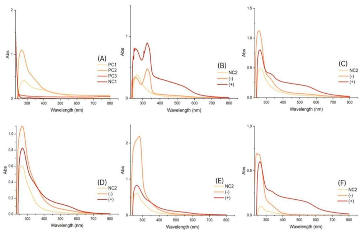
Figure 1. UV-visible absorption spectra of algal-derived Se-NPs with ascorbic acid (+) and without (-). (A) controls including (PC1) 2% bovine serum albumin, selenious acid, and ascorbic acid; (PC2) selenious acid and ascorbic acid; (PC3) commercially synthesised Se-NPs; (NC1) selenious acid, with y-axis range: 0-2 Abs; (B) NC2, P. palmata; (C) U. intestinalis; (D) Fucus vesiculosus; (E) F. serratus; (F) L. digitata.
Preprints 225435 g001
Figure 2. X-ray diffraction (XRD) patterns of macroalgal-based Se-NPs synthesised with (+) or without (-) ascorbic acid. The peaks correspond to hexagonal and monoclinic β-selenium phases, confirming the crystalline structure of the NPs: (A) P. palmata (+), (B) P. palmata (-), (C) U. intestinalis (+), (D) U. intestinalis (-), (E) F. vesiculosus (+), (F) F. vesiculosus (-), (G) F. serratus (+), (H) F. serratus (-), (I) L. digitata (+), and (J) L. digitata (-).
Preprints 225435 g002
Figure 3. TEM micrographs of Se-NPs derived from macroalgae extracts. Images on the left are groups with ascorbic acid (+); images on the right are groups without ascorbic acid (-). Control groups: (A) PC3 (scale bar: 100 nm); (B&C) ˆ(scale bar: 100 nm); (D&E) ˆ; (F&G) ˆ(+) (scale bar: 200 nm left, 500 nm right); (H&I) ˆ(scale bar: 500 nm left, 200 nm right); (J&K) ˆ(scale bar: 200 nm left, 100 nm right); the inset histograms represent the corresponding particle size distribution.
Preprints 225435 g003
Figure 4. SEM micrographs of macroalgae-mediated Se-NPs under ascorbic acid assisted conditions with their corresponding EDX for elemental compositions: (A) P. palmata, (B) U. intestinalis, (C) F. vesiculosus, (D) F. serratus, (E) L. digitata.
Preprints 225435 g004
Figure 5. Dose-response curves for anti-M. tuberculosis REMA assay against five M. tuberculosis strains. A: mc27902; B: mc28245 (INHR); C: mc28247 (RIFR); D: mc28250 (RIFR & INHR); and E: mc28258 (RIFR & INHR). Se-NPs were synthesised using Palmaria palmata, Ulva intestinalis, Fucus vesiculosis, F. serratus, and Laminaria digitata. Rifampicin (Rif), isoniazid (Inh), and ethambutol (Emb), alongside commercially synthesised Se-NPs (PC3), and Ag-NPs were included as reference antimicrobial agents. Curves represent the means and standard errors for three combined independent replicates; flat-fitted lines indicate the inability to fit a sigmoidal model and no inhibitory activity.
Preprints 225435 g005aPreprints 225435 g005b
Figure 6. TEM images of studying the actual interaction of Se-NP with M. tuberculosis; A) untreated cells showing the thick, dense cell envelope and overlapping cells; B) untreated cell with maximised magnification; C) M. tuberculosis exposed to biogenic selenium nanoparticles (Se-NPs); D) maximised exposure of a single M. tuberculosis to Se-NPs, showing small, dark particles potentially attached or embedded.
Preprints 225435 g006
Table 1. Comparative XRD analysis of algal-mediated Se-NPs synthesised with and without ascorbic acid: phase composition and crystallinity based on the International Centre for Diffraction Data (ICDD).
Algal species Treated group Corresponding ICDD reference
Hexagonal
Corresponding ICDD reference
Monoclinic
P. palmata (+) 01-073-0465 00-024-0714
(-) 01-073-0465 00-054-0500
U. intestinalis (+) - 01-075-1162
(-) - 00-024-0714
F. vesiculosus (+) 00-006-0362 01-075-1162
(-) - 01-076-1865
F. serratus (+) 03-056-1876 00-024-0714
(-) - 01-076-1865
L. digitata (+) 01-073-0465 01-076-1865
(-) 01-073-0465 01-076-1865
Table 2. Summary of DLS and zeta potential measurements of macroalgal-mediated Se-NPs. .
Algal species or control Panel Treated group Avg. Size (nm) PDI Zeta Potential (mV)
PC3 A NA 187 ± 2.15 0.09 ± 0.03 -27.2 ± 0.72
P. palmata B (+) 53 ± 0.5 0.24 ± 0.01 - 20 ± 2
C (-) 83.2 ± 0.8 0.28 ± 0.007 -29 ± 2.6
U. intestinalis D (+) 138.5 ± 3.8 0.32 ± 0.04 25.4 ± 1.3
E (-) 249 ± 1.9 0.14 ± 0.14 -27.4
F. vesiculosus F (+) 119.7 ± 0 0.17 ± 0.016 -26.4
G (-) 232.6 ± 1.5 0.26 ± 0.016 -26.6 ± 6.3
F. serratus H (+) 174.8 ± 3.4 0.2 ± 0.009 -20.2 ± 0.6
I (-) 426.6 ± 8.7 0.24 ± 0.04 -26.3 ± 1.6
L. digitata J (+) 330 ± 7.2 0.27 ± 0.5 -28.5 ± 1.7
K (-) 302 ± 22 0.36 ± 0.09 -24.7 ± 1
Table 3. Comparing REMA assay results of macroalgae-based Se-NP of both treated groups (+, -) against targeted M. tuberculosis strains, including mc27902, mc28245, mc28247, mc28250, mc28258, and common anti-TB; both MIC₅₀ and MIC₉₀ (ranging from 1280 to 0.05 μg/mL) are expressed as mean ± standard error (SEM); ND activities were not determined (no activity was determined); NC1 selenious acid; NC2algal extract; Ag-NP silver nanoparticles; Rif rifampicin; Inh isoniazid; Emb ethambutol; PC3 commercially synthesised selenium nanoparticles.
Algae species Treated group M. tuberculosis strains
mc27902 mc28245 (INHR) mc28247 (RIFR) mc28250 (RIFR & INHR) mc28258 (RIFR & INHR)
MIC₅₀ MIC₉₀ MIC₅₀ MIC₉₀ MIC₅₀ MIC₉₀ MIC₅₀ MIC₉₀ MIC₅₀ MIC₉₀
P. palmata NC2 ND ND ND ND ND ND ND ND ND ND
(+) 14.64 ± 1.35 846.14 ± 3.72 ND ND 48.3 ± 1.31 > 1280 8.45 ± 1.32 516 ± 4.3 11.6 ± 1.2 119.7 ± 2.4
(-) 27.85 ± 1.11 48.02 ± 1.23 2.73 ± 2.6 179.9 ± 67 66.7 ± 1.5 > 1280 17.5 ± 1.2 180.7 ± 2.1 16.7 ± 1.2 171.4 ± 2.4
U. intestinalis NC2 ND ND ND ND ND ND ND ND ND ND
(+) 15.06 ± 1.27 304.26 ± 2.58 ND ND 48.5 ± 1.3 > 1280 7.85 ± 1.2 75.2 ± 2 11 ± 1.3 728.6 ± 4.1
(-) 18.34 ± 1.11 40.21 ± 1.31 ND ND 60.8 ± 1.3 > 1280 5.5 ± 1.3 288 ± 3.6 13 ± 1.3 511 ± 4
F. vesiculosus NC2 ND ND ND ND ND ND ND ND ND ND
(+) 59 ± 1.13 175.6 ± 1.3 10.5 ± 2 > 1280 198.9 ± 1.3 > 1280 25.1 ± 1.3 > 1280 36 ± 1.1 88 ± 1.3
(-) 131.2 ±
1.2
324.5 ± 1.6 10.22 ± 1.2 101 ± 2.07 203.3 ± 1.1 995 ± 1.6 58.4 ± 1.1 148.4 ± 1.3 34.9 ± 1.55 > 1280
F. serratus NC2 ND ND ND ND ND ND ND ND ND ND
(+) 19 ± 1.2 192.1 ± 2.1 ND ND 60.6 ± 1.3 > 1280 19.2 ± 1.3 324.9 ± 2.6 33.5 ± 1.1 93.6 ± 1.4
(-) 28.3 ± 1.1 51.3 ± 1.36 1.22 ± 1.5 153.3 ± 695 59.4 ± 1.3 > 1280 21.7 ± 1.3 > 1280 30.5 ± 1.2 174.7 ± 1.7
L. digitata NC2 ND ND ND ND ND ND ND ND ND ND
(+) 192.10 ± 2.1 906.6 ± 3.04 2.51 ± 3.61 > 1280 54.4 ± 1.15 237 ± 1.4 12.6 ± 1.1 46.6 ± 1.6 26.6 ± 1.2 43.4 ± 1.4
(-) 294.3 ± 1.1 673.4 ± 1.48 24.6 ± 1.35 > 1280 ND ND 168 ± 1.1 321.1 ± 1.3 227.3 ± 1.18 519 ± 1.4
NC1 ND ND ND ND ND ND ND ND ND ND
Ag-NP 35.6 ± 1.15 1076 ± 1.77 39.3 ± 1.2 697 ± 2.3 88.2 ± 1.2 1043 ± 1.9 282.4 ± 1.11 320.2 ± 1.3 258.7 ± 1.4 > 1280
RIF 0.07 0.61 0.07 0.58 ND ND ND ND ND ND
INH 3.62 13.23 ND ND 5.19 8.97 ND ND 336.02 ND
EMB 6.88 12.54 8.47 12.20 14.34 24.91 8.79 20.80 14.33 24.84
PC3 3.64 ± 1.5 1011.8 ± 31.3 7.57 ± 2.9 > 1280 123.4 ± 2.3 > 1280 ND ND 1.7 ± 1 > 1280
Table 4. Mann-Whitney U test results comparing inhibitory effects of macroalgae-derived Se-NPs for both MIC₅₀ and MIC₉₀ with and without ascorbic acid; n.s., non-significant (p ≥ 0.05); *, significant (p < 0.05); **, highly significant (p < 0.01); ***, very highly significant (p < 0.001). Values are presented as medians. .
Algae species
(Median)
With AA
(Median)
Without AA
(Median)
U Z p-value Significance
MIC₅₀ MIC₉₀ MIC₅₀ MIC₉₀ MIC₅₀ MIC₉₀ MIC₅₀ MIC₉₀ MIC₅₀ MIC₉₀ MIC₅₀ MIC₉₀
P. palmata 1.1 2.75 1.39 1.86 56 116 -1.44 1.58 0.15 0.11 n.s. n.s.
U. intestinalis 1.10 1.89 1.25 1.69 70 84 -0.72 0 0.47 1.00 n.s. n.s.
F. vesiculosus 1.51 2.16 1.89 2.23 78 106 -1.41 0.02 0.16 0.98 n.s. n.s.
F. serratus 1.43 1.71 1.49 2.17 74 55 -0.51 -1.22 0.61 0.22 n.s. n.s.
L. digitata 1.39 2.78 2.24 2.80 19 71 -3.59 -0.94 0.0003 0.34 *** n.s.
Table 5. Mycobacterium tuberculosis strains; (INHR), isoniazid resistant; (RIFR), rifampicin resistant.
Plasmid and Strain Description (including genotype) Source or reference
Plasmids
pMV261 E. coli-mycobcaterial shuttle plasmid, hsp60 promoter, KanR Stover et al., 1991
Strains
Mtb mc27902 ΔleuCD ΔpanCD ΔargB, Leucine, pantothenate and arginine triple auxotroph Vilcheze et al., 2018
Mtb mc28245 (INHR) mc27902 derived Δ2116169–2162530; Δ2116169–2162530 genome deletion, INHR Vilcheze et al., 2018
Mtb mc28247 (RIFR) mc27902 derived, rpoB (H445Y); rpoB His445 → Lys, RIFR Vilcheze et al., 2018
Mtb mc28250 (RIFR & INHR) mc28247 derived, rpoB (H445Y) Δ2122397–2170320; rpoB His445 → Lys, RIFR, Δ2122397–2170320 genome deletion, INHR Vilcheze et al., 2018
Mtb mc28258 (RIFR & INHR) mc28247 derived, rpoB (H445Y) katG (W438R); rpoB His445 → Lys, RIFR, katG Trp438 → Arg, INHR Vilcheze et al., 2018
Mtb mc27902 pMV261 Mtb mc27902 containing pMV261, KanR Lu et al. 2025
Mtb mc28245 pMV261 Mtb mc28245 containing pMV261, KanR Lu et al. 2025
Mtb mc28247 pMV261 Mtb mc28247 containing pMV261, KanR Lu et al. 2025
Mtb mc28250 pMV261 Mtb mc28250 containing pMV261, KanR Lu et al. 2025
Mtb mc28258 pMV261 Mtb mc28258 containing pMV261, KanR Lu et al. 2025
Disclaimer/Publisher’s Note: The statements, opinions and data contained in all publications are solely those of the individual author(s) and contributor(s) and not of MDPI and/or the editor(s). MDPI and/or the editor(s) disclaim responsibility for any injury to people or property resulting from any ideas, methods, instructions or products referred to in the content.
Copyright: This open access article is published under a Creative Commons CC BY 4.0 license, which permit the free download, distribution, and reuse, provided that the author and preprint are cited in any reuse.
Prerpints.org logo

Preprints.org is a free preprint server supported by MDPI in Basel, Switzerland.

Subscribe

© 2026 MDPI (Basel, Switzerland) unless otherwise stated

Accessibility

Disclaimer

Terms of Use

Privacy Policy

Privacy Settings