
Submitted:
28 July 2026
Posted:
29 July 2026
You are already at the latest version
Abstract
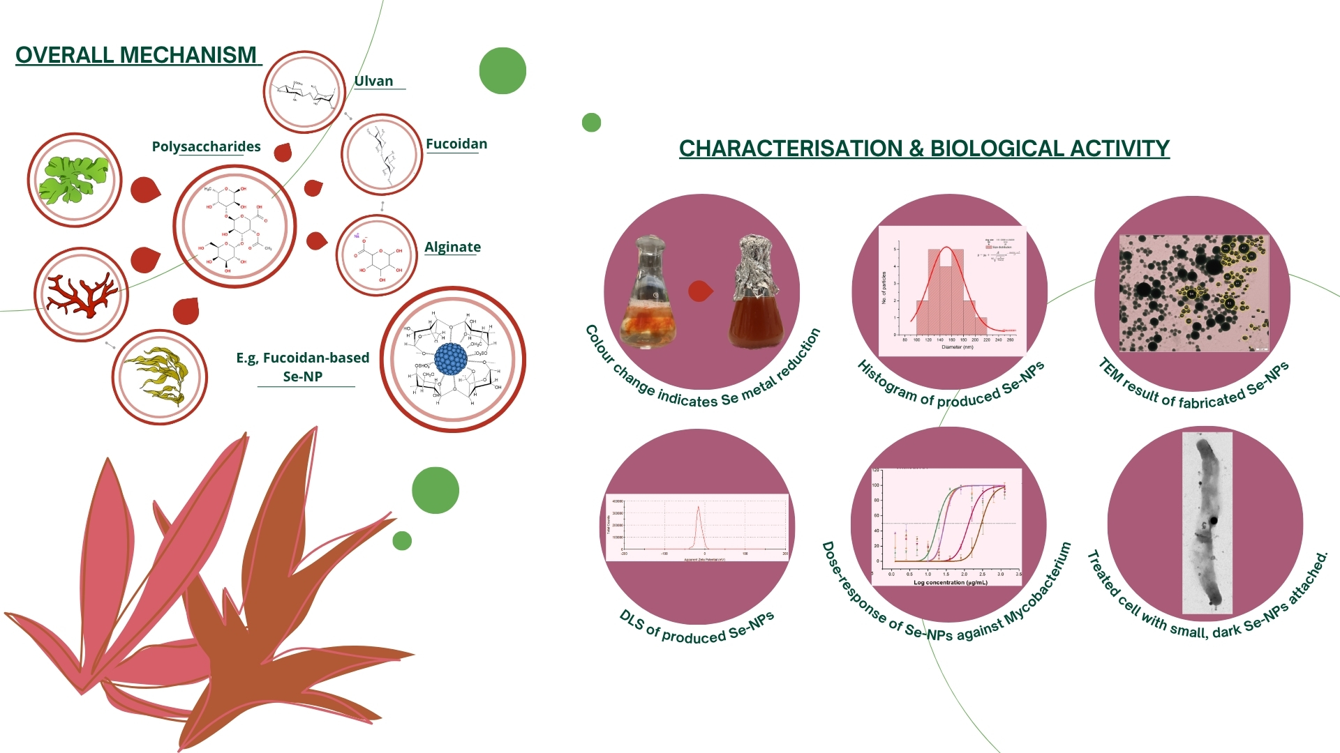
The global rise of multidrug-resistant tuberculosis has intensified the need for novel and sustainable therapeutics that can overcome the limitations of conventional antibiotics, including toxicity and declining efficacy. Selenium nanoparticles (Se-NPs) are a biocompatible and multifunctional alternative, yet most studies rely on chemically synthesised forms. This study investigated the potential of extracts from marine macroalgae (Palmaria palmata, Ulva intestinalis, Fucus vesiculosus, F. serratus, and Laminaria digitata), with and without ascorbic acid supplementation, to function as reducing and stabilising agents. Se-NPs were successfully synthesised without chemical reductants, maintaining comparable size, morphology, and stability to their ascorbic acid-aided counterparts and were systematically characterised using UV-Vis spectroscopy, FTIR, XRD, DLS, zeta potential, TEM, SEM, and EDX techniques. Biological efficacy was assessed against five Mycobacterium tuberculosis strains with varying resistance phenotypes. Crude macroalgae extracts displayed no intrinsic antimycobacterial activity, whereas Se-NPs exhibited robust and broad-spectrum inhibition, with minimum inhibitory concentrations (MIC50 and MIC90) in several cases comparable to reference antibiotics. Se-NPs derived from P. palmata and U. intestinalis were the most effective, including against resistant strains. This is, to the best of our knowledge, the first study to evaluate Se-NPs synthesised from macroalgae against M. tuberculosis. The findings highlight their potential as sustainable, low-cost, and biocompatible nanotherapeutics for combating wild-type and multidrug-resistant tuberculosis.
Keywords:
1. Introduction
2. Results and Discussion
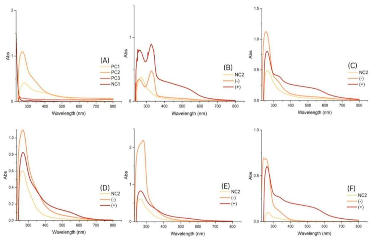
2.1. UV-Vis Spectroscopy
2.2. Fourier Transform Infrared Spectroscopy
2.3. Powder X-Ray Diffraction
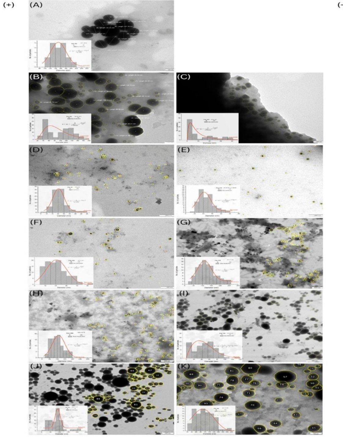
2.4. Transmission Electron Microscopy (TEM)
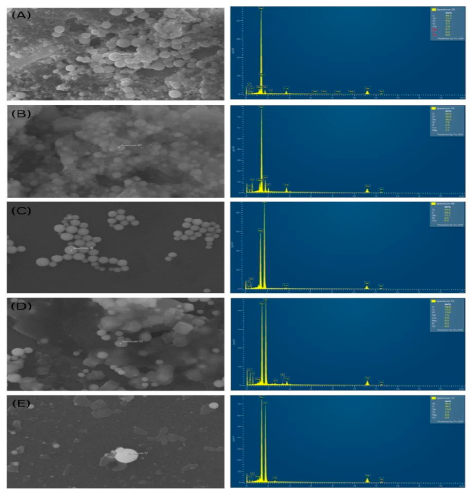
2.5. Scanning Electron Microscopy-Energy-Dispersive X-Ray Spectroscopy (SEM-EDX)
2.6. Dynamic Light Scattering (DLS) and Zeta-Potential
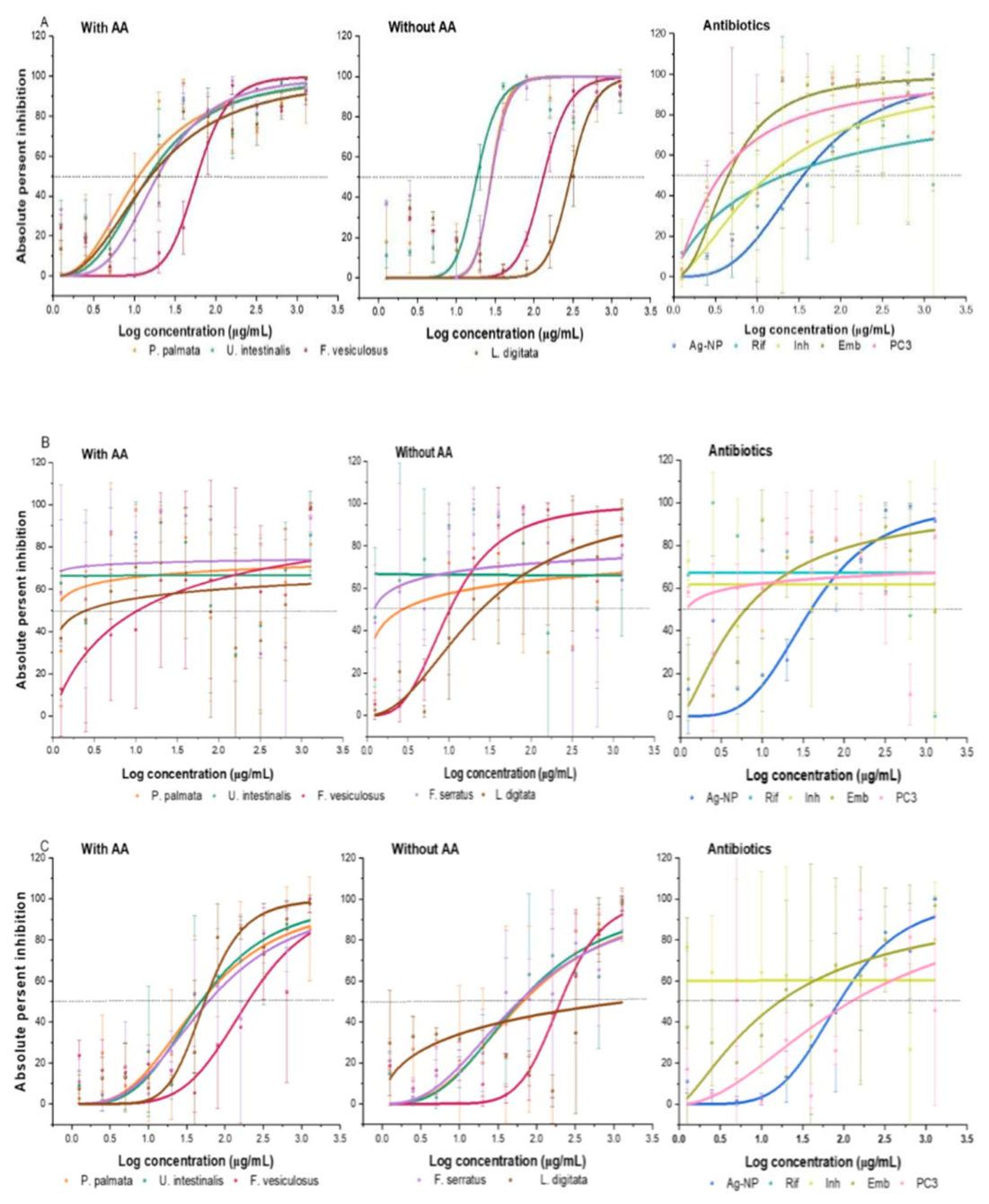
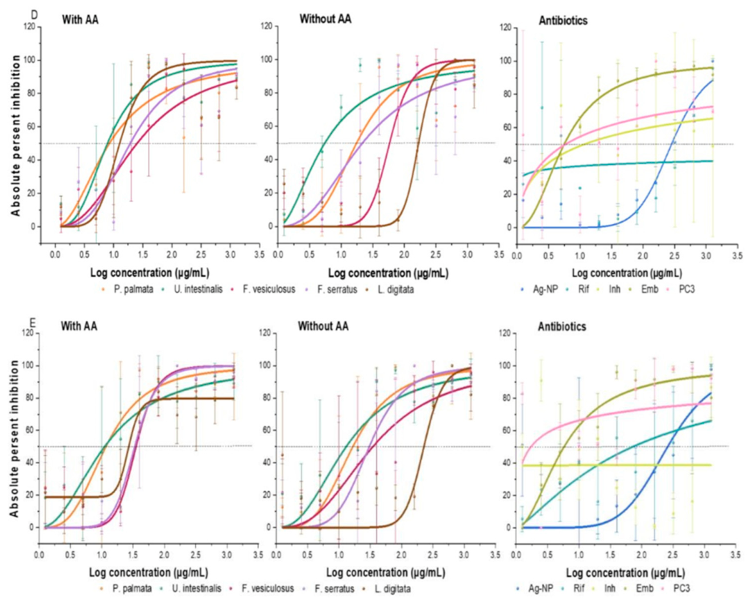
2.7. Antimycobacterial Activity
2.8. Comparative Benchmarking
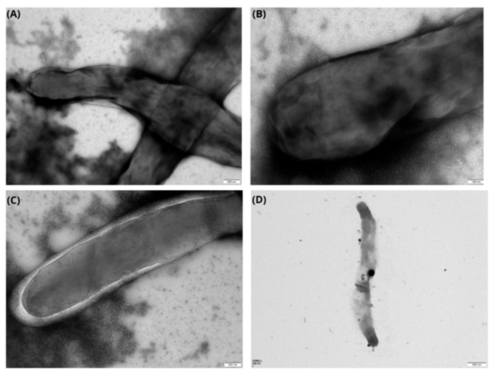
2.9. Se-NPs Mode of Action
3. Materials and Methods
3.1. Sample Collection and Preparation
3.2. Biosynthesis of Selenium Nanoparticles
3.3. Nanoparticle Characterisation: UV-Vis Spectroscopy
3.4. Fourier Transform Infrared Spectroscopy
3.5. X-Ray Diffraction Spectroscopy
3.6. Dynamic Light Scattering and Zeta Potential
3.7. Transmission Electron Microscopy
3.8. Scanning Electron Microscopy and Energy-Dispersive X-Ray Spectroscopy
3.9. Antibacterial Efficacy
3.10. Resazurin Microtiter Assay
3.11. Se-NPs with M. tuberculosis Interaction
3.12. Statistical Analysis
- y is the observed response (percentage inhibition);
- x is the concentration of inhibitor;
- START is the baseline response (bottom asymptote); END is the maximum response (top asymptote);
- k represents the IC50 value (the concentration at half-maximal effect);
- n is the Hill slope, indicating the steepness of the curve.
4. Conclusion
Supplementary Materials
Author Contributions
Conflicts of Interest
Acknowledgments
Abbreviations
| (+) | Targeted group 1 (with ascorbic acid) |
| (–) | Targeted group 2 (without AA) |
| FTIR | Fourier transform infrared spectroscopy |
| MDR | Multi-drug-resistance pathogens |
| MDR-TB | Multidrug-resistant TB |
| OADC | Oleic acid, albumin, dextrose, and catalase |
| PBS | Phosphate-buffered saline |
| PC1 | Positive control (2% bovine serum albumin + SeA + A |
| PC2 | (Chemical approach) 30 mM SeA (donating) + 40 mM AA (reducing agent) |
| PC3 | Commercially synthesised selenium nanoparticles |
References
- WHO. Global tuberculosis report 2025. 2025.
- Nair, A.; Greeny, A.; Nandan, A.; Sah, R.K.; Jose, A.; Dyawanapelly, S.; Junnuthula, V.; Sadanandan, P. Advanced drug delivery and therapeutic strategies for tuberculosis treatment. J. Nanobiotech. 2023, 21, 414. [Google Scholar] [CrossRef] [PubMed]
- Velayati, A.A.; Masjedi, M.R.; Farnia, P.; Tabarsi, P.; Ghanavi, J.; ZiaZarifi, A.H.; Hoffner, S.E. Emergence of new forms of totally drug-resistant tuberculosis bacilli: super extensively drug-resistant tuberculosis or totally drug-resistant strains in Iran. CHES 2009, 136, 420–425. [Google Scholar] [CrossRef] [PubMed]
- Rajput, A.; Mandlik, S.; Pokharkar, V. Nanocarrier-based approaches for the efficient delivery of anti-tubercular drugs and vaccines for management of Tuberculosis. Front. Pharma. 2021, 12. [Google Scholar] [CrossRef]
- Kia, P.; Ruman, U.; Pratiwi, A.R.; Hussein, M.Z. Innovative therapeutic approaches based on nanotechnology for the treatment and management of tuberculosis. IJN Vol. 2023(18), 1159–1191. [CrossRef] [PubMed]
- Ramalingam, V.; Sundaramahalingam, S.; Rajaram, R. Size-dependent antimycobacterial activity of titanium oxide nanoparticles against Mycobacterium tuberculosis. J. Mater. Chem. B 2019, 7, 4338–4346. [Google Scholar] [CrossRef]
- Guo, X.; Zuo, X.; Zhou, Z.; Gu, Y.; Zheng, H.; Wang, X.; Wang, G.; Xu, C.; Wang, F. PLGA- based micro/nanoparticles: an overview of their applications in respiratory diseases. Int. J. Mol. Sci. 2023, 24, 4333. [Google Scholar] [CrossRef] [PubMed]
- Valea, A.; Georgescu, C.E. Selenoproteins in Human Body: Focus on Thyroid Pathophysiology. Hormo 2018, 17, 183–196. [Google Scholar] [CrossRef] [PubMed]
- Srivastava, N.; Mukhopadhyay, M. Green synthesis and structural characterisation of selenium nanoparticles and assessment of their antimicrobial property. Bioproc. Biosys. Eng. 2015, 38, 1723–1730. [Google Scholar] [CrossRef] [PubMed]
- Mikhailova, E.O. Selenium nanoparticles: green synthesis and biomedical application. Molec 2023, 28, 8125. [Google Scholar] [CrossRef] [PubMed]
- Bisht, N.; Phalswal, P.; Khanna, P.K. Selenium nanoparticles: a review on synthesis and biomedical applications. Mater. Adv. 2022, 3, 1415–1431. [Google Scholar] [CrossRef]
- Mostafavi, E.; Medina-Cruz, D.; Truong, L.B.; Kaushik, A.; Iravani, S. Selenium-based nanomaterials for biosensing applications. Mater. Adv. 2022, 3, 7742–7756. [Google Scholar] [CrossRef] [PubMed]
- Prasad, R.; Jha, A.; Prasad, K. (Eds.) Exploring the Realms of Nature for Nanosynthesis, 1st ed.; Spring. Cham., 2018. [Google Scholar] [CrossRef]
- Yadav, R.K.; Jangeer, S.; Parashar, R.; Sharma, G.; Meena, P.; Kumar, M.; Patel, D.D. A critical review on nanoparticles synthesis: Physical, chemical and biological perspectives. IJCRT 2023, 11. [Google Scholar]
- Cruz, D.M.; Mi, G.; Webster, T.J. Synthesis and characterization of biogenic selenium nanoparticles with antimicrobial properties made by Staphylococcus aureus, methicillin-resistant Staphylococcus aureus (MRSA), Escherichia coli, and Pseudomonas aeruginosa. J. Biomed. Mate. Res. Part A 2018, 106, 1400–1412. [Google Scholar] [CrossRef]
- Greeshma, B.C.; Mahesh, M. Biosynthesis of selenium nanoparticles from Bacillus species and its applications. J. Appl. Natu. Sci. 2019, 11, 810–815. [Google Scholar] [CrossRef]
- Huang, Y.; Hsu, J.C.; H. Koo, D.P. Cormode, repurposing ferumoxytol: diagnostic and therapeutic applications of an FDA-approved nanoparticle. Theranost 2022, 12, 796–816. [Google Scholar] [CrossRef] [PubMed]
- Shakeri, F.; Zaboli, F.; Fattahi, E.; Babavalian, H. Biosynthesis of selenium nanoparticles and evaluation of its antibacterial activity against Pseudomonas aeruginosa Adva. Mater. Sci. Engin. 2022, 2022, e4118048. [Google Scholar] [CrossRef]
- Hariharan, S.; Chauhan, S.; Velu, K.; Dharmaraj, S.; C M, V.K.; Ganesan, S. Biological activities of selenium nanoparticles synthesized from Camellia sinensis (L) Kuntze leaves. Appl. Bioch. Biotechnol. 2023, 195, 5823–5837. [Google Scholar] [CrossRef] [PubMed]
- Asaad, A.M.; Saied, S.A.; Torayah, M.M.; Abu-Elsaad, N.I.; Awad, S.M. Antibacterial activity of selenium nanoparticles/copper oxide (SeNPs/CuO) nanocomposite against some multi- drug resistant clinical pathogens. BMC Microbio 2025, 25 33. [Google Scholar] [CrossRef]
- LewisOscar, F.; Vismaya, S.; Arunkumar, M.; Thajuddin, N.; Dhanasekaran, D.; Nithya, C. Algal Nanoparticles: Synthesis and Biotechnological Potentials. In Algae - Organisms for Imminent Biotechnology, 1st ed.; Intech., 2016; p. 342. [Google Scholar] [CrossRef] [PubMed]
- Behera, M.; Behera, P.R.; Bhuyan, P.P.; Singh, L.; Pradhan, B. Algal nanoparticles and their antibacterial activity: current research status and future prospectives. Drug. Dru. Candid. 2023, 2, 554–570. [Google Scholar] [CrossRef]
- Uzair, B.; Liaqat, A.; Iqbal, H.; Menaa, B.; Razzaq, A.; Thiripuranathar, G.; Fatima Rana, N.; Menaa, F. Green and cost-effective synthesis of metallic nanoparticles by algae: safe methods for translational medicine. Bioengin 2020, 7, 129. [Google Scholar] [CrossRef] [PubMed]
- El-Kassas, H.Y.; El-Sheekh, M.M. Cytotoxic activity of biosynthesized gold nanoparticles with an extract of the red seaweed Corallina officinalis on the MCF-7 human breast cancer cell line, Asia. Pacif. J. Canc. Preven. 2014, 15, 4311–4317. [Google Scholar] [CrossRef] [PubMed]
- Costa, L.H.; Hemmer, J.V.; Wanderlind, E.H.; Gerlach, O.M.S.; Santos, A.L.H.; Tamanaha, M.S.; Bella-Cruz, A.; Corrêa, R.; Bazani, H.A.G.; Radetski, C.M.; et al. Green synthesis of gold nanoparticles obtained from algae Sargassum cymosum: Optimization, characterization and stability. BioNanoSci 2020, 10, 1049–1062. [Google Scholar] [CrossRef]
- El-Rafie, H.M.; El-Rafie, M.H.; Zahran, M.K. Green synthesis of silver nanoparticles using polysaccharides extracted from marine macro algae. Carbohyd. Polym. 2013, 96, 403–410. [Google Scholar] [CrossRef] [PubMed]
- Mohandass, C.; Vijayaraj, A.S.; Rajasabapathy, R.; Satheeshbabu, S.; Rao, S.V.; Shiva, C.; De-Mello, L. Biosynthesis of silver nanoparticles from marine seaweed Sargassum cinereum and their antibacterial activity. Indi. J. Pharmace. Sci. 2013. [Google Scholar] [CrossRef]
- Abdel-Raouf, N.; Alharbi, R.; Al-Enazi, N.; Alkhulaifi, M.; Ibraheem, I. Rapid biosynthesis of silver nanoparticles using the marine red alga Laurencia catarinensis and their characterization. Beni-Suef Uni. J. Basi. Appli. Sci. 2017, 7. [Google Scholar] [CrossRef]
- Pugazhendhi, A.; Prabakar, D.; Jacob, J.M.; Karuppusamy, I.; Saratale, R.G. Synthesis and characterization of silver nanoparticles using Gelidium amansii and its antimicrobial property against various pathogenic bacteria. Microb. Pathog. 2018, 114, 41–45. [Google Scholar] [CrossRef] [PubMed]
- Ishwarya, R.; Vaseeharan, B.; Kalyani, S.; Banumathi, B.; Govindarajan, M.; Alharbi, N.S.; Kadaikunnan, S.; Al-anbr, M.N.; Khaled, J.M.; Benelli, G. Facile green synthesis of zinc oxide nanoparticles using Ulva lactuca seaweed extract and evaluation of their photocatalytic, antibiofilm and insecticidal activity. J. Photochem. Photobio. B Biol. 2018, 178, 249–258. [Google Scholar] [CrossRef] [PubMed]
- Prasad, E.R.; Sivakumar, S.R. Synthesis of zinc oxide nanoparticles (ZNO-NPS) via green approach for enhanced antimicrobial activity against human pathogenic bacteria (HPB) strains. Int. J. Sci. Techno. Rese. 2020, 9, 3403–3409. [Google Scholar]
- Modafer, Y.A. Synthesis of zinc oxide nanoparticles via biomass of Hypnea pannosa as a green mediator and their biological applications. BioRe 2024, 19, 3771–3792. [Google Scholar] [CrossRef]
- Estevez, H.; Palacios, A.; Gil, D.; Anguita, J.; Vallet-Regi, M.; González, B.; Prados- Rosales, R.; Luque-Garcia, J.L. Antimycobacterial effect of selenium nanoparticles on Mycobacterium tuberculosis Front. Microbiol. 2020, 11. [Google Scholar] [CrossRef] [PubMed]
- Abubakar, A.R.; Haque, M. Preparation of medicinal plants: basic extraction and fractionation procedures for experimental purposes. J. Pharma. Bioall. Sci. 2020, 12 1. [Google Scholar] [CrossRef] [PubMed]
- González-Ballesteros, N.; Prado-López, S.; Rodríguez-González, J.B.; Lastra, M.; Rodríguez-Argüelles, M.C. Green synthesis of gold nanoparticles using brown algae Cystoseira baccata: Its activity in colon cancer cells. Coll. Surf. B Biointerfaces 2017, 1, 190–198. [Google Scholar] [CrossRef] [PubMed]
- Deans, B.J.; Just, J.; Smith, J.A.; Bissember, A.C. Development and applications of water- based extraction methods in natural products isolation chemistry, Asia. J. Orga. Chem. 2020, 9, 1144–1153. [Google Scholar] [CrossRef]
- Vikneshan, M.; Saravanakumar, R.; Mangaiyarkarasi, R.; Rajeshkumar, S.; Samuel, S.R.; Suganya, M.; Baskar, G. Algal biomass as a source for novel oral nano-antimicrobial agent. Saud. J. Biolo. Sci. 2020, 27, 3753–3758. [Google Scholar] [CrossRef] [PubMed]
- Siddique, M.A.R.; Khan, M.; Bokhari, S.A.I.; Ismail, M.; Ahmad, K.; Haseeb, H.A.; Kayani, M.M.; Khan, S.; Zahid, N.; Khan, S.B. Ascorbic acid-mediated selenium nanoparticles as potential antihyperuricemic, antioxidant, anticoagulant, and thrombolytic agents, Gree. Proce. Synth. 2024, 13. [Google Scholar] [CrossRef]
- Gangadoo, S.; Stanley, D.; Hughes, R.; Moore, R.; Chapman, J. The synthesis and characterisation of highly stable and reproducible selenium nanoparticles. Inorg. Nano-Metal. Chem. 2017, 47. [Google Scholar] [CrossRef]
- Infrared Spectroscopy Absorption Table. Chemistry LibreTexts. Available online: https://chem.libretexts.org/Ancillary_Materials/Reference/Reference_Tables/Spectroscopic_Refe.
- FTIR Functional Group Database Table with Search, InstaNANO. Available online: https://instanano.com/all/characterization/ftir/ftir-functional-group-search/.
- Javad, S.; Butt, A. (Eds.) Nanobotany, 1st ed.; Springer Cham: Cham, 2018. [Google Scholar] [CrossRef]
- Vilchèze, C.; Copeland, J.L.; Keiser, T.L.; Weisbrod, T.; Washington, J.; Jain, P.; Malek, A.; Weinrick, B.; Jacobs, W.R. Rational design of biosafety level 2-approved, multidrug-resistant strains of Mycobacterium tuberculosis through nutrient auxotrophy. mBio 2018, 9, e00938-18. [Google Scholar] [CrossRef] [PubMed]
- Lu, Y.; Partleton, D.; Gugu, F.M.; Alhejaili, A.Y.G.; Norris, S.; Harburn, J.J.; Gill, J.H.; Sellars, J.D.; Brown, A.K. Structural isomerisation affects the antitubercular activity of adamantyl- isoxyl adducts. J. Enzy. Inhibi. Medic. Chem. 2025, 40, 2502600. [Google Scholar] [CrossRef] [PubMed]
- Glauert, A.M.; Lewis, P.R. Biological Specimen Preparation for Transmission Electron Microscopy, Princ. Uni. Pres; 1998; Available online: https://www.jstor.org/stable/j.ctt7ztxpn.
- Srinivasan, B.; Lloyd, M.D. Quantitation and error measurements in dose–response curves. J. Med. Chem. 2025, 68, 2052–2056. [Google Scholar] [CrossRef] [PubMed]
- Dytham, Choosing and Using Statistics: a Biologist’s Guide, 3rd ed.; Wiley-Blackwell: Hoboken, NJ, 2011.
- Bartosiak, M.; Giersz, J.; Jankowski, K. Analytical monitoring of selenium nanoparticles green synthesis using photochemical vapor generation coupled with MIP-OES and UV–Vis spectrophotometry. Microchem. J. 2019, 145, 1169–1175. [Google Scholar] [CrossRef]
- Mansouri-Tehrani, H.A.; Keyhanfar, M.; Behbahani, M.; Dini, G. Synthesis and characterization of algae-coated selenium nanoparticles as a novel antibacterial agent against Vibrio harveyi, a Penaeus vannamei pathogen. Aquacul 2021, 534. [Google Scholar] [CrossRef]
- Chapman, J.; Weir, E.; Fiona, R. Period four metal nanoparticles on the inhibition of biofouling. Coll. Sur. B. Biointerf. 2010, 78, 208–16. [Google Scholar] [CrossRef] [PubMed]
- Princy, K.F.; Gopinath, A. Spectroscopic investigation on the catalytic efficacy of biofabricated gold nanoparticles using marine macroalgae. Mater. Tod. Proce. 2019, 9, 38–45. [Google Scholar] [CrossRef]
- Hashem, A.H.; Salem, S.S. Green and ecofriendly biosynthesis of selenium nanoparticles using Urtica dioica (Stinging nettle) leaf extract: antimicrobial and anticancer activity. Biotech. J. 2022, 17, 2100432. [Google Scholar] [CrossRef] [PubMed]
- Vahdati, M.; Tohidi Moghadam, T. Synthesis and characterization of selenium nanoparticles-lysozyme nanohybrid system with synergistic antibacterial properties. Sci. Rep. 2020, 10, 510. [Google Scholar] [CrossRef] [PubMed]
- Shar, A.H.; Lakhan, M.N.; Wang, J.; Ahmed, M.; Alali, K.T.; Ahmed, R.; Ali, I.; Dayo, A.Q. Facile synthesis and characterization of selenium nanoparticles by the hydrothermal approach. Dig. J. Nanomater. Biostruct. 2019, 14. [Google Scholar]
- Hentati, F.; Delattre, C.; Gardarin, C.; Desbrières, J.; Le Cerf, D.; Rihouey, C.; Michaud, P.; Abdelkafi, S.; Pierre, G. Structural features and rheological properties of a sulfated xylogalactan-rich fraction isolated from Tunisian red seaweed Jania adhaerens Appli. Sci. 2020, 10, 1655. [Google Scholar] [CrossRef]
- Padman, A.J.; Henderson, J.; Hodgson, S.; Rahman, P.K.S.M. Biomediated synthesis of silver nanoparticles using Exiguobacterium mexicanum. Lett. 2014, 36, 2079–2084. [Google Scholar] [CrossRef] [PubMed]
- Belattmania, Z.; Kaidi, S.; El Atouani, S.; Katif, C.; Bentiss, F.; Jama, C.; Reani, A.; Sabour, B.; Vasconcelos, V. Isolation and FTIR-ATR and 1H NMR characterization of alginates from the main alginophyte species of the Atlantic coast of Morocco. Molec 2020, 25, 4335. [Google Scholar] [CrossRef] [PubMed]
- Menazea, A.A.; Ismail, A.M.; Awwad, N.S.; Ibrahium, H.A. Physical characterization and antibacterial activity of PVA/Chitosan matrix doped by selenium nanoparticles prepared via one- pot laser ablation route. J. Mater. Res. Tech. 2020, 9, 9598–9606. [Google Scholar] [CrossRef]
- Alzoubi, F.Y.; Alzouby, J.Y.; Alqadi, M.K.; Alshboul, H.A.; Aljarrah, K.M. Synthesis and characterization of colloidal gold nanoparticles controlled by the pH and ionic strength. Chin. J. Phys. 2015, 53, 100801-1-100801–9. [Google Scholar] [CrossRef]
- Naghavi, M.; Vollset, S.E.; Ikuta, K.S.; Swetschinski, L.R.; Gray, A.P.; Wool, E.E.; Aguilar, G.R.; Mestrovic, T.; Smith, G.; Han, C.; et al. Global burden of bacterial antimicrobial resistance 1990– 2021: a systematic analysis with forecasts to 2050. The Lanc. 2024, 404, 1199–1226. [Google Scholar] [CrossRef] [PubMed]
- Thanh, N.T.K.; Green, L.A.W. Functionalisation of nanoparticles for biomedical applications. Nan. Tod. 2010, 5, 213–230. [Google Scholar] [CrossRef]
- Sharma, G.; Sharma, A.R.; Bhavesh, R.; Park, J.; Ganbold, B.; Nam, J.S.; Lee, S.S. Biomolecule-mediated synthesis of selenium nanoparticles using dried Vitis vinifera (Raisin) Extract. Molec 2014, 19, 2761–2770. [Google Scholar] [CrossRef] [PubMed]
- Alagesan, V.; Venugopal, S. Green synthesis of selenium nanoparticle using leaves extract of Withania somnifera and its biological applications and photocatalytic activities. BioNanoSci 2019, 9, 105–116. [Google Scholar] [CrossRef]
- Xu, M.; Zhang, Y.; Wu, B.; Zhang, Y.; Qiao, M.; Singh, G.; Ólafsdóttir, E.S.; Pálsson, S.; Heiðmarsson, S.; De Boer, H.; et al. A critical review of the edible seaweed Palmaria palmata (L.) Weber & Mohr and its bioactive compounds in the “omics” era. Alg. Res. 2024, 82. [Google Scholar] [CrossRef]
- Saraeva, I.N.; Tolordava, E.R.; Khmelnitsky, R.A.; Shelygina, S.N.; Pozdnyakova, D.S.; Nastulyavichus, A.A.; Rimskaya, E.N.; Rupasov, A.E. Effect of selenium, copper and silver microparticles obtained in viscous media on the Gram-positive and Gram-negative bacteria survival. Jetp. Lett. 2025, 121, 306–314. [Google Scholar] [CrossRef]
- Barciela, P.; Carpena, M.; Li, N.Y.; Liu, C.; Jafari, S.M.; Simal-Gandara, J.; Prieto, M.A. Macroalgae as biofactories of metal nanoparticles; biosynthesis and food applications. Adv. Coll. Interf. Sci. 2023, 311. [Google Scholar] [CrossRef] [PubMed]
- Alvi, G.B.; Iqbal, M.S.; Ghaith, M.M.S.; Haseeb, A.; Ahmed, B.; Qadir, M.I. Biogenic selenium nanoparticles (SeNPs) from citrus fruit have anti-bacterial activities. Sci. Rep. Sci. Rep. 2021, 11. [Google Scholar] [CrossRef] [PubMed]
- Spavieri, J.; Allmendinger, A.; Kaiser, M.; Casey, R.; Hingley-Wilson, S.; Lalvani, A.; Guiry, M.D.; Blunden, G.; Tasdemir, D. Antimycobacterial, antiprotozoal and cytotoxic potential of twenty-one brown algae (phaeophyceae) from British and Irish waters. Phytother. Res. 2010, 24, 1724–1729. [Google Scholar] [CrossRef] [PubMed]
- Akbari, V.; Zafari, S.; Yegdaneh, A. Anti-tuberculosis and cytotoxic evaluation of the seaweed Sargassum boveanum Res. Pharma. Sci. 2018, 13 30. [Google Scholar] [CrossRef] [PubMed]
- Nielsen, C.W.; Rustad, T.; Holdt, S.L. Vitamin C from Seaweed: A review assessing seaweed as contributor to daily intake. Foo 2021, 10, 198. [Google Scholar] [CrossRef] [PubMed]
- Ifijen, I.H.; Atoe, B.; Ekun, R.O.; Ighodaro, A.; Odiachi, I.J. Treatments of Mycobacterium tuberculosis and Toxoplasma gondii with selenium nanoparticles. BioNanoSci 2023, 13, 249–277. [Google Scholar] [CrossRef] [PubMed]
- Pi, J.; Shen, L.; Yang, E.; Shen, H.; Huang, D.; Wang, R.; Hu, C.; Jin, H.; Cai, H.; Cai, J.; et al. Macrophage-targeted isoniazid–selenium nanoparticles promote antimicrobial Immunity and synergize bactericidal bestruction of Tuberculosis bacilli Angewan. Chem. 2020, 59, 3226–3234. [Google Scholar] [CrossRef] [PubMed]
- Escobar-Ramírez, M.C.; Castañeda-Ovando, A.; Pérez-Escalante, E.; Rodríguez- Serrano, G.M.; Ramírez-Moreno, E.; Quintero-Lira, A.; Contreras-López, E.; Añorve-Morga, J.; Jaimez- Ordaz, J.; González-Olivares, L.G. Antimicrobial activity of Se-nanoparticles from bacterial biotransformation. Ferment 2021, 7, 130. [Google Scholar] [CrossRef]
- Filipović, N.; Ušjak, D.; Milenković, M.T.; Zheng, K.; Liverani, L.; Boccaccini, A.R.; Stevanović, M.M. Comparative study of the antimicrobial activity of selenium nanoparticles with different surface chemistry and structure. Front. Bioeng. Biotech. 2021, 8. Available online: https://www.frontiersin.org/articles/10.3389/fbioe.2020.624621. [CrossRef]
- Lin, W.; Fan, S.; Liao, K.; Huang, Y.; Cong, Y.; Zhang, J.; Jin, H.; Zhao, Y.; Ruan, Y.; Lu, H.; et al. Engineering zinc oxide hybrid selenium nanoparticles for synergetic anti-tuberculosis treatment by combining Mycobacterium tuberculosis killings and host cell immunological inhibition. Front. Cell. Infect. Microbiol. 2023, 12. [Google Scholar] [CrossRef] [PubMed]
- Almeida Da Silva, P.E.; Palomino, J.C. Molecular basis and mechanisms of drug resistance in Mycobacterium tuberculosis: classical and new drugs. J. Antimicrob. Chemother. 2011, 66, 1417–1430. [Google Scholar] [CrossRef] [PubMed]
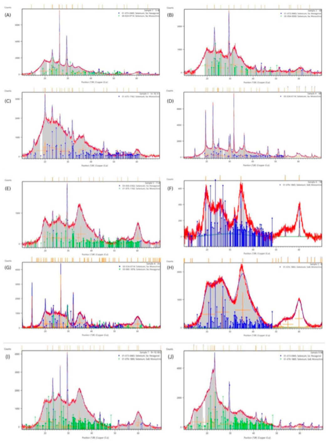

| Algal species | Treated group | Corresponding ICDD reference Hexagonal |
Corresponding ICDD reference Monoclinic |
|---|---|---|---|
| P. palmata | (+) | 01-073-0465 | 00-024-0714 |
| (-) | 01-073-0465 | 00-054-0500 | |
| U. intestinalis | (+) | - | 01-075-1162 |
| (-) | - | 00-024-0714 | |
| F. vesiculosus | (+) | 00-006-0362 | 01-075-1162 |
| (-) | - | 01-076-1865 | |
| F. serratus | (+) | 03-056-1876 | 00-024-0714 |
| (-) | - | 01-076-1865 | |
| L. digitata | (+) | 01-073-0465 | 01-076-1865 |
| (-) | 01-073-0465 | 01-076-1865 |
| Algal species or control | Panel | Treated group | Avg. Size (nm) | PDI | Zeta Potential (mV) |
|---|---|---|---|---|---|
| PC3 | A | NA | 187 ± 2.15 | 0.09 ± 0.03 | -27.2 ± 0.72 |
| P. palmata | B | (+) | 53 ± 0.5 | 0.24 ± 0.01 | - 20 ± 2 |
| C | (-) | 83.2 ± 0.8 | 0.28 ± 0.007 | -29 ± 2.6 | |
| U. intestinalis | D | (+) | 138.5 ± 3.8 | 0.32 ± 0.04 | 25.4 ± 1.3 |
| E | (-) | 249 ± 1.9 | 0.14 ± 0.14 | -27.4 | |
| F. vesiculosus | F | (+) | 119.7 ± 0 | 0.17 ± 0.016 | -26.4 |
| G | (-) | 232.6 ± 1.5 | 0.26 ± 0.016 | -26.6 ± 6.3 | |
| F. serratus | H | (+) | 174.8 ± 3.4 | 0.2 ± 0.009 | -20.2 ± 0.6 |
| I | (-) | 426.6 ± 8.7 | 0.24 ± 0.04 | -26.3 ± 1.6 | |
| L. digitata | J | (+) | 330 ± 7.2 | 0.27 ± 0.5 | -28.5 ± 1.7 |
| K | (-) | 302 ± 22 | 0.36 ± 0.09 | -24.7 ± 1 |
| Algae species | Treated group | M. tuberculosis strains | |||||||||
|---|---|---|---|---|---|---|---|---|---|---|---|
| mc27902 | mc28245 (INHR) | mc28247 (RIFR) | mc28250 (RIFR & INHR) | mc28258 (RIFR & INHR) | |||||||
| MIC₅₀ | MIC₉₀ | MIC₅₀ | MIC₉₀ | MIC₅₀ | MIC₉₀ | MIC₅₀ | MIC₉₀ | MIC₅₀ | MIC₉₀ | ||
| P. palmata | NC2 | ND | ND | ND | ND | ND | ND | ND | ND | ND | ND |
| (+) | 14.64 ± 1.35 | 846.14 ± 3.72 | ND | ND | 48.3 ± 1.31 | > 1280 | 8.45 ± 1.32 | 516 ± 4.3 | 11.6 ± 1.2 | 119.7 ± 2.4 | |
| (-) | 27.85 ± 1.11 | 48.02 ± 1.23 | 2.73 ± 2.6 | 179.9 ± 67 | 66.7 ± 1.5 | > 1280 | 17.5 ± 1.2 | 180.7 ± 2.1 | 16.7 ± 1.2 | 171.4 ± 2.4 | |
| U. intestinalis | NC2 | ND | ND | ND | ND | ND | ND | ND | ND | ND | ND |
| (+) | 15.06 ± 1.27 | 304.26 ± 2.58 | ND | ND | 48.5 ± 1.3 | > 1280 | 7.85 ± 1.2 | 75.2 ± 2 | 11 ± 1.3 | 728.6 ± 4.1 | |
| (-) | 18.34 ± 1.11 | 40.21 ± 1.31 | ND | ND | 60.8 ± 1.3 | > 1280 | 5.5 ± 1.3 | 288 ± 3.6 | 13 ± 1.3 | 511 ± 4 | |
| F. vesiculosus | NC2 | ND | ND | ND | ND | ND | ND | ND | ND | ND | ND |
| (+) | 59 ± 1.13 | 175.6 ± 1.3 | 10.5 ± 2 | > 1280 | 198.9 ± 1.3 | > 1280 | 25.1 ± 1.3 | > 1280 | 36 ± 1.1 | 88 ± 1.3 | |
| (-) | 131.2 ± 1.2 |
324.5 ± 1.6 | 10.22 ± 1.2 | 101 ± 2.07 | 203.3 ± 1.1 | 995 ± 1.6 | 58.4 ± 1.1 | 148.4 ± 1.3 | 34.9 ± 1.55 | > 1280 | |
| F. serratus | NC2 | ND | ND | ND | ND | ND | ND | ND | ND | ND | ND |
| (+) | 19 ± 1.2 | 192.1 ± 2.1 | ND | ND | 60.6 ± 1.3 | > 1280 | 19.2 ± 1.3 | 324.9 ± 2.6 | 33.5 ± 1.1 | 93.6 ± 1.4 | |
| (-) | 28.3 ± 1.1 | 51.3 ± 1.36 | 1.22 ± 1.5 | 153.3 ± 695 | 59.4 ± 1.3 | > 1280 | 21.7 ± 1.3 | > 1280 | 30.5 ± 1.2 | 174.7 ± 1.7 | |
| L. digitata | NC2 | ND | ND | ND | ND | ND | ND | ND | ND | ND | ND |
| (+) | 192.10 ± 2.1 | 906.6 ± 3.04 | 2.51 ± 3.61 | > 1280 | 54.4 ± 1.15 | 237 ± 1.4 | 12.6 ± 1.1 | 46.6 ± 1.6 | 26.6 ± 1.2 | 43.4 ± 1.4 | |
| (-) | 294.3 ± 1.1 | 673.4 ± 1.48 | 24.6 ± 1.35 | > 1280 | ND | ND | 168 ± 1.1 | 321.1 ± 1.3 | 227.3 ± 1.18 | 519 ± 1.4 | |
| NC1 | ND | ND | ND | ND | ND | ND | ND | ND | ND | ND | |
| Ag-NP | 35.6 ± 1.15 | 1076 ± 1.77 | 39.3 ± 1.2 | 697 ± 2.3 | 88.2 ± 1.2 | 1043 ± 1.9 | 282.4 ± 1.11 | 320.2 ± 1.3 | 258.7 ± 1.4 | > 1280 | |
| RIF | 0.07 | 0.61 | 0.07 | 0.58 | ND | ND | ND | ND | ND | ND | |
| INH | 3.62 | 13.23 | ND | ND | 5.19 | 8.97 | ND | ND | 336.02 | ND | |
| EMB | 6.88 | 12.54 | 8.47 | 12.20 | 14.34 | 24.91 | 8.79 | 20.80 | 14.33 | 24.84 | |
| PC3 | 3.64 ± 1.5 | 1011.8 ± 31.3 | 7.57 ± 2.9 | > 1280 | 123.4 ± 2.3 | > 1280 | ND | ND | 1.7 ± 1 | > 1280 | |
| Algae species (Median) |
With AA (Median) |
Without AA (Median) |
U | Z | p-value | Significance | |||||||||||
|---|---|---|---|---|---|---|---|---|---|---|---|---|---|---|---|---|---|
| MIC₅₀ | MIC₉₀ | MIC₅₀ | MIC₉₀ | MIC₅₀ | MIC₉₀ | MIC₅₀ | MIC₉₀ | MIC₅₀ | MIC₉₀ | MIC₅₀ | MIC₉₀ | ||||||
| P. palmata | 1.1 | 2.75 | 1.39 | 1.86 | 56 | 116 | -1.44 | 1.58 | 0.15 | 0.11 | n.s. | n.s. | |||||
| U. intestinalis | 1.10 | 1.89 | 1.25 | 1.69 | 70 | 84 | -0.72 | 0 | 0.47 | 1.00 | n.s. | n.s. | |||||
| F. vesiculosus | 1.51 | 2.16 | 1.89 | 2.23 | 78 | 106 | -1.41 | 0.02 | 0.16 | 0.98 | n.s. | n.s. | |||||
| F. serratus | 1.43 | 1.71 | 1.49 | 2.17 | 74 | 55 | -0.51 | -1.22 | 0.61 | 0.22 | n.s. | n.s. | |||||
| L. digitata | 1.39 | 2.78 | 2.24 | 2.80 | 19 | 71 | -3.59 | -0.94 | 0.0003 | 0.34 | *** | n.s. | |||||
| Plasmid and Strain | Description (including genotype) | Source or reference |
|---|---|---|
| Plasmids | ||
| pMV261 | E. coli-mycobcaterial shuttle plasmid, hsp60 promoter, KanR | Stover et al., 1991 |
| Strains | ||
| Mtb mc27902 | ΔleuCD ΔpanCD ΔargB, Leucine, pantothenate and arginine triple auxotroph | Vilcheze et al., 2018 |
| Mtb mc28245 (INHR) | mc27902 derived Δ2116169–2162530; Δ2116169–2162530 genome deletion, INHR | Vilcheze et al., 2018 |
| Mtb mc28247 (RIFR) | mc27902 derived, rpoB (H445Y); rpoB His445 → Lys, RIFR | Vilcheze et al., 2018 |
| Mtb mc28250 (RIFR & INHR) | mc28247 derived, rpoB (H445Y) Δ2122397–2170320; rpoB His445 → Lys, RIFR, Δ2122397–2170320 genome deletion, INHR | Vilcheze et al., 2018 |
| Mtb mc28258 (RIFR & INHR) | mc28247 derived, rpoB (H445Y) katG (W438R); rpoB His445 → Lys, RIFR, katG Trp438 → Arg, INHR | Vilcheze et al., 2018 |
| Mtb mc27902 pMV261 | Mtb mc27902 containing pMV261, KanR | Lu et al. 2025 |
| Mtb mc28245 pMV261 | Mtb mc28245 containing pMV261, KanR | Lu et al. 2025 |
| Mtb mc28247 pMV261 | Mtb mc28247 containing pMV261, KanR | Lu et al. 2025 |
| Mtb mc28250 pMV261 | Mtb mc28250 containing pMV261, KanR | Lu et al. 2025 |
| Mtb mc28258 pMV261 | Mtb mc28258 containing pMV261, KanR | Lu et al. 2025 |
Disclaimer/Publisher’s Note: The statements, opinions and data contained in all publications are solely those of the individual author(s) and contributor(s) and not of MDPI and/or the editor(s). MDPI and/or the editor(s) disclaim responsibility for any injury to people or property resulting from any ideas, methods, instructions or products referred to in the content. |
© 2026 by the authors. Licensee MDPI, Basel, Switzerland. This article is an open access article distributed under the terms and conditions of the Creative Commons Attribution (CC BY) license (http://creativecommons.org/licenses/by/4.0/).


