

Submitted:
27 July 2026
Posted:
28 July 2026
You are already at the latest version
Abstract
Chlamydospores are stress-resistant propagules that improve the stability and shelf life of Trichoderma-based biocontrol products, but the genetic basis of their production remains poorly understood. Previous transcriptomic analyses of Trichoderma virens GV29-8 identified the hypothetical gene TRIVIDRAFT_211280 as a candidate regulator of chlamydospore development. To investigate its role, researchers generated a gene deletion mutant using CRISPR/Cas9-mediated homologous recombination and constructed a complemented strain. The wild-type, mutant, and complemented strains were compared for growth, morphology, biomass production, conidiation, and chlamydospore formation under both nutrient-rich and chlamydospore-inducing conditions. Deletion of TRIVIDRAFT_211280 had no effect on colony morphology, radial growth, biomass accumulation in inducing medium, or the timing and progression of chlamydospore development, indicating that the gene is not essential for chlamydospore morphogenesis. However, under nutrient-rich conditions, the mutant exhibited significantly greater biomass accumulation (11.8% increase) and conidial production (34.1% increase) than the wild type. Chlamydospore yield was also higher in the mutant at day 6, although overall differences across time points were not significant. These findings suggest that TRIVIDRAFT_211280 functions as a condition-dependent negative regulator of biomass and propagule production and may represent a target for improving spore yields in Trichoderma biocontrol formulations.
Keywords:
biomass
; chlamydospore
; conidiation
; CRISPR/Cas9
; gene knockout
; Trichoderma virens GV29-8
1. Introduction
Plant diseases impose substantial constraints on agricultural productivity worldwide. Although chemical pesticides remain widely used due to their rapid efficacy and ease of application, their negative impacts on non-target organisms, environmental health, and the development of resistance have motivated the search for sustainable alternatives. Biological control, based on the use of beneficial microorganisms, offers a more environmentally compatible approach by suppressing pathogens and enhancing plant defenses [1,2]. Among microbial biocontrol agents, fungi of the genus Trichoderma are particularly important due to their broad-spectrum antagonistic activity, ability to colonize plant roots, and capacity to induce systemic resistance [3,4,5,6,7,8].
For commercial application, biocontrol products must maintain high viability, stability, and long shelf life. In this context, the type of fungal propagule used is critical. While conidia are easy to produce under laboratory and industrial conditions, they are often sensitive to environmental stresses such as temperature and pH fluctuations, which can limit product stability and field performance [9,10,11]. In contrast, chlamydospores are thick-walled, stress-resistant structures that accumulate storage reserves and exhibit enhanced tolerance to adverse conditions, making them highly attractive for formulation purposes [12,13,14]. However, the large-scale exploitation of chlamydospores has been hindered by their relatively low natural production and the complexity of inducing their formation under controlled conditions [15,16].
Environmental factors influencing chlamydospore differentiation, including nutrient limitation and optimal temperature regimes, have been described [16,17,18]. Nevertheless, the genetic mechanisms underlying chlamydospore formation and yield remain poorly characterized. Identifying key regulatory genes would not only improve understanding of fungal developmental biology but also provide molecular targets to engineer strains with enhanced propagule production and improved industrial performance. In Trichoderma spp., only a limited number of genes have been functionally validated in this context, and their roles suggest that chlamydospore formation is governed by complex regulatory networks involving nutrient sensing, metabolism, and stress responses [3,4].
In previous work, chlamydospore formation in Trichoderma virens GV29-8 was characterized across developmental stages using transcriptomic analysis, leading to the identification of several candidate genes whose expression profiles correlate with sporulation [16]. Among them, the hypothetical protein-encoding gene TRIVIDRAFT_211280 displayed increased expression during the transition from active mycelial growth to early pre-sporulation, followed by reduced expression at later stages, suggesting a potential role in this process. Here, we tested this hypothesis by generating a CRISPR/Cas9-mediated deletion mutant and a complemented strain, and comparing their phenotypic traits, including growth, biomass accumulation, conidiation, and chlamydospore production. This approach allows us to assess whether TRIVIDRAFT_211280 contributes to chlamydospore development and/or regulates propagule yield under different culture conditions.
2. Materials and Methods
2.1. Strains, Plasmids and Culture Conditions
Trichoderma virens GV29-8 was used as the wild-type strain in this study. Plasmids pKH-KO and pKH-KO-G418, carrying hygromycin B and G418 resistance markers, respectively, were used for gene deletion and complementation. The CRISPR/Cas9 vector pHZZCas9TELace1_gRNA was used for guide RNA expression. Escherichia coli Fast-T1 competent cells were used for plasmid propagation.
Fungal strains were routinely cultured on potato dextrose agar (PDA) at 28 °C. For liquid cultures, incubation was carried out with shaking at 200 rpm. Chlamydospore formation was induced using a previously described inducing medium [16].
2.2. Construction of the ΔTRIVIDRAFT_211280 Mutant
The coding sequence of TRIVIDRAFT_211280 was retrieved from NCBI and used to design guide RNAs using the E-CRISP tool. The selected sgRNA was cloned into the pHZZCas9TELace1_gRNA vector and sequence-verified (Figures S1 and S2). The resulting plasmid was linearized prior to transformation.
A deletion cassette was constructed by amplifying the upstream and downstream genomic regions flanking TRIVIDRAFT_211280 and ligating them to a hygromycin B resistance cassette using overlap PCR (Figures S3–S9).
Protoplasts of T. virens GV29-8 were prepared from actively growing mycelia and co-transformed with the linearized CRISPR/Cas9 plasmid and the deletion cassette using a polyethylene glycol (PEG)-mediated protocol. Transformants were selected on hygromycin B-containing medium and purified through successive subcultures.
2.3. Construction of the Complemented R-TRIVIDRAFT_211280 Strain
To generate the complemented strain, the TRIVIDRAFT_211280 coding sequence was amplified from wild-type cDNA and placed under the control of the constitutive Aspergillus nidulans gpdA promoter and the trpC terminator in a G418-resistant vector (Figures S10–S13).
The resulting construct was sequence-verified and introduced into the ΔTRIVIDRAFT_211280 strain via PEG-mediated protoplast transformation. G418-resistant transformants were isolated and purified for further analysis.
2.4. Molecular Validation of Transformants and Quantification of Gene Expression
Putative transformants were screened by diagnostic PCR using primers specific for the TRIVIDRAFT_211280 locus and the selectable marker genes (Table S1). Successful deletion of the target gene was confirmed by the absence of the wild-type allele and the presence of the hygromycin-resistance cassette (Figures S4–S7).
For the complemented strain, integration of the complementation construct and the presence of the TRIVIDRAFT_211280 gene were verified by PCR (Figures S11 and S12). Gene expression in both the mutant and complemented strains was evaluated by RT-PCR to confirm the loss and restoration of the TRIVIDRAFT_211280 transcript, respectively (Figures S8 and S13, respectively). Primer sequences are provided in Table S1.
Total RNA was extracted from samples collected at eight time points during chlamydospore formation using FreeZol Reagent (Vazyme, Nanjing, China) according to the manufacturer’s instructions. RNA integrity and purity were assessed by 1% agarose gel electrophoresis and spectrophotometric measurements at 260 and 280 nm with a NanoPhotometer NP80 Touch (IMPLEN GmbH, Munich, Germany). Only RNA samples with an A260/A280 ratio between 1.8 and 2.1 and exhibiting intact electrophoretic bands were used for subsequent reverse-transcription analyses.
First-strand complementary DNA (cDNA) was synthesized from high-quality total RNA using HiScript III 1st Strand cDNA Synthesis Kit (+ gDNA wiper) (Vazyme) according to the manufacturer’s instructions. The resulting cDNA was diluted appropriately and stored at −20 °C until use in RT-qPCR assays.
RT-qPCR was performed using a Taq Pro Universal SYBR qPCR Master Mix (Vazyme) on a QuantStudio 5 qPCR (Applied Biosystems, Foster City CA, USA). GAPDH was used as the reference gene. Gene-specific primers for TRIVIDRAFT_211280 and GAPDH were designed and synthesized (Table S1). For each of the eight sampling time points during chlamydospore formation, qPCR reactions were prepared according to the manufacturer’s protocol. Three independent biological replicates and three technical replicates were included for each sample to ensure the reliability and reproducibility of the results. Relative gene expression levels were calculated using the method [19] (Figure S14).
2.5. Protein Functional Domain and Structure Predictions
The predicted amino acid sequence of TRIVIDRAFT_211280 was analyzed using InterPro to identify conserved domains, protein-family signatures, signal peptides, and membrane-topology features [20]. Subcellular localization and sorting signals were predicted using DeepLoc 2.0 [21]. Potential effector-like properties were assessed using EffectorP [22], which classifies fungal proteins as non-effectors or putative effectors and further distinguishes predicted apoplastic and cytoplasmic effector classes. Predictions from these tools were interpreted collectively to infer whether TRIVIDRAFT_211280 encodes a secreted, membrane-associated, or soluble protein. EffectorP predictions were considered as computational evidence of effector-like features and not as experimental confirmation of effector function.
The three-dimensional structure of TRIVIDRAFT_211280 was predicted using AlphaFold 3 [23] through the AlphaFold Server (https://alphafoldserver.com/) with default settings. The amino acid sequence of TRIVIDRAFT_211280 was submitted as a single protein chain, without specifying additional interacting molecules, ligands, ions, or nucleic acids. The highest-ranked model was retained for visualization and qualitative structural inspection. Predicted structural features were visualized as cartoon/ribbon representations to inspect secondary-structure organization and as molecular surface representations to assess the spatial distribution of charged regions. Because TRIVIDRAFT_211280 was predicted to contain an N-terminal signal peptide, structural interpretations focused primarily on the mature predicted secreted region after the signal peptide.
2.6. Phenotypic Assays
Colony morphology and radial growth: Mycelial plugs (0.5 cm diameter) from actively growing colonies were transferred to fresh PDA plates and incubated at 28 °C. Colony morphology was recorded at defined time points. Radial growth was measured over time by recording colony diameter.
Biomass accumulation and conidial production in PDA: For biomass determination, standardized conidial suspensions (1×10⁷ spores/mL) were inoculated into liquid PDA and cultured for 3 days. Mycelia were collected, dried to constant weight, and weighed. For conidial production, colonies were cultured for 7 days, harvested in sterile water, and filtered. Conidia were quantified using a hemocytometer.
Chlamydospore formation and yield: Conidial suspensions (1×10⁷ spores/mL) were inoculated into chlamydospore-inducing medium and cultured under standard conditions. Samples were taken at multiple time points and examined microscopically to assess the developmental progression of chlamydospore formation. For quantification, aliquots were collected at defined time points, diluted, and chlamydospores were counted using a hemocytometer.
Biomass accumulation in inducing medium: To assess biomass dynamics during chlamydospore induction, cultures were sampled at multiple time points. Mycelia were collected, dried, and weighed to obtain dry biomass.
2.7. Statistical Analysis
Data were analyzed using R (version 4.5.3) in RStudio version 2026.04.0-526 (Posit PBC, Boston MA, USA). Longitudinal measurements (e.g., mycelium growth curves and chlamydospore biomass trajectories and yield) were analyzed using generalized linear mixed-effects models (GLMM) with strain as a fixed factor and biological replicate as a nested random effect; time was treated as a repeated-measures factor where appropriate.
Endpoint measurements (e.g., conidial biomass and yield) were analyzed using generalized linear models (GLM) with strain as a fixed factor. Chlamydospore yield data were analyzed using mixed-effects models including strain and time, and their interaction.
Post hoc pairwise comparisons were conducted using sequential Bonferroni correction. All experiments included three biological replicates and were repeated with three technical replicates.
Differences in expression data () along developmental stages between wild-type, ΔTRIVIDRAFT_211280 and R-TRIVIDRAFT_211280 alleles, were evaluated using non-parametric Friedman rank sum test. Pairwise comparisons of the expression levels were done using Wilcoxon signed-rank tests with sequential Bonferroni correction.
3. Results
3.1. In Silico Analysis of the TRIVIDRAFT_211280-Encoded Protein
Bioinformatic analysis indicated that TRIVIDRAFT_211280 encodes a small protein of 171 amino acids. InterPro/Phobius predicted an N-terminal signal peptide spanning residues 1 - 24, with the remaining region classified as non-cytoplasmic. DeepLoc 2.0 also predicted extracellular localization with high confidence and identified a signal peptide, while classifying the protein as soluble rather than transmembrane-associated (Figure 1A). No conserved catalytic domains or known protein-family signatures were detected. EffectorP classified TRIVIDRAFT_211280 as a putative cytoplasmic effector. Together, these analyses suggest that TRIVIDRAFT_211280 is a small predicted secreted hypothetical protein with effector-like features.
AlphaFold 3 structural prediction suggested that the N-terminal signal peptide is poorly structured, whereas the mature region is predicted with higher confidence (Figure 1B left). The mature protein is predicted to adopt a compact β-rich fold composed of two opposing β-sheet-like regions and a short α-helix near the C-terminal region. The electrostatic surface representation suggests a predominance of negatively charged surface regions (Figure 1B right). These structural features are consistent with the signal-peptide and localization predictions, supporting residues 1 - 24 as a putative signal peptide and residues 25 - 171 as the mature predicted secreted protein.
3.2. Generation and Validation of ΔTRIVIDRAFT_211280 and Complemented Strain
The ΔTRIVIDRAFT_211280 deletion mutant was generated by CRISPR/Cas9-mediated gene replacement with a hygromycin resistance cassette. Among 173 hygromycin-resistant transformants, three candidates lacked the wild-type TRIVIDRAFT_211280 locus as determined by diagnostic PCR. Genomic PCR confirmed the absence of the target gene and the presence of the hygromycin resistance cassette. RT-PCR further demonstrated loss of TRIVIDRAFT_211280 transcript and expression of the selectable marker, confirming successful gene deletion.
To complement the mutation, the TRIVIDRAFT_211280 coding sequence was reintroduced into the deletion mutant under the control of a constitutive promoter. Several G418-resistant transformants carried both the selectable marker and the TRIVIDRAFT_211280 sequence, and RT-PCR confirmed restoration of gene expression. These strains were used for subsequent phenotypic analyses.
Overall differences in expression levels along developmental stages were observed between the three genotypes (Figure S14: χ2 = 10.750, 2 d.f., P = 0.005), mostly due to the lack of expression in the deletion mutant ΔTRIVIDRAFT_211280. Pairwise comparisons also show that the complemented R-TRIVIDRAFT_211280 strain showed consistently lower expression levels than wild-type (W = 35, adjusted P = 0.021).
3.3. Colony Morphology and Radial Growth
No visible differences in colony morphology were observed among the wild-type, ΔTRIVIDRAFT_211280, and complemented strains at either early (35 h) or late (115 h) time points (Figure 2A). Similarly, radial growth rates showed no statistically significant differences among the three strains (Figure 2B; GLMM test of slope: F1,16.170 = 1.191, P = 0.329), indicating that TRIVIDRAFT_211280 does not play a major role in hyphal extension under nutrient-rich conditions.
3.4. Biomass Accumulation and Conidial Production in Nutrient-Rich Medium
Despite the lack of an effect on radial growth, deletion of TRIVIDRAFT_211280 resulted in a significant increase in biomass accumulation in PDA (Figure 2C; GLM: F2,24 = 9.300, P = 0.001). The ΔTRIVIDRAFT_211280 strain produced 11.8% more biomass than the wild-type (Figure 2C; post hoc Bonferroni sequential test: P = 0.026), whereas R-TRIVIDRAFT_211280 strain did not differ from the wild-type (Figure 2C; post hoc Bonferroni sequential tests: P = 0.682), confirming that the phenotype was specifically associated with loss of TRIVIDRAFT_211280.
Consistent with this result, conidial production also differed among strains (Figure 2D; GLM: F2,6 = 15.646, P = 0.004). The deletion mutant produced 34.1% more conidia than the wild-type (Figure 2D; post hoc Bonferroni sequential test: P = 0.009), while the complemented strain again showed no significant difference from the wild-type (Figure 2D; post hoc Bonferroni sequential tests: P = 0.521). Biomass and conidial yield were positively correlated (Figure 2E; partial correlation test controlling for strain: rp = 0.668, 24 d.f., P < 0.001), suggesting that increased propagule production in the deletion mutant is associated with increased overall growth.
3.5. Chlamydospore Formation Dynamics
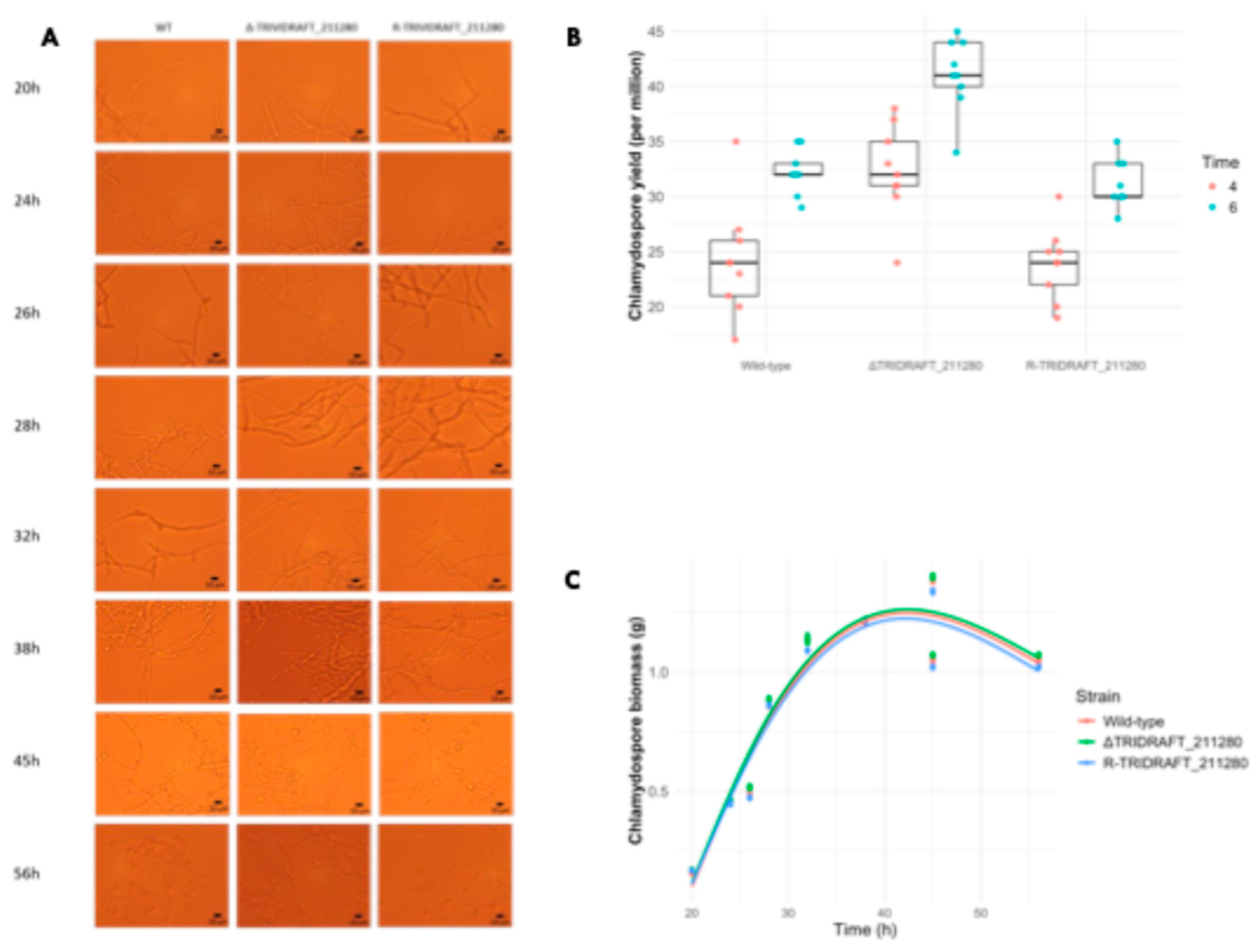
Microscopic observations revealed that the sequence and timing of chlamydospore development were similar across all three strains (Figure 3A). Initial swelling of hyphal tips was observed at approximately 28 h, followed by widespread chlamydospore formation at around 38 h, and partial hyphal autolysis at later stages. These results indicate that deletion of TRIVIDRAFT_211280 does not alter the qualitative developmental program of chlamydospore formation.
3.6. Chlamydospore Yield
Quantitative analysis of chlamydospore production showed no significant overall effect of strain (Figure 3B; GLMM test of strain: F2,6.039 = 0.322, P = 0.737) or strain-by-time interaction (Figure 3B; GLMM test of interaction: F2,6.156 = 0.107, P = 0.900). However, pairwise comparisons revealed that the ΔTRIVIDRAFT_211280 mutant produced more chlamydospores than the wild-type at day 6 (Figure 3B; post hoc Bonferroni sequential test: P = 0.020), whereas the difference at day 4 was not statistically significant (Figure 3B; post hoc Bonferroni sequential test: P = 0.110). The complemented strain did not differ from the wild-type at either time point (Figure 3B, post hoc Bonferroni sequential tests: P = 1.000 in both cases).
These results suggest that TRIVIDRAFT_211280 does not strongly control chlamydospore formation but may influence yield in a time-dependent manner.
3.7. Biomass Dynamics in Chlamydospore-Inducing Medium
To determine whether differences in chlamydospore production were associated with changes in growth under inducing conditions, biomass accumulation was measured over time in chlamydospore-inducing medium (Figure 3C). No significant differences among strains were detected in biomass trajectories or their temporal dynamics (Figure 3C; GLMM test of strain-by-time interaction term: F2,237 = 0.061, P = 0.941).
In all strains, biomass accumulation slowed after approximately 32 h, reached a maximum at around 45 h, and declined thereafter, consistent with the onset of differentiation and autolysis. These results indicate that TRIVIDRAFT_211280 does not affect overall growth under chlamydospore-inducing conditions.
4. Discussion
In this study, we used CRISPR/Cas9-mediated gene editing to investigate the function of the previously uncharacterized gene TRIVIDRAFT_211280 in T. virens GV29-8. Comparative analysis of the deletion mutant, complemented strain, and wild-type revealed a consistent pattern: loss of TRIVIDRAFT_211280 did not affect colony morphology, radial growth, or the qualitative sequence of chlamydospore formation, but it increased biomass accumulation and propagule production under nutrient-rich conditions. These results indicate that TRIVIDRAFT_211280 is not essential for the morphogenetic program of chlamydospore development but instead influences quantitative aspects of growth and sporulation.
The absence of detectable changes in the timing and structure of chlamydospore formation suggests that TRIVIDRAFT_211280 is unlikely to be a core developmental regulator. Instead, its effects on biomass, conidiation, and, to a lesser extent, chlamydospore yield point to a role in modulating resource allocation. Under nutrient-rich PDA conditions, deletion of the gene resulted in increased biomass and conidial production, whereas in chlamydospore-inducing medium, biomass dynamics were indistinguishable among strains. This context-dependent phenotype suggests that TRIVIDRAFT_211280 may act as a negative regulator of growth and propagule production under nutrient sufficiency, possibly by constraining metabolic fluxes or influencing signaling pathways linked to nutrient sensing. Such conditional regulatory effects are consistent with the complex interplay between growth, differentiation, and environmental cues described in Trichoderma and other filamentous fungi [3,4].
The effect of TRIVIDRAFT_211280 on chlamydospore yield appears to be more subtle. Although the deletion mutant tended to produce more chlamydospores, a significant difference relative to the wild-type was only observed at a single time point, and the overall strain effect was not significant. This indicates that the gene does not strongly control chlamydospore formation per se but may indirectly influence yield through its effects on biomass accumulation or metabolic state. Similar patterns have been reported for other genes in T. virens, where perturbations in nutrient transport or metabolism led to increased propagule production without altering developmental progression [16,24]. Together, these findings support a model in which chlamydospore formation is robust at the morphological level but sensitive to upstream physiological conditions that determine the amount of propagule produced.
The in silico characterization of TRIVIDRAFT_211280 provides useful clues about its possible biological role. The predicted N-terminal signal peptide, extracellular localization, soluble classification, and EffectorP prediction suggest that TRIVIDRAFT_211280 may act as a secreted regulatory or effector-like protein rather than as a conventional intracellular enzyme. The AlphaFold 3 model further supports the idea that the mature region forms a folded extracellular protein after removal of the signal peptide. This interpretation is consistent with a possible role in extracellular signaling, cell-wall-associated processes, or interactions with the surrounding environment. However, because no localization, secretion, host-translocation, or biochemical assays were performed, these predictions should be considered provisional and should not be interpreted as experimental evidence of effector function.
From an applied perspective, the identification of genes that negatively regulate propagule production is of particular interest for improving Trichoderma-based biocontrol products. Increased biomass and spore yield under nutrient-rich conditions could facilitate industrial-scale fermentation and reduce production costs. In this context, TRIVIDRAFT_211280 represents a potential genetic target for strain improvement. However, enhanced yield alone is not sufficient for practical applications. Future work should evaluate whether deletion of this gene also affects key formulation traits, such as stress tolerance, storage stability, and field performance, which ultimately determine product efficacy. In addition, combining multiple genetic modifications targeting independent negative regulators may provide additive or synergistic effects on propagule production.
A major limitation of the present study is that the molecular function of TRIVIDRAFT_211280 remains unknown. The gene encodes a hypothetical protein, and no mechanistic link to known regulatory pathways can be inferred at present. Further characterization, including analysis of conserved domains, subcellular localization, and transcriptional or metabolomic profiling of the deletion mutant, will be necessary to elucidate its role. Comparative analyses across Trichoderma species could also determine whether this regulatory function is conserved. Such studies would help place TRIVIDRAFT_211280 within the broader network of genes governing fungal growth and differentiation and clarify how its activity integrates environmental signals with developmental outputs.
In summary, our results show that TRIVIDRAFT_211280 acts as a condition-dependent negative regulator of biomass accumulation and propagule production in T. virens GV29-8, while having little or no effect on the developmental sequence of chlamydospore formation. These findings refine our understanding of the genetic control of propagule yield and identify a candidate target for improving the production of durable biocontrol formulations.
Supplementary Materials
The following supporting information can be downloaded at the website of this paper posted on Preprints.org. Figure S1: Fragment amplification of plasmid 0608-pHZZCas9TELace1_gRNA.; Figure S2: The monoclonal validation of plasmid 0608-pHZZCas9TELace1_gRNA; Figure S3: Schematic diagram of plasmid pKH-KO; Figure S4: Amplification of homologous arms of the TRIVIDRAFT_211280 gene and the resistance gen; Figure S5: Amplification of the homologous arm of TRIVIDRAFT_211280 gene knockout; Figure S6: Preliminary validation of the knockout mutant strain of TRIVIDRAFT_211280 gene (664 bp); Figure S7: Verification of DNA-level in the ΔTRIVIDRAFT_211280 strain; Figure S8: Verification of the mRNA level expression in the ΔTRIVIDRAFT_211280 strain; Figure S9: Amplification of the CDS region of the target gene, promoter gpdA, and terminator trpC; Figure S10: Schematic diagram of plasmid pKH-KO-G418; Figure S11: Construction of the R-TRIVIDRAFT_211280 strain; Figure S12: Verification of DNA-level in the R-TRIVIDRAFT_211280 strain; Figure S13: Verification of the mRNA level expression in the R-TRIVIDRAFT_211280 strain; Figure S14: Expression levels of TRIVIDRAFT_211280 along development in the three different genotypes used in this study; Table S1: Primers used in this study.
Author Contributions
Conceptualization, B.W. and X.J.; methodology, Y.Z., Z.T., W.C., R.W., S.Z., I.S.D., and S.F.E.; software, S.F.E.; validation, S.F.E., B.W. and X.J.; formal analysis, Y.Z., Z.T. and S.F.E.; investigation, I.S.D., M.A.A. and S.F.E.; resources, Y.Z., Z.T., W.C., R.W., and S.Z.; data curation, Y.Z., Z.T. and S.F.E.; writing—original draft preparation, Y.Z.; writing—review and editing, M.A.A., S.F.E., B.W., and X.J.; visualization, S.F.E.; supervision, B.W. and X.J.; project administration, B.W. and X.J.; funding acquisition, X.J. All authors have read and agreed to the published version of the manuscript.
Funding
This research was funded by National Natural Science Foundation of China General Project (32272625) granted to X.J.
Data Availability Statement
The raw data and R code supporting the conclusions of this article will be made available by the authors, without undue reservation.
Conflicts of Interest
The authors declare no conflicts of interest. The funders had no role in the design of the study; in the collection, analyses, or interpretation of data; in the writing of the manuscript; or in the decision to publish the results.
Abbreviations
The following abbreviations are used in this manuscript:
| CRISPR | Clustered regularly interspaced short palindromic repeats |
| GAPDH | Glyceraldehyde 3-phosphate dehydrogenase |
| GLM | Generalized linear model |
| GLMM | Generalized linear mixed model |
| PCR | Polymerase chain reaction |
| PDA | Potato dextrose agar |
| PEG | Polyethylene glycol |
| RT-PCR | Reverse transcription PCR |
References
- Dong, Q.E.; Zhang, Z.R.; Tian, Y.X.; Tong, J.Y. Application of compound microbial preparation in controlling strawberry powdery mildew. Northern Horticulture 2020, 15, 38–41.
- Zhou, Q.; Shen, Y.; Wang, J.Y.; Zhang, Y.X.; Zhu, X.Y, Yan, T.T. Research progress on application of Trichoderma in biological control of plant diseases and pests. Mod. Agric. Sci. Technol. 2024, 15, 70–74.
- Harman, G.E.; Howell, C.R.; Viterbo, A.; Chet, I.; Lorito, M. Trichoderma species—Opportunistic, avirulent plant symbionts. Nat. Rev. Microbiol. 2004, 2, 43–56. [CrossRef]
- Druzhinina, I.S.; Seidl-Seiboth, V.; Herrera-Estrella, A.; Horwitz, B.A.; Kenerley, C.M.; Monte, E.; Mukherjee, P.K.; Zeilinger, S.; Grigoriev, I.V.; Kubicek, C.P. Trichoderma: The genomics of opportunistic success. Nat. Rev. Microbiol. 2011, 9, 749–759.
- Chen, J. Advances in Trichoderma-induced plant resistance. Chin. J. Biol. Control 2015, 31, 733–741.
- Huo, X.X.; Wang, Q.L.; Zhang, H.; Miao, Y; Tian, Y.; Yang, J.; Wang, H.; Zhang, Z.X. Effects of Trichoderma viride fermentation broth and conidial suspension on Meloidogyne incognita J2. J. Nanjing Agric. Univ. 2022, 45, 553–561.
- Lin, C.W.; Liu, J.L. Biological control effects of a Trichoderma strain against several pathogenic fungi. Plant Doctor 2021, 34, 29–33.
- Tan, Y.; Fan, H.; Zhang, Z.Y.; Li, Y.P.; Zhou, X.F. Effects of T. viride fermentation broth and conidial suspension on M. incognita. Jiangsu Agric. Sci. 2022, 50, 114–120.
- Agosin, E. Culture conditions and spore shelf life of T. harzianum. World J. Microbiol. Biotechnol. 1997, 13, 225–232.
- Chen, J.A.; Xie, A.R.; Zhu, W.T. Protection and growth of Trichoderma conidia. Southwest Chin. J. Agric. Sci. 2015, 28, 2563–2567.
- Chi, Y.J.; Yi, H.W.; Ji, H.L. Optimal conditions for T. longibrachiatum T05 to produce conidia and chlamydospores. J. Northeast For. Univ. 2016, 44, 110–113.
- Lian, H.; Chen, Y.R.; Li, M.; Liang, X.; Ma, G.S. The dffect of Trichoderma conidia and chlamydospores on physiological characteristics of cucumber seedlings and control effect against Fusarium wilt. Agric. Res. Arid. Areas 2021, 39, 71–79.
- Zhang, J.J. Storage Ability and Application of Trichoderma Tr-92 Chlamydospore Wettable Powder; Master's Thesis, Hebei University of Technology, Hebei, China, 2015.
- Zhuang, J.H.; Gao, Z.G.; Liu, X.; Liu, Y.; Zhang, Y.F. Fermentation factors affecting spore types of Trichoderma strain 23. Chin. J. Biol. Control 2005, 1, 37–40.
- Bissett, J. Revision of Trichoderma. III. Pachybasium. Can. J. Bot. 1991, 69, 2372–2417.
- Peng, X.H. Molecular Mechanism of Chlamydospore Formation of T. virens GV29-8; Ph.D. Dissertation, Chinese Academy of Agricultural Sciences, Beijing, China, 2021.
- Qin, Y.; Ye, H.Z.; Huang, Y. Factors influencing Trichoderma chlamydospore development. J. Yunnan Agric. Univ. 2002, 17, 416–417.
- Yang, X.Y. Transcriptome Analysis of T. harzianum Th-33 during Chlamydospore Formation; Ph.D. Dissertation, Chinese Academy of Agricultural Sciences, Beijing, China, 2014.
- Livak, K.J.; Schmittgen, T.D. Analysis of relative gene expression data using real-time quantitative PCR and the 2−ΔΔCT method. Methods 2001, 25, 402–408. doi:10.1006/meth.2001.1262.
- Blum, M.; Andreeva, A.; Florentino, L.C.; Chuguransky, S.R.; Grego, T.; Hobbs, E.; Pinto, B.L.; Orr, A.; Paysan-Lafosse, T.; Ponamareva, I.; Salazar, G.A.; Bordin, N.; Bork, P.; Bridge, A.; Colwell, L.; Gough, J.; Haft, D.H.; Letunic, I.; Llinares-López, F.; Marchler-Bauer, A.; Meng-Papaxanthos, L.; Mi, H.; Natale, D.A.; Orengo, C.A.; Pandurangan, A.P.; Piovesan, D.; Rivoire, C.; Sigrist, C.J.A.; Thanki, N.; Thibaud-Nissen, F.; Thomas, P.D.; Tosatto, S.C.E.; Wu, C.H.; Bateman, A. InterPro: The protein sequence classification resource in 2025. Nucleic Acids Res. 2025, 53, D444–D456. [CrossRef]
- Thumuluri, V.; Almagro Armenteros, J.J.; Johansen, A.R.; et al. DeepLoc 2.0: Multi-label subcellular localization prediction using protein language models. Nucleic Acids Res. 2022, 50, W228–W234. [CrossRef]
- Sperschneider, J.; Dodds, P.N. EffectorP 3.0: Prediction of apoplastic and cytoplasmic effectors in fungi and oomycetes. Mol. Plant Microbe Interact. 2022, 35, 146–156. [CrossRef]
- Abramson, J.; Adler, J.; Dunger, J.; Evans, R.; Green, T.; Pritzel, A.; Ronneberger, O.; Willmore, L.; Ballard, A.J.; Bambrick, J.; Bodenstein, S.W.; Evans, D.A.; Hung, C.C.; O'Neill, M.; Reiman, D.; Tunyasuvunakool, K.; Wu, Z.; Žemgulytė, A.; Arvaniti, E.; Beattie, C.; Bertolli, O.; Bridgland, A.; Cherepanov, A.; Congreve, M.; Cowen-Rivers, A.I.; Cowie, A.; Figurnov, M.; Fuchs, F.B.; Gladman, H.; Jain, R.; Khan, Y.A.; Low, C.M.R.; Perlin, K.; Potapenko, A.; Savy, P.; Singh, S.; Stecula, A.; Thillaisundaram, A.; Tong, C.; Yakneen, S.; Zhong, E.D.; Zielinski, M.; Žídek, A.; Bapst, V.; Kohli, P.; Jaderberg, M.; Hassabis, D.; Jumper, J.M. Accurate structure prediction of biomolecular interactions with AlphaFold 3. Nature 2024, 630, 493–500. [CrossRef]
- Zhang, S.H. Cloning and Functional Analysis of the ptr2 Peptide Transporter Gene in T. virens GV29-8; Dissertation, Chinese Academy of Agricultural Sciences, Beijing, China, 2023.
Figure 1.
Bioinformatic analysis of the TRIVIDRAFT_211280-encoded peptide. (A) Predicted sorting signals. InterPro/Phobius and SignalP tools predicted an N-terminal signal peptide spanning approximately residues 1 - 24. Phobius further divides this region into the typical signal peptide N-region at residues, hydrophobic H-region and C-region, corresponding to residues 1 - 5, 6 - 16 and 17 - 24, respectively. (B) AlphaFold 3 predicted structure. Left, cartoon representation of the predicted protein structure, with β-strands shown as arrows, α-helices as helical ribbons, and loop regions as connecting lines. Right, molecular surface representation colored by electrostatic potential, with red indicating negative potential and blue indicating positive potential.
Figure 1.
Bioinformatic analysis of the TRIVIDRAFT_211280-encoded peptide. (A) Predicted sorting signals. InterPro/Phobius and SignalP tools predicted an N-terminal signal peptide spanning approximately residues 1 - 24. Phobius further divides this region into the typical signal peptide N-region at residues, hydrophobic H-region and C-region, corresponding to residues 1 - 5, 6 - 16 and 17 - 24, respectively. (B) AlphaFold 3 predicted structure. Left, cartoon representation of the predicted protein structure, with β-strands shown as arrows, α-helices as helical ribbons, and loop regions as connecting lines. Right, molecular surface representation colored by electrostatic potential, with red indicating negative potential and blue indicating positive potential.
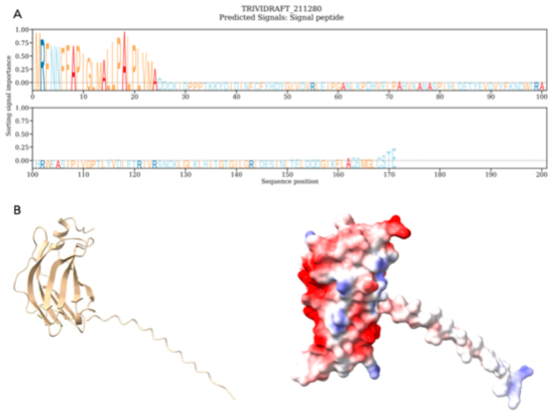
Figure 2.
Effects of TRIVIDRAFT_211280 deletion on growth and conidial production in T. virens GV29-8. (A) Colony morphology of wild-type, ΔTRIVIDRAFT_211280 and complemented (R-TRIVIDRAFT_211280) strains at 35 h and 115 h after inoculation on PDA. (B) Radial growth over time on PDA. Solid lines are shown to illustrate growth trajectories. (C) Biomass accumulation (dry weight) after 3 days of growth in liquid PDA. (D) Conidial yield after 7 days on PDA. (E) Relationship between biomass and conidial yield. The solid line represents the fitted regression, and the shaded area indicates the 95% confidence interval. In (C) and (D), boxplots represent the median, interquartile range and 95% confidence intervals.
Figure 2.
Effects of TRIVIDRAFT_211280 deletion on growth and conidial production in T. virens GV29-8. (A) Colony morphology of wild-type, ΔTRIVIDRAFT_211280 and complemented (R-TRIVIDRAFT_211280) strains at 35 h and 115 h after inoculation on PDA. (B) Radial growth over time on PDA. Solid lines are shown to illustrate growth trajectories. (C) Biomass accumulation (dry weight) after 3 days of growth in liquid PDA. (D) Conidial yield after 7 days on PDA. (E) Relationship between biomass and conidial yield. The solid line represents the fitted regression, and the shaded area indicates the 95% confidence interval. In (C) and (D), boxplots represent the median, interquartile range and 95% confidence intervals.
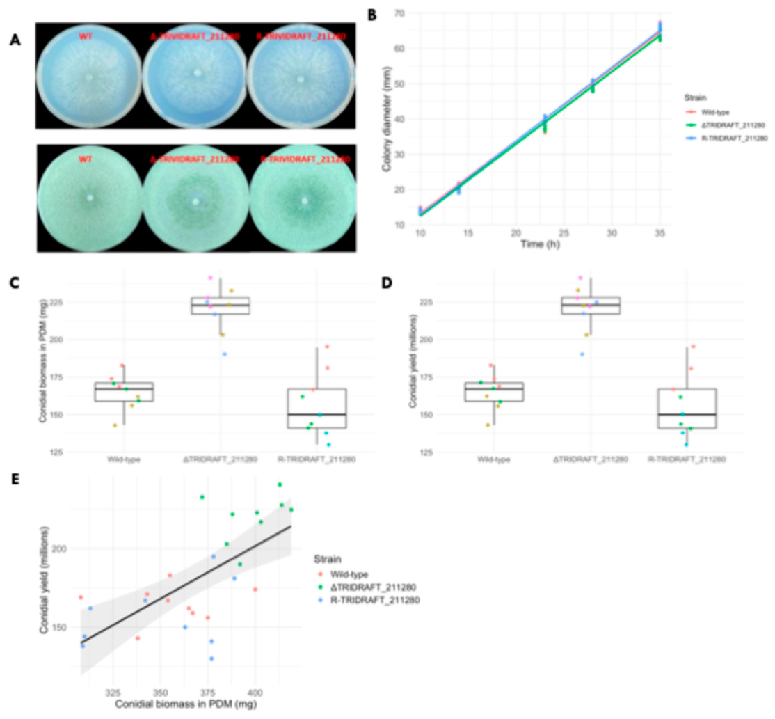
Figure 3.
Effects of TRIVIDRAFT_211280 deletion on chlamydospore formation and biomass dynamics. (A) Microscopic observations of chlamydospore development in wild-type, ΔTRIVIDRAFT_211280 and complemented (R-TRIVIDRAFT_211280) strain at indicated time points under inducing conditions. Representative stages include hyphal swelling, chlamydospore formation, and late-stage autolysis. (B) Chlamydospore yield at days 4 and 6 under inducing conditions. Boxplots represent the median, interquartile range and 95% confidence intervals. (C) Biomass accumulation, measured as dry weight, over time in chlamydospore-inducing medium. Solid lines illustrate temporal trends.
Figure 3.
Effects of TRIVIDRAFT_211280 deletion on chlamydospore formation and biomass dynamics. (A) Microscopic observations of chlamydospore development in wild-type, ΔTRIVIDRAFT_211280 and complemented (R-TRIVIDRAFT_211280) strain at indicated time points under inducing conditions. Representative stages include hyphal swelling, chlamydospore formation, and late-stage autolysis. (B) Chlamydospore yield at days 4 and 6 under inducing conditions. Boxplots represent the median, interquartile range and 95% confidence intervals. (C) Biomass accumulation, measured as dry weight, over time in chlamydospore-inducing medium. Solid lines illustrate temporal trends.
Disclaimer/Publisher’s Note: The statements, opinions and data contained in all publications are solely those of the individual author(s) and contributor(s) and not of MDPI and/or the editor(s). MDPI and/or the editor(s) disclaim responsibility for any injury to people or property resulting from any ideas, methods, instructions or products referred to in the content. |
© 2026 by the authors. Licensee MDPI, Basel, Switzerland. This article is an open access article distributed under the terms and conditions of the Creative Commons Attribution (CC BY) license (http://creativecommons.org/licenses/by/4.0/).
Copyright: This open access article is published under a Creative Commons CC BY 4.0 license, which permit the free download, distribution, and reuse, provided that the author and preprint are cited in any reuse.


