

Submitted:
25 July 2026
Posted:
27 July 2026
You are already at the latest version
Abstract
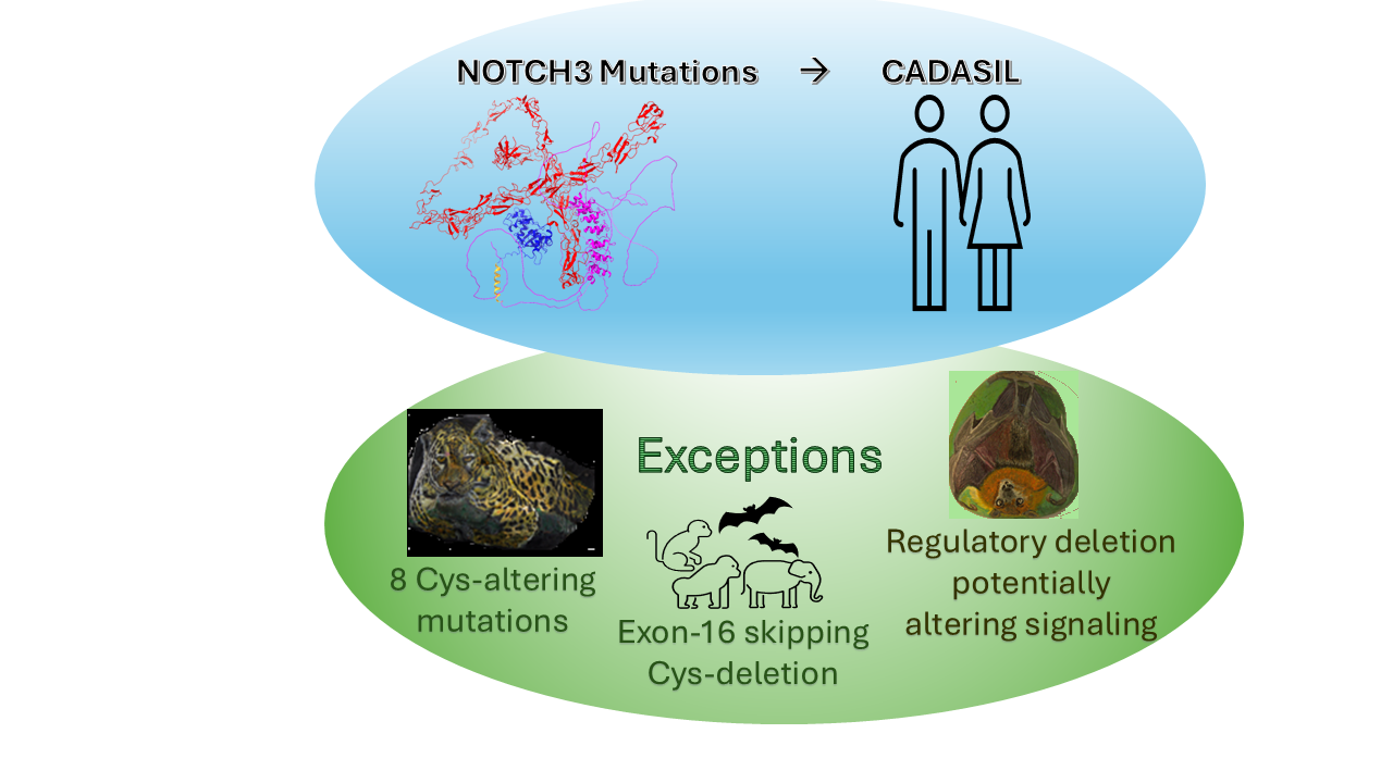
NOTCH3 is a highly conserved transmembrane receptor implicated in CADASIL, a hereditary small-vessel disease caused predominantly by mutations affecting its extracellular EGF-like repeats. The mechanisms by which these muta-tions cause pathology remain incompletely understood. We present the first large-scale comparative bioinformatics and molecular dynamics (MD) analysis of NOTCH3 across 113 mammalian species, revealing three novel findings: (i) near-complete conservation of all 204 cysteine residues, with the only exception being eight naturally occurring cysteine substitutions in jaguar EGFr13–15; (ii) a unique deletion within the regulatory region of Brandt’s bat that may increase protease accessibility and alter receptor signaling; and (iii) a rare human NOTCH3-X1 isoform, absent from most mam-mals but shared with select primates, a bat, and elephants, involving a cysteine-depleting deletion spanning EGFr20–22. These naturally occurring exceptions demonstrate that alterations predicted to be deleterious under current pathogenic-ity models can be tolerated in specific evolutionary contexts. Together, these findings provide new insights into the structure–function relationships of NOTCH3, refine the interpretation of CADASIL-associated variants, and identify testable targets for future experimental studies. Our study highlights the power of comparative bioinformatics to uncover previously unrecognized functional features in disease-associated mammalian proteins.
Keywords:
protein structure–function
; NOTCH3
; CADASIL
; comparative genomics
; evolutionary conservation
; EGF-like repeats
; molecular dynamics simulation
; cysteine substitutions
1. Introduction
“It is notorious that man is constructed on the same general type or model as other mammals. All the bones in his skeleton can be compared with corresponding bones in a monkey, bat, or seal. So it is with his muscles, nerves, blood-vessels and internal viscera. The brain, the most important of all the organs, follows the same law” [1]
NOTCH3 is a large transmembrane receptor essential for cell signaling and development in mammals. It comprises five domains: signal peptide, extracellular domain (ECD), negative regulatory region (NRR), transmembrane (TM), and intracellular domain (ICD). The ECD’s 34 epidermal growth factor repeats (EGFr) enable ligand binding and stability, with cysteines forming disulfide bonds critical for integrity [2].
Cerebral autosomal dominant arteriopathy with subcortical infarcts and leukoencephalopathy (CADASIL) is a hereditary small-vessel disease caused by mutations in NOTCH3 EGFr [3]. Cysteine mutations disrupt disulfide bonds, leading to misfolding and granular osmiophilic material (GOM) aggregation in brain microvasculature. Aggregation is a key factor in CADASIL pathogenesis, though its precise mechanisms remain unclear [4,5,6]. Notably, Cys>aa mutations are common and linked to more severe symptoms [7].
Recent large-scale efforts such as the Zoonomia Project have highlighted the value of mammalian comparative genomics for understanding gene function and disease evolution [8,9]. Genes that are highly conserved across diverse species are likely essential for normal biological function and, when altered, may contribute to disease. In contrast, genes that exhibit lineage-specific divergence may reflect adaptive evolution to ecological or physiological pressures [10].
Building on these large-scale efforts, we present the first comprehensive comparative bioinformatics analysis of NOTCH3 across 113 mammalian species, focusing on cysteines’ roles in function and disease. Key findings include unique deletion isoforms disrupting disulfide bonds, a bat-specific deletion likely affecting the NRR activation, and jaguar-specific mutations eliminating disulfide bonds in an EGFr cluster. These results refine our understanding of the sequence–structure–function–pathogenicity relationship and challenge the notion that all cysteine disruptions are pathogenic. By highlighting cysteine’s nuanced role in NOTCH3 stability, the study informs CADASIL research and may guide future experimental and therapeutic strategies.
2. Materials and Methods
Bioinformatics Analysis
More than 100 mammalian NOTCH3 protein sequences were retrieved from the NCBI database. MSA were carried out using Clustal-Omega available at www.ebi.ac.uk/jdispatcher/msa/clustalo [11]. The mammalian NOTCH3 MSA from Clustal-Omega was exported to Jalview 2.11.3.3 available at www.jalview.org for editing and analysis [12]. The Jalview interactive alignment file is available online (see Supplementary Software 1). Percentage conservation data for all 2,321 amino acids between human and mammalian NOTCH3 was imported into Origin v2020b (OriginLab Corporation, Northampton, MA, USA). All plots feature interactive tooltips for direct data visualization. The Origin interactive file is available online (see Supplementary Software2). Prediction of intrinsically disordered and binding regions in NOTCH3 was carried out using ANCHOR2 and AIUPRED tools available at https://aiupred.elte.hu [13,14]. The structures of various extracellular and cytoplasmic domains of NOTCH3 were generated using AlphaFold3, available at https://alphafoldserver.com/about [15,16]. Protein structures were analyzed and visualized using ChimeraX available at https://www.rbvi.ucsf.edu/chimerax [17]. The pathogenic mutations of NOTCH3 were retrieved from the respective publications. NOTCH3 structures, including the native form and those harboring mutations and deletions, were generated for EGFr domains using AF3 in segments of shorter repeats, as well as for the NRR region. These models were evaluated for stability and flexibility resulting from vibrational entropy changes upon missense mutations using DynaMut suite (https://biosig.lab.uq.edu.au/dynamut/) described [18]. The propensity to aggregate and stability for the WT vs deletion and missense mutant EGFr domains was analyzed using Aggrescan4D (https://biocomp.chem.uw.edu.pl/a4d/#) that includes dynamic mode into the analysis [19]. The formation of new disulfide bonds was analysed using ChimeraX and Disulfide by Design2 [20] tools. The solvent accessible surface area (SASA) of residues in protein structures was calculated using GETAREA available at https://curie.utmb.edu/getarea.html [21].
Molecular Dynamics Simulations (MDS)
The crystal structure of the human NRR domain was obtained from PDB entry 4ZLP, with missing residues supplemented using AlphaFold3 (AF3) predictions. The open conformation of the human ADAM10 metalloprotease was derived from PDB entry 8ESV, from which both the heavy and light Fab chains, as well as tetraspanin-15, were removed [22]. The NRR sequence of Brandt’s bat was obtained from the genome project by [23], and its structure was predicted using AF3. The ADAM10 metalloprotease sequence of Brandt’s bat (XP_005883346.1) was retrieved from the NCBI Database, and its structure was modelled based on the human open-form ADAM10 structure (8ESV).
The docking conformations between the NRR and ADAM10 metalloprotease of both human and Brandt’s bat were predicted using AlphaFold3 multimer modeling [16]. Input files for molecular dynamics simulations were generated using the CHARMM-GUI platform [24]. Protein structures were prepared with the Amber ff14SB force field [25], and the protonation state was set to pH 7.0 using CHARMM-GUI. Water molecules were modeled using the TIP3P model [26]. All systems were solvated in a cubic box of TIP3P water with dimensions of 300 Å per side.
All simulations and trajectory analyses were performed using the GROMACS 2024.3-gpuvolta software package [27]. Each system was first subjected to 1000 steps of steepest-descent energy minimization. Equilibration was conducted in two phases. Initially, constant-volume (NVT) equilibration was carried out at 303.15 K using the V-rescale thermostat [28] for 4 nanoseconds, with a time step of 1 femtosecond (fs).
This was followed by constant-pressure (NPT) equilibration, with pressure maintained at 1 bar using the Parrinello–Rahman barostat [29] and semi-isotropic pressure coupling. NPT equilibration was conducted in four stages, during which position restraints were applied to both backbone and side chains, beginning with force constants of 400 and 40 kJ/mol/nm2, respectively (as used in the initial NVT phase), and gradually reduced by 100 and 10 kJ/mol/nm2 at each stage.
The Verlet cutoff scheme [30] was employed, with a cutoff radius of 1.2 nm for short-range van der Waals interactions. Long-range electrostatics were treated using the Particle Mesh Ewald (PME) method [31]. All bonds involving hydrogen atoms were constrained using the LINCS algorithm [32].
After equilibration, three independent 200 ns production runs were performed using a 2 fs time step. The SHAKE algorithm was employed to constrain all bonds involving hydrogen atoms [33].
Following the simulations, trajectories were corrected for periodic boundary conditions using the gmx trjconv tool with the flags -pbc mol -center. Trajectory analyses included the calculation of backbone root mean square deviation (RMSD) using the gmx rms tool. The solvent-accessible surface area (SASA) was computed using gmx sasa, and the distance between the Zn atom of ADAM10 and the cleavage site of the NRR was measured using gmx dist.
3. Results
Using a comprehensive multiple-sequence alignment (MSA) of 113 mammalian NOTCH3 sequences, we uncovered novel patterns of conservation and divergence. By analyzing sequence and structural features, we gained insights into how deletions and missense mutations may impact function. Our findings reveal links between structural conservation, integrity, and disease, challenging the assumption that all EGFr cysteine mutations cause cerebrovascular pathology [34].
3.1. Mammalian NOTCH3 Shows High Sequence and Structure Conservation
We assessed conservation using MSA of NOTCH3 from 113 mammalian species (Supplementary Figure S1) via Jalview (Supplementary Software 1). Combined with published pathogenicity data and domain annotations, results were visualized in Origin plots (Supplementary Software 2). Among primates, identity with human NOTCH3 ranged from 94.7% to 99.8%, while most other mammals showed ≥90% identity, except armadillo (89.7%). EGFr1–12 showed near-perfect identity, and the ICD was also highly conserved (95–100%) (Figure 1a). The AlphaFold3-predicted full-length human NOTCH3 structure illustrates domain organization and model confidence (Figure 1b,c), though 3D interpretation requires caution due to low-confidence inter-domain positioning. These conserved regions likely support NOTCH3’s structural and functional stability across a broad range of mammals, implies a diversity of potential targets for exploring pathogenic mechanisms, and is consistent with a very large number of pathogenic mutations associated with CADASIL [34].
To explore evolutionary insights relevant to CADASIL, we selected four species—human, rhesus macaque, mouse, and naked mole-rat—for focused comparison based on biomedical relevance and unique traits (Supplementary Figure S3). The mouse is widely used in CADASIL models; the rhesus macaque is phylogenetically close to humans; and the naked mole-rat is a long-lived rodent with notable anti-aging traits [35]. Interestingly, naked mole-rat NOTCH3 shares higher sequence identity with human (92.8%) than with mouse (91.0%), a notable exception within the typical 90–99% mammalian identity range. This trend persists in the ECD (EGFr1–34): 92.7% identity with human vs. 89.2% with mouse (Supplementary Software 2), suggesting the naked mole-rat may be a more suitable rodent model. Given the importance of structural fidelity in NOTCH3 signaling, this similarity likely preserves 3D architecture (Supplementary Figure S3). Structural superposition confirms this: the human ECD aligns better with rhesus than with mouse, consistent with sequence identity (Figure 2a,b). The first 11 EGF repeats in human and rhesus superimpose closely, reflecting conserved structure in CADASIL-prone and ligand-binding regions, while divergence beyond EGFr11 suggests differences in inter-repeat orientation, loop flexibility, or species-specific indels in more tolerant mid- and C-terminal regions.
The AlphaFold3-predicted structure of the human NOTCH3 ECD (Figure 2c) shows high confidence for individual EGFr domains but low confidence in their relative orientation. A domain-wise conservation map across >113 mammals (Figure 2d) highlights strong conservation in many EGFrs, aligning with sequence similarity and supporting their structural and functional significance. Sequence identity between corresponding EGFr domains in the human and mouse ECD was remarkably high (Figure 2e, diagonal), suggesting evolutionary conservation driven by structural and functional constraints (Figure 2a,b). However, comparing all 34 human and mouse EGFr domains revealed lower pairwise identity (21.6%–68.4%), with high variability across domains (Figure 2e diagonal; Supplementary Figure S4). The mean identity across EGFr1–34 was 41.1%; EGFr6 showed the lowest identity (21.6%) to EGFr31, while EGFr4 had the highest (68.4%) to EGFr10.
To complement sequence analysis, we examined the 3D structures of the extracellular domain (ECD; Figure 2a, b) and intracellular domain (ICD; Figure 3a), along with IUPRED disorder predictions. AF3 predicted high confidence (pLDDT 70–90) for most of the first 30 EGFr domains, while EGFr20–21 and EGFr30–34 showed lower confidence (Figure 2c), aligning with IUPRED disorder predictions (Figure 3b). Despite high sequence conservation, the human NOTCH3 ICD appears largely disordered (Figure 3a, orange wire), confirmed by IUPRED (Figure 3b). Similar disorder was predicted in the ICDs of mouse (Figure 3a), rhesus macaque, and naked mole-rat (Extended Data, Figure 3). ANCHOR2 suggests the ICD may undergo disorder-to-order transitions upon binding partner proteins (Figure 3b, red plot)—a dynamic feature often overlooked in classical structural studies [36]. In contrast, the EGFr1–34 and NRR domains are predicted to be largely ordered (Figure 2c).
3.2. Exon 16 Skipping Produces ECD Deletion Isoform
The MSA revealed key differences in NOTCH3 ECD sequences across mammals, particularly in missense mutations and deletions. These deletions provide a natural evolutionary framework for examining how structural disruptions, particularly those involving cysteine residues and disulfide bonds, are accommodated within NOTCH3. A notable deletion corresponding to exon 16 was identified in several mammals, including higher primates, marmoset, elephants, and a vampire bat, but was absent in monkeys, capuchins, bats, and rodents. This suggests exon 16 skipping may exist in other, yet-unidentified, mammalian isoforms. Two whale isoforms showed a distinct upstream deletion (Figure 4a). A conserved Gly>Asp missense mutation precedes this deletion in all affected species (Figure 4a, arrow).
The deletion spans the final four residues of EGFr20, all of EGFr21, and the first eight of EGFr22—eliminating eight cysteines: one each in EGFr20 and EGFr22, and all six in EGFr21 (Figure 4 abc). This disrupts all three disulfide bonds in EGFr21 and one each in EGFr20 and EGFr22. Although C1798 and C10812 appear unpaired in sequence, the deletion alters spacing and may permit novel disulfide bonding (Figure 4c, inset). ChimeraX and Disulfide by Design 2 [20] analysis showed the C1798–C10812 bond is geometrically feasible: bond distance 1.895 Å, χ3 angle –80.04°, and bond energy 2.70 kcal/mol—within tolerable limits, despite mild strain. The sigma B-factor (174.36) indicates reduced flexibility, possibly affecting redox sensitivity. Environmental factors like redox potential and pH may further modulate its stability. Thus, despite the removal of eight cysteine residues, the deletion does not necessarily result in persistent free thiols, as structural rearrangement permits formation of a compensatory disulfide bond.
Despite preservation of disulfide bonding capacity through formation of a compensatory C798–C812 disulfide bond and the absence of free cysteines, the AF3 modelling of the deletion variant predicted local destabilization of the fused EGFr20–22 region (Figure 4c, cyan/orange/yellow) together with increased aggregation propensity (A4D score –0.34 vs. WT –0.59). These findings suggest that preservation of cysteine pairing alone may be insufficient to maintain native structural stability, and that higher order domain architecture contributes importantly to NOTCH3 folding and aggregation behaviour. The structural and biological implications of this human-associated exon 16-skipping isoform, originally reported by the Hirose group in a CADASIL pedigree [37], warrant further investigation. Interestingly, full-length ECD modeling with AF3 did not predict the C798–C812 disulfide bond, likely due to low domain-to-domain spatial confidence in large models. Collectively, these observations demonstrate that substantial reductions in cysteine number can be accommodated through local structural reorganization, highlighting the importance of structural context and domain architecture when interpreting NOTCH3 variation.
3.3. Jaguar NOTCH3 Cysteine Variants Disrupt EGFr Integrity
In humans, the three disulfide bonds in each EGFr domain are essential for stabilizing the NOTCH3 protein, and mutations in cysteine residues are linked to CADASIL disease [34]. The prevailing hypothesis posits that loss of a disulfide bond or an odd number of cysteines in ECD promotes high-molecular-weight S–S-linked aggregation [38]. However, the evolutionary observations described here suggest that the relationship between cysteine disruption, structural stability, and aggregation may be more complex than previously appreciated.
To assess natural variation in the 224 highly conserved NOTCH3 cysteines, we analyzed ECD sequences from 113 mammalian species using Clustal and JALVIEW (Supplementary Software 1). Unexpectedly, the jaguar sequence exhibited seven cysteine missense mutations and one deletion across EGFr13–15, eliminating all six cysteines in EGFr14. Additionally, the last cysteine of EGFr13 (C542) and the first of EGFr15 (C589) were mutated (Figure 5a). This marks the first known case of eight consecutive missing cysteines in any mammal, in sharp contrast to their complete conservation in all other species (Supplementary Software 1). Remarkably, despite extensive disruption of canonical disulfide-bond architecture, no obvious species-level fitness disadvantage is apparent in jaguar, suggesting the existence of compensatory structural adaptations.
Using AF3, we modelled the structural impact of these mutations in the jaguar and compared them with structures from humans and lions (Figure 5b–d). In humans and lions, EGFr12–16 maintained compact, folded conformations consistent with intact disulfide bonds. In contrast, the jaguar’s EGFr14 appeared completely disordered, with low pLDDT scores reflecting the loss of all three disulfide bonds. EGFr13 and EGFr15 showed partial disorder due to unpaired C533 and C600, while EGFr12 and EGFr16 retained compact structures, consistent with preserved bonds (Figure 5d).
When the WT lion EGFr12–16 region was mutated to carry all eight jaguar Cys>aa substitutions, it showed marked destabilization (ΔΔG = 34.85 kcal/mol), aligning with pLDDT-based disorder. However, A4D scores indicated either no change (WT vs. mutated lion: –0.84) or a slight decrease (WT jaguar: –0.68) in aggregation tendency. Restoring seven Cys residues and the original mutations in jaguar reduced ΔΔG to 16.34 kcal/mol and aggregation score to –0.90. Reverting all seven Cys>aa mutations and restoring the deleted cysteine further reduced destabilization to 4 kcal/mol. Yet, disulfide bridges were not reformed, as confirmed by DynaMut, which consistently indicated destabilization and increased flexibility across all aa>Cys reversions (Supplementary Table S1). These findings imply that compensatory mutations, insertions, and deletions in jaguar EGFr13–15 may block disulfide bond reformation—even with full cysteine restoration. The jaguar ortholog therefore provides a naturally occurring example in which extensive disruption of canonical disulfide-bond architecture is tolerated without increased aggregation propensity, suggesting that structural context may be as important as cysteine status in determining NOTCH3 behavior.
3.4. NRR Deletions in Brandt’s Bat Enhance Protease Access
The NRR domain of human NOTCH3 prevents premature receptor activation. It includes three Lin12-Notch repeats (LNRs) and a heterodimerization (HD) domain, which maintain NOTCH3 in an autoinhibited state in the absence of ligand binding (Figure 6a). The NRR shields the S2 cleavage site from ADAM proteases, ensuring NOTCH3 activation only occurs upon ligand (e.g., Jagged or Delta-like) engagement. Ligand binding induces mechanical or allosteric disruption of the NRR, exposing the S2 site for ADAM cleavage, followed by γ-secretase cleavage and release of the NOTCH3 ICD [39,40] (Figure 6b).
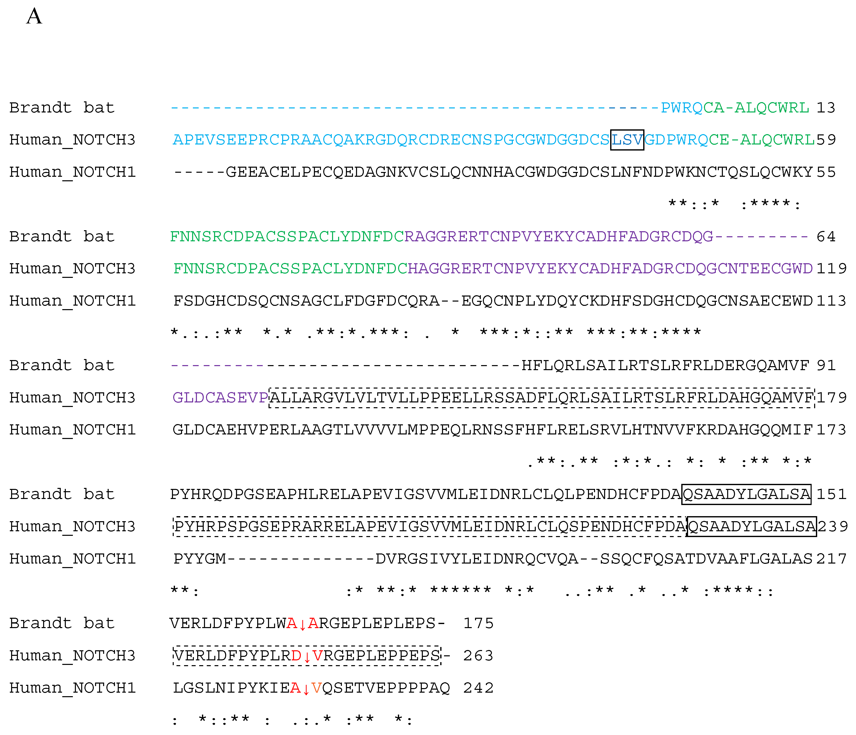
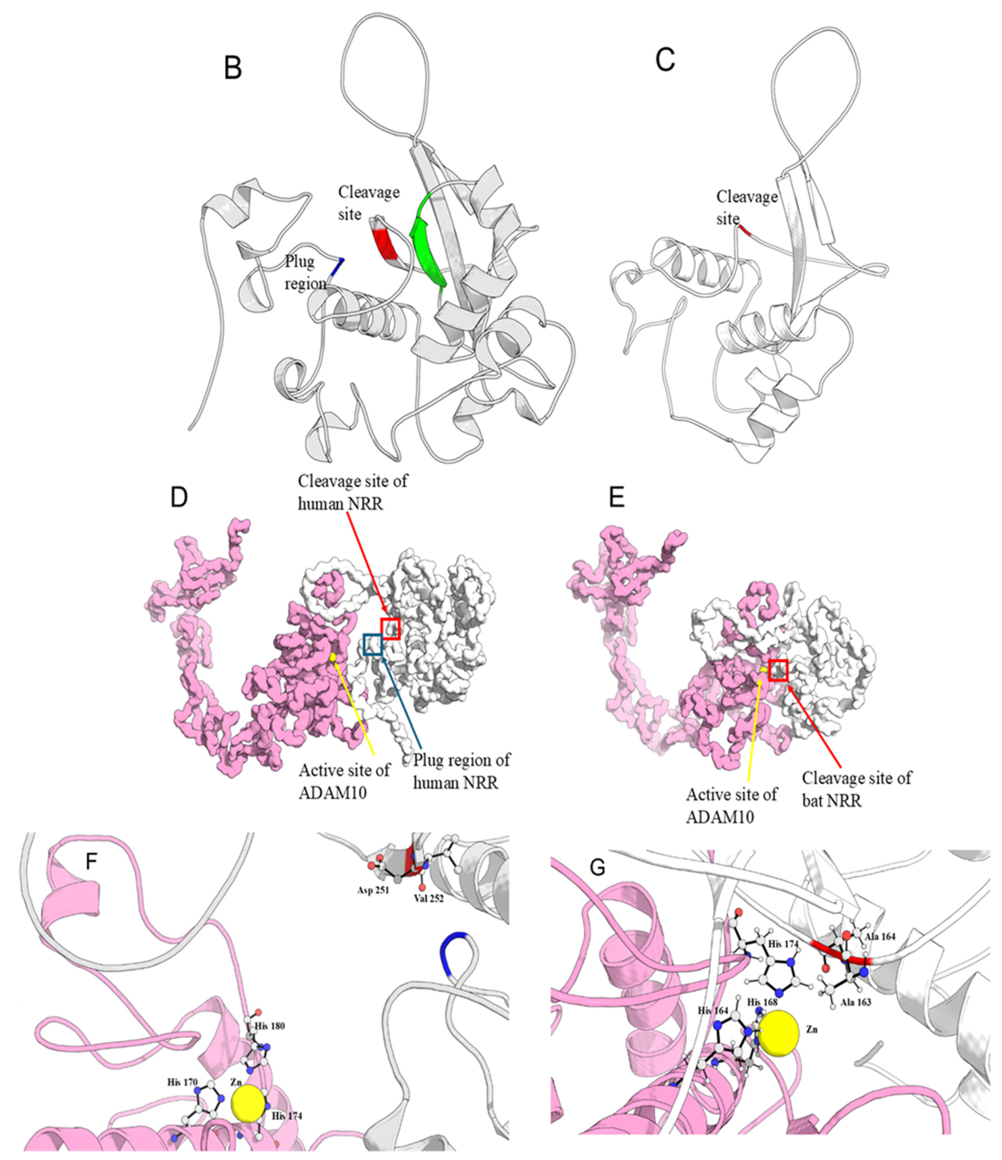
Multiple sequence alignment (MSA) of mammalian NOTCH3 (Supplementary Software 1) shows that the long-lived bat Myotis brandtii lacks two peptides in its NRR domain—within LNR-A and LNR-C (Figure 6a). One missing segment in LNR-A (LNF/LSV), normally forming a loop plug that shields the S2 site, is absent in M. brandtii, potentially enabling easier access for ADAM10 metalloprotease (Figure 6b, c). GETAREA analysis further supports this: human S2 residues D152 and V153 show SASA values of 21.9 Å2 and 1.8 Å2, while the corresponding bat residues A163 and A164 exhibit higher values of 90.3 Å2 and 44.1 Å2. All other examined mammals, including vampire bats and Myotis daubentonii, M. myotis, and M. kuhlii, retain these sequences.
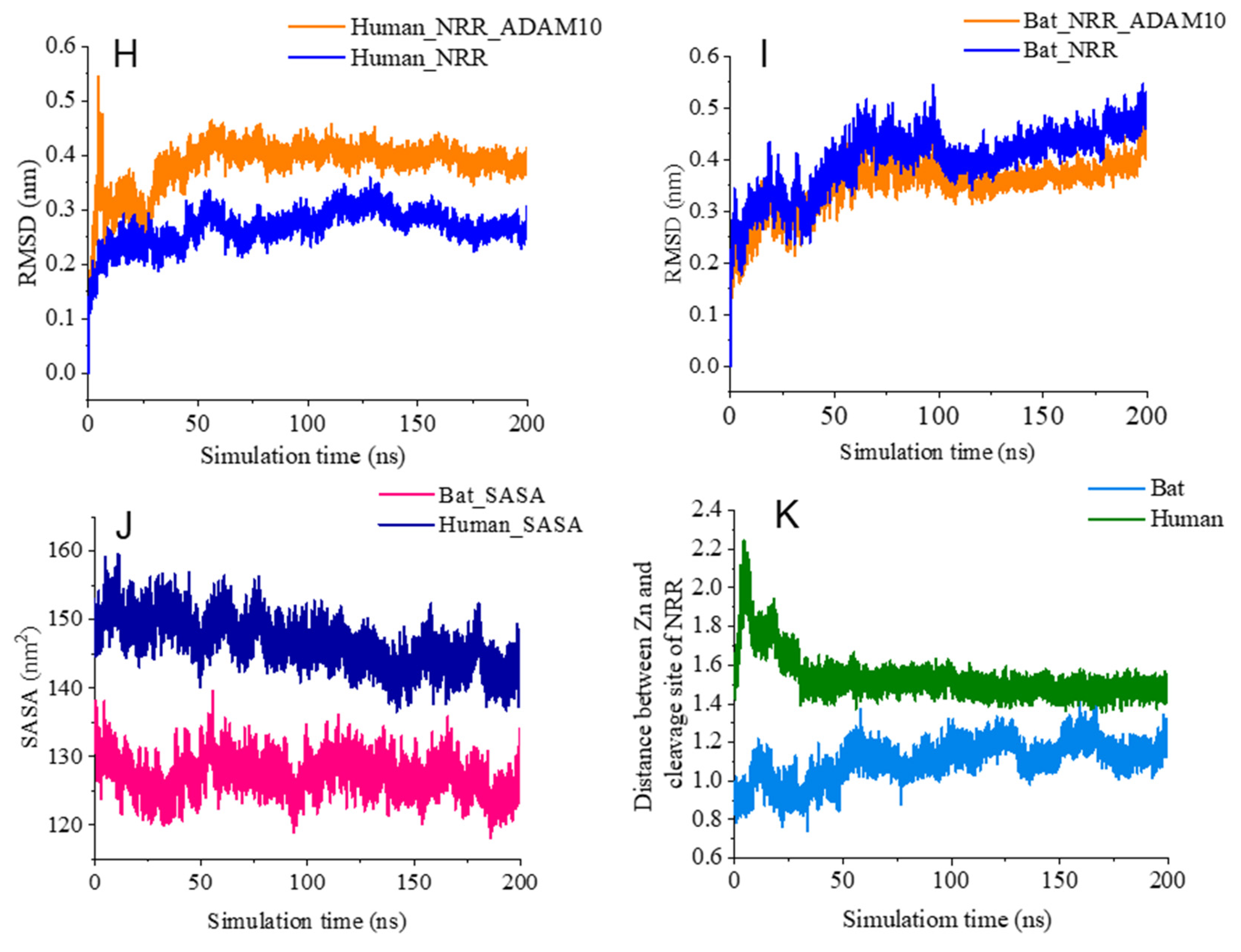
Simulations of the NRR–ADAM10 complex revealed that in the bat (Figure 6e, g), the S2 site lies closer to the catalytic Zn ion than in the human complex (Figure 6d, f), likely due to the LNR-A deletion. Both human and bat complexes remained stable over 200 ns (Figure 6h, i). The human complex showed higher solvent-accessible surface area (SASA), suggesting a less compact structure and weaker interaction (Figure 6j). The Zn–S2 cleavage site distance is consistently shorter in the bat complex (initial 8.4 Å; final 11.8 Å; average 11 Å; Figure 6e, g, k) than in the human (initial 15.24 Å; final 15 Å; average 15.33 Å; Figure 6d, f, k), supporting a conformation more favourable for proteolysis in M. brandtii.
4. Discussion
Cross-species protein analyses are essential for uncovering the evolutionary conservation and pathogenic potential of vascular proteins such as NOTCH3. While aged domestic and wild animals, such as dogs and cats, can exhibit cognitive decline and microvascular changes resembling human small vessel disease, no naturally occurring animal model has been definitively identified for Vascular Cognitive Impairment and Dementia (VCID) or CADASIL. In contrast, several rodent models have been developed to replicate key features of these conditions, including inflammation, white matter damage, and cognitive impairment. Transgenic mice carrying NOTCH3 mutations further recapitulate hallmark CADASIL features such as vascular smooth muscle cell (VSMC) degeneration, white matter lesions, and GOM deposition [41]. Advances in MRI across larger mammals have also enabled cerebrovascular comparisons, laying the groundwork for translational research into how vascular defects contribute to neuronal injury and cognitive decline. These efforts may ultimately guide novel preventative or therapeutic strategies for VCID [42].
In this study, we analyzed NOTCH3 sequences from 113 mammalian species to assess conservation, structural variability, and pathological relevance. Recent large-scale efforts such as the Zoonomia Project have highlighted the power of mammalian comparative genomics for uncovering essential genes and disease mechanisms [8,10]. Our results fit within this broader framework: while NOTCH3 shows strong conservation across mammals (>90% identity), we also uncovered naturally tolerated variation, including eight cysteine mutations in jaguar (Panthera onca) and an EGFr XI deletion present in multiple species, including humans, without apparent ill effects. These findings emphasize that conservation alone does not dictate pathogenicity; context and compensatory mechanisms are also important.
We focused on three naturally occurring scenarios with direct implications for human CADASIL: two involving cysteine loss in EGFrs (via missense mutations or deletions), and one involving a novel deletion in the NRR. These findings form a clear basis for testable hypotheses. While sequence identity below 25% typically signals functional divergence, our MSA revealed strong conservation of NOTCH3 across mammals (>90% identity), underscoring its critical role—particularly the integrity of disulfide bonds that stabilize the 34 EGFr domains (Figure 1). Disruption of these bonds through mutation or deletion, and the resulting unpaired cysteines in the ECD, is increasingly recognized as a key driver of CADASIL [4,5,7].
All NOTCH3 domains were well-structured except for a large portion of the intracellular domain (ICD), which appeared intrinsically disordered (Figure 3). Intrinsically disordered proteins (IDPs) lack a stable tertiary structure under native conditions. IUPRED predicts disorder based on weak inter-residue interactions insufficient for stable folding, while ANCHOR2 identifies regions that become structured upon binding specific partners [13]. The disordered nature of the ICD allows it to adopt multiple conformations depending on binding partners, broadening its functional scope—as seen in other IDPs [43].
The human isoform X1, which likely restores the C1798–C10812 disulfide bond and eliminates free cysteines, presents a paradox: patients still show hallmark CADASIL symptoms such as GOM deposits, vascular smooth muscle degeneration, and cerebral white matter lesions [37]. This suggests that unpaired cysteines alone may not fully explain disease pathology. The recurrence of this deletion across multiple mammals implies a conserved evolutionary mechanism that may contribute to adaptive or pathological traits and warrants experimental investigation.
The discovery of cysteine mutations in the jaguar’s NOTCH3 is intriguing, suggesting that suggesting that not all cysteine residues are equally constrained across mammals. Despite complete disulfide bond loss in EGFr14 and two unpaired cysteines in EGFr13 and EGFr15, the sequenced jaguar appeared healthy [44], though any late-onset neurological consequences remains unknown. Structurally, this mutation cluster may affect ligand binding: EGFr13–15 forms an extended loop over the compact EGFr10–11 ligand-binding domains in jaguar, unlike the more sequential, compact configuration in humans and lions (Figure 5), potentially altering ligand interactions.
Each disulfide bond contributes ~5–6 kcal/mol to protein stability [45]. The loss of three such bonds via Cys>aa substitutions in jaguar NOTCH3 led to a predicted destabilization of ~15–18 kcal/mol. Reverting all seven missense mutations in EGFr12–16 back to cysteines and restoring the deleted residue did not re-establish disulfide bonding, as destabilization persisted in A4D and DynaMut predictions (Supplementary Table S1). This implies that EGFr13–15, which harbors unique insertions, deletions, and mutations, has undergone broader sequence adaptations that influence its structural properties beyond cysteine content alone. We speculate this potentially disordered region near the ligand-binding domains (EGFr10–11) may fine-tune NOTCH3 signaling in ways adapted to the jaguar’s muscular, semi-aquatic, and predatory lifestyle. Further investigation is needed to determine if these mutations promote muscle development, ecological adaptation, or offer insights into CADASIL-relevant structural variation.
Positive selection in jaguar genes for craniofacial and limb development has been reported [44]. Since NOTCH3 loss enhances skeletal muscle in mice [46], similar effects might underlie the jaguar’s exceptional musculature. Jaguars possess the highest bite force quotient among felids—capable of cracking turtle shells and caiman skulls [47]—potentially driven by adaptive NOTCH3 alterations, including loss of five disulfide bonds in EGFr13–15 and the labile DP bond in EGFr14, which exists in humans and lions. These DP bonds may promote ECD aggregation via non-enzymatic autolysis at Asp-Pro sites, a CADASIL hallmark [48]. The combined loss of eight cysteines and a DP bond is unlikely due to drift alone, though lineage-specific drift, compensatory mutations, or other evolutionary forces cannot be excluded.
Most CADASIL-associated mutations occur within the 34 EGFrs or the NRR, leading to either ECD aggregation or ligand-independent receptor activation. Although ECD aggregation is a hallmark of CADASIL, NOTCH3 signaling often remains intact, and the NRR still undergoes normal cleavage [49]. Mutations—particularly in the HD or LNR domains can destabilize the NRR, exposing the S2 cleavage site even without ligand binding.
Interestingly, a unique deletion in Brandt’s bat removes a protective NRR peptide, including the LSV plug from LNR-A (Figure 6a–c), suggesting species-specific modulation of ligand-free activation. While loss of LNR-A might appear to fully expose the S2 site, MD simulations suggest this alone is insufficient for constitutive activation. Rather, the deletion likely increases S2 accessibility and cleavability by ADAM10 compared to the human NRR.
Normally, ligand binding to the ECD initiates endocytosis, pulling LNR-A away [39]. In the bat, the absence of LNR-A implies ligand binding may no longer be necessary. With no LNR-A to displace, activation could proceed more readily [39]. Still, this deletion may not suffice, as Helix3 continues to shield the S2 site, potentially requiring subtle displacement [18]. In bat NRR, Ala143 of α3 forms hydrophobic contacts with Ala164, Arg165, and Gly166 including S2 residue Ala164 likely obstructing ADAM10 access. The shielding function of α3 in preventing cleavage has been previously reported [39,40]. Catalytically active Zn2+ scissile bond distances typically span ~2.1–2.4 Å (QM/MM studies of MMP-3) [50]. In our simulations, Zn–S2 distances remained substantially larger (8.5–11.8 Å), suggesting further conformational changes are needed to bring S2 into alignment with the ADAM10 active site.
A related mechanism involves a conserved calcium-binding His60 in NOTCH1, replaced by arginine in NOTCH3 (Figure 6a). In NOTCH1, mutating this residue to proline disrupts calcium binding, causing a 20-fold increase in ligand-independent activity mimicking LNR deletion [40]. This supports the idea that structural changes in M. brandtii NRR may alter its activation profile, leading to excess NICD production and dysregulated transcription.
Such unregulated signaling disrupts VSMC homeostasis, leading to dysfunction, degeneration, blood–brain barrier leakage, and microangiopathy contributing to cerebral small vessel disease, stroke, vascular cognitive impairment, and dementia [7,34]. Alternatively, the NRR deletion in the bat may support regulated signaling via distinct ligand interactions and subtle conformational changes that displace Helix3. While ligand-independent Notch activation has been associated with cancers, specific oncogenic roles for NOTCH3 remain less studied than for NOTCH1 or NOTCH2 [2].
Bats are renowned for exceptional longevity relative to size [51], attributed to factors like hibernation, efficient DNA repair, mitochondrial function, low IGF-1 signaling, and enhanced autophagy [52,53]. However, the role of altered intercellular signaling—such as via NOTCH3—remains largely unexplored.
Their cerebrovascular anatomy is also unique: a well-developed vertebrobasilar network and robust pial and parenchymal arteries support high cerebral energy demands, while the internal carotid system is comparatively underdeveloped [54,55,56]. This resilience raises the question of why only Brandt’s bat harbors a unique NOTCH3 NRR deletion—and whether it enables ligand-independent activation or reflects alternative regulation.
Recent studies highlight that age-related increases in NOTCH3 expression contribute to vascular aging [53], while elevated signaling promotes narrowing of cerebral arteries and reduced perfusion [34], potentially contributing to cognitive decline. These findings underscore the importance of tightly regulated NOTCH3 activity. The bat-specific NRR deletion may represent an evolutionary adaptation that enables stable, ligand-independent baseline signaling—potentially preserving vascular integrity and supporting bat longevity.
The relationship between NOTCH3 cysteine mutations and pathogenicity remains unclear, highlighting the need for species-specific responses to be validated in model organisms. In jaguars, the absence of disulfide bonds and the presence of unpaired cysteines do not cause disease, suggesting compensatory mechanisms. Disruption of EGFr14 eliminates all disulfide bonds, likely rendering it unstructured (Figure 5). Introducing these mutations into models could clarify how disulfide loss affects NOTCH3 stability, aggregation, pathogenicity, and whether unstructured loops function as ligand-binding sites.
In the human X1 isoform, exon 16 skipping removes EGFr21 and parts of EGFr20 and EGFr22, permitting formation of a new disulfide bond between unpaired cysteines (Figure 4). Despite preservation of cysteine pairing, the deletion remained structurally destabilizing, indicating that domain architecture and local structural context may be as important as cysteine number in determining NOTCH3 stability. Although this deletion occurs in other mammals, its biological significance outside humans remains unknown.
5. Conclusions
Mammalian NOTCH3 sequence–structure–function relationships can reveal hidden clues to the pathogenesis of human vascular disorders, including CADASIL, highlighting how evolutionary variation can inform disease mechanisms. The three mammalian mutations identified here are particularly salient against a background of extreme conservation across 113 mammals. They may not only account for some unique physiological attributes of the three species in which they occur, but also inform the relationship between structure and pathology across species. To deepen understanding of NOTCH3 biology, various cysteine mutations, disulfide rearrangements, and NRR deletions identified in this study should be systematically modeled in cell and animal systems.
Supplementary Materials
The following supporting information can be downloaded at Preprints.org, Figure S1: The multiple sequence alignment of human NOTCH3 with 113 mammals using CLUSTAL-Omega.; S2: The Origin plots showing the NOTCH3 sequence with various domains, pathogenic mutations and % consensus with mammalian protein.; S3: MSA and AF3 structures of NOTCH3.; S4: Plot showing the mean percentage identity and standard deviation for each individual EGFr domain.; Table S1: The stability and flexibility scores of jaguar reverse aa>Cys mutations analysed by DynaMut tool.
Author Contributions
Conceptualization, K.S.S., H.E., P.S. and A.P.; methodology, K.S.S., Y.R., H.E. and A.P.; software, K.S.S., H.E., Y.R; validation, K.S.S., H.E., A.P., T.J. and Y.R.; formal analysis, K.S.S., A.P., T.J. and H.E.; investigation, K.S.S., H.E., P.S., T.J. and A.P; resources, P.S.; data curation, K.S.S. and H.E.; writing—original draft preparation, K.S.S., Y.R. and H.E.; writing—review and editing, A.P., T.J. and P.S.; visualization, K.S.S., H.E. and Y.R; supervision, P.S.; project administration, P.S.; funding acquisition, P.S. All authors have read and agreed to the published version of the manuscript.
Funding
This research was funded by NHMRC of Australia, grant numbers 1196150 and 2006765 and Sachdev Foundation.
Institutional Review Board Statement
Not applicable.
Informed Consent Statement
Not applicable.
Data Availability Statement
The additional data that supports the findings of this study are available in the form of interactive data files and raw data is available on Figshare as follows: Interactives supplementary software data files are available for this paper at https://figshare.com/account/mycontent/items. Multiple Sequence Alignment (MSA) using Custal-Omega and presented using Jalview Software. Identifier: 10.6084/m9.figshare.30223741, Interactive Origin plots show the NOTCH3 sequence with various domains, pathogenic mutations and % consensus with 113 mammalian NOTCH3 sequences. Identifier: 10.6084/m9.figshare.30223747.
Acknowledgments
The authors used ChatGPT5/OpenAI for editing English, word management, and formatting references. The authors reviewed and edited the ChatGPT-generated output and take full responsibility for the content of the publication.
Conflicts of Interest
The authors declare no conflicts of interest.The funders had no role in the design of the study; in the collection, analyses, or interpretation of data; in the writing of the manuscript; or in the decision to publish the results.
Abbreviations
The following abbreviations are used in this manuscript:
| NOTCH3 | Neurogenic locus notch homolog protein 3 |
| CADASIL | Cerebral autosomal dominant arteriopathy with subcortical infarcts and leukoencephalopathy |
| EGFr | epidermal growth factor repeats |
| ECD | extracellular domain |
| NRR | negative regulatory region |
| TM | transmembrane |
| ICD | intracellular cytoplasmic domain |
| GOM | granular osmiophilic material |
| VSMC | vascular smooth muscle cell |
| ADAM | a disintegrin and metalloproteinase |
| MDS | molecular dynamics simulation |
| AF3 | AlphaFold3 |
| pLDDT | predicted local distance difference test |
| SASA | solvent accessible surface area |
| RMSD | root mean square deviation |
| LNR | Lin12-Notch repeats |
| HD | heterodimerization domain |
| IGF | insulin-like growth factor |
| VCID | vascular cognitive impairment and dementia |
References
- Darwin, C. The Descent of Man, and selection in relation to sex, 2nd ed.; John Murray: London, 1874; p. 6. [Google Scholar]
- Hosseini-Alghaderi, S.; Baron, M. Notch3 in development, health and disease. Biomolecules 2020, 10, 485. [Google Scholar] [CrossRef] [PubMed]
- Boston, G.; Jobson, D.; Mizuno, T.; Ihara, M.; Kalaria, R. N. Most common NOTCH3 mutations causing CADASIL or CADASIL-like cerebral small vessel disease: A systematic review. Cereb. Circ.-Cogn. Behav. 2024, 6, 100227. [Google Scholar] [CrossRef] [PubMed]
- Young, K. Z.; et al. Oligomerization, trans-reduction, and instability of mutant NOTCH3 in inherited vascular dementia. Commun. Biol. 2022, 5, 1 331. [Google Scholar] [CrossRef] [PubMed]
- Lee, S. J.; et al. Structural changes in NOTCH3 induced by CADASIL mutations: role of cysteine and non-cysteine alterations. J. Biol. Chem. 2023, 299, 104838. [Google Scholar] [CrossRef] [PubMed]
- Hess, K. L.; et al. Protein aggregates containing wild-type and mutant NOTCH3 are major drivers of arterial pathology in CADASIL. J. Clin. Invest. 2024, 134, e175789. [Google Scholar] [CrossRef] [PubMed]
- Mizuta, I.; et al. Progress to clarify how NOTCH3 mutations lead to CADASIL, a hereditary cerebral small vessel disease. Biomolecules 2024, 14, 127. [Google Scholar] [CrossRef] [PubMed]
- Christmas, M.J.; et al. Zoonomia: Evolutionary constraint and innovation across hundreds of placental mammals. Science. 2023, 380, 366. [Google Scholar] [CrossRef] [PubMed]
- Romero, I.J. Seeing humans through an evolutionary lens: a collection of mammalian genomes provides insights into human biology and evolution. Science. 2023, 380, 360–361. [Google Scholar] [CrossRef] [PubMed]
- Sullivan, P.F.; et al. Zoonomia: Leveraging base-pair mammalian constraint to understand genetic variation and human disease. Science. 2023, 380, 367. [Google Scholar] [CrossRef] [PubMed]
- Madeira, F.; et al. The EMBL-EBI Job Dispatcher sequence analysis tools framework in 2024. Nucleic Acids Res. 2024, 52, W521–W525. [Google Scholar] [CrossRef] [PubMed]
- Waterhouse, A.M.; Procter, J.B.; Martin, D.M.A.; Clamp, M.; Barton, G.J. Jalview Version 2—a multiple sequence alignment editor and analysis workbench. Bioinformatics 2009, 25, 1189–1191. [Google Scholar] [CrossRef] [PubMed]
- Erdős, G.; Dosztányi, Z. AIUPred: combining energy estimation with deep learning for the enhanced prediction of protein disorder. Nucleic Acids Res. 2024, 52, W176–W181. [Google Scholar] [CrossRef] [PubMed]
- Mészáros, B.; Erdős, G.; Dosztányi, Z. IUPred2A: context-dependent prediction of protein disorder as a function of redox state and protein binding. Nucleic Acids Res. 2018, 46, W329–W337. [Google Scholar] [CrossRef] [PubMed]
- Jumper, J.; et al. Highly accurate protein structure prediction with AlphaFold. Nature 2021, 596, 583–589. [Google Scholar] [CrossRef]
- Abramson, J.; et al. Accurate structure prediction of biomolecular interactions with AlphaFold 3. Nature 2024, 630, 493–500. [Google Scholar] [CrossRef] [PubMed]
- Meng, E. C.; et al. UCSF ChimeraX: tools for structure building and analysis. Protein Sci. 2023, 32, e4792. [Google Scholar] [CrossRef] [PubMed]
- Rodrigues, C. H. M.; Pires, D. E. V.; Ascher, D. B. DynaMut2: Assessing changes in stability and flexibility upon single and multiple point missense mutations. Protein Sci. 2021, 30, 60–69. [Google Scholar] [CrossRef] [PubMed]
- Zalewski, M.; Iglesias, V.; Bárcenas, O.; Ventura, S.; Kmiecik, S. Aggrescan4D: A comprehensive tool for pH-dependent analysis and engineering of protein aggregation propensity. Protein Sci. 2024, 33, e5180. [Google Scholar] [CrossRef] [PubMed]
- Craig, D. B.; Dombkowski, A. A. Disulfide by Design 2.0: a web-based tool for disulfide engineering in proteins. BMC Bioinform. 2013, 14, 346. [Google Scholar] [CrossRef] [PubMed]
- Fraczkiewicz, R.; Braun, W. Exact and efficient analytical calculation of the accessible surface areas and their gradients for macromolecules. J. Comp. Chem. 1998, 19, 319–333. [Google Scholar] [CrossRef]
- Lipper, C. H.; Egan, E. D.; Gabriel, K. H.; Blacklow, S. C. Structural basis for membrane-proximal proteolysis of substrates by ADAM10. Cell 2023, 186, 3632–3641.e10. [Google Scholar] [CrossRef] [PubMed]
- Seim, I.; et al. Genome analysis reveals insights into physiology and longevity of the Brandt’s bat Myotis brandtii. Nat. Commun. 2013, 4, 2212. [Google Scholar] [CrossRef] [PubMed]
- Jo, S.; Kim, T.; Iyer, V. G.; Im, W. CHARMM-GUI: A web-based graphical user interface for CHARMM. J. Comput. Chem. 2008, 29, 1859–1865. [Google Scholar] [CrossRef] [PubMed]
- Maier, J. A.; Martinez, C.; Kasavajhala, K.; Wickstrom, L.; Hauser, K. E.; Simmerling, C. ff14SB: Improving the accuracy of protein side chain and backbone parameters from ff99SB. J. Chem. Theory Comput. 2015, 11, 3696–3713. [Google Scholar] [CrossRef] [PubMed]
- Jorgensen, W. L.; Chandrasekhar, J.; Madura, J. D.; Impey, R.; Klein, M. Refined TIP3P model for water. J. Chem. Phys. 1983, 79, 926–935. [Google Scholar]
- Abraham, M. J.; et al. GROMACS: High performance molecular simulations through multi-level parallelism from laptops to supercomputers. SoftwareX 2015, 1, 19–25. [Google Scholar] [CrossRef]
- Bussi, G.; Donadio, D.; Parrinello, M. Canonical sampling through velocity rescaling. J. Chem. Phys. 2007, 126, 014101. [Google Scholar] [CrossRef] [PubMed]
- Ke, Q.; Gong, X.; Liao, S.; Duan, C.; Li, L. Effects of thermostats/barostats on physical properties of liquids by molecular dynamics simulations. J. Mol. Liq. 2022, 365, 120070. [Google Scholar] [CrossRef]
- Grubmüller, H.; Heller, H.; Windemuth, A.; Schulten, K. Generalized Verlet algorithm for efficient molecular dynamics simulations with long-range interactions. Mol. Simul. 1991, 6, 121–142. [Google Scholar] [CrossRef]
- Petersen, H. G. Accuracy and efficiency of the particle mesh Ewald method. J. Chem. Phys. 1995, 103, 3668–3679. [Google Scholar] [CrossRef]
- Hess, B.; Bekker, H.; Berendsen, H. J.; Fraaije, J. G. LINCS: A linear constraint solver for molecular simulations. J. Comput. Chem. 1997, 18, 1463–1472. [Google Scholar] [CrossRef]
- Ryckaert, J.-P.; Ciccotti, G.; Berendsen, H. J. C. Numerical integration of the cartesian equations of motion of a system with constraints: molecular dynamics of n-alkanes. J. Comput. Phys. 1977, 23, 327–341. [Google Scholar] [CrossRef]
- Baron-Menguy, C.; Domenga-Denier, V.; Ghezali, L.; Faraci, F. M.; Joutel, A. Increased Notch3 activity mediates pathological changes in structure of cerebral arteries. Hypertension 2017, 69, 60–70. [Google Scholar] [CrossRef] [PubMed]
- Oka, Kaori; et al. The naked mole-rat as a model for healthy aging. Annu. Rev. Anim. Biosci. 2023, 11, 207–226. [Google Scholar] [CrossRef] [PubMed]
- Morris, O.M.; Torpey, J.H.; Isaacson, R.L. Intrinsically disordered proteins: modes of binding with emphasis on disordered domains. Open Biol. 2021, 11, 210222. [Google Scholar] [CrossRef] [PubMed]
- Saiki, S.; et al. Varicose veins associated with CADASIL result from a novel mutation in the Notch3 gene. Neurology 2006, 67, 337–339. [Google Scholar] [CrossRef] [PubMed]
- Chabriat, H.; et al. CADASIL. Lancet Neurol. 2009, 8, 643–653. [Google Scholar] [CrossRef] [PubMed]
- Gordon, W. R.; Arnett, K. L.; Blacklow, S. C. The molecular logic of Notch signaling—a structural and biochemical perspective. J. Cell Sci. 2008, 121, 3109–3119. [Google Scholar] [CrossRef] [PubMed]
- Gordon, W. R.; et al. Structure of the Notch1-negative regulatory region: implications for normal activation and pathogenic signaling in T-ALL. Blood 2009, 113, 4381–4390. [Google Scholar] [CrossRef] [PubMed]
- Gong, Z.; et al. Analysis of the pathogenicity and pathological characteristics of NOTCH3 gene-sparing cysteine mutations in in vitro and in vivo models. Front. Mol. Neurosci. 2024, 17, 1391040. [Google Scholar] [CrossRef] [PubMed]
- Hainsworth, A. H.; et al. Translational models for vascular cognitive impairment: a review including larger species. BMC Med. 2017, 15, 16. [Google Scholar] [CrossRef] [PubMed]
- Wright, P.; Dyson, H. Intrinsically disordered proteins in cellular signalling and regulation. Nat. Rev. Mol. Cell Biol. 2015, 16, 18–29. [Google Scholar] [CrossRef] [PubMed]
- Figueiró, H. V.; et al. Genome-wide signatures of complex introgression and adaptive evolution in the big cats. Sci. Adv. 2017, 3, e1700299. [Google Scholar] [CrossRef] [PubMed]
- Zavodszky, M.; et al. Disulfide bond effects on protein stability: designed variants of Cucurbita maxima trypsin inhibitor-V. Protein Sci. 2001, 10, 149–160. [Google Scholar] [CrossRef] [PubMed]
- Kitamoto, T.; Hanaoka, K. Notch3 null mutation in mice causes muscle hyperplasia by repetitive muscle regeneration. Stem Cells 2010, 28, 2205–2216. [Google Scholar] [CrossRef] [PubMed]
- Hoogesteijn, R. Wild cats 101: Why jaguars hunt caimans? Panthera Blog. 2023. Available online: https://panthera.org/blog-post/wild-cats-101-why-jaguars-hunt-caimans.
- Lee, S. J.; et al. A midposition NOTCH3 truncation in inherited cerebral small vessel disease may affect the protein interactome. J. Biol. Chem. 2023, 299, 102772. [Google Scholar] [CrossRef] [PubMed]
- Wang, T.; Baron, M.; Trump, D. An overview of Notch3 function in vascular smooth muscle cells. Prog. Biophys. Mol. Biol. 2008, 96, 499–509. [Google Scholar] [CrossRef] [PubMed]
- Feliciano, G. T.; da Silva, A. J. R. Unravelling the reaction mechanism of matrix metalloproteinase 3 using QM/MM calculations. J. Mol. Struct. 2015, 1091, 125–132. [Google Scholar] [CrossRef]
- Brunet-Rossinni, A. K.; Austad, S. N. Ageing studies on bats: a review. Biogerontology 2004, 5, 211–222. [Google Scholar] [CrossRef] [PubMed]
- Ding, Y.; et al. Comprehensive human proteome profiles across a 50-year lifespan reveal aging trajectories and signatures. Cell 2025, S0092-8674(25)00749-4. [Google Scholar] [CrossRef] [PubMed]
- Gorbunova, V.; Seluanov, A.; Kennedy, B. K. The world goes bats: living longer and tolerating viruses. Cell Metab. 2020, 32, 31–43. [Google Scholar] [CrossRef] [PubMed]
- Hasegawa, K.; Kawano, T.; Oishi, M.; Sunagawa, G.; Nishimura, T. The angio-architecture in the brain of bat. Fukuoka Acta Medica 1960, 51, 1251–1266. [Google Scholar]
- Andō, K. A histochemical study on the innervation of the cerebral blood vessels in bats. Cell Tissue Res. 1981, 217, 55–64. [Google Scholar] [CrossRef] [PubMed]
Figure 1.
Sequence conservation plot and structure of human NOTCH3. a, the Origin plot shows the NOTCH3 sequence with various domains, pathogenic mutations and % consensus with 113 mammalian NOTCH3 sequences. Domains are shown on the X-axis as colored segments. 34 EGFrs in ECD, blue; NRR (LNR + HD), red; TM, black; ICD, green; % consensus, pink circle. EGFr10,11 (391-467 aa) are involved in ligand-binding. Lifted bars indicate the EGFr region corresponding to jaguar EGFr13–15 (507-618 aa), and the X1 deletion spanning EGFr20–22 (771-885 aa). Various types of pathogenic mutations are shown as triangles: aa>X, red; aa>C, green; C>aa, blue. The interactive Origin plot features tooltips, where hovering the cursor over data points reveals the exact percent consensus, amino acid sequence, residue name, and mutation information (Supplementary Software 2). The exploded views of EGFr13-15 and EGFr20-22 are shown in Supplementary Figs 2b and 2c. b and c, Human full-length NOTCH3 structure predicted by AlphaFold3. b, color-coded domains—signal peptide (red), EGFr1–34 (cyan), NRR (blue), TM helix (grey), and ICD (pink). c, pLDDT confidence scores—blue: >90 (very high); light blue: 70–90 (confident); yellow: 50–70 (low); orange: <50 (very low). Although local domain structures are well defined, their spatial arrangement in the full-length model is uncertain due to low inter-domain confidence.
Figure 1.
Sequence conservation plot and structure of human NOTCH3. a, the Origin plot shows the NOTCH3 sequence with various domains, pathogenic mutations and % consensus with 113 mammalian NOTCH3 sequences. Domains are shown on the X-axis as colored segments. 34 EGFrs in ECD, blue; NRR (LNR + HD), red; TM, black; ICD, green; % consensus, pink circle. EGFr10,11 (391-467 aa) are involved in ligand-binding. Lifted bars indicate the EGFr region corresponding to jaguar EGFr13–15 (507-618 aa), and the X1 deletion spanning EGFr20–22 (771-885 aa). Various types of pathogenic mutations are shown as triangles: aa>X, red; aa>C, green; C>aa, blue. The interactive Origin plot features tooltips, where hovering the cursor over data points reveals the exact percent consensus, amino acid sequence, residue name, and mutation information (Supplementary Software 2). The exploded views of EGFr13-15 and EGFr20-22 are shown in Supplementary Figs 2b and 2c. b and c, Human full-length NOTCH3 structure predicted by AlphaFold3. b, color-coded domains—signal peptide (red), EGFr1–34 (cyan), NRR (blue), TM helix (grey), and ICD (pink). c, pLDDT confidence scores—blue: >90 (very high); light blue: 70–90 (confident); yellow: 50–70 (low); orange: <50 (very low). Although local domain structures are well defined, their spatial arrangement in the full-length model is uncertain due to low inter-domain confidence.
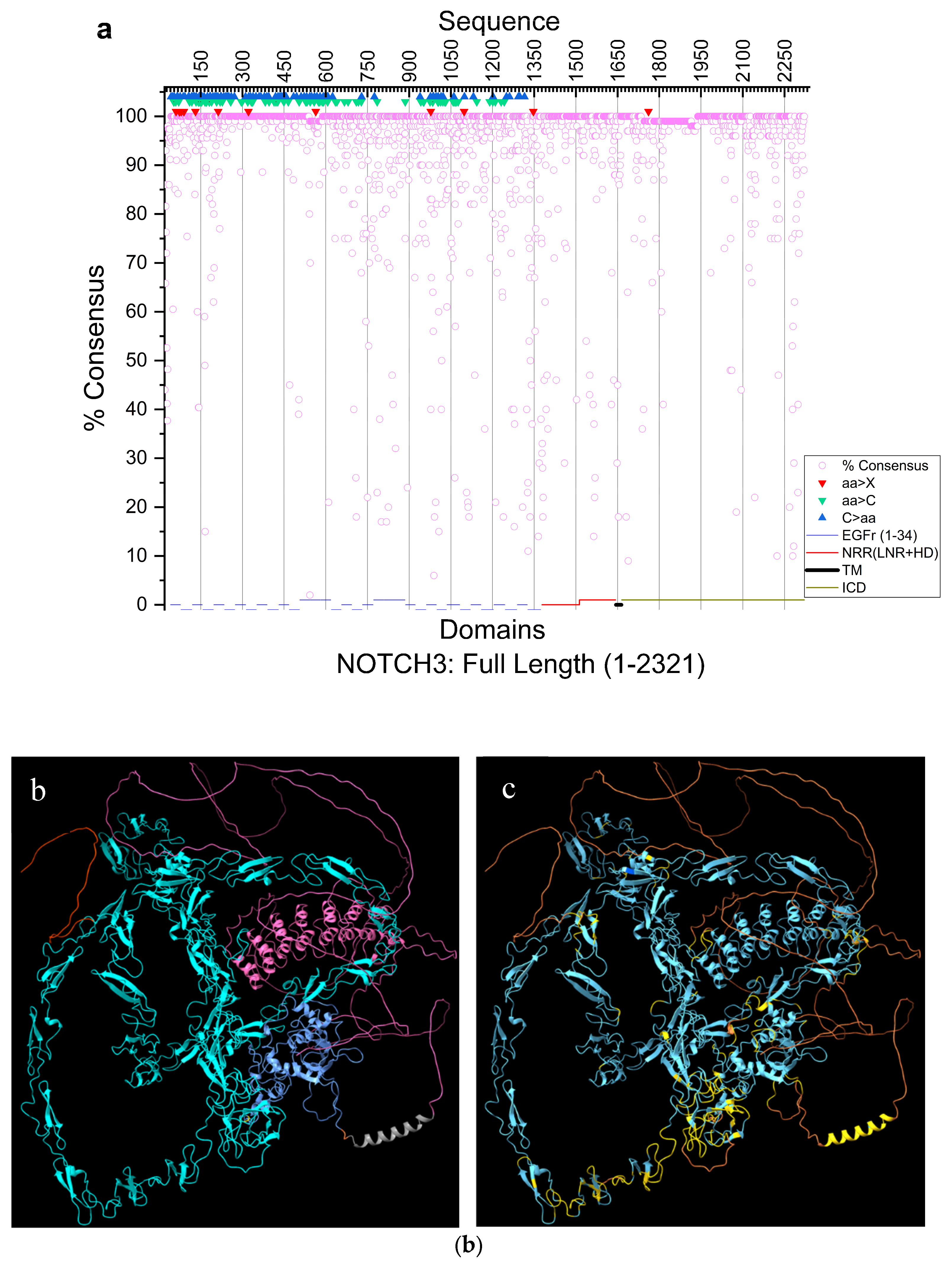
Figure 2.
Sequence and structural similarity in ECD. a, human and rhesus (RMSD across all 1603 residue pairs, 17.34 Å), b, human and mouse (RMSD across all 1603 residue pairs, 23.9 Å). Light orange/dark orange, human; light green/dark green, rhesus, light blue/dark blue, mouse. Darker shades represent EGFr1-11. c, human NOTCH3 extracellular domain structure predicted by AlphaFold3. c, colored according to pLDDT confidence scores as in Figure 1. The AlphaFold3-predicted structure of the human NOTCH3 ECD shows high confidence for individual EGFr domains but low confidence in their relative orientation. d, human ectodomain showing conservation among >113 mammalian species. Blue, 100%; corn-blue, 98-99%, lime, 90-97%; yellow, 50-89%; <50%, orange. Blue-light blue, high conservation; lime-yellow, medium conservation; orange, low conservation. The relative positioning of EGFr and NRR shows low confidence. e, sequence similarity among EGFr1–34 domains in human and mouse NOTCH3. Heatmap showing pairwise sequence identity within human EGFr domains (H vs H), within mouse EGFr domains (M vs M), and between human and mouse EGFr domains (H vs M). The green diagonal represents 100% identity for self-comparisons of the same EGFr domain; the orange-red diagonal indicates sequence identity between corresponding EGFr domains in human and mouse; off-diagonal values above and below the main diagonals show the percentage identity between non-corresponding EGFr domains within and between both species. Structurally, EGFr6 and EGFr31 had an RMSD of 3.815 Å (37 aligned residues), while EGFr4 and EGFr10 had an RMSD of 1.102 Å (38 residues). Other comparisons: EGFr4 vs. EGFr6, 41.7% identity, RMSD 3.81 Å; EGFr6 vs. EGFr10, 35.0%, RMSD 4.2 Å; EGFr4 vs. EGFr31, 50.0%, RMSD 1.0 Å.
Figure 2.
Sequence and structural similarity in ECD. a, human and rhesus (RMSD across all 1603 residue pairs, 17.34 Å), b, human and mouse (RMSD across all 1603 residue pairs, 23.9 Å). Light orange/dark orange, human; light green/dark green, rhesus, light blue/dark blue, mouse. Darker shades represent EGFr1-11. c, human NOTCH3 extracellular domain structure predicted by AlphaFold3. c, colored according to pLDDT confidence scores as in Figure 1. The AlphaFold3-predicted structure of the human NOTCH3 ECD shows high confidence for individual EGFr domains but low confidence in their relative orientation. d, human ectodomain showing conservation among >113 mammalian species. Blue, 100%; corn-blue, 98-99%, lime, 90-97%; yellow, 50-89%; <50%, orange. Blue-light blue, high conservation; lime-yellow, medium conservation; orange, low conservation. The relative positioning of EGFr and NRR shows low confidence. e, sequence similarity among EGFr1–34 domains in human and mouse NOTCH3. Heatmap showing pairwise sequence identity within human EGFr domains (H vs H), within mouse EGFr domains (M vs M), and between human and mouse EGFr domains (H vs M). The green diagonal represents 100% identity for self-comparisons of the same EGFr domain; the orange-red diagonal indicates sequence identity between corresponding EGFr domains in human and mouse; off-diagonal values above and below the main diagonals show the percentage identity between non-corresponding EGFr domains within and between both species. Structurally, EGFr6 and EGFr31 had an RMSD of 3.815 Å (37 aligned residues), while EGFr4 and EGFr10 had an RMSD of 1.102 Å (38 residues). Other comparisons: EGFr4 vs. EGFr6, 41.7% identity, RMSD 3.81 Å; EGFr6 vs. EGFr10, 35.0%, RMSD 4.2 Å; EGFr4 vs. EGFr31, 50.0%, RMSD 1.0 Å.
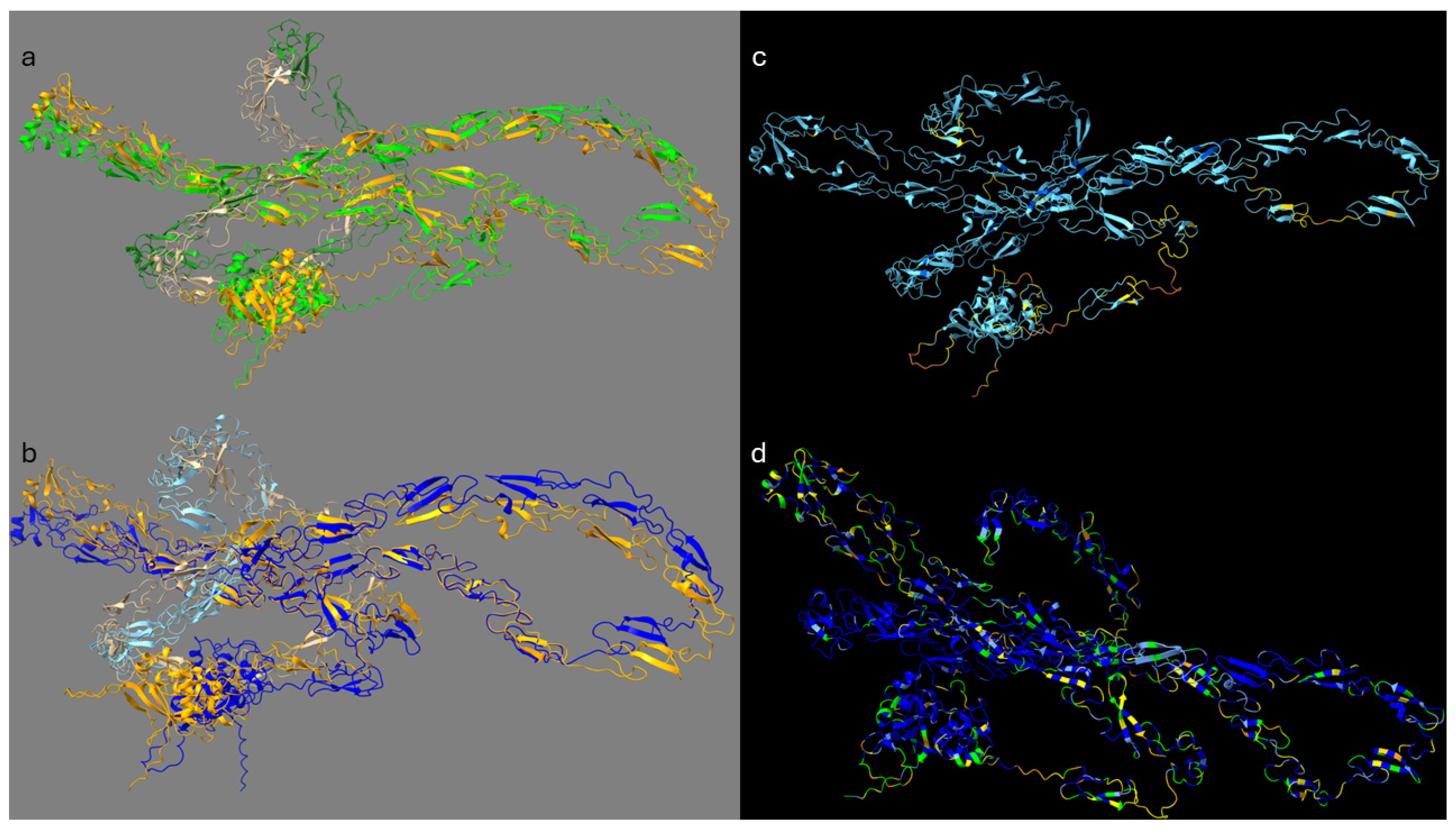
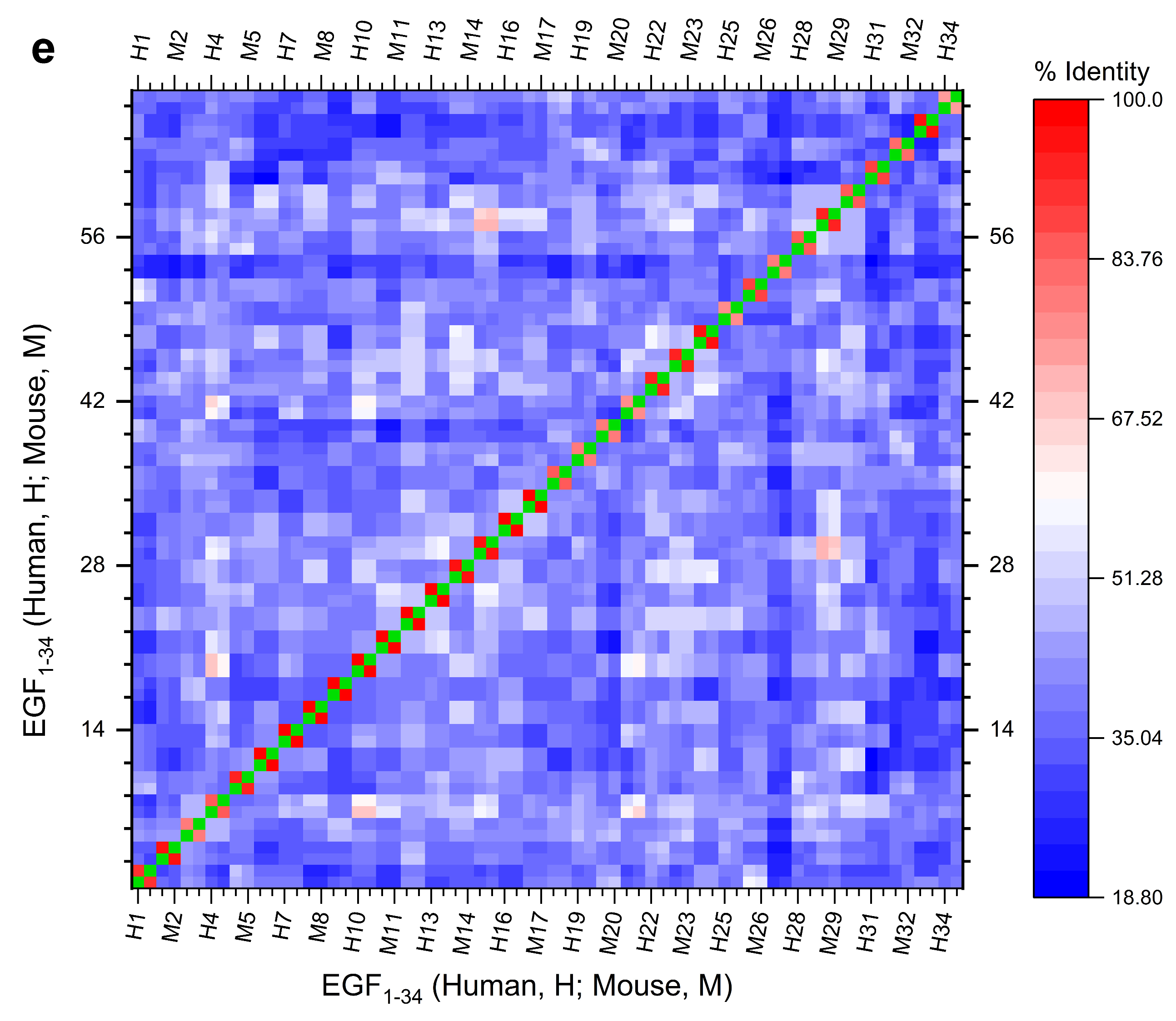
Figure 3.
Structure and analysis of intrinsically disordered regions. a, superimposed human and mouse NOTCH3 ICD predicted by AF3. Refer to Figure 1 for pIDDT confidence score. b, plot showing intrinsically disordered regions in the NOTCH3 sequence using two different tools: IUPred and ANCHOR2.
Figure 3.
Structure and analysis of intrinsically disordered regions. a, superimposed human and mouse NOTCH3 ICD predicted by AF3. Refer to Figure 1 for pIDDT confidence score. b, plot showing intrinsically disordered regions in the NOTCH3 sequence using two different tools: IUPred and ANCHOR2.
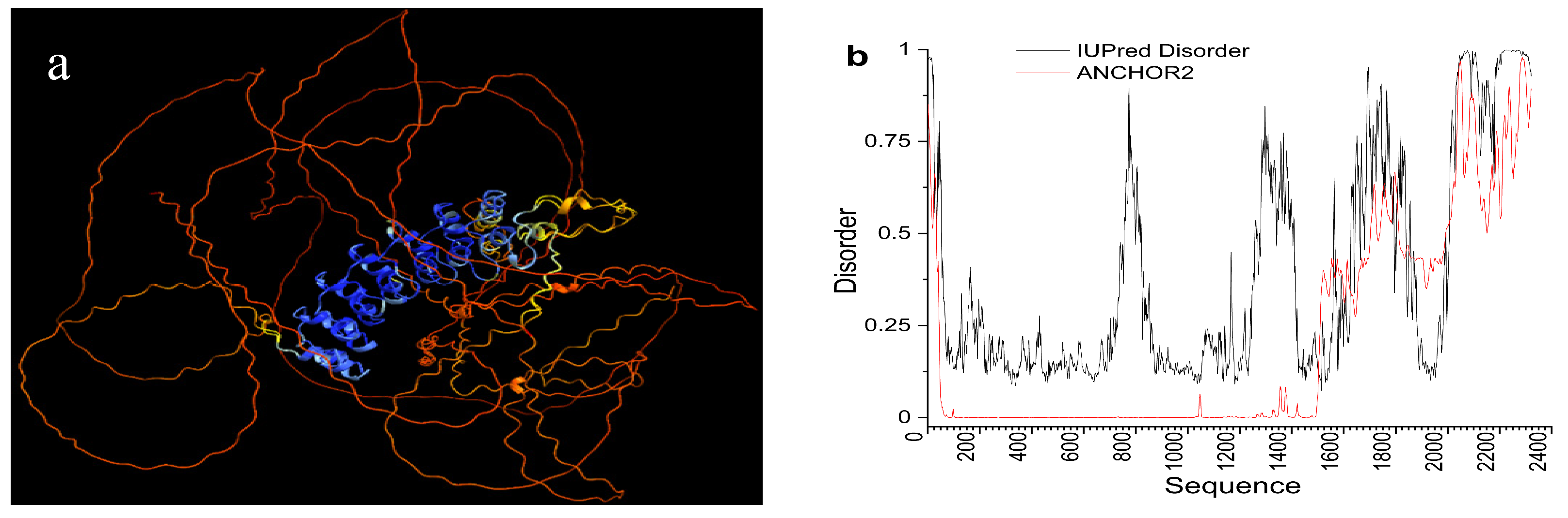
Figure 4.
Sequence and structural analysis of deletion isoforms. a, MSA of isoform (X1) of human NOTCH3 compared to various mammals. Chimpanzee, Pan troglodytes; Pygmy chimpanzee, Pan paniscus; Gorilla, Gorilla gorilla gorilla; Monkey, Rhinopithecus roxellana; Marmoset, Callithrix jacchus; Capuchin, Sapajus apella; Bat, Myotis myotis; vampire bat, Desmodus rotundus; Mouse, Mus musculus; hamster, Cricetulus griseus; Indian elephant (Ind.), Elephas maximus indicus; African elephant (Afr.), Loxodonta Africana; Whale (blue), Balaenoptera musculus. Arrows/bold residue (D) shows the start of the deletion. *Conserved residues and x, where Cys residue is missing in some sequence. 8 Cys residues within the deleted sequence (Human) are shown in bold. There are 8 cysteines in the missing peptide (P805-N856). C3814-C5826, C4820-C6835, and C7837-C8846 S-S bonds are within EGFr21; In full-length NOTCH3, C1798-C2807 and C9853-C10864 S-S bonds are in EGFr20 and EGFr22 respectively. The C1798 and C10812 in X1 deletion mutant form a new disulfide bond and are shown in red bold. b, structure of great apes, NOTCH3, EGFr20-22 (above); c, Great apes NOTCH3 X1 deletion isoform (with EGFr23) is shown in pLDDT colors. Dark blue, >90 (very high); light blue, 90>pLDDT>70 (confident); yellow, 70>pLDDT>50 (low). pLDDT scores served as a qualitative indicator of structural destabilization. All disulfide bonds are shown (green dashes). C798-C807 and C853-C864 are highlighted because C798 and C853 are within the deleted isoform. ↑C798 forms a new disulfide with the C812 (C864 in NOTCH3 without deletion) due to the deletion. ↓G804D mutation.
Figure 4.
Sequence and structural analysis of deletion isoforms. a, MSA of isoform (X1) of human NOTCH3 compared to various mammals. Chimpanzee, Pan troglodytes; Pygmy chimpanzee, Pan paniscus; Gorilla, Gorilla gorilla gorilla; Monkey, Rhinopithecus roxellana; Marmoset, Callithrix jacchus; Capuchin, Sapajus apella; Bat, Myotis myotis; vampire bat, Desmodus rotundus; Mouse, Mus musculus; hamster, Cricetulus griseus; Indian elephant (Ind.), Elephas maximus indicus; African elephant (Afr.), Loxodonta Africana; Whale (blue), Balaenoptera musculus. Arrows/bold residue (D) shows the start of the deletion. *Conserved residues and x, where Cys residue is missing in some sequence. 8 Cys residues within the deleted sequence (Human) are shown in bold. There are 8 cysteines in the missing peptide (P805-N856). C3814-C5826, C4820-C6835, and C7837-C8846 S-S bonds are within EGFr21; In full-length NOTCH3, C1798-C2807 and C9853-C10864 S-S bonds are in EGFr20 and EGFr22 respectively. The C1798 and C10812 in X1 deletion mutant form a new disulfide bond and are shown in red bold. b, structure of great apes, NOTCH3, EGFr20-22 (above); c, Great apes NOTCH3 X1 deletion isoform (with EGFr23) is shown in pLDDT colors. Dark blue, >90 (very high); light blue, 90>pLDDT>70 (confident); yellow, 70>pLDDT>50 (low). pLDDT scores served as a qualitative indicator of structural destabilization. All disulfide bonds are shown (green dashes). C798-C807 and C853-C864 are highlighted because C798 and C853 are within the deleted isoform. ↑C798 forms a new disulfide with the C812 (C864 in NOTCH3 without deletion) due to the deletion. ↓G804D mutation.
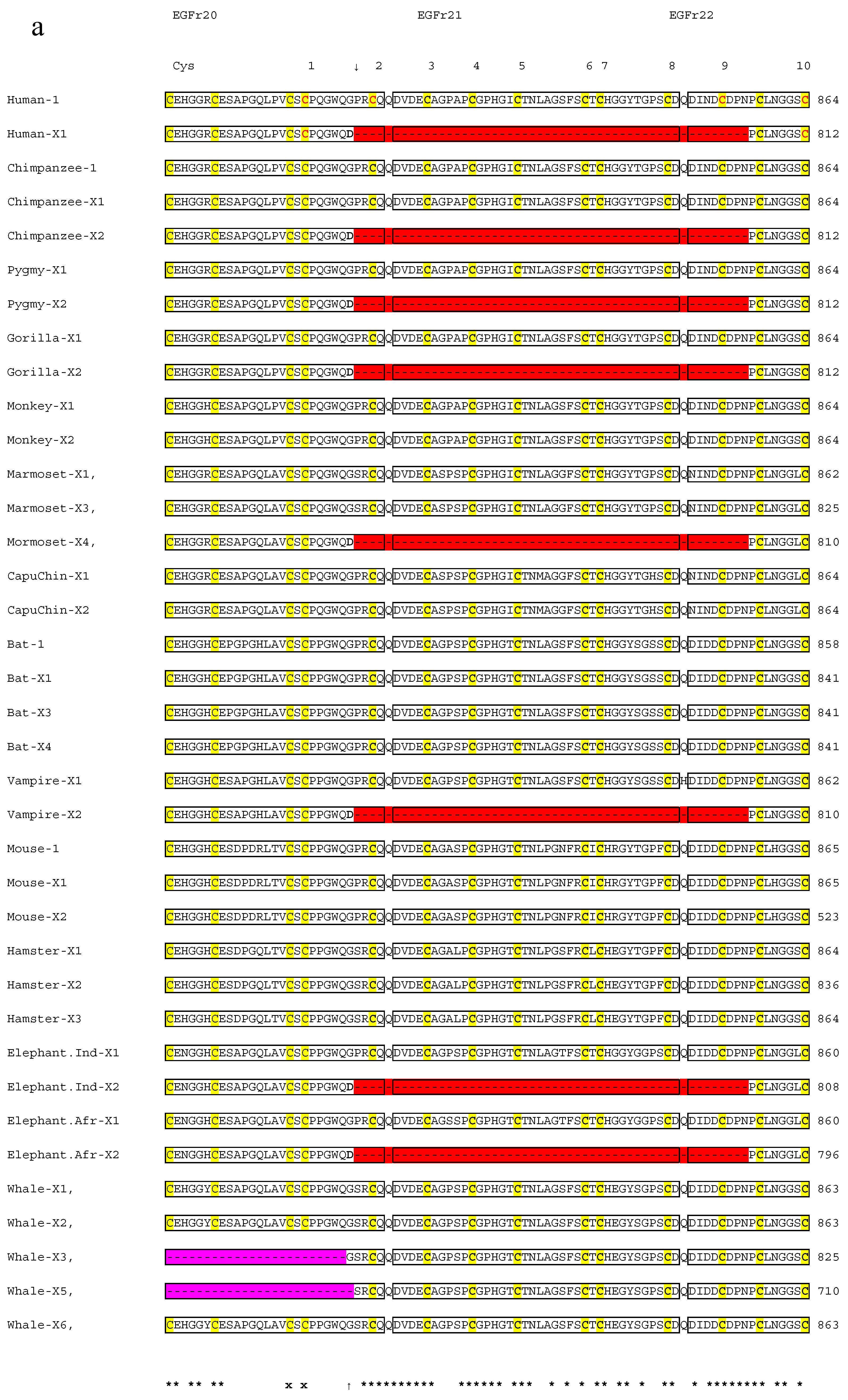
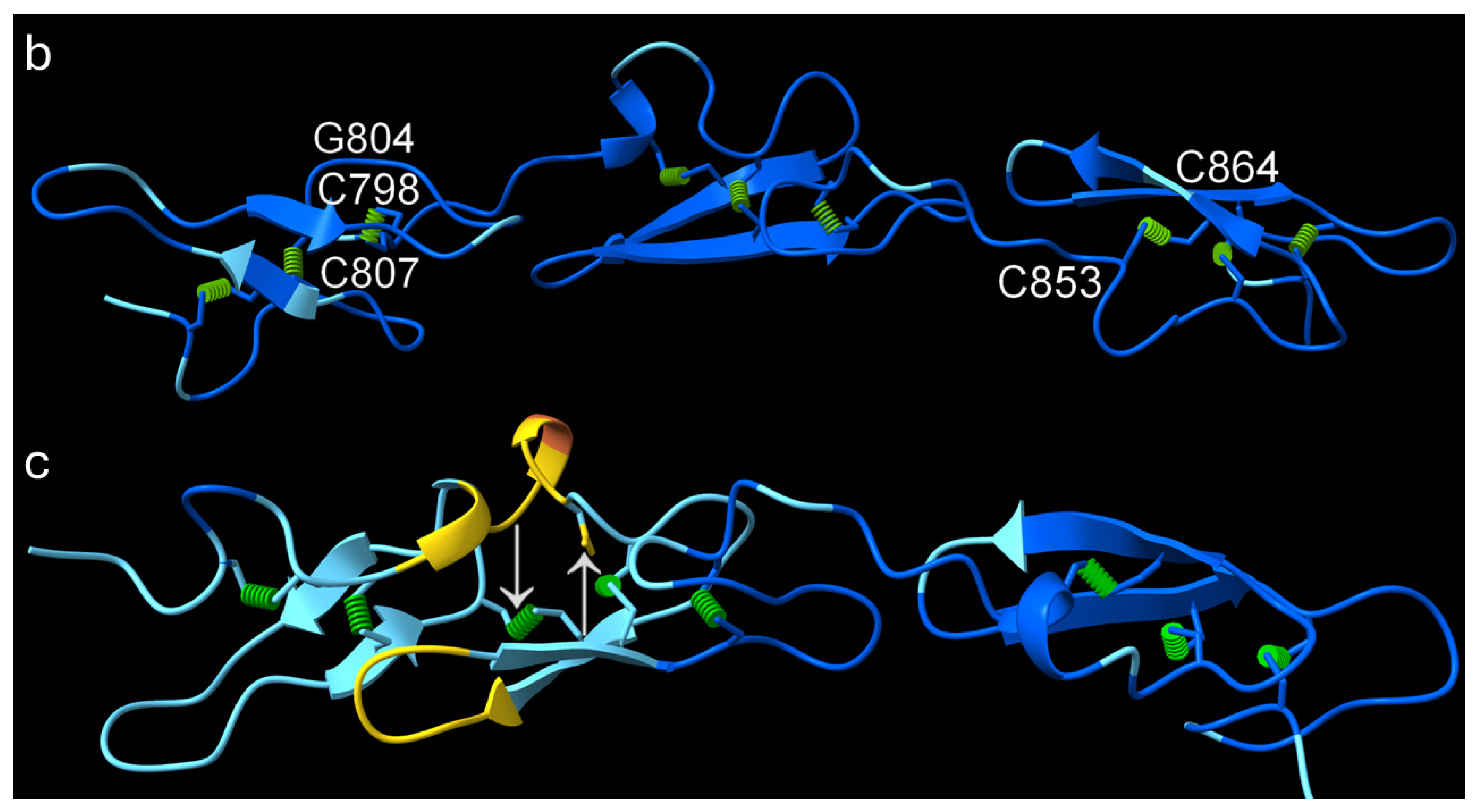
Figure 5.
Sequence and structural analysis of cysteine mutations in jaguar. a, MSA of jaguar EGFr13-15 with human and lion. b, Human; c, lion; d, jaguar AF3 structures. Seven Cys>X mutations and a deletion are shown in bold red in jaguar. In EGFr14 all 6 Cysteines are mutated, therefore, this EGFr is without any disulfide bond. One Cys (blue) residue each in EGFr13 (C533) and EGFr15 (C600) is also unpaired (their corresponding Cys partners mutated to I542 and G589 respectively, leaving both cysteines without any disulfide bridge. b, c, d: AF3 structures of NOTCH3 EGFr12-16 colored according to pLDDT as in the legend of Figure 1. pLDDT scores served as a qualitative indicator of structural destabilization. Jaguar protein shows two unpaired Cys residues (C533 and C600) in EGFr13 and EGFr15. All 6 Cys residues are mutated in EGFr14 (between orange arrows). Disulfide bridges are shown as green cylinders. EGFr12 and EGFr16 with all disulfides intact, are shown as fully-folded references. Disulfide by Design 2 tool (http://cptweb.cpt.wayne.edu/DbD2/) also shows that C533 and C600 are free. Inset figures: space-filled AF3 models of human (upper) and Jaguar (bottom) ECD. Red, EGFr13-15; blue, ligand-binding EGFr10-11; green; first EGFr, cyan and tan, the rest of EGFr. Note the compact EGFr13-15 in humans with all disulfide bonds intact. The jaguar EGFr13-15 shows a disordered loop.
Figure 5.
Sequence and structural analysis of cysteine mutations in jaguar. a, MSA of jaguar EGFr13-15 with human and lion. b, Human; c, lion; d, jaguar AF3 structures. Seven Cys>X mutations and a deletion are shown in bold red in jaguar. In EGFr14 all 6 Cysteines are mutated, therefore, this EGFr is without any disulfide bond. One Cys (blue) residue each in EGFr13 (C533) and EGFr15 (C600) is also unpaired (their corresponding Cys partners mutated to I542 and G589 respectively, leaving both cysteines without any disulfide bridge. b, c, d: AF3 structures of NOTCH3 EGFr12-16 colored according to pLDDT as in the legend of Figure 1. pLDDT scores served as a qualitative indicator of structural destabilization. Jaguar protein shows two unpaired Cys residues (C533 and C600) in EGFr13 and EGFr15. All 6 Cys residues are mutated in EGFr14 (between orange arrows). Disulfide bridges are shown as green cylinders. EGFr12 and EGFr16 with all disulfides intact, are shown as fully-folded references. Disulfide by Design 2 tool (http://cptweb.cpt.wayne.edu/DbD2/) also shows that C533 and C600 are free. Inset figures: space-filled AF3 models of human (upper) and Jaguar (bottom) ECD. Red, EGFr13-15; blue, ligand-binding EGFr10-11; green; first EGFr, cyan and tan, the rest of EGFr. Note the compact EGFr13-15 in humans with all disulfide bonds intact. The jaguar EGFr13-15 shows a disordered loop.
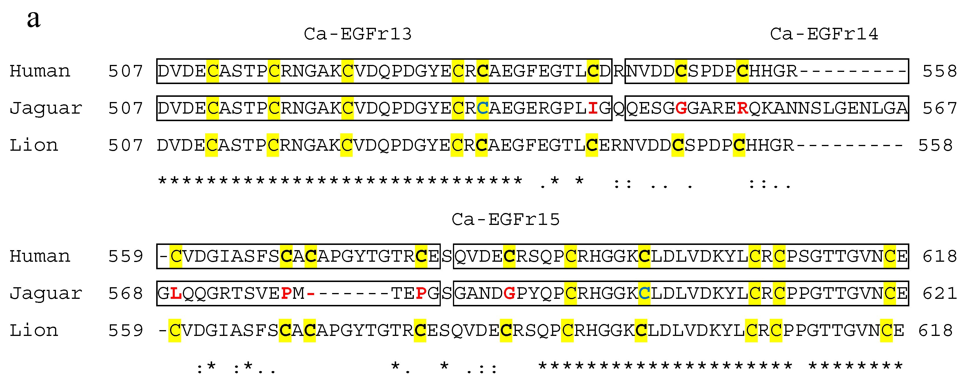
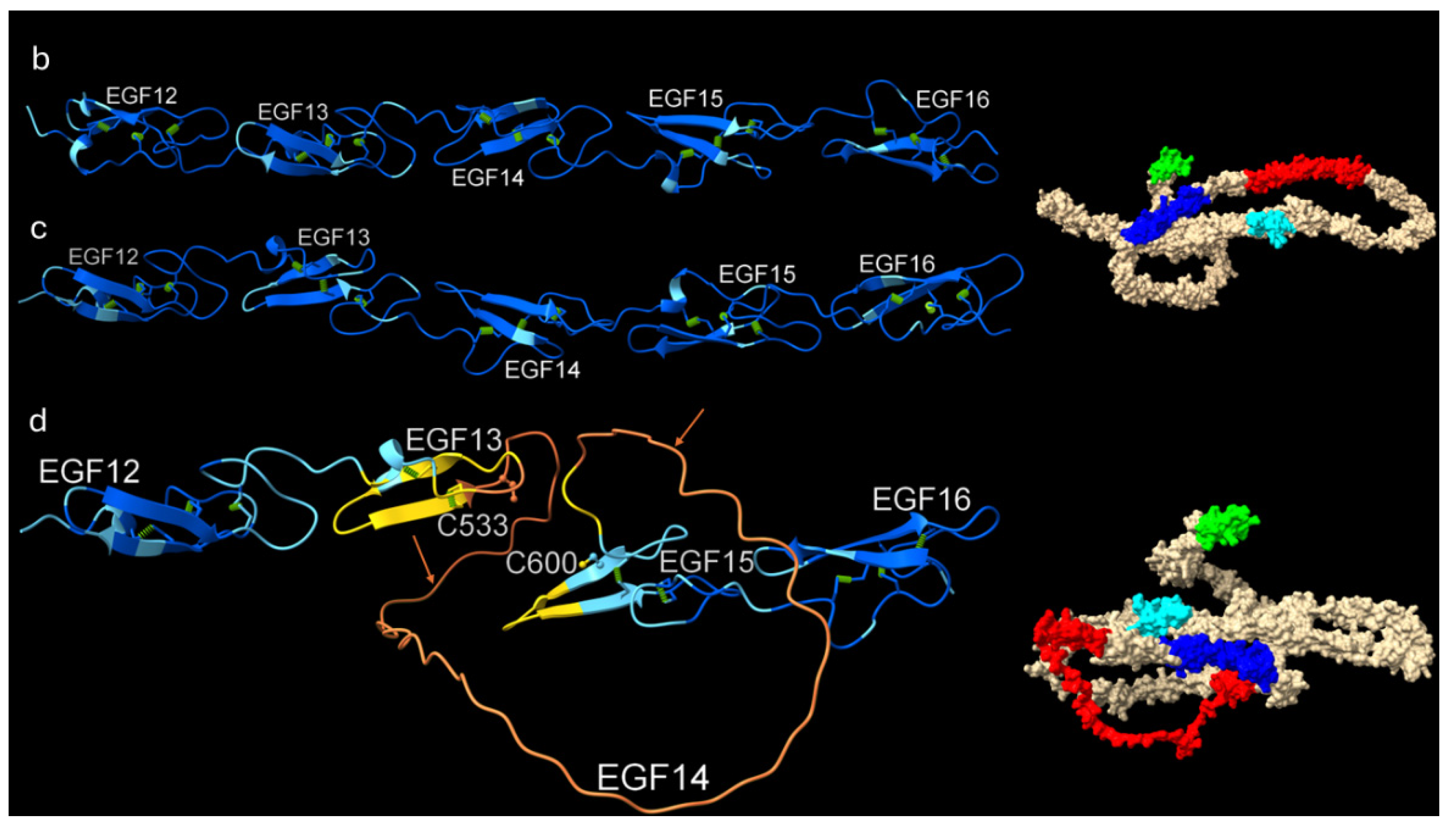
Figure 6.
Sequence, structural, and MDS analysis of the NRR-ADAM 10 metalloprotease complexes from human and Brandt’s bat. a, shows aligned sequences of human NOTCH1 and NOTCH3 alongside the NRR sequence of Brandt’s bat. Blue, LNR-A showing boxed plug; green, LNR-B; purple, LNR-C; broken box, NRR-HD; solid box, α3-helix; and red arrows, S2 cleavage site. b, human NRR; showing the S2 cleavage site (red) between a β-strand (green) and plug (blue); c, bat NRR structure. The α3-helix is shown below the S2 site. d-k, 200 ns simulations for human and bat NRR-ADAM10 complexes. Grey, NRR; pink, ADAM10. d, overall view of Human NRR-ADAM10 complex. e, overall view of the bat NRR-ADAM10 complex. f, close-up of ADAM10-NRR complex in human. g, close-up of ADAM10-NRR complex in bat. Red box, S2 cleavage site in NRR. Blue box, plug region of human NRR. The yellow sphere, Zn ion indicates the location of the active site of ADAM10. All ADAM10 were open-forms. h, the RMSD of backbone atoms of the human NRR-ADAM10 complex and the human NRR. i, the RMSD of backbone atoms of the bat NRR-ADAM10 complex and bat NRR. j, the comparison of the solvent accessible surface area (SASA) of bat and human structures. k, comparison of distances between Zn ion at the active site centre of ADAM10 metalloprotease and the center of mass of the S2 cleavage site in bat (AA) and human (DV).
Figure 6.
Sequence, structural, and MDS analysis of the NRR-ADAM 10 metalloprotease complexes from human and Brandt’s bat. a, shows aligned sequences of human NOTCH1 and NOTCH3 alongside the NRR sequence of Brandt’s bat. Blue, LNR-A showing boxed plug; green, LNR-B; purple, LNR-C; broken box, NRR-HD; solid box, α3-helix; and red arrows, S2 cleavage site. b, human NRR; showing the S2 cleavage site (red) between a β-strand (green) and plug (blue); c, bat NRR structure. The α3-helix is shown below the S2 site. d-k, 200 ns simulations for human and bat NRR-ADAM10 complexes. Grey, NRR; pink, ADAM10. d, overall view of Human NRR-ADAM10 complex. e, overall view of the bat NRR-ADAM10 complex. f, close-up of ADAM10-NRR complex in human. g, close-up of ADAM10-NRR complex in bat. Red box, S2 cleavage site in NRR. Blue box, plug region of human NRR. The yellow sphere, Zn ion indicates the location of the active site of ADAM10. All ADAM10 were open-forms. h, the RMSD of backbone atoms of the human NRR-ADAM10 complex and the human NRR. i, the RMSD of backbone atoms of the bat NRR-ADAM10 complex and bat NRR. j, the comparison of the solvent accessible surface area (SASA) of bat and human structures. k, comparison of distances between Zn ion at the active site centre of ADAM10 metalloprotease and the center of mass of the S2 cleavage site in bat (AA) and human (DV).
Disclaimer/Publisher’s Note: The statements, opinions and data contained in all publications are solely those of the individual author(s) and contributor(s) and not of MDPI and/or the editor(s). MDPI and/or the editor(s) disclaim responsibility for any injury to people or property resulting from any ideas, methods, instructions or products referred to in the content. |
© 2026 by the authors. Licensee MDPI, Basel, Switzerland. This article is an open access article distributed under the terms and conditions of the Creative Commons Attribution (CC BY) license.
Copyright: This open access article is published under a Creative Commons CC BY 4.0 license, which permit the free download, distribution, and reuse, provided that the author and preprint are cited in any reuse.