

Submitted:
24 July 2026
Posted:
24 July 2026
You are already at the latest version
Abstract
The field of skin tissue engineering has rapidly advanced in recent years. This advancement has been driven by the increasing need to address biocompatibility, fabrication techniques, antimicrobial properties, hemostatic properties, and porosity of the materials used for tissue repair and regeneration. This review focuses on three common materials for scaffold fabrication in skin tissue engineering, cellulose, chitosan, and gelatin. As the most common natural organic polymer on Earth, cellulose is a readily available and renewable material that is distinguished by a remarkable combination of advantages. These advantages include biocompatibility, biodegradability, renewability, and good mechanical strength, rendering it a nontoxic and environmentally friendly substance. The benefits of chitosan include its antimicrobial and hemostatic properties, while gelatin enhances cell adhesion and proliferation, contributing to the scaffold’s overall biocompatibility. The inherent porous structure of these materials supports fluid flow, cell infiltration, and nutrient transport, creating a conducive environment for tissue repair and regeneration. In addition, this review highlights improved techniques in fabricating a durable scaffold. It also addresses the challenges in fabrication, such as controlling the pore size and uniformity of the pores and structure within the scaffold. This is crucial for cell growth and tissue regeneration. These insights contribute to a deeper understanding of tissue repair and underscore the importance of using scaffolds and multilayered materials in skin tissue regeneration. Further research in skin tissue engineering will be essential for overcoming existing challenges and realizing the full potential of cellulose, chitosan, and gelatin in tissue engineering.
Keywords:
wound healing
; tissue engineering
; chitosan
; gelatin
; cellulose
; hydrogel
; scaffold fabrication
; skin regeneration
1. Introduction
Tissue engineering is a multidisciplinary field that emerges from the convergence of various scientific and technological domains, including biology, chemistry, engineering, medicine, pharmacy, and materials science. This integration of knowledge and techniques from various areas enables the development of innovative strategies to regenerate, repair, or replace damaged tissues and organs [1]. The primary goal of tissue engineering is to create functional replacements for damaged tissues or organs. Scaffolds are essential as they offer an alternative to traditional organ and tissue transplants. Their primary function is to provide a supportive structure that promotes tissue growth and cell proliferation. Biomaterials are fundamental to tissue engineering, with a wide range of natural and synthetic materials that have been investigated for scaffold fabrication [2]. Tissue engineering applications can be broadly categorized according to the target tissue for repair or regeneration, including skin [3,4], bone [5], cardiac [6], muscle [7], neural [8], and vascular tissue [9]. The skin is made up of the epidermis (i.e., the epithelial layer, which facilitates the physical barrier role) and the dermis (i.e., the connective layer, which facilitates the immunological role of the skin). Tissue-engineered skin substitutes for wound healing have evolved considerably in the last few years. New advances have been made in the development of skin substitutes made of artificial and natural materials. The most widely used natural polymers in biomedical applications are polysaccharides such as alginate, chitosan, starch, and cellulose. There are also common proteins used in biomedical applications, such as collagen, gelatin, and silk fibroin [2]. Skin wound healing, a complex biological process that involves a cascade of cellular and molecular reactions, remains an ongoing clinical problem [10]. Clinically, wounds are commonly categorized as acute or chronic based on whether they follow the expected path of repair with timely progression through the normal healing phases. Although acute wounds usually progress through well-established stages of hemostasis, inflammation, proliferation, and remodeling to achieve complete tissue restoration, chronic wounds generally remain in the inflammatory stage, leading to prolonged healing times, increased sensitivity to infection, and excessive patient morbidity [10,11]. Chronic wounds, such as diabetic foot ulcers, pressure ulcers, and venous leg ulcers, bear a large economic burden on healthcare systems around the world that require new and effective therapeutic regimens (Figure 1). The limitations of conventional wound dressings that have functioned mainly as passive barriers have emphasized the demand for advanced biomaterials capable of actively stimulating regeneration and promoting the cascade of healing [12].
2. The Biology of Wound Healing and Material Requirements
Effective wound care requires a deep understanding of the intricate biological processes that govern tissue repair. Wound healing is a highly orchestrated physiological process, traditionally described as having four overlapping yet distinct phases: hemostasis, inflammation, proliferation, and remodeling [13]. It is initiated immediately upon injury by vasoconstriction and platelet accumulation, forming a fibrin clot to prevent blood loss and providing a temporary matrix for subsequent cellular invasion. This is immediately succeeded by the inflammatory stage with recruitment of neutrophils and macrophages to kill debris and pathogens. During inflammation, cytokines and growth factors are released to coordinate the subsequent stages of repair [14]. The proliferative phase is characterized by angiogenesis, granulation tissue formation, re-epithelialization, and wound contraction as fibroblasts synthesize new extracellular matrix and keratinocytes migrate to restore the epidermal barrier. Finally, the remodeling phase, lasting months to years, involves collagen reorganization, resulting in increased tensile strength and scar maturation [15,16].
Given the complexity of the wound-healing cascade of biological responses, an ideal dressing should provide multiple complementary functions to support and accelerate repair. The key material requirements of the properties most significant are illustrated in Figure 2.
- Biocompatibility: The dressing material should be non-toxic and cause minimal adverse immune response. It should support cell viability, proliferation, and differentiation [18].
- Antimicrobial activity: Wounds are susceptible to microbial colonization, which can lead to infection and delayed healing. Therefore, dressings with inherent antimicrobial properties or those capable of delivering antimicrobial agents are of critical importance.
- Mechanical strength and elasticity: The dressing should protect the wound from external forces while maintaining flexibility. Ideally, its tensile properties should be compatible with the native skin to reduce discomfort and prevent tearing, especially in dynamic anatomical sites [21].
- Porosity and degradation rate: An appropriate porous architecture enables gas exchange, diffusion of nutrients, and cell infiltration. In scaffold-based dressings, the size and distribution of the pores influence cell adhesion and proliferation [22]. The degradation rate should be controllable and aligned with the formation of new tissue, minimizing the need for frequent dressing changes [23].
- Moisture retention: Maintaining a moist environment is essential for successful wound healing, promotes cell migration, and autolytic debridement, while adequate absorption of exudate helps prevent maceration and desiccation [24].
This review focuses on three fundamental biomaterials in scaffold development: cellulose, chitosan, and gelatin. These materials are selected for their porosity, biodegradability, and synergistic potential in composite scaffolds, as well as their cost effective production.
3. Biomaterials in Scaffold Development
3.1. Gelatin: Enhancing Cellular Interactions
The primary objective of tissue engineering is to develop scaffolding systems that provide an optimal environment for cell growth and, ultimately, support tissue repair and regeneration [25]. Among natural biomaterials, gelatin is a collagen-derived biopolymer produced by partial hydrolysis of collagen, a fibrous protein abundant in animal skin, bones, and connective tissues. Industrially, gelatin is most commonly derived from mammalian tissues (e.g., porcine skin, bovine hides, and bones), although fish- and poultry-derived gelatin are also used [26].
Gelatin can reversibly transition between the sol and gel states, which has supported its widespread use in biomedical applications, including as a hemostatis agent. Upon contact with blood, gelatin can swell and provide a matrix that promotes platelet activation and aggregation, accelerating coagulation [27]. In addition, gelatin is enzymatically degradable and generally well tolerated, with minimal immunogenicity, which supports its use in medical products [28]. In wound healing, its biocompatibility and non-toxicity are particularly valuable, as gelatin can support cell adhesion and proliferation to promote repair [29].
A key advantage of gelatin is that it can readily crosslink to form hydrogels and composite networks. This reactivity is mainly attributed to its amino (), carboxyl (), and hydroxyl () functional groups, which participate in reactions with a range of crosslinking agents. Crosslinking can improve mechanical strength, thermal stability, and water-handling properties, making gelatin-based structures attractive for applications such as skin tissue engineering [30]. These gelatin-cell interactions help to create a microenvironment that supports key cellular activities during regeneration [31]. From a sustainability perspective, gelatin is often obtained as a byproduct of the meat industry; converting cartilage, bones, and skins into gelatin can add value to low-cost waste streams and support the development of bio-based products [32].
Beyond these foundational properties, several advances have further expanded gelatin’s role in wound-care scaffold design. Chemical functionalization of gelatin with methacrylate groups yields gelatin methacryloyl (GelMA), a photo-crosslinkable derivative that retains gelatin’s native cell-adhesion and protease-sensitive motifs while allowing precise, light-triggered control over hydrogel stiffness and degradation, making it one of the most extensively studied gelatin derivatives for tissue-engineered constructs [33]. Clinically, gelatin-based biomaterials have been applied directly to chronic, hard-to-heal wounds, where they support hemostasis, provide peri-wound antibacterial and anti-inflammatory effects, and promote vascular and epithelial regeneration [34]. In addition to hydrogels and sponges, gelatin is frequently processed into electrospun nanofiber mats that replicate the fibrous architecture of the native extracellular matrix; such gelatin-based nanofiber dressings combine high porosity and exudate-absorbing capacity with the ability to carry antimicrobial, anti-inflammatory, or pro-angiogenic agents for burn and diabetic wound applications [35].
3.2. Chitosan: A Biopolymer with Antimicrobial and Hemostatic Properties
Chitosan, a natural polysaccharide, is derived by partial deacetylation of chitin, which is a primary component of the exoskeletons of crustaceans such as shrimp and crabs, as well as the cuticles of insects and the cell walls of fungi [36]. The deacetylation process converts chitin to chitosan by removing acetyl groups and forming free amino groups. The molecular structure of chitosan, characterized by glucosamine and N-acetylglucosamine units, is responsible for its versatile chemical and biological properties, making it a promising candidate for various applications, particularly in the biomedical field [37]. Its ability to form hydrogels and films also makes it ideal for creating scaffolds in tissue engineering, where controlled release of bioactive agents and the promotion of cell adhesion are essential for tissue regeneration [38].
Chitosan can effectively mimic the extracellular matrix (ECM), providing a supportive environment for cellular activities, facilitating better integration with host tissues, and promoting healing. Research has demonstrated that incorporation of chitosan into hydrogels can improve mechanical strength, moisture retention, and biocompatibility, underscoring its significance in the advancement of therapeutic strategies for tissue repair and regeneration [39]. Chitosan can naturally degrade into non-toxic byproducts that the body can safely resorb. In addition, its non-toxic nature, which stems from its natural origin, can minimize the risk of adverse reactions and improve compatibility with biological systems [40].
Chitosan’s cationic nature allows it to bind to negatively charged surfaces of microbial cell membranes, leading to increased permeability and eventual cell death [41,42]. This antimicrobial action is effective against a wide spectrum of bacteria, making chitosan particularly useful in wound care products where infection prevention is crucial [43]. Chitosan is also known for its rapid hemostatic properties, which enhance the natural clotting mechanism of the body. This effect is primarily due to the ability of chitosan to interact with blood cells and platelets, facilitating the accelerated formation of blood clots and thereby reducing blood loss. The positively charged chitosan molecules bind with negatively charged components on the surfaces of platelets and red blood cells, promoting aggregation and triggering the clotting cascade. As a result, chitosan has emerged as a promising material for applications in wound dressings and surgical procedures, where adequate hemostasis is critical [44].
Despite its favorable attributes for wound healing applications, chitosan has been limited by inherent shortcomings, namely poor mechanical properties, which have posed serious hurdles to widespread clinical use. In addition, poor mechanical properties, possible toxicity from certain cross-linkers, and the potential for adverse reactions require further research and modification strategies to maximize their performance and safety profile in biomedicine [45].
Recent work has continued to refine both the sourcing and the functional tuning of chitosan for wound-care applications. Because crustacean-derived chitosan can raise allergenicity and supply-chain sustainability concerns, fungal biomass has been investigated as a complementary, lower-allergen source of chitin and chitosan for advanced wound-management materials, including nanoparticles, membranes, hydrogels, sponges, and nanofibrous scaffolds [46]. At the molecular level, both the degree of deacetylation and the molecular weight strongly govern chitosan’s hydrophilicity, degradation rate, and mechanical performance, making these two parameters a key lever for optimizing chitosan-based scaffolds [47]. The same molecular parameters also influence chitosan’s procoagulant efficiency: a moderate degree of deacetylation combined with a high molecular weight has been reported to yield the most pronounced hemostatic effect, providing a theoretical basis for designing more effective chitosan-based hemostatic dressings [48].
3.3. Cellulose: An Abundant Biorenewable Polymer
Cellulose, the most common biopolymer on Earth, is a crucial structural part of plant cell walls and can also be synthesized by certain bacteria and algae [49]. Its availability in nature, biodegradability, and renewability increase its substantial potential as a green material for a wide variety of biomedical applications, particularly in wound therapy. Because cellulose contains a high concentration of hydroxyl groups, it can form strong hydrogen-bonding networks within and between substrates, which can improve hydrogel cohesion and adhesion. In addition, cellulose derivatives can be prepared from natural sources such as algae, fungi, bacteria, and plants [49,50].
Bacterial cellulose (BC), also called bacterial nanocellulose (BNC), is produced by fermentation of certain bacteria, is also a suitable material for use in tissue regeneration. The typical hierarchical structure of cellulose also makes it suitable for processing in different forms, including microcrystalline cellulose (MCC), cellulose nanofibers (CNF), and cellulose nanocrystals (CNC) with varying properties and applications [50]. BC has been used in wound care studies because it can help maintain moistness in the dressing, absorb excess exudate, provide mechanical protection, reduce microbial infection, control allergic reactions, relieve wound pain, and remain easy to remove and replace. Gluconacetobacter xylinus is often reported to be a stong producer of bacterial cellulose, and the cellulose produced using this strain has been associated with improved mechanical strength and biodegradability [51].
Microcrystalline cellulose (MCC) typically consists of highly aggregated cellulose microfibrils that are insoluble in water and most organic solvents [52]. Due to its particulate nature, MCC is used primarily as a filler or an excipient in pharmaceutical formulations and as a structural element in composite systems, providing mechanical integrity without the possibility of continuous network formation [53]. Alternatively, cellulose nanofibers (CNF), produced through mechanical fibrillation of cellulose pulp, possess high aspect ratios and can entangle in a network. CNFs have extremely high mechanical strength, large surface area and satisfactory water retention capacity and are therefore suitable for hydrogel formation and as reinforcement fillers in soft matrices [54]. Strong hydrogen bonding networks between CNFs are responsible for their resilient mechanical behavior, can replicate aspects of the ECM, and can create a desirable microenvironment for cell proliferation and adhesion [55].
CNC derived from acid hydrolysis of cellulose to remove amorphous regions, are highly crystalline rod-shaped nanoparticles of typical nanometer-sized dimensions [56]. CNCs are characterized by high crystallinity, stiffness, and high surface area, making them excellent reinforcement elements in composite materials [57]. Their ability to form stable colloidal suspensions and their innate biocompatibility make CNCs useful for enhancing the mechanical strength of hydrogels and scaffolds and to address the universal problem of limited mechanical strength in many natural polymer-based wound dressings [58,59].
The biomedical functions of cellulose and its nanostructured forms are varied. Their inherent hydrophilicity enables for high water capacity, which is crucial to maintaining a wet wound environment that is important for improved healing and reduced scar formation [60]. Moreover, the high stiffness and mechanical strength of CNF and CNC confer reinforcement in composites and provide the ability to create tunable wound dressings that can withstand exogenous forces but still promote tissue regeneration [61]. Cellulose-based materials such as films, hydrogels, and scaffolds are frequently used in wound care and integrated into composite systems to take advantage of their reinforcing properties [62].
However, nanoparticle aggregation and the challenges of achieving uniform dispersion within polymer matrices must be handled with care during the fabrication process [63]. In addition, it is still difficult for existing hydrogel dressings to achieve a desirable balance between mechanical strength, moisture retention function, and biocompatibility.

Table 1.
Comparison of natural polymers commonly used in wound healing scaffolds.
| Property | Gelatin | Chitosan | Cellulose |
|---|---|---|---|
| Source | Collagen-derived protein | Deacetylated chitin | Plant or bacterial cellulose |
| Biocompatibility | High | High | High |
| Biodegradability | High | High | High |
| Mechanical Strength | Low | Moderate | High |
| Antimicrobial Activity | Limited | Excellent | Limited |
| Hemostatic Activity | Moderate | Excellent | Limited |
| Cell Adhesion | Excellent | Good | Moderate |
| Moisture Retention | High | High | High |
| Primary Limitation | Rapid degradation | Brittleness | Limited bioactivity |
Beyond the forms discussed above, cellulose chemistry has been further diversified to broaden its biomedical utility. The seminal review by Klemm et al. established the foundational classification of nanocelluloses, including microfibrillated cellulose, nanocrystalline cellulose, and bacterial nanocellulose, which underpins much of the subsequent literature on nanocelluloses in tissue engineering and wound care [64]. Within the cellulose nanofiber family, TEMPO-mediated oxidation has become a particularly important production route, introducing C6 carboxylate groups on the fibril surface that promote spontaneous nanofibrillation and produce individualized high-aspect ratio nanofibers with high tensile strength [65]. Clinically oriented reviews have further cataloged the growing range of cellulose-based wound-dressing formats, including films, hydrogels, foams, and nanofiber mats, and their comparative performance in protecting the wound bed, absorbing exudate and modulating inflammation [66]. Cellulose-based scaffolds have also drawn attention for their hemostatic potential, since their surface chemistry, porosity, and mechanical properties can be engineered to promote platelet activation and clot formation, positioning cellulose alongside chitosan as a candidate hemostatic biomaterial for wound-care applications [67].
4. Synergistic Potential of Chitosan, Gelatin, and Cellulose
The synergistic combination of chitosan, gelatin, and cellulose has attracted significant attention because these biopolymers can complement each other. Each polymer offers unique capabilities; chitosan for porosity, antimicrobial activity, and hemostatic properties [68], gelatin for improved cell adhesion [21], and cellulose for mechanical reinforcement [51]. When combined, these materials form composites with enhanced performance that helps overcome the limitations of the individual components. However, the greatest challenge remains the precise control of critical material parameters, including mechanical strength, ideal porosity, and a controlled degradation rate, which are necessary for effective wound-healing outcomes.
Polysaccharides and gelatin are natural biopolymers that have been widely used in biomedical applications [9]. Polysaccharides are found in crabs, lobsters, shrimp, forests (biomass), and bacteria [69], highlighting their wide availability from both animal and plant sources. In this review, polysaccharides such as cellulose and chitosan are discussed as well as their synergy with gelatin in hydrogel formation. In general, blending gelatin with polysaccharides can result in improved properties, and recent studies have expanded these systems from wound healing [70] and cell growth [71] to inhibit bacterial growth and drug, gene, siRNA, and peptide delivery (Figure 3) [72]. Gelatin-polysaccharide hydrogels can absorb large amounts of water, typically more than 100 times their dry weight, supporting in vitro culture systems for studying mammalian-cell behavior in a matrix-like tissue-engineering environment that encourages cell attachment, growth, infiltration, and vascularization (Figure 2) [73]. At the molecular level, carbohydrate-protein interactions can be designed through two major covalent-bonding reactions that emulate ECM proteoglycan [73]: (i) Schiff base formation [74] and (ii) Millard reaction [69], both of which can contribute to hydrogel formation.
Gelatin and chitosan hydrogels, while highly biocompatible and cell-supportive, are typically not mechanically robust and often show poor structural integrity. As a result, they may not be able to support dynamic or load-bearing wound regions [21,75,76]. In these systems, nanostructured cellulose, including cellulose nanofibers (CNF), CNC, and bacterial cellulose (BC), can act as an effective reinforcement material because of its high crystallinity, high aspect ratio, hydrogen-bonding capacity, and ability to form mechanically supportive networks [56,57,64,65]. The crystallinity and high aspect ratio of CNC and CNF can create a robust percolated network within the composite, leading to large increases in compressive modulus and tensile strength without compromising biocompatibility [76]. For example, Li et al. demonstrated that a dual-network chitosan/cellulose nanofiber composite prepared by in situ enzymatic catalysis showed a remarkable improvement in both tensile strength and elongation at break, providing a route to improve strength and toughness at the same time [77]. Similarly, Wegrzynowska-Drzymalska et al. explored dialdehyde cellulose nanocrystals (DCNC) as a crosslinker to chemically reinforce chitosan-gelatin films, improving mechanical properties and adding anti-inflammatory advantages; however, balancing crosslinking density with potential cytotoxicity remains challenging [78]. Furthermore, Bhatnagar et al. reported that CNCs can enable sustained release of platelet lysate in a chitosan-cellulose nanocrystal hydrogel, which in turn enhanced fibroblast proliferation and wound closure in vitro [79].
Studies by Afewerki et al. and Ooi et al. also found that the addition of palm oil-derived nanocellulose improved mechanical characteristics and crystallinity in collagen-nanocellulose scaffolds [80,81]. For a gelatin-based wound dressing to be effective, degradation must be precisely tuned to match the natural pace of tissue regeneration. The rapid degradation of gelatin, which is a drawback in one-component dressings, can be controlled in a composite system; by blending it with slower-degrading chitosan and cellulose, the degradation rate can be adjusted to better match the rate of new tissue growth so that the dressing remains an effective scaffold during healing [82]. In addition, Muzzarelli (2009) utilized the naturally occurring crosslinker genipin to stabilize chitosan/gelatin blends, where thermal degradation, surface wettability, dissolution, and swelling depend both on both composition of the blend and the concentration of genipin used for crosslinking [83].
Beyond mechanical reinforcement, chitosan is also used to tune the porosity of the scaffold in gelatin/cellulose composites. Porosity is another key requirement for tissue-engineering scaffolds, particularly when chitosan is used for structural support and to promote cell proliferation. The scaffold should provide sufficient surface area for cells to thrive with an ideal pore size that allows cell growth and cellular penetration. The objective is a highly interpenetrating pore structure that allows nutrient transport to support growth [84]. The optimum pore size for wound-healing scaffolds varies considerably, but generally falls in the 20 to 300 m range, depending on tissue type, biomaterial, and application. Smaller pores promote cell attachment, while larger pores (60–300 m) facilitate cell migration, vascularization, and nutrient transport; for skin regeneration, 20–125 m is commonly cited as optimal [85].
In gelatin-chitosan-cellulose composites, chitosan can be used to manage porosity by forming a complex with gelatin and cellulose. The optimal porosity reported typically ranges from 60% to 90%, with pore sizes of 50 to 250 m. Charge interactions between chitosan and the other polymers can be regulated to create interconnected porous networks suitable for nutrient transport, gas exchange, and cell penetration [40]. In a study of the gelatin–chitosan scaffold by Han et al., the porosity of the compound increased with the addition of viscous chitosan [3]. The lowest porosity (only gelatin) was shown by 52.81 ± 13.99%), C10CNC (only chitosan) showed the highest porosity (78.42 ± 9.49%), and intermediate ratios such as G3C7CNC also exhibited higher porosity (67.37 ± 9.09%) [82]. Together, the synergistic combination of these polymers in a composite scaffold can offer additional benefits while reducing their respective shortcomings [82]. Table 2 summarizes the main benefits reported for these composite combinations.
5. Fabrication Techniques for Wound Dressing
5.1. Hydrogels
5.1.1. Introduction
A hydrogel is a water-swollen polymer network that can be formed from natural or synthetic polymers. Polymers that compose a hydrogel are cross-linked using different methods, and the selected cross-linking strategy largely determines the final function and performance of the material. In general, cross-linking can be categorized as physical or chemical. Physical cross-linking relied mainly on intermolecular interactions such as hydrogen bonding, ionic interactions, crystallization cross-linking, and hydrophobic association. Physically cross-linked hydrogels are often valued for their high biocompatibility and low molecular toxicity. Chemical crosslinking typically occurs through covalent bonding between polymer chains (e.g., free-radical polymerization or radiation crosslinking). As a result, chemically crosslinked hydrogels generally show improved mechanical properties, and chemical crosslinking is a common approach to enhance hydrogel mechanical strength [84]. Hydrogels have played a central role in tissue engineering because they can provide structural mimics of ECM biomacromolecules, support cell functions, and allow the permeability of oxygen, nutrients, and other water-soluble metabolites [3,40,85]. In aqueous environments, hydrogels absorb and retain water (swell), making them a useful class of biomaterials for 3D cell culture.
5.1.2. Hydrogels for Wound Care
In particular, hydrogels have been a preferred option for wound dressings and for promoting chronic wound healing. In wound healing and soft tissue engineering, hydrogels can easily conform to irregular wound shapes while maintaining moisture and absorptive function. They are also flexible and can be further designed through chemical modifications to reinforce their properties or introduce new biological functions.
For example, Kanimozhi et al. prepared a chitosan/poly(vinyl alcohol)/methylcellulose (CS/PVA/MC) hydrogel scaffold with substantial potential for wound healing. The hydrogel showed biocompatibility and non-toxicity and supported the proliferative functions of fibroblast cells that are critical to tissue healing. Due to its chitosan content, the material also exhibited inherent antibacterial action against common wound pathogens such as S. aureus and E. coli. In its hydrated form, the scaffold functioned as a hydrogel with structural properties suitable for use as a wound dressing. Its highly porous, connected morphology and high swelling capacity allowed it to manage wound exudate while also providing routes for transport. The inclusion of methylcellulose further enhanced hydrogel flexibility. In general, the combination of biocompatibility, antibacterial efficacy, high absorbency, and flexibility makes this CS/PVA/MC hydrogel scaffold a strong candidate for next-generation wound dressings [86].
A nanocomposite hydrogel was also developed by Tanpichai and Oksman through reinforcement of a PVA matrix with CNCs. The purpose of this hydrogel was to improve performance in biomedical and tissue engineering applications, and it showed good elasticity and recovery after deformation, which are crucial for applications such as artificial cartilage or wound dressings. With greatly improved mechanical strength and elastic recovery while still maintaining high water content, approximately 92%, the cross-linked CNC–PVA hydrogel was reported to be a promising material for demanding biomedical applications [87].
In a review by Adelnia et al., PVA hydrogels prepared by the physical freeze–thaw (F–T) process were described as versatile biomaterials due to their unique fabrication pathway and tunable properties. A major advantage of the F–T process is that it can form highly biocompatible and ultrapure hydrogels without the need for chemical cross-linking agents, which makes these systems well suited for biomedical applications. Pure PVA hydrogels provide a wet protective environment, but are often combined with active compounds such as chitosan, alginate, or silver nanoparticles to introduce antimicrobial and wound-stimulatory properties [88].
Figueroa-Pizano et al. also used the F–T method to prepare chitosan–poly(vinyl alcohol) (CS–PVA) hydrogels without relying on potentially toxic chemical cross-linkers. Their study examined how adjusting F–T conditions, including freezing temperature and number of cycles, can modulate the physical structure and drug-release profile of the hydrogel. A key observation was that porosity could be controlled by changing the freezing temperature; SEM micrographs and porosimetry showed that reducing the freezing temperature, from to , produced smaller pore sizes but greater total porosity. This occurs because lower temperatures promote the rapid formation of many small ice crystals, which, upon sublimation, leave behind a more porous network [89].
Moreover, Koosha et al. combined honey and allantoin with physically crosslinked hydrogel films composed of chitosan and PVA for wound healing applications. The resulting hydrogel showed beneficial properties for wound healing, including long-term biocompatibility, antimicrobial activity, and appropriate and accelerated wound closure in in vivo models [90].
Mansur et al. engineered a bioadhesive hydrogel as a therapeutic dressing for chronic wounds by optimizing bioadhesion to improve cellular interaction. They reported the design, synthesis, and characterization of hydrogels obtained from carboxymethylcellulose (CMC) and PVA, which were chemically crosslinked using citric acid (CA) through a biocompatible thermal treatment process. This approach created a stable hybrid network, and the resulting hydrogel displayed a combination of physicochemical and biological properties that support its potential for next-generation wound care [91].
A ternary hydrogel was synthesized by Fan et al. for wound dressing applications by mixing chitosan, gelatin (Gel), and PVA. The goal was to improve the hemostatic and mechanical properties of gelatin/PVA hydrogels by incorporating chitosan as a hemostatic component. The experiment demonstrated that the resulting CS/Gel/PVA hydrogel achieved a satisfactory balance of wound-dressing attributes, particularly blood clotting performance and mechanical strength. The addition of chitosan significantly increased mechanical strength compared to a regular Gel/PVA hydrogel; however, elongation and tensile strength decreased as the proportion of chitosan increased, because the hydrogel became hard and brittle at high concentrations of chitosan [92].
Another study by Shamloo et al. used the F–T method and incorporated various concentrations of honey into hydrogels formed from PVA, chitosan, and gelatin. They reported that honey addition can provide strong therapeutic effects in wound healing. In vivo experiments in rat models showed enhanced wound closure, with contraction approaching 95% on day 12. Honey also improved antibacterial efficacy against S. aureus and P. aeruginosa and increased fibroblast viability, although these benefits were coupled with trade-offs in physicochemical properties. Acting as a plasticizer, honey increased flexibility and maximum strain while reducing ultimate tensile strength and elastic modulus; its high solubility also increased scaffold degradation [93].
Together, these hydrogel examples show that wound-dressing hydrogels can be engineered to support moisture retention, antimicrobial activity, controlled bioactivity, and improved mechanical performance, depending on the selected polymer system and fabrication strategy.
For example, Kanimozhi et al. prepared a chitosan/poly(vinyl alcohol)/methylcellulose (CS/PVA/MC) hydrogel scaffold with substantial potential for wound healing. The hydrogel showed biocompatibility and non-toxicity and supported the proliferative functions of fibroblast cells that are critical to tissue healing. Due to its chitosan content, the material also exhibited an inherent antibacterial action against common wound pathogens such as S. aureus and E. coli.. In its hydrated form, the scaffold functioned as a hydrogel with structural properties suitable for use as a wound dressing. Its highly porous, connected morphology and high swelling capacity allowed it to manage wound exudate while also providing routes for transport. The inclusion of methylcellulose further enhanced the hydrogel flexibility. In general, the combination of biocompatibility, antibacterial efficacy, high absorbency, and flexibility makes this CS/PVA/MC hydrogel scaffold a strong candidate for next-generation wound dressings [86].
A nanocomposite hydrogel was also developed by Tanpichai and Oksman through reinforcement of a PVA matrix with CNCs. The purpose of this hydrogel was to improve the performance in biomedical and tissue engineering applications, and it showed good elasticity and recovery after deformation, which are crucial for applications such as artificial cartilage or wound dressings. With greatly improved mechanical strength and elastic recovery while still maintaining high water content (approximately 92%), the cross-linked CNC-PVA hydrogel was reported to be a promising material for demanding biomedical applications [87].
In a review by Adelnia et al. PVA hydrogels prepared by the physical freeze-thaw (F–T) process were described as versatile biomaterials due to their unique fabrication pathway and tunable properties. A major advantage of the F–T process is that it can form highly biocompatible and ultrapure hydrogels without the need for chemical cross-linking agents, which makes these systems well suited for biomedical applications. Pure PVA hydrogels provide a wet protective environment, but are often combined with active compounds such as chitosan, alginate, or silver nanoparticles to introduce antimicrobial and wound-stimulatory properties [88]. Figueroa-Pizano et al. also used the F–T method to prepare CS–PVA hydrogels without relying on potentially toxic chemical cross-linkers. Their study examined how adjusting F–T conditions, including freezing temperature and number of cycles, can modulate the physical structure and drug-release profile of the hydrogel. A key observation was that porosity could be controlled by changing the freezing temperature; SEM micrographs and porosimetry showed that reducing the freezing temperature, from to , produced smaller pore sizes but greater total porosity. This occurs because lower temperatures promote the rapid formation of many small ice crystals, which, upon sublimation, leave behind a more porous network [89].
Moreover, Koosha et al. combined honey and allantoin with physically crosslinked hydrogel films composed of chitosan and PVA for wound healing applications. The resulting hydrogel showed beneficial properties for wound healing, including long-term biocompatibility, antimicrobial activity, appropriate and accelerated wound closure in in vivo models [90].
Mansur et al. engineered a bioadhesive hydrogel as a therapeutic dressing for chronic wounds by optimizing bioadhesion to improve cellular interaction. They reported the design, synthesis, and characterization of hydrogels obtained from CMC and polyvinylidene (PVA), which were chemically crosslinked using citric acid (CA) through a biocompatible thermal treatment process. This approach created a stable hybrid network and the resulting hydrogel displayed a combination of physicochemical and biological properties that support its potential for next-generation wound care [91].
A ternary hydrogel was synthesized by Fan et al. for wound dressing applications by mixing chitosan, gelatin (Gel), and (PVA). The goal was to improve the hemostatic and mechanical properties of gelatin/PVA hydrogels by incorporating chitosan as a hemostatic component. The experiment demonstrated that the resulting CS/Gel/PVA hydrogel achieved a satisfactory balance of wound-dressing attributes, particularly blood clotting performance and mechanical strength. The addition of chitosan significantly increased mechanical strength compared to a regular Gel/PVA hydrogel; however, the elongation and tensile strength decreased as the proportion of chitosan increased, because the hydrogel became hard and brittle at high concentrations of chitosan [92].
Another study by Shamloo et al. used the F–T method and incorporated various concentrations of honey into hydrogels formed from PVA, chitosan, and gelatin. They reported that honey addition can provide strong therapeutic effects in wound healing: In vivo experiments in rat models showed enhanced wound closure, with contraction approaching 95% on day 12. Honey also improved antibacterial efficacy against S. aureus and P. aeruginosa and increased fibroblast viability, but these benefits were coupled with trade-offs in physicochemical properties. Specifically, honey acts as a plasticizer, increasing flexibility and maximum strain while reducing ultimate tensile strength and elastic modulus; in addition, its high solubility increased scaffold degradation [93].
5.2. Electrospinning
Electrospinning for wound healing uses an electrohydrodynamic process to create fine, nanofiber-based wound dressings from polymer solutions or melts. Figure 4 illustrates the electrospinning process and how it can be applied in the design of new wound dressings for chronic diabetic wounds. In this process, a polymer solution is pumped through a spinneret and drawn into ultra-thin nanofibers under a high electric field, and the nanofibers are then collected to form a dressing. Once applied to a diabetic wound surface, electrospun dressings can provide antibacterial activity to combat infection, promote faster healing, enable drug delivery over extended periods, and enhance mechanical performance. In addition, the resulting porous scaffold can support cell attachment, proliferation, and tissue regeneration by mimicking key features of the ECM of the body [99].
Single-layer dressings conventionally used in practice may not meet all clinical requirements, and the growing demand for functional wound dressings has attracted interest in bilayer designs [96]. Differences in structure and properties between layers can confer several advantages. For example, a denser outer layer can protect the wound against infection and mechanical stress while minimizing dehydration, whereas the inner layer (in direct contact with the wound) can be designed to mimic the ECM and promote cell adhesion and proliferation [97]. In general, combining functionalities within bilayer dressings shows the potential to advance wound care management and improve patient outcomes [98].
A study by Ghafari et al. investigated a bilayer wound dressing consisting of a dense polyurethane/ethanolic extract of propolis (PU/EEP) membrane as the top layer and a polycaprolactone/gelatin (PCL/Gel) scaffold electrospun as the sublayer. The upper layer was designed to impart antimicrobial activity and protect the wound from external contaminants, while the sublayer provided a cell-friendly scaffold. Both layers showed good biocompatibility (non-toxic and cell-viable), and the PCL/Gel scaffold can biodegrade over, potentially avoiding removal after healing. In this bilayer configuration, faster wound healing can be supported through increased cell adhesion and proliferation; however, rapid degradation of gelatin can affect structural stability, which was addressed by combining gelatin with PCL [100].
In a recent study, H. Samadian et al. applied graphene nanosheet-reinforced, electrospun chitosan/gelatin nanofiber scaffolds for wound healing and infection prevention. They reproted high porosity (~90%), which supports fluid absorption and biodegradability, as well as strong antibacterial effects (reported reductions of ~50% and ~80% for E. Coli and S. aureus, respectively). The scaffolds did not exhibit toxicity to human cells and improved cell motility to support faster wound closure; however, at high graphene content, bead formation problems and reduced porosity were observed [101].
Kazeminava et al. also reported electrospun wound dressings based on gelatin and chitosan nanofibers, loaded with a phlorotannin-rich extract of Undaria pinnatifida, aiming to emulate antibacterial and anti-inflammatory functions. The fibers were highly porous (~90%), permeable to water-vapor and allowed controlled release of the extract for up to 160 days, which supports long-term applications in wound care applications. Antibacterial activity was effective against Pseudomonas aeruginosa, while Staphylococcus aureus showed higher resistance; the main limitations were reduced effectiveness against S. aureus and relatively weak mechanical properties [102].
Furthermore, Haghbin et al. assessed electrospun polyurethane–gelatin nanofibrous membranes loaded with honey and zinc oxide nanoparticles for wound dressing applications, focusing on antibacterial properties, mechanical strength, and biocompatibility. PU/Gel/H/ZnO membranes showed strong antibacterial effects, with clear inhibition zones against E. coli, S. aureus, and Bacillus subtilis. However, while gelatin contributes to good biocompatibility and a hydrophilic surface, its fast degradation remains a limitation for long-term applications [5].
The synergistic combination of gelatin (GEL) and cellulose acetate (CA) in electrospun nanofibers is also considered an interesting biomaterial approach for future wound dressings. Cellulose acetate can provide the required mechanical support and structural integrity to help the scaffold maintain its structure, while gelatin contributes biocompatibility and bioactivity to replicate native and support cell adhesion, proliferation, and migration. These CA/GEL nanofibers can form highly porous scaffolds with a well-interconnected architecture and high surface-area-to-volume ratio, which can improve gas exchange, regulate wound exudate, and help establish an optimal microenvironment for cellular infiltration and tissue regeneration, thus accelerating the healing cascade [101].
Finally, electrospun nanofibers of CMC and PVA have also been used to fabricate antimicrobial wound dressings with improved mechanical properties and loading of antibiotics such as colistin (CL). These systems were reported to show suitable biocompatibility with (more than 80% viability in human skin cells) and improved antibacterial activity against pathogens such as S. aureus and E. coli. In addition, the nanofibrous scaffold can be designed to replicate the ECM of the skin and facilitate the migration and proliferation of cells needed for healing [102].
5.3. Freeze-Drying
Freeze-drying is a promising method for converting a polymer scaffold solution into a highly porous 3D structure, and porosity levels of more than 90% are commonly reported. In general, the freeze-drying technique includes three steps: (i) preparation of the polymer solution, (ii) freezing (typically from to ) and (iii) lyophilization of the frozen system reduced under pressure to generate an interconnected porous scaffold [5,103](Figure 5).
In one study, the authors focused on chitosan–gelatin scaffolds loaded with copper nanoparticles for skin tissue engineering. Poor mechanical strength and rapid degradation limit the use of gelatin alone in these systems. The value of chitosan lies in its biodegradability, antimicrobial activity, and hydrogel formation capability, which makes it useful for skin regeneration; however, compared to gelatin, chitosan can be brittle and shows limited cell adhesion. The combination of chitosan and gelatin can therefore provide enhanced biocompatibility and structural features that promote cell growth. The addition of copper nanoparticles further increased porosity and mechanical stability, although at high loadings the scaffold tended to compact and porosity decreased [104]. Overall, the chitosan–gelatin blend still needs to be fine-tuned, particularly due to the rapid degradation of gelatin and the stiffness of chitosan.
Han et al. made Gel/Chi sponges by freeze-drying and reported a uniform porous structure with pore sizes in the range of 120-140 m and porosity greater than 90%. This indicates that the scaffold can be suitable for cell seeding and nutrient transport. Water uptake was higher (more than 1500%) and water retention was also notable; however, the degradation rate increased linearly with the higher gelatin content, which may raise concerns about long-term stability. Although overall biocompatibility was good, cell proliferation decreased with higher chitosan content, which was attributed to excessive cellular electrostatic interactions [3].
In another study, chitosan–gelatin scaffolds were developed for dermal tissue engineering using freeze-drying. A 2:5 chitosan-to-gelatin weight ratio provided a balance of mechanical and cellular properties; scaffolds supported cell proliferation, but still faced challenges such as brittleness and rapid degradation [4].
Wang et al. investigated a wound dressing formulation based on gelatin and chitosan, each reinforced with a piece of nonwoven fabric, and incorporated Centella asiatica extract into the composite hydrogel. The hydrogel was designed as a biocompatible antibacterial dressing that may help counteract the growth of antibiotic resistance. However, incorporation of chitosan tends to reduce elasticity and increase pore size, which can affect structural integrity; its hydrophobic nature can also negatively affect swelling capacity [105].
Zulfiqar et al. reported chitosan-based wound dressings loaded with copper metal organic frameworks (Cu-MOFs) to enhance antibacterial and wound healing properties, support angiogenesis and collagen deposition, and accelerate wound healing in a rat model infected with Pseudomonas aeruginosa [106].
Ghosh et al. prepared and characterized bio-scaffolds based on carboxymethyl chitosan, gelatin, and copper oxide nanoparticles. These scaffolds were fabricated by freeze-drying followed by two crosslinking steps: an initial crosslinking using CuO nanoparticles and a secondary crosslinking using epichlorohydrin in an ammonia-rich atmosphere to enhance water resistance and mechanical stability. The authors evaluated swelling, hydrolytic degradation, moisture transmission, surface roughness, and antibacterial performance, and reported that crosslinking improved structural stability and mechanical properties while maintaining antibacterial activity against Gram-positive and Gram-negative bacteria [107].
5.4. Salt Leaching
Salt leaching is a simple and widely used method for forming 3D porous scaffolds, and it can provide an appropriate microenvironment for cell growth due to the ease of creating interconnected pores. This technique can offer enhanced mechanical support and physical and biochemical stimuli that support cell proliferation. In salt leaching, pores are produced using porogens (soluble particles such as sugar, salt, wax, paraffin, or gelatin) [108,109]. Figure 6 shows a schematic workflow for preparing porous scaffolds through porogens: Porogens are mixed into a polymer solution, the mixture is cast in a mold, and the porogen is then dissolved (typically in water), leaving behind a porous structure [110].
The salt leaching technique was introduced by Mikos et al. [111] and mainly applied to prepare composite scaffolds; later, it also found applications in porous scaffolds designed for the growth of endothelial cells.
A commonly described procedure involves the preparation of a 25% gelatin solution mixed with NaCl crystals, with an average particle size of 300–500 m. The mixture is poured into a mold and dried (e.g., in a vacuum oven at 50 C), followed by cross-linking in an acetone/water mixture containing 1-ethyl-3-(3-dimethylaminopropyl)carbodiimide (EDC). After cross-linking, excess NaCl is washed out with water, leaving uniformly distributed, interconnected macropores with sizes that reflect the original NaCl particle size. One key issue is that insufficient NaCl can lead to a bilayer structure, where a dense layer forms at the bottom due to the sinking of the salt particles. In addition, although salt leaching is effective in creating porosity, an excessive NaCl content does not necessarily improve mechanical properties and may compromise the integrity of the scaffold [112].
Morpara and Wiwatwongwana investigated gelatin/CMC scaffolds fabricated by salt leaching, where gelatin and CMC were dissolved in distilled water and NaCl was used as the porogen. The Scanning Electron Microscopy (SEM) analysis showed interconnected porous structures with pore sizes ranging from to , and the pore size decreased with increasing CMC content. Although salt leaching is straightforward and inexpensive, it still lacks definite control over the pore-size distribution [113]. Kanimozhi et al. also employed salt leaching to fabricate chitosan/poly(vinyl alcohol)/ methylcellulose –ZnO (CS/PVA/ MC3 –ZnO) nanocomposite scaffolds designed for tissue engineering applications. In this approach, sodium chloride particles served as a porogen, resulting in an interconnected porous architecture. The resulting high-porosity structure was advantageous for cellular infiltration and growth, and the reported physicochemical and biological properties were conducive to wound healing applications in skin tisue engineering [86].
5.5. 3D Printing
3D printing, also known as additive manufacturing, is considered a new frontier in scaffold fabrication because it provides unrivaled control of scaffold architecture. This technology builds patient-specific three-dimensional structures layer-by-layer directly from a digital model. Unlike traditional approaches, such as solvent casting or freeze-drying, where definitive control over pore geometry may not be readily achievable, 3D printing enables hydrogels with complex, pre-designed microporous structures to be fabricated in a timely and accurate manner, supporting better mimicry of the natural ECM. This level of control is highly crucial for improved cell proliferation, improved material transport, and, subsequently, tissue regeneration [114].
For example, Hafezi et al. showed that chitosan can be successfully 3D printed using an extrusion-based method to develop skin tissue-regenerating scaffolds. In that work, 3D-printed chitosan film matrices were crosslinked with genipin to produce flexible dressings. However, in their non-plasticized state, the chitosan films were brittle and showed a low elongation at the break (2.44%). Therefore, plasticizers such as polyethylene glycol (PEG) were added and an optimized film was obtained with much higher flexibility (22.67% elongation at break) [115].
Gelatin is also one of the most common biopolymers harnessed for bio-inks, and one strength of 3D printing is the ability to combine materials to create more functional dressings. For example, one study fabricated an antibacterial 3D-printed patch using an extrusion-based process with a gelatin bio-ink that incorporates Manuka honey (MH). In that study, the addition of MH enhanced the printability, print resolution, and structural fidelity of the gelatin hydrogel. The resulting 3D Manuka–gelatin patches were printable and showed promising antibacterial activity against both S. aureus and E. coli [116].
Among all these materials, cellulose is an ideal bio-ink component, especially in its nanostructured forms CNC and CNF), due to its ability to form networks that mimic ECM and provide mechanical reinforcement [94]. In another study, cellulose nanofibrils were combined with casein and chitosan to create a 3D composite scaffold. In that system, TCNFs provided hydrophilicity for blood absorption, while chitosan acted as a cross-linking agent for structural reinforcement [95]. Table 3 provides a comparison of the main fabrication techniques used in the development of wound-healing scaffolds.
6. Conclusion
Combining cellulose and chitosan with gelatin has excellent potential to improve scaffold designs in skin tissue engineering, particularly for wound healing. Cellulose provides biocompatibility, biodegradability, and an excellent capacity to absorb and retain fluids, while chitosan can help fight infection, support blood clotting, contribute to high porosity and biodegradability. Gelatin can promote cell attachment and growth. However, improvement is still needed in how these materials can be designed to form uniform and interconnected pores that are essential for cell growth and nutrient transport. This review highlights the importance of porous structure in scaffolds, where highly porous regions support cell adhesion, proliferation, and differentiation, while less porous regions can provide the mechanical strength required for stability. In general, the influence of preparation parameters on the pore structure and the associated biological consequences highlight the need for careful tuning to achieve optimal scaffold performance.
Future research should focus on improving techniques to produce more uniform pore structures while preserving the functionality of the scaffolds in real wound environments. In addition, studying the degradation behavior of these materials over time will be critical to better match the body’s healing process. Future studies should also control scaffold architecture to balance mechanical stability with biodegradability, supported by extended in vivo studies to verify safety and functionality. Only then can these innovations be translated into clinically viable and effective solutions for skin tissue regeneration.
Abbreviations
The following abbreviations are used in this manuscript:
| BC | Bacterial cellulose |
| BNC | Bacterial nanocellulose |
| CA | Cellulose acetate |
| CMC | Carboxymethylcellulose |
| CNC | Cellulose nanocrystals |
| CNF | Cellulose nanofibers |
| CS | Chitosan |
| ECM | Extracellular matrix |
| EDC | 1-ethyl-3-(3-dimethylaminopropyl)carbodiimide |
| FT | Freeze-thaw |
| Gel | Gelatin |
| GelMA | Gelatin methacryloyl |
| MCC | Microcrystalline cellulose |
| PCL | Polycaprolactone |
| PEG | Polyethylene glycol |
| PVA | Poly(vinyl alcohol) |
| SEM | Scanning Electron Microscopy |
Author Contributions
Conceptualization, H.M. and M.L.C.; writing—original draft preparation, H.M.; writing—review and editing, N.S.K., D.A.F., R.K.M. and M.L.C.; supervision, M.L.C. All authors have read and agreed to the published version of the manuscript.
Funding
This research was funded by the National Science Foundation (NSF) Grant No. NSF-OIA-2430294, the U.S. Department of Agriculture (USDA) Endowment Subaward No. 24-SWFS-248640, the NSF-sponsored eFellows Postdoctoral Fellowship program under Grant Subaward No. 769-2120y, and North Carolina Agricultural and Technical State University Faculty Startup Funds
Institutional Review Board Statement
Not applicable.
Informed Consent Statement
Not applicable.
Data Availability Statement
No new data were created or analyzed in this study. Data sharing is not applicable to this article.
Acknowledgments
This work was partially supported by the National Science Foundation under Grant No. NSF-OIA-2430294 and by the USDA Endowment Subaward No. 24-SWFS-248640 at North Carolina A&T State University. We also acknowledge the USDA Forest Products Laboratory Postdoctoral Training Agreement and personnel support through the NSF-sponsored eFellows Postdoctoral Fellowship program under Grant Subaward No. 769-2120. We extend our thanks to the Intelligent Materials Innovation Lab at JSNN for their lab support and assistance, and we acknowledge the use of materials characterization and computer facilities at the Joint School of Nanoscience and Nanoengineering (JSNN), supported by NSF SENIC. Lastly, the authors acknowledge the Faculty Startup Funds (Title III) provided by North Carolina A&T State University.
Conflicts of Interest
The authors declare no conflicts of interest.
References
- Zhang, L.; Hu, J.; Athanasiou, K.A. The Role of Tissue Engineering in Articular Cartilage Repair and Regeneration. [CrossRef] [PubMed]
- Satchanska, G.; Davidova, S.; Petrov, P.D. Natural and Synthetic Polymers for Biomedical and Environmental Applications; Multidisciplinary Digital Publishing Institute (MDPI), 2024. [Google Scholar] [CrossRef] [PubMed]
- Han, F.; Dong, Y.; Su, Z.; Yin, R.; Song, A.; Li, S. Preparation, characteristics and assessment of a novel gelatin-chitosan sponge scaffold as skin tissue engineering material. Int. J. Pharm. 2014, 476(1), 124–133. [Google Scholar] [CrossRef] [PubMed]
- Tseng, H.J.; Tsou, T.L.; Wang, H.J.; Hsu, S.hui. Characterization of chitosan-gelatin scaffolds for dermal tissue engineering. J. Tissue Eng. Regen. Med. 2013, 7(1), 20–31. [Google Scholar] [CrossRef] [PubMed]
- Haghbin, M.; Esmaeilzadeh, J.; Kahrobaee, S. Freeze Dried Biodegradable Polycaprolactone/Chitosan/Gelatin Porous Scaffolds for Bone Substitute Applications. Macromol. Res. 2020, 28, 1232–1240. [Google Scholar] [CrossRef]
- Amin, D.R.; Sink, E.; Narayan, S.P.; Abdel-Hafiz, M.; Mestroni, L.; Peña, B. Nanomaterials for Cardiac Tissue Engineering. Molecules 2020, 25(21), 5189. [Google Scholar] [CrossRef] [PubMed]
- Nuge, T.; Liu, Z.; Liu, X.; Ang, B.C.; Andriyana, A.; Metselaar, H.S.C.; Hoque, M.E. Recent Advances in Scaffolding from Natural-Based Polymers for Volumetric Muscle Injury. Molecules 2021, 26(3), 699. [Google Scholar] [CrossRef] [PubMed]
- Guan, S.; Wang, Y.; Xie, F.; Wang, S.; Xu, W.; Xu, J.; Sun, C. Carboxymethyl Chitosan and Gelatin Hydrogel Scaffolds Incorporated with Conductive PEDOT Nanoparticles for Improved Neural Stem Cell Proliferation and Neuronal Differentiation. Molecules 2022, 27(23), 8326. [Google Scholar] [CrossRef] [PubMed]
- Bačáková, L.; Novotná, K.; Pařízek, M. Polysaccharides as cell carriers for tissue engineering: The use of cellulose in vascular wall reconstruction. In Czech Academy of Sciences; 2014. [Google Scholar] [CrossRef] [PubMed]
- Frykberg, R.G.; Banks, J. Challenges in the Treatment of Chronic Wounds. Adv. Wound Care (New Rochelle) 2015, 4(9), 560–582. [Google Scholar] [CrossRef] [PubMed]
- Sen, C.K. Human Wound and Its Burden: Updated 2022 Compendium of Estimates; Mary Ann Liebert Inc, 2023. [Google Scholar] [CrossRef] [PubMed]
- Ho, T.T.P.; et al. Natural Polymer-Based Materials for Wound Healing Applications; John Wiley and Sons Inc, 2024. [Google Scholar] [CrossRef]
- Fernandez-Guarino, M.; Naharro-Rodriguez, J.; Bacci, S. Aberrances of the Wound Healing Process: A Review; Multidisciplinary Digital Publishing Institute (MDPI), 2024. [Google Scholar] [CrossRef]
- Mahmoudi, M.; Gould, L. Opportunities and Challenges of the Management of Chronic Wounds: A Multidisciplinary Viewpoint. Chronic Wound Care Manag. Res. 2020, 7, 27–36. [Google Scholar] [CrossRef]
- Reinke, J.M.; Sorg, H. Wound repair and regeneration. 2012. [Google Scholar] [CrossRef] [PubMed]
- Gonzalez, A.C.D.O.; Andrade, Z.D.A.; Costa, T.F.; Medrado, A.R.A.P. Wound healing - A literature review. Soc. Bras. De Dermatol. 2016. [Google Scholar] [CrossRef] [PubMed]
- Vivcharenko, V.; Przekora, A. Modifications of wound dressings with bioactive agents to achieve improved pro-healing properties. Appl. Sci. 2021, 11(9). [Google Scholar] [CrossRef]
- Shahzadi, M.; Hassan, T.; Saeed, S. Application of Natural Polymers in Wound Dressings. Giunti 2022. [Google Scholar] [CrossRef]
- Yousefian, F.; et al. Antimicrobial Wound Dressings: A Concise Review for Clinicians; Multidisciplinary Digital Publishing Institute (MDPI), 2023. [Google Scholar] [CrossRef] [PubMed]
- Pasaribu, K.M.; et al. A review: Current trends and future perspectives of bacterial nanocellulose-based wound dressings. In Elsevier B; 2024. [Google Scholar] [CrossRef] [PubMed]
- Ndlovu, S.P.; Ngece, K.; Alven, S.; Aderibigbe, B.A. Gelatin-based hybrid scaffolds: Promising wound dressings. MDPI 2021. [Google Scholar] [CrossRef] [PubMed]
- Evans, N.D.; Gentleman, E.; Polak, J.M. Scaffolds for stem cells. 2006. [Google Scholar] [CrossRef] [PubMed]
- Mukasheva, F.; Adilova, L.; Dyussenbinov, A.; Yernaimanova, B.; Abilev, M.; Akilbekova, D. Optimizing scaffold pore size for tissue engineering: insights across various tissue types; Frontiers Media SA, 2024. [Google Scholar] [CrossRef] [PubMed]
- Rottmar, M.; et al. In vitro investigations of a novel wound dressing concept based on biodegradable polyurethane. Sci. Technol. Adv. Mater. 2015, 16(3). [Google Scholar] [CrossRef] [PubMed]
- Krishani, M.; Shin, W.Y.; Suhaimi, H.; Sambudi, N.S. Development of Scaffolds from Bio-Based Natural Materials for Tissue Regeneration Applications: A Review. MDPI 2023. [Google Scholar] [CrossRef] [PubMed]
- Rather, J.A.; et al. A comprehensive review on gelatin: Understanding impact of the sources, extraction methods, and modifications on potential packaging applications; Elsevier Ltd, 2022. [Google Scholar] [CrossRef]
- Herliana, H.; Yusuf, H.Y.; Laviana, A.; Wandawa, G.; Cahyanto, A. Characterization and Analysis of Chitosan-Gelatin Composite-Based Biomaterial Effectivity as Local Hemostatic Agent: A Systematic Review. MDPI 2023. [Google Scholar] [CrossRef] [PubMed]
- Lukin, I.; et al. Progress in Gelatin as Biomaterial for Tissue Engineering. MDPI 2022. [Google Scholar] [CrossRef] [PubMed]
- Elgadir, M.A.; Mariod, A.A. Gelatin and Chitosan as Meat By-Products and Their Recent Applications. MDPI 2023. [Google Scholar] [CrossRef] [PubMed]
- Mushtaq, F.; et al. Preparation, properties, and applications of gelatin-based hydrogels (GHs) in the environmental, technological, and biomedical sectors; Elsevier B, 2022. [Google Scholar] [CrossRef] [PubMed]
- Sindhusha, V.B.; Doraiswamy, J.N. Evaluating the Biological Properties of Chitosan and Gelatin-Based Scaffold in Saliva and Blood: An In Vitro Study. Cureus 2023. [Google Scholar] [CrossRef] [PubMed]
- Andreazza, R.; Morales, A.; Pieniz, S.; Labidi, J. Gelatin-Based Hydrogels: Potential Biomaterials for Remediation. MDPI 2023. [Google Scholar] [CrossRef] [PubMed]
- Yue, K.; Trujillo-de Santiago, G.; Alvarez, M.M.; Tamayol, A.; Annabi, N.; Khademhosseini, A. Synthesis, properties, and biomedical applications of gelatin methacryloyl (GelMA) hydrogels. Biomaterials 2015, 73, 254–271. [Google Scholar] [CrossRef] [PubMed]
- Cao, H.; Wang, J.; Hao, Z.; Zhao, D. Gelatin-based biomaterials and gelatin as an additive for chronic wound repair. Front. Pharmacol. 2024, 15, 1398939. [Google Scholar] [CrossRef] [PubMed]
- Li, T.; Sun, M.; Wu, S. State-of-the-Art Review of Electrospun Gelatin-Based Nanofiber Dressings for Wound Healing Applications. Nanomaterials 2022, 12(5), 784. [Google Scholar] [CrossRef] [PubMed]
- Elieh-Ali-Komi, D.; Hamblin, M.R.; Daniel, E.-A.-K. Chitin and Chitosan: Production and Application of Versatile Biomedical Nanomaterials HHS Public Access. 2016. [Google Scholar] [CrossRef] [PubMed]
- Santos, V.P.; Marques, N.S.S.; Maia, P.C.S.V.; Lima, M.A.B.de; Franco, L.deO.; Campos-Takaki, G.M.de. Seafood waste as attractive source of chitin and chitosan production and their applications. In MDPI AG; 2020. [Google Scholar] [CrossRef] [PubMed]
- Singh, R.; Shitiz, K.; Singh, A. Chitin and chitosan: biopolymers for wound management. Int. Wound J. 2017, 14(6), 1276–1289. [Google Scholar] [CrossRef] [PubMed]
- Guo, W.; et al. Recent Advances of Chitosan-Based Hydrogels for Skin-Wound Dressings; Multidisciplinary Digital Publishing Institute (MDPI), 2024. [Google Scholar] [CrossRef] [PubMed]
- Rodríguez-Vázquez, M.; Vega-Ruiz, B.; Ramos-Zúñiga, R.; Saldaña-Koppel, D.A.; Quiñones-Olvera, L.F. Chitosan and Its Potential Use as a Scaffold for Tissue Engineering in Regenerative Medicine; Hindawi Limited, 2015. [Google Scholar] [CrossRef] [PubMed]
- Guarnieri, A.; et al. Antimicrobial properties of chitosan from different developmental stages of the bioconverter insect Hermetia illucens. Sci. Rep. 2022, 12(1). [Google Scholar] [CrossRef] [PubMed]
- Tamara, F.R.; Lin, C.; Mi, F.L.; Ho, Y.C. Antibacterial effects of chitosan/cationic peptide nanoparticles. Nanomaterials 2018, 8(2). [Google Scholar] [CrossRef] [PubMed]
- Confederat, L.G.; Tuchilus, C.G.; Dragan, M.; Sha’at, M.; Dragostin, O.M. Preparation and Antimicrobial Activity of Chitosan and Its Derivatives: A Concise Review. NLM (Medline) 2021. [Google Scholar] [CrossRef] [PubMed]
- Huang, Z.; et al. Protonated-chitosan sponge with procoagulation activity for hemostasis in coagulopathy. Bioact. Mater. 2024, 41, 174–192. [Google Scholar] [CrossRef] [PubMed]
- Ebhodaghe, S.O. A short review on chitosan and gelatin-based hydrogel composite polymers for wound healing; Taylor and Francis Ltd, 2022. [Google Scholar] [CrossRef] [PubMed]
- Guan, R.; Zhang, X.; Gao, H.; Wang, F.; Li, J.; Du, M.; He, P.; Li, J.; Zhang, S. Fungal-Derived Chitin and Chitosan: Sustainable Biomaterials for Advanced Wound Management. ACS Biomater. Sci. Eng. 2026, 12(6), 2851–2877. [Google Scholar] [CrossRef] [PubMed]
- Hsu, S.-H.; Whu, S.W.; Tsai, C.-L.; Wu, Y.-H.; Chen, H.-W.; Hsieh, K.-H. Chitosan as Scaffold Materials: Effects of Molecular Weight and Degree of Deacetylation. J. Polym. Res. 2004, 11(2), 141–147. [Google Scholar] [CrossRef]
- Hu, Z.; Lu, S.; Cheng, Y.; Kong, S.; Li, S.; Li, C.; Yang, L. Investigation of the Effects of Molecular Parameters on the Hemostatic Properties of Chitosan. Molecules 2018, 23(12), 3147. [Google Scholar] [CrossRef] [PubMed]
- Noack, L.C.; Persson, S. Cellulose synthesis across kingdoms. 2023. [Google Scholar] [CrossRef] [PubMed]
- Chen, C.; Xi, Y.; Weng, Y. Recent Advances in Cellulose-Based Hydrogels for Tissue Engineering Applications. MDPI 2022. [Google Scholar] [CrossRef] [PubMed]
- He, W.; Wu, J.; Xu, J.; Mosselhy, D.A.; Zheng, Y.; Yang, S. Bacterial Cellulose: Functional Modification and Wound Healing Applications; Mary Ann Liebert Inc, 2021. [Google Scholar] [CrossRef] [PubMed]
- Poudel, J.; Bhattarai, S.; Nath, N.; Tanti, B. An exclusive review of microcrystalline cellulose: Structure and applications, and limitations; Elsevier Ltd, 2025. [Google Scholar] [CrossRef]
- Janvalkar, M.; Shettigar, R. |Review. Int. J. Pharm. Sci. 2025, 3, 3098–3112. [Google Scholar] [CrossRef]
- Yang, J.; Xu, F. Synergistic Reinforcing Mechanisms in Cellulose Nanofibrils Composite Hydrogels: Interfacial Dynamics, Energy Dissipation, and Damage Resistance. Biomacromolecules 2017, 18(8), 2623–2632. [Google Scholar] [CrossRef] [PubMed]
- Smyth, M.; Fournier, C.; Driemeier, C.; Picart, C.; Foster, E.J.; Bras, J. Tunable Structural and Mechanical Properties of Cellulose Nanofiber Substrates in Aqueous Conditions for Stem Cell Culture. Biomacromolecules 2017, 18(7), 2034–2044. [Google Scholar] [CrossRef] [PubMed]
- Habibi, Y.; Lucia, L.A.; Rojas, O.J. Cellulose nanocrystals: Chemistry, self-assembly, and applications. Chem. Rev. 2010, 110(6), 3479–3500. [Google Scholar] [CrossRef] [PubMed]
- Xu, X.; Liu, F.; Jiang, L.; Zhu, J.Y.; Haagenson, D.; Wiesenborn, D.P. Cellulose nanocrystals vs. Cellulose nanofibrils: A comparative study on their microstructures and effects as polymer reinforcing agents. ACS Appl. Mater. Interfaces 2013, 5(8), 2999–3009. [Google Scholar] [CrossRef] [PubMed]
- Afewerki, S.; Alimohammadzadeh, R.; Osong, S.H.; Tai, C.W.; Engstrand, P.; Córdova, A. Sustainable Design for the Direct Fabrication and Highly Versatile Functionalization of Nanocelluloses. Glob. Chall. 2017, 1(7). [Google Scholar] [CrossRef] [PubMed]
- George, J.; Sabapathi, S.N. Cellulose nanocrystals: Synthesis, functional properties, and applications. Nanotechnol. Sci. Appl. 2015, 8, 45–54. [Google Scholar] [CrossRef] [PubMed]
- Abdelhamid, H.N.; Mathew, A.P. Cellulose-Based Nanomaterials Advance Biomedicine: A Review. MDPI 2022. [Google Scholar] [CrossRef] [PubMed]
- Sun, F.; Nordli, H.R.; Pukstad, B.; Gamstedt, E.Kristofer; Chinga-Carrasco, G. Mechanical characteristics of nanocellulose-PEG bionanocomposite wound dressings in wet conditions. J. Mech. Behav. BioMed Mater. 2017, 69, 377–384. [Google Scholar] [CrossRef] [PubMed]
- Tudoroiu, E.E.; et al. An overview of cellulose derivatives-based dressings for wound-healing management. MDPI 2021. [Google Scholar] [CrossRef] [PubMed]
- Chu, Y.; Sun, Y.; Wu, W.; Xiao, H. Dispersion Properties of Nanocellulose: A Review; Elsevier Ltd, 2020. [Google Scholar] [CrossRef] [PubMed]
- Klemm, D.; Kramer, F.; Moritz, S.; Lindström, T.; Ankerfors, M.; Gray, D.; Dorris, A. Nanocelluloses: A New Family of Nature-Based Materials. Angew. Chem. Int. Ed. 2011, 50(24), 5438–5466. [Google Scholar] [CrossRef] [PubMed]
- Isogai, A.; Saito, T.; Fukuzumi, H. TEMPO-oxidized cellulose nanofibers. Nanoscale 2011, 3(1), 71–85. [Google Scholar] [CrossRef] [PubMed]
- Abazari, M.F.; Gholizadeh, S.; Karizi, S.Z.; Birgani, N.H.; Abazari, D.; Paknia, S.; Derakhshankhah, H.; Allahyari, Z.; Amini, S.M.; Hamidi, M.; Delattre, C. Recent Advances in Cellulose-Based Structures as the Wound-Healing Biomaterials: A Clinically Oriented Review. Appl. Sci. 2021, 11(17), 7769. [Google Scholar] [CrossRef]
- Sawant, T.R.; Wang, X.; Fan, Z.; Liu, Z.; Xu, P.; Cheng, L.; Zhong, C.; Zhong, Y.; Guo, J.; Wang, Q.; Liu, H.; Liu, J. Interfacial property-dependent hemostatic regulation in cellulose-based scaffold: mechanism and design strategies. Cellulose 2025, 32(17), 9819–9853. [Google Scholar] [CrossRef]
- Dai, T.; Tanaka, M.; Huang, Y.Y.; Hamblin, M.R. Chitosan preparations for wounds and burns: Antimicrobial and wound-healing effects. 2011. [Google Scholar] [CrossRef] [PubMed]
- Yang, L.; Zhang, L.M. Chemical structural and chain conformational characterization of some bioactive polysaccharides isolated from natural sources. 2009. [Google Scholar] [CrossRef]
- Pei, Y.; et al. Effectively promoting wound healing with cellulose/gelatin sponges constructed directly from a cellulose solution. J. Mater. Chem. B 2015, 3(38), 7518–7528. [Google Scholar] [CrossRef] [PubMed]
- Ehrenfreund-Kleinman, T.; Domb, A.J.; Golenser, J. Polysaccharide Scaffolds Prepared by Crosslinking of Polysaccharides with Chitosan or Proteins for Cell Growth. J. Bioact. Compat. Polym. 2003, 18(5), 323–338. [Google Scholar] [CrossRef]
- Elzoghby, A.O. Gelatin-based nanoparticles as drug and gene delivery systems: Reviewing three decades of research. 2013. [Google Scholar] [CrossRef] [PubMed]
- Theocharis, A.D.; Skandalis, S.S.; Gialeli, C.; Karamanos, N.K. Extracellular matrix structure; Elsevier B.V, 2016. [Google Scholar] [CrossRef] [PubMed]
- Jia, Y.; Li, J. Molecular assembly of Schiff base interactions: Construction and application; American Chemical Society, 2015. [Google Scholar] [CrossRef] [PubMed]
- Gharaie, S.Samimi; Habibi, S.; Nazockdast, H. Fabrication and characterization of chitosan/gelatin/thermoplastic polyurethane blend nanofibers. J. Text. Fibrous Mater. 2018, 1. [Google Scholar] [CrossRef]
- Gazzotti, S.; et al. Cellulose nanofibrils as reinforcing agents for PLA-based nanocomposites: An in situ approach. Compos Sci. Technol. 2019, 171, 94–102. [Google Scholar] [CrossRef]
- Li, Y.; Miao, W.; You, C.; Wang, Z. Preparation and properties of chitosan/cellulose nanofiber dual-network composite by in-situ catalysis of recombinant enzyme. Carbohydr. Polym. 2025, 356. [Google Scholar] [CrossRef] [PubMed]
- Wegrzynowska-Drzymalska, K.; Mlynarczyk, D.T.; Chelminiak-Dudkiewicz, D.; Kaczmarek, H.; Goslinski, T.; Ziegler-Borowska, M. Chitosan-Gelatin Films Cross-Linked with Dialdehyde Cellulose Nanocrystals as Potential Materials for Wound Dressings. Int. J. Mol. Sci. 2022, 23(17). [Google Scholar] [CrossRef] [PubMed]
- Bhatnagar, P.; Law, J.X.; Ng, S.F. Chitosan reinforced with kenaf nanocrystalline cellulose as an effective carrier for the delivery of platelet lysate in the acceleration of wound healing. Polymers 2021, 13(24). [Google Scholar] [CrossRef] [PubMed]
- Ooi, K.S.; et al. Physicochemical characterization of bilayer hybrid nanocellulose-collagen as a potential wound dressing. Materials 2020, 13(19), 1–17. [Google Scholar] [CrossRef] [PubMed]
- Afewerki, S.; Sheikhi, A.; Kannan, S.; Ahadian, S.; Khademhosseini, A. Gelatin-polysaccharide composite scaffolds for 3D cell culture and tissue engineering: Towards natural therapeutics. Bioeng. Transl. Med. 2019, 4(1), 96–115. [Google Scholar] [CrossRef] [PubMed]
- Cheah, Y.J.; et al. Gelatin–chitosan–cellulose nanocrystals as an acellular scaffold for wound healing application: fabrication, characterisation and cytocompatibility towards primary human skin cells. Cellulose 2023, 30(8), 5071–5092. [Google Scholar] [CrossRef]
- Muzzarelli, R.A.A. Genipin-crosslinked chitosan hydrogels as biomedical and pharmaceutical aids. 2009. [Google Scholar] [CrossRef]
- Ho, M.H.; et al. Preparation of porous scaffolds by using freeze-extraction and freeze-gelation methods. Biomaterials 2004, 25(1), 129–138. [Google Scholar] [CrossRef] [PubMed]
- Zhang, Z.; Feng, Y.; Wang, L.; Liu, D.; Qin, C.; Shi, Y. A review of preparation methods of porous skin tissue engineering scaffolds; Elsevier Ltd, 2022. [Google Scholar] [CrossRef]
- Kanimozhi, K.; Basha, S.K.; Kaviyarasu, K.; SuganthaKumari, V. Salt Leaching Synthesis, Characterization and In Vitro Cytocompatibility of Chitosan/Poly(vinyl alcohol)/Methylcellulose – ZnO Nanocomposites Scaffolds Using L929 Fibroblast Cells. J. Nanosci. Nanotechnol. 2019, 19(8), 4447–4457. [Google Scholar] [CrossRef] [PubMed]
- Tanpichai, S.; Oksman, K. Cross-linked nanocomposite hydrogels based on cellulose nanocrystals and PVA: Mechanical properties and creep recovery. Compos. Part A Appl. Sci. Manuf. 2016, 88, 226–233. [Google Scholar] [CrossRef]
- Adelnia, H.; Ensandoost, R.; Moonshi, S.S.; Gavgani, J.N.; Vasafi, E.I.; Ta, H.T. Freeze/thawed polyvinyl alcohol hydrogels: Present, past and future. Eur. Polym. J. 2022, 164, 110974. [Google Scholar] [CrossRef]
- Figueroa-Pizano, M.D.; Vélaz, I.; Peñas, F.J.; Zavala-Rivera, P.; Rosas-Durazo, A.J.; Maldonado-Arce, A.D.; Martínez-Barbosa, M.E. Effect of freeze-thawing conditions for preparation of chitosan-poly(vinyl alcohol) hydrogels and drug release studies. Carbohydr. Polym. 2018, 195, 476–485. [Google Scholar] [CrossRef] [PubMed]
- Koosha, M.; Aalipour, H.; Sarraf Shirazi, M.J.; Nouri Khorasani, S.; Rashidi, A. Physically crosslinked chitosan/PVA hydrogels containing honey and allantoin with long-term biocompatibility for skin wound repair: An in vitro and in vivo study. J. Funct. Biomater. 2021, 12(4), 61. [Google Scholar] [CrossRef] [PubMed]
- Mansur, A.A.P.; Rodrigues, M.A.; Capanema, N.S.V.; Carvalho, S.M.; Gomes, D.A.; Mansur, H.S. Functionalized bioadhesion-enhanced carboxymethyl cellulose/polyvinyl alcohol hybrid hydrogels for chronic wound dressing applications. RSC Adv. 2023, 13(19), 13156–13168. [Google Scholar] [CrossRef] [PubMed]
- Fan, L.; Yang, H.; Yang, J.; Peng, M.; Hu, J. Preparation and characterization of chitosan/gelatin/PVA hydrogel for wound dressings. Carbohydr. Polym. 2016, 146, 427–434. [Google Scholar] [CrossRef] [PubMed]
- Shamloo, A.; Sarmadi, M.; Aghababaie, Z.; Vossoughi, M. Fabrication and evaluation of chitosan/gelatin/PVA hydrogel incorporating honey for wound healing applications: An in vitro, in vivo study. Int. J. Pharm. 2021, 592, 120068. [Google Scholar] [CrossRef] [PubMed]
- Ganpisetti, R.; Lalatsa, A. Cellulose Bio–ink on 3D Printing Applications. J. Young Pharm. 2021, 13(1), 1–6. [Google Scholar] [CrossRef]
- Biranje, S.S.; et al. Development of Cellulose Nanofibril/Casein-Based 3D Composite Hemostasis Scaffold for Potential Wound-Healing Application. ACS Appl. Mater. Interfaces 2022, 14(3), 3792–3808. [Google Scholar] [CrossRef] [PubMed]
- Kato, A. Review Industrial Applications of Maillard-Type Protein-Polysaccharide Conjugates. 2002. [Google Scholar] [CrossRef] [PubMed]
- Fahimirad, S.; Ajalloueian, F. Naturally-derived electrospun wound dressings for target delivery of bio-active agents; Elsevier B, 2019. [Google Scholar] [CrossRef] [PubMed]
- Soylu, Z.; Oktay, B.; Erarslan, A.; Özerol; Ahlatcıoğlu, E. Multifunctional polymeric wound dressings; Springer Science and Business Media Deutschland GmbH, 2025. [Google Scholar] [CrossRef]
- Jang, E.J.; Patel, R.; Patel, M. Electrospinning Nanofibers as a Dressing to Treat Diabetic Wounds; Multidisciplinary Digital Publishing Institute (MDPI), 2023. [Google Scholar] [CrossRef] [PubMed]
- Eskandarinia, A.; et al. A Novel Bilayer Wound Dressing Composed of a Dense Polyurethane/Propolis Membrane and a Biodegradable Polycaprolactone/Gelatin Nanofibrous Scaffold. Sci. Rep. 2020, 10(1). [Google Scholar] [CrossRef] [PubMed]
- Samadian, H.; et al. Electrospun cellulose acetate/gelatin nanofibrous wound dressing containing berberine for diabetic foot ulcer healing: in vitro and in vivo studies. Sci. Rep. 2020, 10(1). [Google Scholar] [CrossRef] [PubMed]
- Kazeminava, F.; et al. Electrospun nanofibers based on carboxymethyl cellulose/polyvinyl alcohol as a potential antimicrobial wound dressing. Int. J. Biol. Macromol. 2022, 214, 111–119. [Google Scholar] [CrossRef] [PubMed]
- Zarith, N.Z.; Sultana, N. Polycaprolactone(PCL)/Chitosan(Cs)-Based Scaffold by Freeze Drying Technique for Tissue Engineering and Drug Delivery Application. Appl. Mech. Mater. 2014, 695, 203–206. [Google Scholar] [CrossRef]
- Kumari, S.; Singh, B.N.; Srivastava, P. Effect of copper nanoparticles on physico-chemical properties of chitosan and gelatin-based scaffold developed for skin tissue engineering application. 3 Biotech. 2019, 9(3). [Google Scholar] [CrossRef] [PubMed]
- Wang, L.; et al. Preparation of Centella asiatica loaded gelatin/chitosan/nonwoven fabric composite hydrogel wound dressing with antibacterial property. Int. J. Biol. Macromol. 2021, 192, 350–359. [Google Scholar] [CrossRef] [PubMed]
- Zulfiqar, S.; et al. Cu-MOF loaded chitosan based freeze-dried highly porous dressings with anti-biofilm and pro-angiogenic activities accelerated Pseudomonas aeruginosa infected wounds healing in rats. Int. J. Biol. Macromol. 2024, 271. [Google Scholar] [CrossRef] [PubMed]
- Ghosh, T.; Deveswaran, R.; Murahari, M.; Bharath, S. Development and Characterization of Copper Cross-Linked Freeze-Dried Bioscaffolds for Potential Wound Healing Activity. J. Pharm. Innov. 2023, 18(1), 2–12. [Google Scholar] [CrossRef]
- Zhang, J.; Zhang, H.; Wu, L.; Ding, J. Fabrication of three dimensional polymeric scaffolds with spherical pores. J. Mater. Sci. 2006, 41(6), 1725–1731. [Google Scholar] [CrossRef]
- Draghi, L.; Resta, S.; Pirozzolo, M.G.; Tanzi, M.C. Microspheres leaching for scaffold porosity control. [CrossRef] [PubMed]
- Bencherif, S.A.; Braschler, T.M.; Renaud, P. Advances in the design of macroporous polymer scaffolds for potential applications in dentistry. 2013. [Google Scholar] [CrossRef] [PubMed]
- Mikos, A.G.; et al. Preparation and characterization of poly(L-lactic acid) foams. 1994. [Google Scholar] [CrossRef] [PubMed]
- Lee, S.B.; Kim, Y.H.; Chong, M.S.; Hong, S.H.; Lee, Y.M. Study of gelatin-containing artificial skin V: Fabrication of gelatin scaffolds using a salt-leaching method. Biomaterials 2005, 26(14), 1961–1968. [Google Scholar] [CrossRef] [PubMed]
- Morpara, Y.; Wiwatwongwana, F. Characterization of gelatin/cmc scaffold fabricated by using salt leaching technique. In in Materials Science Forum; Trans Tech Publications Ltd, 2019; p. 129–136 129–136. [Google Scholar] [CrossRef]
- Uchida, D.T.; Bruschi, M.L. 3D Printing as a Technological Strategy for the Personalized Treatment of Wound Healing; Springer Science and Business Media Deutschland GmbH, 2023. [Google Scholar] [CrossRef] [PubMed]
- Hafezi, F.; Scoutaris, N.; Douroumis, D.; Boateng, J. 3D printed chitosan dressing crosslinked with genipin for potential healing of chronic wounds. Int. J. Pharm. 2019, 560, 406–415. [Google Scholar] [CrossRef] [PubMed]
- Brites, A.; et al. Fabrication of antibacterial and biocompatible 3D printed Manuka-Gelatin based patch for wound healing applications. Int. J. Pharm. 2023, 632. [Google Scholar] [CrossRef] [PubMed]
Figure 1.
Schematic illustration of human skin anatomy and the divergent biological pathways of acute versus chronic wound healing.
Figure 1.
Schematic illustration of human skin anatomy and the divergent biological pathways of acute versus chronic wound healing.
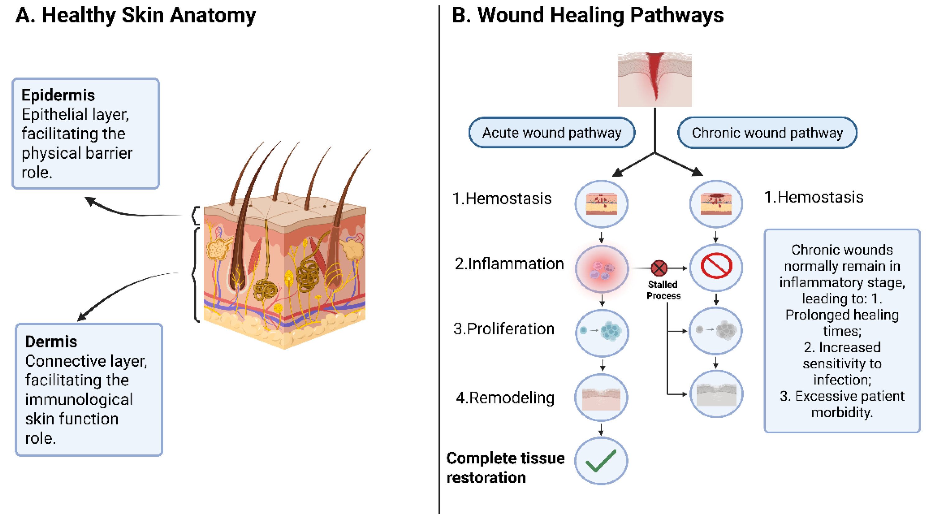
Figure 2.
The features of the ideal wound dressing.
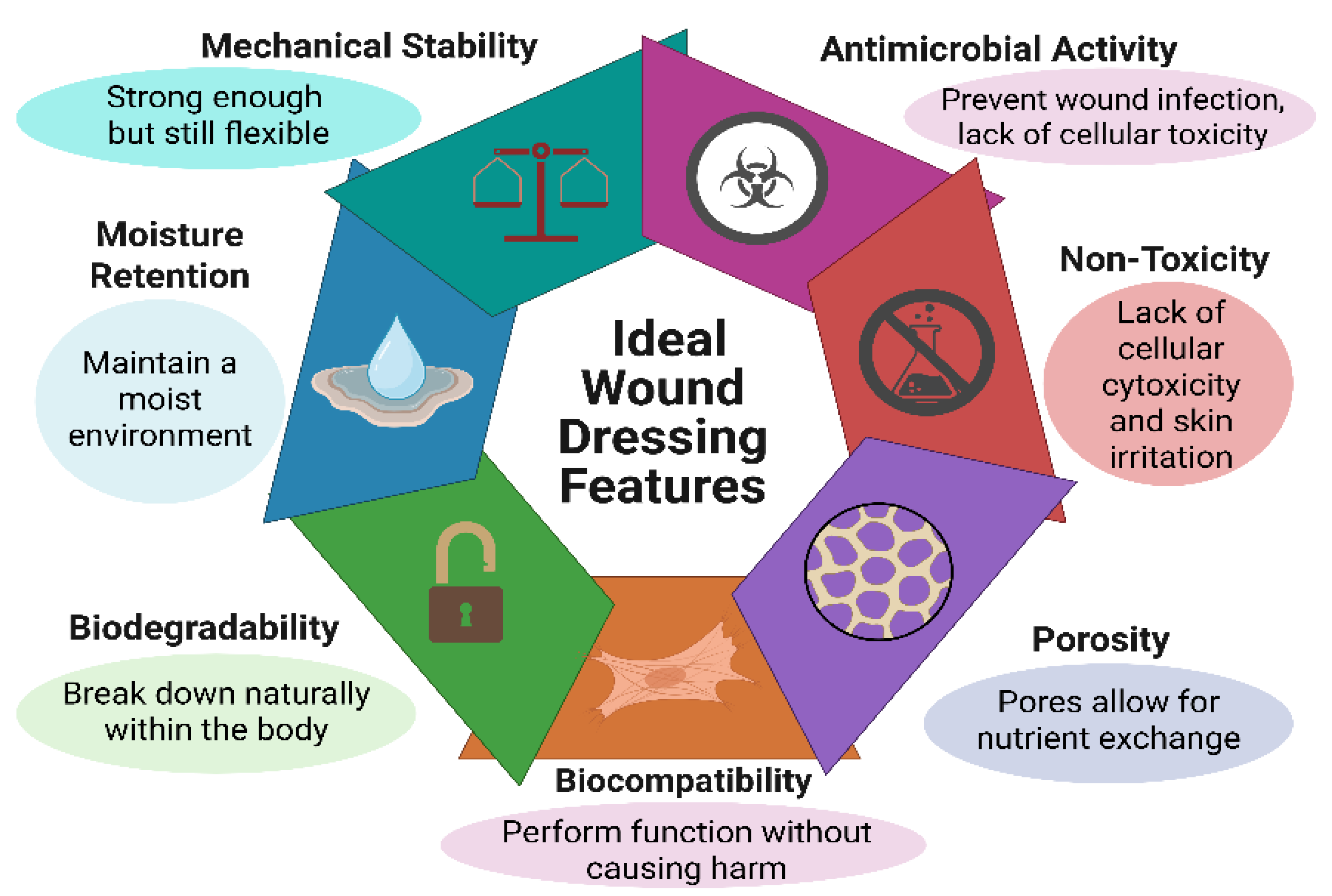
Figure 3.
An overview of the origin and significance of hydrogels prepared from gelatin and polysaccharides, along with their biomedical applications.
Figure 3.
An overview of the origin and significance of hydrogels prepared from gelatin and polysaccharides, along with their biomedical applications.
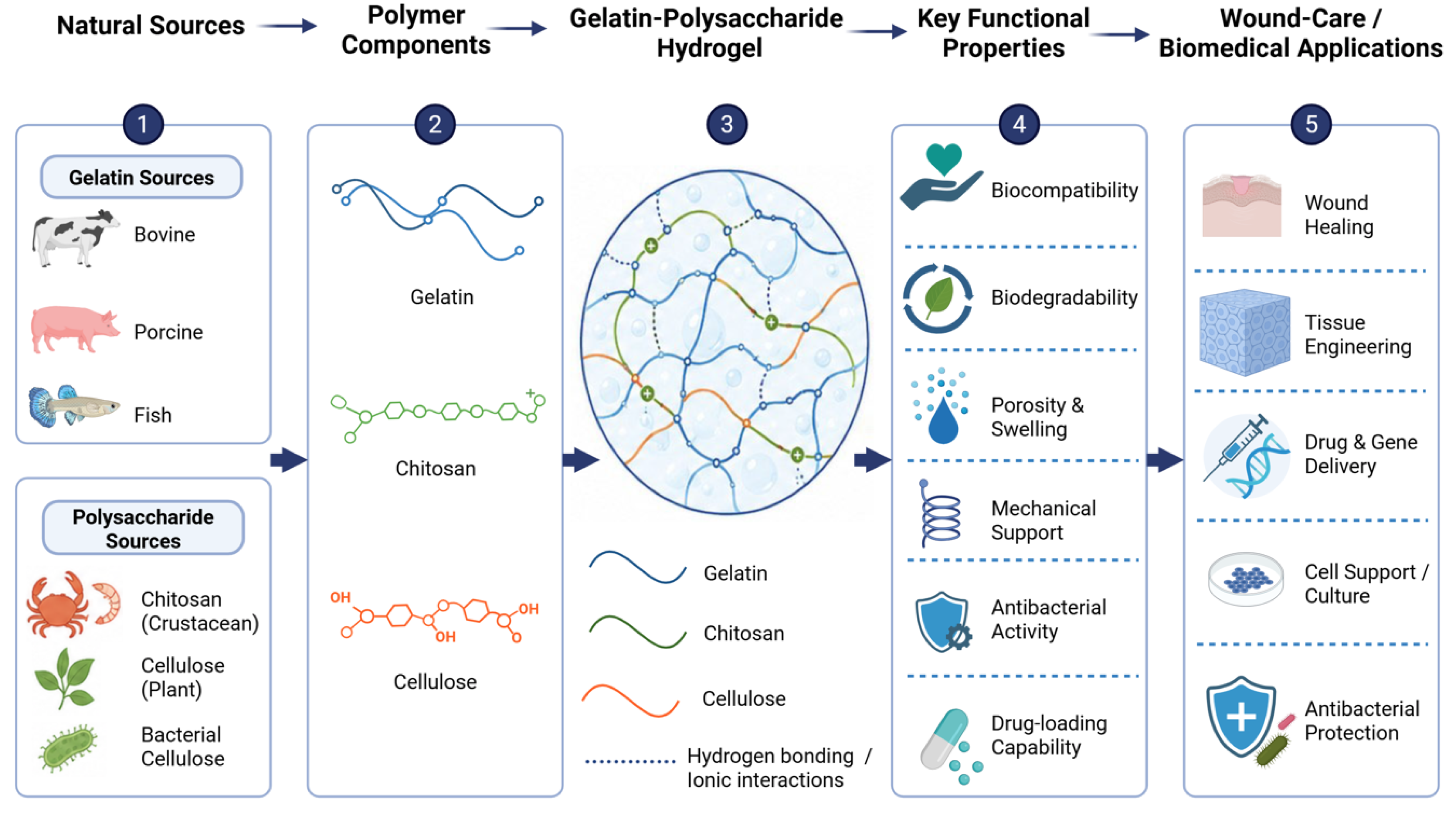
Figure 4.
Schematic diagram showing electrospun nanofibers for diabetic wound healing.
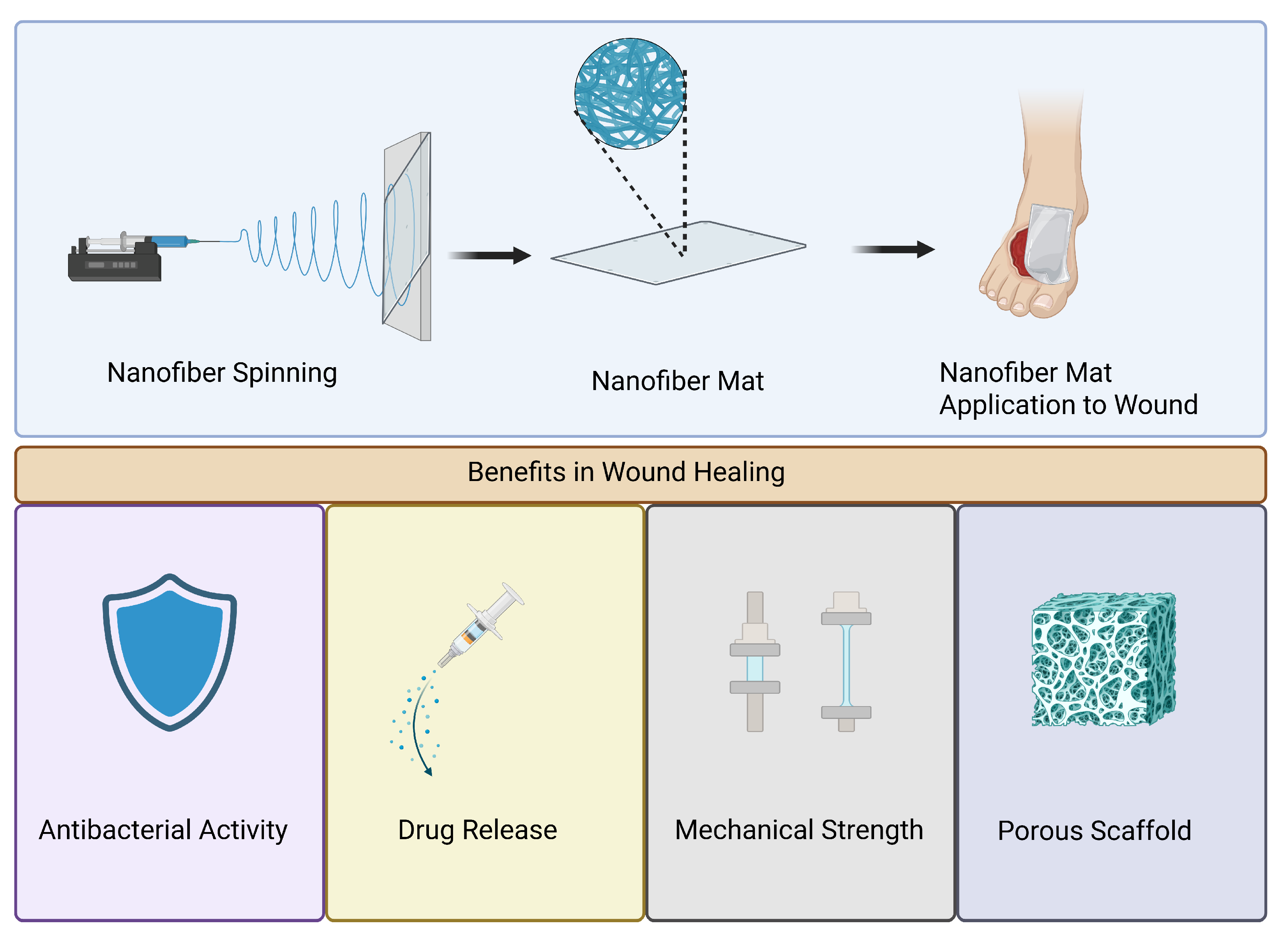
Figure 5.
Freeze-drying process.
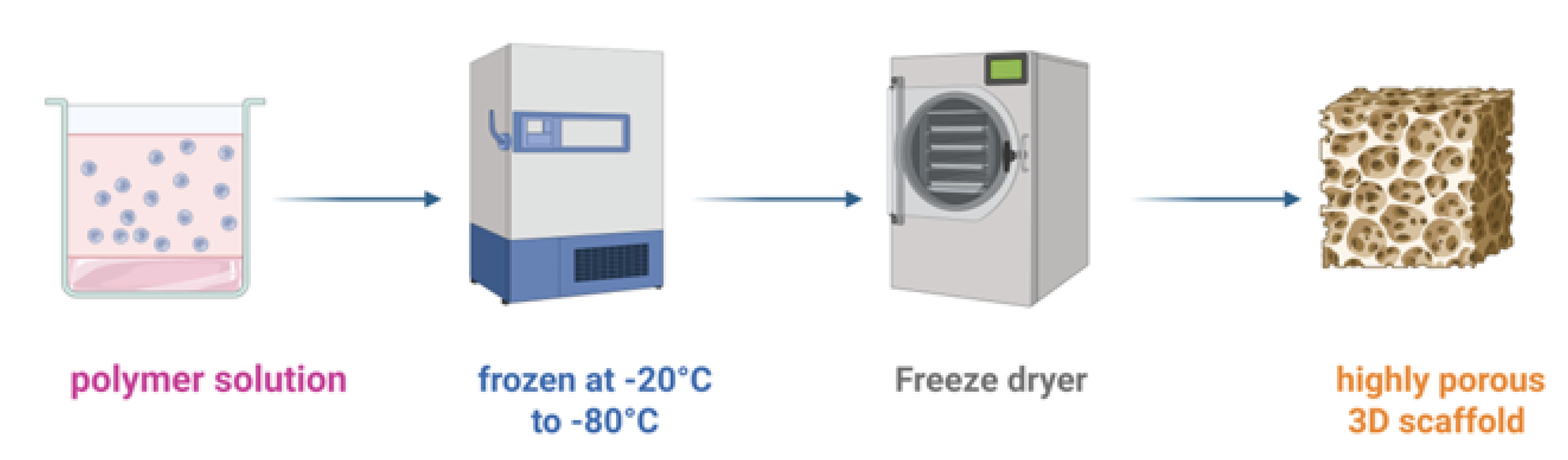
Figure 6.
A schematic workflow of the preparation of porous scaffolds via porogens.
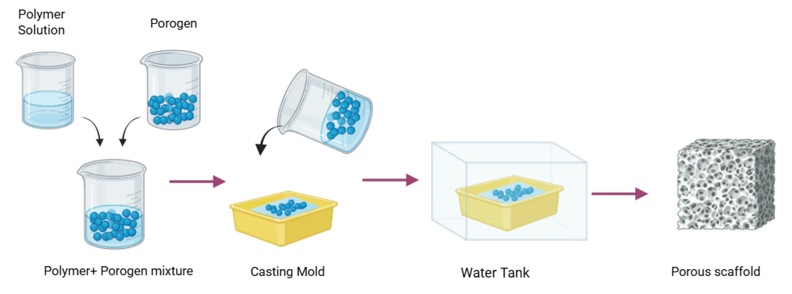
Table 2.
Advantages of composite scaffold systems based on gelatin, chitosan, and cellulose.
| Composite System | Primary Benefit |
|---|---|
| Gelatin–Chitosan | Enhanced cell adhesion and antimicrobial activity |
| Gelatin–Cellulose | Improved mechanical stability and moisture retention |
| Chitosan–Cellulose | Enhanced strength, porosity, and wound protection |
| Gelatin–Chitosan–Cellulose | Balanced biocompatibility, mechanical performance, and regenerative functionality |
Table 3.
Comparison of fabrication techniques used in wound healing scaffold development.
| Method | Advantages | Limitations | Typical Applications |
|---|---|---|---|
| Hydrogels | Excellent moisture retention; adaptable chemistry | Limited mechanical strength | Chronic wound care |
| Electrospinning | ECM-mimicking nanofibers; high surface area | Solvent limitations; scaling challenges | Nanofibrous wound dressings |
| Freeze-Drying | Highly porous structures | Mechanical weakness | Porous tissue scaffolds |
| Salt Leaching | Simple and inexpensive | Limited pore-size control | Porous scaffold fabrication |
| 3D Printing | Precise architectural control | Equipment cost; bioink constraints | Patient-specific wound dressings |
Disclaimer/Publisher’s Note: The statements, opinions and data contained in all publications are solely those of the individual author(s) and contributor(s) and not of MDPI and/or the editor(s). MDPI and/or the editor(s) disclaim responsibility for any injury to people or property resulting from any ideas, methods, instructions or products referred to in the content. |
© 2026 by the authors. Licensee MDPI, Basel, Switzerland. This article is an open access article distributed under the terms and conditions of the Creative Commons Attribution (CC BY) license (http://creativecommons.org/licenses/by/4.0/).
Copyright: This open access article is published under a Creative Commons CC BY 4.0 license, which permit the free download, distribution, and reuse, provided that the author and preprint are cited in any reuse.


