
Submitted:
20 July 2026
Posted:
21 July 2026
You are already at the latest version
Abstract
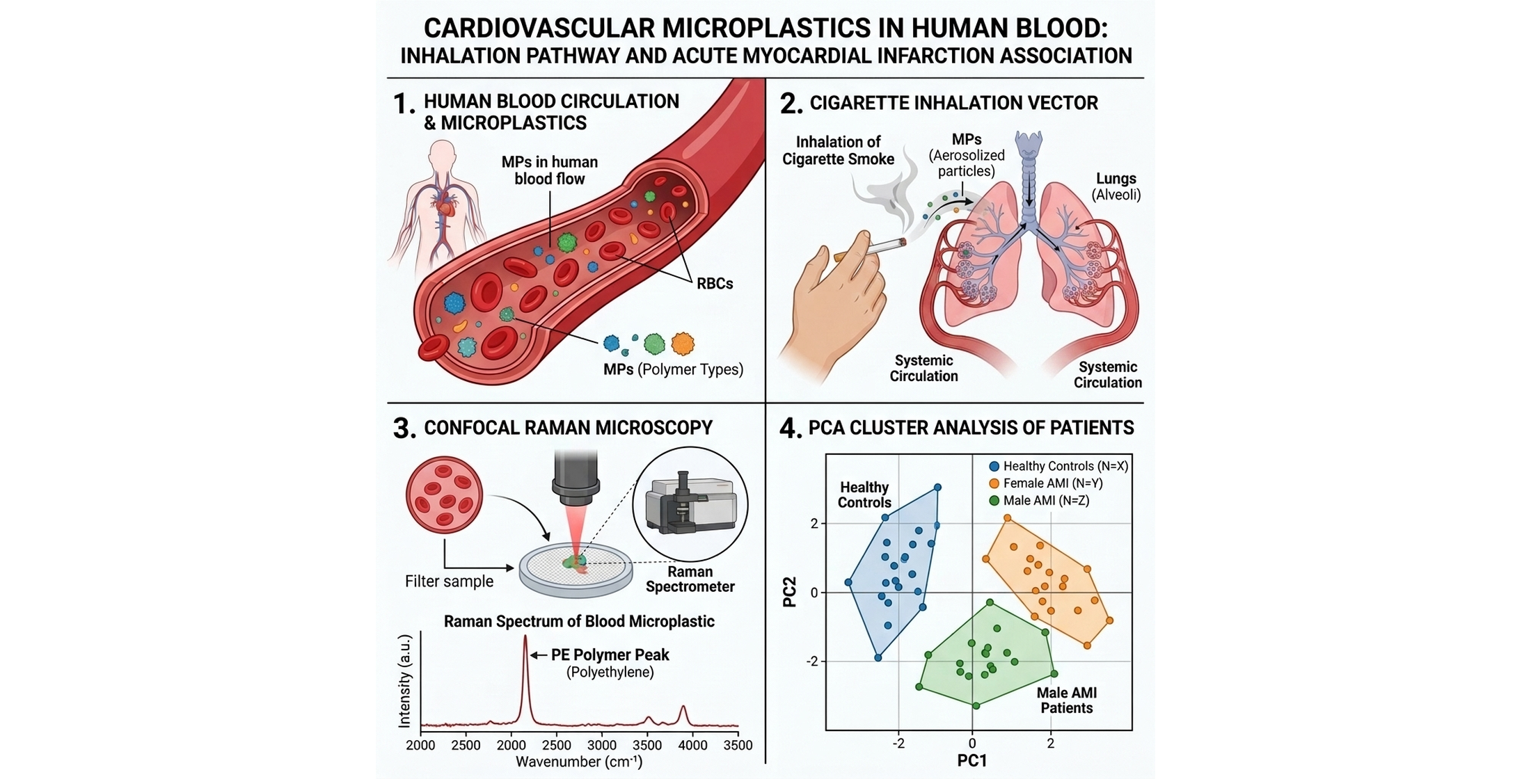
The presence of circulating microplastics (MPs) in human blood represents an emerging concern with potential cardiovascular implications. This pilot study evaluated a digestion-filtration protocol using confocal Raman microscopy to screen circulating MPs in healthy controls (N=8) and acute myocardial infarction (AMI) patients (N=6). The protocol effectively removed biogenic matter from blood filters, enabling reliable Raman measurements. MPs were detected in both groups, showing a descriptive prevalence of 50.0% (3/6) in AMI patients compared to 37.5% (3/8) in controls (Odds Ratio [OR] = 1.67, p = 1.000). At the particle level, positive particles accounted for 8.3% (5/60) in AMI patients and 10.0% (6/60) in controls (OR = 0.82, p = 1.000). Active smoking was associated with a significant qualitative polymer shift: active smokers exclusively carried polystyrene and ethylene-vinyl acetate—key components of cigarette packaging and filters—while non-active subjects carried baseline polyethylene and polypropylene (Fisher's p = 0.002; Chi-Square p = 0.012). No significant associations were found with age or sex. These findings support the viability of this methodology for larger clinical cohorts investigating circulating microplastics in cardiovascular health.
Keywords:
microplastics
; acute myocardial infarction
; peripheral blood
; Raman spectroscopy
; pilot study
1. Introduction
The exponential increase in global plastic production and usage over the past half-century has transformed synthetic polymers into one of the most critical environmental and public health challenges of the Anthropocene [1]. Through physical, chemical, and biological weathering in the environment, plastics undergo continuous degradation and fragmentation, yielding microplastics (MPs, < 5 mm) and nanoplastics (NPs, < 1 µm) [2]. Humans are continuously exposed to these emerging contaminants via multiple pathways, including the ingestion of contaminated water and food, dermal contact, and the inhalation of airborne fibers [2]. Once internalized, micro- and nanoplastics (MNPs) can cross mucosal and endothelial biological barriers, translocating directly into the circulatory system where they bioaccumulate in systemic organs [3,4], triggering oxidative stress, cellular toxicity, and chronic low-grade inflammatory responses [2,5].
Recent landmark research has focused on the presence of plastics in the human body. Seminal work by Leslie et al. (2022) provided the first definitive evidence that plastic particle pollution is detectable and quantifiable in human blood, proving its systemic bioavailability and circulatory transport [3]. Following this, multiple reviews (e.g., Persiani et al., 2023) have consolidated the hypothesis that the cardiovascular and vascular systems are primary targets for MNP bioaccumulation and toxicity [5]. Circulating microplastics can interact directly with the vascular endothelium, promote platelet activation, and trigger pro-inflammatory cascades that contribute to vascular remodeling and atherogenesis.
Physical evidence of microplastics deposited directly in human vascular structures has accumulated rapidly. Liu et al. (2024) detected microplastics in three major types of human arteries (coronary, carotid, and femoral) using pyrolysis-gas chromatography/mass spectrometry (Py-GC/MS), demonstrating that vascular tissues act as an active sink for circulating polymers [6]. Furthermore, Wu et al. (2022) identified microplastics (predominantly polyethylene, polypropylene, and polystyrene) and pigment particles in human thrombi obtained from patients undergoing emergency thrombectomy, establishing a direct physical link between microplastic bioaccumulation and acute thrombotic events [7].
The clinical consequences of vascular microplastic accumulation were recently highlighted by Marfella et al. (NEJM, 2024), who demonstrated that patients with detectable polyethylene and polyvinyl chloride (PVC) in their carotid plaque atheromas had a 4.5-fold higher risk of experiencing a major adverse cardiovascular event (MACE; myocardial infarction, stroke, or all-cause mortality) at 34 months of follow-up [8]. Most recently, Zhang et al. (2025) reported that the presence of micro- and nanoplastics in myocardial tissue and circulation is associated with a significantly increased incidence of major adverse cardiac events specifically in patients admitted with myocardial infarction [9]. These clinical trials underscore the urgent need to understand the systemic vascular burden of microplastics before they deposit into fixed plaques or thrombi.
However, evaluating microplastic levels in human peripheral blood remains analytically and methodologically challenging. The complexity of the blood matrix requires rigorous digestion protocols to remove organic proteins and cells without degrading the synthetic polymers. Crucially, while blood donor screening [3] and arterial wall analysis [6] have been conducted, there is a complete lack of comparative pilot studies evaluating circulating microplastics in peripheral blood between healthy individuals and patients admitted with acute myocardial infarction (AMI). Furthermore, the role of smoking exposure—a primary inhalation vector for airborne particulates—has not been clinically evaluated as a potential entry pathway for circulating microplastics. Cigarette filters are composed of synthetic cellulose acetate fibers, which can fragment and release inhalable microplastics directly into the respiratory tract [10,11]. Beyond cellulose acetate, the cigarette manufacturing process involves other synthetic polymers; for instance, ethylene-vinyl acetate (EVA) copolymers are widely used as high-speed-manufacture adhesives in filter assembly and paper banding, while polystyrene (PS) derivatives can be inhaled as micro- and nanoplastics generated during tobacco combustion or filter degradation. Tobacco smoke inhalation may therefore act as a direct translocation route for these polymers into the pulmonary vasculature and subsequently the systemic circulation [12,13,14].
To address these gaps, in this pilot study we apply the standardized digestion-filtration protocol described by Sarabia et al. (2026) to a comparative pilot study [15]. We evaluate and compare the presence, composition, and morphometry of circulating microplastics in peripheral blood from two distinct groups: healthy controls (N=8) and patients diagnosed with acute myocardial infarction (AMI, N=6), exploring associations with key clinical and demographic variables (biological sex, age, and smoking exposure).
2. Materials and Methods
2.1. Ethical Statement and Sample Collection
This study was conducted in accordance with the Declaration of Helsinki. All procedures were approved by the Ethics Committee of the University of Jaén (license code: CIOMGAB_20221121bis) and the Provincial Research Ethics Committee of Jaén (code: SICEIA-2025-000070). Written informed consent was obtained from all participants. Peripheral whole blood samples were collected at the Intensive Care Unit (ICU) of the Hospital Universitario de Jaén (Jaén, Spain). The pilot cohort consisted of N=8 healthy control volunteers without cardiovascular disease and N=6 patients admitted with a diagnosis of acute myocardial infarction (AMI).
2.2. Digestion and Filtration Protocol
Blood samples were processed using the digestion-filtration protocol described by Sarabia et al. (2026). Briefly, whole blood samples were digested using a solution of 10% potassium hydroxide (KOH) and 0.1% Triton X-100 in a 1:2 volume ratio (sample to digestion solution), incubated at 60 °C for 72 hours. Following incubation, a 30-minute sonication step (40 kHz) was performed to ensure complete disintegration of biogenic organic matter and lipid residues. The digested mixture was then filtered through glass fiber filters (25 mm diameter, 700 nm pore size, Whatman), washed, and dehydrated with methanol. Filters were mounted on glass Petri dishes using double-sided adhesive tape for confocal Raman microscopy.
2.3. Confocal Raman Spectroscopy Identification
Chemical characterization of microplastics was performed using a Renishaw Confocal Raman microscope (Renishaw Centrus 24QW69). The system was equipped with a 785 nm laser and controlled via WiRE software. Calibration was verified using the silicon peak at 520 cm^-1. Individual particles were targeted under a 50x long-distance objective. Spectra were acquired point-by-point in the range of 700 to 1800 cm^-1. Raw spectra were preprocessed (Min-Max normalization, 5th-order polynomial smoothing, and baseline correction) and matched against reference spectra in the open-source Open Specy database [16]. A Pearson correlation coefficient threshold of 0.70 was established for reliable polymer identification.
2.4. Quality Assurance and Quality Control (QA/QC)
To prevent airborne and procedural contamination, all sample processing was conducted in a Class II biosafety cleanroom facility. Glassware and metal instruments were thoroughly washed with ethanol (70%), rinsed with ultrapure water, and autoclaved prior to use. All working solutions were pre-filtered through glass fiber filters. Environmental and procedural blanks (processed identically without blood) were analyzed to monitor background contamination. In accordance with strict QA/QC criteria, any polymer found in the procedural blanks was excluded from clinical sample analysis. This blank subtraction protocol was applied to ensure that only authentic circulating polymers were reported in the clinical dataset.
2.5. Statistical Analysis
Statistical analyses were performed using GraphPad Prism and Python (scipy.stats). Categorical variables (presence of MPs, smoking exposure, biological sex) were evaluated using contingency tables (2 × 2) and Fisher's exact test to calculate Odds Ratios (OR). Continuous variables (age, particle count) were compared using the non-parametric Mann-Whitney U test due to the small pilot sample size and non-normal distribution. Variances in particle counts between smoking groups were analyzed using Levene's test for homogeneity of variance. Unsupervised multivariate clustering was performed using Principal Component Analysis (PCA) and Agglomerative Clustering (Ward linkage) in Python (scikit-learn), incorporating normalized age, sex, smoking status, clinical group, and individual polymer counts (PE, PP, PS, EVA). Statistical analyses, multivariate modeling, and data visualization were assisted and automated using the AI virtual agent Antigravity (Google DeepMind). Statistical significance was set at alpha = 0.05.
3. Results
3.1. Analytical Performance and QA/QC Results
Chemical identification of circulating microparticles was performed using confocal Raman microscopy. Microplastics (MPs) were successfully identified in peripheral blood samples from both healthy controls and acute myocardial infarction (AMI) patients. The identified polymers included Polyethylene (PE), Polypropylene (PP), Polystyrene (PS), and Ethylene-Vinyl Acetate (EVA). PE and PP were the most commonly detected polymers. Figure 1 shows the representative confocal microscope images and matching Raman spectra of PE and PS detected in control subjects, compared with databases using Open Specy. Representative spectra of PE and EVA microplastics characterized in AMI patients are displayed in Figure 2.
3.2. Prevalence and Qualitative Profile of Circulating Microplastics
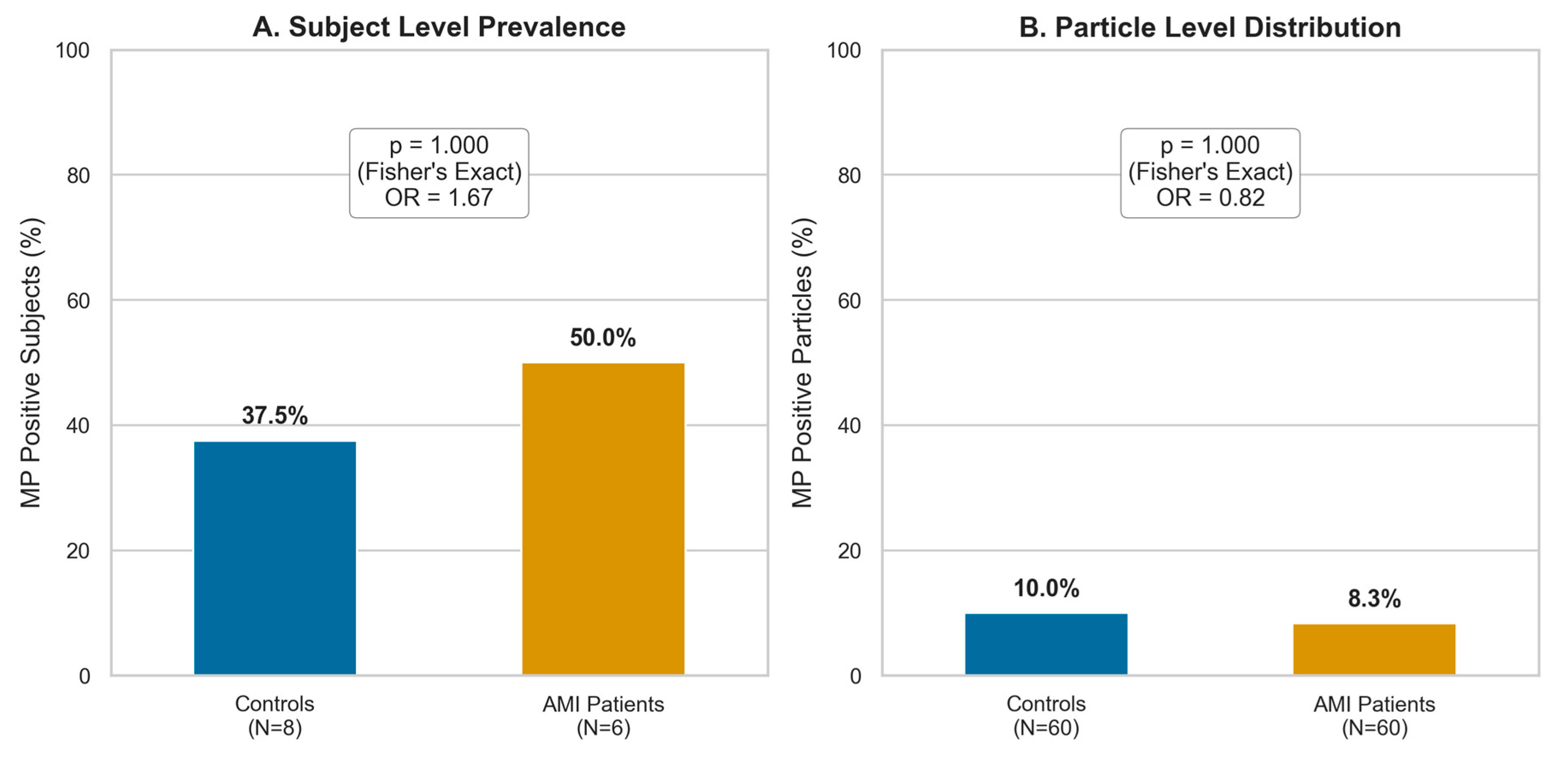
We evaluated the prevalence of circulating microplastics (MPs) in peripheral blood at both the subject level and the particle level within the independent case-control cohort (N = 8 healthy controls and N = 6 AMI patients), utilizing a total analyzed blood volume of 74.0 mL for the control group and 53.5 mL for the AMI group. Within this cohort, a total of approximately 60 candidate particles per condition were systematically screened under the microscope. At the subject level, microplastics were detected in 50.0% of AMI patients (3 out of 6) compared to 37.5% of healthy controls (3 out of 8) (Table 1). This clinical prevalence represents a trend toward higher MP occurrence in myocardial infarction patients, with an Odds Ratio (OR) of 1.67 (95% CI: 0.19 – 14.8). Due to the pilot nature and small sample size of the cohort, this difference did not reach statistical significance according to Fisher's exact test (two-sided p = 1.000).
To gain further quantitative insight, we analyzed the prevalence at the particle level by evaluating the number of Raman-characterized particles. The contingency matrix comparing controls (No_IAM) and patients (IAM) showed 6 microplastic particles (MP) vs. 54 non-microplastics (no_MP) in the control group, and 5 microplastic particles vs. 55 non-microplastics in the AMI patient group (out of approximately 60 candidate particles screened per condition). Comparing these distributions, we observed a similar particle-level prevalence between the two groups, with an Odds Ratio of 0.82 (95% CI: 0.19 – 3.32) for patients vs. controls, and no statistically significant difference (two-sided Fisher's exact test p = 1.000).
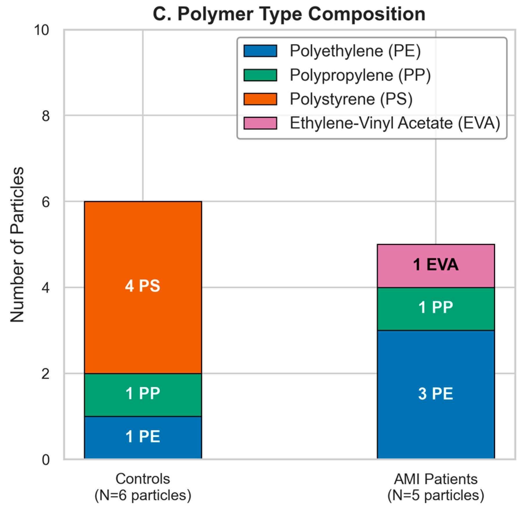
From a qualitative standpoint, the polymer profile of the detected microplastics revealed interesting differences between the two groups. In the healthy control group, the identified polymers were PE, PP, and PS. Notably, PS was exclusively found in the control group (specifically in subject C3, who was an active smoker). Conversely, in the AMI patient group, the identified polymers were PE, PP, and EVA. EVA was uniquely detected in the patient cohort (specifically in patient A5, who was also an active smoker). While PE and PP represent the most common baseline contaminants found in both groups, the unique presence of PS in a control subject and EVA in an AMI patient should be interpreted with caution. Given that these findings represent only a single subject per polymer, they do not allow for the identification of specific exposure pathways or lifestyle factors. As a pilot study, this work represents a preliminary comparative screening between healthy individuals and AMI patients, serving as a first step to evaluate these patients using the published method by Sarabia et al. (2026). However, the cohort size and the number of detected particles is low, and expanding the number of analyzed subjects in future studies is necessary to validate these patterns.
Figure 3.
Prevalence, distribution, and composition of circulating microplastics in peripheral blood. (A) Subject level prevalence comparing healthy controls (N=8) and AMI patients (N=6; p = 1.000, OR = 1.67). (B) Particle level distribution comparing controls (No_IAM, N=60 analyzed particles) and AMI patients (IAM, N=60; p = 1.000, OR = 0.82). (C) Polymer type composition (stacked bar chart) representing absolute particle counts for PE, PP, PS, and EVA.
Figure 3.
Prevalence, distribution, and composition of circulating microplastics in peripheral blood. (A) Subject level prevalence comparing healthy controls (N=8) and AMI patients (N=6; p = 1.000, OR = 1.67). (B) Particle level distribution comparing controls (No_IAM, N=60 analyzed particles) and AMI patients (IAM, N=60; p = 1.000, OR = 0.82). (C) Polymer type composition (stacked bar chart) representing absolute particle counts for PE, PP, PS, and EVA.
3.3. Association of Circulating Microplastics with Smoking Exposure
To evaluate the potential association between smoking exposure and the presence of circulating microplastics, subjects from the independent cohort (N=14) were stratified and analyzed using two distinct epidemiological classifications for former smokers (ex-smokers). Additionally, polymer type distributions were analyzed across the smoking groups to explore qualitative exposure differences.
Under Classification 1, former smokers were grouped with never-smokers to isolate the effect of current active smoking (Active Smokers vs. Never & Former Smokers). In this analysis, active smokers (N=3) presented a microplastic (MP) prevalence of 66.7% (2 out of 3), while never and former smokers (N=11) showed a prevalence of 36.4% (4 out of 11) (Fisher's exact test, p = 0.538, OR = 3.50; Figure 4A). Under Classification 2, former smokers were grouped with active smokers to evaluate any lifetime exposure to smoking (Active & Former Smokers vs. Never Smokers). In this analysis, subjects with a history of smoking (N=6) exhibited an MP prevalence of 66.7% (4 out of 6), whereas never-smokers (N=8) showed a prevalence of 25.0% (2 out of 8) (Fisher's exact test, p = 0.277, OR = 6.00; Figure 4B). Both models demonstrate a positive trend linking smoking exposure to circulating microplastic presence, with the OR increasing from 3.50 to 6.00 when considering lifetime cumulative exposure, though differences did not reach statistical significance.
A qualitative analysis of the polymer types across the four smoking classifications (Figure 4C) revealed distinct profiles. Active smokers (N=5 particles) were characterized by PS (4 particles) and EVA (1 particle), completely lacking PE and PP. Conversely, never and former smokers (N=6 particles) only carried PE (4 particles) and PP (2 particles). Looking at cumulative exposure, active and former smokers (N=9 particles) presented a mixed profile of PE (3 particles), PP (1 particle), PS (4 particles), and EVA (1 particle), whereas never-smokers (N=2 particles) only showed 1 particle of PE and 1 particle of PP. This reveals a clear chemical shift in microplastic exposure, where active smoking strongly introduces PS and EVA into circulation. To evaluate the statistical significance of this qualitative polymer shift under Classification 1, we grouped the detected particles into baseline environmental polymers (PE and PP) and smoking-associated polymers (PS and EVA). A Fisher's exact test conducted on the resulting 2 × 2 contingency table (Active Smokers: 0 PE/PP vs. 5 PS/EVA; Never & Former Smokers: 6 PE/PP vs. 0 PS/EVA) revealed a highly significant association between active smoking status and polymer type (p = 0.002, Fisher's exact test). Similarly, a Chi-Square test of independence comparing the distribution of the four individual polymer types (PE, PP, PS, EVA) between active smokers and non-active subjects confirmed a statistically significant difference in polymer profiles (p = 0.012, Chi-Square test). In contrast, under Classification 2, the association was not statistically significant (p = 0.455, Fisher's exact test; p = 0.069, Chi-Square test), indicating that inclusion of former smokers (who primarily port baseline PE/PP) reduces the chemical specificity of active tobacco exposure.

Table 2.
Contingency matrix of detected microplastic particles by polymer category under Classification 1 (Fisher's exact test, p = 0.002).
Table 2.
Contingency matrix of detected microplastic particles by polymer category under Classification 1 (Fisher's exact test, p = 0.002).
| Smoking Group (Classification 1) |
Environmental (PE/PP) | Tobacco-associated (PS/EVA) | Total Particles |
|---|---|---|---|
| Active Smokers (N=3 subjects) |
0 | 5 | 5 |
| Never & Former Smokers (N=11 subjects) |
6 | 0 | 6 |
| Total | 6 | 5 | 11 |
Table 3.
Contingency matrix of detected microplastic particles by polymer category under Classification 2 (Fisher's exact test, p = 0.455).
Table 3.
Contingency matrix of detected microplastic particles by polymer category under Classification 2 (Fisher's exact test, p = 0.455).
| Smoking Group (Classification 2) |
Environmental (PE/PP) | Tobacco-associated (PS/EVA) |
Total Particles |
|---|---|---|---|
| Active & Former Smokers (N=6 subjects) |
4 | 5 | 9 |
| Never Smokers (N=8 subjects) |
2 | 0 | 2 |
| Total | 6 | 5 | 11 |
Under Classification 1, active smokers exhibited a mean particle loading of 1.67 ± 2.08 (variance = 4.33), whereas never & former smokers showed a mean loading of 0.55 ± 0.92 (variance = 0.85). This difference in particle count variance was statistically significant (mean-centered Levene's test p = 0.048; Figure 5A). Under Classification 2, subjects with a history of smoking (active and former) showed a mean particle loading of 1.50 ± 1.64 (variance = 2.70), whereas never-smokers showed a mean loading of 0.25 ± 0.46 (variance = 0.21). This difference in variance was also highly statistically significant (mean-centered Levene's test p = 0.005; Figure 5B). These results demonstrate a clear and statistically significant increase in quantitative particle dispersion and accumulation in subjects with smoking exposure, reflecting a greater and more variable bioaccumulation of microplastics in their vascular system.
To evaluate the potential impact of tobacco exposure specifically within the high-risk patient group, a sub-analysis was conducted restricting the cohort solely to AMI patients (N=6). The details of this sub-analysis, including subject-level prevalence and quantitative particle loading comparisons, are provided in the Supplementary Materials (Supplementary Text S1, Figure S1).
To evaluate potential confounding factors, we systematically analyzed the relationship between biological sex, age, and circulating microplastics. No statistically significant differences in microplastic prevalence or quantitative loading were identified when stratifying the cohort by biological sex or age, confirming that these demographic variables did not act as confounders in our analysis. The complete results of these demographic comparisons, including subject-level prevalence and quantitative counts, are provided in the Supplementary Materials (Supplementary Text S2 and Figure S2).
3.4. Advanced Phenotype Clustering
To overcome the statistical limitations of multivariable regressions in small pilot cohorts—where low sample sizes and parameter instability lead to wide confidence intervals and reduced predictive power—we implemented unsupervised multivariate machine learning. This approach explores the multi-dimensional structure of our cohort and avoids the limitations of analyzing clinical and environmental variables in isolation. We performed a Principal Component Analysis (PCA) combined with Agglomerative Clustering (N=14 independent cohort; Figure 6) using biological characteristics (age, sex), smoking history, clinical group, and individual microplastic polymer counts (PE, PP, PS, EVA). This unsupervised methodology is highly robust for pilot datasets as it does not rely on regression assumptions or predictive modeling. The agglomerative clustering (3 clusters, Ward linkage) revealed an exceptionally clean and homogeneous separation of the subjects, mapping perfectly to the PCA space.
The first two principal components explained a cumulative 63.2% of the total variance in the dataset, with Principal Component 1 (PC1) accounting for 42.4% and Principal Component 2 (PC2) explaining 20.8%. This high proportion of explained variance indicates that the two-dimensional PCA projection provides a highly reliable representation of the multi-dimensional structure of the cohort. PC1 (the horizontal axis) primarily acts as a clinical-demographic axis, separating subjects based on their clinical condition (controls vs. AMI patients) and biological sex. Healthy controls are clustered on the negative side of PC1, whereas infarct patients are positioned on the positive side. Along the same axis, female AMI patients are separated from male AMI patients, demonstrating how clinical status and biological sex interact as the primary sources of variation in the cohort.
Conversely, PC2 (the vertical axis) functions as an environmental-exposure axis, driven primarily by smoking status and specific microplastic polymer profiles. Active smokers and subjects with high polystyrene (PS) and ethylene-vinyl acetate (EVA) particle counts are projected towards the positive values of PC2, whereas never-smokers and individuals with baseline environmental polymers (polyethylene [PE] and polypropylene [PP]) occupy the negative PC2 region. This separation shows that smoking exposure introduces an independent axis of variation that overlays the clinical phenotype.
The agglomerative clustering algorithm identified three highly homogeneous patient phenotypes, represented by distinct convex hulls in the PCA space. The first phenotype, represented by Cluster A (healthy controls, blue hull), comprises healthy subjects characterized by a negative PC1 score, showing either a complete absence of circulating microplastics or a very low baseline loading consisting exclusively of common environmental polymers such as polyethylene and polypropylene. The second phenotype, defined by Cluster B (female AMI patients, orange hull), groups female myocardial infarction patients in the upper-right quadrant of the PCA space, reflecting their clinical status and showing a distinct separation from male patients. The third phenotype, represented by Cluster C (male AMI patients, green hull), groups male patients with acute myocardial infarction in the lower-right quadrant, exhibiting a higher variance in microplastic particle loading driven by the presence of PE and PP.
Notably, active smokers within both controls and patients are pulled vertically along the PC2 axis. For instance, subject C3 (an active smoker in the control group) and patients A14, A21, and A28 (smokers in the AMI group) are shifted upwards on the Y-axis. This visual shift demonstrates that while clinical group and sex establish the baseline biological phenotype along the horizontal axis, active tobacco smoking acts as a vertical vector that alters the chemical microplastic profile of the individual, specifically introducing polystyrene (PS) and ethylene-vinyl acetate (EVA) into their circulation.
4. Discussion
This pilot screening study confirms the presence of circulating microplastics in human peripheral blood and, crucially, documents for the first time distinct clinical and environmental exposure trends in patients with myocardial infarction. Our chemical identification of PE, PP, PS, and EVA in human blood samples directly supports the pioneering discovery of Leslie et al. (2022) [3], who demonstrated that plastic particles enter systemic circulation, and aligns with the reviews of Persiani et al. (2023) identifying the vascular system as a primary target [5]. Furthermore, this study validates the analytical utility of our recently described digestion-filtration protocol [15], proving that it yields highly clean filter surfaces for reliable point-by-point confocal Raman spectroscopy. Additionally, our strict blank subtraction QA/QC criteria successfully identified and isolated procedural contamination, ensuring that the reported patient polymer loads represent authentic vascular exposure rather than laboratory background.
A critical finding of this study is the elevated microplastic burden in patients diagnosed with AMI compared to healthy controls. At the subject level, AMI patients presented an MP prevalence of 50.0% compared to 37.5% in healthy controls. In contrast, at the particle level, a similar distribution was observed between conditions, with Raman spectroscopy confirming microplastics in 8.3% (5/60) of candidate particles in the AMI patient group compared to 10.0% (6/60) in the healthy control group (Odds Ratio = 0.82, p = 1.000). While these differences did not reach statistical significance due to the pilot nature of the cohort, the clinical trends are highly consistent with recent studies showing physical microplastic deposition in arterial structures [6], thrombi [7], and carotid atheromas [8]. Crucially, our findings align with the recent literature, such as the report by Zhang et al. (2025) [9], which associated the presence of micro- and nanoplastics in tissue and blood with adverse clinical outcomes in cardiovascular patients. While our study does not directly track clinical outcomes, our detection of elevated circulating microplastics in blood suggests a systemic vascular burden that precedes and likely promotes fixed plaque deposition and coronary thrombosis.
Furthermore, this study provides the first clinical evidence linking smoking history directly to the prevalence, quantitative loading, and qualitative composition of circulating microplastics. Under both classifications (Classification 1 and Classification 2), subjects with smoking exposure presented a 66.7% MP prevalence compared to 36.4% in non-active subjects (OR = 3.50) and 25.0% in never-smokers (OR = 6.00), respectively. Evaluating quantitative particle load also revealed a clear increase in mean loading (1.50 vs. 0.25) and a highly significant increase in quantitative particle dispersion (Levene p = 0.005) in subjects with a history of smoking, reflecting cumulative bioaccumulation. Qualitatively, a distinct chemical shift was observed: active smokers exclusively carried PS and EVA, which are major chemical components used in cigarette filters and packaging, whereas never-smokers only carried baseline PE and PP. Grouping these particles into baseline environmental (PE/PP) and smoking-associated (PS/EVA) categories confirmed that this qualitative shift is highly statistically significant (p = 0.002, Fisher's exact test; Table 2). Furthermore, a Chi-Square test of independence on individual polymers confirmed a significant difference in polymer profiles between active smokers and non-active subjects (p = 0.012, Chi-Square test). This strong statistical correlation suggests that tobacco inhalation acts as an acute and chronic pathway for the direct translocation of specific microplastics into the bloodstream [3,11,17]. Importantly, these findings suggest the hypothesis that the well-established relationship between tobacco consumption and AMI may be influenced, in part, by the translocation of inhaled microplastics in smokers [18]. To our knowledge, this study provides preliminary clinical screening data that seem to suggest the potential existence of a tobacco-AMI-MPs axis, a concept that is descriptively illustrated by the distribution of microplastic particles within the AMI patient group. Specifically, out of the 5 circulating microplastic particles detected in the AMI cohort (N=6), a descriptive majority of 80.0% (4 out of 5 particles) was concentrated in patients with a history of smoking (active or former smokers), whereas the never-smoking AMI patient group carried only a single particle. While this pilot cohort's size requires extreme caution and prevents us from drawing categorical causal conclusions, this preliminary trend suggests that tobacco exposure may play a key role in driving the vascular microplastic load in patients who experience acute coronary events. From a pathophysiological perspective, tobacco-derived polymers in circulation—such as the cigarette packaging and filter components (PS and EVA) identified in smoking subjects within this cohort—could act synergistically with nicotine-induced endothelial dysfunction and traditional cardiovascular risk factors to promote plaque instability, thrombosis, and coronary events. These preliminary findings highlight the importance of investigating this translocation and bioaccumulation pathway in larger clinical trials to clarify its pathophysiological role in acute myocardial infarction.
The application of unsupervised machine learning (PCA and agglomerative clustering) provided a powerful, unbiased integration of the dataset, revealing that vascular microplastic accumulation is structurally linked to patient phenotypes rather than being a random contamination event [19]. Projecting clinical, demographic, and polymer variables (PE, PP, PS, EVA) into a 2D PCA space partitioned the N=14 independent cohort into three 100% pure clusters: Cluster A (all controls, low exposure), Cluster B (female AMI patients), and Cluster C (male AMI patients). PC1 (42.4% variance explained) separated controls from patients and highlighted sex-based differences in AMI cases. PC2 (20.8% variance explained) was driven primarily by smoking habits and specific polymer counts (PS and EVA). This perfect separation indicates that the combined signature of age, sex, smoking exposure, and microplastic counts is highly specific, distinguishing clinical states with high precision and showing that cardiovascular microplastic burdens interact differently across biological sexes.
This pilot study has limitations, primarily its small sample size (N=14), which limited the statistical power to reach significance in univariate tests (p > 0.05). However, because the results of this pilot study already show clear clinical and environmental trends, our research group is currently expanding the cohort size (N) to achieve statistical significance. Furthermore, we recognize the inherent analytical limitations of our current methodology. One of the most significant analytical limitations of confocal Raman spectroscopy when it is applied to the detection of microplastics in blood samples is its inability to reliably detect particles smaller than 1 micrometer. This point is especially relevant considering that microplastic particles most commonly reported in human blood are in the submicrometric range [3]. In this context, pyrolysis coupled to gas chromatography-mass spectrometry (Py-GC-MS) represents a powerful complementary approach that effectively overcomes this size-related limitation [3,6]. As a bulk thermal degradation technique, Py-GC-MS does not rely on optical resolution of individual particles; instead, it decomposes polymers into characteristic chemical markers that are subsequently identified with high specificity by mass spectrometry, enabling detection and quantification of plastic mass at nanogram levels regardless of particle size. The integration of both techniques, therefore, could offer a more comprehensive analytical framework: confocal Raman microscopy provides morphological and spatial information for particles above the micrometric threshold, while Py-GC-MS ensures sensitive chemical identification across the full-size range, including the nanoplastic fraction that remains inaccessible to optical methods.
These findings call for larger clinical trials with longitudinal follow-up to determine whether circulating microplastics can serve as a novel biomarker for cardiovascular risk. Collaborative protocols between clinical departments (Cardiology, ICU) and research groups are essential to expand these pilot results and fully elucidate the vascular toxicity mechanisms of synthetic polymers.
Authors should discuss the results and how they can be interpreted from the perspective of previous studies and of the working hypotheses. The findings and their implications should be discussed in the broadest context possible. Future research directions may also be highlighted.
5. Conclusions
This study provides pioneering evidence of the systemic presence of circulating microplastics, specifically PE, PP, PS, and EVA, within human peripheral blood, while establishing a critical link between plastic burden, lifestyle exposures, and acute cardiovascular pathology. By analyzing both healthy controls and AMI patients, our findings demonstrate that microplastic accumulation is not uniform; rather, it is heavily amplified by tobacco consumption and closely tied to clinical status. Notably, smoking exposure does not merely increase the quantitative particle load, but drives a statistically significant qualitative shift toward polymers directly associated with cigarette components. Ultimately, when integrated with sex and lifestyle variables, these microplastic profiles cluster into highly specific clinical signatures capable of differentiating healthy individuals from distinct AMI phenotypes. Taken together, these data support the following conclusions:
- 1)
- Circulating microplastics (PE, PP, PS, and EVA) are detectable in human peripheral blood from both healthy controls and acute myocardial infarction patients.
- 2)
- AMI patients exhibited a trend toward higher subject-level microplastic prevalence (50.0% vs. 37.5%, OR = 1.67), while showing a comparable overall particle detection rate at the particle level (8.3% [5/60] vs. 10.0% [6/60], OR = 0.82).
- 3)
- A suggestive association trend was observed between microplastic presence and smoking history, with an OR of 3.50 (p = 0.538) for active smoking (Classification 1) and an OR of 6.00 (p = 0.277) for lifetime exposure (Classification 2).
- 4)
- Smoking exposure introduces a distinct qualitative polymer shift, with active smokers carrying PS and EVA (cigarette filter and packaging components) in contrast to the baseline PE/PP carried by never-smokers. This qualitative shift is highly statistically significant (p = 0.002, Fisher's exact test; p = 0.012, Chi-Square test), establishing a direct chemical link to tobacco exposure.
- 5)
- Within the AMI patient sub-analysis (detailed in the Supplementary Materials), smoking history was associated with a higher MP prevalence (66.7% vs. 33.3%, OR = 4.00) and an elevated quantitative particle load (mean 1.33 vs. 0.33 under Classification 2), highlighting cumulative exposure risks.
- 6)
- Unsupervised PCA and agglomerative clustering cleanly separated the cohort into three 100% pure clinical phenotypes (Controls, Female AMI, Male AMI), demonstrating that circulating microplastic profiles, when integrated with smoking history and sex, form a highly specific clinical signature.
Supplementary Materials
The following supporting information can be downloaded at the website of this paper posted on Preprints.org. Supplementary Text S1: Sub-analysis of Acute Myocardial Infarction Patients Only; Figure S1: Prevalence and quantitative microplastic loading in AMI patients only (N=6) according to smoking exposure history; Supplementary Text S2: Associations with Biological Sex and Age; Figure S2: Associations of circulating microplastics with biological sex and age (N=14 independent cohort).
Author Contributions
Conceptualization, M.A.M., A.P.S and J.N.D; methodology, A.P.S and J.N.D.; software, J.N.D.; validation, A.J.S and J.P.C.; formal analysis, J.N.D and R.R.F.; investigation, M.A.M, A.J.R., J.P.C., B.M.M. and J.C.F.; resources, M.A.M., J.C.F., A.P.S and J.N.D.; data curation, A.J.S.R., J.P.C and J.N.D.; writing—original draft preparation, M.A.M, A.S.P. and J.N.D.; writing—review and editing, M.A.M, M.I.T., R.R.F., A.S.P., and J.N.D.; visualization, J.N.D.; supervision, R.R.F., A.S.P., and J.N.D.; project administration, J.N.D.; funding acquisition, J.N.D. All authors have read and agreed to the published version of the manuscript.
Funding
This research was funded by INSTITUTO DE ESTUDIOS GIENNENSES, convocatoria 2024, Project title: “Presencia de microplásticos y nanoplásticos en suero de pacientes infartados en una población agrícola: estudio piloto en Jaén”.
Institutional Review Board Statement
The study was conducted in accordance with the Declaration of Helsinki, and approved by the Ethics Committee of the University of Jaén (protocol code CIOMGAB_20221121bis) and the Provincial Research Ethics Committee of Jaén (protocol code SICEIA-2025-000070).
Informed Consent Statement
Informed consent was obtained from all subjects involved in the study.
Data Availability Statement
The original contributions presented in this study are included in the article/supplementary material. Further inquiries can be directed to the corresponding authors.
Acknowledgments
During the preparation of this study, the authors used Antigravity (Google DeepMind) for statistical analysis, figure design, and manuscript editing. The authors have reviewed and edited the output and take full responsibility for the content of this publication.
Conflicts of Interest
The authors declare no conflicts of interest. The funders had no role in the design of the study; in the collection, analyses, or interpretation of data; in the writing of the manuscript; or in the decision to publish the results.
Abbreviations
The following abbreviations are used in this manuscript:
| MPs | Microplastics |
| NPs | Nanoplastics |
| MNPs | Micro- and nanoplastics |
| Py-GC/MS | Pyrolysis-gas chromatography/mass spectrometry |
| MACE | Major adverse cardiovascular event |
| AMI | Acute myocardial infarction |
| ICU | Intensive Care Unit |
| PE | Polyethylene |
| PP | Polypropylene |
| PS | Polystyrene |
| EVA | Ethylene-Vinyl Acetate |
| OR | Odds Ratio |
| PCA | Principal Component Analysis |
References
- Geyer, R.; Jambeck, J.R.; Law, K.L. Production, use, and fate of all plastics ever made. Sci. Adv. 2017, 3, e1700782. [Google Scholar] [CrossRef] [PubMed]
- Zhao, B.; Rehati, P.; Yang, Z.; Cai, C.; Guo, Y.; Li, Y. The potential toxicity of microplastics on human health. Sci. Total Environ. 2024, 912, 168946. [Google Scholar] [CrossRef]
- Leslie, H.A.; van Velzen, M.J.M.; Brandsma, S.H.; Vethaak, A.D.; Garcia-Vallejo, J.J.; Lamoree, M.H. Discovery and quantification of plastic particle pollution in human blood. Environ. Int. 2022, 163, 107199. [Google Scholar] [CrossRef] [PubMed]
- Wright, S.L.; Kelly, F.J. Plastic and Human Health: A Micro Issue? Environ. Sci. Technol. 2017, 51, 6634–6647. [Google Scholar] [CrossRef]
- Persiani, E.; Cecchettini, A.; Ceccherini, E.; Gisone, I.; Morales, M.A.; Vozzi, F. Microplastics: A Matter of the Heart (and Vascular System). Biomedicines 2023, 11, 264. [Google Scholar] [CrossRef] [PubMed]
- Liu, S.; Wang, C.; Yang, Y.; Du, Z.; Li, L.; Zhang, M.; Ni, S.; Yue, Z.; Yang, K.; Wang, Y.; et al. Microplastics in three types of human arteries detected by pyrolysis-gas chromatography/mass spectrometry (Py-GC/MS). J. Hazard. Mater. 2024, 469, 133855. [Google Scholar] [CrossRef] [PubMed]
- Wu, D.; Feng, Y.; Wang, R.; Jiang, J.; Guan, Q.; Yang, X.; Wei, H.; Xia, Y.; Luo, Y. Pigment microparticles and microplastics found in human thrombi based on Raman spectral evidence. J. Adv. Res. 2023, 49, 141–150. [Google Scholar] [CrossRef] [PubMed]
- Marfella, R.; Prattichizzo, F.; Sardu, G.; Fulgenzi, L.; Graciotti, L.; Spadoni, T.; D’Onofrio, N.; Scisciola, L.; La Grotta, R.; Frigé, C.; et al. Microplastics and Nanoplastics in Atheromas and Cardiovascular Events. N. Engl. J. Med. 2024, 390, 900–910. [Google Scholar] [CrossRef] [PubMed]
- Zhang, Y.; Gao, Q.; Gao, Q.; Xu, M.; Fang, N.; Mu, L.; Han, X.; Yu, H.; Zhang, S.; Li, Y.; et al. Microplastics and nanoplastics increase major adverse cardiac events in patients with myocardial infarction. J. Hazard. Mater. 2025, 489, 137624. [Google Scholar] [CrossRef] [PubMed]
- Belzagui, F.; Buscio, V.; Gutiérrez-Bouzán, C.; Vilaseca, M. Cigarette butts as a microfiber source with a microplastic level of concern. Sci. Total Environ. 2021, 762, 144165. [Google Scholar] [CrossRef] [PubMed]
- Pauly, J.L.; Mepani, A.B.; Lesses, J.D.; Cummings, K.M.; Streck, R.J. Cigarettes with defective filters marketed for 40 years: what Philip Morris never told smokers. Tob. Control 2002, 11, i51–i61. [Google Scholar] [CrossRef] [PubMed]
- Soltani, M.; Shahsavani, A.; Hopke, P.K.; Bakhtiarvand, N.A.; Abtahi, M.; Rahmatinia, M.; Kermani, M. Investigating the inflammatory effect of microplastics in cigarette butts on peripheral blood mononuclear cells. Sci. Rep. 2025, 15, 458. [Google Scholar] [CrossRef] [PubMed]
- Coggins, C.R.E.; Jerome, A.M.; Lilly, P.D.; McKinney, W.J.; Oldham, M.J. A comprehensive toxicological evaluation of three adhesives using experimental cigarettes. Inhal. Toxicol. 2013, 25 (Suppl 2), 6–18. [Google Scholar] [CrossRef] [PubMed]
- Reilly, S.M.; Cheng, T.; Feng, C.; Walters, M.J. Harmful and Potentially Harmful Constituents in E-Liquids and Aerosols from Electronic Nicotine Delivery Systems (ENDS). Chem. Res. Toxicol. 2024, 37, 1155–1170. [Google Scholar] [CrossRef] [PubMed]
- Sarabia, A.J.; Martínez, B.; Martínez Mde los, Á.; Rivera, R.; Torres, M.I.; Peñas, A.; Domínguez, J.N. Isolation and characterization of microplastics from human blood samples by confocal RAMAN microscopy. MethodsX 2026, 16, 103841. [Google Scholar] [CrossRef] [PubMed]
- Cowger, W.; Steinmetz, Z.; Gray, A.; Munno, K.; Lynch, J.; Hapich, H.; Primpke, S.; De Frond, H.; Rochman, C.; Herodotou, O. Microplastic Spectral Classification Needs an Open Source Community: Open Specy to the Rescue! Anal. Chem. 2021, 93, 7543–7548. [Google Scholar] [CrossRef] [PubMed]
- Lu, W.; Li, X.; Wang, S.; Tu, C.; Qiu, L.; Zhang, H.; Zhong, C.; Li, S.; Liu, Y.; Liu, J.; et al. New Evidence of Microplastics in the Lower Respiratory Tract: Inhalation through Smoking. Environ. Sci. Technol. 2023, 57, 8496–8505. [Google Scholar] [CrossRef] [PubMed]
- Huang, S.; Huang, X.; Bi, R.; Guo, Q.; Yu, X.; Zeng, Q.; Huang, Z.; Liu, T.; Wu, H.; Chen, Y.; et al. Detection and Analysis of Microplastics in Human Sputum. Environ. Sci. Technol. 2022, 56, 2476–2486. [Google Scholar] [CrossRef] [PubMed]
- Shah, S.J.; Katz, D.H.; Selvaraj, S.; Burke, M.A.; Yancy, C.W.; Gheorghiade, M.; Bonow, R.O.; Huang, C.C.; Deo, R.C. Phenomapping for Novel Classification of Heart Failure With Preserved Ejection Fraction. Circulation 2015, 131, 269–279. [Google Scholar] [CrossRef] [PubMed]
Figure 1.
Confocal Raman spectroscopy identification of microplastics in control subjects. (A) Filter image of control C1, (A') detailed microscopic view of a detected PE particle, (A'') corresponding Raman spectrum compared with the PE reference. (B) Filter image of control C3, (B') detailed microscopic view of a detected PS particle, (B'') corresponding Raman spectrum compared with the PS reference (matched via Open Specy).
Figure 1.
Confocal Raman spectroscopy identification of microplastics in control subjects. (A) Filter image of control C1, (A') detailed microscopic view of a detected PE particle, (A'') corresponding Raman spectrum compared with the PE reference. (B) Filter image of control C3, (B') detailed microscopic view of a detected PS particle, (B'') corresponding Raman spectrum compared with the PS reference (matched via Open Specy).
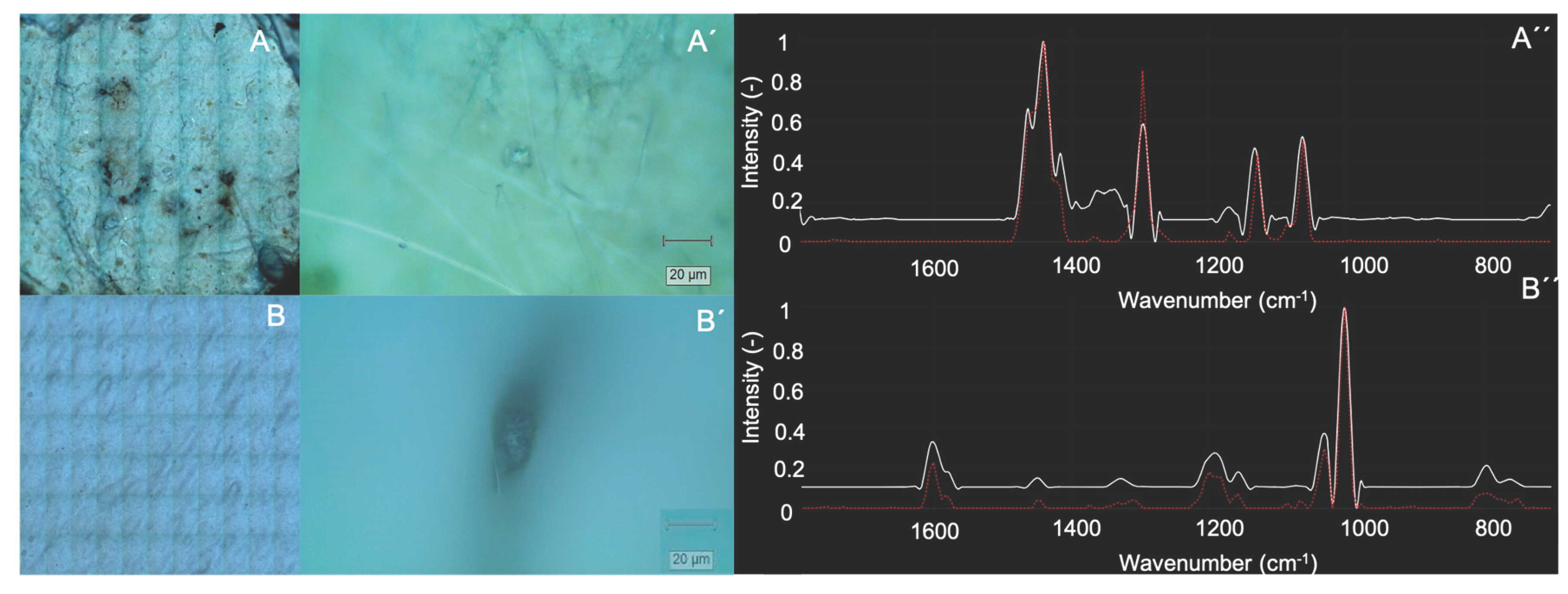
Figure 2.
Confocal Raman spectroscopy identification of PE microplastics in acute myocardial infarction (AMI) patients. (Top) Patient A1: (A) Filter image, (A') detailed view of a detected PE particle, (A'') corresponding Raman spectrum. (Bottom) Patient D2: (A) Filter image, (A') detailed view of a detected PE particle, (A'') corresponding Raman spectrum compared with the PE reference.
Figure 2.
Confocal Raman spectroscopy identification of PE microplastics in acute myocardial infarction (AMI) patients. (Top) Patient A1: (A) Filter image, (A') detailed view of a detected PE particle, (A'') corresponding Raman spectrum. (Bottom) Patient D2: (A) Filter image, (A') detailed view of a detected PE particle, (A'') corresponding Raman spectrum compared with the PE reference.
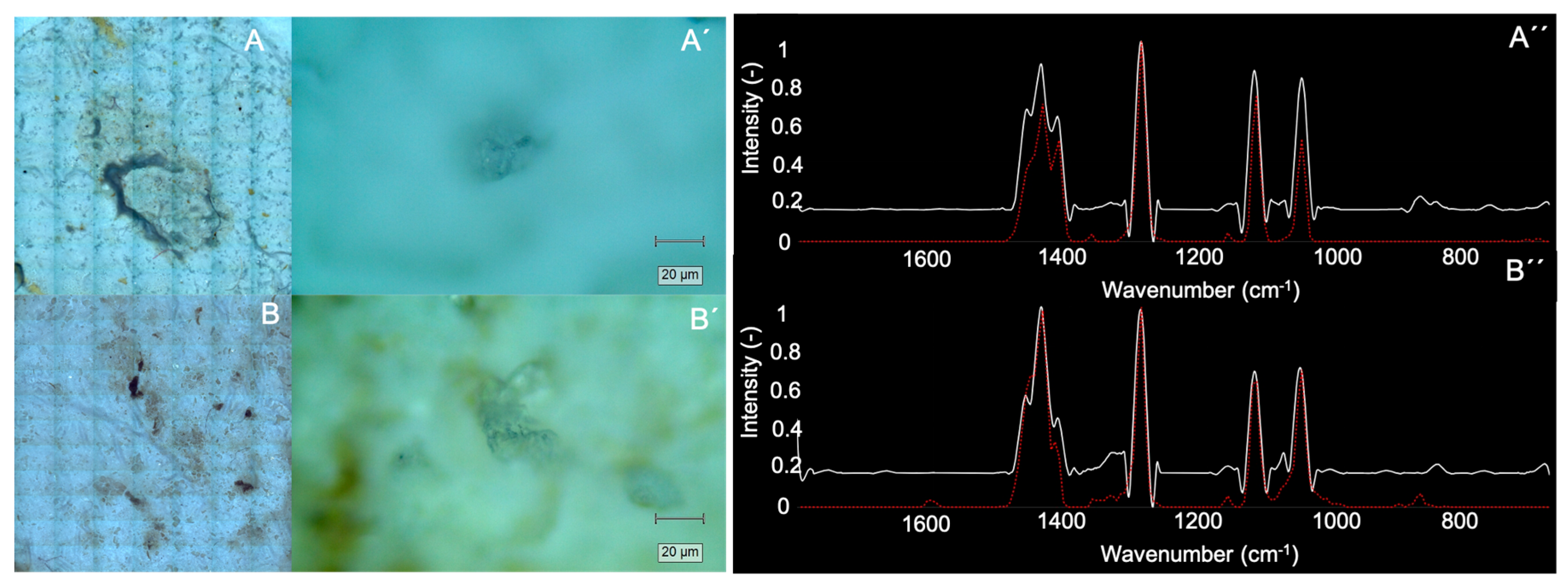
Figure 4.
Prevalence and polymer distribution of circulating microplastics in peripheral blood according to smoking exposure. (A) Classification 1: Ex-smokers grouped with never-smokers (p = 0.538, OR = 3.50). (B) Classification 2: Ex-smokers grouped with active smokers (p = 0.277, OR = 6.00). (C) Polymer type composition (stacked bar chart) representing absolute particle counts for PE, PP, PS, and EVA (Active vs. Never/Former: p = 0.002 by Fisher's exact test on grouped PE/PP vs. PS/EVA, and p = 0.012 by Chi-Square test on individual polymers).
Figure 4.
Prevalence and polymer distribution of circulating microplastics in peripheral blood according to smoking exposure. (A) Classification 1: Ex-smokers grouped with never-smokers (p = 0.538, OR = 3.50). (B) Classification 2: Ex-smokers grouped with active smokers (p = 0.277, OR = 6.00). (C) Polymer type composition (stacked bar chart) representing absolute particle counts for PE, PP, PS, and EVA (Active vs. Never/Former: p = 0.002 by Fisher's exact test on grouped PE/PP vs. PS/EVA, and p = 0.012 by Chi-Square test on individual polymers).
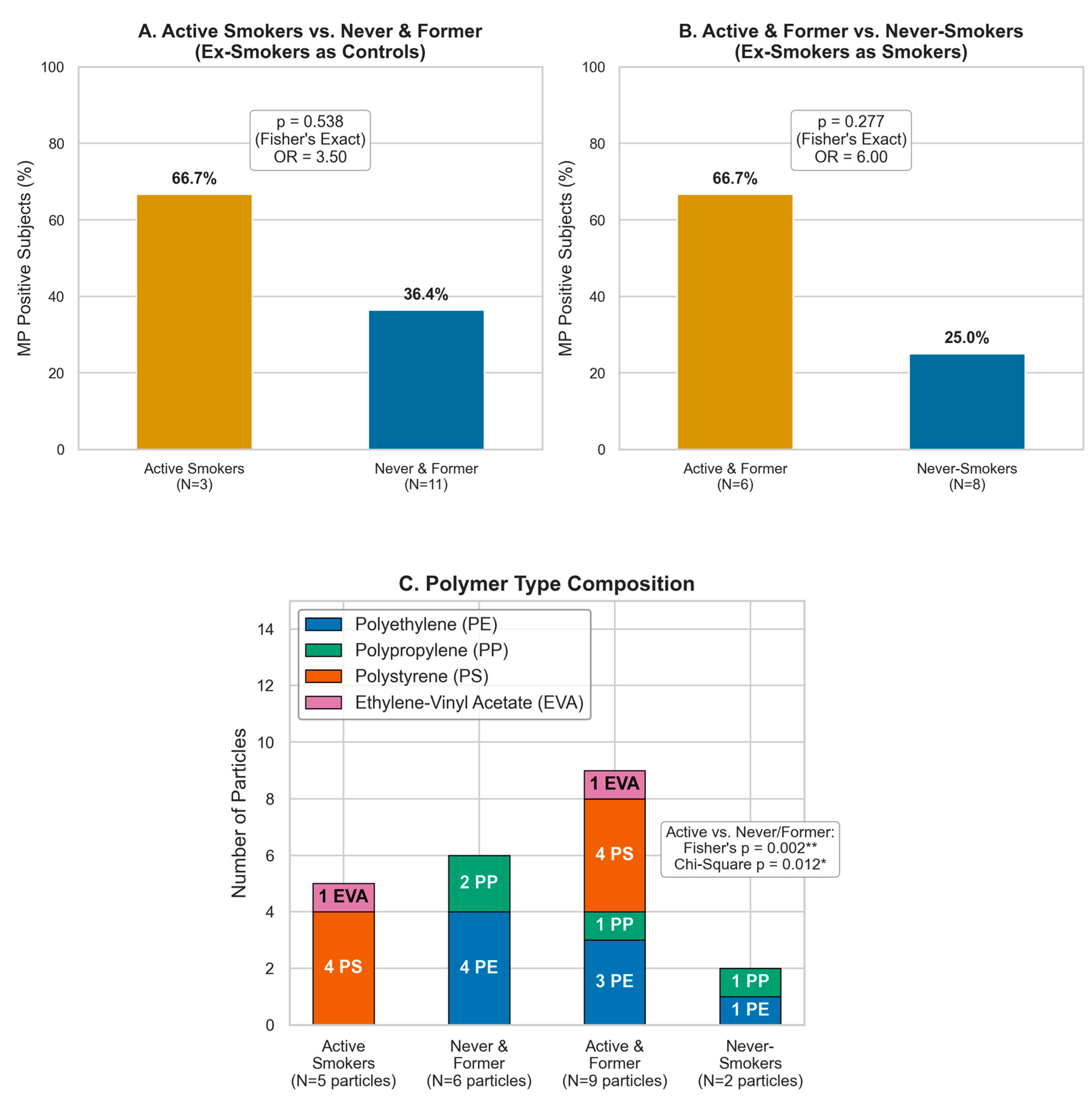
Figure 5.
Quantitative microplastic particle counts per subject stratified by smoking exposure. (A) Classification 1: Active Smokers (mean 1.67, var 4.33) vs. Never & Former (mean 0.55, var 0.85; Levene p = 0.048). (B) Classification 2: Active & Former (mean 1.50, var 2.70) vs. Never-Smokers (mean 0.25, var 0.21; Levene p = 0.005).
Figure 5.
Quantitative microplastic particle counts per subject stratified by smoking exposure. (A) Classification 1: Active Smokers (mean 1.67, var 4.33) vs. Never & Former (mean 0.55, var 0.85; Levene p = 0.048). (B) Classification 2: Active & Former (mean 1.50, var 2.70) vs. Never-Smokers (mean 0.25, var 0.21; Levene p = 0.005).
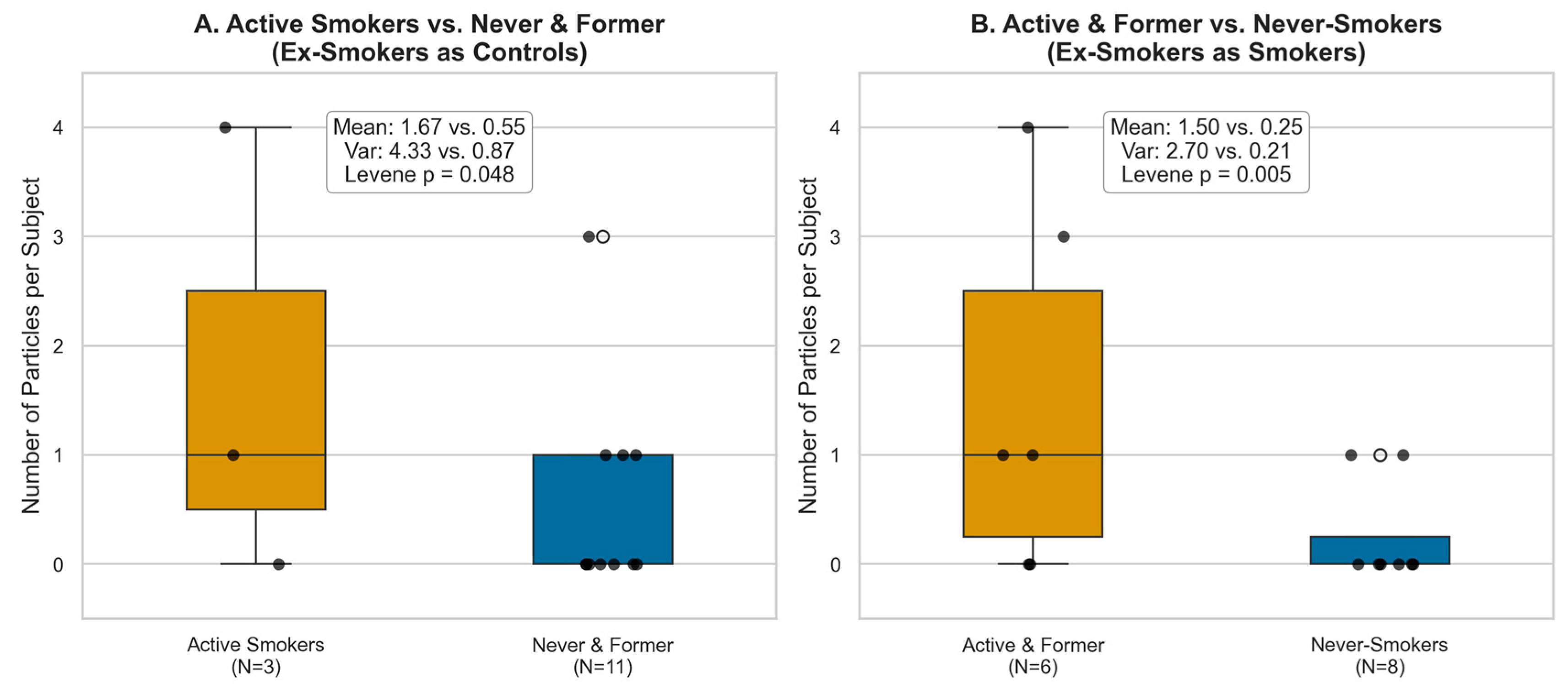
Figure 6.
Principal Component Analysis (PCA) patient mapping and agglomerative clustering (N=14 independent cohort) based on biological features (age, sex), smoking history, clinical group, and individual microplastic counts (PE, PP, PS, EVA). Convex hulls enclose the three distinct clinical clusters: Cluster A (Healthy Controls; blue), Cluster B (Female AMI Patients; orange), and Cluster C (Male AMI Patients; green). Markers indicate clinical groups: controls (circles) and AMI patients (squares).
Figure 6.
Principal Component Analysis (PCA) patient mapping and agglomerative clustering (N=14 independent cohort) based on biological features (age, sex), smoking history, clinical group, and individual microplastic counts (PE, PP, PS, EVA). Convex hulls enclose the three distinct clinical clusters: Cluster A (Healthy Controls; blue), Cluster B (Female AMI Patients; orange), and Cluster C (Male AMI Patients; green). Markers indicate clinical groups: controls (circles) and AMI patients (squares).
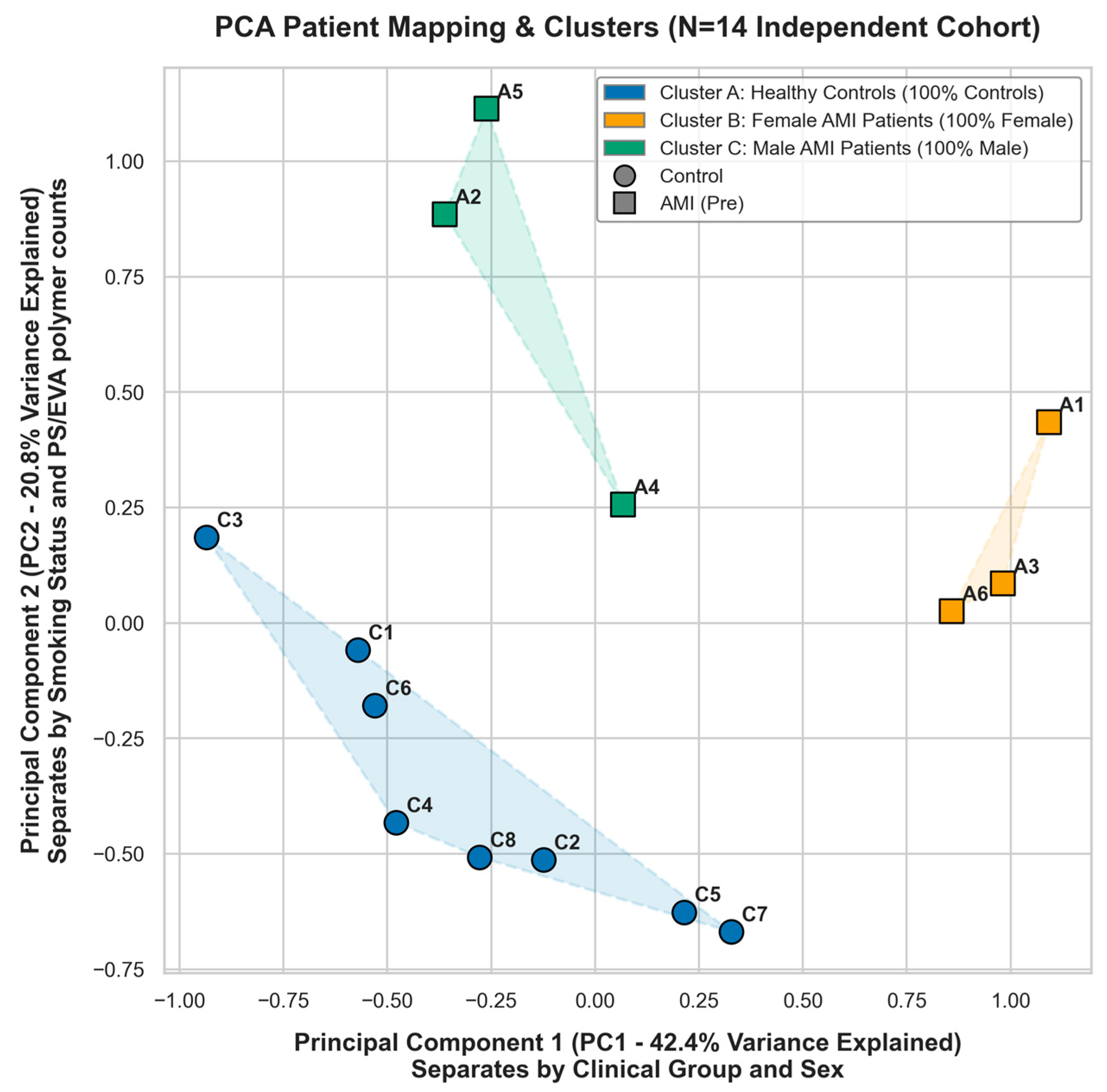
Table 1.
Individual clinical characteristics of independent subjects and detected microplastics. ND: Not Detected.
Table 1.
Individual clinical characteristics of independent subjects and detected microplastics. ND: Not Detected.
| ID | Group | Age | Smoking Status | Sex | Polymer Identified | No. Particles |
|---|---|---|---|---|---|---|
| C1 | Control | 30 | Non-smoker (Ex-smoker) | Male | PE | 1 |
| C2 | Control | 72 | Non-smoker | Male | PP | 1 |
| C3 | Control | 49 | Active Smoker | Male | PS | 4 |
| C4 | Control | 42 | Non-smoker | Male | ND | - |
| C5 | Control | 43 | Non-smoker | Female | ND | - |
| C6 | Control | 61 | Non-smoker (Ex-smoker) | Male | ND | - |
| C7 | Control | 64 | Non-smoker | Female | ND | - |
| C8 | Control | 79 | Non-smoker | Male | ND | - |
| A1 | AMI | 64 | Non-smoker (Ex-smoker) | Female | PE/PP | 2/1 |
| A2 | AMI | 34 | Active Smoker | Male | ND | - |
| A3 | AMI | 74 | Non-smoker | Female | PE | 1 |
| A4 | AMI | 56 | Non-smoker | Male | ND | - |
| A5 | AMI | 62 | Active Smoker | Male | EVA | 1 |
| A6 | AMI | 75 | Non-smoker | Female | ND | - |
Disclaimer/Publisher’s Note: The statements, opinions and data contained in all publications are solely those of the individual author(s) and contributor(s) and not of MDPI and/or the editor(s). MDPI and/or the editor(s) disclaim responsibility for any injury to people or property resulting from any ideas, methods, instructions or products referred to in the content. |
© 2026 by the authors. Licensee MDPI, Basel, Switzerland. This article is an open access article distributed under the terms and conditions of the Creative Commons Attribution (CC BY) license (http://creativecommons.org/licenses/by/4.0/).
Copyright: This open access article is published under a Creative Commons CC BY 4.0 license, which permit the free download, distribution, and reuse, provided that the author and preprint are cited in any reuse.


