

Submitted:
21 July 2026
Posted:
21 July 2026
You are already at the latest version
Abstract
Maternal psychological stress during pregnancy is linked to adverse birth outcomes, but the biochemical pathways involved remain poorly defined. We previously reported that the human placenta buffers the fetus from several effects of maternal perceived stress in a cohort of term pregnancies. Here, we extend that cohort with a broader steroid panel and, for the first time, a targeted bile acid metabolomic panel across four maternal–fetal compartments: maternal plasma, placental tissue, cord blood, and maternal hair. Nulliparous women were recruited during the third trimester at UCSF Mission Bay Hospital and stratified into Low- and High-Perceived Stress Scale (PSS) groups using the 10-item PSS. Steroid and bile acid metabolites were quantified by UPLC–MS/MS and compared between groups using nonparametric methods. Most steroid and bile acid metabolites did not differ significantly between High-PSS and Low-PSS groups, consistent with a broadly preserved placental metabolic barrier. However, three metabolites were selectively reduced in the High-PSS group: placental estrone (E1), cord blood glycochenodeoxycholic acid (GCDCA), and maternal plasma ursodeoxycholic acid (UDCA) (all p < 0.05), while estradiol and estriol concentrations were unaffected. Maternal hair showed poor con-cordance with plasma and placental compartments, suggesting limited value as a non-invasive proxy for these analytes in this setting. These findings reinforce the concept that placental buffering of maternal stress is broadly selective rather than absolute and identify E1, GCDCA, and UDCA as candidate stress-sensitive metabolites for validation in larger, longitudinal cohorts.
Keywords:
bile acids
; cord blood
; centering pregnancy
; estrone
; maternal hair
; mass spectrometry
1. Introduction
1.1. Maternal Stress & Pregnancy Outcomes
The placenta is a temporary organ that develops in the uterus during pregnancy. It connects the developing fetus via the umbilical cord and attaches to the uterine wall, facilitating nutrient exchange, oxygen transport, waste removal, and endocrine signaling between pregnant woman and fetus. In addition to its vascular functions, the placenta also serves as a complex endocrine and immune organ that supports fetal growth, regulates pregnancy-related physiology in females, and protects the fetus from harmful environmental exposures and infections. The human placenta takes over the primary production of progesterone and estrogen from the ovaries around the first 9 weeks of gestation. These include hormones such as estrone (E1), estradiol (E2), and estriol (E3), which play essential roles in maintaining pregnancy, regulating placental function, and supporting fetal development [1,2,3].
Although estrogens are frequently grouped in clinical and research settings, E1, E2, and E3 possess distinct biological roles and metabolic pathways. However, little is known about whether maternal stress selectively alters specific estrogen metabolites across maternal and fetal compartments. Progesterone and estrogen signaling are critical for maintaining a healthy pregnancy and supporting fetal development. Progesterone concentrations rise steadily throughout gestation and are essential for maintaining uterine quiescence, modulating maternal immune tolerance, and supporting placental function.
Longitudinal hormonal profiling has demonstrated that both progesterone and E2 increase substantially across pregnancy before rapidly declining postpartum, reflecting major endocrine adaptations required to sustain gestation [4]. Estrogen metabolites also increase throughout pregnancy and contribute to placental growth, fetal organ maturation, and preparation for labor. Dysregulation of these hormonal pathways has been associated with adverse pregnancy outcomes, including preterm birth and fetal growth restriction. Studies have identified distinct plasma metabolomic signatures in women with preeclampsia and preterm delivery, including disruptions in lipid metabolism and inflammatory pathways associated with long-term maternal cardiovascular risk [5]. Despite their biological importance, the relationship between maternal perceived stress and compartment-specific steroid metabolite profiles remains poorly understood.
Maternal psychological stress during pregnancy, driven by various factors like socioeconomic disparities, trauma, or physiological and psychosocial stressors, has been associated with adverse maternal and neonatal outcomes, including preterm birth, low birth weight, and altered neurodevelopmental trajectories in offspring. Psychological stress during pregnancy is common, with a substantial proportion of pregnant individuals reporting significant life stressors during gestation. Nearly three-quarters of pregnant individuals report experiencing at least one significant stressor in the year before delivery, highlighting the widespread prevalence of stress during pregnancy and the importance of understanding its biological consequences [6]. In metabolomic studies of pregnant individuals with depression, significant alterations in amino acid, lipid, and energy metabolism pathways have been observed during pregnancy and postpartum, suggesting that psychological stress may produce measurable biochemical changes across multiple metabolic systems [7]. To measure this stress, the Perceived Stress Scale (PSS) is used because it is the most widely used psychological instrument for measuring the perception of stress. Created by Sheldon Cohen and colleagues in 1983, it evaluates how unpredictable, uncontrollable, and overloaded individuals find their lives [8,9].
Although cortisol has frequently been used as a biomarker of stress during pregnancy, studies examining the relationship between cortisol, perceived stress, and adverse birth outcomes have produced inconsistent findings, suggesting that additional biomarkers may be needed to characterize stress-related physiological changes during pregnancy. The placenta plays a critical role in protecting the fetus from excessive maternal stress signaling by regulating the transfer and metabolism of glucocorticoids and steroid hormones. As part of the maternal–fetal stress response system, the placenta responds dynamically to maternal cortisol levels and contributes to the development of the fetal hypothalamic–pituitary–adrenal (HPA) axis. Excessive or chronic maternal stress may disrupt placental endocrine and metabolic pathways, potentially altering steroid hormone synthesis and fetal exposure to bioactive metabolites. However, the biochemical mechanisms linking maternal perceived stress to altered pregnancy outcomes remain incompletely understood. One of the primary biological pathways linking maternal stress to fetal outcomes involves activation of the HPA axis, which alters circulating endocrine and metabolic signaling during pregnancy. Activation of the maternal HPA axis increases glucocorticoid signaling and may alter downstream steroidogenesis and metabolic pathways within the placenta [10]. Because the placenta functions as a major endocrine organ throughout gestation, stress-associated hormonal changes may influence placental metabolism and fetal exposure to bioactive metabolites. The placenta naturally acts as a protective barrier that shields the fetus from excess maternal hormones, functioning not just as a physical interface but as an active biochemical regulator of fetal exposure to stress-associated metabolites. Chronic or excessive maternal stress can compromise this buffering capacity, potentially altering fetal brain development, endocrine regulation, and stress reactivity later in life.
1.2. Centering Pregnancy vs. Individual Prenatal Care
Centering Pregnancy is a group-based prenatal care model that replaces traditional 15-minute doctor visits with 90-minute to 2-hour group sessions. Groups of 8-12 expectant women with similar gestational ages meet with healthcare providers throughout their pregnancy to check vitals, receive prenatal education, and receive peer support. This is different from the traditional one-on-one appointments. Centering Pregnancy has been associated in some studies with improved prenatal education, increased patient engagement, and reduced risk of adverse birth outcomes, potentially through enhanced social support and stress reduction [11]. UCSF Mission Bay Hospital offered both Centering and individual prenatal care; participants came from both settings.
1.3. Bile Acids in Pregnancy and the Study Aim
In addition to steroid hormones, bile acids have emerged as important signaling molecules during pregnancy. Bile acids are synthesized in the liver from cholesterol and play central roles in fat digestion, gut signaling, and metabolic regulation. Primary bile acids such as cholic acid and chenodeoxycholic acid also act as signaling molecules through receptors, including FXR and TGR5, which regulate glucose homeostasis, inflammation, and lipid metabolism [12]. Beyond their role in lipid digestion, bile acids also function as signaling molecules involved in inflammation, glucose metabolism, and endocrine regulation. Primary bile acids, including cholic acid (CA) and chenodeoxycholic acid (CDCA), are synthesized in the liver from cholesterol and may be converted into secondary bile acids through intestinal microbial metabolism. Conjugated bile acids, including glycochenodeoxycholic acid (GCDCA) and ursodeoxycholic acid (UDCA), participate in lipid digestion and function as signaling molecules involved in metabolic and inflammatory regulation.
Altered bile acid homeostasis during pregnancy has been associated with adverse maternal and fetal outcomes, yet its relationship with maternal psychological stress remains poorly understood. In pregnancy, bile acid metabolism shifts significantly; elevated bile acids are the hallmark of intrahepatic cholestasis of pregnancy (ICP), associated with preterm birth and stillbirth. Certain bile acids, including ursodeoxycholic acid (UDCA), have also been investigated therapeutically in ICP because of their anti-inflammatory and hepatoprotective effects. Studies have reported that UDCA therapy reduced serum bile acid concentrations and improved liver function markers in patients with intrahepatic cholestasis of pregnancy [13]. Systematic metabolomic analyses of ICP pregnancies have demonstrated widespread alterations in both bile acid and steroid metabolic pathways, suggesting close biochemical interactions between hepatic function and endocrine regulation during pregnancy [14]. Despite growing recognition of the importance of bile acid signaling during pregnancy, the extent to which maternal stress influences bile acid metabolism across maternal and fetal compartments remains largely unexplored.
Although maternal stress has been associated with adverse pregnancy outcomes, little is known about how perceived stress alters compartment-specific steroid and bile acid metabolism across the maternal–fetal interface. Most prior studies have focused on isolated hormones or single biospecimens, leaving the broader metabolomic response to maternal stress poorly characterized. Previous metabolomic investigations in pregnancy complications have primarily relied on maternal plasma samples alone, with limited studies simultaneously evaluating maternal, placental, fetal, and noninvasive biospecimens across the maternal–fetal interface [5,7]. This paper provides the first systematic description of bile acid metabolites across all four compartments: maternal plasma, cord blood, placental tissue, and maternal hair in a cohort of term pregnancies. We further evaluated whether maternal perceived stress, measured using the PSS-10 questionnaire, was associated with alterations in specific metabolites across maternal and fetal compartments. Finally, we explored whether maternal hair metabolites reliably reflected plasma metabolite levels as a potential noninvasive biomarker approach. By integrating steroid and bile acid metabolomics across multiple maternal–fetal compartments, this study seeks to provide new insight into the biochemical pathways associated with maternal perceived stress during pregnancy. Simultaneous analysis of maternal plasma, cord blood, placental tissue, and maternal hair may provide insight into how stress-associated metabolic changes differ between maternal and fetal environments
2. Materials and Methods
2.1. Study Participants
Nulliparous pregnant women (age ≥18, nonsmokers) were recruited during their third trimester at UCSF Mission Bay Hospital under UCSF IRB protocol #16-10957 [3]. Inclusion criteria were age ≥18, English or Spanish fluency, and consent for questionnaires and tissue collection. Exclusion criteria included any history of adrenal or endocrine disease, use of exogenous steroids (e.g., prednisone), smoking, or cognitive impairment. Perceived stress was measured using the 10-item Perceived Stress Scale (PSS-10) during the clinic visit, and sociodemographic data (age, education, income, etc.) were collected via a self-report questionnaire. Of 52 women who consented, 17 completed the study procedures (including the PSS questionnaire); final biospecimen availability was placenta (n=12), maternal hair (n=7), cord plasma (n=6), and maternal plasma (n=6). All 17 participants had their birth weight and gestational age recorded from medical records [3].
2.2. Biospecimen Collection
At the third-trimester clinic visit, a small hair sample (~1–3 cm in length) was cut from the posterior scalp and stored in a foil-wrapped bag at room temperature. None of the women had recent hair treatments. At delivery, placentas were kept at 4 °C and collected within 2–12 hours postpartum. Maternal peripheral blood and umbilical cord blood were drawn into EDTA tubes at delivery and processed to plasma within 12 hours. All collected samples (plasma, tissue, hair) were aliquoted and stored at –80 °C until analysis.
2.3. Metabolite Analysis
Metabolomic profiling included targeted quantification of steroid hormones and bile acids by ultra-performance liquid chromatography–tandem mass spectrometry. In brief, ~15 mg of placental tissue (or equivalent plasma/hair sample) was homogenized in an antioxidant solution (0.2 mg/mL BHT/EDTA in 1:1 methanol: water), centrifuged, and subjected to serial solvent washes. Extracts were lyophilized and reconstituted in 100 μL of a methanol/acetonitrile (50:50) solvent containing phenyl-hexadecanoic acid/urate (PHAU/CUDA) as internal standards. Tritiated steroids were added to monitor HPLC retention and recovery, and deuterated steroid analogs were spiked into each sample to calibrate quantification. Concentrations were calculated from the peak area ratio to the corresponding deuterated internal standard and normalized to tissue weight (pg/mg) or plasma volume (ng/mL) as described before [3].
For bile acids, a parallel UPLC-MS/MS assay was used to quantify a targeted panel of conjugated bile acids. Analytes were detected by negative-ion MRM on the same mass spectrometer. Calibration curves were generated using authentic bile acid standards (e.g., cholic acid, chenodeoxycholic acid, glycochenodeoxycholic acid, ursodeoxycholic acid, etc.), and concentrations in samples were calculated relative to internal standards of known quantity.
2.4. Statistical Analysis
Data was analyzed using Aseesa Stars 2.0 (https://www.aseesa.com/). Due to the small sample size and non-normal metabolite distributions, nonparametric statistical analyses were performed. Mann–Whitney U tests were used to compare metabolite concentrations between low- and high-PSS groups. Spearman correlation analyses were used to evaluate associations between metabolite levels and continuous clinical variables, including PSS score, gestational age, and birth weight. Statistical significance was defined as p < 0.05.
3. Results
3.1. Centering vs. Individual Care: PSS Was Not Different
Because Centering Pregnancy has previously been associated with reduced stress and improved psychological support, we first evaluated whether perceived stress levels differed between prenatal care models in this cohort. Determining whether prenatal care type influenced PSS scores was important to assess whether subsequent metabolomic analyses should be stratified by care model or by perceived stress alone. Participant demographic and clinical characteristics are summarized in Table 1. The cohort included nulliparous women receiving either Centering Pregnancy group prenatal care or traditional individual prenatal care at UCSF Mission Bay Hospital. Clinical variables collected from medical records included gestational age at delivery, maternal body mass index (BMI), infant birth weight, and delivery mode.
Mean PSS scores did not significantly differ between participants enrolled in Centering Pregnancy and those receiving individual prenatal care (Centering: mean PSS ~12.75; Individual care: mean PSS ~11.85; p = n.s.) (Figure 2A). Due to the small number of participants enrolled in the Centering Pregnancy cohort (n = 4), this comparison was considered exploratory and insufficiently powered for definitive statistical interpretation.
Because perceived stress scores were comparable between prenatal care models, all subsequent analyses were stratified by perceived stress status rather than prenatal care type. Participants were divided into Low-PSS (≤11) and High-PSS (≥12) groups using cohort-based PSS thresholds as shown in Figure 2B, resulting in equal group sizes (Low PSS, n = 8; High PSS, n = 8). Sample sizes differed across individual metabolite analyses because not all participants contributed every biospecimen type.
3.2. Overview of Metabolite in Four Tissues
Before evaluating stress-associated metabolomic differences, it was necessary to determine which steroid and bile acid metabolites were reliably detected across maternal and fetal samples. This study characterized steroid and bile acid metabolites across four maternal–fetal compartments: maternal plasma, cord blood, placental tissue, and maternal hair. In the steroid metabolite panel, 40.0% (12/30) of steroid metabolites were present above the limit of detection (LOD) levels in at least one biospecimen (Table 2). Cord blood and maternal plasma each contained 33.3% (10/30) detectable steroid metabolites, whereas placental tissue contained 30.0% (9/30) detectable steroid metabolites. In contrast, only 3.3% of steroid metabolites (1/30) were above the LOD in maternal hair samples.
In the bile acid panel, 72% (18/23) of metabolites exceeded the LOD in at least one compartment (Table 3). Maternal plasma demonstrated the broadest bile acid detectability, with 60% (14/23) of bile acids above LOD, followed by cord blood and placental tissue with 48% (11/23) detectable bile acids each. Maternal hair demonstrated lower analyte recovery, with only 30% (7/23) of bile acids above LOD. Overall, placental tissue and maternal plasma exhibited the greatest metabolite detectability across both steroid and bile acid panels, whereas maternal hair showed markedly reduced analyte recovery.
3.3. PSS-Associated Metabolite Changes
To identify metabolites potentially associated with maternal perceived stress, we compared metabolite abundance between High-PSS and Low-PSS pregnancies across all maternal and fetal compartments. This analysis aimed to determine whether maternal stress was associated with compartment-specific alterations in steroid or bile acid metabolism. To evaluate global metabolomic differences associated with maternal perceived stress, volcano plot analysis was performed to compare the High-PSS and Low-PSS groups across all detectable metabolites (Figure 3). Three metabolites exceeded the nominal significance threshold (p < 0.05): placental estrone (E1), cord blood glycochenodeoxycholic acid (GCDCA), and maternal plasma ursodeoxycholic acid (UDCA). All three metabolites were lower in the High-PSS group than in the Low-PSS group.
Placental estrone was significantly reduced in participants with higher perceived stress scores. Cord blood GCDCA and maternal plasma UDCA concentrations were also significantly lower in the High-PSS group. Although cord blood 17-OH pregnenolone (CORDB~17OH_Plen) is above the significance threshold, it was excluded from differential analyses because concentrations were below the limit of detection in most samples. A pie chart visualization highlighting significant metabolites and relative abundance differences between Low- and High-PSS groups is shown in Figure 4.
3.4. Estrone (E1), Not Estradiol (E2), Is the Stress-Sensitive Estrogen
The three principal estrogens during pregnancy are E1, E2, and E3. Because estrogens are often evaluated collectively despite possessing distinct biological functions, we next examined whether specific estrogen metabolites demonstrated differential associations with maternal perceived stress. A key finding of this study was the significant reduction in placental E1 concentrations among participants with higher perceived stress scores (p < 0.05) (Figure 5). In contrast, neither E2 nor E3 demonstrated significant PSS-associated differences in any maternal or fetal compartment.
3.5. Bile Acid Profiles and PSS-Associated Changes
Given the emerging role of bile acids as signaling molecules involved in metabolic and inflammatory regulation during pregnancy, we evaluated whether maternal perceived stress was associated with alterations in bile acid profiles across maternal and fetal compartments. This analysis aimed to explore whether stress-related metabolic changes extended beyond steroid hormones alone. This study characterized bile acid metabolite profiles across maternal plasma, placental tissue, cord blood, and maternal hair in relation to maternal perceived stress. Bile acid concentrations and metabolite composition differed substantially across maternal and fetal compartments, suggesting compartment-specific regulation of bile acid metabolism during pregnancy.
Two bile acids demonstrated significant PSS-associated differences consistent with the volcano plot analysis: glycochenodeoxycholic acid (GCDCA) in cord blood and ursodeoxycholic acid (UDCA) in maternal plasma. Cord blood GCDCA concentrations and maternal plasma UDCA concentrations were reduced in participants with higher perceived stress scores (Figure 6).
3.6. Hair Is Not a Reliable Proxy for Plasma Metabolite Levels
Because maternal hair has been proposed as a noninvasive biomarker of long-term hormonal exposure [15], we evaluated whether metabolite concentrations detected in hair reflected those observed in maternal plasma. Determining the agreement between hair and plasma metabolite profiles is important for assessing the feasibility of hair-based metabolomics in pregnancy research. In this study, the majority of steroid and bile acid metabolites detectable in plasma or placental tissue were below the limit of detection in maternal hair samples. Furthermore, among metabolites measurable in hair, directional agreement with plasma concentrations was inconsistent. Metabolites elevated in maternal plasma were not necessarily elevated in hair, suggesting that metabolite incorporation into hair may depend on analyte-specific biochemical properties rather than directly reflecting circulating concentrations. For several detectable metabolites, including GCDCA and UDCA, directional trends in maternal hair did not consistently mirror those observed in plasma or placental tissue (Figure 7).
4. Discussion
4.1. The Placenta Continues to Buffer
Maternal psychological stress during pregnancy has been associated with adverse maternal and neonatal outcomes, including preterm birth, low birth weight, and altered neurodevelopmental trajectories in offspring. Previous metabolomic studies have demonstrated that psychological distress during pregnancy is associated with measurable alterations in lipid, amino acid, and inflammatory metabolic pathways [3,7]. Because the placenta functions as the primary endocrine and metabolic interface between the mother and fetus, understanding how maternal stress alters placental metabolism is essential for identifying biological pathways associated with these outcomes. In the present study, most steroid and bile acid metabolites remained relatively stable across low- and high-PSS groups, suggesting that healthy term placentas may effectively maintain endocrine and metabolic homeostasis despite maternal psychological stress exposure. These findings are consistent with prior work proposing that the placenta serves as a protective biochemical buffer that regulates fetal exposure to maternal stress-associated hormones and metabolites [3].
Our earlier work in this same cohort first proposed that the human placenta buffers the fetus from many effects of maternal perceived stress [3], and the present findings, drawn from a different set of metabolites in the same participants, reinforce that conclusion rather than standing apart from it. The placenta has no real analog elsewhere in human physiology: it is built anew with each pregnancy, discarded within minutes of delivery, and yet for nine months carries out the combined work of a maternal–fetal lung, gut, kidney, and endocrine gland. Its buffering role is also not specific to psychological stress. The same transport and metabolic machinery that governs fetal hormone exposure is also responsible for limiting fetal exposure to circulating xenobiotics and environmental toxicants, and disruption of placental transport and metabolic pathways has been implicated in adverse outcomes following maternal exposure to air pollution and other chemical stressors [21]. Read alongside our prior report, the present results support a broader view in which a healthy placenta acts as a general-purpose buffer against maternal stressors of different kinds, psychological and chemical alike, and in which the integrity of that buffering capacity, rather than the stressor itself, may be what most directly shapes fetal outcomes.
Importantly, however, placental E1, cord blood GCDCA, and maternal plasma UDCA demonstrated significant PSS-associated changes, indicating that placental buffering is not absolute and may occur selectively across specific metabolic pathways. Rather than broad dysregulation of steroidogenesis or bile acid metabolism, maternal perceived stress appeared to influence a limited subset of metabolites. This selective pattern may reflect compensatory placental regulatory mechanisms that preserve overall hormonal balance while allowing subtle alterations in stress-sensitive pathways. Prior studies in both humans and animal models suggest that relative metabolite balance may be more biologically important than absolute metabolite concentrations during pregnancy [16]. Our findings support this concept and suggest that healthy pregnancies may tolerate moderate maternal stress exposure while maintaining overall fetal endocrine stability.
These findings may also help explain why adverse birth outcomes are not uniformly observed in all pregnancies affected by maternal stress. Even in the presence of measurable maternal psychological stress, placental metabolic regulation may partially protect the fetus from large-scale endocrine disruption. At the same time, selective alterations in metabolites such as E1, GCDCA, and UDCA may represent early biochemical indicators of stress-associated physiological adaptation that could become more pronounced in pregnancies complicated by placental dysfunction, preterm birth, or metabolic disease. Distinct metabolomic alterations have previously been reported in pregnancies complicated by preeclampsia and preterm delivery, supporting the possibility that metabolic dysregulation becomes more evident in pathologic pregnancies [5]. Together, these findings suggest that maternal perceived stress may influence highly specific metabolic pathways rather than causing broad endocrine disruption during healthy term pregnancy.
4.2. Why E1 and Not E2/E3?
One of the most unexpected findings in this study was the selective reduction in placental E1 in participants with higher perceived stress scores, whereas E2 and E3 did not differ significantly between PSS groups. Most prior studies investigating estrogen signaling during pregnancy have focused primarily on E2 or have grouped estrogens together rather than examining individual estrogen metabolites separately. As a result, the possibility that maternal stress may selectively alter E1 rather than E2 has received little attention.
Although E2 is generally considered the dominant biologically active circulating estrogen in nonpregnant women, E1 possesses distinct metabolic and physiological roles. During pregnancy, placental steroidogenesis involves dynamic interconversion between E1 and E2 through the activity of hydroxysteroid 17-beta dehydrogenase enzymes, including HSD17β1 [17]. Maternal stress-associated alterations in placental enzyme activity could therefore shift the balance between E1 and E2 production without necessarily altering total estrogen abundance. This possibility is biologically plausible given that glucocorticoid signaling has been shown to influence placental steroidogenic pathways and endocrine regulation.
Selective depletion of E1 may have important implications for maternal and fetal physiology. Estrogens contribute to placental vascular function, cervical remodeling, fetal organ maturation, and preparation for labor [4]. If maternal stress preferentially alters E1 metabolism, these changes could affect downstream estrogen-sensitive processes in ways that would not be detected when measuring only E2 or total estrogen levels. Interestingly, the directionality of E1 changes differed between placental and fetal compartments, with placental E1 reduced in high-PSS pregnancies while cord blood E1 appeared directionally increased. Although the number of matched maternal–fetal samples was too small for meaningful statistical analysis, this pattern raises the possibility that maternal stress may differentially influence placental versus fetal estrogen regulation.
These findings also highlight the importance of modern mass spectrometry approaches capable of measuring steroid metabolites individually rather than grouping hormones together. Earlier immunoassay-based studies often lacked the specificity required to distinguish compartment-specific estrogen alterations [18]. Our results suggest that future pregnancy metabolomics studies should evaluate E1, E2, and E3 separately, particularly in the context of maternal stress and placental function.
4.3. Bile Acids (Novel Territory)
In addition to steroid hormones, this study characterized bile acid metabolites across maternal plasma, placental tissue, cord blood, and maternal hair in relation to maternal perceived stress. To our knowledge, this represents one of the first studies to simultaneously evaluate bile acid profiles across multiple maternal–fetal compartments in the context of maternal psychological stress. Bile acids are increasingly recognized not only as regulators of lipid digestion but also as signaling molecules involved in inflammation, glucose metabolism, liver function, and endocrine regulation. Bile acids exert many of these regulatory effects through signaling receptors, including FXR and TGR5, which influence glucose homeostasis, inflammatory signaling, and lipid metabolism [12]. Despite growing interest in bile acid metabolism during pregnancy, little is known about how maternal stress may influence bile acid homeostasis across maternal and fetal tissues.
Two bile acids demonstrated significant PSS-associated changes in this cohort: glycochenodeoxycholic acid (GCDCA) in cord blood and ursodeoxycholic acid (UDCA) in maternal plasma. GCDCA is a conjugated primary bile acid involved in hepatic cholesterol metabolism and bile acid transport. Reduced cord blood GCDCA levels in high-PSS pregnancies may reflect altered maternal–fetal bile acid transfer or stress-associated changes in placental bile acid regulation [19]. This is consistent with evidence from a pregnant swine showing that placental bile acid handling normally limits transfer of maternal bile acids into the fetal compartment, implying that fetal bile acid levels depend as much on placental transport capacity as on maternal supply [20]. One plausible route by which psychological stress could alter that capacity is inflammatory signaling: in a pregnant rodent model, acute systemic inflammation downregulated placental expression of several bile acid and drug transporters, altering maternal-to-fetal disposition of endogenous compounds [21]. If a similar inflammatory pathway operates in chronically stressed human pregnancies, it will offer a testable mechanistic link between perceived stress and the reduced cord blood GCDCA observed here, though this remains speculative until directly examined. Because bile acids can cross the placental interface, alterations in placental transport mechanisms may contribute to compartment-specific bile acid accumulation or depletion. Altered fetal bile acid exposure may be biologically important because bile acids function not only in hepatic metabolism but also as signaling molecules involved in inflammatory regulation, glucose homeostasis, and cellular stress responses. Comprehensive bile acid profiling studies have shown that bile acid dysregulation in pregnancy is associated with widespread metabolic and endocrine pathway alterations [14]. Consequently, even modest stress-associated changes in bile acid composition could influence fetal metabolic programming or placental inflammatory signaling.
The observed reduction in maternal plasma UDCA is particularly interesting given the established anti-inflammatory and hepatoprotective properties of UDCA. Beyond its role in bile flow, UDCA modulates inflammatory and immune signaling through several distinct mechanisms, including antioxidant, anti-apoptotic, endoplasmic-reticulum stress-relieving, and direct immunomodulatory effects, which together make it a plausible buffer against stress-associated inflammatory changes rather than a purely hepatic actor [22]. Clinically, UDCA is widely used as a therapeutic treatment for intrahepatic cholestasis of pregnancy, where it helps reduce maternal bile acid accumulation and improve fetal outcomes [13]. Lower UDCA concentrations in participants with higher perceived stress scores may therefore indicate stress-associated disruption of protective bile acid regulatory pathways. Psychological stress has previously been linked to inflammatory and metabolic dysregulation, including altered liver-associated signaling pathways, making stress–bile acid interaction biologically plausible. Metabolomic analyses of pregnant individuals with depression have identified disruptions in multiple metabolic and inflammatory pathways associated with psychological stress exposure [3,7].
The identification of stress-associated bile acid alterations across maternal and fetal compartments provides preliminary evidence that maternal psychological stress may influence pathways beyond classical glucocorticoid signaling.
4.4. Hair: A Cautionary Biomarker Tale
Maternal hair has increasingly been used as a proposed noninvasive biomarker of cumulative hormonal exposure during pregnancy because hair growth may reflect long-term metabolite incorporation over weeks to months [15]. However, the validity of hair as a surrogate for circulating steroid and bile acid levels remains incompletely understood and is often assumed rather than experimentally validated for individual analytes. These detection patterns (Section 3.6) point to a basic mismatch between what hair and plasma actually measure, not simply a sensitivity problem. If hair reliably tracked circulating levels, metabolites abundant in plasma would also turn up in hair, and the direction of change would match. Neither was consistently true in this cohort.
Several biological factors may explain these discrepancies, including differences in metabolite incorporation into the hair shaft, metabolite degradation over time, keratin binding affinity, and differences in the biological time windows represented by plasma versus hair samples [23]. Plasma metabolites reflect relatively acute physiological states, whereas hair may capture longer-term cumulative exposure. As a result, these tissues may provide fundamentally different biological information rather than interchangeable measures of the same process. These findings highlight the importance of validating tissue-specific biomarker relationships rather than assuming direct correspondence between circulating and hair metabolite levels. Although hair-based metabolomics remains promising as a noninvasive biomarker strategy, our results suggest that analyte-specific validation is necessary before hair measurements can reliably substitute for plasma-based metabolite assessment during pregnancy.
4.5. Limitations
Several limitations should be considered when interpreting the findings of this study, and most stem from a constraint that is characteristic of this area of research rather than specific to our design: simultaneously collecting matched maternal plasma, hair, placental tissue, and cord blood from term pregnancies is logistically demanding, and cohorts with all four compartments represented remain uncommon in the literature. The overall cohort size was small, and biospecimen availability differed across compartments, resulting in unequal subgroup sizes for several analyses. This limited statistical power and reduced the ability to detect smaller stress-associated metabolite differences. We view this as a reason for caution in how far we extend biological interpretation of any single effect size, not as a reason to set the findings aside: three metabolites converged on the same direction of effect across two distinct chemical classes (steroids and bile acids) and three separate compartments, a pattern less likely to arise from noise alone in a dataset of this size. Second, this study used a cross-sectional design with sample collection occurring at delivery, preventing longitudinal assessment of changes in perceived stress and metabolite dynamics throughout gestation. Maternal stress exposure likely fluctuates during pregnancy, and a single third-trimester PSS measurement may not fully capture cumulative psychological stress burden.
An additional limitation is the lack of well-established reference ranges for postpartum placental E1 concentrations in healthy pregnancies. Although placental E1 was significantly lower in participants with higher perceived stress scores, limited prior literature characterizing compartment-specific postpartum E1 dynamics makes interpretation of the biological directionality uncertain. Consequently, it remains unclear whether reduced placental E1 reflects altered placental synthesis, increased fetal transfer, compensatory metabolism, or other stress-associated regulatory mechanisms. Larger studies establishing normative compartment-specific estrogen profiles across pregnancy and delivery are needed to better contextualize these findings.
These constraints call for restraint in how far we extend biological interpretation, but they are not, in our view, a reason to set the findings aside. Given how rarely all four maternal-fetal compartments are sampled in the same cohort, even a modest, well-characterized signal has value in pointing toward where larger studies should look. This study provides a novel multi-compartment characterization of steroid and bile acid metabolites in relation to maternal perceived stress and identifies three candidate stress-sensitive metabolites, placental E1, cord blood GCDCA, and maternal plasma UDCA, that warrant targeted follow-up in larger, longitudinal cohorts.
5. Conclusion
This study provides the first integrated characterization of steroid and bile acid metabolites across maternal plasma, placental tissue, cord blood, and maternal hair in relation to maternal perceived stress during term pregnancy. Although most metabolites remained stable across PSS groups, selective alterations in placental E1, cord blood GCDCA, and maternal plasma UDCA suggest that maternal stress may influence specific endocrine and metabolic pathways despite overall preservation of placental buffering. Together with our earlier report in this same cohort [3], these results reinforce a consistent picture: the human placenta is a uniquely capable buffering organ, one built anew each pregnancy to moderate fetal exposure not only to maternal psychological stress but, as the broader chemical-exposure literature suggests, to a range of environmental and toxicant stressors as well [24]. Placental health, in this framework, is not simply a marker of fetal growth but an active determinant of how much of the mother’s internal and external environment reaches the fetus at all. These findings support the concept that the placenta functions as an active metabolic regulator during pregnancy while also identifying potentially stress-sensitive metabolites that require further investigation. Larger longitudinal studies are needed to determine whether these metabolite alterations contribute to adverse pregnancy outcomes or may serve as biomarkers of maternal stress-related physiological adaptation.
Author Contributions
Conceptualization, A.B. and J.L.; methodology, A.B. and J.L.; formal analysis, R.G.J AND A.B.; investigation, A.B. and J.L; resources, A.B.; data curation, A.B.; writing—original draft preparation, R.G.J.; writing—review and editing, R.G.J. and A.B.; supervision, A.B.; project administration, A.B.; funding acquisition, A.B. All authors have read and agreed to the published version of the manuscript.
Funding
This work was supported by UCSF’s Research Allocation Program grants.
Institutional Review Board Statement
The study was conducted according to the guidelines of the Declaration of Helsinki, and approved by UCSF’s Institutional Review Board protocol # 16-10957.
Informed Consent Statement
Informed consent was obtained from all subjects involved in the study.
Data Availability Statement
Data is contained within the article. De-identified individual level data can be requested from the corresponding author and will be made available as per IRB approvals.
Acknowledgments
The authors thank Dr. Burcu Hasdemir and wish to thank the UCSF Mission Bay Hospital Obstetrics clinic for help with identifying and distributing consent forms to participants.
Conflicts of Interest
The authors declare no conflicts of interest. The funders had no role in the design of the study; in the collection, analyses, or interpretation of data; in the writing of the manuscript; or in the decision to publish the results.
Abbreviations
The following abbreviations are used in this manuscript:
| E1 | Estrone |
| E2 | Estradiol |
| E3 | Estriol |
| HPA axis | Hypothalamic-pituitary-adrenal axis |
| CA | Cholic acid |
| CDCA | Chenodeoxycholic acid |
| GCDCA | Glycochenodeoxycholic acid |
| UDCA | Ursodeoxycholic acid |
| ICP | Intrahepatic cholestasis of pregnancy |
| IRB | Institutional Review Board |
| HPLC | High-Performance Liquid Chromatography |
| UPLC-MS/MS | Ultra-Performance Liquid Chromatography with Tandem Mass Spectrometry |
| PSS | Perceived Stress Scale |
| BMI | Body Mass Index |
| LOD | Limit of Detection |
| EDTA | Ethylenediaminetetraacetic Acid |
| BHT | Butylated Hydroxytoluene |
| MRM | Multiple Reaction Monitoring |
| FXR | Farnesoid X Receptor |
| TGR5 | Takeda G-protein-coupled Bile Acid Receptor 5 (also known as GPBAR1) |
| HSD17β1 | Hydroxysteroid 17-beta Dehydrogenase 1 |
| PHAU | Phenylhexadecanoic Acid |
| CUDA | 12-(3-Cyclohexylureido)dodecanoic Acid |
| CORDB | Cord Blood |
| PLACT | Placenta |
| MATB | Maternal Blood |
References
- Berkane, N.; Ansaldi, Y.; Pluchino, N. The Role of Estrogens in Pregnancy.; Cambridge University Press: 2022; pp. 42-49.
- Fuentes, N.; Silveyra, P. Estrogen receptor signaling mechanisms. Adv Protein Chem Struct Biol 2019, 116, 135-170. [CrossRef]
- Vuppaladhadiam, L.; Lager, J.; Fiehn, O.; Weiss, S.; Chesney, M.; Hasdemir, B.; Bhargava, A. Human Placenta Buffers the Fetus from Adverse Effects of Perceived Maternal Stress. Cells 2021, 10. [CrossRef]
- Dukic, J.; Johann, A.; Henninger, M.; Ehlert, U. Estradiol and progesterone from pregnancy to postpartum: a longitudinal latent class analysis. Front Glob Womens Health 2024, 5, 1428494. [CrossRef]
- Hong, X.; Zhang, B.; Liang, L.; Zhang, Y.; Ji, Y.; Wang, G.; Ji, H.; Clish, C.B.; Burd, I.; Pearson, C.; et al. Postpartum plasma metabolomic profile among women with preeclampsia and preterm delivery: implications for long-term health. BMC Med 2020, 18, 277. [CrossRef]
- Burns, E.R.; Farr, S.L.; Howards, P.P.; Control, C.f.D.; Prevention. Stressful life events experienced by women in the year before their infants’ births—United States, 2000–2010. MMWR Morb Mortal Wkly Rep 2015, 64, 247-251.
- Yu, Z.; Matsukawa, N.; Saigusa, D.; Motoike, I.N.; Ono, C.; Okamura, Y.; Onuma, T.; Takahashi, Y.; Sakai, M.; Kudo, H.; et al. Plasma metabolic disturbances during pregnancy and postpartum in women with depression. iScience 2022, 25, 105666. [CrossRef]
- Cohen, S.; Kamarck, T.; Mermelstein, R. A global measure of perceived stress. J Health Soc Behav 1983, 24, 385-396.
- Harris, K.M.; Gaffey, A.E.; Schwartz, J.E.; Krantz, D.S.; Burg, M.M. The Perceived Stress Scale as a Measure of Stress: Decomposing Score Variance in Longitudinal Behavioral Medicine Studies. Ann Behav Med 2023, 57, 846-854. [CrossRef]
- Volqvartz, T.; Andersen, H.H.B.; Pedersen, L.H.; Larsen, A. Obesity in pregnancy-Long-term effects on offspring hypothalamic-pituitary-adrenal axis and associations with placental cortisol metabolism: A systematic review. Eur J Neurosci 2023, 58, 4393-4422. [CrossRef]
- Park, C.H.; Driver, N.; Richards, R.C., Jr.; Ward, P. The Effects of CenteringPregnancy: A Quasi-Experimental Evaluation. Healthcare (Basel) 2025, 13. [CrossRef]
- Liu, Y.; Rong, Z.; Xiang, D.; Zhang, C.; Liu, D. Detection technologies and metabolic profiling of bile acids: a comprehensive review. Lipids Health Dis 2018, 17, 121. [CrossRef]
- Meng, L.J.; Reyes, H.; Axelson, M.; Palma, J.; Hernandez, I.; Ribalta, J.; Sjovall, J. Progesterone metabolites and bile acids in serum of patients with intrahepatic cholestasis of pregnancy: effect of ursodeoxycholic acid therapy. Hepatology 1997, 26, 1573-1579. [CrossRef]
- Xu, H.; Xu, Y.; Zhao, G.; Fu, X.; Zhao, J.; Wang, H.; Cai, Y.; Lin, H. The complete change in bile acids and steroids in systematic metabolomics applied to the intrahepatic cholestasis of pregnancy. Mol Omics 2023, 19, 418-428. [CrossRef]
- Wright, K.D.; Hickman, R.; Laudenslager, M.L. Hair Cortisol Analysis: A Promising Biomarker of HPA Activation in Older Adults. Gerontologist 2015, 55 Suppl 1, S140-145. [CrossRef]
- Lowe, W.L., Jr.; Karban, J. Genetics, genomics and metabolomics: new insights into maternal metabolism during pregnancy. Diabet Med 2014, 31, 254-262. [CrossRef]
- Zheng, S.; Feng, W.; Sun, Z.; Xu, P.; Dong, S.; Pan, L.; Shen, H.; He, J.; Chen, P.; Shu, C. HSD17B1-mediated trophoblast differentiation lowers estrogen levels in early-onset preeclampsia. Sci Rep 2025, 15, 17448. [CrossRef]
- Stanczyk, F.Z.; Clarke, N.J. Advantages and challenges of mass spectrometry assays for steroid hormones. J Steroid Biochem Mol Biol 2010, 121, 491-495. [CrossRef]
- Dai, S.; Zhao, H.; Xie, Y.; Chen, Q.; Chen, Q.; Li, Y.; Shan, D.; Tan, X.; Hu, Y. Intrahepatic cholestasis of pregnancy and offspring neurodevelopment: bile acid-mediated mechanisms and long-term neurodevelopmental outcomes. Arch Gynecol Obstet 2026, 313. [CrossRef]
- Wang, P.; Zhong, H.; Song, Y.; Yuan, P.; Li, Y.; Lin, S.; Zhang, X.; Li, J.; Che, L.; Feng, B.; et al. Targeted metabolomics analysis of maternal-placental-fetal metabolism in pregnant swine reveals links in fetal bile acid homeostasis and sulfation capacity. Am J Physiol Gastrointest Liver Physiol 2019, 317, G8-G16. [CrossRef]
- Petrovic, V.; Piquette-Miller, M. Impact of polyinosinic/polycytidylic acid on placental and hepatobiliary drug transporters in pregnant rats. Drug Metab Dispos 2010, 38, 1760-1766. [CrossRef]
- Bessone, F.; Hillotte, G.L.; Tamagnone, N.; Arnedillo, D.; Roma, M.G. Ursodeoxycholic Acid for the Management of Drug-induced Liver Injury: Role of Hepatoprotective and Anti-cholestatic Mechanisms. J Clin Transl Hepatol 2025, 13, 162-168. [CrossRef]
- Sulek, K.; Han, T.L.; Villas-Boas, S.G.; Wishart, D.S.; Soh, S.E.; Kwek, K.; Gluckman, P.D.; Chong, Y.S.; Kenny, L.C.; Baker, P.N. Hair metabolomics: identification of fetal compromise provides proof of concept for biomarker discovery. Theranostics 2014, 4, 953-959. [CrossRef]
- Hahad, O.; Kuntic, M.; Al-Kindi, S.; Lelieveld, J.; Cheng, Y.; Schmitt, V.H.; Hobohm, L.; Keller, K.; Kerahrodi, J.G.; Faridi, S.; et al. Air pollution and adverse birth outcomes: a narrative review of epidemiological and mechanistic findings. J Environ Health Sci Eng 2026, 24, 22. [CrossRef]
Figure 2.
Legend: (A) Violin plot: PSS score by care type (Centering (n = 4) vs. Individual (n = 13)). Individual data points shown, mean ± SD, and note no significant difference (p = n.s.). (B) Perceived Stress Scale (PSS) scores in Low-PSS (n = 8) and High-PSS (n = 8) participant groups following cohort stratification. Participants in the High-PSS group had significantly higher mean PSS scores than those in the Low-PSS group (p < 0.001).
Figure 2.
Legend: (A) Violin plot: PSS score by care type (Centering (n = 4) vs. Individual (n = 13)). Individual data points shown, mean ± SD, and note no significant difference (p = n.s.). (B) Perceived Stress Scale (PSS) scores in Low-PSS (n = 8) and High-PSS (n = 8) participant groups following cohort stratification. Participants in the High-PSS group had significantly higher mean PSS scores than those in the Low-PSS group (p < 0.001).
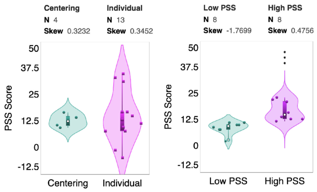
Figure 3.
Legend. Volcano plot comparing metabolite abundance between High-PSS and Low-PSS pregnancies across maternal plasma, placental tissue, cord blood, and maternal hair compartments. The x-axis represents log2-fold change in metabolite abundance between groups, and the y-axis represents statistical significance as −log10 (p-value). Metabolites above the horizontal significance threshold (green line; p < 0.05) were considered significantly associated with perceived stress status. Placental estrone (PLACT~E1), cord blood glycochenodeoxycholic acid (CORDB~GCDCA), and maternal plasma ursodeoxycholic acid (MATB~UDCA) were significantly reduced in High-PSS pregnancies. Positive fold-change values indicate metabolites elevated in High-PSS pregnancies, whereas negative values indicate metabolites reduced in High-PSS pregnancies. Bubble size reflects relative metabolite abundance.
Figure 3.
Legend. Volcano plot comparing metabolite abundance between High-PSS and Low-PSS pregnancies across maternal plasma, placental tissue, cord blood, and maternal hair compartments. The x-axis represents log2-fold change in metabolite abundance between groups, and the y-axis represents statistical significance as −log10 (p-value). Metabolites above the horizontal significance threshold (green line; p < 0.05) were considered significantly associated with perceived stress status. Placental estrone (PLACT~E1), cord blood glycochenodeoxycholic acid (CORDB~GCDCA), and maternal plasma ursodeoxycholic acid (MATB~UDCA) were significantly reduced in High-PSS pregnancies. Positive fold-change values indicate metabolites elevated in High-PSS pregnancies, whereas negative values indicate metabolites reduced in High-PSS pregnancies. Bubble size reflects relative metabolite abundance.
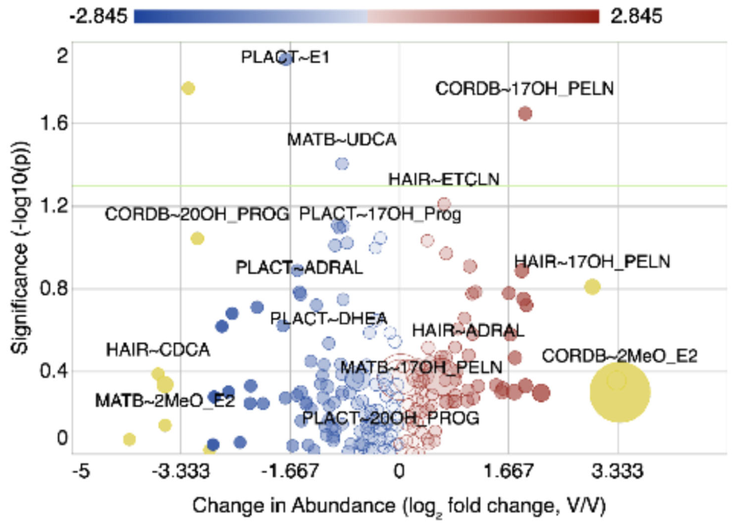
Figure 4.
Legend. Pie chart visualization of metabolites significantly associated with maternal perceived stress (PSS) change between Low- and High-PSS groups (p < 0.05). Rectangle size and color intensity represent the magnitude of the log2 fold-change in metabolite abundance. Negative fold-change values indicate reduced metabolite abundance in the High-PSS group relative to the Low-PSS group. Three metabolites met the nominal significance threshold: placental estrone (PLACT~E1), maternal plasma ursodeoxycholic acid (MATB~UDCA), and cord blood glycochenodeoxycholic acid (CORDB~GCDCA). CORDB~GCDCA demonstrated the largest reduction in abundance among High-PSS pregnancies. Asterisks indicate statistically significant differences (p < 0.05).
Figure 4.
Legend. Pie chart visualization of metabolites significantly associated with maternal perceived stress (PSS) change between Low- and High-PSS groups (p < 0.05). Rectangle size and color intensity represent the magnitude of the log2 fold-change in metabolite abundance. Negative fold-change values indicate reduced metabolite abundance in the High-PSS group relative to the Low-PSS group. Three metabolites met the nominal significance threshold: placental estrone (PLACT~E1), maternal plasma ursodeoxycholic acid (MATB~UDCA), and cord blood glycochenodeoxycholic acid (CORDB~GCDCA). CORDB~GCDCA demonstrated the largest reduction in abundance among High-PSS pregnancies. Asterisks indicate statistically significant differences (p < 0.05).
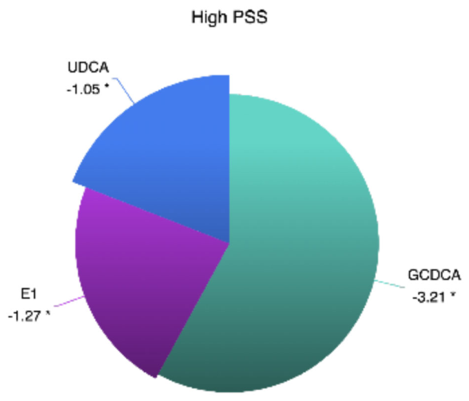
Figure 5.
Legend. Placental estrone (E1), estradiol (E2), and estriol (E3) concentrations in Low-PSS and High-PSS pregnancies. Placental E1 abundance was significantly reduced in the High-PSS group compared with the Low-PSS group (p < 0.05), whereas placental E2 and E3 concentrations did not significantly differ between groups.
Figure 5.
Legend. Placental estrone (E1), estradiol (E2), and estriol (E3) concentrations in Low-PSS and High-PSS pregnancies. Placental E1 abundance was significantly reduced in the High-PSS group compared with the Low-PSS group (p < 0.05), whereas placental E2 and E3 concentrations did not significantly differ between groups.
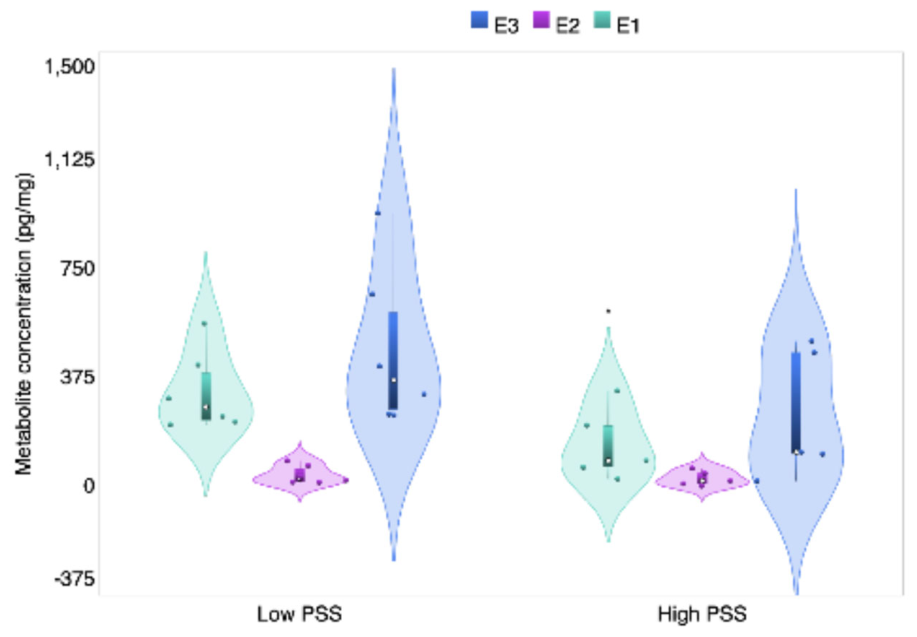
Figure 6.
Legend: Stack chart showing glycochenodeoxycholic acid (GCDCA) in cord blood and ursodeoxycholic acid (UDCA) in maternal plasma abundance by compartment and PSS group.
Figure 6.
Legend: Stack chart showing glycochenodeoxycholic acid (GCDCA) in cord blood and ursodeoxycholic acid (UDCA) in maternal plasma abundance by compartment and PSS group.
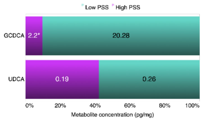
Figure 7.
Legend: Heatmap depicting side-by-side comparison figure: maternal plasma vs. hair for metabolites detectable in hair.
Figure 7.
Legend: Heatmap depicting side-by-side comparison figure: maternal plasma vs. hair for metabolites detectable in hair.
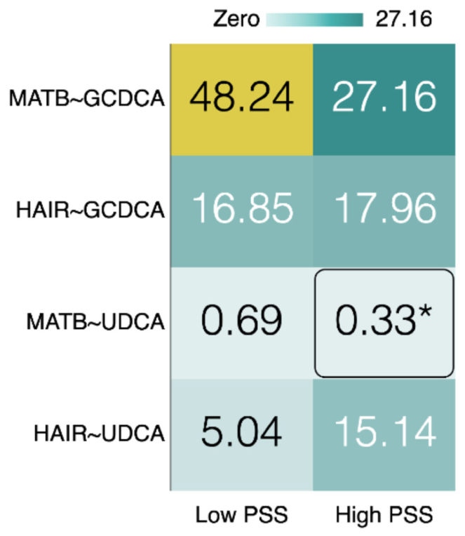

Table 1.
Study population characteristics. BMI: body mass index; PSS: Perceived Stress Score. F: female; M: male.
Table 1.
Study population characteristics. BMI: body mass index; PSS: Perceived Stress Score. F: female; M: male.
| Patient | BMI | Maternal Weight (kg) | PSS | Care Type | Gestational Age | Infant Weight (g) | Infant Sex |
|---|---|---|---|---|---|---|---|
| P-002 | 24.8 | 63.5 | 11 | Individual | 39 + 4 | 2710 | F |
| P-006 | 27.6 | 66.2 | 14 | Centering | 39 + 2 | 2772 | F |
| P-016 | 26.3 | 63 | 10 | Centering | 39 + 6 | 3330 | M |
| P-022 | 26.9 | 68.9 | 3 | Individual | 37 + 0 | 2360 | F |
| P-023 | 23.3 | 69.6 | 16 | Centering | 41 + 3 | 3600 | M |
| P-026 | 27 | 64.7 | 1 | Individual | 41 + 1 | 3390 | M |
| P-030 | 26.7 | 64 | 13 | Individual | 40 + 0 | 3500 | F |
| P-032 | 29 | 83.9 | 11 | Centering | 40 + 0 | 3835 | F |
| P-034 | 52.2 | 125.2 | 23 | Individual | 41 + 0 | 4260 | F |
| P-037 | 24.7 | 75.8 | 8 | Individual | 39 + 3 | 3525 | M |
| P-038 | 33.8 | 100.7 | 12 | Individual | 41 + 6 | 3460 | F |
| P-041 | 36.6 | 86.2 | 21 | Individual | 39 + 6 | 2895 | F |
| P-043 | 27.1 | 71.7 | 9 | Individual | 40 + 4 | 3605 | F |
| P-046 | 27.1 | 62.9 | 13 | Individual | 39 + 1 | 2850 | F |
| P-048 | 22.3 | 8 | Individual | 39 + 6 | 2870 | F | |
| P-049 | 24.2 | 71.2 | 22 | Individual | 39 + 1 | 3040 | M |
| P-051 | 35.5 | 99.8 | 10 | Individual | 39 + 3 | 4160 | M |
Table 2.
Steroid metabolite concentrations across compartments.
| Metabolite | LOD Plasma (ng/mL) | Cord Plasma (ng/mL) | Maternal Plasma (ng/mL) | LOD Tissue (pg/mg) | Placenta Mean (pg/mg) | Hair Mean (pg/mg) |
|---|---|---|---|---|---|---|
| 17-OH pregnenolone | 332.235 | <LOD | <LOD | 6644.703 | <LOD | <LOD |
| 17α-Hydroxyprogesterone | 0.826 | 7.04 | 7.68 | 16.511 | 64.21 | <LOD |
| 2-methoxyestradiol | 0.755 | <LOD | <LOD | 15.109 | <LOD | <LOD |
| 20-hydroxyprogesterone | 79.060 | <LOD | <LOD | 1581.201 | <LOD | <LOD |
| aldosterone | 0.900 | <LOD | <LOD | 18.010 | <LOD | <LOD |
| allo-pregnanolone | 4.774 | <LOD | <LOD | 95.477 | 168.88 | <LOD |
| androstanediol | 1.461 | <LOD | <LOD | 29.224 | <LOD | <LOD |
| androstenediol | 7.256 | <LOD | <LOD | 145.112 | <LOD | <LOD |
| androstenedione | 0.715 | <LOD | <LOD | 14.310 | <LOD | <LOD |
| beta-pregnanolone | 3.183 | <LOD | <LOD | 63.651 | <LOD | <LOD |
| cis-androsterone | 1.451 | <LOD | <LOD | 29.022 | <LOD | <LOD |
| cortexolone | 0.866 | 0.98 | 1.71 | 17.311 | 21.79 | <LOD |
| cortexone | 0.495 | <LOD | <LOD | 9.907 | 19.9 | <LOD |
| corticosterone | 0.866 | 1.01 | 2.6 | 17.311 | <LOD | <LOD |
| cortisol | 1.811 | 21.12 | 52.2 | 36.221 | <LOD | <LOD |
| cortisol sulfate | 11.054 | <LOD | <LOD | 221.083 | <LOD | <LOD |
| cortisone | 0.360 | 57.49 | 38.6 | 7.204 | 370.82 | <LOD |
| dehydroepiandrosterone | 7.205 | <LOD | <LOD | 144.104 | <LOD | <LOD |
| dehydroepiandrosterone sulfate | 18.424 | 571.51 | 457.71 | 368.489 | <LOD | <LOD |
| dihydroprogesterone | 39.530 | <LOD | <LOD | 790.601 | <LOD | <LOD |
| dihydrotestosterone | 0.726 | <LOD | <LOD | 14.511 | <LOD | <LOD |
| estradiol | 0.680 | 1.38 | 4.82 | 13.609 | 34.32 | <LOD |
| estriol | 2.882 | 57.65 | 57.53 | 57.635 | 356.34 | <LOD |
| estrone | 2.702 | <LOD | 5.94 | 54.032 | 232.78 | <LOD |
| etiocholanolone | 1.451 | <LOD | <LOD | 29.022 | <LOD | <LOD |
| pregnenolone | 158.120 | <LOD | <LOD | 3162.402 | <LOD | <LOD |
| progesterone | 0.393 | 226.85 | 56.77 | 7.856 | 4884 | 15.92 |
| testosterone | 0.144 | 0.02 | <LOD | 2.882 | <LOD | <LOD |
| testosterone glucuronide | 23.212 | <LOD | <LOD | 464.241 | <LOD | <LOD |
| trans-androsterone | 7.256 | <LOD | <LOD | 145.112 | <LOD | <LOD |
Table 3.
Bile acid metabolite concentrations across compartments.
| Metabolite | LOD Plasma (ng/mL) | Cord Plasma (ng/mL) | Maternal Plasma (ng/mL) | LOD Tissue (pg/mg) | Placenta Mean (pg/mg) | Hair Mean (pg/mg) |
|---|---|---|---|---|---|---|
| Chenodeoxycholic acid | 0.3923 | <LOD | 1.09 | 7.8459 | 13.31 | 26.51 |
| Cholic acid | 0.4083 | <LOD | 3.13 | 8.1658 | 15.78 | 22.07 |
| Deoxycholic acid | 0.3923 | 0.48 | 7.47 | 7.8459 | 28.3 | 62.28 |
| Glycochenodeoxycholic acid | 0.4493 | 17.36 | 37.7 | 8.9863 | 32.34 | 17.32 |
| Glycocholic acid | 0.4653 | 19.95 | 16.42 | 9.3062 | 35.15 | <LOD |
| Glycodeoxycholic acid | 0.4493 | <LOD | 26.01 | 8.9863 | 12.51 | <LOD |
| Glycohyodeoxycholic acid | 0.4493 | 0.53 | <LOD | 8.9863 | <LOD | <LOD |
| Glycolithocholic acid | 0.4333 | <LOD | <LOD | 8.6664 | <LOD | <LOD |
| Glycoursodeoxycholic acid | 0.4493 | <LOD | 0.47 | 8.9863 | <LOD | <LOD |
| Lithocholic acid | 0.3763 | 0.73 | 0.87 | 7.5260 | 18.67 | 23.23 |
| Tauro-α-Muricholic acid | 0.5153 | <LOD | 0.56 | 10.3058 | <LOD | <LOD |
| Tauro-β-Muricholic acid | 0.5153 | <LOD | <LOD | 10.3058 | <LOD | <LOD |
| Tauro-ω-Muricholic acid | 0.5153 | 0.53 | <LOD | 10.3058 | 11.8 | <LOD |
| Taurochenodeoxycholic acid | 0.4993 | 54.41 | 33.04 | 9.9859 | 143.3 | 11.58 |
| Taurocholic acid | 0.5153 | 42.66 | 25.83 | 10.3058 | 94.22 | <LOD |
| Taurodehydrocholate | 0.5092 | <LOD | <LOD | 10.1849 | <LOD | <LOD |
| Taurodeoxycholic acid | 0.4993 | <LOD | 16.35 | 9.9859 | <LOD | <LOD |
| Taurolithocholic acid | 0.4833 | <LOD | 0.49 | 9.6660 | <LOD | <LOD |
| Tauroursodeoxycholic acid | 0.4993 | 1.03 | <LOD | 9.9859 | 12.7 | <LOD |
| Ursodeoxycholic acid | 0.3923 | <LOD | 0.51 | 7.8459 | <LOD | 9.37 |
| α-Muricholic acid | 0.4083 | <LOD | <LOD | 8.1658 | <LOD | <LOD |
| β-Muricholic acid | 0.4083 | 0.01 | <LOD | 8.1658 | <LOD | <LOD |
| ω-Muricholic acid | 0.4083 | 1 | <LOD | 8.1658 | <LOD | <LOD |
Disclaimer/Publisher’s Note: The statements, opinions and data contained in all publications are solely those of the individual author(s) and contributor(s) and not of MDPI and/or the editor(s). MDPI and/or the editor(s) disclaim responsibility for any injury to people or property resulting from any ideas, methods, instructions or products referred to in the content. |
© 2026 by the authors. Licensee MDPI, Basel, Switzerland. This article is an open access article distributed under the terms and conditions of the Creative Commons Attribution (CC BY) license (http://creativecommons.org/licenses/by/4.0/).
Copyright: This open access article is published under a Creative Commons CC BY 4.0 license, which permit the free download, distribution, and reuse, provided that the author and preprint are cited in any reuse.


