

Submitted:
19 July 2026
Posted:
21 July 2026
You are already at the latest version
Abstract
Individuals with Laron syndrome, a rare condition characterized by congenital insulin-like growth factor 1 (IGF-1) deficiency, display a remarkably low incidence of cancer, suggesting the existence of protective mechanisms linking reduced IGF-1 signaling to decreased cancer susceptibility. Consistent with this observation, IGF-1 is a recognized promoter of prostate cancer (PCa) progression, although the underlying mechanisms remain incompletely understood. Methylglyoxal (MG)-derived glycative stress, reflected by the accumulation of MG-derived hydroimidazolone 1 (MG-H1), has been implicated in PCa progression but has never been investigated in Laron syndrome. We found that liver tissues from Laron mice exhibited lower MG-H1 levels, suggesting reduced MG-derived glycative stress associated with low IGF-1 signaling. These findings prompted us to investigate whether MG-derived glycative stress contributes to IGF-1-driven PCa progression. Compared with the less aggressive LNCaP cells, PC3 cells displayed higher basal IGF-1 and MG-H1 levels, consistent with a potential association between IGF-1 and MG-derived glycative stress in PCa progression. Moreover, IGF-1 stimulation of LNCaP cells increased MG-H1 accumulation, proliferation, colony formation, invasiveness, and expression of matrix metalloproteinase (MMP)-1, MMP-7, MMP-9, receptor for advanced glycation end-products (RAGE), and Osteopontin (OPN), all of which were markedly attenuated by the MG scavenger, aminoguanidine (AG). Collectively, these findings support a potential contribution of MG-derived glycative stress to IGF-1-driven PCa progression.
Keywords:
Laron syndrome
; IGF-1
; methylglyoxal
; MG-H1
; glycative stress
; glyoxalase 1
; prostate cancer
; LNCaP
; PC3
‡ These authors share last authorship.
1. Introduction
Prostate cancer (PCa) is the most frequently diagnosed malignancy among men in Western countries and remains a leading cause of cancer-related mortality worldwide [1]. Although localized disease can often be successfully treated, progression toward aggressive, metastatic, and therapy-resistant stages remains the major clinical challenge and is associated with poor patient outcomes [2]. A better understanding of the molecular mechanisms driving PCa progression is therefore essential for identifying novel biomarkers and therapeutic targets to limit PCa progression.
Among the biological processes implicated in PCa progression, metabolic reprogramming has emerged as a hallmark of tumor evolution [3]. One consequence of this metabolic rewiring is the increased production of highly reactive metabolic by-products capable of modifying cellular macromolecules and influencing tumor behavior. One of these metabolites is methylglyoxal (MG), a highly reactive dicarbonyl compound mainly generated as a by-product of glycolysis [4]. Under physiological conditions, MG is efficiently detoxified by the glyoxalase system, composed of glyoxalase 1 (GLO1) and glyoxalase 2 (GLO2), using glutathione (GSH) as a cofactor [5]. However, excessive MG production or impaired detoxification results in the accumulation of advanced glycation end products (AGEs), among which MG-derived hydroimidazolone 1 (MG-H1) is the predominant MG-derived adduct, accounting for more than 90% of MG-derived protein modifications [6]. Accumulation of MG-H1 is widely regarded as the hallmark of MG-derived glycative stress, which has been implicated in the pathogenesis of several diseases, including cancer [7,8]. Notably, MG-H1 has been associated with PCa progression, at least in part through activation of the receptor for advanced glycation end products (RAGE), thereby enhancing cell migration, invasion, and interactions with the tumor microenvironment [9,10,11,12,13].
Nevertheless, the upstream molecular mechanisms responsible for the induction of MG-derived glycative stress during PCa progression remain poorly understood.
Insulin-like growth factor 1 (IGF-1) is a key regulator of cell growth, survival, differentiation, and metabolism and a recognized promoter of PCa progression [14]. Elevated circulating IGF-1 levels have been associated with increased disease aggressiveness, while activation of IGF-1 signaling enhances proliferation, survival, invasion, and metastatic potential through multiple downstream pathways [14]. In addition to these well-established functions, IGF-1 stimulates glucose uptake and glycolytic metabolism through activation of the PI3K/AKT pathway [15,16,17,18], raising the possibility that it may also promote MG generation. However, whether this metabolic effect contributes to MG-derived glycative stress during PCa progression has not been investigated. Despite extensive evidence supporting the role of IGF-1 in PCa progression, the molecular mechanisms through which IGF-1 promotes its tumor-promoting effects remain incompletely understood. Identifying additional downstream pathways may therefore provide new opportunities for therapeutic intervention in advanced PCa.
An opportunity to address this question is provided by Laron syndrome, a rare autosomal recessive disorder caused by mutations in the growth hormone receptor (GHR) gene [19]. Because of growth hormone resistance, affected individuals exhibit congenital IGF-1 deficiency and display a remarkably low incidence of cancer despite several metabolic abnormalities [19]. This distinctive phenotype suggests the existence of protective mechanisms linking lifelong reduction of IGF-1 signaling to decreased cancer susceptibility, although the molecular basis of this protection remains largely unknown. To our knowledge, MG-derived glycative stress has never been investigated in Laron syndrome. We therefore reasoned that this rare condition could represent a valuable biological model to explore whether reduced IGF-1 signaling is associated with attenuation of MG-derived glycative stress and whether this pathway contributes to the relationship between IGF-1 signaling and PCa progression.
Based on these considerations, we hypothesized that MG-derived glycative stress represents a previously unrecognized mechanism contributing to IGF-1-driven PCa progression. To test this hypothesis, we combined in vivo analyses in the Laron syndrome mouse model [19] with in vitro studies using two well-established PCa cell models differing in aggressiveness, LNCaP and PC3. Finally, using aminoguanidine as an MG scavenger [20], we causatively investigated whether MG-derived glycative stress contributes to the pro-tumorigenic effects of IGF-1, including enhanced proliferation, colony formation, invasiveness, and expression of molecules associated with PCa progression, namely RAGE, osteopontin (OPN), and matrix metalloproteinases (MMPs).
2. Materials and Methods
2.1. Material and Treatments
RPMI-1640 culture medium, fetal bovine serum (FBS), penicillin/streptomycin, and recombinant human IGF-1 (cat. no. PHG0071) were purchased from Thermo Fisher Scientific (Milan, Italy). Recombinant human IGF-1 was reconstituted in sterile water according to the manufacturer’s instructions. Working aliquots were prepared in the presence of 0.1% bovine serum albumin (BSA) and stored at −80 °C to avoid repeated freeze–thaw cycles.
Aminoguanidine bicarbonate (AG; cat. no. 396494) was purchased from Merck (Milan, Italy). Cells were pretreated with AG at a final concentration of 1 mM for 6 h [21,22]. Recombinant human IGF-1 was then added directly to the culture medium at a final concentration of 150 ng/mL without removing AG, and cells were incubated for an additional 72 h. Thus, AG remained present throughout the entire period of IGF-1 stimulation. Untreated cells and cells treated with AG or IGF-1 alone served as controls.
The concentration of recombinant IGF-1 (150 ng/mL) was selected to ensure robust activation of the IGF-1 signaling pathway throughout the experimental period. Similar concentrations have been widely used in mechanistic in vitro studies investigating IGF-1-dependent responses in PCa cells [23,24,25]. The aim of the present study was not to reproduce physiological circulating IGF-1 levels but rather to determine whether sustained activation of IGF-1 signaling promotes MG-derived glycative stress and the associated pro-tumorigenic phenotype.
2.2. Laron Mouse Model and Tissue Collection
Male and female heterozygous growth hormone receptor knockout (GHR+/−) mice, originally generated in the laboratory of Prof. J. J. Kopchick (Edison Biotechnology Institute, Ohio University, Athens, OH, USA), were bred in the animal facility of the University of Perugia. Homozygous GHR−/− mice, an established experimental model of Laron syndrome characterized by congenital growth hormone resistance and markedly reduced circulating IGF-1 levels [26,27], were obtained from these breeding pairs and used in the present study.
All animal procedures were conducted in accordance with the Italian legislation governing the protection of animals used for scientific purposes and were approved by the Italian Ministry of Health (authorization no. 605/2019-PR, issued on August 1, 2019, pursuant to Legislative Decree No. 26/2014 implementing Directive 2010/63/EU). Animals were euthanized under deep anesthesia, and liver samples were immediately collected.
Liver samples were collected following anesthesia induced by intraperitoneal administration of ketamine (100 mg/kg) and xylazine (10 mg/kg). Tissue samples were immediately processed for protein and RNA extraction.
For protein extraction, approximately 100 mg of tissue were homogenized in 1 mL of RIPA buffer (Santa Cruz Biotechnology, Dallas, TX, USA; cat. no. sc-24948) using an Ultra-Turrax homogenizer (IKA-Werke GmbH & Co. KG, Staufen, Germany). Homogenates were centrifuged at 2,500 × g for 10 min at 4 °C to obtain the soluble protein fraction. Total protein concentration was determined using the Bradford protein assay (Bio-Rad Laboratories, Hercules, CA, USA; cat. no. 5000006).
For RNA extraction, an additional 100 mg of tissue were homogenized in 1 mL of TRIzol™ Reagent (Thermo Fisher Scientific, Waltham, MA, USA; cat. no. 15596026), and total RNA was isolated according to the manufacturer’s instructions.
2.3. Cell Cultures
Human PCa cell lines LNCaP and PC3 were obtained from the American Type Culture Collection (ATCC, Manassas, VA, USA). LNCaP cells, representative of an androgen-dependent and less aggressive PCa phenotype, and PC3 cells, representative of an androgen-independent and highly aggressive phenotype, were cultured in RPMI-1640 medium (Thermo Fisher Scientific, Milan, Italy) supplemented with 10% fetal bovine serum (FBS), 100 U/mL penicillin, and 100 μg/mL streptomycin. Cells were maintained in a humidified incubator at 37 °C in an atmosphere containing 5% CO₂ and were routinely passaged at 70–80% confluence. Cell cultures were regularly tested and confirmed to be free of mycoplasma contamination.
2.4. Measurement of MG-H1 Levels
Intracellular levels of methylglyoxal-derived hydroimidazolone (MG-H1) were quantified in cell and tissue lysates using the OxiSelect™ Methylglyoxal (MG) Competitive ELISA Kit (Cell Biolabs, Inc., San Diego, CA, USA; cat. no. STA-811), according to the manufacturer’s instructions. Cell and tissue lysates were prepared using the same volume of RIPA buffer, and equal amounts of total protein were analyzed for each sample to ensure direct comparison among experimental groups. Absorbance was measured using a microplate reader at the recommended wavelength, and MG-H1 concentrations were calculated from standard curves and expressed as μg/mL.
2.5. Determination of Glyoxalase 1 (GLO1) Enzymatic Activity
GLO1 specific activity was determined spectrophotometrically as previously described [12,13]. Briefly, GLO1 activity was measured by monitoring the formation of S-D-lactoylglutathione from the hemithioacetal spontaneously generated by MG and GSH. The reaction was carried out in 100 mM sodium phosphate buffer (pH 6.6) containing 2 mM MG and 2 mM GSH. The increase in absorbance was monitored at 240 nm, and enzyme activity was calculated using the molar extinction coefficient of S-D-lactoylglutathione (ε = 3.3 mM⁻¹ cm⁻¹). GLO1 activity was normalized to total protein content, determined by the Bradford assay, and expressed as µmol/min per milligram of protein.
2.6. Measurement of IGF-1 Levels
Intracellular IGF-1 protein levels were determined in LNCaP and PC3 cell lysates using the Human IGF-1 ELISA Kit (Thermo Fisher Scientific, Milan, Italy; cat. no. EH250RB). The assay was performed according to the manufacturer’s instructions and adapted for the analysis of cell lysates. Cell lysates were prepared under identical experimental conditions using the same volume of RIPA buffer, and equal amounts of total protein were analyzed for each sample to ensure direct comparison between the two cell lines. Absorbance was measured at 450 nm using a microplate reader, and IGF-1 concentrations were calculated from standard calibration curves and expressed as ng/mL.
2.7. RNA Isolation, cDNA Synthesis, and Quantitative Real-Time PCR (qRT-PCR)
Total RNA was extracted from cultured cells and tissue samples using TRIzol™ Reagent (Thermo Fisher Scientific, Milan, Italy; cat. no. 15596026) according to the manufacturer’s instructions. RNA concentration and purity were determined spectrophotometrically by measuring the absorbance at 260 and 280 nm, while RNA integrity was verified by agarose gel electrophoresis.
First-strand cDNA was synthesized from 1 μg of total RNA using the RevertAid First Strand cDNA Synthesis Kit (Thermo Fisher Scientific, Milan, Italy; cat. no. K1622) according to the manufacturer’s instructions.
Quantitative real-time PCR (qRT-PCR) was performed on an Mx3000P Real-Time PCR System (Agilent Technologies, Milan, Italy). PCR reactions were carried out in a final volume of 20 μL containing 25 ng of cDNA, PowerTrack SYBR Green Master Mix (Thermo Fisher Scientific, Milan, Italy; cat. no. A46109) and 600 nM of each primer. Thermal cycling conditions consisted of an initial denaturation step at 95 °C for 5 min, followed by 45 amplification cycles at 95 °C for 20 s and 60 °C for 30 s. Melting curve analysis was performed for all reactions to verify amplification specificity and the absence of nonspecific PCR products.
2.8. RNA Isolation, cDNA Synthesis, and Quantitative Real-Time PCR (qRT-PCR)
Thermo Scientific™ RIPA Lysis and Extraction Buffer (cat. no. 89901; Thermo Fisher Scientific, Milan, Italy) supplemented immediately before use with Thermo Scientific™ Halt™ Protease and Phosphatase Inhibitor Cocktail (100×; cat. no. 78440; Thermo Fisher Scientific, Milan, Italy).
Western blot analysis was performed as previously described [12], with minor modifications. Equal amounts of protein (30 μg) were denatured in Laemmli sample buffer, separated by SDS-PAGE, and transferred onto nitrocellulose membranes using the iBlot™ Dry Blotting System (Thermo Fisher Scientific, Milan, Italy) according to the manufacturer’s instructions. Non-specific binding sites were blocked with Roti®-Block (Carl Roth, Karlsruhe, Germany) for 1 h at room temperature. Membranes were incubated overnight at 4 °C with the appropriate primary antibodies, followed by incubation for 1 h at room temperature with the corresponding horseradish peroxidase (HRP)-conjugated secondary antibodies. The antibodies used were: mouse anti-Glo1 mAb (dilution 1:1000, Santa Cruz, cat. sc-133144, DBA Italia S.r.l., Milan, Italy) and mouse anti-β-actin mAb (dilution 1:1000, Santa Cruz, cat. sc-517582, DBA Italia S.r.l., Milan, Italy). Immunoreactive bands were visualized using enhanced chemiluminescence (ECL; Amersham Pharmacia, Milan, Italy) in iBright Imaging Systems for western blot (Thermo Fisher Scientific, Milan, Italy). β-Actin was used as the loading control.
2.9. Cell Proliferation Assay
Cell proliferation was assessed by direct counting of viable cells following the indicated treatments. Cells were harvested, stained with Trypan Blue, and counted using a hemocytometer. Results were expressed as the percentage of viable cells relative to untreated controls.
2.10. Colony Formation Assay
Following the indicated treatments, cells were seeded at low density and allowed to grow for 10 days. Colonies were then fixed with methanol, stained with crystal violet, and quantified using ImageJ software (National Institutes of Health, Bethesda, MD, USA). Only colonies containing more than 50 cells were included in the analysis.
2.11. Cell Invasion Assay
Cell invasion was assessed using the CytoSelect 24-Well Cell Invasion Assay kit (cat. no. CBA-110, DBA Italia S.r.l., Milan, Italy) according to the manufacturer’s instructions. Following the indicated treatments, cells were seeded at a density of 5 × 10⁴ cells per insert in serum-free medium into the upper chamber, while medium containing 10% fetal bovine serum (FBS) was added to the lower chamber as a chemoattractant. Cells were allowed to invade through the matrix-coated membrane for 16 h. Non-invading cells were removed from the upper surface of the membrane, whereas invading cells were stained with crystal violet and quantified by measuring the optical density (OD) at 560 nm according to the manufacturer’s protocol.
2.12. Statistical Analysis
Data are presented as the mean ± standard deviation (SD) of three independent experiments. Statistical analyses were performed using GraphPad Prism version 11 (GraphPad Software, Boston, MA, USA). Comparisons between two groups were performed using Student’s t-test. When appropriate, comparisons among multiple groups were performed using one-way analysis of variance (ANOVA) followed by Tukey’s multiple comparisons test. Differences were considered statistically significant at p < 0.05.
3. Results
3.1. MG-Derived Glycative Stress Is Reduced in the Laron Syndrome Mouse Model
To investigate whether MG-derived glycative stress is altered in the Laron syndrome mouse model, MG-H1, a specific marker of MG-derived glycative stress [6], and the specific activity of glyoxalase 1 (GLO1), the rate-limiting enzyme responsible for MG detoxification [5], were evaluated in liver homogenates from Laron (GHR−/−) and control (GHR+/+) mice. Laron (GHR−/−) mice, a model of congenital IGF-1 deficiency [29], exhibited significantly lower MG-H1 levels and GLO1 specific activity than control (GHR+/+) mice (Figure 1).
These findings indicate that reduced IGF-1 signaling is associated with lower MG-derived glycative stress in the liver of Laron mice. Since Laron syndrome is associated with reduced cancer susceptibility, these observations raised the possibility that attenuation of MG-derived glycative stress may represent one of the mechanisms linking low IGF-1 signaling to cancer protection. Conversely, in PCa, where both IGF-1 signaling and MG-derived glycative stress have been independently implicated in disease progression [9,10,11,12,13,14], these findings prompted us to hypothesize that the two processes may be mechanistically linked and jointly contribute to tumor progression. Based on this rationale, we next investigated whether MG-derived glycative stress contributes to the pro-tumorigenic effects of IGF-1 in PCa cells.
3.2. Basal IGF-1 Expression, MG-H1 Levels, and GLO1 Activity in LNCaP and PC3 Cells
We first investigated whether basal IGF-1 expression is associated with MG-derived glycative stress in two established PCa cell models displaying different degrees of aggressiveness, LNCaP and PC3. To this end, IGF-1 mRNA expression, intracellular IGF-1 protein levels, MG-H1 levels, and GLO1 specific activity were evaluated. As shown in Figure 2, both IGF-1 mRNA expression (Figure 2a) and intracellular IGF-1 protein levels (Figure 2b), together with MG-H1 levels (Figure 2c), were significantly higher in the more aggressive PC3 cells than in the less aggressive LNCaP cells. Consistent with the increased MG-derived glycative stress, GLO1 specific activity was also significantly higher in PC3 cells (Figure 2d), suggesting an adaptive response to the greater MG burden.
Collectively, these findings reveal a positive association between IGF-1 expression and MG-derived glycative stress in PCa cells with different degrees of aggressiveness, providing the rationale to investigate whether IGF-1 promotes PCa progression through induction of MG-derived glycative stress.
3.3. IGF-1 Induces MG-Derived Glycative Stress in LNCaP Cells
To investigate whether IGF-1 promotes MG-derived glycative stress, LNCaP cells were exposed to IGF-1 (150 ng/mL) for 72 h. As shown in Figure 3a, IGF-1 treatment significantly increased intracellular MG-H1 accumulation compared with untreated cells. In parallel, GLO1 expression was significantly upregulated at both the mRNA and protein levels, accompanied by a marked increase in GLO1 specific activity (Figure 3b–d). Collectively, these findings indicate that IGF-1 enhances MG-derived glycative stress in LNCaP cells and induces a concomitant upregulation of the glyoxalase system, likely as an adaptive response to the increased MG burden.
3.4. IGF-1 Enhances the Proliferative and Invasive Phenotype of LNCaP Cells
To investigate the biological consequences of IGF-1 stimulation, cell proliferation and invasive behavior were evaluated following exposure of LNCaP cells to IGF-1. IGF-1 treatment significantly increased cell proliferation, as demonstrated by increased cell number and colony-forming ability compared with untreated cells (Figure 4a,b). In addition, IGF-1 markedly enhanced the invasive capacity of LNCaP cells (Figure 4c).
To further characterize this phenotype, the expression of matrix metalloproteinases (MMPs) involved in extracellular matrix remodeling and tumor invasion was evaluated. IGF-1 significantly increased the expression of MMP-1, MMP-7, and MMP-9 (Figure 4d–f), consistent with the acquisition of a more invasive phenotype. Collectively, these findings demonstrate that IGF-1 promotes proliferative and invasive properties in LNCaP cells.
3.5. IGF-1 Increases RAGE and OPN Expression in LNCaP Cells
Since MG-H1 is a well-established ligand of the receptor for advanced glycation end products (RAGE) [30], we first investigated whether the increase in MG-derived glycative stress induced by IGF-1 was associated with changes in RAGE expression and subsequently evaluated its potential association with the expression of osteopontin (OPN). Both molecules have previously been implicated in PCa progression [13,31,32,33]. Moreover, OPN has been identified as a downstream effector of RAGE signaling in several pathological contexts [34], and the RAGE/OPN axis has been implicated in inflammatory responses associated with proliferative disorders [35]. Furthermore, an MG-H1/OPN axis has recently been proposed to contribute to the acquisition of a more aggressive phenotype in LNCaP cells [12]. As shown in Figure 5a, IGF-1 significantly increased RAGE mRNA expression in LNCaP cells. Likewise, OPN expression was markedly upregulated following IGF-1 treatment (Figure 5b).
Collectively, these findings demonstrate that IGF-1 increases the expression of both RAGE and OPN in LNCaP cells and support the hypothesis that these molecules may participate in downstream molecular events associated with PCa progression.
3.6. Inhibition of MG-Derived Glycative Stress by Aminoguanidine Prevents the Pro-Tumorigenic Effects of IGF-1
To determine whether MG-derived glycative stress mediates the pro-tumorigenic effects induced by IGF-1, LNCaP cells were pretreated with aminoguanidine (AG), a well-established methylglyoxal scavenger [20], that consequently limits the formation of MG-H1), before IGF-1 stimulation. As shown in Figure 6a, AG markedly reduced the intracellular accumulation of MG-H1 induced by IGF-1. Consistently, the IGF-1-mediated upregulation of GLO1 expression was also significantly attenuated by AG (Figure 6b-d), confirming the effective suppression of MG-derived glycative stress under our experimental conditions.
We next investigated whether inhibition of glycative stress affected the molecular changes associated with IGF-1 treatment. Pretreatment with AG significantly prevented the IGF-1-induced increase in both RAGE (Figure 7a) and OPN expression (Figure 7b), indicating that the upregulation of these molecules is dependent, at least in part, on MG-derived glycative stress.
We then evaluated whether suppression of glycative stress influenced the biological responses elicited by IGF-1. AG significantly reduced IGF-1-induced cell proliferation (Figure 8a), clonogenic growth (Figure 8b), and invasive capacity (Figure 8c). Likewise, AG abolished the induction of the invasion-associated genes MMP-1, MMP-7, and MMP-9 observed following IGF-1 stimulation (Figure 8d).
Collectively, these findings demonstrate that MG-derived glycative stress is a key mediator of the pro-tumorigenic effects elicited by IGF-1 in LNCaP cells. Pharmacological scavenging of methylglyoxal with AG effectively suppressed MG-H1 accumulation, prevented the induction of RAGE and OPN, and markedly attenuated the acquisition of an aggressive phenotype. These results support a mechanistic link between IGF-1-induced glycative stress and the activation of molecular pathways associated with PCa progression.
4. Discussion
The present study identifies MG-derived glycative stress, specifically mediated by MG-H1, as a novel downstream component of IGF-1 signaling in PCa cells. While IGF-1 is a well-established driver of PCa progression [36,37], MG-derived glycative stress has independently been associated with tumor aggressiveness through MG-H1/RAGE-dependent mechanisms [9,10,11,12,13]. Our findings support the hypothesis that MG-H1-mediated glycative stress represents a previously unrecognized mechanistic component linking IGF-1 signaling to the acquisition of a pro-tumorigenic phenotype. The observation that AG effectively prevented the IGF-1-induced increase in MG-H1 together with the associated proliferative and invasive phenotype provides proof-of-concept that pharmacological targeting of MG-derived glycative stress may represent a feasible strategy for limiting IGF-1-driven PCa progression. Future studies should evaluate whether MG scavengers or other pharmacological approaches targeting this pathway may have translational potential, either alone or in combination with existing therapies [8].
Although the mechanism underlying the increase in MG-H1 following IGF-1 stimulation was not directly investigated in the present study, a plausible explanation is that enhanced IGF-1 signaling promotes metabolic conditions, thus favoring MG generation. IGF-1 is known to promote metabolic reprogramming and enhance glycolytic metabolism to sustain cell growth and proliferation [16], thereby potentially increasing the generation of MG from glycolytic intermediates. Under these conditions, MG-H1 accumulation may reflect increased MG production that exceeds the detoxification capacity of the glyoxalase system.
The simultaneous increase in MG-H1 and GLO1 is particularly intriguing. Although GLO1 detoxifies MG, its upregulation in IGF-1-treated cells likely represents an adaptive response to increased MG production rather than effective suppression of glycative stress, as also reported in highly glycolytic tumors [38]. Accordingly, the persistence of elevated MG-H1 levels despite increased GLO1 activity indicates that activation of the glyoxalase system is insufficient to counterbalance the increased MG burden generated by IGF-1, thereby allowing glycative stress to contribute to the pro-tumorigenic phenotype [8].
In line with the literature, our findings indicate that IGF-1 not only stimulates tumor cell growth [23], but also promotes the acquisition of a more aggressive phenotype by enhancing invasive capacity [23] and increasing the expression of MMP-1, MMP-7, and MMP-9, key mediators of extracellular matrix remodeling and metastatic dissemination [39,40,41]. A previous study has demonstrated that IGF-1/IGF-1R signaling promotes PCa invasiveness through regulation of MMP-14 (MT1-MMP), a membrane-bound MMP critically involved in tumor invasion and metastasis [42]. Our findings further extend these observations by identifying MMP-1, MMP-7, and MMP-9 as additional metalloproteinases induced by IGF-1, suggesting that the pro-invasive effects of IGF-1 involve a broader extracellular matrix remodeling program than previously recognized.
MG-H1 is a well-established ligand of RAGE [30], and both RAGE and OPN have been extensively implicated in PCa progression [13,31,32,33]. Moreover, OPN has been identified as a downstream effector of RAGE signaling in several pathological contexts [34], while the RAGE/OPN axis has been associated with inflammatory responses that support proliferative disorders [35]. More recently, an MG-H1/OPN axis has also been proposed to contribute to the acquisition of a more aggressive phenotype in LNCaP cells [12]. Consistent with this evidence, we found that both RAGE and OPN were upregulated following IGF-1 stimulation and that their induction was markedly attenuated by MG scavenging. These findings support the hypothesis that MG-H1-mediated glycative stress contributes to the activation of RAGE- and OPN-associated signaling pathways downstream of IGF-1, thereby promoting tumor cell aggressiveness. However, because neither RAGE nor OPN were directly inhibited or genetically manipulated in the present study, their precise mechanistic contribution remains to be established.
The rationale of this work originated from observations obtained in experimental models of Laron syndrome, a rare disorder characterized by congenital IGF-1 deficiency and remarkable protection against cancer development [19,43,44,45]. In these models, reduced MG-H1 levels together with lower GLO1 activity suggested an attenuation of MG-derived glycative stress in the setting of chronic IGF-1 deficiency. This observation led us to hypothesize that IGF-1 may positively regulate MG-derived glycative stress pathways and that this mechanism could contribute to tumor progression. Our findings in PCa cells support this hypothesis by identifying MG-H1-mediated glycative stress as a previously unrecognized downstream component of IGF-1 signaling associated with a pro-tumorigenic phenotype. To the best of our knowledge, this is the first study to propose that attenuation of MG-derived glycative stress may contribute to the cancer-protective phenotype associated with congenital IGF-1 deficiency.
Although the reduced MG-derived glycative stress was observed in an experimental model of Laron syndrome and therefore cannot be directly extrapolated to humans, these findings raise the intriguing possibility that attenuation of MG-H1 accumulation may represent one of the mechanisms contributing to the remarkable cancer protection associated with IGF-1 deficiency. This hypothesis warrants further investigation in appropriate experimental and clinical settings.
More broadly, the present study illustrates the value of rare diseases as biological models for generating mechanistic hypotheses relevant to common non-communicable diseases [46]. By revealing a previously unrecognized link between IGF-1 signaling and MG-derived glycative stress, the Laron syndrome model provided the conceptual framework that led to the identification of MG-H1 as a potential contributor to PCa progression. Overall, these findings further support the concept that the metabolic reprogramming associated with IGF-1 signaling extends beyond enhanced glycolysis to include activation of MG-derived glycative stress, thereby identifying this pathway as a functionally relevant contributor to PCa progression.
5. Conclusions
Our findings identify MG-H1-mediated glycative stress as a previously unrecognized downstream component of IGF-1 signaling that contributes to the acquisition of pro-tumorigenic phenotype in PCa cells. By demonstrating that pharmacological scavenging of MG attenuates the pro-tumorigenic effects of IGF-1, this study provides proof-of-concept that targeting MG-derived glycative stress may represent a promising strategy for limiting prostate cancer progression.
Beyond its implications for PCa biology, this work highlights the value of rare diseases as biological models for generating mechanistic hypotheses relevant to common non-communicable diseases. The observations obtained in the Laron syndrome model provided the conceptual framework for uncovering a previously unrecognized connection between IGF-1 signaling and MG-derived glycative stress, illustrating how research on rare disorders can reveal novel molecular pathways with potential translational implications extending beyond the rare disease itself.
Supplementary Materials
The following supporting information can be downloaded at: https://www.mdpi.com/article/doi/s1, Figure S1: Original images of Western blots and colony formation assays.
Author Contributions
The following statements should be used “Conceptualization, C.A.; methodology, D.M., C.T. and F.M.; validation, D.M., C.T. and C.A.; formal analysis, D.M, C.T. and C.A.; investigation, D.M., C.T, C.L. C.B and F.M.; resources, D.M., C.T, C.L. C.B and F.M.; writing—original draft preparation, C.A.; writing—review and editing, F.M., V.N.T., T.B; visualization, C.A., F.M., V.N.T., T.B; funding acquisition, C.A. and T.B. All authors have read and agreed to the published version of the manuscript.
Funding
This research was funded by the University of Perugia - University Research Fund, 2023 (“University Research Project”: ISPIRARE) granted to C.A.
Institutional Review Board Statement
The animal study protocol was approved by the Ministero della Salute with authorization no. 605/2019-PR issued on 01/08/2019 (pursuant to Art. 31, Legislative Decree 2672014).
Informed Consent Statement
Not applicable.
Data Availability Statement
The original contributions presented in this study are included in the article/Supplementary Material. Further inquiries can be directed to the corresponding author.
Acknowledgments
We thank Roberta Frosini for her excellent technical assistance.
Conflicts of Interest
The authors declare no conflicts of interest. .
Abbreviations
The following abbreviations are used in this manuscript:
| IGF-1 | Insulin-like growth factor 1 |
| MG | Methylglyoxal |
| MG-H1 | MG-derived hydroimidazolone 1 |
| PCa | Prostate cancer |
| MMP | Matrix metalloproteinase |
| RAGE | Receptor for advanced glycation end-products |
| OPN | Osteopontin |
| AG | Aminoguanidine |
| GLO1 | Glyoxalase 1 |
| GLO2 | Glyoxalase 2 |
| GSH | Glutathione |
| GHR | Growth hormone receptor |
References
- Al-Assil, T.; et al. Theranostic strategies in prostate cancer: Advances in PSMA PET imaging and radioligand therapy. Cancer Treat. Res. Commun. 2026. 48, 101293. [Google Scholar] [CrossRef]
- Cheng, Y.; et al. METTL3/YTHDF1-Driven SURF6 Promotes Prostate Cancer Stemness via CDK4. J. Cell Mol. Med. 2026, 30(12), e71259. [Google Scholar] [CrossRef] [PubMed]
- Ding, H.; et al. Revisiting the role of metabolic reprogramming as a contributor to prostate cancer disease progression. Chin. Med. J. (Engl) 2025, 138(24), 3381–3391. [Google Scholar] [CrossRef] [PubMed]
- Koike, S.; et al. Capsaicin attenuates methylglyoxal-induced neurotoxicity via the ATF4-Sestrin2 signaling and direct scavenging of methylglyoxal. Neurotoxicology 2026, 114, 103444. [Google Scholar] [CrossRef] [PubMed]
- Aragonès, G.; et al. The Glyoxalase System in Age-Related Diseases: Nutritional Intervention as Anti-Ageing Strategy. Cells 2021, 10(8). [Google Scholar] [CrossRef] [PubMed]
- Schalkwijk, C.G.; Stehouwer, C.D.A. Methylglyoxal, a Highly Reactive Dicarbonyl Compound, in Diabetes, Its Vascular Complications, and Other Age-Related Diseases. Physiol. Rev. 2020, 100(1), 407–461. [Google Scholar] [CrossRef] [PubMed]
- Maessen, D.E.; Stehouwer, C.D.; Schalkwijk, C.G. The role of methylglyoxal and the glyoxalase system in diabetes and other age-related diseases. Clin. Sci. (Lond) 2015, 128(12), 839–61. [Google Scholar] [CrossRef] [PubMed]
- Wang, Z.; et al. Dual roles of methylglyoxal in cancer. Front Oncol. 2025, 15, 1557162. [Google Scholar] [CrossRef] [PubMed]
- Antognelli, C.; et al. Glyoxalase 1 sustains the metastatic phenotype of prostate cancer cells via EMT control. J. Cell Mol. Med. 2018, 22(5), 2865–2883. [Google Scholar] [CrossRef] [PubMed]
- Antognelli, C.; et al. Glyoxalase-1-Dependent Methylglyoxal Depletion Sustains PD-L1 Expression in Metastatic Prostate Cancer Cells: A Novel Mechanism in Cancer Immunosurveillance Escape and a Potential Novel Target to Overcome PD-L1 Blockade Resistance. Cancers 2021, 13(12). [Google Scholar] [CrossRef] [PubMed]
- Antognelli, C.; et al. Metastatic Prostate Cancer Cells Secrete Methylglyoxal-Derived MG-H1 to Reprogram Human Osteoblasts into a Dedifferentiated, Malignant-like Phenotype: A Possible Novel Player in Prostate Cancer Bone Metastases. Int. J. Mol. Sci. 2021, 22(19). [Google Scholar] [CrossRef] [PubMed]
- Manfredelli, D.; et al. Acetylcholine Sustains LNCaP Prostate Cancer Cell Migration, Invasion and Proliferation Through Glyoxalase 1/MG-H1 Axis with the Involvement of Osteopontin. Int. J. Mol. Sci. 2025, 26(9). [Google Scholar] [CrossRef] [PubMed]
- Manfredelli, D.; et al. PTEN/PKM2/ERα-Driven Glyoxalase 1 Overexpression Sustains PC3 Prostate Cancer Cell Growth Through MG-H1/RAGE Pathway Desensitization Leading to H(2)O(2)-Dependent KRIT1 Downregulation. Antioxidants 2025, 14(9). [Google Scholar] [CrossRef] [PubMed]
- Liu, G.; et al. Emerging Role of IGF-1 in Prostate Cancer: A Promising Biomarker and Therapeutic Target. Cancers 2023, 15(4). [Google Scholar] [CrossRef] [PubMed]
- Rajoria, B.; Zhang, X.; Yee, D. IGF-1 Stimulates Glycolytic ATP Production in MCF-7L Cells. Int. J. Mol. Sci. 2023, 24(12). [Google Scholar] [CrossRef] [PubMed]
- Kasprzak, A. Insulin-Like Growth Factor 1 (IGF-1) Signaling in Glucose Metabolism in Colorectal Cancer. Int. J. Mol. Sci. 2021, 22(12). [Google Scholar] [CrossRef] [PubMed]
- Manfrini, S.; et al. Ketogenic and Low-Carbohydrate Diets in Prostate Cancer: Metabolic Rationale, Preclinical Evidence, and Preliminary Clinical Data. J. Clin. Med. 2026, 15(10). [Google Scholar] [CrossRef] [PubMed]
- Mir, M.A.; et al. Carbohydrates and cancer: A metabolic and epidemiological overview. Adv. Carbohydr. Chem. Biochem 2025, 87, 1–24. [Google Scholar] [CrossRef] [PubMed]
- Werner, H.; et al. Laron Syndrome Research Paves the Way for New Insights in Oncological Investigation. Cells 2020, 9(11). [Google Scholar] [CrossRef] [PubMed]
- Tang, H.; et al. Multifaceted elucidation of aminoguanidine in protecting against diabetes-induced vascular endothelial injury. Acta Biochim Biophys. Sin. 2026. [Google Scholar] [CrossRef] [PubMed]
- Xin, Y.; et al. Mechanism study on the inhibition of lactoferrin glycation and AGEs production by pineapple Peel polyphenols based on molecular interaction and antioxidant synergy. Food Chem. 2026, 517, 149423. [Google Scholar] [CrossRef] [PubMed]
- Antognelli, C.; et al. Testosterone and Follicle Stimulating Hormone-Dependent Glyoxalase 1 Up-Regulation Sustains the Viability of Porcine Sertoli Cells through the Control of Hydroimidazolone- and Argpyrimidine-Mediated NF-κB Pathway. Am. J. Pathol. 2018, 188(11), 2553–2563. [Google Scholar] [CrossRef] [PubMed]
- Siech, C.; et al. Insulin-like Growth Factor-1 Influences Prostate Cancer Cell Growth and Invasion through an Integrin α3, α5, αV, and β1 Dependent Mechanism. Cancers 2022, 14(2). [Google Scholar] [CrossRef] [PubMed]
- Ortiz-Hernández, G.L.; et al. CYR61 Expression Is Induced by IGF1 and Promotes the Proliferation of Prostate Cancer Cells Through the PI3/AKT Signaling Pathway. Int. J. Mol. Sci. 2025. 26, 18. [Google Scholar]
- Niu, X.B.; et al. Insulin-like growth factor-I induces chemoresistence to docetaxel by inhibiting miR-143 in human prostate cancer. Oncotarget 2017, 8(63), 107157–107166. [Google Scholar] [CrossRef] [PubMed]
- Young, J.; et al. Mouse models of growth hormone insensitivity. Rev. Endocr. Metab. Disord. 2021, 22(1), 17–29. [Google Scholar] [CrossRef] [PubMed]
- Zhou, Y.; et al. A mammalian model for Laron syndrome produced by targeted disruption of the mouse growth hormone receptor/binding protein gene (the Laron mouse). Proc. Natl. Acad. Sci. U S A 1997, 94(24), 13215–20. [Google Scholar] [CrossRef] [PubMed]
- Schmittgen, T.D.; Livak, K.J. Analyzing real-time PCR data by the comparative C(T) method. Nat. Protoc. 2008, 3(6), 1101–8. [Google Scholar] [CrossRef] [PubMed]
- Mora-Criollo, P.; et al. High growth hormone serum partially protects mice against Trypanosoma cruzi infection. FEBS Open Bio 2023, 13(7), 1346–1356. [Google Scholar] [CrossRef] [PubMed]
- Kim, G.; et al. Methylglyoxal-derived hydroimidazolone-1/RAGE axis induces renal oxidative stress and renal fibrosis in vitro and in vivo. Toxicology 2024, 507, 153887. [Google Scholar] [CrossRef] [PubMed]
- Silver, S.V.; Popovics, P. The Multifaceted Role of Osteopontin in Prostate Pathologies. Biomedicines 2023, 11(11). [Google Scholar] [CrossRef] [PubMed]
- Zhao, C.B.; et al. Co-expression of RAGE and HMGB1 is associated with cancer progression and poor patient outcome of prostate cancer. Am. J. Cancer Res. 2014, 4(4), 369–77. [Google Scholar] [PubMed]
- Khodavirdi, A.C.; et al. Increased expression of osteopontin contributes to the progression of prostate cancer. Cancer Res. 2006, 66(2), 883–8. [Google Scholar] [CrossRef] [PubMed]
- Palanissami, G.; Paul, S.F.D. RAGE and Its Ligands: Molecular Interplay Between Glycation, Inflammation, and Hallmarks of Cancer-a Review. Horm. Cancer 2018, 9(5), 295–325. [Google Scholar] [CrossRef] [PubMed]
- El-Asrar, A.M.; Missotten, L.; Geboes, K. Expression of high-mobility groups box-1/receptor for advanced glycation end products/osteopontin/early growth response-1 pathway in proliferative vitreoretinal epiretinal membranes. Mol. Vis. 2011, 17, 508–18. [Google Scholar] [PubMed]
- Adzavon, Y.M.; Culig, Z.; Sun, Z. Interactions between androgen and IGF1 axes in prostate tumorigenesis. Nat. Rev. Urol. 2025, 22(5), 268–275. [Google Scholar] [PubMed]
- Nandakumar, A.M.; et al. IGF-1 regulates cancer cell immune evasion in prostate cancer. Sci. Rep. 2025, 15(1), 38422. [Google Scholar] [CrossRef] [PubMed]
- Wang, J.; et al. Role of the Glyoxalase System in Breast Cancer and Gynecological Cancer-Implications for Therapeutic Intervention: a Review. Front Oncol. 2022, 12, 857746. [Google Scholar] [CrossRef] [PubMed]
- Gong, Y.; Chippada-Venkata, U.D.; Oh, W.K. Roles of matrix metalloproteinases and their natural inhibitors in prostate cancer progression. Cancers 2014, 6(3), 1298–327. [Google Scholar] [CrossRef] [PubMed]
- Yuan, J.; et al. Effects of various treatment approaches for treatment efficacy for late stage breast cancer and expression level of TIMP-1 and MMP-9. Cancer Biomark. 2018, 23(1), 1–7. [Google Scholar] [CrossRef] [PubMed]
- Lipka, U.; Orywal, K.; Gudowska-Sawczuk, M. Matrix Metalloproteinases and Pro-Inflammatory Cytokines in Bladder Cancer: Diagnostic and Prognostic Perspectives: Narrative Review. Int. J. Mol. Sci. 2026, 27(7). [Google Scholar] [CrossRef] [PubMed]
- Sroka, I.C.; et al. Differential localization of MT1-MMP in human prostate cancer tissue: role of IGF-1R in MT1-MMP expression. Prostate 2008, 68(5), 463–76. [Google Scholar] [CrossRef] [PubMed]
- Lapkina-Gendler, L.; et al. Identification of signaling pathways associated with cancer protection in Laron syndrome. Endocr. Relat. Cancer 2016, 23(5), 399–410. [Google Scholar] [CrossRef] [PubMed]
- Laron, Z.; et al. IGF-I deficiency, longevity and cancer protection of patients with Laron syndrome. Mutat. Res. Rev. Mutat. Res. 2017, 772, 123–133. [Google Scholar] [CrossRef] [PubMed]
- Laron, Z.; Werner, H. Congenital IGF-1 deficiency protects from cancer: lessons from Laron syndrome. Endocr. Relat. Cancer 2023, 30(9). [Google Scholar] [CrossRef] [PubMed]
- Reinholdt, L.; et al. The rare-to-common disease journey: a winding road to new therapies. Trends Genet 2025, 41(9), 762–773. [Google Scholar] [CrossRef] [PubMed]
Figure 1.
MG-H1 levels and GLO1 specific activity in the Laron syndrome mouse model. Liver homogenates from Laron (GHR−/−; n = 5) and control (GHR+/+; n = 5) mice were analyzed for (a) MG-H1 levels by enzyme-linked immunosorbent assay (ELISA) and (b) GLO1 specific activity by spectrophotometric assay. Data are presented as the mean ± standard deviation (SD). *p < 0.05 versus GHR+/+ mice.
Figure 1.
MG-H1 levels and GLO1 specific activity in the Laron syndrome mouse model. Liver homogenates from Laron (GHR−/−; n = 5) and control (GHR+/+; n = 5) mice were analyzed for (a) MG-H1 levels by enzyme-linked immunosorbent assay (ELISA) and (b) GLO1 specific activity by spectrophotometric assay. Data are presented as the mean ± standard deviation (SD). *p < 0.05 versus GHR+/+ mice.
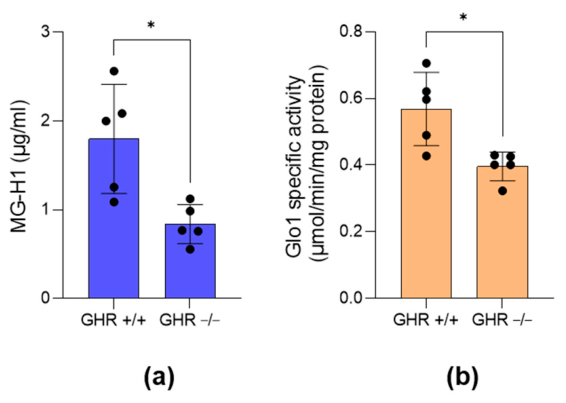
Figure 2.
Basal IGF-1 expression is associated with increased MG-derived glycative stress in aggressive prostate cancer (PCa) cells. LNCaP and PC3 cells, representing less aggressive, androgen-dependent and more aggressive, androgen-independent human prostate cancer (PCa) cell models, respectively, were analyzed for IGF-1 expression at the (a) mRNA level by quantitative real-time PCR (qRT-PCR) and (b) intracellular protein level by enzyme-linked immunosorbent assay (ELISA), (c) MG-H1 levels by ELISA, and (d) GLO1 specific activity by spectrophotometric assay. Data are presented as the mean ± standard deviation (SD) of three independent experiments. **p < 0.01; ****p < 0.0001.
Figure 2.
Basal IGF-1 expression is associated with increased MG-derived glycative stress in aggressive prostate cancer (PCa) cells. LNCaP and PC3 cells, representing less aggressive, androgen-dependent and more aggressive, androgen-independent human prostate cancer (PCa) cell models, respectively, were analyzed for IGF-1 expression at the (a) mRNA level by quantitative real-time PCR (qRT-PCR) and (b) intracellular protein level by enzyme-linked immunosorbent assay (ELISA), (c) MG-H1 levels by ELISA, and (d) GLO1 specific activity by spectrophotometric assay. Data are presented as the mean ± standard deviation (SD) of three independent experiments. **p < 0.01; ****p < 0.0001.
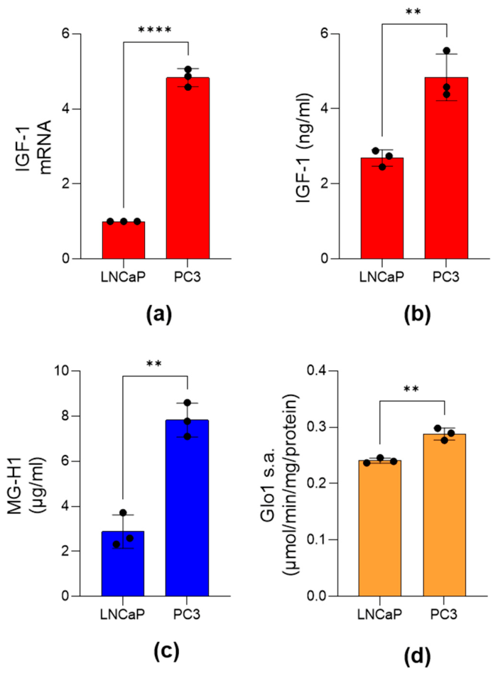
Figure 3.
Effect of IGF-1 on MG-derived glycative stress in LNCaP cells. LNCaP cells were treated with IGF-1 (150 ng/mL) for 72 h or left untreated (control). Markers of MG-derived glycative stress were then evaluated by measuring (a) intracellular MG-H1 levels by enzyme-linked immunosorbent assay (ELISA), and GLO1 expression at the (b) mRNA level by quantitative real-time PCR (qRT-PCR), (c) protein level by Western blot analysis, and (d) functional level as GLO1 specific activity by spectrophotometric assay. β-Actin was used as the loading control for Western blot normalization. Data are presented as the mean ± standard deviation (SD) of three independent experiments. **p < 0.01; ***p < 0.001; ****p < 0.0001 versus untreated cells.
Figure 3.
Effect of IGF-1 on MG-derived glycative stress in LNCaP cells. LNCaP cells were treated with IGF-1 (150 ng/mL) for 72 h or left untreated (control). Markers of MG-derived glycative stress were then evaluated by measuring (a) intracellular MG-H1 levels by enzyme-linked immunosorbent assay (ELISA), and GLO1 expression at the (b) mRNA level by quantitative real-time PCR (qRT-PCR), (c) protein level by Western blot analysis, and (d) functional level as GLO1 specific activity by spectrophotometric assay. β-Actin was used as the loading control for Western blot normalization. Data are presented as the mean ± standard deviation (SD) of three independent experiments. **p < 0.01; ***p < 0.001; ****p < 0.0001 versus untreated cells.
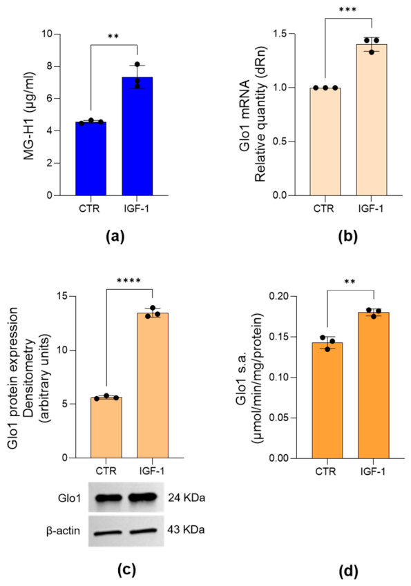
Figure 4.
Effect of IGF-1 on the proliferative and invasive properties of LNCaP cells. LNCaP cells were treated with IGF-1 (150 ng/mL) for 72 h or left untreated (control). Cell proliferation was assessed by (a) cell counting and (b) colony formation assay. Invasive potential was evaluated by (c) an invasion assay and by analyzing the expression of the invasion-related matrix metalloproteinases (d) MMP-1, MMP-7, and MMP-9. Data are presented as the mean ± standard deviation (SD) of three independent experiments. *p < 0.05; ***p < 0.001; ****p < 0.0001 versus untreated cells.
Figure 4.
Effect of IGF-1 on the proliferative and invasive properties of LNCaP cells. LNCaP cells were treated with IGF-1 (150 ng/mL) for 72 h or left untreated (control). Cell proliferation was assessed by (a) cell counting and (b) colony formation assay. Invasive potential was evaluated by (c) an invasion assay and by analyzing the expression of the invasion-related matrix metalloproteinases (d) MMP-1, MMP-7, and MMP-9. Data are presented as the mean ± standard deviation (SD) of three independent experiments. *p < 0.05; ***p < 0.001; ****p < 0.0001 versus untreated cells.
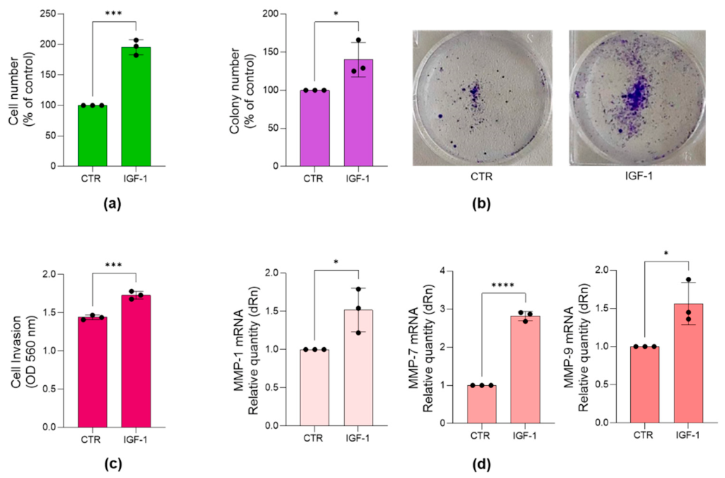
Figure 5.
Effect of IGF-1 on RAGE and OPN expression in LNCaP cells. LNCaP cells were treated with IGF-1 (150 ng/mL) for 72 h or left untreated (control). (a) RAGE and (b) OPN mRNA expression were evaluated by quantitative real-time PCR (qRT-PCR). Data are presented as the mean ± standard deviation (SD) of three independent experiments. **p < 0.01, ***p < 0.001 versus untreated cells.
Figure 5.
Effect of IGF-1 on RAGE and OPN expression in LNCaP cells. LNCaP cells were treated with IGF-1 (150 ng/mL) for 72 h or left untreated (control). (a) RAGE and (b) OPN mRNA expression were evaluated by quantitative real-time PCR (qRT-PCR). Data are presented as the mean ± standard deviation (SD) of three independent experiments. **p < 0.01, ***p < 0.001 versus untreated cells.
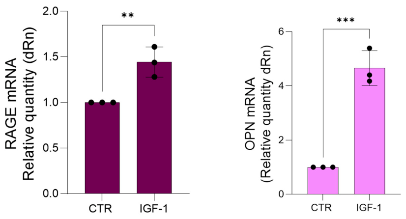
Figure 6.
Effect of aminoguanidine on IGF-1-induced MG-derived glycative stress in LNCaP cells. LNCaP cells were pretreated with aminoguanidine (AG, 50 ng/mL) for 6 h before exposure to IGF-1 (150 ng/mL) for 72 h. MG-derived glycative stress was evaluated by measuring (a) intracellular MG-H1 levels using a specific ELISA assay; GLO1 expression at the (b) mRNA level by qRT-PCR and (c) protein level by Western blot analysis; and (d) GLO1 enzymatic activity using a spectrophotometric assay. β-Actin was used as the loading control for normalization of Western blot data. Data are presented as the mean ± SD of three independent experiments. *** p < 0.001; ****p < 0.0001.
Figure 6.
Effect of aminoguanidine on IGF-1-induced MG-derived glycative stress in LNCaP cells. LNCaP cells were pretreated with aminoguanidine (AG, 50 ng/mL) for 6 h before exposure to IGF-1 (150 ng/mL) for 72 h. MG-derived glycative stress was evaluated by measuring (a) intracellular MG-H1 levels using a specific ELISA assay; GLO1 expression at the (b) mRNA level by qRT-PCR and (c) protein level by Western blot analysis; and (d) GLO1 enzymatic activity using a spectrophotometric assay. β-Actin was used as the loading control for normalization of Western blot data. Data are presented as the mean ± SD of three independent experiments. *** p < 0.001; ****p < 0.0001.
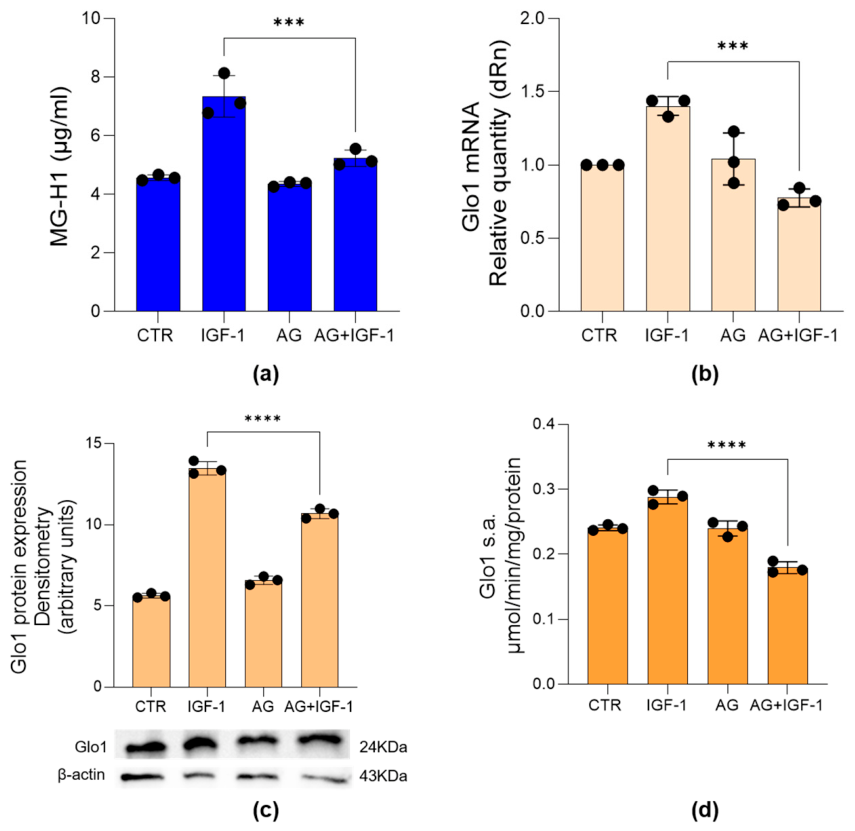
Figure 7.
Effect of aminoguanidine on IGF-1-induced RAGE and OPN expression in LNCaP cells. LNCaP cells were pretreated with aminoguanidine (AG, 50 ng/mL) for 6 h before exposure to IGF-1 (150 ng/mL) for 72 h. The expression of (a) RAGE and (b) osteopontin (OPN) was evaluated at the mRNA level by qRT-PCR. Data are presented as the mean ± SD of three independent experiments. ** p < 0.01; **** p < 0.0001.
Figure 7.
Effect of aminoguanidine on IGF-1-induced RAGE and OPN expression in LNCaP cells. LNCaP cells were pretreated with aminoguanidine (AG, 50 ng/mL) for 6 h before exposure to IGF-1 (150 ng/mL) for 72 h. The expression of (a) RAGE and (b) osteopontin (OPN) was evaluated at the mRNA level by qRT-PCR. Data are presented as the mean ± SD of three independent experiments. ** p < 0.01; **** p < 0.0001.
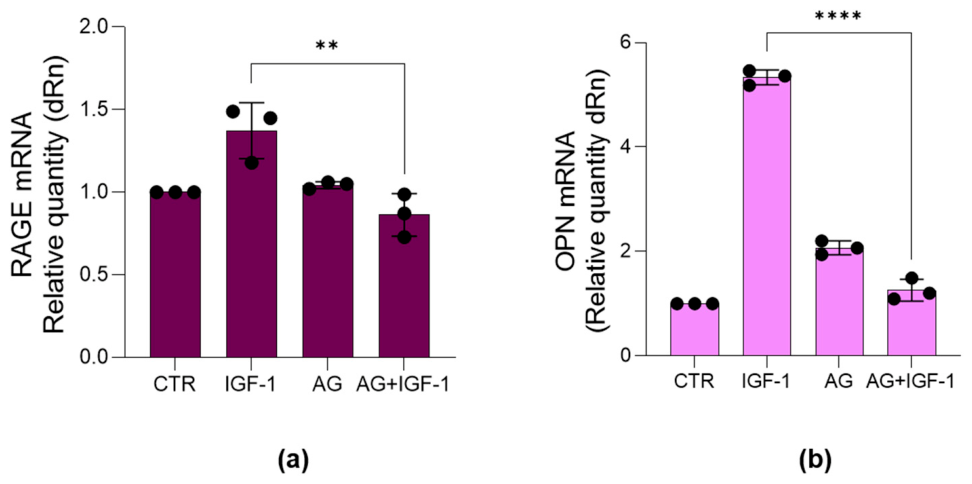
Figure 8.
Effect of aminoguanidine on IGF-1-induced cell proliferation and invasive capacity in LNCaP cells. LNCaP cells were pretreated with aminoguanidine (AG, 50 ng/mL) for 6 h before exposure to IGF-1 (150 ng/mL) for 72 h. Cell proliferation was evaluated by (a) cell counting and (b) colony formation assay. Invasive capacity was assessed by (c) a cell invasion assay and by evaluating the mRNA expression of the invasion-related metalloproteinases (d) MMP-1, MMP-7, and MMP-9 using qRT-PCR. Data are presented as the mean ± SD of three independent experiments. * p < 0.05; ** p < 0.01; *** p < 0.001, **** p < 0.0001.
Figure 8.
Effect of aminoguanidine on IGF-1-induced cell proliferation and invasive capacity in LNCaP cells. LNCaP cells were pretreated with aminoguanidine (AG, 50 ng/mL) for 6 h before exposure to IGF-1 (150 ng/mL) for 72 h. Cell proliferation was evaluated by (a) cell counting and (b) colony formation assay. Invasive capacity was assessed by (c) a cell invasion assay and by evaluating the mRNA expression of the invasion-related metalloproteinases (d) MMP-1, MMP-7, and MMP-9 using qRT-PCR. Data are presented as the mean ± SD of three independent experiments. * p < 0.05; ** p < 0.01; *** p < 0.001, **** p < 0.0001.
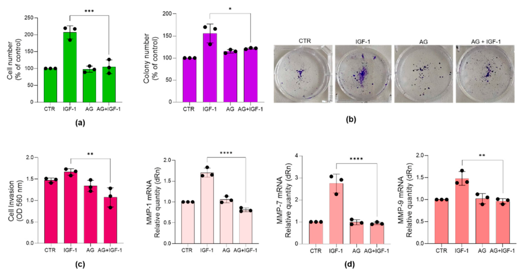

Table 1.
Primer sequences used for quantitative real-time PCR (qRT-PCR).
| Gene | Forward primer (5’→3’) | Reverse primer (5’→3’) |
|---|---|---|
| IGF-1 | GCTCTTCAGTTCGTGTGTGGA | GCCTCCTTAGATCACAGCTCC |
| GLO1 | CTCTCCAGAAAAGCTACACTTTGAG | CGAGGGTCTGAATTGCCATTG |
| RAGE | TGAAGGAACAGACCAGGAGACAC | GCACAGGCTCCCAGACAC |
| OPN | GCCGAGGTGATAGTGTGGTT | TGAGGTGATGTCCTCGTCTG |
| MMP-1 | TTGGGCTGAAAGTGACTGGGAAAC | GGCATGGTCCACATCTGCTCTTG |
| MMP-7 | GCATTTCAGGAAAGTTGTATGGG | CATCCGTCCAGCGTTCATCC |
| MMP-9 | GGCAAGGGCGTCGTGGTTCC | CGGTCGTCGGTGTCGTAGTTGG |
| β-actin | CACTCTTCCAGCCTTCCTTCC | ACAGCACTGTGTTGGCGTAC |
Disclaimer/Publisher’s Note: The statements, opinions and data contained in all publications are solely those of the individual author(s) and contributor(s) and not of MDPI and/or the editor(s). MDPI and/or the editor(s) disclaim responsibility for any injury to people or property resulting from any ideas, methods, instructions or products referred to in the content. |
© 2026 by the authors. Licensee MDPI, Basel, Switzerland. This article is an open access article distributed under the terms and conditions of the Creative Commons Attribution (CC BY) license.
Copyright: This open access article is published under a Creative Commons CC BY 4.0 license, which permit the free download, distribution, and reuse, provided that the author and preprint are cited in any reuse.