

Submitted:
17 July 2026
Posted:
20 July 2026
You are already at the latest version
Abstract
Cellular identity determination and lineage tracing is a pivotal technique in modern biological research. Conceptually simple yet efficient cell labeling techniques offering broader applicability are warranted despite many methods requiring intricate construction procedures do exist. One such technique is ADAR (adenosine deaminase acting on RNA)-mediated RNA sensing, a live cell labeling technique that is based on the expression and abundance of cell-type-specific RNAs. Here, we utilized the optimized version, CellREADR (Cell access through RNA sensing by Endogenous ADAR)1, to establish a feasible tracing system for mouse spermatogonial stem cells (mSSCs) which are refractory to CRISPR-based reporter gene knock-in. We identified several previously unreported features of CellREADR, including RNA interference induced by double-strand RNA formation that is obligatorily generated during the normal operation of the CellREADR system. More importantly, we established its application for long-term labelling of mSSCs and monitoring mSSCs differentiation induced by retinoic acid (RA) treatment in vitro. This work offers a practical solution for dynamic monitoring of mSSCs self-renewal and differentiation and supports that CellREADR can be developed into more versatile and efficient tools in stem cell research.
Keywords:
live-cell labeling
; RNA sensing
; cell type identification
; spermatogonial stem cells
Introduction
The diverse cell types and their specialized functions in a multicellular organism are determined by the expression products of the same genome including transcripts, proteins, and epigenetic modifications[2,3]. Accurate discrimination and tracing of cellular identity based on their intrinsic molecular profiles represent an important aspect of modern cell biology. Contemporary methods are mainly based on the direct detection of gene products using RT-PCR, immunostaining, or various omics technologies. Although these methods provide greater accuracy than methods based on morphological descriptions[4], they usually compromise cell integrity and viability[5,6,7,8]. In contrast, live-cell labeling technologies offer a promising alternative by enabling long-term, real-time monitoring of cells.
Live-cell labeling relies on the engineering of gene expression circuits, resulting in novel physical or chemical features that allow cells to be monitored noninvasively[9,10,11]. The most commonly used labeling technology involves employing the CRISPR/Cas9 system to insert reporter genes into specified genome loci, thereby reflecting endogenous gene expression through the activity of reporter genes to indicate cell states. However, this approach is hindered by several limitations including low success rates, time-consuming procedures, and the potential risk for the inserted reporter gene to disrupt the function of endogenous genes[12,13,14]. As the expression levels of microRNAs exhibit significant variability across different cell types[15], a microRNA-based live-cell labeling method known as microRNA switch, has been developed to identify cellular identity transitions and to purify specific cell types derived from induced pluripotent stem cells (iPSCs)[16,17,18]. Despite their potential, microRNA switch is plagued by issues such as high background noise.
In the past several years, several ADAR-mediated RNA sensing methods have been developed including CellREADR[1], RADAR (RNA sensors using ADAR)[19], RADARS (reprogrammable ADAR sensors)[20], ADAR-recruiting hairpin RNA sensor[21]. These technologies exploit the endogenous RNA-editing activity of ADAR to convert cell-type-specific RNA expression into detectable reporter signals. The principle involves designing sensor RNA (sesRNA) sequences that are partially complementary to the endogenous target RNAs. A stop codon “TAG” in frame is introduced near the center of the sesRNA sequence to create an A:C mismatch within the endogenous sequence “ACC”. This partial double-strand RNA (dsRNA) recruits the ADAR to convert “A” to “I” by deamination in the mismatched base pair (Figure 1a). “I” is recognized as “G” within the cell, allowing the subsequent translation of the effector gene following the sesRNA[22,23]. The ubiquitous presence of ADAR in cells eliminates the need to introduce exogenous components analogous to the CAS9 protein of the CRISPR-Cas9 system. Moreover, ADAR-based RNA sensing exhibits low cytotoxicity[1]. However, many aspects of CellREADR technology remain unclear, and its application in tracing stem cell differentiation has not been explored.
Spermatogonial stem cells (SSCs) hold great promise for applications in human reproduction and animal breeding[24,25]. To date, credible reports of stable SSC line establishment are limited to mouse, rat, and tree shrew[26,27,28]. Among these, mouse SSCs (mSSCs) are widely considered the most representative model, owing to their well-established culture systems and extensive biological characterization. In contrast, genetic labeling approaches are still difficult to implement in mSSCs[29]. Given the simplicity and versatility of CellREADR, we evaluated its potential as a practical strategy for labeling and monitoring mSSCs.
We firstly investigated the effects of various factors on the efficiency of CellREADR, including the Woodchuck Hepatitis Virus Posttranscriptional Regulatory Element (WPRE), different mismatch types, and stop codon selection in sesRNA. Subsequently, we demonstrated the cell-type-specific discrimination capability of CellREADR in human embryonic kidney 293FT (HEK293FT) and HeLa cell lines. We further evaluated the accuracy on similar sequence detection. Finally, we implemented this technology in mouse embryonic stem cells (mESCs) and mSSCs to explore its potential in identifying cell types and monitoring differentiation.
Methods
sesRNA Design
All sesRNA sequences in this article were designed strictly following the steps outlined by Qian, Y, et.al[1]. It should be noted that, based on our results, replacing the functional stop codon in sesRNA 5'-TAG-3' with 5'-TGA-3' is also feasible, if it could reduce the formation of complex secondary structures. The specific sequences are detailed in Supplementary Table 3.
Plasmid and Clone
All primers used in this study were synthesized by Tsingke Biotechnology Co. Ltd (Beijing, China). The sesRNA plasmid backbones were derived from pCMV-PE2 (Addgene plasmid # 132775) and PLV-huGDNF (Addgene plasmid # 182946). All plasmid constructions followed standard molecular cloning procedures. The molecular reagents used included but not limited NheI-HF® (NEB, R3131), BamH1-HF® (NEB, R3136), BsaI-HF®v2 (NEB, R3733), KpnI-HF® (NEB, R3142), 2×TransStart® FastPfu Fly PCR SuperMix(-dye) (TransGen Biotech, AS231), Escherichia coli Trans1-T1 (TransGen Biotech, CD501), Zymoclean Gel DNA Recovery Kit (ZYMO RESEARCH, D4002), EndoFree Plasmid Midi Kit (CWBIO, CW2105S), ClonExpress Ultra One Step Cloning Kit (Vazyme,C115), and DNA Ligation Kit Ver.2.1 (Takara,6022).
Cell Culture
All cells were cultured at 37°C with 5% CO2 and mycoplasma-free. Different cell lines were maintained in diverse media compositions. Briefly, HeLa, HEK293FT, STO, STO-GDNF and MEF cells were cultured in DMEM (Gibco, 12100046) supplemented with 10% FBS (cytiva, sv30160) and 1% penicillin-streptomycin (Gibco, 15140122). Cells were digested by 0.25% trypsin-EDTA (Gibco, 25200) then passaged at a 1:3 ratio upon reaching confluence. MEF cell at passage 5 treated by 10 μg/ml mitomycin C (Sigma, M0503) for 2.5h could be feeder cells to support the growth of mESCs and mSSCs. mESCs were typically cultured on MEF feeder using N2B27 medium consist of DMEM/F12 (Thermo Fisher Scientific, 11330-032), neurobasal (Thermo Fisher Scientific, 21103-049), 1× N2 supplement (Thermo Fisher Scientific, 17502-048), 1× B27 supplement (Thermo Fisher Scientific, 12587-010), 2 mM L-glutamine (Gibco, 25030), 1× NEAA (Gibco, 15140), 100 μM 2-mercaptoethanol (Sigma, M7522), 1% penicillin-streptomycin (Gibco, 15140122), 103 units/mL mLIF (Milllipore, ESG1107), 3 μM CHIR 99021 (Tocris, 4423) and 1 μM PD 0325901 (Tocris, 4192). When transfection is required, mESCs originally cultured on MEF feeder need firstly to be pre-incubated after terminating digestion by DMEM supplemented with 10% FBS and 1% penicillin-streptomycin to remove feeder cells and then planted into poly-L-ornithine-laminin-coated wells. For preparing poly-L-ornithine-laminin-coated wells, briefly, the wells were coated with 0.01% poly-L-ornithine at 25°C for more than 1h, then continuously coated with 300 ng/mL laminin (Yeasen, 92602ES) again. About mSSCs, we have exactly described the establishment and culture method before[29]. Briefly, mSSCs were established from 3-month-old C57BL/6 mouse through two-step digestion method, first digestion system is 1 mg/mL Dnase1 (AppliChem, A3778) plus 1 mg/mL collagenase1 (Gibco, 17100-017), second is 0.25% trypsin-EDTA. mSSCs were typically cultured on mitomycin C-treated MEF feeders with specific culture medium consisted of MEMα (Gibco, 12571), 2 mg/mL BSA (MP, 9078-46-8), 5 μg/mL insulin (Sigma, I2643), 30 nM Na2SeO3 (Sigma, 2114485), 25 μg/mL transferrin (Sigma, I2643), 10 μg/mL putrescine (Sigma, P5780), 2mM L-glutamate (Gibco, 25030), 1×lipoprotein (MP, 191476), 7.6 μeq/L free fatty acid cocktail, 5 ng/mL human bFGF (peprotech, 400-29), 20 ng/mL human GDNF (R&D, 212-GD), 20 μg/mL L-ascorbic acid (Sigma, A4544), 5.5 μM 2-mercaptoethanol (Sigma, M7522) and 1× Penicillin streptomycin. Fresh medium changes every two days and cell was passaged at a 1:2 every four days.
Plasmid transfection
All transfection experiments were conducted in 24-well plates when the cell density was approximately 80%, and the fresh culture medium was replaced before transfection. Unless otherwise specified, the total amount of DNA transfected per well was 1 μg. When HEK293FT cells were used exclusively, they were transfected with TransIntro® PEI Transfection Reagent (GMP Grade) (TransGen Biotech, FT401). The transfection mixture consisted of 1 μg DNA + 3 μL PEI + 25 μL PEI buffer, incubated at room temperature for 15 minutes before transfection. For mESC transfections or co-transfections of HEK293 and HeLa cells, Lipofectamine™ 2000 (Thermo Fisher Scientific, 11668019) was used according to the manufacturer's instructions. For STO and STO-G cell lines, Lipofectamine LTX (Invitrogen, 15338100) were used according to the manufacturer's protocol, and the optimized reagent composition is 2μL LTX + 1μg DNA +1μL PLUS™ Reagent.
Flow Cytometry Analysis and Sorting
Forty-eight hours after transfection, cells were digested with 0.25% Trypsin-EDTA for 2 minutes at 37°C, then terminated with DMEM + 10% FBS + 1% penicillin-streptomycin. The digested cell suspension was filtered through a 400-mesh filter membrane into specific flow cytometry tubes and transported and stored on ice until analysis. The raw data were analyzed using FlowJo V10 software. For sorting, the same procedure was followed, except that the collected cells were centrifuged at 600g for 6 minutes, the supernatant was discarded, and the cells were reseeded into culture plates.
RNA Extraction and RT-qPCR
Total RNA was extracted from cells using TRIzol reagent (Thermo Fisher Scientific,15596026CN) following the manufacturer's instructions. 1 μg of RNA underwent reverse transcription following the protocol (Vazyme, R323). The resulting cDNA was diluted fivefold and used as a template for qPCR. RT-qPCR analysis was performed using SYBR dye (Vazyme, Q712) in a LightCycler® 96 Instrument. Gene-specific primers were designed by PrimerBank (harvard.edu) and are listed in the Supplementary Table 2.
Western Blot
Primary antibody to ADAR1 (Beyotime, AF6153, 1:1000) and β-actin antibody (Sigma, A5316, 1:2500) were used in this study. The cells from one well of a 12-well plate were collected and lysed using 190μL 2× Laemmli Sample Buffer (Bio-Rad, 1610737) supplement with 10μL 355mM 2-mercaptoethanol and 1× protease inhibitor cocktail (Mei5bio, MF182), followed by heating at 100°C for 10 minutes. 5μL lysate was loaded for SDS-PAGE (LabLEAD, P41212) and run at 120V for 70min. Then the protein was transferred onto 0.45μm PVDF membrane (Vazyme, E801-02-AA) using Trans-Blot Turbo Transfer Starter System (Bio-Rad, 17001915). The PVDF membrane was blocked by 5% DIFCOTM Skim Milk (BD, 232100) for 1h and then incubated overnight by primary antibody at 4°C. Secondary antibody including HRP-labeled goat anti-rabbit IgG(H+L) (Biodragon, BF03008, 1:2000) and HRP-labeled goat anti-mouse IgG(H+L) (Biodragon, BF03001, 1:2000) was used in this study. After completion of primary antibody incubation and washed three times by TBST (Lablead, T7209), the PVDF membrane was incubated secondary antibodies for 1 hour at room temperature and then washed with TBST three times. The result was imaged using BeyoECL Plus (Biyotime, P0018) with ChemiDOx XRS+ system.
Multiple Sequence Alignment (MSA) and Visualization
The cDNA and protein sequences of mGdnf, huGDNF, and opti-cod huGDNF were aligned by Clustal Omega < EMBL-EBI. The visualization of the similarity matrix and sequence annotation derived from MSA was implemented by the pheatmap package in R and the Jalview software.
Lentivirus Preparation
HEK293 cells were seeded in 10 cm dishes and grown until they reached approximately 80% confluence. The culture medium was replaced with antibiotic-free medium before transfection. The cells were transiently transfected with PEI using 8 μg of the sesRNA plasmid, 2 μg of pMD2.G (Addgene plasmid # 12259), and 4 μg of psPAX2 (Addgene plasmid # 12260). The medium was changed to fresh medium 16 hours post-transfection. The supernatant was collected at 48- and 72-hours post-transfection and centrifuged at 600g for 6 minutes. The collected supernatant was filtered through a 0.45 μm filter and transferred to specific lentivirus centrifuge tubes. An adequate volume of PEG8000 (Beyotime, ST483) was incorporated to reach a final concentration of 8.5% (m/v). The mixture was then thoroughly combined at 4°C for 4 hours, followed by centrifugation at 10,000 rpm at 4°C for 20 minutes. The supernatant was discarded, and the pellet was resuspended in one-tenth of the volume of the collected supernatant using MEMα. The lentivirus was aliquoted into 50 μL volumes and stored at -80°C.
mSSC Lentivirus Infection
The digested mSSCs suspension was seeded into new wells at a 1:2 ratio based on the original culture plate area and pre-incubated at 37°C with 5% CO2 for 15 minutes to remove some of the MEF feeders. The pre-incubated cell suspension was collected, centrifuged, and the supernatant was discarded. The cells were then seeded at 500,000 cells per well in laminin-coated 12-well plates. After 1 day of culture, the lentivirus suspension mixed with polybrene (1:1000) (Yeasen, 40804ES) was added to the mSSC culture medium. The medium was replaced 1 day after infection.
RA treat mSSCs
20,000 feeder-free mSSCs was seeded into laminin coated 24-well dish, then kept culturing for 24h then replacing new medium with a final concentration of 100nM RA (Sigma, 302-79-4). After treatment for 24h, the fresh medium was replaced. Notably, RA is photosensitive and easy to degrade. Therefore, RA needs to be prepared just before use, and protected from light throughout the experiment.
Cell Plating and Immunofluorescence Staining
For immunofluorescence in 24-well plates, pre-place coverslips into wells and coat with laminin. 20,000 cells were seeded per well and kept culturing for 24 hours, and then using 200 μL of 4% (m/v) paraformaldehyde (Thermo Fisher Scientific, 169650010) per well to fix cells for 15 minutes at room temperature. Wash wells 3 times with DPBS (Gibco, 21600010), 5 minutes per wash. Permeabilize cells with 200 μL of 0.1% (m/v) Triton X-100 (Sigma, 234729) per well for 15–20 minutes at RT. Wash 3 times. Each well was blocked with 200 μL of 5% BSA for 1 hour at room temperature. Primary antibody to PLZF (R&D, AF2944, 1:200) was used in this study. 50 μL of diluted primary antibody solution was used per well then incubating overnight at 4°C. Secondary antibody used was Donkey anti-Goat IgG (H+L) Cross-Adsorbed Secondary Antibody, Alexa Fluor™ 488 (Thermo Fisher Scientific, A11055,1:1000). 200 μL of diluted secondary antibody solution was added per well and incubated for 1 hour at room temperature in the dark followed by DAPI (Beyotime, C1002, 1:1000) incubation for 10min. After completion, coverslips were carefully transferred onto glass slides dripped with antifade mounting medium (Beyotime, P0126).
Statistical Analysis
All data were obtained from three independent biological replicates. Data analysis was performed using GraphPad Prism 8. Data are presented as mean ± SD. Independent sample two-tailed t-tests or one-way analysis of variance (ANOVA) were conducted to assess significance. Statistical significance was defined as *P < 0.05, **P < 0.01, ***P < 0.001, and ns (not significant) for P > 0.05.
Results
Validation and Further Characterization of CellREADR
Based on the design principles outlined in a previous study, we constructed the reporter system with mCherry as the indicator and copGFP as the effector (Figure 1b), sesRNA targeting the endogenous EEF1A1 (ses-EEF1A1) as the primary test case[1]. We replaced the sesRNA sequence with a short linker containing or lacking the stop codon in frame as the negative (ses-negative) and positive control (ses-positive), respectively. We noted that the WPRE, which is usually used in viral vectors to enhance gene expression by post-transcriptional regulation[30], is present in the CellREADR plasmid construct[1]. Its absence in RADAR, RADARS, ADAR-recruiting hairpin RNA sensor technologies may stem from the typical use of WPRE to enhance virus-related infectivity[31,32], while its effect on the performance of ADAR-mediated RNA sensing remains undiscussed. To explore this, we further incorporated the WPRE into both the negative control (ses-negative-W) and the ses-EEF1A1 system (ses-EEF1A1-W).
It was expected that the ses-negative construct should result in low percentage of green cells in the red population, which could be a result of the leaky expression of copGFP, when the plasmid was delivered into HEK293FT cells. By allowing at most 1% of cell conversion rates generated by the ses-negative construct. Accordingly, we achieved cell conversion rates of 3.99 ± 0.50%, 18.50 ± 0.95%, 26.23 ± 3.15% and almost 100% for the ses-negative-W, ses-EEF1A1, ses-EEF1A1-W and ses-positive constructs, respectively (Figure 1c, d and Supplementary Figure S1). These results not only validated the effect of CellREADR but also demonstrated that WPRE integration significantly enhances the positive signal generated by CellREADR, despite that it also slightly increases the background signal. Based on this, we keep WPRE in the plasmid for subsequent experiments.
Additionally, we observed that, compared with what was observed in ses-positive cells, mCherry fluorescence intensity in ses-EEF1A1 and ses-EEF1A1-W cells but not in ses-negative and ses-negative-W (Figure 1e). We reasoned that RNA sensing process likely triggered RNA degradation, supported by the observation that ADAR1 participates in RNAi by forming a complex with Dicer[33]. To investigate this further, we transfected the ses-negative-W, ses-EEF1A1-W, and ses-positive plasmids into HEK293FT cells and sorted cells with strong mCherry signal. RT-qPCR with validated copGFP-targeted primers revealed the expression of the plasmid-derived exogenous sequence in the ses-EEF1A1-W group was reduced 2.6- to 4.0-fold relative to the ses-negative-W and ses-positive groups. However, no significant differences in endogenous EEF1A1 expression among these groups (Figure 1f and Supplementary Figure S2). These data suggest that RNA sensing preferentially destabilizes the exogenous sesRNA-containing transcript rather than the endogenous target.
Mismatch Optimization and Mutation Detection of CellREADR
ADAR mediated adenosine deamination preferentially occurs in the A-C mismatch site[34,35], but alternative mismatches must be considered when endogenous RNA sequence lacks suitable “CCA” site to design sesRNA (Figure 2a). To investigate this, we designed a single sesRNA variant harboring both A:A and A:G mismatches. This design ensures that the two mismatch types share an identical sequence context and secondary structure, thus eliminates confounding effects arising from differences in nucleotide abundance or variations in RNA secondary structure (Figure 2b).
The cell conversion rates of A:A and A:G mismatches were 16.50 ± 0.95% and 15.80 ± 1.75%, respectively, significantly lower than that of the A:C mismatch, which was 29.60 ± 0.35% (Figure 2c). These was no significant difference between A:A and A:G mismatches. This shows that A:C mismatches are optimal, but A:A and A:G mismatches are viable alternatives when A:C mismatches are not feasible. Moreover, sesRNA of the A:A and A:G mismatches also resulted in the attenuation of mCherry fluorescence intensity like the sesRNA of the A:C mismatch (Figure 2e and Supplementary Figure S3a).
Additionally, we noted that both the “TAG” and “TGA” stop codons can form A:C mismatches with “CCA” (Figure 2a). To test whether these two stop codons can be used in sesRNA and whether their uses result in any difference, we modified the ses-EEF1A1 sequence by replacing “TAG” of sesRNA with “TGA” (Figure 2b) and found no difference in the cell conversion ratio (Figure 2d). This suggests that the “TGA” stop codon can be tried when the use of “TAG” is not satisfying.
Previous studies have demonstrated that RADAR can detect mutations at specific sites despite of low sensitivity [19]. To explore the ability of CellREADR in mutation detection (Figure 2f), we selected two housekeeping genes in HEK293FT, FANCF and EMX1, which commonly serve as the targets in prime editing[36,37]. We first generated plasmid constructs from which mRNA fragments of the gene of interest (GOI) containing a single-base mutation can be transcribed under the U6 promoter. Constructs of sesRNA targeting the mutant region of GOI mRNAs, ses-FANCF-W and ses-EMX1-W, were accordingly designed and generated. HEK293FT cells were transfected with the ses-RNA plasmid with or without the plasmid for the mutant GOI (Figure 2g). In the absence of the mutant plasmids, the ses-EMX1-W and ses-FANCF-W groups exhibited cell conversion ratio of 0.85% ± 0.19% and 2.90% ± 0.04%, respectively. In the presence of the mutant plasmids, the positive rates significantly increased to 3.07% ± 0.77% and 11.30% ± 0.61% for ses-EMX1-W and ses-FANCF-W, respectively (Figure 2h and Supplementary Figure S3b). These results indicate that mutant mRNAs can be detected by CellREADR although the sensitivity warrants further investigation.
Distinguishing Two General Cell Lines by CellREADR
The sesRNA designed targeting Fezf2 (zinc-finger transcription factor) and Ctip2 (B cell leukemia/lymphoma 11B) achieved >90% specificity in distinguishing two types of neurons in mouse cortex[1]. We assessed the performance of CellREADR in distinguishing HeLa and HEK293FT cell lines to further assess its suitability for detecting cell identity in common cell lines (Figure 3a). By comparing the transcriptomic profiles of these two cell lines, we selected six candidate genes that display high specificity in their expression patterns: KRT7, SPINK6, and PED2A exhibiting HeLa-specific expression, while LCP1, NEFM, and CSNK2B showing preferential expression in HEK293 cells (Supplementary Table 1)[38]. Based on both expression specificity and sesRNA design feasibility, KRT7 was selected as the HeLa-specific biomarker, whereas NEFM was chosen as the HEK293FT-specific biomarker (Figure 3b).
We subsequently designed ses-KRT7-W and ses-NEFM-W, constructed the corresponding expression vectors, and delivered the plasmids into both Hela and HEK293FT cells. While ses-KRT7-W showed comparable positivity rates in both cell types, ses-NEFM-W exhibited clear cell-type specificity. Specifically, ses-NEFM-W demonstrated only background-level signals (similar to ses-negative-W group) in HeLa cells, but achieved an average 12% positivity rate in HEK293FT cells (Figure 3c, d). These results indicate that CellREADR can be used to discriminate different cell lines given that marker genes are reasonably selected and the performance of corresponding sesRNAs are experimentally validated.
Limited Accuracy of CellREADR for Targeting Similar Sequences
It is a common practice in molecular biology to express and detect homologous and/or codon-optimized transgenes in cells, and also to readily distinguish expressing cells from the bulk population. We wondered whether CellREADR can be used for this application. We first derived a STO cell line that expresses a codon-optimized human GDNF (opti-cod huGDNF) stably, here referred to as STO-GDNF (Figure 4a and Supplementary Figure4a, b). Based on the opti-cod huGDNF sequence, we designed the corresponding sesRNA (ses-GDNF-W). By aligning the sequences of mouse Gdnf (mGdnf), human GDNF (huGDNF), and opti-cod huGDNF, we found that the sequence similarities between mGdnf and opti-cod huGDNF, as well as between huGDNF and opti-cod huGDNF, were 71.54% and 74.53%, respectively (Figure 4b). Alignment of ses-GDNF-W with endogenous human and mouse mRNA sequences identified many mismatched sites within the coding regions. The mismatch formed between huGDNF and ses-GDNF-W was A:A (Supplementary Figure 4c), and RT-qPCR confirmed abundant expression of endogenous levels of endogenous Gdnf/GDNF in STO, STO-GDNF, and HEK293FT cells (Figure 4c). Moreover, both STO and STO-GDNF cells exhibited abundant and comparable ADAR1 protein expression (Figure 4d), indicating that differences in CellREADR activity would be unlikely to arise from variations in ADAR availability.
To determine whether CellREADR could selectively detect the codon-optimized transcript, ses-GDNF-W was introduced into both STO and STO-GDNF cells using the optimized transfection system (Supplementary Figure 5a, b). Surprisingly, both cell lines exhibited nearly identical positive rates, 8.73 ± 0.74% in STO and 8.83 ± 1.97% in STO-GDNF (Figure 4e and Supplementary Figure 5c). We concluded that multiple mismatches between sesRNA and the endogenous target sequence do not prevent dsRNA formation or impair the efficiency of the RNA sensing system.
To determine whether this lack of discrimination was specific to STO cells, we sought to validate the finding in an independent cellular context, we conducted experiments in HEK293FT cells. As no HEK293FT-GDNF cell line analogous to STO-GDNF was available, we transiently expressed opti-cod huGDNF in these cells (Figure 4f). Results showed no significant difference in cell conversion rate between HEK293FT cells transiently expressing opti-cod huGDNF and wild-type (WT) HEK293FT cells. The positive rates for these two groups were 46.27 ± 2.74% and 44.00 ± 2.82%, respectively (Figure 4g, h and Supplementary Figure 5d). These results reflect the tolerance of the system to extensive sequence mismatches independent of cell types.
Stem Cell Lines Identification by CellREADR
To assess the feasibility of CellREADR in stem cell identification, we first tested its performance in mESCs with mouse embryonic fibroblast (MEF) cells as a negative control. We first examined the mRNA and protein expression of ADAR1 and ADAR2 in several cell lines including mESCs and mSSCs (Figure 5a). Both ADAR1 and ADAR2 exhibited high expression in mESCs, indicating that mESCs possess the molecular machinery necessary for effective CellREADR application. Then the classic marker genes Oct4 and Klf4 of mESCs was chosen as targets[39], and the corresponding sesRNAs, ses-Oct4-W and ses-Klf4-W, were designed. Two days after transfection of sesRNA plasmids into mESCs and MEF cells separately, we observed the cell conversion signal exclusively present in mESCs using ses-Klf4-W (Figure 5b), but ses-Oct4-W was invalid in both cell types (data not shown).
Subsequently, we tested the feasibility of CellREADR in mSSCs, in which the two isoforms of ADAR1 are also detected as in mESCs (Figure 5a). Transcription factor PLZF is a specific marker for mouse undifferentiated spermatogonia including mSSCs and is not expressed in somatic cells in the testis or MEF cells that is used as feeder cells in mSSCs culture[40], and we designed ses-Plzf-W according to its mRNA. Since SSCs cannot be transfected efficiently using common lipid-based methods, we used lenti-virus to delivery ses-Plzf-W into mSSCs[41]. Moreover, the CMV promoter has low activity in mSSCs, we replaced it with the EF1α promoter (Figure 5c).
mSSCs are routinely maintained on MEF feeder cells. At the beginning of the experiment, mSSCs were plated on laminin-coated dish with a small number of MEF cells co-cultured as the negative control. Three days after twice lentiviral infections, copGFP expression was observed in a small number of mSSCs but not in any of the MEF cells (Figure 5c, d), which means mSSCs were specifically labeled by CellREADR. Given that the expression cassettes in lentiviral vector can be integrated into the host genome[42], durable long-term expression of the exogenous gene is achieved. We next assessed whether the copGFP-positive mSSCs could be maintained during long-term culture. After three passages, small clusters of mSSCs remained positive for both mCherry and copGFP (Figure 5e), demonstrating stable reporter expression. Collectively, these results indicate that mSSCs can be specifically labeled by CellREADR with long-term stability.
Monitoring mSSCs Differentiation by CellREADR
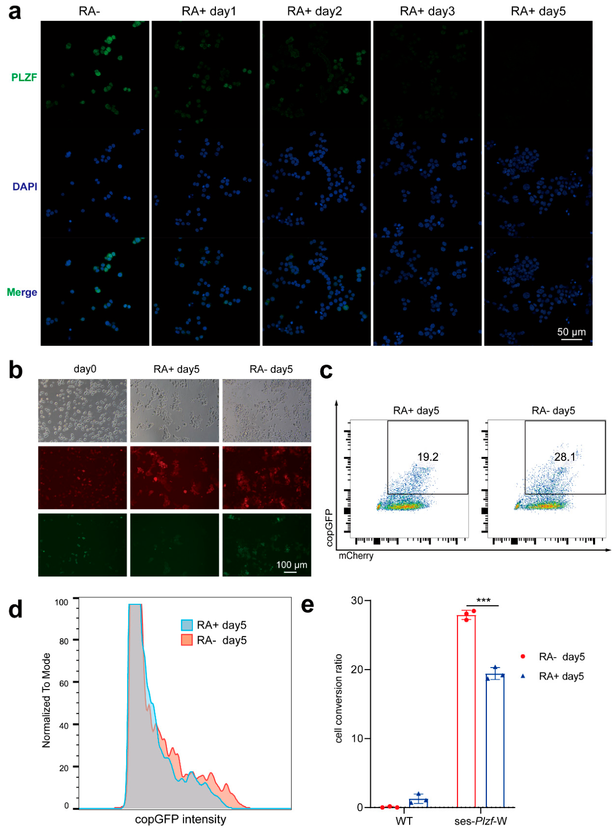
Our previous study showed that the cultured mSSCs could be induced by RA to differentiate into spermatocytes, with Plzf RNA expression gradually reduced[43]. Consistent with this, PLZF protein expression diminished after 5 days of induction (Figure 6a). To assess the sensitivity of CellREADR in monitoring cellular differentiation, we plated labeled mSSCs (Figure 5e) onto laminin-coated plate. These cells were treated with RA to induce differentiation, with untreated cells as a control. WT mSSCs served as a parallel control.
Five days after inducing, fluorescence microscopy and flow cytometry analysis revealed a significant decrease in both the percentage of copGFP-positive cells and the mean fluorescence intensity in the RA-treated mSSCs compared to controls (Figure 6b, c, d). Moreover, the cell conversion rate in the RA-treated group was 19.43 ± 0.87%, markedly lower than the 27.96 ± 0.67% observed in the untreated group (Figure 6e). The persistence of residual copGFP signal in cells 5 days after RA treatment when PLZF is undetectable probably attributes to the asynchronous degradation process and long life-span of copGFP. Collectively, these results indicate that CellREADR enables monitoring of mSSCs differentiation status, even in the presence of delayed effector signal clearance.
Discussion
Cellular identity characterization and dynamic monitoring, also known as lineage tracing, is a common technology in biomedical research. We are particularly interested in how to identify mammalian SSCs and monitor their self-renewal and differentiation in vitro. Although mSSCs can be readily expanded and induced to differentiation into early spermatocytes[43,44], SSCs from large animals such as humans and pigs are extremely hard to culture due to slow growth and low abundance among all testicular cells[45,46]. Therefore, it is urgent to for us to develop live cell tracking techniques that are simple and reliable. To this end, we test the applicability of the CellREADR system that has been reported recently[1]. We first tested its performance in commonly used cell lines, HEK293FT and Hela cells, and then in stem cells such as mESCs and mSSCs. Our results confirmed its potential values in future application and also revealed some problems that should be solved in the future for this technology becomes more practical.
We investigated the effects of other features of CellREADR such as the use of A:A and A:G mismatches instead of A:C, and the use of stop codon TGA instead of TAG. Our results show that CellREADR performs well under these conditions, therefore, we provided more choices to satisfy the basic requirement of the system. Moreover, we confirmed that CellREADR has the potential as an effective approach for genetic diagnosis through specific detection of mutations at defined loci. Although CellREADR cannot distinguish between highly homologous sequences, our data show that an efficient sesRNA can be used to detect homologous sequences from different species or designed artificially as long as the homology between the sesRNA and a target RNA are high enough, the threshold may need to be determined in a case-dependent way.
One concern is that the effector copGFP in the ses-RNA construct can be precociously expressed due to non-specific targeting of the ses-RNA by other partially complementary RNAs or by alternative translation initiation. In this study, we repeatedly found that the ratios of copGFP+ cells among mCherry+ cells (conversion rates) are satisfyingly low in the absence of target RNAs even if the ATG start codon is included at the beginning of copGFP coding sequence (Figure 1b). This means that this method is highly specific. However, it is not sensitive enough as the conversion rates less than 50% for all cases examined. The low sensitivity of CellREADR may results from the low-level expression of ses-RNA. We found that incorporating WPRE in sesRNAs can enhance their stability and/or translation efficiency. Indeed, the conversion rate increased from 18% to 26% (Figure 1d). One possible cause for the low-level expression of sesRNAs is RNAi-induced RNA degradation as double stand RNA structure is inevitable. We not only confirmed the existence of RNAi between sesRNAs and target RNAs but found that the expression level of sesRNAs but not that of the endogenous target RNAs was significantly reduced as indicated by the reduced mCherry intensity and reduced transcript abundance of copGFP (Figure 1e and Figure 1f). Other explanations for the low sensitivity include the heterogeneous expression of the cellular RNAs due to cell cycle progression or other random events inherent to the system that warrants further investigations.
In the present study, we first showed that HEK293FT and HeLa cells can be identified by using CellREADR if target genes are carefully selected based on their differential expression and experimentally tested. However, its performance in stem cells has not been evaluated. We then reported that mESCs and mSSCs can be identified based on the expression of certain marker genes. In particular, mSSCs can be identified among co-cultured MEF cells and their RA-induced differentiation can be observed based on the reduction of the conversion rate. Our results suggested that CellREADR should not be intended as a universal method for detecting gene expression as it often failed on a particular target gene that seemed promising before experimental test. For example, Oct4, the well-known stem cell marker gene failed in mESC identification. However, CellREADR can be used as a valuable tool for cell type/state identification as long as the right target gene is selected.
Another concern for CellREADR is that using RNA-level rather than protein-level differences as the basis for cell identity annotation can be inappropriate under certain specific circumstances. Protein expression depends on RNA templates transcribed from DNA, meaning that the RNA of a cell marker must appear prior to the protein. Yet in some physiological events, RNA translation significantly lags behind DNA transcription—a delay caused by dedicated translational repression mechanisms rather than simply reflecting the sequential order of transcription and translation. Hence, the presence of RNA cannot be equated with the presence of protein. For instance, genes required for late spermatid development are transcribed in advance and stored in a translationally repressed state as messenger ribonucleoproteins (mRNPs)[47]. This specialized phenomenon is typically considered only in studies of specific cell types and particular developmental processes. In general, under steady-state conditions, cellular RNA and protein levels largely correspond, making it acceptable to infer protein levels from mRNA abundance. Thus, specific detection and recognition of marker RNAs represents another reasonable strategy. It should be noted that “steady state” is broadly defined here: during cell proliferation, differentiation, or other biological events, as long as intracellular RNA or protein levels remain relatively stable over an extended period, the cell can be considered to be at steady state[48].
Finally, the future optimizations of this promising system include the following: First, sensitivity enhancement by more careful selection of marker genes and more rational design of sesRNAs to endure higher level expression of sesRNAs with reduced RNAi; Second, more accurate cell type/state identification by using combination of multiple marker genes; Third, better acid delivery methods such as the use of lipid nano-particles for tough cells such as mSSCs so that the plasmids or in vitro transcribed sesRNAs can be delivered into cells more efficiently.
Supplementary Materials
The following supporting information can be downloaded at the website of this paper posted on Preprints.org.
Author Contributions
Shuaitao Hu: conceptualization, investigation, methodology, formal analysis, writing – original draft, visualization. Lin Yan: investigation, methodology. Gulistan Khan: methodology, writing – original draft. Xiaowei Liu: methodology. Chunsheng Han: supervision, conceptualization, funding acquisition, investigation, project administration, resources, writing—review and editing. All authors have read and approved the final manuscript.
Funding
This work was supported by the National Key Research and Development Program of China(2024YFA1106900), the Strategic Priority Research Program of the Chinese Academy of Sciences (XDC0200000), the National Natural Science Foundation of China (32370790), the Beijing Institute for Stem Cell and Regenerative Medicine (2022FH207), and the Initiative Scientific Research Program, Institute of Zoology, Chinese Academy of Sciences (2023IOZ0102, 2023IOZ0301).
Institutional Review Board Statement
All animals used for cell line
establishment
were approved (Approval No. IOZ20190024) by the Animal Ethics Committee of the Institute of Zoology, Chinese Academy of Sciences.
Informed Consent Statement
Not applicable.
Data Availability Statement
The original contributions presented in this study are included in the article. Further inquiries can be directed to the corresponding author.
Acknowledgments
The original contributions presented in this study are included in the article. Further inquiries can be directed to the corresponding author.
Conflicts of Interest
The authors have no relevant financial or non-financial interests to disclose.
Abbreviations
| ADAR | Adenosine deaminase acting on RNA |
| CellREADR | Cell access through RNA sensing by Endogenous ADAR |
| mSSCs | Mouse spermatogonial stem cells |
| mESCs | Mouse embryonic stem cells |
| RA | Retinoic acid |
| WPRE | Woodchuck Hepatitis Virus Posttranscriptional Regulatory Element |
| GOI | Gene of interest |
| MEF | Mouse embryonic fibroblast |
| mRNPs | Messenger ribonucleoproteins |
References
- Qian, Y.; et al. Programmable RNA sensing for cell monitoring and manipulation. Nature 2022, 610, 713–721. [Google Scholar] [CrossRef] [PubMed]
- Gulati, G. S.; et al. Single-cell transcriptional diversity is a hallmark of developmental potential. Science 2020, 367, 405. [Google Scholar] [CrossRef] [PubMed]
- Lister, R.; et al. Human DNA methylomes at base resolution show widespread epigenomic differences. Nature 2009, 462, 315–322. [Google Scholar] [CrossRef] [PubMed]
- Gulati, G. S.; D'Silva, J. P.; Liu, Y.; Wang, L.; Newman, A. M. Profiling cell identity and tissue architecture with single-cell and spatial transcriptomics. Nat. Rev. 2025, 26, 11–31. [Google Scholar] [CrossRef] [PubMed]
- Cao, J.; et al. The single-cell transcriptional landscape of mammalian organogenesis. Nature 2019, 566, 496–502. [Google Scholar] [CrossRef] [PubMed]
- Han, X.; et al. Mapping the Mouse Cell Atlas by Microwell-Seq. Cell 2018, 172, 1091–1107 e1017. [Google Scholar] [CrossRef] [PubMed]
- Tang, F.; et al. mRNA-Seq whole-transcriptome analysis of a single cell. Nat. Methods 2009, 6, 377–382. [Google Scholar] [CrossRef] [PubMed]
- Speicher, M. R.; Carter, N. P. The new cytogenetics: blurring the boundaries with molecular biology. Nat. Rev. Genet. 2005, 6, 782–792. [Google Scholar] [CrossRef] [PubMed]
- Cenik, B. K.; et al. TurboCas: A method for locus-specific labeling of genomic regions and isolating their associated protein interactome. Mol. Cell 2024, 84, 4929–4944 e4928. [Google Scholar] [CrossRef] [PubMed]
- Caliendo, F.; et al. Customizable gene sensing and response without altering endogenous coding sequences. Nat. Chem. Biol. 2024. [Google Scholar] [CrossRef] [PubMed]
- Ilia, K.; et al. Synthetic genetic circuits to uncover the OCT4 trajectories of successful reprogramming of human fibroblasts. Sci. Adv. 2023, 9. [Google Scholar] [CrossRef]
- Cong, L.; et al. Multiplex genome engineering using CRISPR/Cas systems. Science 2013, 339, 819–823. [Google Scholar] [CrossRef] [PubMed]
- Pacesa, M.; Pelea, O.; Jinek, M. Past, present, and future of CRISPR genome editing technologies. Cell 2024, 187, 1076–1100. [Google Scholar] [CrossRef] [PubMed]
- Villiger, L.; et al. CRISPR technologies for genome, epigenome and transcriptome editing. Nat. Rev. 2024, 25, 464–487. [Google Scholar] [CrossRef] [PubMed]
- Xie, Z.; Wroblewska, L.; Prochazka, L.; Weiss, R.; Benenson, Y. Multi-input RNAi-based logic circuit for identification of specific cancer cells. Science 2011, 333, 1307–1311. [Google Scholar] [CrossRef] [PubMed]
- Wang, L.; et al. Sensing and guiding cell-state transitions by using genetically encoded endoribonuclease-mediated microRNA sensors. Nat. BioMed Eng. 2024, 8, 1730–1743. [Google Scholar] [CrossRef] [PubMed]
- Miki, K.; et al. Efficient Detection and Purification of Cell Populations Using Synthetic MicroRNA Switches. Cell Stem Cell 2015, 16, 699–711. [Google Scholar] [CrossRef] [PubMed]
- Tsujisaka, Y.; et al. Purification of human iPSC-derived cells at large scale using microRNA switch and magnetic-activated cell sorting. Stem Cell Rep. 2022, 17, 1772–1785. [Google Scholar] [CrossRef] [PubMed]
- Kaseniit, K. E.; et al. Modular, programmable RNA sensing using ADAR editing in living cells. Nat. Biotechnol. 2022, 41, 482–487. [Google Scholar] [CrossRef] [PubMed]
- Jiang, K.; et al. Programmable eukaryotic protein synthesis with RNA sensors by harnessing ADAR. Nat. Biotechnol. 2023, 41, 698–707. [Google Scholar] [CrossRef] [PubMed]
- Qin, P. P.; et al. Programming ADAR-recruiting hairpin RNA sensor to detect endogenous molecules. Nucleic Acids Res. 2024. [Google Scholar] [CrossRef] [PubMed]
- Nishikura, K. A-to-I editing of coding and non-coding RNAs by ADARs. Nat. Rev. 2016, 17, 83–96. [Google Scholar] [CrossRef] [PubMed]
- Eisenberg, E.; Levanon, E. Y. A-to-I RNA editing - immune protector and transcriptome diversifier. Nat. Rev. Genet 2018, 19, 473–490. [Google Scholar] [CrossRef] [PubMed]
- Kubota, H.; Brinster, R. L. Spermatogonial stem cells. Biol. Reprod. 2018, 99, 52–74. [Google Scholar] [CrossRef] [PubMed]
- Fayomi, A. P.; Orwig, K. E. Spermatogonial stem cells and spermatogenesis in mice, monkeys and men. Stem Cell Res. 2018, 29, 207–214. [Google Scholar] [CrossRef] [PubMed]
- Hamra, F. K.; et al. Self renewal, expansion, and transfection of rat spermatogonial stem cells in culture. Proc. Natl. Acad. Sci. USA 2005, 102, 17430–17435. [Google Scholar] [CrossRef] [PubMed]
- Kanatsu-Shinohara, M.; et al. Long-term proliferation in culture and germline transmission of mouse male germline stem cells. Biol. Reprod. 2003, 69, 612–616. [Google Scholar] [CrossRef] [PubMed]
- Li, C. H.; et al. Long-term propagation of tree shrew spermatogonial stem cells in culture and successful generation of transgenic offspring. Cell Res. 2017, 27, 241–252. [Google Scholar] [CrossRef] [PubMed]
- Wang, Y.; Hu, S.; Han, C. A Simple and Efficient Procedure for Developing Mouse Germline Stem Cell Lines with Gene Knock-in via CRISPR/Cas9 Technology. Curr. Protoc. 2024, 4, e70002. [Google Scholar] [CrossRef] [PubMed]
- Donello, J. E.; Loeb, J. E.; Hope, T. J. Woodchuck Hepatitis Virus Contains a Tripartite Posttranscriptional Regulatory Element. J. Virol. 1998, 72, 5085–5092. [Google Scholar] [CrossRef] [PubMed]
- Zufferey, R.; Donello, J. E.; Trono, D.; Hope, T. J. Woodchuck hepatitis virus posttranscriptional regulatory element enhances expression of transgenes delivered by retroviral vectors. J. Virol. 1999, 73, 2886–2892. [Google Scholar] [CrossRef] [PubMed]
- Xu, Z. L.; Mizuguchi, H.; Mayumi, T.; Hayakawa, T. Woodchuck hepatitis virus post-transcriptional regulation element enhances transgene expression from adenovirus vectors. Biochim. Biophys. Acta 2003, 1621, 266–271. [Google Scholar] [CrossRef] [PubMed]
- Ota, H.; et al. ADAR1 forms a complex with Dicer to promote microRNA processing and RNA-induced gene silencing. Cell 2013, 153, 575–589. [Google Scholar] [CrossRef] [PubMed]
- Bass, B. L.; Weintraub, H. An unwinding activity that covalently modifies its double-stranded RNA substrate. Cell 1988, 55, 1089–1098. [Google Scholar] [CrossRef] [PubMed]
- Wong, S. K.; Sato, S.; Lazinski, D. W. Substrate recognition by ADAR1 and ADAR2. RNA 2001, 7, 846–858. [Google Scholar] [CrossRef] [PubMed]
- Anzalone, A. V.; et al. Search-and-replace genome editing without double-strand breaks or donor DNA. Nature 2019, 576, 149–157. [Google Scholar] [CrossRef] [PubMed]
- Chen, P. J.; et al. Enhanced prime editing systems by manipulating cellular determinants of editing outcomes. Cell 2021, 184, 5635–5652 e5629. [Google Scholar] [CrossRef] [PubMed]
- Jin, H.; et al. Systematic transcriptional analysis of human cell lines for gene expression landscape and tumor representation. Nat. Commun. 2023, 14, 5417. [Google Scholar] [CrossRef] [PubMed]
- Takahashi, K.; Yamanaka, S. Induction of Pluripotent Stem Cells from Mouse Embryonic and Adult Fibroblast Cultures by Defined Factors. Cell 2006, 126, 663–676. [Google Scholar] [CrossRef]
- Buaas, F. W.; et al. Plzf is required in adult male germ cells for stem cell self-renewal. Nat. Genet. 2004, 36, 647–652. [Google Scholar] [CrossRef] [PubMed]
- Wu, Y.; et al. Correction of a genetic disease by CRISPR-Cas9-mediated gene editing in mouse spermatogonial stem cells. Cell Res. 2015, 25, 67–79. [Google Scholar] [CrossRef] [PubMed]
- Zufferey, R.; et al. Self-inactivating lentivirus vector for safe and efficient in vivo gene delivery. J. Virol. 1998, 72, 9873–9880. [Google Scholar] [CrossRef] [PubMed]
- Wang, S.; et al. Retinoic Acid Is Sufficient for the In Vitro Induction of Mouse Spermatocytes. Stem Cell Rep. 2016, 7, 80–94. [Google Scholar] [CrossRef] [PubMed]
- Shi, Y. Q.; et al. In vitro propagation of spermatogonial stem cells from KM mice. Front. Biosci.-Landmrk 2006, 11, 2614–2622. [Google Scholar] [CrossRef]
- Ibtisham, F.; Honaramooz, A. Spermatogonial Stem Cells for In Vitro Spermatogenesis and In Vivo Restoration of Fertility. Cells 2020, 9. [Google Scholar] [CrossRef] [PubMed]
- Sun, Y. Z.; Liu, S. T.; Li, X. M.; Zou, K. Progress in in vitro culture and gene editing of porcine spermatogonial stem cells. Zool. Res. 2019, 40, 343–348. [Google Scholar] [CrossRef] [PubMed]
- Idler, R. K.; Yan, W. Control of messenger RNA fate by RNA-binding proteins: an emphasis on mammalian spermatogenesis. J. Androl. 2012, 33, 309–337. [Google Scholar] [CrossRef] [PubMed]
- Liu, Y.; Beyer, A.; Aebersold, R. On the Dependency of Cellular Protein Levels on mRNA Abundance. Cell 2016, 165, 535–550. [Google Scholar] [CrossRef] [PubMed]
Figure 1.
Further characterization of CellREADR. (a) Schematic diagram of the RNA sensing technology principle. After the plasmid is transfected into cells then transcription. If there is a target RNA in the cell that can form a double-stranded RNA with sesRNA, the RNA sensing system is activated, leading to the expression of the effector gene. By contrast, translation of the effector signal does not occur. (b) Schematic diagram of the plasmid structure for testing the effectiveness of the RNA sensing system and the WPRE. In the ses-positive and ses-negative groups, a short linker is used to express RNA that cannot target endogenous RNA within the cell. The red vertical lines indicate that a stop codon has been artificially introduced to create a mismatch. The “-W” indicates that the WPRE has been additionally added to the original plasmid structure. (c) Brightfield, fluorescence of image and flow cytometry results for each group after plasmid transfer into cells, Scale bar is 100 μm. (d) The positive rate of each group was calculated according to the results of flow cytometry. (e) The fluorescence intensity of the reporter mCherry was compared among groups corresponding to the plasmid vectors in (a). (f) One targeting copGFP primer pairs designed for the sesRNA plasmid are shown. RT-qPCR was performed to quantify the relative expression levels of endogenous EEF1A1 and the exogenously transfected sesRNA plasmid in mCherry-positive cells sorted from the three groups.
Figure 1.
Further characterization of CellREADR. (a) Schematic diagram of the RNA sensing technology principle. After the plasmid is transfected into cells then transcription. If there is a target RNA in the cell that can form a double-stranded RNA with sesRNA, the RNA sensing system is activated, leading to the expression of the effector gene. By contrast, translation of the effector signal does not occur. (b) Schematic diagram of the plasmid structure for testing the effectiveness of the RNA sensing system and the WPRE. In the ses-positive and ses-negative groups, a short linker is used to express RNA that cannot target endogenous RNA within the cell. The red vertical lines indicate that a stop codon has been artificially introduced to create a mismatch. The “-W” indicates that the WPRE has been additionally added to the original plasmid structure. (c) Brightfield, fluorescence of image and flow cytometry results for each group after plasmid transfer into cells, Scale bar is 100 μm. (d) The positive rate of each group was calculated according to the results of flow cytometry. (e) The fluorescence intensity of the reporter mCherry was compared among groups corresponding to the plasmid vectors in (a). (f) One targeting copGFP primer pairs designed for the sesRNA plasmid are shown. RT-qPCR was performed to quantify the relative expression levels of endogenous EEF1A1 and the exogenously transfected sesRNA plasmid in mCherry-positive cells sorted from the three groups.
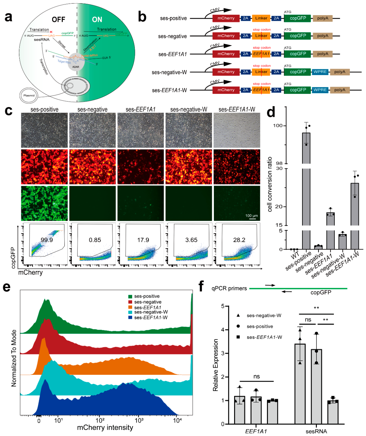
Figure 2.
The optimal mismatch designs of CellREADR and its ability on gene mutation detection. (a) Schematic diagram of A:N mismatches and the design of the stop codon UGA. (b) According to the CDS region of EEF1A1, the designed sesRNA of A:A, A:G, A:C mismatches and UGA stop codon correspond to the positions on the sequence. (c-d) A: N mismatch and different stop codons statistics according to the results of flow cytometry. (e) A comparison of the fluorescence intensities of mCherry among A:A, A:G, A:C mismatch groups. (f) Schematic diagram of detecting genetic mutation in cells using CellREADR. (g) EMX1 and FANCF genes were selected for mutation detection simulation, The left figures indicate cells transfected only with the sesRNA vector, while the right figures indicate cells co-transfected with both the sesRNA and gene mutation simulation vectors. GOI denotes the gene of interest, which refers to EMX1 and FANCF in this context, scale bar is 100 μm. (h) Cell conversion ratio was counted from flow cytometry analysis in (g).
Figure 2.
The optimal mismatch designs of CellREADR and its ability on gene mutation detection. (a) Schematic diagram of A:N mismatches and the design of the stop codon UGA. (b) According to the CDS region of EEF1A1, the designed sesRNA of A:A, A:G, A:C mismatches and UGA stop codon correspond to the positions on the sequence. (c-d) A: N mismatch and different stop codons statistics according to the results of flow cytometry. (e) A comparison of the fluorescence intensities of mCherry among A:A, A:G, A:C mismatch groups. (f) Schematic diagram of detecting genetic mutation in cells using CellREADR. (g) EMX1 and FANCF genes were selected for mutation detection simulation, The left figures indicate cells transfected only with the sesRNA vector, while the right figures indicate cells co-transfected with both the sesRNA and gene mutation simulation vectors. GOI denotes the gene of interest, which refers to EMX1 and FANCF in this context, scale bar is 100 μm. (h) Cell conversion ratio was counted from flow cytometry analysis in (g).
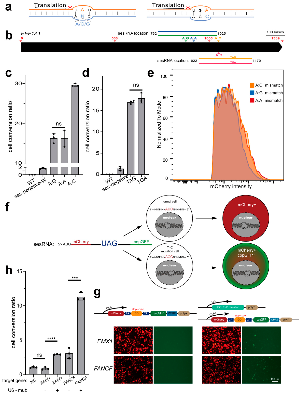
Figure 3.
Distinguishing two general cell lines by CellREADR. (a) Schematic diagram of the design of distinguishing HEK293FT and HeLa cells using CellREADR. (b) Relative expression levels of NEFM, LCP1, CSNK2B, SPINK6, KRT7, and PDE2A genes in HEK293FT and HeLa cells. (c) The flow analysis result of assessing the cell conversion ratio of ses-KRT7-W and ses-NEFM-W in HEK293FT and HeLa cells. (d) Statistical results of the cell conversion ratio of ses-KRT7-W and ses-NEFM-W in HEK293FT and HeLa cells based on (c), n =3.
Figure 3.
Distinguishing two general cell lines by CellREADR. (a) Schematic diagram of the design of distinguishing HEK293FT and HeLa cells using CellREADR. (b) Relative expression levels of NEFM, LCP1, CSNK2B, SPINK6, KRT7, and PDE2A genes in HEK293FT and HeLa cells. (c) The flow analysis result of assessing the cell conversion ratio of ses-KRT7-W and ses-NEFM-W in HEK293FT and HeLa cells. (d) Statistical results of the cell conversion ratio of ses-KRT7-W and ses-NEFM-W in HEK293FT and HeLa cells based on (c), n =3.
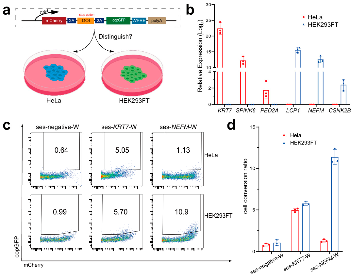
Figure 4.
Measuring the accuracy of similar sequences detection by CellREADR. (a) Schematic diagram of the design of STO and STO-GDNF cell distinguish. (b) DNA sequence similarity matrix of mGdnf, huGDNF and opti-cod huGDNF. (c) Expression statistics of endogenous Gdnf/GDNF in STO, STO-GNDF and HEK293FT cells, meanwhile the expression level of Actb/ACTB as reference. (d) The protein expression level of ADAR1 was detected with ACTB as reference. (e) Statistics of cell conversion rate after ses-GDNF-W transfection of STO and STO-GDNF cells, n = 3. (f) Schematic diagram of the design in HEK293FT. Here, in one group, the PLV-huGDNF-2A-puroR plasmid was transiently transfected into cells along with other sesRNA-related plasmids which include ses-positive, ses-negative-W and ses-GDNF-W. (g) Flow cytometry results for ses-negative-W and ses-GDNF-W groups, illustrating the cell conversion ratio and comparing fluorescence intensity within the same group across the two cell populations. (h) Statistical results of the cell conversion ratio, n=3.
Figure 4.
Measuring the accuracy of similar sequences detection by CellREADR. (a) Schematic diagram of the design of STO and STO-GDNF cell distinguish. (b) DNA sequence similarity matrix of mGdnf, huGDNF and opti-cod huGDNF. (c) Expression statistics of endogenous Gdnf/GDNF in STO, STO-GNDF and HEK293FT cells, meanwhile the expression level of Actb/ACTB as reference. (d) The protein expression level of ADAR1 was detected with ACTB as reference. (e) Statistics of cell conversion rate after ses-GDNF-W transfection of STO and STO-GDNF cells, n = 3. (f) Schematic diagram of the design in HEK293FT. Here, in one group, the PLV-huGDNF-2A-puroR plasmid was transiently transfected into cells along with other sesRNA-related plasmids which include ses-positive, ses-negative-W and ses-GDNF-W. (g) Flow cytometry results for ses-negative-W and ses-GDNF-W groups, illustrating the cell conversion ratio and comparing fluorescence intensity within the same group across the two cell populations. (h) Statistical results of the cell conversion ratio, n=3.
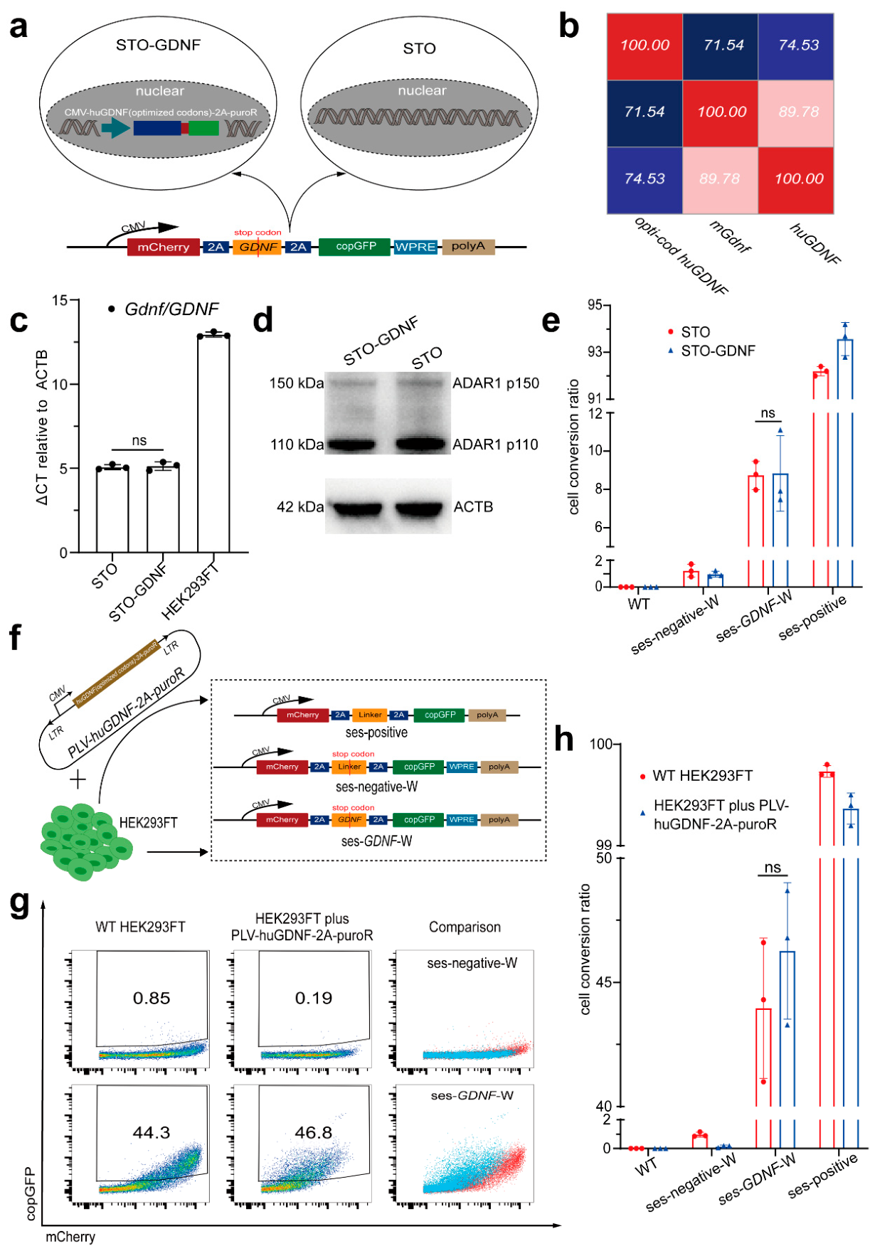
Figure 5.
Exploring the feasibility of CellREADR in stem cell lines. (a) Relative RNA expression statistics of endogenous Adar1 and Adar2 in mESCs and mSSCs comparison with STO and STO-GDNF cells, and the protein expression level of ADAR1 in mESCs and mSSCs meanwhile the ACTB as reference. (b) The ses-Klf4-W is transfected into cells for specifically marking mESCs meanwhile the MEF cell as the negative control. The white arrows point to the location of the mESCs that are specifically labeled, whereas in MEF cells, there is no effector signal at a scale of 100 μm. (c) The sesRNA targeting Plzf was designed to prepare lentivirus for mSSCs infection. The day of cell preparation of mSSCs was recorded as day 0, followed by lentiviral infection on day 1 and day 4, and recorded and observed on day 7 and day 19, respectively.(d) Fluorescence images on day 7, white arrows indicate the locations of mSSCs that are specifically labeled by CellREADR. And the MEF cell, serving as the feeder layer and negative control at the bottom of the well and exhibiting a fibrous morphology, shows no cell conversion signal, scale bar is 100 μm. (e) Fluorescence images on day 19 of the labeled mSSCs. Those cells formed clusters during extended culture while maintaining fluorescence labeling, scale bar is 100 μm.
Figure 5.
Exploring the feasibility of CellREADR in stem cell lines. (a) Relative RNA expression statistics of endogenous Adar1 and Adar2 in mESCs and mSSCs comparison with STO and STO-GDNF cells, and the protein expression level of ADAR1 in mESCs and mSSCs meanwhile the ACTB as reference. (b) The ses-Klf4-W is transfected into cells for specifically marking mESCs meanwhile the MEF cell as the negative control. The white arrows point to the location of the mESCs that are specifically labeled, whereas in MEF cells, there is no effector signal at a scale of 100 μm. (c) The sesRNA targeting Plzf was designed to prepare lentivirus for mSSCs infection. The day of cell preparation of mSSCs was recorded as day 0, followed by lentiviral infection on day 1 and day 4, and recorded and observed on day 7 and day 19, respectively.(d) Fluorescence images on day 7, white arrows indicate the locations of mSSCs that are specifically labeled by CellREADR. And the MEF cell, serving as the feeder layer and negative control at the bottom of the well and exhibiting a fibrous morphology, shows no cell conversion signal, scale bar is 100 μm. (e) Fluorescence images on day 19 of the labeled mSSCs. Those cells formed clusters during extended culture while maintaining fluorescence labeling, scale bar is 100 μm.
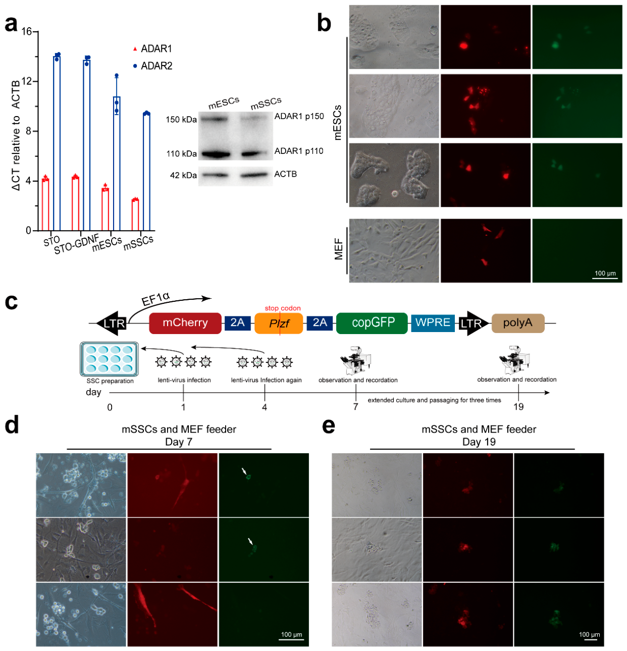
Figure 6.
Monitoring mSSCs states transition by CellREADR. (a) Assessing the protein levels of PLZF in mSSCs before and after RA induction for day 1, 2, 3 and 5 using immunofluorescence staining, scale bar is 50 μm. (b) Fluorescence images of CellREADR-labeled mSSCs (Figure 5) on day 0, and on day 5 with or without RA treatment. After 5 days of RA treatment, mSSCs formed larger colonies and became more flattened, scale bar is 100 μm. (c) Flow cytometry results for RA treated and untreated groups. (d) Comparison of copGFP fluorescence intensity between RA treatment group and non-treatment group. (e) Statistical results of the cell conversion ratio in RA treated and untreated groups.
Figure 6.
Monitoring mSSCs states transition by CellREADR. (a) Assessing the protein levels of PLZF in mSSCs before and after RA induction for day 1, 2, 3 and 5 using immunofluorescence staining, scale bar is 50 μm. (b) Fluorescence images of CellREADR-labeled mSSCs (Figure 5) on day 0, and on day 5 with or without RA treatment. After 5 days of RA treatment, mSSCs formed larger colonies and became more flattened, scale bar is 100 μm. (c) Flow cytometry results for RA treated and untreated groups. (d) Comparison of copGFP fluorescence intensity between RA treatment group and non-treatment group. (e) Statistical results of the cell conversion ratio in RA treated and untreated groups.
Disclaimer/Publisher’s Note: The statements, opinions and data contained in all publications are solely those of the individual author(s) and contributor(s) and not of MDPI and/or the editor(s). MDPI and/or the editor(s) disclaim responsibility for any injury to people or property resulting from any ideas, methods, instructions or products referred to in the content. |
© 2026 by the authors. Licensee MDPI, Basel, Switzerland. This article is an open access article distributed under the terms and conditions of the Creative Commons Attribution (CC BY) license (http://creativecommons.org/licenses/by/4.0/).
Copyright: This open access article is published under a Creative Commons CC BY 4.0 license, which permit the free download, distribution, and reuse, provided that the author and preprint are cited in any reuse.


