

Submitted:
15 July 2026
Posted:
17 July 2026
You are already at the latest version
Abstract
Many anticancer drugs are hydrophobic in nature. This affects the bioavailability and therapeutic efficacy of a drug to a large extent as the solubility of drug in biological system is reduced, resulting in its fast clearance from the body. Also, such drugs readily cross cell membranes and can accumulate in fatty tissues and organs. Nanoparticle-mediated drug delivery has attracted considerable attention to enhance the delivery of such hydrophobic, as well as hydrophilic, drugs. Among the various nanocarriers studied so far, liposomes, polymeric micelles, and exosomes are three of the most potential nanocarriers. This review provides a comparative account of different types of drug carriers with respect to the parameters affecting the delivery of hydrophobic drugs. In addition, the biological interactions of the nanocarriers have also been summarized. The performance trade-offs for the various carrier systems and the differences in structural and biological characteristics have been discussed. The review highlight the use of polymeric micelles for drug loading and tumor penetration, the value of liposomes, and aptamers, for clinical applications and pharmacokinetic control, and the promise of exosomes for targeted delivery and personalized medicine.
Keywords:
nanocarrier-based drug delivery
; liposomes
; polymeric micelles
; exosomes
; clinical translation
; aptamers
; nanomedicine
1. Introduction
Cancer remains one of the major health concerns worldwide, causing millions of new cases and deaths annually, even with the advancement of early detection methods and therapeutic regimen. Among the most commonly used therapeutic regimen for the treatment of a wide range of cancer types is chemotherapy. Despite the clinical benefits of many chemotherapeutic drugs, their effectiveness has been marred by their hydrophobicity, poor water solubility, systemic clearance, and non-specific distribution, limiting their therapeutic window and clinical utility.
A significant proportion of contemporary anticancer agents, such as like paclitaxel, docetaxel, doxorubicin and etoposide fall into the class of poor water-soluble drugs. In many cases, the traditional formulation approaches have not provided adequate solutions to the problems associated with the formulation of such drugs. There is a critical need to develop advanced drug delivery systems to enhance the solubility, stability, and targeting ability of hydrophobic anticancer drugs to the site of action while minimizing the side effects on the body.
Recently, nanotechnology-based drug delivery systems have been recognized as a revolutionary tool to address these challenges. It is now possible to encapsulate hydrophobic drugs using nanotechnology-based drug delivery systems and protect them from degradation. Additionally, it is also possible to exploit various biological phenomena, such as the enhanced permeability and retention effect, to prolong their circulation and facilitate their intracellular delivery. In recent years, a tremendous amount of scientific research has been performed to explore various nanocarrier-based drug delivery systems. Among them, liposomes, polymeric micelles, and exosomes have been found to be of great significance due to their structural characteristics.
The most advanced nanocarrier platform is liposomes, which are made up of a membrane of phospholipids. The ability to encapsulate hydrophobic drugs into the membrane, along with surface engineering techniques such as PEGylation, has led to several FDA-approved products with favorable PK properties and reduced toxicity. Another well-studied nanocarrier platform is polymeric micelles, which are self-assembled structures made up of amphiphilic copolymers. The small size and hydrophobicity of polymeric micelles have made them an attractive platform for extremely hydrophobic small molecule drugs. Additionally, polymeric micelles have been observed to have increased penetration into tumors. Exosomes, on the other hand, are naturally occurring vesicles that are secreted by cells. The inherent biological activity of exosomes, including immune evasion, membrane fusion, and cell specificity, has made them an attractive emerging platform for next-generation drug delivery.
Despite the availability of various types of nanocarrier platforms, none of them can be regarded as optimal for the delivery of hydrophobic anticancer drugs. Each type of nanocarrier has its advantages and disadvantages regarding the delivery of drugs, such as the amount of drug that can be carried, its stability in blood circulation, and its efficiency in entering cells. The current literature is mainly focused on specific types of drug delivery platforms and specific drugs, and it is not easy to conclude their efficacy and suitability for use.
Under these circumstances, the present article aims to offer a systematic, comparative, and translational evaluation of liposomes, polymeric micelles, and exosomes as vehicles for hydrophobic anti-cancer drug delivery. Representative data on essential characteristics of these vehicles, such as size, molecular weight, loading capacity, plasma half-life, and clearance, are analyzed and graphically presented to facilitate a comparative evaluation. Additionally, clinical development status and manufacturing aspects are also addressed to bridge the gap between experimental research and clinical application. The purpose of this article is to offer a holistic overview of anti-cancer drug delivery vehicles to facilitate the development of clinically relevant and personalized nanomedicine. Unlike previous reviews, which address anti-cancer drug delivery vehicles individually, this study offers an integrated translational comparison of anti-cancer drug delivery vehicles based on physicochemical characteristics, biological interactions, clinical development status, and manufacturing aspects.
2. Comparative Analysis of Nanocarriers
Nanocarrier-mediated delivery has emerged as an important approach to overcome the formulation and therapeutic challenges faced with hydrophobic anticancer drug candidates. However, the efficacy of a nanocarrier-mediated system is not dictated by a single factor but rather a combination of factors including its physicochemical properties, biological interactions, pharmacokinetics, and translationability. In this section, liposomes, polymeric micelles, and exosomes are compared and evaluated on the basis of some literature-derived parameters to present a logical and application-oriented approach to drug delivery systems.

Table 1.
A comparison of physicochemical, pharmacokinetic, and translational characteristics of liposomes, polymeric micelles, and exosomes for hydrophobic anticancer drug delivery.
Table 1.
A comparison of physicochemical, pharmacokinetic, and translational characteristics of liposomes, polymeric micelles, and exosomes for hydrophobic anticancer drug delivery.
| Parameter | Liposomes | Polymeric Micelles | Exosomes | References |
|---|---|---|---|---|
| Drug Type Best Suited | Hydrophobic drugs with dose limiting toxicity | Extremely hydrophobic small-molecule drugs | Hydrophobic drugs requiring cell-specific delivery | [1,2] |
| Drug Loading Capacity | Moderate | Relatively high | Variable | [5,16] |
| Protection of Drug | Very good due to lipid bilayer | Limited compared to liposomes | Very good due to natural membrane | [7,9] |
| Stability in Blood Circulation | Good, especially after PEGylation | Can be unstable upon dilution | Generally stable | [10,11] |
| Tumor Penetrability | Moderate | Better due to smaller particle size | Good | [15,21] |
| Targeting Feasibility | Well-established ligand or antibody conjugation | Possible but formulation dependent | Intrinsic targeting ability | [2,7] |
| Interaction with Immune System | Possible uptake by RES | Usually low | Minimal | [4,11] |
| Suitability for Personalized Therapy | Limited | Limited | High | [19] |
| Clinical Translation Status | Clinically established | Several systems under clinical evaluation | Early clinical stage | [3,14] |
| Manufacturing | Scalable and standardized | Scalable | Technically challenging | [14,18] |
| Typical Particle Size (nm) | 80-200 nm | 10-80 nm | 30-150 nm | [1,5] |
| Approximate Molecular Weight (Da) | 10⁶–10⁹ | 10⁴–10⁵ | 10⁹–10¹² | [5,7] |
| Drug Loading Capacity (% w/w) | 5–15% | 10–30% | 5–25% | [5,8] |
| Cellular Uptake Efficiency | Moderate | High | Very high |
[7,18] |
| Plasma Half-Life (h) | 10-50h | 2-10h | 12-48h | [9,10] |
| Bioavailability Enhancement | Moderate–High | High | High | [1,20] |
| Bioaccumulation Tendency | Moderate (liver, spleen) | Low–Moderate | Low |
[4,21] |
| Drug Leakage Risk | Low–Moderate | Moderate | Low | [12,16] |
3. A Physicochemical Characteristics
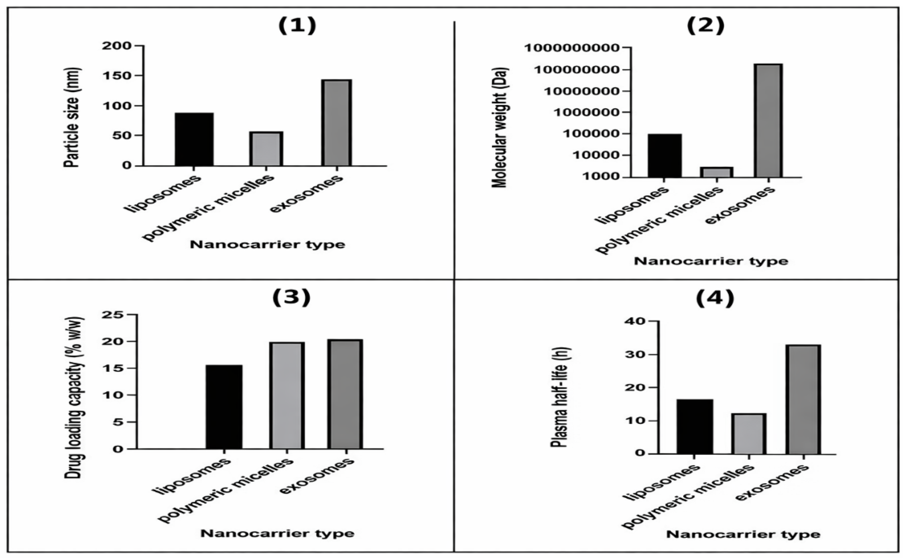
Size of the particles is a major factor in the biodistribution of nanocarriers, penetration of the nanocarrier into the tumor, and internalization of the nanocarrier into the cells. As depicted in Figure 1, the size of polymeric micelles is the smallest compared to the other three nanocarriers, followed by exosomes and liposomes. The smaller size of polymeric micelles may provide better penetration into the tumor and better diffusivity into the extracellular matrix.
The molecular weight of the nanocarrier is a major factor in the circulation of the nanocarrier and the mechanism of clearance. As depicted in Figure 2, the comparative molecular weight trend of the nanocarriers indicates that the molecular weight of polymeric micelles is the smallest compared to the other three nanocarriers. The molecular weight of liposomes is the highest compared to the other three nanocarriers. The molecular weight of exosomes is the highest compared to the other three nanocarriers because of the complexity of the exosome nanocarrier.
The smaller the molecular weight of the nanocarrier, the better the circulation of the nanocarrier. The nanocarrier with the smallest molecular weight may have a better mechanism of clearance.
Figure 1.
Comparative physicochemical and pharmacokinetic characteristics of liposomes, polymeric micelles, and exosomes for hydrophobic anticancer drug delivery. (A) Particle size distribution. (B) Molecular weight comparison. (C) Drug loading capacity. (D) Plasma half-life.
Figure 1.
Comparative physicochemical and pharmacokinetic characteristics of liposomes, polymeric micelles, and exosomes for hydrophobic anticancer drug delivery. (A) Particle size distribution. (B) Molecular weight comparison. (C) Drug loading capacity. (D) Plasma half-life.
4. Drug Loading Capacity and Cellular Uptake Behaviour
Efficiently loading the drugs into the carrier is an important step to achieve the desired therapeutic dose without overloading the carrier. Figure 3 illustrates the relative amount of drugs that different nanocarrier systems can carry. For instance, polymeric micelles can carry the maximum amount of drug, especially for highly hydrophobic small molecules, since the core of the polymeric micelles is hydrophobic. The core of the polymeric micelles is useful for solubilizing drugs that are sparingly soluble in water. Liposomes, on the other hand, can carry a moderate amount of drug, but the amount is limited by the physical and chemical properties of the lipid bilayer. The amount of drug carried by exosomes is variable, depending on the method of loading. The amount of drug carried by exosomes is highly dependent on the method of loading, whether passive or active.
In addition to the amount of drug carried by the nanocarriers, the uptake of the drug by the cell is another important aspect. From the results presented in Table 1, it is clear that exosomes take the lead with regard to the uptake of the drug by the cell. This is attributed to the natural composition of the cell membrane. On the other hand, polymeric micelles are usually uptaken by the cell via the endocytosis pathway, which is efficient. Liposomes, on the other hand, require modification of the liposome surface with ligands or antibodies to improve the uptake of the drug by the cell.
5. Pharmacokinetic Behaviour and Clearance Pathways
The way a nanocarrier functions inside the body will affect the drug exposure, the dosing schedule, and the systemic toxicity risk. If we look at the plasma half-life results provided by the data in Figure 1, we can see that the PEGylated liposomes and exosomes have a longer plasma half-life than the polymeric micelles. This is because the tendency of the micelles to self-assemble and dilute in the blood often causes them to lose stability, leading them to clear faster.
The way a nanocarrier is cleared from the body is another factor that will distinguish it from others. As we have seen from the results provided by the data in Table 1, liposomes are cleared by the reticuloendothelial system, mainly by the liver and spleen. The polymeric micelles, on the other hand, are cleared by the kidneys and the liver, depending on the size of the micelles and the polymer used. Exosomes are mainly cleared by the liver, mainly because of their natural origins. The way a nanocarrier is cleared will determine the way we can adjust the drug exposure to minimize the risk of toxicity.
6. Clinical Translation and Manufacturing Considerations
Clinical applicability is one of the major considerations when selecting a nanocarrier. Liposomes are the most clinically advanced nanocarriers, with several FDA-approved formulations showing improved safety and efficacy profiles. Polymeric micelles are being explored extensively in the clinic, with several systems at various stages of Phase I, II, and III clinical trials, although stability problems and reproducibility at large scales still plague these nanocarriers. Exosomes are showing promise from a biological perspective, although these are still at a very early stage of clinical translation, facing problems of reproducibility, standardization, and FDA approval.
The manufacturing feasibility of nanocarriers is another important consideration, with varying levels of complexity involved in the manufacture of the nanocarriers, as illustrated in Table 1, where liposomes have the advantage of a standardized large-scale manufacturing process, whereas polymeric micelles require precise control over the polymerization reaction, and exosomes are still a challenge due to problems associated with their isolation and purification.
Table 2.
Overview of clinically approved, withdrawn, and late-stage nanotechnology-based anticancer drug formulations, highlighting active drug, nanocarrier platform, regulatory status, year of approval, geographic region, and primary clinical indication.
Table 2.
Overview of clinically approved, withdrawn, and late-stage nanotechnology-based anticancer drug formulations, highlighting active drug, nanocarrier platform, regulatory status, year of approval, geographic region, and primary clinical indication.
| Product (Brand) | Active Drug | Nanocarrier Platform | Regulatory Status | Year | Region | Primary Indication | References | ||
|---|---|---|---|---|---|---|---|---|---|
| Doxil® / Caelyx® | Doxorubicin | PEGylated Liposome | Approved | 1995 | US/EU | Ovarian cancer, Kaposi’s sarcoma | [9,28] | ||
| DaunoXome® | Daunorubicin | Liposome | Approved | 1996 | US | AIDS-related Kaposi's sarcoma | [9,28] | ||
| Myocet® | Doxorubicin | Non-PEG Liposome | Approved | 2000 | EU/Canada | Metastatic breast cancer | [14,28] | ||
| Abraxane® | Paclitaxel | Albumin-bound NP | Approved | 2005 | US/EU | Breast, NSCLC, Pancreatic cancer | [28] | ||
| Genexol-PM® | Paclitaxel | Polymeric Micelle | Approved | 2007 | South Korea | Breast cancer, NSCLC | [34] | ||
| Onivyde® | Irinotecan | PEGylated Liposome | Expanded | 2015/2024* | US | 1st-line Metastatic Pancreatic cancer | [9,14] | ||
| Hensify® | Hafnium Oxide | Inorganic NP | Approved | 2019 | EU | Soft tissue sarcoma (Radioenhancer) | [35] | ||
| Zolsketil® | Doxorubicin | PEGylated Liposome | Approved | 2022 | EU | Kaposi’s, Breast, Ovarian cancer | [14] | ||
| mRNA-4157 | mRNA (Neoantigen) | Lipid NP (LNP) | Phase 3 | 2026 | Global | Adjuvant Melanoma (KEYNOTE-942) | [23] | ||
| Marqibo® | Vincristine | Liposomal | Withdrawn | 2022 | US | (Approval withdrawn May 2022) | [28,36] | ||
7. Brief Overview of Nanocarrier Fabrication Methods
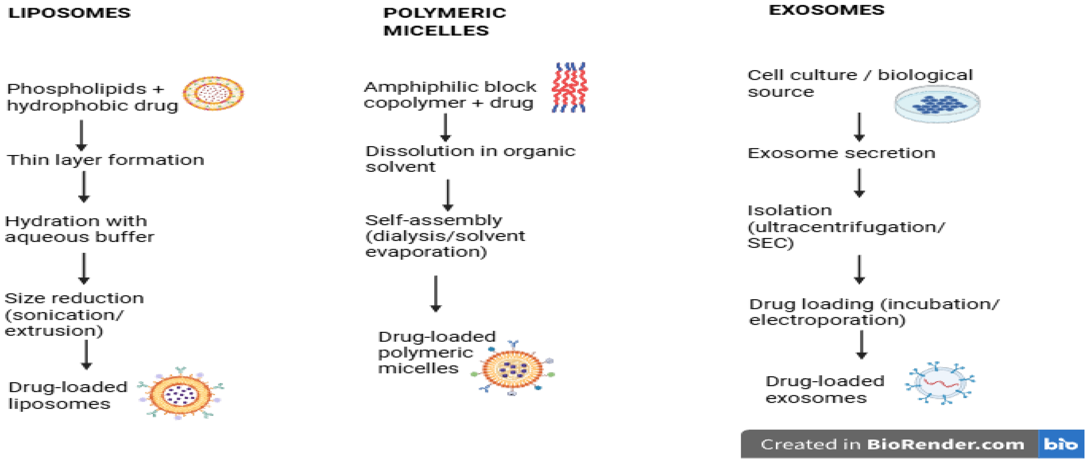
The process by which we make nanocarriers is a big deal. The process affects the particle size, the amount of drug we can incorporate into the system, the stability of the system, the ease of scale-up, and so on. All of these affect the overall process of moving the drug therapy forward. While liposomes, polymeric micelles, and exosomes have very different starting points, each has an established pathway to the process of formulation, as a brief overview of the process is represented in Figure 2.
Liposomes are derived from a process involving lipids. We make a mixture of the phospholipid and the hydrophobic drug, then form a thin film. We then rehydrate the film with an aqueous buffer. The resulting structure is a multilamellar structure. The process is spontaneous. To make the liposomes nanometric with a uniform particle size, we use processes to reduce the particle size.
The polymeric micelles are formed through the self-assembly of amphiphilic diblock copolymers in an aqueous environment. In most cases, the polymer and the drug are dissolved in an organic solvent, and then the solvent is evaporated or changed through a process called dialysis to induce micelle formation. The hydrophobic drug molecules are located inside the micelles, while the hydrophilic polymer chains are located on the micelle surface. This method is simple and easily scalable to accommodate various formulations, but the stability of micelles can depend on the concentration and environmental factors.
Exosome fabrication, on the other hand, is not a physicochemical process but rather a biological process. Exosomes are naturally secreted by various cell types and are collected from cell cultures or body fluids and isolated through ultracentrifugation or size-exclusion chromatography. The drug molecules are then loaded into exosomes through passive loading or permeabilization techniques like electroporation. Exosomes are highly compatible with drug molecules and have good targeting ability, but the process of exosome fabrication is technically challenging, with difficulties in exosome production, purification, and reproducibility between batches.
Overall, the differences in liposomes, polymeric micelles, and exosome fabrication highlight the balance between the ease of drug formulation and its application in drug development. Liposomes and polymeric micelles are advantageous in terms of the ease of drug formulation, while exosome fabrication needs to be optimized to overcome the challenges in its development and application.
Figure 2.
Schematic illustration of representative fabrication and drug-loading processes for liposomes, polymeric micelles, and exosomes employed in hydrophobic anticancer drug delivery. The figure summarizes the key preparation steps and highlights differences in formulation approach and manufacturing complexity among the nanocarrier systems.
Figure 2.
Schematic illustration of representative fabrication and drug-loading processes for liposomes, polymeric micelles, and exosomes employed in hydrophobic anticancer drug delivery. The figure summarizes the key preparation steps and highlights differences in formulation approach and manufacturing complexity among the nanocarrier systems.
8. Comparative Study Of Drug Delivery Performance Using Nanocarriers
A comparison of the effectiveness of liposomes, polymeric micelles, and exosomes as drug delivery agents would provide an insight into the practical applications of the three agents, apart from the physical and chemical properties. The effectiveness of the agents is not only dependent on the structure and pharmacokinetic properties of the agents; the therapeutic effect of the agents depends on the encapsulation efficiency, the tumor accumulation of the drug, the release of the drug, the therapeutic index, and the reduction of systemic toxicity.
8.1. Drug Encapsulation and Solubilization Efficiency
Hydrophobic anticancer agents such as paclitaxel, docetaxel, and camptothecin derivatives need solid solubility for the drugs to achieve sufficient bioavailability in the human system. Liposomes encapsulate the anticancer agents within the hydrophobic region of the lipid bilayer structure and provide moderate loading and excellent defense against degradation [1,9]. Polymeric micelles have the advantage of higher loading capacity compared to other systems because of the hydrophobic region that can effectively dissolve very hydrophobic small molecules [5,16].
Exosomes can encapsulate anticancer agents in variable amounts depending on the method of loading: passive loading, electroporation, or permeabilizing the membrane. The composition of the exosome membrane protects the encapsulated agents against degradation because of its natural composition [7,8].
In summary, polymeric micelles have the advantage of higher loading capacity compared to other systems for hydrophobic small molecules, while liposomes have the advantage in providing structural stability and defense against degradation.
8.2. Tumor Accumulation and Biodistribution
The nanocarriers exploit the EPR effect, allowing for the targeted delivery of the drug. Liposomes, especially the PEGylated ones, have shown a greater circulation time, thereby increasing the exposure of the drug at the tumor site, as observed with the clinically approved formulation Doxil® [9,13].
The smaller size of the polymeric micelles, ranging from 10-80 nm, has often resulted in a greater penetration ability through the dense tumor mass [15,21]. However, the shorter circulation half-life of the polymeric micelles may affect the exposure time at the tumor site, especially when compared to the PEGylated liposomes.
8.3. Intracellular Uptake and Endosomal Escape
Efficient intracellular delivery is critical for the effectiveness of cytotoxic drugs. Micelles are taken up by the cell through the process of endocytosis, after which the drug is efficiently delivered to the cell. The efficiency of cellular uptake for micelles has been reported to be high.
For liposomes, surface functionalization is critical for the efficient delivery of drugs, particularly through the conjugation of ligands or antibodies, allowing for efficient receptor-mediated uptake. Without functionalization, the efficiency of cellular uptake is moderate.
For exosomes, the efficiency of cellular uptake is high, given their ability to fuse with the cell membrane, thereby enhancing intracellular drug delivery. This is a critical factor for the effectiveness of drugs, particularly for drug-resistant cancer phenotypes.
8.4. Pharmacokinetic and Therapeutic Index Improvement
One of the main goals of nanocarrier-mediated drug delivery is the improvement of the therapeutic index. PEG liposomes have been shown to significantly improve the plasma half-life of the drug (10-50 hours), reduce peak plasma toxicity, and decrease cardiotoxicity associated with the free form of anthracyclines [9,13]. In contrast, polymeric micelles have a shorter plasma half-life of 2-10 hours but tend to increase the drug concentration in the tumor compared to the free form of the drug, reducing systemic toxicity [16]. Exosomes have a long plasma half-life with reduced immunogenicity, which may provide therapeutic potential with reduced immune activation [7,18]. However, the pharmacokinetic variability is an important consideration.
8.5. Reduction of Systemic Toxicity
In traditional formulations of hydrophobic anticancer agents, toxic excipients are often necessary to achieve solubilization (e.g., Cremophor EL in paclitaxel formulations), contributing to hypersensitivity reactions and organ toxicities. Nanocarrier formulations circumvent the need for these excipients, thus improving the safety profiles of the anticancer agents [2,20]. Efficacy in human patients with liposomal formulations has shown a decrease in cardiotoxicity and an increase in tolerability compared to the free drug [9,14]. In addition, solvent-associated toxicities are minimized in polymeric micelles. The exosome-based delivery system, with its endogenous composition, holds promise to further reduce immune-related adverse effects. However, clinical validation is in process.
8.6. Comparative Summary of Therapeutic Performance
When evaluated across multiple performance parameters:
∙ Liposomes provide clinically validated pharmacokinetic enhancement and toxicity reduction, with strong regulatory acceptance.
∙ Polymeric micelles offer superior solubilization and tumor penetration for highly hydrophobic drugs.
∙ Exosomes demonstrate promising biological targeting and cellular uptake capabilities, supporting future personalized therapeutic applications.
No single nanocarrier system universally outperforms others across all parameters. Instead, drug physicochemical properties, tumor microenvironment characteristics, dosing strategy, and translational feasibility must collectively guide nanocarrier selection.
9. Emerging Trends in Nanocarrier Design
9.1. Ai-Guided Nanocarrier Design
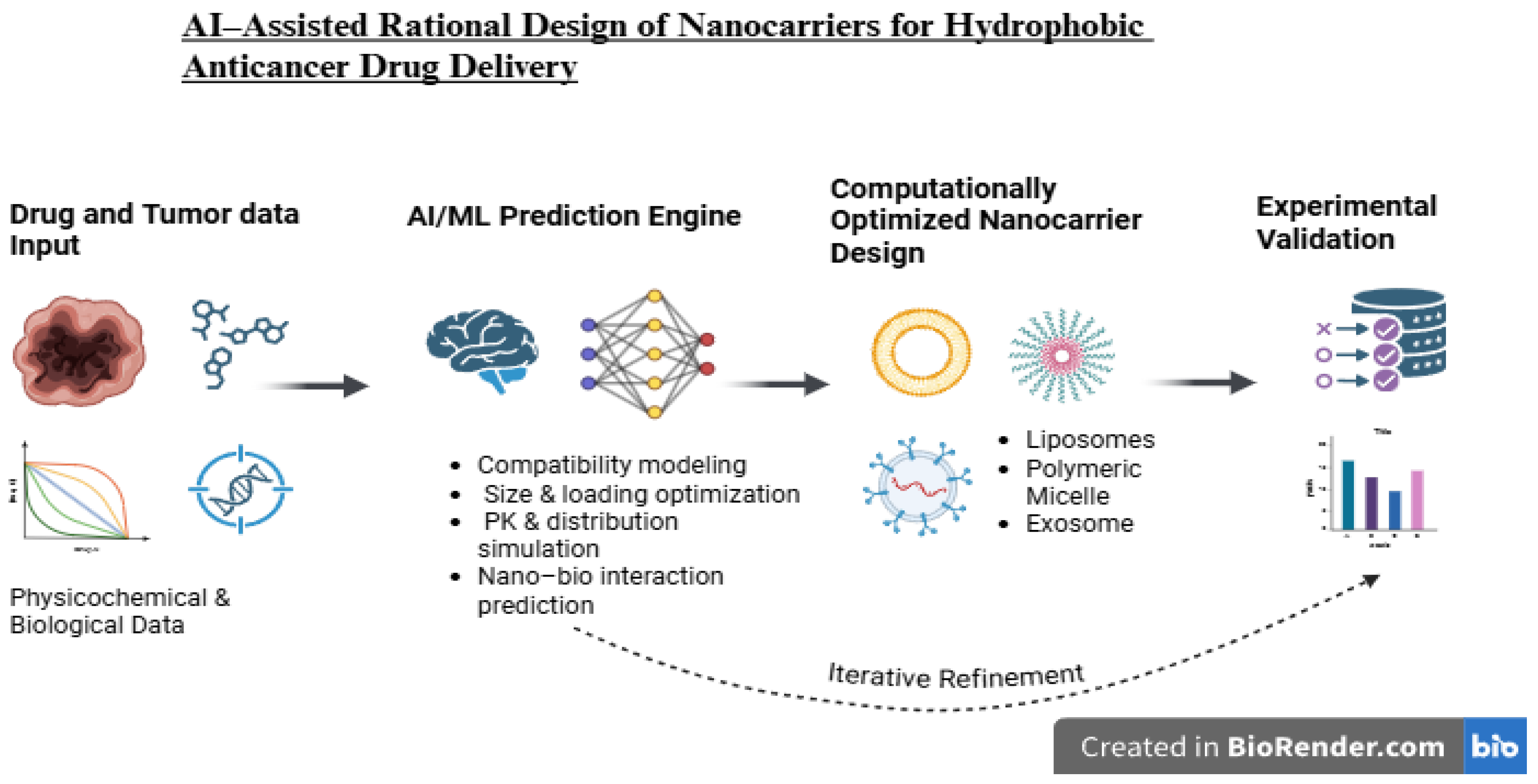
Artificial intelligence (AI) and machine learning (ML) techniques are increasingly recognized as promising tools for the development of nanomedicine. Data-driven techniques can predict the compatibility of drugs with nanocarriers, optimize the composition of the polymer, predict the particle size distribution of the nanocarriers, and forecast the pharmacokinetic profiles of the drug using physicochemical parameters and biological interaction data. Modern computational techniques, such as deep neural networks and graph neural networks, have been found to be useful tools for the prediction of nano-bio interactions and bio-distribution profiles, thus reducing the need for experimental screening. The integration of AI with high-throughput experimental techniques is expected to improve the design of nanocarriers, reproducibility of results, and the overall translation of nanomedicine. However, there are challenges to be overcome with regard to the use of AI techniques.
9.2. Personalized Nanomedicine
The move towards precision oncology demands flexible nanocarrier systems that can be tailored to specific patient requirements. Personalized nanomedicine involves tumor molecular profiling, pharmacogenomics, and biomarker-based therapy to optimize various drug delivery strategies [19,25].
The use of exosomes is considered to be beneficial for developing personalized medicine because they are naturally occurring entities. The autologous exosome-based drug delivery systems can lead to reduced immunogenicity. Moreover, ligand-targeted liposomes and polymeric micelles can be designed to target specific receptors on cancer cells, which can be determined using genomic sequencing. The integration of nanocarrier-based systems with companion diagnostics is likely to improve therapeutic outcomes.
9.3. Hybrid Nanocarriers: Liposome–exosome Hybrids
The development of hybrid nanocarriers, where synthetic and biological components are combined, is a promising advancement in the field of drug delivery engineering. Hybrid liposome-exosome nanocarriers aim to combine the advantage of the reproducibility of liposome formulation with the biological activity associated with exosomes.
The hybrid nanocarriers have the potential to overcome the limitations associated with membrane fusion efficiency, cell uptake, and targeting efficiency, particularly for the isolation and purification of exosomes, while maintaining structural stability, membrane proteins, and reproducibility, which is a major challenge associated with the development of hybrid nanocarriers.
10. Regulatory and Translational Challenges
10.1. Regulatory Bottlenecks
Despite the significant advancements made in technology, the current regulatory guidelines for the regulation of complex nanomedicines are still not harmonized. The regulatory guidelines for liposomal formulations are already well established, with clear guidelines on the physicochemical characterization of the formulation. In the case of polymeric micelles, as well as exosome-based nanomedicines, the regulatory guidelines are still evolving [3,27].
The key issues to be addressed include the physicochemical characterization of the formulation, potency tests, immunogenicity tests, long-term biodistribution studies, and quality control tests. The formulation of new regulatory guidelines will be critical to the future translation of nanomedicines.
10.2. GMP-Scale Manufacturing Challenges
Large-scale production in a Good Manufacturing Practice environment is a major obstacle to translation on a wider scale. In the case of liposomes and polymeric micelles, it is easier to scale up production. However, the problem of maintaining the uniformity of the vesicles, the level of loading, and stability remains a technical challenge [14,16]. For exosome-based systems, there are additional technical hurdles, which include low yields, purification difficulties, heterogeneity, and the potential for contamination [18]. There are emerging technologies, such as microfluidization, continuous manufacturing, and automated purification systems, which have the potential to make the production processes more reproducible. There is a need to invest in the development of standardized production protocols and analytical systems to ensure quality and safety.
10.3. Cost–Benefit Analysis and Health Economics
The long-term clinical utility of such advanced nanocarrier systems will depend on evidence of therapeutic benefit relative to costs of manufacturing or healthcare. While more sophisticated systems such as exosome-based or hybrid nanocarrier systems offer greater potential for targeting efficacy and reduced systemic toxicity, costs associated with manufacturing such systems may also limit accessibility to such systems in resource-constrained healthcare systems [3,28]. Survival benefit, toxicity reduction, hospitalization rates, and quality-adjusted life years gained are some of the key aspects that need to be considered during cost-effectiveness evaluations of such systems.
Figure 3.
AI-assisted workflow for rational design and optimization of nanocarriers for hydrophobic anticancer drug delivery integrating physicochemical drug properties, machine learning-based prediction, nanocarrier engineering, and experimental validation.
Figure 3.
AI-assisted workflow for rational design and optimization of nanocarriers for hydrophobic anticancer drug delivery integrating physicochemical drug properties, machine learning-based prediction, nanocarrier engineering, and experimental validation.
11. Aptamer-Mediated Targeted Drug Delivery Applications
Aptamer-mediated targeting has been recognized as an efficient method to enhance the specificity of nanocarrier-mediated drug delivery systems. Aptamers are short single-stranded DNA or RNA molecules with the potential to form specific three-dimensional shapes, thus allowing for the binding of specific molecules. Aptamers are also referred to as “chemical antibodies.” Aptamers have been recognized to possess a number of advantages over other molecules, such as the use of monoclonal antibodies, owing to the fact that they are small, non-immunogenic, chemically stable, and easy to synthesize. Aptamers are highly suitable for the functionalization of nanocarriers such as liposomes, polymeric nanoparticles, and exosomes [29].
Among the various aptamers studied for cancer targeting, AS1411 has emerged as an important one. AS1411 is a guanine-rich DNA aptamer that forms a stable G-quadruplex structure and binds to nucleolin, a multifunctional protein that is often overexpressed on the cell surface of many cancer cells. Nucleolin is implicated in several cellular functions, including the synthesis of ribosomal RNA, remodeling of chromatin, and the regulation of cell proliferation. The overexpression of nucleolin in cancer cells makes it an important target for selective cancer therapy [30]. AS1411 has been demonstrated to selectively target nucleolin-expressing cancer cells and deliver therapeutic molecules intracellularly by receptor-mediated endocytosis [31].
Recently, researchers have attempted to develop targeted drug-delivery systems by functionalizing AS1411 with various nanocarriers to target cancer cells. AS1411-functionalized nanocarriers were observed to deliver anticancer drugs such as doxorubicin to nucleolin-overexpressing cancer cells more effectively, thus enhancing the therapeutic efficacy of cancer treatment [32]. The functionalization of biologically derived drug-delivery systems such as exosomes with AS1411 was also observed to enhance the precision of cancer targeting by directing exosomes to nucleolin-expressing cancer cells, thus improving the therapeutic specificity of the drug-delivery system [33].
12. Discussion and Conclusion
The concept of nanocarrier-based drug delivery has greatly impacted the approaches taken to overcome the limitations of traditional chemotherapy, especially for hydrophobic anticancer drugs. Hydrophobicity, low solubility, and systemic clearance are the major limitations of traditional chemotherapy, leading to the decreased therapeutic efficiency of many anticancer drugs. Nanocarriers, such as liposomes, micelles, and exosomes, have been used to overcome these limitations, especially for hydrophobic drugs, by improving their solubility, systemic circulation, and tumor targeting through passive and active targeting approaches [1,2].
Liposomes are the most advanced nanocarriers, showing the highest clinical translation, especially for drug delivery approaches. Their structure, consisting of a lipid bilayer, can efficiently entrap the drug, protecting it from premature degradation. Liposomal formulations, such as liposomal doxorubicin, have shown improved pharmacokinetics, including decreased systemic toxicity, compared to traditional drug formulations, showing the clinical translation of lipid-based nanocarriers for drug delivery approaches. Despite their success, liposomes have limitations, including their interaction with the reticuloendothelial system, their size, and their ability to penetrate the tumor site, due to their large size.
Another option for the delivery of extremely hydrophobic small molecule drugs is provided by polymeric micelles. Their structure, consisting of a hydrophobic core, has the ability to dissolve water-insoluble drugs, thus increasing the drug loading capacity, penetration into the tumor site, and the smaller size of the drug delivery system, which is beneficial for drug targeting [5,16]. Despite the above-mentioned benefits, instability of the drug delivery system during systemic circulation, especially after dilution, has been observed, leading to decreased drug efficiency [16].
Exosomes are a biologically derived drug delivery system, and their advantages lie in their biocompatibility, targeting ability, and interaction with the cell membrane, allowing them to efficiently deliver therapeutic molecules into the cell by interacting with the cell membrane, owing to their membrane structure, which is composed of membrane proteins, making them a promising drug delivery system for the development of personalized medicine.
Significant advancements in the field of nanomedicine have also been made to improve the specificity of targeted delivery using ligand-mediated strategies. The use of ligands such as antibodies, peptides, and aptamers to functionalize nanoparticles has been promising to improve targeted delivery to tumor tissues. For instance, the AS1411 aptamer recognizes a specific protein called nucleolin, which is overexpressed on the surface of cancer cells, thereby allowing targeted delivery of drugs to tumor tissues [30,31]. The use of such targeted delivery strategies along with nanocarrier technology is likely to improve the efficacy and safety of drugs.
Each of the nanocarrier delivery systems has its advantages and disadvantages. For instance, liposomes have good clinical validation and manufacturing processes, polymeric micelles have good drug solubilization and tumor penetration properties, and exosomes have good biological targeting properties. The future of nanocarrier technology is likely to be a hybrid system that uses the advantages of both synthetic and biological delivery systems and also computational models such as artificial intelligence to improve the formulation and interaction of drugs with biological tissues [22,23].
The use of nanocarriers in drug delivery systems has been found to significantly improve the therapeutic index of hydrophobic anticancer drugs. Among the different types of drug delivery systems reviewed in the current study, liposomes are the most tested system, polymeric micelles hold great promise in terms of their efficiency in delivering hydrophobic drugs to cancer sites, and exosomes are a biological system that also holds great promise because of their inherent targeting ability. However, before the use of such drug delivery systems is approved for use in cancer therapy, several issues need to be addressed. Further research is required to explore targeted strategies using nanocarriers, hybrid technology, and artificial intelligence technology to advance the field of nanomedicine.
Conflicts of Interest
The authors declare that they have no competing interests
Funding
No funds, grants, or other support was received.
Financial Interest
Piyush Bhati acknowledge the financial support provided by Council for Scientific and Industrial Research as fellowship.
Author Contributions
Sresi Singh and Mohd. Tashfeen Ashraf: conceptualization, methodology and writing the Original Draft. Sresi Singh, Anjali Mehra, Gunjan Garg, Piyush Bhati, Sonal Roy and Kanu Priya: Formal analysis and data curation. Mohd. Tashfeen Ashraf, Sresi Singh and Abdul Arif Khan: supervision and writing – review & editing.
Acknowledgments
Authors acknowledge the host institutes, Department School of Biotechnology, Gautam Buddha University, Greater Noida; VRDL, Government Institute of medical Sciences, Greater Noida; and National Institute of. Translational Virology and AIDS Research (ICMR-NITVAR), Pune for providing institutional support, facilities, and conducive environment for the carrying out this work,
References
- Torchilin, V. P. Recent advances with liposomes as pharmaceutical carriers. Nat. Rev. Drug Discov. 2005, vol. 4(no. 2), 145–160. [Google Scholar] [CrossRef]
- Peer, D.; et al. Nanocarriers as an emerging platform for cancer therapy. Nat. Nanotechnol. 2007, vol. 2, 751–760. [Google Scholar] [CrossRef]
- Anselmo, A. C.; Mitragotri, S. Nanoparticles in the clinic. Bioeng. Transl. Med. 2016, vol. 1, 10–29. [Google Scholar] [CrossRef]
- Dobrovolskaia, M. A.; Aggarwal, P.; Hall, J. B.; McNeil, S. E. Preclinical studies to understand nanoparticle interaction with the immune system. Mol. Pharm. 2008, vol. 5, 487–495. [Google Scholar] [CrossRef]
- Kataoka, K.; Harada, A.; Nagasaki, Y. Block copolymer micelles for drug delivery. Adv. Drug Deliv. Rev. 2012, vol. 64, 37–48. [Google Scholar] [CrossRef]
- Kwon, G.; Kataoka, K. Block copolymer micelles as long-circulating drug vehicles. Adv. Drug Deliv. Rev. 2012, vol. 64, 237–245. [Google Scholar] [CrossRef]
- EL Andaloussi, S.; Mäger, I.; Breakefield, X. O.; Wood, M. J. A. Extracellular vesicles: biology and emerging therapeutic opportunities. Nat. Rev. Drug Discov. 2013, vol. 12, 347–357. [Google Scholar] [CrossRef]
- Luan, L.; et al. Exosomes as drug delivery systems: biological functions and clinical applications. J. Control. Release 2017, vol. 266, 331–350. [Google Scholar]
- Allen, T. M.; Cullis, P. R. Liposomal drug delivery systems: from concept to clinical applications. Adv. Drug Deliv. Rev. 2013, vol. 65, 36–48. [Google Scholar] [CrossRef]
- Harris, J. M.; Chess, R. B. Effect of PEGylation on pharmaceuticals. Nat. Rev. Drug Discov. 2003, vol. 2, 214–221. [Google Scholar] [CrossRef]
- Owens, D. E., III; Peppas, N. A. Opsonization, biodistribution, and pharmacokinetics of polymeric nanoparticles. Int. J. Pharm. 2006, vol. 307, 93–102. [Google Scholar] [CrossRef]
- Mura, S.; Nicolas, J.; Couvreur, P. Stimuli-responsive nanocarriers for drug delivery. Nat. Mater. 2013, vol. 12, 991–1003. [Google Scholar] [CrossRef]
- FDA. Doxil (doxorubicin liposome) prescribing information; U.S. Food and Drug Administration, 2013.
- Bulbake, M.; et al. Liposomes in clinical use: an updated review. J. Control. Release 2017, vol. 250, 21–43. [Google Scholar]
- Shi, J.; Votruba, A. R.; Farokhzad, O. C.; Langer, R. Nanotechnology in drug delivery and tissue engineering. Nano Lett. 2010, vol. 10, 3223–3230. [Google Scholar] [CrossRef]
- Bae, S.; Kataoka, K. Polymeric micelles for drug delivery: design and clinical translation. Pharm. Res. 2009, vol. 26, 2197–2209. [Google Scholar]
- Yáñez-Mó, M.; et al. Biological properties of extracellular vesicles and their physiological functions. J. Extracell. Vesicles 2015, vol. 4. [Google Scholar]
- Elsharkasy, S.; et al. Extracellular vesicles as drug delivery systems: why and how? Adv. Drug Deliv. Rev. 2020, vol. 159, 332–343. [Google Scholar] [CrossRef]
- van der Meel, R.; et al. Personalized nanomedicine: principles and clinical translation. Nat. Nanotechnol. 2019, vol. 14, 1007–1017. [Google Scholar] [CrossRef]
- Farokhzad, O. C.; Langer, R. Impact of nanotechnology on drug delivery. ACS Nano 2009, vol. 3, 16–20. [Google Scholar] [CrossRef]
- Decuzzi, P.; et al. Size and shape effects in the biodistribution of intravascularly injected nanoparticles. J. Control. Release 2010, vol. 141, 320–327. [Google Scholar]
- Cavalcanti, A.; Shirinzadeh, B.; Freitas, R.A., Jr.; Hogg, T. Machine learning and nanotechnology: accelerating nanocarrier design. npj Comput Mater. 2021, 7, 44. [Google Scholar]
- Tang, J.; Xie, Y.; Li, J.; et al. Artificial intelligence in drug delivery and biomedical applications: advances and challenges. Adv. Drug Deliv. Rev. 2023, 190, 114482. [Google Scholar]
- Chen, R.J.; Lu, M.Y.; Wang, J.; et al. Graph neural networks for biological and pharmaceutical interaction prediction. Nat. Mach. Intell. 2022, 4, 676–687. [Google Scholar]
- Corbo, C.; Molinaro, R.; Taraballi, F.; et al. Personalized nanomedicine: emerging principles and clinical translation. Chem. Soc. Rev. 2020, 49, 1353–1368. [Google Scholar]
- Zhuang, X.; Xiang, X.; Grizzle, W.; et al. Hybrid exosome–liposome nanocarriers for enhanced drug delivery. J. Control Release. 2022, 350, 370–383. [Google Scholar]
- Sosa, S.; Noriega, P.; García, K.P.; et al. Regulatory considerations for nanomedicines: progress and challenges. Nanomedicine. 2022, 51, 102346. [Google Scholar]
- Ventola, C.L. Progress in nanomedicine: approved and investigational nanodrugs. Pharm. Ther. 2012, 37(9), 512–525. [Google Scholar]
- Zhou, J.; Rossi, J. Aptamers as targeted therapeutics: current potential and challenges. Nat. Rev. Drug Discov. 2017, 16, 181–202. [Google Scholar] [CrossRef] [PubMed]
- Bates, P.J.; Laber, D.A.; Miller, D.M.; Thomas, S.D.; Trent, J.O. Discovery and development of the G-rich oligonucleotide AS1411 as a novel treatment for cancer. Exp. Mol. Pathol. 2009, 86, 151–164. [Google Scholar] [CrossRef] [PubMed]
- Van den Avont, A.; et al. Anti-nucleolin aptamer AS1411: an advancing therapeutic. Frontiers in Molecular Biosciences. 2023. [Google Scholar]
- Seo, K.; Kim, D.; Park, J. Nucleolin-targeting AS1411 aptamer-conjugated nanospheres for targeted cancer therapy. Pharmaceutics. 2024. [Google Scholar] [CrossRef] [PubMed]
- Tong, X.; Lu, G.; Ai, J.; Wang, Y. Progress in cancer drug delivery based on AS1411-oriented nanomaterials. J. Nanobiotechnology. 2022, 20, 57. [Google Scholar] [CrossRef] [PubMed]
- Kim, T.Y.; Kim, D.W.; Chung, J.Y.; et al. Phase I and pharmacokinetic study of Genexol-PM, a polymeric micelle-formulated paclitaxel, in patients with advanced malignancies. Clin. Cancer Res. 2004, 10, 3708–3716. [Google Scholar] [CrossRef] [PubMed]
- Bonvalot, S.; Rutkowski, P.L.; Thariat, J.; et al. NBTXR3, a first-in-class radioenhancer hafnium oxide nanoparticle plus radiotherapy versus radiotherapy alone in soft-tissue sarcoma. Lancet Oncol. 2019, 20, 1148–1159. [Google Scholar] [CrossRef] [PubMed]
- Silverman, J.A.; Deitcher, S.R. Marqibo (vincristine sulfate liposome injection): a review of pharmacology and clinical development. Clinical Pharmacology. 2013. [Google Scholar] [CrossRef]
Disclaimer/Publisher’s Note: The statements, opinions and data contained in all publications are solely those of the individual author(s) and contributor(s) and not of MDPI and/or the editor(s). MDPI and/or the editor(s) disclaim responsibility for any injury to people or property resulting from any ideas, methods, instructions or products referred to in the content. |
© 2026 by the authors. Licensee MDPI, Basel, Switzerland. This article is an open access article distributed under the terms and conditions of the Creative Commons Attribution (CC BY) license (http://creativecommons.org/licenses/by/4.0/).
Copyright: This open access article is published under a Creative Commons CC BY 4.0 license, which permit the free download, distribution, and reuse, provided that the author and preprint are cited in any reuse.


