

Submitted:
10 July 2026
Posted:
13 July 2026
You are already at the latest version
Abstract
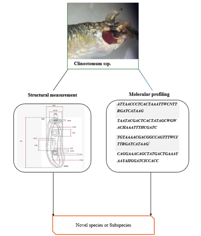
Clinostomum spp. are digenetic trematodes characterized by high genetic diversity and complex life cycles involving three hosts: snails, fish, and birds. These parasites signifi-cantly impact freshwater ecosystems, particularly when infecting fish as the second in-termediate host. Despite their ecological importance, no studies have examined the morpho-molecular characteristics of Clinostomum spp. from freshwater fish species in Oman, limiting our understanding of their life cycles and potential threats to the local aquatic environment. The present study investigated Clinostomum spp. infecting killifish (Aphaniops sp.) from four locations in the Muscat region of the Sultanate of Oman (Al Amirat, Al Khoud, Darsait, and Al Bahayes) using integrated morphological and mo-lecular approaches. Metacercariae were morphologically characterized through organ measurements and molecularly identified using mitochondrial cytochrome c oxidase subunit I (COI) sequences. Morphometric analysis revealed that Clinostomum parasites from Omani freshwater bodies were consistently smaller than the established species C. complanatum and C. marginatum reported from other regions. DNA sequence analysis indicated that the Omani specimens were closely related to, but distinct from, Turkish C. complanatum, suggesting they may represent a potentially novel species or subspecies.
Keywords:
metacercaria
; morphometry
; molecular phylogeny
; killifish
; Oman
1. Introduction
In arid and semi-arid regions, Aphaniops (Aphaniidae) species have attracted research interest due to their rapid diversification and adaptability across varied aquatic habitats [1,2]. Aphaniops stoliczkanus is particularly widespread across the Arabian Peninsula and South Asia, thriving in environments ranging from freshwater lakes to hypersaline waters [3,4]. In Oman, A. stoliczkanus and A. kruppi [5] inhabit seasonal water bodies such as Wadi Adai in Al Amerat [5]. While several studies have explored these species' morphology, habitat range, and phenotypic variability, in-formation on their parasitic infections in Oman remains limited. Understanding parasite-host dynamics in these endemic fish is crucial for freshwater conservation and fisheries management. Clinostomum spp. (Trematoda: Digenea), commonly known as "yellow grub," represent an important parasite group with a complex life cycle involving snails (first intermediate host), fish (second intermediate host), and piscivorous birds (definitive host) [6]. Metacercariae encyst in various fish tissues, including gills, muscle, and the oral cavity, causing physical damage and economic losses in aquaculture [7]. Human infection can occur through consumption of raw or undercooked fish, resulting in halzoun syndrome [8]. Clinostomum spp. have been reported globally, with increasing attention due to their zoonotic potential [9].
Taxonomic identification of Clinostomum spp. has traditionally relied on morphological traits. However, the soft, small, and often overlapping structures of these trematodes complicate species-level differentiation [11]. Recent molecular techniques have emerged as valuable tools to resolve these diagnostic challenges, providing deeper insights into parasite life cycles, genetic diversity, and host specificity [12,13]. Despite these advances, no morpho-molecular studies have characterized Clinostomum spp. infecting freshwater fish in Oman, leaving their prevalence, distribution, and pathogenic impacts unknown in the region. Given the ecological and public health implications of Clinostomum infections, documenting their occurrence and genetic identity within Oman's freshwater ecosystems is essential.
Therefore, the present study investigated Clinostomum spp. infecting Aphaniops spp. by combining morphological examination with molecular techniques to identify the species involved and establish baseline data for future freshwater ecological studies in Oman.
2. Materials and Methods
2.1. Study Sites and Sample Collection
The A total of 237 live fish specimens were collected from four distinct locations in the Muscat region: three land-locked sites Al Amirat (23° 31′ 42″ N, 58° 29′ 00″ E), Al Khoud (23° 34′ 33” N, 58° 07′ 06″ E), and Darsait (23°37′13.1″N 58°32′ 22.1″E) and one coastal lo-cation (Al Bahayes , 23° 40′ 47” N, 58° 11′ 36″ E) (Figure 1). Sampling was conducted using foldable shrimp and crab fishing traps (mesh size 3×3 mm, 3 nets deployed for 2-3 hours per site). Live fish were transported to the Marine Sciences Re-search Laboratory at Sultan Qaboos University (SQU) in containers equipped with aeration pumps. In the laboratory, four 70 cm³ tanks with aeration were established, one for each site sample. Total and standard lengths (mm) and body weight (g) were recorded for each fish. All specimens underwent thorough examination of skin, gills, muscles, and internal organs for Clinostomum spp.
2.2. Parasite Identification
2.2.1. Morphological Analysis
Sixty live Aphaniops sp. were randomly sampled from each site and transported to the laboratory. Collected metacercaria parasites were placed in 0.8% NaCl and refrigerated for 1 hour to prevent shrinkage and maintain their shape. After three washes with saline (0.8% NaCl), 10ml of Berland's fluid solution was added to relax the specimen; then parasites were preserved in 80% ethanol for >48 hours, then stained using Mayer's paracarmine for 2-6 hours following standard carmine staining protocols, dehydrated through graded alcohol series to 100%, then xylene, and then mounted in Canada balsam. As shown in Figure 2, Digital images and measurements of the cercaria were obtained using an Axio Cam HRc attached to an Axio microscope with Rising View software (Germany). Additional photographs were taken using an Olympus SZ61 microscope (Japan).
2.2.2. Molecular Analysis
A Total of 60 live parasites were collected from the live host Aphaniops sp at Al Khoud, Darsait, and Al Amirat, 20 live parasites respectively. After washing with distilled water, parasites were preserved in 50% ethanol in labelled 1.5 mL Eppendorf tubes and stored at -80°C overnight. The mitochondrial cytochrome c oxidase subunit 1 (cox1) gene was targeted for analysis. Frozen with -80°C parasites were ground into fine pieces, and 600 µL of lysis buffer was added and thoroughly mixed. The mixtures were incubated at 65°C for 1 hour, followed by the addition of 600 µL phenol: chloroform: isoamyl alcohol (25:24:1) and centrifugation at 10,000 g for 15 minutes. The supernatant was transferred to new 1.5 mL Eppendorf tubes, and the extraction process was repeated once more. Next, 10 µL of 3M Sodi-um-Acetate-NaAc and volumes of isopropanol were added to the supernatant. Tubes were gently inverted to visualize DNA threads and then incubated at -20°C overnight. After centrifugation at 10,000 g for 2 minutes, supernatants were discarded, and pellets were washed with 70% ethanol and centrifuged again. Ethanol was removed, and the tubes were air-dried. DNA pellets were stored at -20°C and subsequently suspended in 100 µL TE 0.1 buffer
2.2.3. PCR Amplification and Sequencing
DNA concentrations were determined using Qubit Fluorometric Quantitation (Invitrogen, Life Technologies). PCR was conducted to amplify the ITS-2 and mt-COI regions using OPHET_Fwd/OPHET_Rev primers [13] and Dice1F/Dice11R primers, respectively. PCR reactions were performed in 25 μL volumes containing 12.5 μL commercial master mix (Maxima Hot Start PCR Master Mix, Thermo Scientific), 10 μM of each primer, and 10-50 ng genomic DNA. Amplification occurred in a BIO-RAD T100 thermal cycler following conditions outlined by Van Steenkiste et al., 2015 [14], for mt-COI, with modifications for ITS-2 (initial denaturation at 95°C for 4 minutes; 30 cycles of 95°C for 30 seconds, 52°C for 35 seconds, 73°C for 1 minute; final extension at 73°C for 10 minutes). Amplicons were examined on 1.5% agarose gels stained with Safe View and visualized under UV light. Purified mt-COI amplicons were sequenced by Macrogen (Korea). Sequences were compared with reference sequences in NCBI using BLAST to detect genetic variations according to Altschul et al. (1990) [15].
2.3. Data Analysis
Statistical analyses included two-way ANOVA, one-way ANOVA, and Student's t-tests to assess regional and host effects on parasite numbers. All tests were conducted using Excel 2024, with significance set at p ≤ 0.05.
3. Results
3.1. Parasite Morphology
Clinostomum spp. specimens exhibited stout, tongue-shaped bodies with flat, unsegmented structures lacking hooks and possessing smooth lateral edges. The oral sucker (OS) was situated apically, while the ventral sucker (VS) occupied the anterior third of the body. The ventral sucker appeared large and well-defined, while the oral sucker was comparatively small. No pharynx was observed. The caecum and uterus were well-developed. Testes were arranged in tandem between the middle and posterior third of the body. The oral sucker displayed two distinct collar-like rings, and the ventral sucker extended to the posterior end. The testes exhibited branching structures.
3.2. Morphometric Analysis
Comprehensive comparative morphometric parameters—encompassing both the dimensional ranges (Min–Max) and localized means (± SD) for the Clinostomum spp. populations across the three surveyed sites in Oman, alongside the synthesized national baseline (N=180), are presented in Table 1. Phenotypic variations in total body length (BL) were observed across the geographic zones; specimens collected from Al Amirat were the longest (2390–2600 μm, mean: 2405±819 μm), followed by those from Al Khoud (2430–2910 μm, mean: 2320±142 μm) and Darsait (1520–2040 μm, mean: 2005±102 μm), establishing a consolidated national BL range of 1520–2910 μm (2243.3±510.8 μm). Consistent with these structural dimensions, the mean body length-to-width ratio (BL/BW) reached its peak in the Al Amirat population (2.19–4.02; 3.1±0.48), followed closely by Al Khoud (3.13–3.96; 3.0±0.26) and Darsait (1.52–3.81; 2.83±1.0).
Diagnostic sucker ratios and organ proportions further highlighted distinct phenotypic matrices. The ratio of oral sucker width to oral sucker length (OSW/OSL) was highest in the Al Amirat population (0.08–0.19; 0.14±0.04). Conversely, the Al Khoud population displayed uniquely elevated values for both the ventral-to-oral sucker width ratio (VSW/OSW: 1.33–3.50; 2.6±0.75) and the oral sucker width-to-body width index (OSW/BW: 0.13–0.36; 0.27±0.08) compared to their regional counterparts.
Regarding internal structural dynamics, oral collar width (OCW) spanned a combined national range of 170–1080 μm (509.3±241.3 μm), with the maximum localized mean recorded in Darsait (580±277 μm). The protective uterine sac length (USL) and width (USW) maintained consistent development across all three systems, settling at a collective national average of 621.0±156.7 μm and 73.7±21.3 μm, respectively. Despite these descriptive phenotypic gradients across the three Omani host populations, a one-way analysis of variance (ANOVA) confirmed that the localized morphometric discrepancies were not statistically significant (P≥0.05).
Ultimately, by formally integrating the combined dataset from all three locations (N = 180), these figures establish the first comprehensive, country-wide morphological profile for Clinostomum spp. within Oman.
3.3. Molecular Characterization
To reveal the taxonomic positioning of the Omani specimens, mitochondrial cytochrome c oxidase subunit I (COI) gene fragments were amplified using the specific primer sets outlined in Table 2. Nucleotide sequencing and subsequent phylogenetic analyses revealed notable directional topology discrepancies between the forward and reverse reads (Figure 3 and Figure 4).
Phylogenetic reconstruction using the forward sequence generated via the Dice1F primer resolved the studied specimens into a novel, monophyletic clade distinct from all previously cataloged Clinostomum species, indicating substantial genetic divergence (Figure 3). Conversely, the topology obtained from the reverse sequence via the Dice11R primer positioned the specimens in close evolutionary proximity to Clinostomum complanatum, suggesting that the Omani populations may represent a novel regional subspecies or a distinct evolutionary lineage within the C. complanatum complex rather than an entirely independent species (Figure 4).
4. Discussion
4.1. Morphological Differentiation and Phenotypic Plasticity
The morphometric datasets generated in this study indicate that the Clinostomum spp. populations from Oman possess a distinctly compact body size compared to previously documented congeners [11,18,19,20]. This pronounced structural diminution falls largely outside the standard metrics recorded for established Clinostomum species, indicating a unique regional phenotypic profile. Morphologically, while the specimens harvested from Aphaniops sp. exhibit a general structural affinity to Clinostomum complanatum, they remain consistently and significantly smaller than Clinostomum lineages described from neighboring regions, including Egypt [11] and Iran [21].
Although the inter-site morphometric variances did not reach statistical significance (P ≥ 0.05), the minor discrepancies observed in structural ratios across the sampling sites likely reflect localized phenotypic adaptations or the early stages of genetic drift within geographically isolated freshwater systems. Notably, the Al Amirat population exhibited both the highest mean body length-to-width ratio 3.1 ± 0.48) and an elevated anterior testis width-to-length ratio (1.34 ± 0.49). These variations underscore a degree of morphological elasticity that may be actively driven by microenvironmental fluctuations, differences in host lookouts, or specific host-parasite dynamics unique to each Omani drainage system. Consequently, comprehensive large-scale sampling is warranted to fully map these macrostructural trends.
4.2. Resolution of Molecular Ambiguity and Evolutionary Dynamics
Phylogenetic reconstruction and mitochondrial cytochrome c oxidase subunit I (COI) gene profiling provided critical, albeit complex, insights into the taxonomic positioning of the Omani Clinostomum populations. The distinct topological conflict observed between the forward and reverse sequencing tracks necessitates cautious and rigorous interpretation. The resolution of the forward reads (generated via the Dice1F primer) into a novel, independent monophyletic branch separate from known standard sequences supports a hypothesis of localized speciation. Conversely, the close evolutionary proximity to C. complanatum revealed by the reverse reads (via the Dice11R primer) strongly points toward an ongoing subspecific designation or a regional variant within the C. complanatum species complex.
This directional discrepancy in sequence topology can be logically attributed to several underlying biological and methodological factors:
1. Technical constraints or quality gradients inherent to unidirectional Sanger sequencing execution;
2. The deployment of broad or degenerate primer frameworks rather than highly restricted, taxon specific markers [16].
3. Intragenomic variation or high intraspecific genetic heterogeneity within the localized parasite population [21]; or
4. Ongoing, incomplete lineage sorting driven by rapid, contemporary evolutionary divergence.
4.3. Taxonomic Status and Cryptic Diversity
When evaluated together, the integrated morphological and molecular data suggest that the Clinostomum parasites infecting endemic Aphaniops sp. in Omani freshwater systems represent either an uncatalogued, novel species or a highly specialized regional subspecies of C. complanatum adapted to the extreme arid environments of the Arabian Peninsula. This finding strongly aligns with an expanding body of global parasitological literature documenting extensive cryptic diversity within the genus Clinostomum [10]. Increasingly, populations that appear morphologically identical or highly challenging to differentiate via traditional microscopy are resolved as phylogenetically distinct lineages when subjected to high-resolution molecular analysis across distinct geographical boundaries [17,22].
4.4. Ecological Impacts and Zoonotic Potential
The potential discovery of an endemic or unique Clinostomum lineage carries important ecological implications for the region. Possessing a complex heteroxenous life cycle that requires sequential transmission through multiple intermediate and definitive hosts, Clinostomum species serve as vital indicators of ecosystem connectivity and food web health in freshwater habitats [23]. The documentation of a potentially endemic lineage in Oman substantially enriches our understanding of parasite biogeography and biodiversity within the Arabian Peninsula, emphasizing the need for structured wildlife-parasitology surveillance in these fragile aquatic ecosystems.
From a veterinary and public health perspective, the zoonotic potential of these trematodes cannot be ignored, even though documented human clinostomiasis remains relatively rare globally [24]. Because these parasites reside as metacercaria within the musculature and tissues of freshwater fish, identifying and characterizing localized wild parasite reservoirs is highly critical. Establishing these biological baselines is vital for conducting accurate epidemiological risk assessments and formulating appropriate food safety and preventative frameworks, particularly in regions or communities where the consumption of raw, lightly preserved, or undercooked fish products occurs
5. Conclusions
This study provides a foundational contribution to the expanding body of research concerning parasite dynamics and biodiversity within arid aquatic ecosystems. By employing an integrated morpho-molecular diagnostic framework, this investigation demonstrated that Clinostomum parasites infecting endemic Aphaniops sp. in Omani freshwater systems exhibit a distinctly compact phenotypic profile and unique genetic attributes compared to previously described global congeners. Phylogenetic reconstruction revealed a directional discrepancy within the mitochondrial COI gene target; forward sequence analysis resolved the Omani specimens into an independent monophyletic branch supporting potential speciation, whereas reverse sequence analysis indicated a close evolutionary proximity to C. complanatum, suggesting a novel regional subspecies or distinct evolutionary lineage within the C. complanatum complex.
These findings underscore the critical importance of sustained parasitological monitoring and high resolution surveillance to safeguard both aquatic ecosystem integrity and regional public health. By elucidating the complex biological interactions between wild fish hosts and their associated metazoan parasites, this research highlights the necessity of ongoing investigations to better understand, model, and mitigate the long-term ecological impacts of parasitic infections on the resilience of fragile desert drainage systems, the sustainability of natural environments, and the safety of human populations.
Author Contributions
Conceptualization, G.Y. and M.A.; methodology, M.A., S.A., and A.S.; formal analysis, M.A. and H.A.; investigation, M.A, G.Y., S.A., A.A., S.A., A.S, and S.A.; data curation, M.A.; writing—original draft preparation, M.A.; writing—review and editing, G.Y., H.A. and A.S.; visualization, M.A.; supervision, G.Y.; project administration, G.Y. All authors have read and agreed to the published version of the manuscript.
Funding
This research was funded by Sultan Qaboos University internal grant number IG/AGR/FISH/24/02.
Institutional Review Board Statement
Not applicable.
Informed Consent Statement
Not applicable.
Data Availability Statement
The data presented in this study are available from the corresponding author upon request.
Acknowledgments
We would like to express our sincere gratitude to Dr. Ali Al Subhi for his invaluable support throughout the course of this research. We extend our heartfelt thanks to Aziza Al Adhoubi for generously providing the samples that were essential to this study. We also appreciate the guidance and support from our colleagues and mentors throughout the research process. Their valuable insights and encouragement were instrumental in completing this project.
Conflicts of Interest
The authors declare no conflicts of interest.
Abbreviations
The following abbreviations are used in this manuscript:
| BL | Body Length |
| BW | Body Width |
| OCW | Oral Collar Width |
| OSL | Oral Sucker Length |
| OSW | Oral Sucker Width |
| VSL | Ventral Sucker Length |
| VSW | Ventral Sucker Width |
| USL | Uterine Sac Length |
| USW | Uterine Sac Width |
References
- Freyhof, J., Weissenbacher, A., & Geiger, M. F. Aphaniops teimorii, a new species of tooth carp from the Dasht River drainage in Iran (Teleostei: Aphaniidae). Zootaxa. 2017, 4363(1), 117-126.
- Esmaeili, H. R., Teimori, A., Zarei, F., & Sayyadzadeh, G. DNA barcoding and species delimitation of the Old-World tooth-carps, family Aphaniidae Hoedeman, 1949 (Teleostei: Cyprinodontiformes). PLoS One. 2020,15(4), e0231717.
- Gonzalez, E. G., Cunha, C., Ghanavi, H. R., Oliva-Paterna, F. J., Torralva, M., & Doadrio, I. Phylogeography and population genetic analyses in the Iberian toothcarp (Aphanius iberus Valenciennes, 1846) at different time scales. Journal of Heredity. 2018, 109(3), 253-263.
- Clavero, M., Blanco-Garrido, F., & Prenda, J. Population and microhabitat effects of interspecific interactions on the endangered Andalusian toothcarp (Aphanius baeticus). Environmental Biology of Fishes. 2007, 78, 173-182.
- Freyhof, J., Weissenbacher, A., & Geiger, M. Aphanius kruppi, a new killifish from Oman with comments on the A. dispar species group (Cyprinodontiformes: Aphaniidae). Zootaxa. 2017, 4338(3), 557-573.
- Al Jufaili, S., Esmaeili, H. R., & Reichenbacher, B. Review of the freshwater fishes of Oman, with an updated check-list. Journal of Fish Biology. 2021, 98(3), 600–617.
- Caffara, M., Locke, S. A., Gustinelli, A., Marcogliese, D. J., & Fioravanti, M. L. Morphological and molecular differentiation of Clinostomum complanatum and Clinostomum marginatum (Digenea: Clinostomidae) metacercariae and adults. Journal of Parasitology. 2011, 97(5), 884-891.
- Iaria, C., Lo Monaco, D., & Marino, F. Pathology of Clinostomum spp. infections in wild and farmed fish. Journal of Fish Diseases. 2019, 42(9), 1215-1224.
- Park, C. W., Kim, J. S., Joo, K. H., & Kim, J. A human case of Clinostomum complanatum infection in Korea. The Korean Journal of Parasitology. 2009, 47(4), 401-404.
- Sohn, W. M., Na, B. K., & Cho, S. H. Morphological and molecular characteristics of Clinostomid metacercariae and adults. The Korean Journal of Parasitology. 2020, 58(4), 415-423.
- Gustinelli, A., Caffara, M., Florio, D., Otachi, E. O., Wathuta, E. M., & Fioravanti, M. L. First description of the adult stage of Clinostomum cutaneum Paperna, 1964 (Digenea: Clinostomidae) from grey herons Ardea cinerea L. and a redescription of the metacercaria from the Nile tilapia Oreochromis niloticus (L.) in Kenya. Systematic Parasitology. 2010, 76, 39-51.
- Dias, M. L. G. G., Eiras, J. C., Machado, M. H., Souza, G. T. R., & Pavanelli, G. C. The life cycle of Clinostomum complanatum Rudolphi, 1814 (Digenea, Clinostomidae) on the floodplain of the high Paraná river, Brazil. Parasitology Research. 2003, 89, 506-508.
- Blasco-Costa, I., Montero, F. E., Gibson, D. I., Balbuena, J. A., & Kostadinova, A. Structural validation of ribosomal DNA markers for the molecular identification of Clinostomum and related digenean complexes. Parasitology International. 2010, 59(2), 142–150.
- Van Steenkiste, N. W., Stephenson, E. W., & Locke, S. A. Evaluation of new degenerate primer sets for DNA barcoding of metazoan parasites: Application to the mitochondrial cytochrome c oxidase subunit I (COI) gene in Trematoda. Molecular Ecology Resources. 2015, 15(4), 812–821. [CrossRef]
- Altschul, S. F., Gish, W., Miller, W., Myers, E. W., & Lipman, D. J. Basic local alignment search tool. Journal of Molecular Biology. 1990, 215(3), 403–410. [CrossRef]
- Moszczynska, A., Locke, S. A., McLaughlin, J. D., Marcogliese, D. J., & Crease, T. J. Development of primers for the mitochondrial cytochrome c oxidase I gene in digenetic trematodes (Platyhelminthes) illustrates the challenge of barcoding parasitic helminths. Molecular Ecology Resources. 2009, 9(s1), 75–82. [CrossRef]
- Caffara, M., Bruni, G., Paoletti, C., Gustinelli, A., & Fioravanti, M. L. Cryptic diversity of Clinostomum spp. (Digenea: Clinostomidae) in European fish and amphibians. Parasites & Vectors. 2014, 7(1), 1–11. [CrossRef]
- Caffara, M., Locke, S. A., Echi, P. C., Halajian, A., Benini, D., Luus-Powell, W. J., & Fioravanti, M. L. A morphological and molecular study of Clinostomid metacercariae from African fish with a redescription of Clinostomum tipiae. Parasitology. 2017, 144(11), 1519-1529.
- Briosio-Aguilar, R., García-Varela, M., Hernández-Mena, D. I., Rubio-Godoy, M., & De León, G. P. P. Morphological and molecular characterization of an enigmatic clinostomid trematode (Digenea: Clinostomidae) parasitic as metacercariae in the body cavity of freshwater fishes (Cichlidae) across Middle America. Journal of Helminthology. 2019, 93(4), 461-474.
- Gholami, Z., Mobedi, I., Esmaeili, H. R., & Kia, E. B. Occurrence of Clinostomum complanatum in Aphanius dispar (Actinoptrygii: Cyprinodontidae) collected from Mehran River, Hormuzgan Province, South of Iran. Asian Pacific journal of tropical biomedicine. 2011, 1(3), 189-192.
- Locke, S. A., McLaughlin, J. D., & Marcogliese, D. J. Diversity and specificity in Diplostomum spp. metacercariae in freshwater fishes revealed by cytochromes oxidase I and internal transcribed spacer sequences. International Journal for parasitology. 2010, 1;40(3):333-343. [CrossRef]
- Emrah Simsek, Alparslan Yildirim, Erdal Yilmaz, Abdullah Inci, Onder Duzlu, Zuhal Onder, Arif Ciloglu, Gamze Yet-ismis & Gokmen Zafer Pekmezci. Occurrence and molecular characterization of Clinostomum complanatum (Trematoda: Clinostomidae) in freshwater fishes caught from Turkey. Parasitology Research. 2018, 117(7), 2111–2119. [CrossRef]
- Marcogliese, D. J. Parasites of the superorganism: Are they indicators of ecosystem health? International Journal for Parasitology. 2005, 35(7), 705–716. [CrossRef]
- Park, C. W., Kim, J. S., Joo, H. S., & Kim, J. A human case of Clinostomum complanatum infection in Korea. The Korean Journal of Parasitology. 2009, 47(4), 401–404. [CrossRef]
Figure 1.
The map shows four different sites of the sampling points in this study. The three inland freshwater wadis and one brackish water wadi (Al Bahayes).
Figure 1.
The map shows four different sites of the sampling points in this study. The three inland freshwater wadis and one brackish water wadi (Al Bahayes).
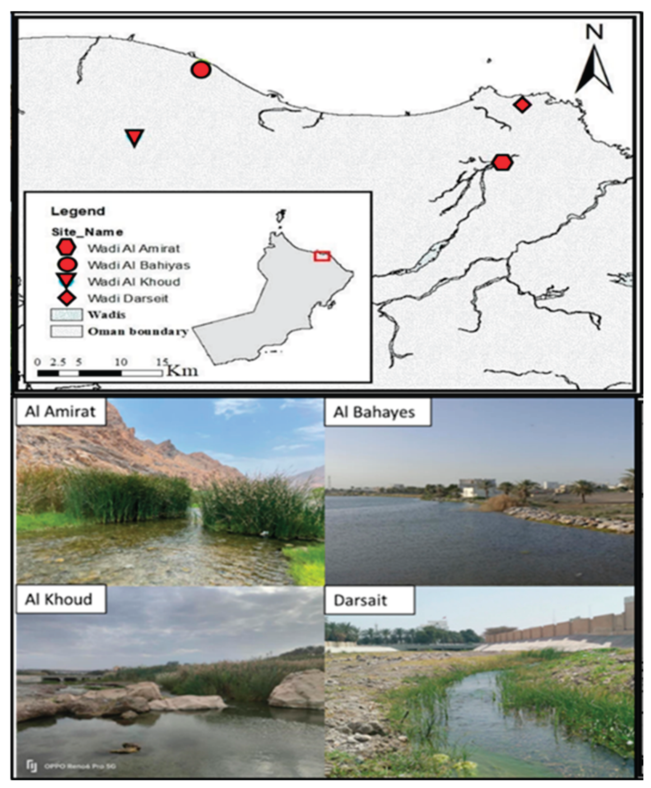
Figure 1.
Morphological features of Clinostomum metacercaria stained using Mayer’s paracarmine. Abbreviations: AT = anterior testis; C = caecum; O = ovary; OC = oral collar; OS = oral sucker; PT = posterior testis; US = uterus; VS = ventral sucker.
Figure 1.
Morphological features of Clinostomum metacercaria stained using Mayer’s paracarmine. Abbreviations: AT = anterior testis; C = caecum; O = ovary; OC = oral collar; OS = oral sucker; PT = posterior testis; US = uterus; VS = ventral sucker.
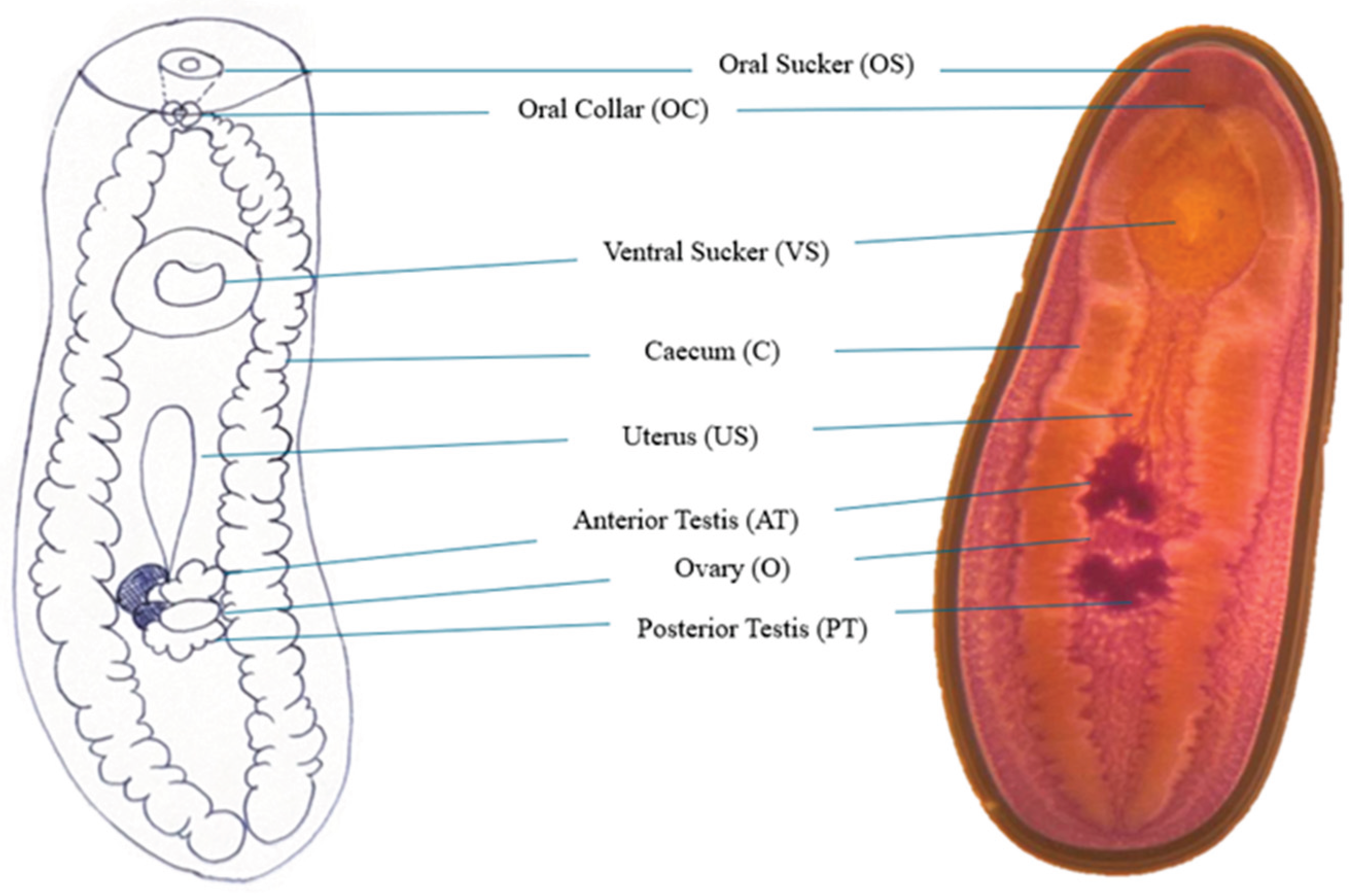
Figure 3.
Phylogeny of Clinostomum complanatum based on a neighbor-joining model using COI mtDNA (Direction: Forward).
Figure 3.
Phylogeny of Clinostomum complanatum based on a neighbor-joining model using COI mtDNA (Direction: Forward).
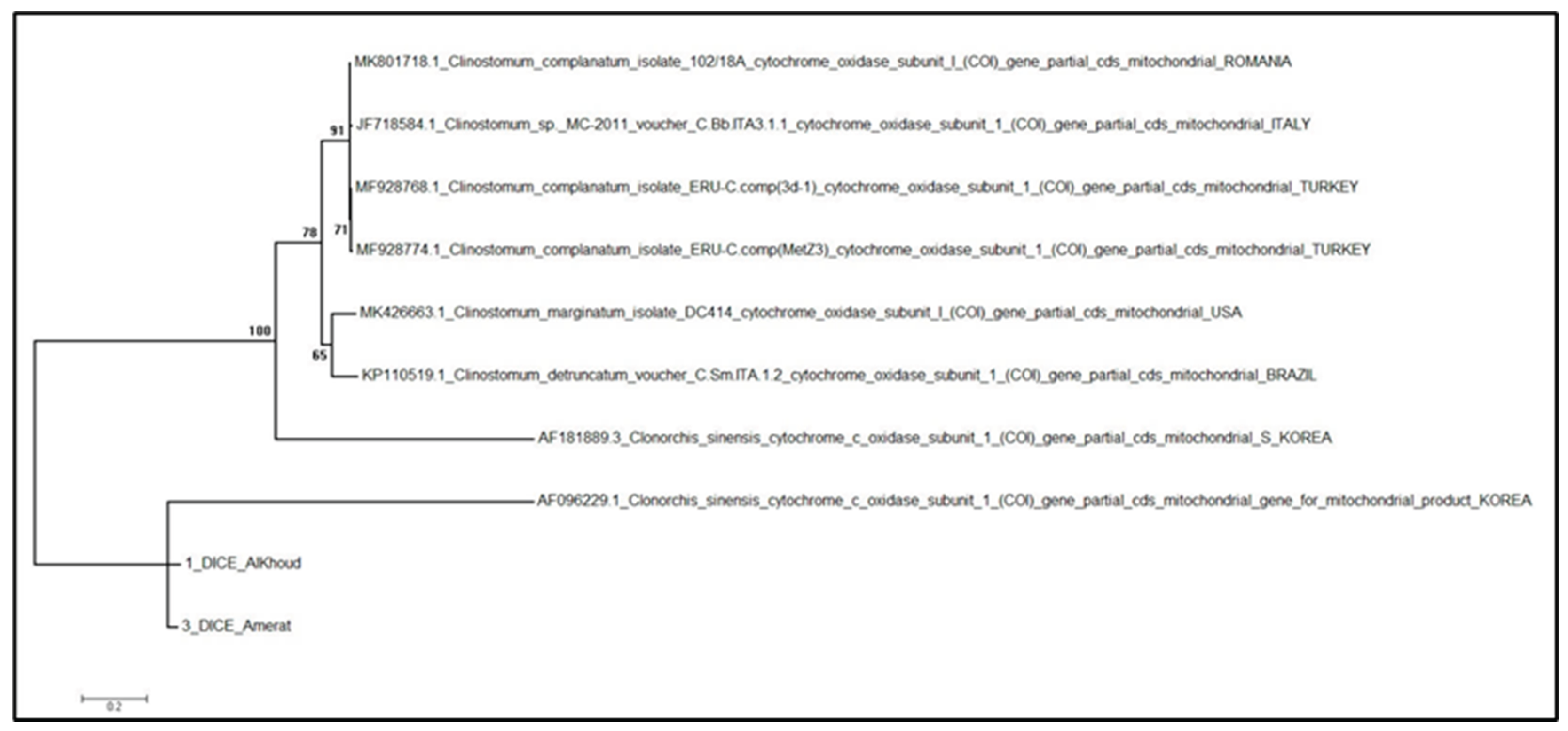
Figure 4.
Phylogeny of Clinostomum complanatum based on a neighbor-joining model using COI mtDNA (Direction: Reverse).
Figure 4.
Phylogeny of Clinostomum complanatum based on a neighbor-joining model using COI mtDNA (Direction: Reverse).
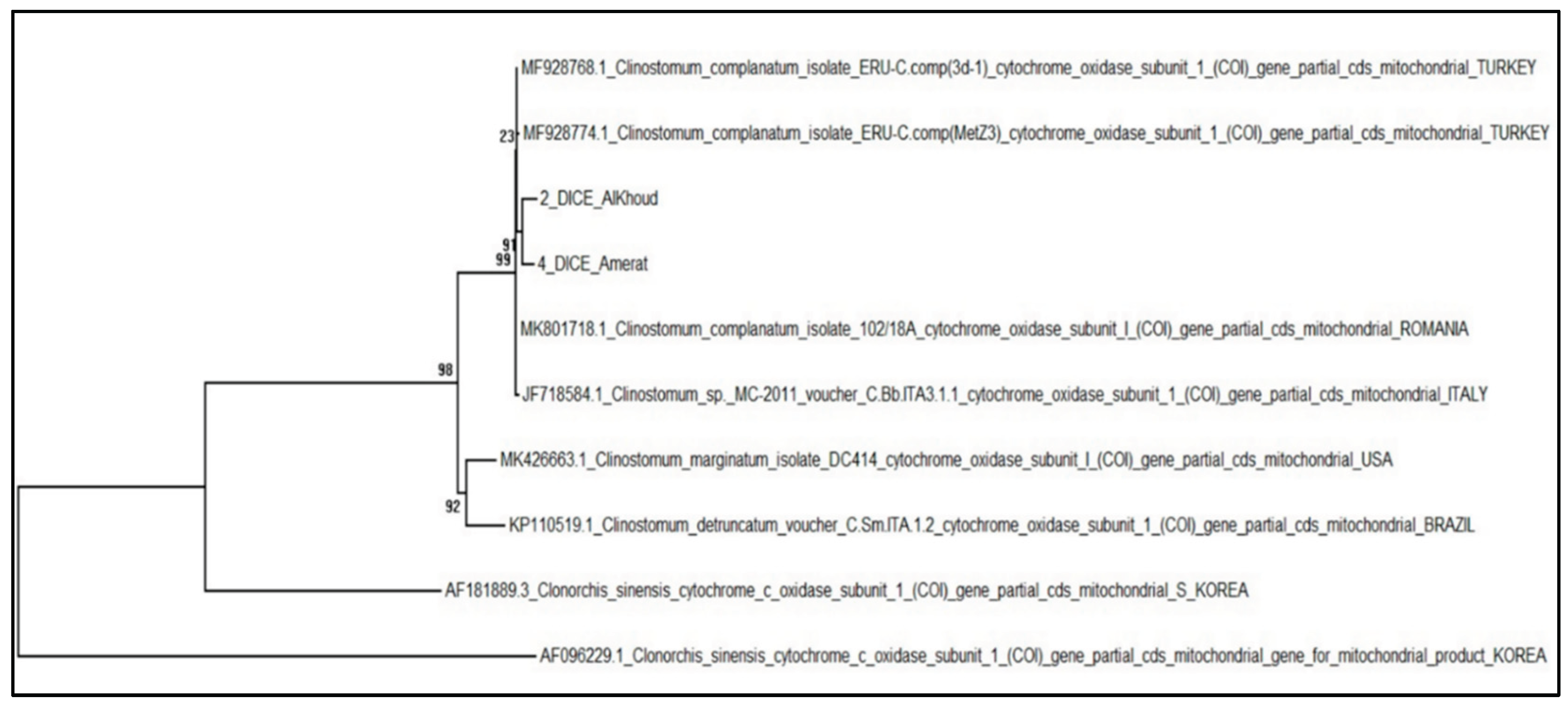

Table 1.
Measurements (in µm) of species of Clinostomum spp. from the three locations (Body length (BL), Body width (BW), Oral Collar Width (OCW), Oral Sucker Length (OSL), Oral Sucker Width (OSW), Ventral Sucker Length (VSL), Ventral Sucker Width (VSW), Uterine Sac Length (USL), Uterine Sac Width (USW).
Table 1.
Measurements (in µm) of species of Clinostomum spp. from the three locations (Body length (BL), Body width (BW), Oral Collar Width (OCW), Oral Sucker Length (OSL), Oral Sucker Width (OSW), Ventral Sucker Length (VSL), Ventral Sucker Width (VSW), Uterine Sac Length (USL), Uterine Sac Width (USW).
|
Clinostomun spp. Al Khoud (n= 60) |
Clinostomum spp. Darsat (n=60) |
Clinostomum spp. Al Amirat (n=60) |
Clinostomum spp. Oman (Total n=180) |
|||||
|---|---|---|---|---|---|---|---|---|
| Min–Max | (Mean ± SD) | Min–Max | (Mean ± SD) | Min–Max | (Mean ± SD) | Min–Max | (Mean ± SD) | |
| BL | 2430-2910 | 2320 ± 142a | 1520-2040 | 2005 ± 102a | 2390-2600 | 2405 ± 819a | 1520-2910 | 2243.3 ± 510.8 |
| BW | 700-1080 | 985 ± 64a | 1000-2500 | 1653 ± 482a | 920-2480 | 1514 ± 482a | 700-2500 | 1384.0 ± 487.6 |
| BL/BW | 3.13-3.96 | 3 ± 0.26a | 1.52-3.81 | 2.83 ± 1a | 2.19-4.02 | 3.1 ± 0.48a | 1.52-4.02 | 2.98 ± 0.66 |
| OCW | 230-830 | 404 ± 151a | 170-1010 | 580 ± 277a | 270-1080 | 544 ± 244a | 170-1080 | 509.3 ± 241.3 |
| OSL | 90-210 | 151 ± 31a | 65-270 | 130 ± 80a | 60-230 | 130 ± 62a | 60-270 | 137.0 ± 61.6 |
| OSW | 40-140 | 84 ± 26a | 100-320 | 186 ± 30a | 100-310 | 191 ± 55a | 40-320 | 153.7 ± 62.9 |
| OSW/OSL | 0.06-0.17 | 0.11 ± 0.03a | 0.05-0.18 | 0.10 ± 0.05a | 0.08-0.19 | 0.14 ± 0.04a | 0.05-0.19 | 0.12 ± 0.04 |
| VSL | 170-280 | 217 ± 30a | 130-420 | 226 ± 92a | 110-400 | 226 ± 92a | 110-420 | 223.0 ± 76.8 |
| VSW | 100-300 | 210 ± 68a | 120-400 | 221 ± 91a | 100-510 | 221 ± 91a | 100-510 | 217.3 ± 83.7 |
| VSW/ OSW | 1.33-3.50 | 2.6 ± 0.75a | 0.67- 1.89 | 1.16 ± 0.30a | 0.71-3.00 | 1.17 ± 0.36a | 0.67-3.50 | 1.64 ± 0.85 |
| OSW/ BW | 0.13-0.36 | 0.27 ± 0.08a | 0.08-0.19 | 0.11 ± 0.02a | 0.08-0.30 | 0.13 ± 0.03a | 0.08-0.36 | 0.17 ± 0.09 |
| USL | 470-900 | 650 ± 147a | 400-900 | 600 ± 159a | 400-940 | 613 ± 162a | 400-940 | 621.0 ± 156.7 |
| USW | 38-110 | 79 ± 21a | 30-97 | 69 ± 20a | 38-100 | 73 ± 22a | 30-110 | 73.7 ± 21.3 |
Table 2.
PCR primers that used to amplify mitochondrial COI gene fragments from samples of the parasites.
Table 2.
PCR primers that used to amplify mitochondrial COI gene fragments from samples of the parasites.
| Primer name | Direction | Primer sequence (5′-3′) | Gene | Product size | Reference |
|---|---|---|---|---|---|
| Dice1F | Forward | ATTAACCCTCACTAAATTWCNTTRGATCATAAG | COI | 800- to 820-bp | [14] |
| Dice11R | Reverse | TAATACGACTCACTATAGCWGWACHAAATTTHCGATC | |||
| MplatCOX1dF | Forward | TGTAAAACGACGGCCAGTTTWCITTRGATCATAAG | COI | 600 bp | [16] |
| MplatCOX1dR | Reverse | CAGGAAACAGCTATGACTGAAAYAAYAIIGGATCICCACC |
Disclaimer/Publisher’s Note: The statements, opinions and data contained in all publications are solely those of the individual author(s) and contributor(s) and not of MDPI and/or the editor(s). MDPI and/or the editor(s) disclaim responsibility for any injury to people or property resulting from any ideas, methods, instructions or products referred to in the content. |
© 2026 by the authors. Licensee MDPI, Basel, Switzerland. This article is an open access article distributed under the terms and conditions of the Creative Commons Attribution (CC BY) license (http://creativecommons.org/licenses/by/4.0/).
Copyright: This open access article is published under a Creative Commons CC BY 4.0 license, which permit the free download, distribution, and reuse, provided that the author and preprint are cited in any reuse.


