
Submitted:
08 July 2026
Posted:
10 July 2026
You are already at the latest version
Abstract
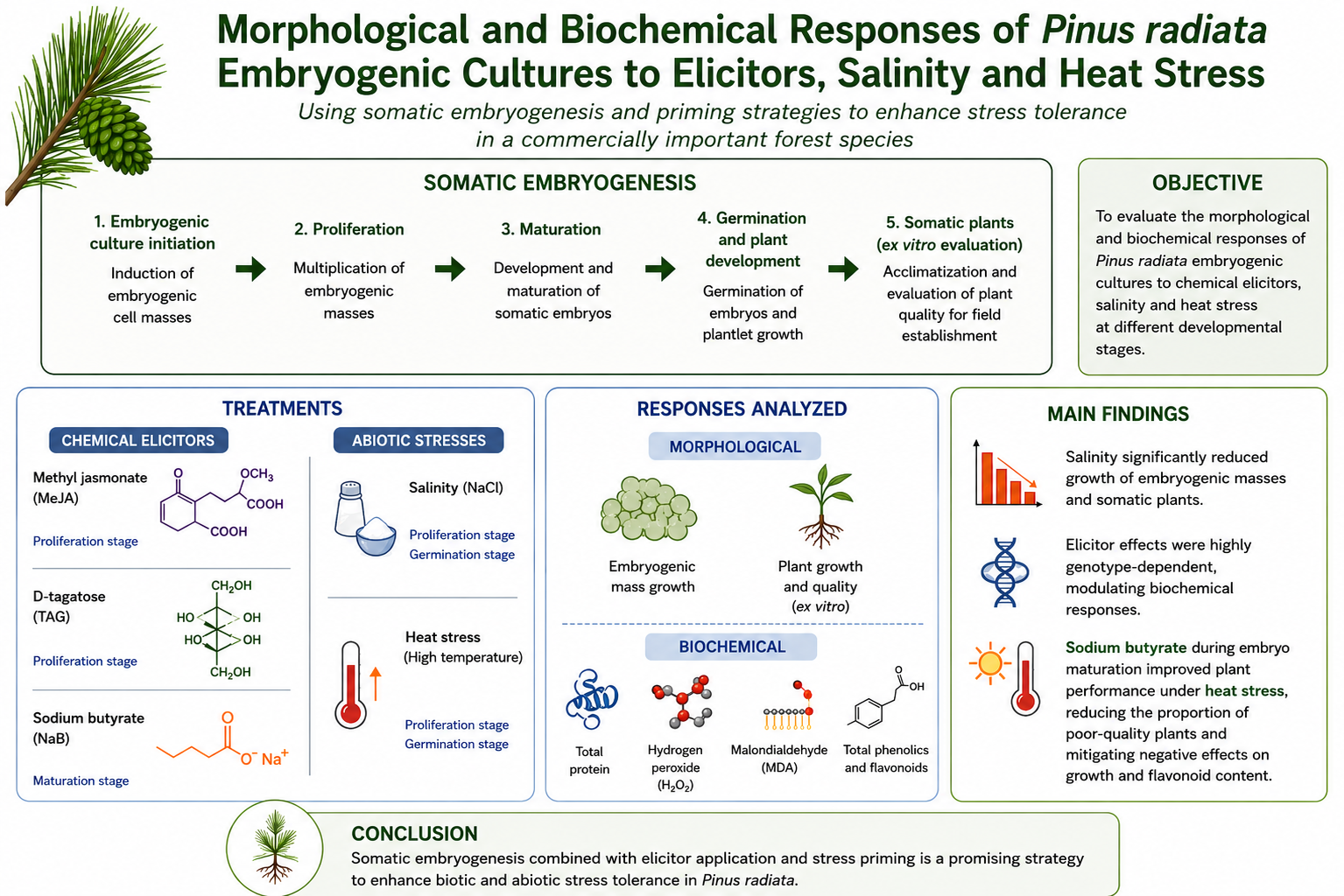
Pinus radiata is a commercially valuable forest species whose productivity is increasingly threatened by climate change-related biotic and abiotic stresses such as diseases, soil salinization and rising temperatures. Taking as a starting point a sufficiently optimized somatic embryogenesis propagation protocol for radiata pine, this study aimed to evaluate the morphological and biochemical responses embryogenic cell lines subjected to different elicitors such as methyl jasmonate and tagatose during proliferation or sodium butyrate at maturation stage. The effect of various stresses such as salinity and high temperatures was evaluated at proliferation and germination stages. Then, biochemical analyses (total protein, hydrogene peroxide and malondialdehide contents) were carried out in embryogenic tissues and plants and the growth was also assessed in these tissues and in somatic plants. This latter growth assessment led to a classification of plants depending on their suitability to be planted ex vitro. Several embryogenic cell lines were tested and the response to elicitation or stress was found highly genotype-dependent however some trends could be observed particularly when assessing growth. These results suggest that somatic embryogenesis combined with stress priming or elicitor application may be a viable strategy to enhance biotic and abiotic stress tolerance in radiata pine.
Keywords:
heat
; methyl jasmonate
; sodium butyrate
; sodium chloride
; tagatose
1. Introduction
Climate change is exposing the necessity of adapting agriculture and forestry sectors to new challenges by implementing scientific knowledge to produce more resilient plants. To this aim, fundamental research is the first step on the path; uncovering the physiological and biochemical mechanisms underlying plant stress response and adaptation is crucial to developing cutting edge biotechnological approaches.
In this regard, thanks to the basic knowledge generated during the last 50 years, alternative plant improvement approaches such as priming for enhanced defence are gaining special attention. This phenomenon is based on the biochemical mechanism in which plants pre-exposed to a certain stressor or stimulus, activate faster and/or stronger immune and stress response in the future [1]. Some molecules, commonly known as elicitors or priming agents, have the potential to mimic the presence of a stress factor or pathogen, triggering plant signalling and an advanced “warning” physiological state as well as activating the systemic acquired resistance (Tripathi et al. 2019). Well documented examples of elicitors include some phytohormones such as methyl jasmonate (MeJa) [2,3] and salycilic acid [4] or some rare sugars such as D-tagatose (TAG) [5]. Multiple studies have demonstrated that priming entails complex epigenetic modifications [6,7] ensuring the maintenance of a mid-to-long-term stress memory. These findings have also promoted research about the use of epigenetic modulators (e.g., azacitidine, sodium butyrate, trichostatin A to improve the behaviour of plants under different stress factors [8].
Somatic embryogenesis (SE) is a powerful and versatile micropropagation technique. Not only it is useful for producing a high number of genetically uniform, disease-free plants, but it can also be combined with long-term cryopreservation protocols [9] and genome-editing technologies [10] or just be used as a research model for the study of plant stress response [11] and the implementation of priming procedures under controlled and sterile environmental conditions [12]. Furthermore, as demonstrated by [13,14], and afterwards corroborated in our laboratory [15], the environmental conditions during plant embryo development (e.g., temperature, water availability) can determine the behaviour of the resulting plants years later.
In our region, the Basque Country (Spain), radiata pine (Pinus radiata D. Don) is a conifer species of great economic importance. Native to the Central Coast of California and Mexico, its cultivation has spread to numerous countries, including Spain, Australia, Chile, New Zealand, and South Africa [16]. It is considered the most widely planted exotic forest species worldwide, with over four million hectares planted globally [17,18], thanks to its rapid growth, adaptability, ease of plantation establishment, forest management and versatile wood properties [19]. However, this species is vulnerable to several biotic and abiotic stressors, which have been intensified by global climate change [20]. For instance, salinity due to poor soil management and is leading to an increase in barren land (about 40% of the terrestrial area), negatively impacting forest plantations [21]. Also, high salt concentrations cause physiological drought by lowering the osmotic potential of the soil [22].
One of the main consequences of climate change, the increase in temperatures, also provokes adverse effects on plant growth and development [23]. Elevated temperatures have negative effects on various physiological processes such as photosynthesis, primary and secondary metabolism, water relations and lipid metabolism [24]. Specifically, heat stress generates damage to the cell membrane due to the overproduction of reactive oxygen species (ROS), causing senescence and cell death and leading to altered plant growth and reproduction [25,26].
In this context, the aim of this study was to exploit the advantages that SE offers as a research tool and study 1) the in vitro response of P. radiata embryonal masses (EMs) and somatic plants (SPs) to high sodium chloride (NaCl) concentrations, 2) the effect of exposing EMs to two different priming agents (MeJa and tagatose), and 3) the long-term effect of a histone deacetylase inhibitor [sodium butyrate (NaB)] applied during the proliferation of EMs on the performance of the resulting SPs under heat stress. This first approach has been based on the measurement of different biochemical parameters [total protein, hydrogen peroxide and malondialdehyde (MDA) contents] and morphological parameters.
2. Materials and Methods
2.1. Plant Material
Embryonal masses (EMs) and somatic plants (SPs) of P. radiata were obtained following the protocols outlined by Montalbán and Moncaleán [27,28]. Briefly, one-year-old green female cones, enclosing immature zygotic embryos at precotyledonary stage were collected from genetically distinct mother trees. Megagametophytes were isolated from seeds and cultured on embryo development medium (EDM) [29] for 4 to 8 weeks. After that, the resulting EMs were separated from the megagametophytes and fortnightly subcultured onto EDM proliferation medium [28]. After four subcultures, the EMs growing actively were considered as embryogenic cell lines (ECLs).
For experiments with SPs, EMs at the proliferation stage were suspended in liquid EDM, filtered and cultured on EDM maturation medium [30] to promote somatic embryo development. After 15 weeks on maturation medium, somatic embryos (ses) were transferred to Petri dishes containing RG medium [28] for 6 weeks, followed by additional 6 weeks in OV80+OVD80 microbox (SacO2) culture boxes with the same medium to produce SPs. Unless stated otherwise, initiation, proliferation and maturation were carried out in darkness at 22ºC. Germination was carried out at the same temperature but under a 16 h photoperiod provided by cool white fluorescent tubes (TLD 58 W/33; Philips, France) at 120 μmolm− 2s−1.
2.2. Experiment 1: Salt Stress Response in EMs and SPs
For experiments carried out with EMs, five ECLs (R101, R330, R55, R156 and R4) at proliferation stage were used as initial material. The EMs were transferred to EDM proliferation media supplemented with different concentrations of NaCl (0, 50 or 100 mM). Each treatment comprised 5 Petri dishes and 4 calli were cultured on each plate.
To assess growth, photographs of each EM were taken on days 1 and 14 of proliferation. Then, with the program ImageJ V. 1.54g, the diameter of each callus was determined, and growth was estimated as the difference between the final and initial measure.
For the determination of dry weight, 0.2 g of EM was collected and oven dried at 70 °C for 48 h; 3 replicates per ECL and salt concentration were performed. The percentage of dry weight was calculated by applying the formula: Dry weight percentage (DW%) = dry weight (DW; g)/fresh weight (FW; g*100). The remaining material was crushed into liquid nitrogen and stored at -80 °C until the biochemical analyses were performed.
Total protein content was determined colorimetrically using the Bradford dye-binding assay with bovine serum albumin (BSA) following the protocol described in [31]. Three technical replicates for each ECL and salt concentration were measured.
For hydrogen peroxide (H2O2) determination, the protocols described by Alexieva et al. [32] were used as a reference. Three technical replicates of 0.5 g were processed for each treatment and ECL (except for R55 due to lack of material enough for the analyses in 100 mM treatment).
For experiments with SPs, three different ECLs (R23-94, R23-11 and R23-125), previously cultured for six weeks in Petri dishes containing RG germination medium, were transferred to OV80+OVD80 culture boxes (SacO2) with 80 mL of RG medium and supplemented with different treatments of NaCl (0, 25 and 50 mM). Eight SPs per microbox and three microboxes per treatment and ECL were cultivated under 16 h photoperiod at 120 μmols-1m-2 and 22 °C for seven weeks.
Photographs were taken of SPs from every treatment and embryogenic cell line (ECL) at the start and at the end of the culture in microboxes. Root and shoot lengths were measured using ImageJ V. 1.54 g. Then, growth was estimated as the difference between the final and initial length measurements. According to final root and shoot lengths SPs were classified into different categories as described in Table 1.
DW (two plants per replicate), total protein content and H2O2 (0.2 g per replicate) were calculated as previously described.
MDA content was measured following the protocol reported by Morcillo et al. [33], using 0.2 g tissue per replicate.
Chlorophyll a was quantified following the procedure described by López-Hidalgo et al. [34]. The absorbance was measured at 649 and 664 nm and a total of three technical replicates per treatment and ECL were analyzed. The concentration of chlorophyll a (mgDW-1) was calculated as 13.36*A664–5.19 *A649.
2.3. Experiment 2: Elicitation of EMs with MeJa and TAG
Three ECLs (RE22-25, RE22-51 and R23-126) were used as starting material. A total of 0.9 g was placed in a sterile 50 mL tube for each treatment and ECL. In each tube, 15 mL of liquid EDM [29] supplemented with MeJa (0, 5 or 20 µM) or TAG (0, 0.5 or 1.5 gL-1) were added. Cultures were incubated in darkness at 22 °C and 70 rpm for 3 days. After that, 5 mL of the suspension containing 0.3 g of embryogenic tissue were poured on a filter paper (Whatman, nº2) and the liquid medium was drained in a Büchner funnel; then, the filter paper with the attached tissue was transferred onto proliferation medium. Three Petri dishes for each treatment and genotype were cultured for three weeks. The whole procedure (culture of EMs with MeJa or TAG and subsequent proliferation) was repeated two more times to induce three elicitations of plant material. Then, morphological and biochemical analyses were performed.
Tissue growth was calculated indirectly weighing the Petri dish with the proliferating tissue at the start and at the end of the proliferation period.
The same protocol used to determine dry weight in Experiment 1 was followed, but using 0.5 g of EM for each analysis. Total protein, H2O2 and MDA were determined for all treatments and ECLs as described above. Three technical replicates were performed for each treatment and ECL.
2.4. Experiment 3: Culture of EMs on Media with NaB and Heat Stress Application on the Resulting SPs
Actively proliferating EMs from three different ECLs (RD21-27, R20-52, R23-54) were divided into three treatments based on NaB application, following the methodology of Sandoval et al. [35]. In NaB 1 treatment, EMs were suspended in liquid EDM with 0.5 µM NaB for 1 h before transferring to EDM maturation medium. For NaB 2 treatment, EMs were cultured directly on semisolid EDM maturation medium supplemented with 0.5 µM NaB. The control group followed the standard maturation procedure. After 15 weeks, ses were germinated as explained above. Then, for each NaB treatment, seven microboxes per ECL and temperature regime, each containing ten SPs, were subjected to heat stress consisting of exposure to 35 ºC for 3 h daily over 3 days, followed by 42 ºC for 3 h daily over 2 additional days. Control microboxes were kept at 22 ºC for 5 days.
Survival per ECL and treatment was registered weekly for 6 weeks. Initial and final longitude of the root and shoot were measured for each treated somatic plant on day 1 and 42 of the experiment using ImageJ software V. 1.54g. Classification of somatic plants was conducted according to final root and shoot lengths as described above (Table 1).
Six weeks after the start of heat stress application, extraction and quantification of total phenolic compunds and flavonoids was carried out as described by Kim et al. [36] with some modifications. Briefly, 50 mg of the grounded plant material was suspended in 1 mL 80% (v:v) methanol, homogenized and centrifuged at 11000 g for 10 min. The supernatant was collected and evaporated. Afterwards, 1 mL methanol 50% (v:v) was added to the tube and homogenized. For phenol quantification, 100 µL plant extract was diluted in 900 µL distilled water. Subsequently, 100 µL Folin & Ciocalteu’s phenol reagent were added, followed by incubation for 5 minutes. Then, 1 mL 7% (m:v) Na₂CO₃ solution was added. For flavonoid quantification, 100 µL of the extract were mixed with 400 µL distilled water, 30 µL 5% (m:v) NaNO₂, 30 µL 10% (m:v) AlCl₃, 200 µL 1M NaOH, and 240 µL distilled water. Then, total phenol and flavonoid contents were measured spectrophotometrically, and measurements were expressed as mg gallic acid and catechin equivalents (for total phenolic compounds and flavonoids, respectively) per g of fresh plant tissue after construction of standard curves. Three technical replicates for each treatment and ECL were analysed.
2.5. Statistical Analysis
Experiments were conducted in a completely randomized block design. The assumptions of normality and homoscedasticity were determined and linear model or generalized linear model fittings were performed followed by Tukey’s test of multiple comparisons (α = 0.05) to determine significant statistical differences.
3. Results
3.1. Experiment 1: Salt Stress Response in EMs and SPs
After 14 days in proliferation medium, the EMs of each treatment showed significant differences in size from day 1 to 14; the growth decreased significantly as the concentration of salt in the medium increased in all the ECLs assayed (Figure 1 and Figure 2A), although this reduction was most noticeable in those cultured with100 mM NaCl. DW percentage of EMs was significantly higher as the salt concentration in the medium increased (Figure 2B).
Total protein content was significantly lower in EMs cultured at the highest concentration of salt. The amount of protein per dry weight followed a tendency to decrease in the EM from the lowest concentration to the highest of sodium chloride and was significantly lower at 50 mM NaCl (Figure 3A). On the contrary, H2O2 contents did not show a clear pattern, moreover a significant interaction between treatments and ECLs was found. (Figure 3B).
The relative percentage of SPs categorized as 1 from 25 mM NaCl treatment was equal or higher than control treatment for lines R23-11 and R23-125. However, this percentage decreased as NaCl increased in R23-94. Plants from class 2 and 3 showed a different tendency among treatments and ECLs. The relative percentage of class 4 SPs coming from different ECLs was always highest after culture at the highest salt concentration (Figure 4).
The growth depended significantly on the genotype tested; the root growth was not affected by salinity. Whereas at the highest NaCl concentration, plants displayed shorter shoots. (Figure 5A). DW percentage was one of the few parameters analysed not genotype-dependent, showing significantly higher percentages the plants cultured at control conditions (Figure 5B).
A significant interaction between the genotype and the treatments was found for the total protein contents. Within each cell line, SPs cultured at 25 mM NaCl showed significantly higher protein contents than control, whereas culture at the highest salinity concentration led to different results depending on the genotype (Figure 6A). When H2O2 content was analysed, although significant differences were found for the ECLs and treatments, the contents (ranging from 40.6 to 60.8 µg g DW-1) did not follow a clear pattern (Figure 6B).
In the same way, chlorophyll a content did not follow a trend; however, the highest (R23-125, control) and lowest values (R23-94 50 mM NaCl) were observed in the same ECLs and treatments (Figure 7A) in relation to H2O2 accumulation (Figure 6B). The MDA content was similar in line R23-11 regardless of the treatment applied, whereas in the other lines tested it was lower at control conditions (Figure 7B).
Figure 8 shows that most SP from R23-11 and R23-94 exhibited yellowish colors under 25 and 50 mM treatments compared to 0 mM NaCl. In contrast, SP from R23-125 ECL did not show the same trend.
3.2. Experiment 2: Elicitation of EMs with MeJa and TAG
The growth of EMs was similar regardless of the treatment, only significant differences were found depending on the ECL tested (Figure 9). A significant effect of the ECL, elicitation treatment and their interaction was found. Embryonal masses tissue of R23-126 at the end of the experiment in control (a), 5 µM methyl jasmonate (b), 20 µM methyl jasmonate (c), 0.5 g L-1 tagatose (d) and 1.5 g L-1 tagatose (e).
The effect of elicitors on the total protein content was genotype dependent, but no significant interactions were found between them and the treatments assayed. The EMs at control conditions exhibited a significantly higher total protein content than those subjected to MeJa treatments (Figure 10 A). In the case of TAG, significant differences were found between all treatments, the highest protein content was shown in control treatment, followed by TAG at 1.5 and 0.5 g L-1 (Figure 10B).
3.3. Experiment 3: Culture of EMs on Media with NaB and Heat Stress Application on the Resulting SPs
The addition of NaB did not imply an improvement in the quality of SPs (Figure 13); however, under thermal stress conditions the lowest number of low-quality somatic plants (class 4) were observed in plants from both NaB treatments (Figure 13).
The biggest SPs with the highest flavonoid content were those from control conditions (Figure 14A, B). The growth (Figure 14A) and total flavonoid content (Figure 14B) were significantly reduced in non-primed SPs subjected to heat stress, these values were similar to those observed in plants from sodium butyrate treatments regardless of their culture conditions during germination.
The total phenol content was significantly affected by all the factors studied separately (Figure 14B). However, no significant effect of the interaction between NaB treatments and temperature stress was observed, ranging from 3.5 and 4.5 mg of gallic acid equivalents per gram of fresh weight plant tissue.
4. Discussion
After analyzing the results of salt stress experiment, the growth of both EMs and SPs was negatively affected by increasing salt concentrations. This effect was particularly evident at concentrations above 50 mM NaCl and reached its maximum at 100 mM, where the growth of EMs was almost completely suppressed. The results are consistent with previous studies on SE in Medicago truncatula [37], in which a slight reduction in cell growth was observed at 50 mM NaCl when compared with the control treatment (0 mM NaCl), which was attributed to osmotic stress. At higher concentrations (100 and 150 mM NaCl), the authors reported a reduction in cell size, which was associated with plasmolysis. Similar responses have also been described in embryogenic cultures of other plant species; for example, callus and cell suspension cultures of Stevia rebaudiana exposed to salt stress exhibited lower growth rates than control cultures [38]. In Psidium guajava SPs, shoot growth decreased when 50 and 100 mM NaCl treatments were applied, which was attributed to limitations in water uptake and detrimental ionic effects on biochemical processes [39].
Regarding dry weight, EMs presented a tendency to increase their dry matter content at high NaCl concentrations. This pattern indicates that all ECLs were affected by the osmolarity of the culture medium [40]. In SPs, however, the opposite behavior was detected. This trend is in accordance with other studies conducted at the whole-plant level, where salinity reduces dry weight presumably because of water deficit, nutritional imbalance and osmotic stress, ultimately leading to stomatal closure [41]. Since the present study was performed in vitro, where plants rely largely on heterotrophic nutrition, the reduction in dry weight may be more closely related to impaired water uptake, and consequently, reduced carbohydrate absorption from the culture medium rather than stomatal dysfunction. Nonetheless, total dry weight alone may provide a limited view of plant responses to salinity, as some species tend to allocate a greater proportion of biomass to the root system under stress conditions [43].
Total protein content in EMs showed a general decreasing trend with increasing NaCl concentrations. This reduction can be attributed to the detrimental effect of salinity on mRNA biosynthesis and nutrient uptake required for protein synthesis, leading to alterations in this metabolic process [41]. Similarly, Jasim et al. [44], reported that increasing NaCl concentrations in the culture medium resulted in lower protein contents in the embryogenic calli of Phoenix dactylifera. On the other hand, the total protein content of SPs increased after culture at 25 mM NaCl for all ECLs, and for two of the three ECLs at 50 mM NaCl. It is well-known that moderate salinity can promote the synthesis of specific stress response proteins [45]. For example, in a study carried out by Azizi et al. [46], an increase in the total protein of all Rosa damascena seedling genotypes treated with 25 mM NaCl was observed, whereas only a few genotypes showed that response at 50 mM treatment.
The levels of H2O2, chlorophyll a and MDA appeared to be genotype-dependent under salt stress. In the case of H2O2, this behavior is consistent with Hasanuzzaman et al. [47] and Wang et al. [48], who reported that the degree of oxidative stress caused by ROS like H2O2 in plants under saline conditions is highly dependent on the characteristics of each ECL and their genetic background. Despite the absence of a clear pattern, combining phenotypic observations with biochemical analyses enabled the differentiation of salt tolerance levels among the different ECLs. Two of the three genotypes presented a yellowish color under salt stress, suggesting higher susceptibility to elevated NaCl concentrations. These ECLs exhibited reduced chlorophyll content under stress conditions, following trends observed in other species [49,50]. Interestingly, the most susceptible genotype presented almost double the malondialdehyde content when growth under stress conditions, indicating increased lipid peroxidation and cellular damage, as observed in Solanum melongena seedlings at 30 mM NaCl [51]. Together, these findings support the usefulness of in vitro systems as effective tools for the early selection and propagation of stress-tolerant plant material [52].
In relation to Experiment 2, the growth of EMs did not differ significantly among elicitor treatments; this indicates that the treatment neither promoted nor inhibited cell proliferation. These results are consistent with those reported by Yazdanian et al. [53], who observed that elicitors such as MeJa at a concentration of 25 µM did not significantly affect the growth of Allium jesdianum EMs compared with the control treatment. Likewise, these treatments had no effect on the dry biomass of embryogenic cultures. In contrast, both MeJa and TAG treatments negatively affected total protein content, probably because of alterations in protein biosynthesis or enhanced protein degradation, as previously suggested for EMs of P. radiata [54]. When MeJa or TAG treatments were applied to EMs, no increase in H2O2 content was recorded, suggesting that these elicitors did not generate oxidative stress in treated EMs. Similar results were reported in EMs of Quercus ilex, where the lowest H2O2 content was obtained following treatment with 25 µM MeJa [12]. MDA is a product of the secondary degradation of lipid peroxidation and is widely used as an indicator of oxidative damage [55]. In the present study, a highly genotype-dependent response was observed for MDA content. Some studies have suggested that elicitors such as MeJa can increase MDA levels as part of the stress response MeJa [56]. However, Yazdanian et al. [53] reported that treatment with 25 µM MeJa did not produce significant changes in MDA content compared with the control in Allium jesdianum EMs. Similarly, in Rosmarinus officinalis, MeJa treatment resulted in reduced MDA levels, which was attributed to an enhanced capacity to mitigate and repair oxidative damage [57].
In Experiment 3, in which EMs were treated with NaB and the resulting plants were exposed to heat stress, no clear effect of the histone deacetylase inhibitor was observed on either the quality or the growth of the SPs under control conditions. On the other hand, high temperatures caused a noticeable decline in the shoot growth of plants originating from non-treated EMs. It is well documented that heat provokes a decrease in photosynthesis, cell membrane dysfunction, and oxidative damage, impairing plant development [24,26]. The detrimental effect of elevated temperatures could not be observed in NaB derived SPs, suggesting that the elicitor could be playing a determinant role in plant homeostasis under stress conditions [58]. Multiple studies have addressed the positive effects of NaB on different stages of SE. For example, the addition of 0.5 mM NaB to the induction culture medium improved the embryogenic response in Vitis vinifera [59], or concentrations up to 1 mM on tomato and tabaco protoplast derived-calli increased the frequency of adventitious shoot formation [60]. Less information is available about the implications of this histone deacetylase inhibitor in stress-response processes. Some authors have observed that NaB treatments can alter the expression of stress-related genes, modulating the immune response of plants against certain pathogens [61]. In some medicinal plants, such as Platycodon grandiflorus, NaB-induced changes in histone acetylation led to alterations in secondary metabolism, particularly in the accumulation of polyphenolic compounds in the roots of the plants [62].
In this regard, in the present study, elevated temperatures provoked a significant decrease in total flavonoid levels, which, as observed for shoot growth, was not detected in SPs derived from NaB treatments. It is noteworthy that the basal levels of these compounds under control conditions were lower than those of non-treated plants. Under abiotic stress conditions, an increase in the synthesis of certain groups of secondary metabolites is expected, since these molecules play crucial roles in oxidative stress mitigation, osmoprotection, and overall stress adaptation [63]. Specifically, heat stress promotes the production of antioxidant phenolic compounds such as flavonoids, phenylpropanoids, anthocyanins and lignans which are related to the suppression of stress-induced oxidation of cellular molecules [64]. Nonetheless, some authors have also identified a slight reduction in the levels of these metabolites under heat stress, attributed to overall metabolic malfunction [65]. In tomato seedlings, lower flavonoid levels were detected after heat stress, but contrastingly, the plants showed improved antioxidant capacity in antioxidant assays [66]. Similarly, in Pinus sylvestris, elevated temperatures altered the composition of the flavonoid profile, and specific groups such as acylated flavanols and flavan-3-ols were more affected than others [67]. These findings suggest that long-term epigenetic modifications induced by NaB may be implicated in the fine-tuning of certain secondary metabolism pathways, resulting in improved performance of SPs under stress conditions according with Zhao et al. [68].
5. Conclusions
The growth and weight of EMs and SPs was affected by salinity; on the contrary, elicitated ECLs did not grow differently from the controls. The total protein content was negatively affected by salinity, MeJa or TAG treatments at proliferation stage of the cultures. Other biochemical parameters tested seemed to be more genotype-dependent in both experiments 1 and 2. When elicitation was carried out at maturation stage, the resulting SPs showed lower flavonoid contents after being subjected to heat stress, finally SPs generated at control conditions grew less after stress application that those elicitated with NaB.
Author Contributions
Conceptualization: I.A.M. and P.M.; Funding acquisition: P.M.; Investigation: M.G., D.V., D.B., A.C., I. A. M.; Data curation: M.G., D.V., D.B. Formal analysis: M.G., D.V., D.B., A.C., I.A.M., J.P.; Visualization: A.C., I.A.M., J.P.; Supervision: A.C., I.A.M., P.M.; Writing—original draft: M.G., D.V., D.B., A.C., I.A.M., J.P. and P.M.; Writing—review and editing: A.C. I.A.M., J.P., P.M. Resources: I. A. M., P.M. Project administration: P. M. Funding adqusition: P.M. All authors have read and agreed to the published version of the manuscript.
Funding
This research was performed through Project PID2020-112627RB-C32 and Project PID2024-156422OB-C32 funded by MICIU/AEI/10.13039/501100011033. The authors would like to acknowledge COST Action CA21157 “European Network for Innovative Woody Plant Cloning”, www.copytree.eu (accessed on 12 March 2025), supported by COST (European Cooperation in Science and Technology) www.cost.eu (accessed on 12 March 2025).
Conflicts of Interest
The authors declare no conflicts of interest.
Abbreviations
The following abbreviations are used in this manuscript:
| BSA Bovine serum albumin DW Dry weight ECL Embryogenic cell line ECLs Embryogenic Cell ines EMs Embryonal masses FW Fresh weight H2O2 Hydrogen peroxide MDA Malondialdehyde MeJa Methyl jasmonate NaB Sodium butyrate NaCl Sodium chloride ROS Reactive oxygen species SE Somatic Embryogenesis Ses Somatic embryos SPs Somatic plants TAG D-Tagatose |
References
- Conrath, U.; Beckers, G. J. M.; Langenbach, C. J. G.; Jaskiewicz, M. Priming for enhanced defense. Ann. Rev. Phytopathol. 2015, 53, 97-119. [CrossRef]
- Sheteiwy, M.S.; Gong, D.; Gao, Y.; Pan, R.; Hu, J.; Guan, Y. Priming with methyl jasmonate alleviates polyethylene glycol-induced osmotic stress in rice seeds by regulating the seed metabolic profile. Env. Exp. Bot. 2018, 153, 236-248. [CrossRef]
- Mageroy, M.H.; Wilkinson, S.W.; Tengs, T.; Cross, H.; Almvik, M.; Erbilgin, N. Molecular underpinnings of methyl jasmonate-induced resistance in Norway spruce. Plant Cell Environ. 2020, 43(8), 1827-1843. [CrossRef]
- Alam, P.; Karabulut, F.; Iqbal, J.; Sharma, R.; Fathi, A.; Bagci, E.; Baran, M. F.; Albalawi, T.; Faizan, M. Role of salicylic acid and jasmonic acid in salinity stress mitigation in plants. Front. Plant Sci. 2026, 17, 1824082. [CrossRef]
- Mijailovic, N.; Nesler, A.; Perazzolli, M.; Aziz, A.; Aït Barka, E. D-Tagatose-based product triggers sweet immunity and resistance of grapevine to downy mildew, but not to gray mold disease. Plants 2022, 11(3), 296. [CrossRef]
- Catoni, M.; Alvarez-Venegas, R.; Worrall, D.; Holroyd, G.; Barraza, A.; Luna, E.; Ton, J.; Roberts, M. R. Long-lasting defence priming by β-aminobutyric acid in tomato is marked by genome-wide changes in DNA methylation. Front. Plant Sci. 2022, 13, 836326. [CrossRef]
- Wilkinson, S. W.; Hannan Parker, A.; Muench, A.; Wilson, R.S.; Hooshmand, K.M.; Henderson A.; Moffat E.K.; Rocha, P.S.C.F.; Hipperson, H.; Stassen JHM, López Sánchez, A.; Fomsgaard, I.S.; Krokene, P. M.; Mageroy, H.; Ton J. Long-lasting memory of jasmonic acid-dependent immunity requires DNA demethylation and ARGONAUTE1. Nat. Plants 2023, 9, 81-95. [CrossRef]
- Xu, Y.; Wang, Y.; Chen, Y.; Li, H.; Zhang, J. Histone deacetylase inhibitors (including trichostatin A) enhance plant tolerance to abiotic stress by regulating epigenetic modifications and stress-responsive genes. J. Exp. Bot. 2022, 73(15), 5231-5246. [CrossRef]
- Sandoval, K.P.; Castander-Olarieta, A.; Moncaleán, P.; Montalbán, I.A. Assessment of alternative freezing methods for preservation at −80 °C of radiata pine embryogenic cultures: A six-year study. Cryobiol. 2025, 119, 105217. [CrossRef]
- Pavese, V.; Moglia, A.; Corredoira, E.; Martínez, M.T.; Torello Marinoni, D.; Botta, R. First Report of CRISPR/Cas9 gene editing in Castanea sativa Mill. Front. Plant Sci. 2021, 12:728516. [CrossRef]
- Eliášová, K.; Vondráková, Z.; Gemperlová, L.; Nedela, V.; Runštuk, J.; Fischerová, L.; Malbeck, J.; Trávnícková, A.; Cvikrová, M.; Vágner, M. The Response of Picea abies Somatic Embryos to UV-B Radiation Depends on the Phase of Maturation. Front. Plant Sci. 2018, 9, 1736. [CrossRef]
- Morcillo, M.; Sales, E.; Ponce, L.; Guillén, A.; Segura, J.; Arrillaga, I. Effect of elicitors on holm oak somatic embryo development and efficacy inducing tolerance to Phytophthora cinnamomi. Sci. Rep. 2020, 10(1), 15166. [CrossRef]
- Kvaalen, H.; Johnsen, Ø. Timing of bud set in Picea abies is regulated by a memory of temperature during zygotic and somatic embryogenesis. New Phytol. 2008, 177(1), 49-59. [CrossRef]
- Castander-Olarieta, A.; Pereira, C.; Montalbán, I.A.; Pěnčík, A.; Petřík, I.; Pavlović, I.; Novák, O.; Strnad, M.; Moncaleán P. Quantification of endogenous aromatic cytokinins in Pinus radiata embryonal masses after application of heat stress during initiation of somatic embryogenesis. Trees-Struct. Func. 2021, 35(3), 1075-1080. [CrossRef]
- Hurtado, M.; Suarez-Álvarez, S.; Castander-Olarieta, A.; Montalbán, I.A.; Goicoechea, P.G.; López de Heredia, U.; Marino, D.; Moncaleán P. Physiological and molecular response to drought in somatic plants from Pinus radiata embryonal masses induced at high temperatures. Plant Physiol. Biochem. 2025, 224, 109886. [CrossRef]
- Brockerhoff, E.G.; Gresham, B.A.; Meurisse, N.; Nahrung, H.F.; Perret-Gentil, A.; Pugh, A.R.; Sopow, S.L.; Turner, R.M. Pining away and at home: global utilisation of Pinus radiata by native and non-native insects. NeoBiota 2023, 84, 137-167. [CrossRef]
- Wei, J.: Rao, F.; Huang, Y.; Zhang, Y.; Qi, Y.; Yu, W.; Hse, C. Y. Structure, mechanical performance, and dimensional stability of radiata pine (Pinus radiata D. Don) scrimbers. Adv. Polym. Tech. 2019, 1, 5209624. [CrossRef]
- Bellingham, P.J.; Arnst, E.A.; Clarkson, B.D.; Etherington, T.R.; Forester, L.J.; Shaw, W.B.; Sprague, R.; Wiser, S.K.; Peltzer, D.A. The right tree in the right place? A major economic tree species poses major ecological threats. Biol. Invasions 2022, 25, 39-60. [CrossRef]
- Rojas-Vargas, A.; Castander-Olarieta, A.; Montalbán, I.A.; Moncaleán, P. Influence of physico-chemical factors on the efficiency and metabolite profile of adult Pinus radiata D. Don bud organogenesis. Forests 2022, 13(9), 1455. [CrossRef]
- Carnegie, A.J.; Kathuria, A.; Nagel, M.; Mitchell, P.J.; Stone, C.; Sutton, M. Current and future risks of drought-induced mortality in Pinus radiata plantations in New South Wales, Australia. Aust. Forestry 2022, 85(4), 161-177. [CrossRef]
- Huang, J.; Yu, H.; Guan, X.; Wang, G.; Guo, R. Accelerated dryland expansion under climate change. Nat. Clim. Change 2016, 6, 166-172. [CrossRef]
- Ahanger, M.A.; Gul, F.; Ahmad, P.; Akram, N.A.; Alamri, S. Insights into the physiological and biochemical impacts of salt stress on plant growth and development. Agronomy 2020, 10(7), 938. [CrossRef]
- Goraya, G.K.; Kaur, B.; Asthir, B.; Bala, S.; Kaur, G.; Farooq, M. Rapid injuries of high temperature in plants. J. Plant Biol. 2017, 60, 298-305. [CrossRef]
- Zhao, J.; Zhang, H.; Zhu, J.K. Abiotic stress responses in plants. Nat. Rev. Genet. 2021, 22, 104-119. [CrossRef]
- Liu, J.; Feng, L.; Li, J.; He, Z. Plant responses to heat stress: Physiology, transcription, noncoding RNAs, and epigenetics. Int. J. Mol. Sci. 2021, 22(1), 117. [CrossRef]
- Escandón, M.; Meijón, M.; Pascual, J.; Pinto, G.; Cañal, M.J.; Valledor, L. Integrated physiological and hormonal profile of heat-induced thermotolerance in Pinus radiata. Tree Physiol. 2015, 36(1), 63-77. [CrossRef]
- Montalbán, I.A.; Moncaleán, P. Pinus radiata (D. Don) somatic embryogenesis. In Step Wise Protocols for Somatic Embryogenesis of Important Woody Plants, 2nd ed.; Jain, S.M., Gupta, P., Eds.; Springer International Publishing, Cham, Switzerland, 2018; Volume 1, pp. 1–11. [CrossRef]
- Castander-Olarieta, A.; Montalbán, I.A.; Moncaleán, P. Multi-strategy approach towards optimization of maturation and germination in radiata pine somatic embryogenesis. Plant Cell Tiss. Org. 2023, 153(1), 173-190. [CrossRef]
- Walter, C.; Find, J.I.; Grace, L.J. Somatic embryogenesis and genetic transformation in Pinus radiata. In Protocol for Somatic Embryogenesis in Woody Plants; Jain, S.M., Gupta, P.K., Eds.; Springer, Dordretch, The Netherlands, 2005; pp. 11-24. [CrossRef]
- Montalbán, I.A.; De Diego, N.; Moncaleán, P. Bottlenecks in Pinus radiata somatic embryogenesis: improving maturation and germination. Trees 2010, 24(6), 1061-1071. [CrossRef]
- Pérez-Oliver, M.A.; González-Mas, M.C.; Renau-Morata, B.; Arrillaga, I.; Sales, E. Heat-priming during somatic embryogenesis increased resilience to drought stress in the generated maritime pine (Pinus pinaster) plants. Int. J.Mol. Sci. 2023, 24(11), 9299. [CrossRef]
- Alexieva, V.; Sergiev, I.; Mapelli, S.; Karanov, E. The effect of drought and ultraviolet radiation on growth and stress markers in pea and wheat. Plant Cell Environ. 2001, 24(12), 1337-1344. [CrossRef]
- Morcillo, M.; Sales, E.; Ponce, L.; Guillén, A.; Segura, J.; Arrillaga, I. Effect of elicitors on holm oak somatic embryo development and efficacy inducing tolerance to Phytophthora cinnamomi. Sci. Rep. 2020, 10, 14371. [CrossRef]
- López-Hidalgo, C.; Meijón, M.; Lamelas, L.; Valledor, L. The rainbow protocol: A sequential method for quantifying pigments, sugars, free amino acids, phenolics, flavonoids and MDA from a small amount of sample. Plant Cell Environ. 2021, 44(6), 1977-1986. [CrossRef]
- Sandoval, K.P.; Castander-Olarieta, C.; Moncaleán, P.; Montalbán, I.A. Assessment of alternative freezing methods for preservation at -80 ◦C of radiata pine embryogenic cultures: A six-year study. Cryobiology 2025, 119, 105217. [CrossRef]
- Kim, D.-O.; Jeong, W.; Lee, C.Y. Antioxidant capacity of phenolic phytochemicals from various cultivars of plums. Food Chem. 2003, 81, 321-326. [CrossRef]
- Elmaghrabi, A. M.; Ochatt, S.; Rogers, H.J.; Francis, D. Enhanced tolerance to salinity following cellular acclimation to increasing NaCl levels in Medicago truncatula. Plant Cell Tiss. Org. Cult. 2013, 114(1), 61-70. [CrossRef]
- Gupta, P.; Sharma; S.; Saxena, S. Effect of salts (NaCl and Na2CO3) on callus and suspension culture of Stevia rebaudiana for steviol glycoside production. Appl. Biochem. Biotech. 2014, 172, 2894-2906. [CrossRef]
- Zulfiqar, F.; Nafees, M.; Chen, J.; Darras, A.; Ferrante, A.; Hancock, J.; Ashraf, M.; Zaid, A.; Latif, N.; Corpas, F.; Altaf, M.; Siddique, K. Chemical priming enhances plant tolerance to salt stress. Front. Plant Sci. 2022, 13, 946922. [CrossRef]
- Ochatt, S.J. The role of stress on unravelling of somatic embryogenesis competence. Acta Hortic. 2017, 1155, 1-14. [CrossRef]
- Taybi, T.; Alyahya, N. Comparative analysis of physiological and biochemical responses to salt stress reveals important mechanisms of salt tolerance in wheat. Int. J. Mol. Sci. 2025, 26(8), 3742. [CrossRef]
- Rasheed, F.; Bakhsh, R.; Qadir, I. Trade-off between shoot and root dry weight along with a steady CO2 assimilation rate ensures the survival of Eucalyptus camaldulensis under salt stress. J. For. Sci. 2020, 66(11), 452-460. [CrossRef]
- Saadallah, K.; Drevon, J.J.; Hajji, M.; Abdelly, C. Genotypic variability for tolerance to salinity of N2-fixing common bean (Phaseolus vulgaris). Agronomie 2001, 21, 675-682. [CrossRef]
- Jasim, A.M.; Abbas, M. F.; Alzubaidy, B. H. Effect of salt stress and proline on chemical content of embryogenic callus and somatic embryos of date palm (Phoenix dactylifera L. ’Ashkar’). Acta Hortic. 2010, 882, 219-224. [CrossRef]
- Solís-Marroquín, D.; Lecona-Guzmán, C.A.; Canto-Flick, A.; Bello-Bello, J. J.; Santana-Buzzy, N., Iglesias-Andreu, L. In vitro selection of a salt-tolerant embryogenic line in Habanero pepper (Capsicum chinense Jacq.). HortSci. 2011, 46(12), 1666-1671. [CrossRef]
- Azizi, S.; Hajizadeh, H.; Aghaee, A.; Kaya, O. In vitro assessment of physiological traits and ROS detoxification pathways involved in tolerance of Damask rose genotypes under salt stress. Sci. Rep. 2023, 13(1), 17795. [CrossRef]
- Hasanuzzaman, M.; Bhuyan, M.H.M.B.; Zulfiqar, F.; Raza, A.; Mohsin, S.M.; Mahmud, J.A.; Fujita, M.; Fotopoulos, V. Reactive oxygen species and antioxidant defense in plants under abiotic stress: revisiting the crucial role of a universal defense regulator. Antioxidants 2020, 9(8), 681. [CrossRef]
- Wang, P.; Wang, S.; Bai, M.Y.; Song, C.P. Reactive oxygen species: Multidimensional regulators of plant adaptation to abiotic stress and development. J. Int. Plant Biol. 2024, 66(3), 330-367. [CrossRef]
- Dogan, M. Effect of salt stress on in vitro organogenesis from nodal explant of Limnophila aromatica (Lamk.) Merr. and Bacopa monnieri (L.) Wettst. and their physio-morphological and biochemical responses. Physiol. Mol. Biol. Plants 2020, 26(4), 803-816.
- El Maataoui, S.; Bouchiha, F.; Kharbouche, H.A.; Radi, H.; Mazri, M.A. In vitro salt and drought stress modulate somatic embryogenesis and plantlet development in date palm (Phoenix dactylifera L.) cv. Mejhoul. Plant Cell Tiss. Org. Cult. 2026, 165, 165–176. [CrossRef]
- Hannachi, S.; Werbrouck, S.; Bahrini, I.; Abdelgadir, A.; Affan Siddiqui, H. Agronomical, physiological and biochemical characterization of in vitro selected eggplant somaclonal variants under NaCl Stress. Plants 2021, 10(11), 2544. [CrossRef]
- Wijerathna-Yapa, A.; Hiti-Bandaralage, J. Tissue Culture—A Sustainable Approach to Explore Plant Stresses. Life 2023, 13(3), 780. [CrossRef]
- Yazdanian, E.; Golkar, P.; Vahabi, M.R.; Taghizadeh, M. Optimization of seed germination, callus induction, and secondary metabolites production in Allium jesdianum: Elicitation by methyl jasmonate and putrescine. Research Square 2021, 1-25. [CrossRef]
- Shahabinejad, M.; Shojaaddini, M.; Maserti, B.; Arvin, S.M.J.; Seyedi, S.M. Exogenous application of methyl jasmonate and salicylic acid increases antioxidant activity in the leaves of pistachio (Pistacia vera L. cv. Fandoughi) trees and reduces the performance of the phloem-feeding psyllid Agonoscena pistaciae. Arthropod-Plant Interact. 2014, 8, 525-530. [CrossRef]
- Jbir-Koubaa, R.; Charfeddine, S.; Ellouz, W.; Saidi, M.; Drira, N.; Gargouri-Bouzid, R.; Nouri-Ellouz, O. Investigation of the response to salinity and to oxidative stress of interspecific potato somatic hybrids grown in a greenhouse. Plant Cell Tiss. Org. Cult. 2015, 120, 933-947. [CrossRef]
- Ho, T.; Murthy, H.; Park, S. Methyl jasmonate induced oxidative stress and accumulation of secondary metabolites in plant cell and organ cultures. Int. J. Mol. Sci. 2020, 21(3), 716. [CrossRef]
- Yao, D.; Zhang, Z.; Chen, Y.; Lin, Y.; Xu, X.; Lai, Z. Transcriptome analysis reveals differentially expressed genes that regulate biosynthesis of the active compounds with methyl jasmonate in rosemary suspension cells. Genes 2021, 13(1), 67. [CrossRef]
- Nievola, C.; Carvalho, C.P.; Carvalho, V.M.; Rodrigues, E. Plant responses to temperature stress: adaptation and acclimation mechanisms. J. Plant Physiol. 2017, 214, 97–103. [CrossRef]
- Martínez Troncoso, Ó.; Arjones, V.; González, M.V.; Rey, M. Histone deacetylase inhibitors increase the embryogenic potential and alter the expression of embryogenesis-related and HDAC-encoding genes in grapevine (Vitis vinifera L., cv. Mencía). Plants 2021, 10(6), 1164. [CrossRef]
- Lee, Y.; Park, S.Y.; Kim, S.H.; Choi, D. Effects of histone deacetylase inhibitor sodium butyrate on plant regeneration from protoplast-derived calli in tomato and tobacco. Plant Cell Tiss. Org. Cult. 2020, 141, 1-12. [CrossRef]
- Xu, Y.; Miao, Y.; Cai, B.; Yi, Q.; Tian, X.; Wang, Q.; Ma, D.; Luo, Q.; Tan, F.; Hu, Y. A histone deacetylase inhibitor enhances rice immunity by depressing the expression of defense-related genes. Front. Plant Sci. 2022, 13: 1041095. https://doi: 10.3389/fpls.2022.1041095.
- Ahn, M.-A.; Lee, J.; Hyun, T.K. Histone deacetylase inhibitor, sodium butyrate-induced metabolic modulation in Platycodon grandifloras roots enhances anti-melanogenic properties. Int. J. Mol. Sci. 2023, 24, 11804. [CrossRef]
- Naik, P.M.; Al-Khayri, J.M. Abiotic and biotic elicitors – Role in secondary metabolites production through in vitro culture of medicinal plants. In Abiotic and Biotic Stress in Plants: Recent Advances and Future Perspectives, Shanker, A.K., Shanker, C., Eds.; IntechOpen, London, 2016; pp. 247-277. [CrossRef]
- Escandón, M.; Meijón, M.; Valledor, L.; Pascual, J.; Pinto, G.; Cañal, M.J. Metabolome integrated analysis of high-temperature response in Pinus radiata. Front. Plant Sci. 2018, 9, 485. [CrossRef]
- Jaakola, L.; Hohtola, A. Effect of latitude on flavonoid biosynthesis in plants. Plant Cell Env. 2010, 33(8), 1239-1247. [CrossRef]
- Alhaithloul, H.A.; Soliman, M.H.; Ameta, K.L.; El-Esawi, M.A.; Elkelish, A. Changes in ecophysiology, osmolytes, and secondary metabolites of medicinal plants subjected to drought and heat stress. Biomolecules 2021, 11(1), 43. [CrossRef]
- Räisänen, T.; Ryyppö, A.; Julkunen-Tiitto, R.; Kellomäki, S. Effects of elevated CO₂ and temperature on secondary compounds in the needles of Scots pine (Pinus sylvestris L.). Trees 2008, 22(1), 121–135. [CrossRef]
- Zhao, Y.; Liu, G.; Yang, F.; Liang, Y.; Gao, Q.; Xiang, C.; Li, X.; Yang, R.; Zhang, G.; Jiang, H.; Yu, L.; Yang, S. Multilayered regulation of secondary metabolism in medicinal plants. Mol. Hort. 2023, 3, 11. [CrossRef]
Figure 1.
Pinus radiata embryonal masses of five embryogenic cell lines after 14 days of proliferation in medium with different sodium chloride concentrations. From left to right, sodium chloride concentrations (0 mM, 50 mM and 100 mM); from top to bottom, embryogenic cell lines (R4, R55, R101, R156, R330).
Figure 1.
Pinus radiata embryonal masses of five embryogenic cell lines after 14 days of proliferation in medium with different sodium chloride concentrations. From left to right, sodium chloride concentrations (0 mM, 50 mM and 100 mM); from top to bottom, embryogenic cell lines (R4, R55, R101, R156, R330).

Figure 2.
Embryonal mass growth (A) and dry weight (B) in response to sodium chloride treatments (0, 50 and 100 mM) in five radiata pine embryogenic cell lines (M±S.E.). Different letters indicate significant differences according to Tukey HSD test (α = 0.05).
Figure 2.
Embryonal mass growth (A) and dry weight (B) in response to sodium chloride treatments (0, 50 and 100 mM) in five radiata pine embryogenic cell lines (M±S.E.). Different letters indicate significant differences according to Tukey HSD test (α = 0.05).

Figure 3.
Total protein (mg, A) and hydrogen peroxide content (µg, B) per gram of dry weight in response to sodium chloride treatments (0, 50 and 100 mM) in embryogenic cell lines of radiata pine (M±S.E.). Different letters indicate significant differences according to Tukey HSD test (α = 0.05).
Figure 3.
Total protein (mg, A) and hydrogen peroxide content (µg, B) per gram of dry weight in response to sodium chloride treatments (0, 50 and 100 mM) in embryogenic cell lines of radiata pine (M±S.E.). Different letters indicate significant differences according to Tukey HSD test (α = 0.05).

Figure 4.
Radiata pine somatic plants (%) classified as 1 (root length ≥ 1 cm and shoot length ≥ 2.5 cm), 2 (root length < 1 cm and shoot length ≥ 2.5 cm), 3 (root length ≥ 1 cm and shoot length < 2.5 cm), and 4 (root length < 1 cm and shoot length < 2.5 cm) according to sodium chloride treatments (0, 25 and 50 mM) in somatic plants from three embryogenic cell lines.
Figure 4.
Radiata pine somatic plants (%) classified as 1 (root length ≥ 1 cm and shoot length ≥ 2.5 cm), 2 (root length < 1 cm and shoot length ≥ 2.5 cm), 3 (root length ≥ 1 cm and shoot length < 2.5 cm), and 4 (root length < 1 cm and shoot length < 2.5 cm) according to sodium chloride treatments (0, 25 and 50 mM) in somatic plants from three embryogenic cell lines.

Figure 5.
Shoot growth (cm, A) and dry weight (%, B) in somatic plants from three radiata pine embryogenic cell lines in response to sodium chloride treatments (0, 25 and 50 mM; M±S.E.). Different letters indicate significant differences according to Tukey HSD test (α = 0.05).
Figure 5.
Shoot growth (cm, A) and dry weight (%, B) in somatic plants from three radiata pine embryogenic cell lines in response to sodium chloride treatments (0, 25 and 50 mM; M±S.E.). Different letters indicate significant differences according to Tukey HSD test (α = 0.05).

Figure 6.
Total protein (A) and hydrogen peroxide (B) content per gram of dry weight in Pinus radiata somatic plants in response to sodium chloride treatments (0, 25 and 50 mM; M±S.E.). Different letters indicate significant differences according to Tukey HSD test (α = 0.05).
Figure 6.
Total protein (A) and hydrogen peroxide (B) content per gram of dry weight in Pinus radiata somatic plants in response to sodium chloride treatments (0, 25 and 50 mM; M±S.E.). Different letters indicate significant differences according to Tukey HSD test (α = 0.05).

Figure 7.
Chlorophyll a (A) and malondialdehyde (MDA, B) contents per gram of dry weight in Pinus radiata somatic plants in response to sodium chloride treatments (0, 25 and 50 mM; M±S.E.). Different letters indicate significant differences according to Tukey HSD test (α = 0.05).
Figure 7.
Chlorophyll a (A) and malondialdehyde (MDA, B) contents per gram of dry weight in Pinus radiata somatic plants in response to sodium chloride treatments (0, 25 and 50 mM; M±S.E.). Different letters indicate significant differences according to Tukey HSD test (α = 0.05).

Figure 8.
Pinus radiata somatic plants from three embryogenic cell lines at the end of culture in medium with different sodium chloride concentrations. From up to down (R23-11, R23-125 and R23-94 ECL); from left to right (0 mM, 25 mM and 50 mM NaCl).
Figure 8.
Pinus radiata somatic plants from three embryogenic cell lines at the end of culture in medium with different sodium chloride concentrations. From up to down (R23-11, R23-125 and R23-94 ECL); from left to right (0 mM, 25 mM and 50 mM NaCl).

Figure 9.
Embryonal masses tissue of R23-126 at the end of the experiment in control (a), 5 µM methyl jasmonate (b), 20 µM methyl jasmonate (c), 0.5 g L-1 tagatose (d) and 1.5 g L-1 tagatose (e).
Figure 9.
Embryonal masses tissue of R23-126 at the end of the experiment in control (a), 5 µM methyl jasmonate (b), 20 µM methyl jasmonate (c), 0.5 g L-1 tagatose (d) and 1.5 g L-1 tagatose (e).

Figure 10.
Total protein content per gram of dry weight in response to methyl jasmonate treatments (0, 5 and 20 µM, A) and tagatose treatments (0, 0.5 and 1.5 g L-1, B) in radiata pine embryogenic cell lines (M±S.E.). Different letters indicate significant differences according to Tukey HSD test (α = 0.05).
Figure 10.
Total protein content per gram of dry weight in response to methyl jasmonate treatments (0, 5 and 20 µM, A) and tagatose treatments (0, 0.5 and 1.5 g L-1, B) in radiata pine embryogenic cell lines (M±S.E.). Different letters indicate significant differences according to Tukey HSD test (α = 0.05).

Figure 11.
Hydrogen peroxide content per gram of dry weight in response to methyl jasmonate treatments (0, 5 and 20 µM, A) and tagatose treatments (0, 0.5 and 1.5 g L-1, B) radiata pine embryogenic cell lines (M±S.E.). Different letters indicate significant differences according to Tukey HSD test (α = 0.05).
Figure 11.
Hydrogen peroxide content per gram of dry weight in response to methyl jasmonate treatments (0, 5 and 20 µM, A) and tagatose treatments (0, 0.5 and 1.5 g L-1, B) radiata pine embryogenic cell lines (M±S.E.). Different letters indicate significant differences according to Tukey HSD test (α = 0.05).

Figure 12.
Malondialdehyde (MDA) content per gram of dry weight in response to methyl jasmonate treatments (0,5 and 20 µM, A) and tagatose treatments (0, 0.5 and 1.5 g L-1, B) in radiata pine embryogenic cell lines (M±S.E.). Different letters indicate significant differences according to Tukey HSD test (α = 0.05).
Figure 12.
Malondialdehyde (MDA) content per gram of dry weight in response to methyl jasmonate treatments (0,5 and 20 µM, A) and tagatose treatments (0, 0.5 and 1.5 g L-1, B) in radiata pine embryogenic cell lines (M±S.E.). Different letters indicate significant differences according to Tukey HSD test (α = 0.05).

Figure 13.
Radiata pine somatic plants (%) classified as 1 (root length ≥ 1 cm and shoot length ≥ 2.5 cm), 2 (root length < 1 cm and shoot length ≥ 2.5 cm), 3 (root length ≥ 1 cm and shoot length < 2.5 cm), and 4 (root length < 1 cm and shoot length < 2.5 cm) from control conditions and sodium butyrate treatments (NaB 1 and NaB 2) after 6 weeks of in vitro culture at 22 °C (control) or after exposure to 3h at 35 ºC for 3 days, 3 h at 42 ºC for 2 days and the resting culture period at 22 ºC (stressed).
Figure 13.
Radiata pine somatic plants (%) classified as 1 (root length ≥ 1 cm and shoot length ≥ 2.5 cm), 2 (root length < 1 cm and shoot length ≥ 2.5 cm), 3 (root length ≥ 1 cm and shoot length < 2.5 cm), and 4 (root length < 1 cm and shoot length < 2.5 cm) from control conditions and sodium butyrate treatments (NaB 1 and NaB 2) after 6 weeks of in vitro culture at 22 °C (control) or after exposure to 3h at 35 ºC for 3 days, 3 h at 42 ºC for 2 days and the resting culture period at 22 ºC (stressed).

Figure 14.
Growth (cm, A) and total flavonoid content (TFC, mg of catechin equivalents per g of fresh weight, B) in Pinus radiata somatic plantlets from control conditions and sodium butyrate treatments (NaB 1 and NaB 2) after 6 weeks of in vitro culture at 22 °C (control) and or after exposure to 3h at 35 ºC for 3 days, 3 h at 42 ºC for 2 days and the resting culture period at 22 ºC (stressed) (M±S.E.) Different letters indicate statistically significant differences according to Tukey HSD post hoc test (α = 0.05).
Figure 14.
Growth (cm, A) and total flavonoid content (TFC, mg of catechin equivalents per g of fresh weight, B) in Pinus radiata somatic plantlets from control conditions and sodium butyrate treatments (NaB 1 and NaB 2) after 6 weeks of in vitro culture at 22 °C (control) and or after exposure to 3h at 35 ºC for 3 days, 3 h at 42 ºC for 2 days and the resting culture period at 22 ºC (stressed) (M±S.E.) Different letters indicate statistically significant differences according to Tukey HSD post hoc test (α = 0.05).

Table 1.
Morphological characteristics of Pinus radiata somatic plants categorized as 1, 2, 3 or 4 according to final shoot and root length (cm).
Table 1.
Morphological characteristics of Pinus radiata somatic plants categorized as 1, 2, 3 or 4 according to final shoot and root length (cm).
![]() |
Disclaimer/Publisher’s Note: The statements, opinions and data contained in all publications are solely those of the individual author(s) and contributor(s) and not of MDPI and/or the editor(s). MDPI and/or the editor(s) disclaim responsibility for any injury to people or property resulting from any ideas, methods, instructions or products referred to in the content. |
© 2026 by the authors. Licensee MDPI, Basel, Switzerland. This article is an open access article distributed under the terms and conditions of the Creative Commons Attribution (CC BY) license (http://creativecommons.org/licenses/by/4.0/).
Copyright: This open access article is published under a Creative Commons CC BY 4.0 license, which permit the free download, distribution, and reuse, provided that the author and preprint are cited in any reuse.
