

Submitted:
04 July 2026
Posted:
06 July 2026
You are already at the latest version
Abstract
Spinal cord injury (SCI) is a devastating neurological condition characterized by irreversible primary damage followed by a complex secondary injury cascade involving oxidative stress, neuroinflammation, iron dysregulation, and regulated cell death. Among these mechanisms, ferroptosis a distinct, iron-dependent form of regulated cell death driven by lipid peroxidation and redox imbalance is increasingly recognized as a critical mediator of neurodegeneration after SCI. The miRNAs play essential roles in neural injury responses by modulating inflammation, oxidative stress, and cell death pathways. Growing evidence indicates that miRNAs closely regulate ferroptosis-related signaling networks following SCI, influencing key molecular targets including iron metabolism, antioxidant defense systems, and lipid peroxidation pathways. Conversely, ferroptotic stress may alter miRNA expression profiles, suggesting a bidirectional regulatory relationship. In addition, ferritinophagy a selective autophagy pathway degrading ferritin via nuclear receptor coactivator 4 (NCOA4) has emerged as an important yet underexplored regulator of intracellular iron homeostasis and ferroptosis susceptibility in SCI. This review systematically summarizes current evidence on the molecular mechanisms linking miRNAs and ferroptosis in SCI, highlights how miRNA-mediated regulation of ferroptosis contributes to neuronal death, glial responses, and impaired regeneration, and discusses emerging therapeutic strategies targeting this axis to promote neuroprotection and functional recovery. By integrating recent experimental findings, we aim to provide mechanistic insight and identify translational opportunities for miRNA and ferroptosis-based interventions in SCI.
Keywords:
MicroRNAs
; ferroptosis
; spinal cord injury
; neurodegeneration
; neuroprotection
; lipid peroxidation
; iron accumulation
; ferritinophagy
; oxidative stress
; extracellular vesicles
1. Introduction
Spinal cord injury (SCI) is a devastating insult to the central nervous system (CNS) that results in profound and often permanent loss of sensory, motor, and autonomic function below the level of injury [1]. This injury imposes severe functional deficits and immense financial and psychological burdens on affected individuals and society [2]. Figure 1 shows the pathophysiology of SCI is characterized by a biphasic process: the primary injury involving direct mechanical damage, followed by a secondary injury cascade of biochemical and cellular events that exacerbate initial damage over time [3].
The hallmarks of SCI include neuronal loss, axonal degeneration, reactive gliosis, and a pronounced neuroinflammatory response [4]. A significant challenge in treating SCI lies in the CNS's inherently limited regenerative capacity, making spontaneous neuronal recovery rare after injury [5]. This lack of intrinsic repair mechanisms underscores the need to explore novel therapeutic strategies that protect the remaining neural tissue or actively promote the survival and regeneration of damaged neurons.
MicroRNAs (miRNAs) are a class of small, non-coding RNA molecules, typically 20-24 nucleotides in length, that play a critical role in the post-transcriptional regulation of gene expression [7]. They exert their regulatory effects by binding to the 3'-untranslated region (3'-UTR) of target messenger RNAs (mRNAs), leading to either the inhibition of protein translation or the degradation of the mRNA transcript [7]. These small RNA molecules are involved in a wide array of biological processes that are highly relevant to SCI pathology, including inflammation, oxidative stress, apoptosis, and neurodevelopment [8]. Given their broad regulatory roles in processes critical to SCI, miRNAs are recognized as key players in the disease and potential therapeutic targets.
Ferroptosis represents a distinct form of regulated cell death, characterized as non-apoptotic and dependent on iron, with a primary feature being the peroxidation of lipids [9,10]. Key factors in this cell death mechanism include reliance on ferric ions, accumulation of reactive oxygen species (ROS), iron overload, and lipid peroxidation [2,11]. Morphologically, cells undergoing ferroptosis exhibit characteristic changes such as mitochondrial shrinkage, increased membrane density, and a reduction in or absence of mitochondrial cristae [12]. The process of ferroptosis is tightly regulated by key molecules, including glutathione peroxidase 4 (GPX4) and ferroptosis suppressor protein 1 (FSP1), which function to inhibit this form of cell death [13]. Fsp1, previously referred to as apoptosis-inducing factor 2 (Aifm2), has been highlighted in studies [14] and [15] as a key component that works in conjunction with GPX4. Given its unique mechanisms and morphological features, which are distinct from those of other cell death pathways, ferroptosis represents a specific and potentially crucial target for therapeutic intervention in SCI.
Emerging research suggests a potential interplay between miRNAs and ferroptosis in the context of SCI, indicating that these two biological processes may be interconnected and contribute to both neurodegeneration and the potential for repair following SCI. This review aims to explore this complex interaction, synthesizing current findings from various research articles to provide a thorough understanding of the existing knowledge in this field. The scope of this report includes an in-depth analysis of the molecular mechanisms involved in this interplay, the evidence supporting their interaction in the context of SCI, and the therapeutic implications of targeting this axis for the treatment of SCI.
2. Extracellular Vesicles, MicroRNA, and Ferroptosis
Extracellular vesicles (EVs) have emerged as critical, highly coordinated vehicles for cell-to-cell communication within the CNS [16]. These membrane-bound structures encapsulate diverse bioactive molecular payloads, including genomic DNA, messenger RNAs (mRNAs), microRNAs (miRNAs), structural lipids, and signaling proteins [17].By serving as protective envelopes, EVs transport these regulatory molecules safely through the extracellular space to recipient cells. miRNAs are critical regulators of fundamental cellular processes, including proliferation, differentiation, signaling, cell growth, and cell death (Figure 2).
Among the distinct subpopulations of EVs, exosomes nanosized vesicles ranging from approximately 30 to 150 nm in diameter—play highly specialized roles in maintaining neural tissue homeostasis and coordinating multicellular responses to pathological stress [18]. Secreted dynamically by neurons, astrocytes, microglia, and oligodendrocytes, exosomes cross biological barriers to modulate synaptic plasticity, direct immunomodulatory pathways, stimulate local angiogenesis, and promote neurogenesis [19]. Under physiological conditions, their cargo carefully regulates critical signaling cascades required for neural development. However, following trauma, these exosomal communication networks shift, often delivering altered instructions that modulate inflammation, autophagy, oxidative stress, and cell death pathways within the surrounding injury microenvironment [20].
MicroRNAs transferred via EVs act as post-transcriptional master regulators of recipient cell transcriptomes. Under standard physiological constraints, stable baseline miRNA expression is essential for orchestrating vital cellular processes, including proliferation, structural differentiation, and cell survival [21]. Conversely, abnormal variations in miRNA synthesis, sorting, or activity are fundamentally linked to the development and progression of diverse neurodegenerative disorders and systemic diseases [21].
Mechanistically, once an EV or exosome fuses with internalized by a recipient cell, its regulatory miRNA cargo is released directly into the cytoplasm [22]. These mature single-stranded miRNAs track and bind sequence-specifically to the 3′-untranslated regions (3′-UTRs) of complementary target messenger RNAs (mRNAs) [23]. Depending on the degree of sequence complementarity, this binding recruits the RNA-induced silencing complex (RISC), resulting in either translational repression or direct mRNA degradation [22]. Because a single miRNA can target multiple genes across interconnected metabolic pathways, exosomal miRNA delivery offers a powerful mechanism to simultaneously silence detrimental pro-death machinery or upregulate protective survival networks within injured tissues (Figure 2).
Importantly, research indicates that the abnormal expression of miRNAs is associated with the development and progression of many diseases [24,25]. Therefore, modulating the production and activity of these miRNAs holds potential for therapeutic intervention in a wide range of diseases. The cellular response to oxidative stress is a crucial factor in determining cell fate. ROS, oxygen-derived free radicals generated during metabolism, can induce lipid peroxidation, ultimately leading to ferroptosis [26]. Ferroptosis is a distinct, iron-dependent cell death characterized by excessive lipid peroxidation caused by agents like ROS, erastin, and RAS-selective lethal compound 3 (RSL-3). It occurs when the cellular antioxidant system is insufficient to counteract the increased peroxidation, activating genes and pathways that promote ferroptosis [27,28]. Recognizing that EVs encapsulate various molecules, including miRNAs, which are important regulators of cellular functions to the connection between miRNAs present in EVs and pyroptosis, another form of cell death [29]. In a study by Xia et al., EVs from adipose-derived stem cells (ADSCs) overexpressing miR-19b-3p were shown to reduce ferroptosis and neuronal damage following intracerebral hemorrhage. In essence, EVs loaded with regulatory miRNAs play a critical role in mediating intercellular communication. Given that miRNAs regulate key cellular functions and their dysregulation contributes to disease pathogenesis, they represent promising therapeutic targets.
3. Role of MicroRNAs in Pathways Related Spinal Cord Injury-Induced Ferroptosis
Numerous miRNAs show altered expression levels in animal models following SCI [30]. These changes are not static; certain miRNAs are upregulated, downregulated, or show bidirectional changes at different time points after injury [31]. This dynamic regulation suggests that miRNAs are involved in various phases of the injury and repair processes, highlighting the importance of considering the temporal context when exploring them as therapeutic targets.
Several specific miRNAs have been identified as playing critical roles in the pathological events that unfold following SCI. In the context of inflammation and immune responses, miRNA-543-5p and miRNA-940 have been shown to regulate inflammation [32,33]. Notably, miRNA-155 acts as a pro-inflammatory miRNA, contributing to the M1 polarization of microglia, a phenotype associated with the release of pro-inflammatory cytokines [34]. Conversely, the downregulation of miRNA-24-3p after SCI has been observed, and its upregulation or the knockdown of MAPK-activated protein kinase 2 (MK2) has been shown to reduce microglial cell activation and the expression of inflammatory cytokines such as TNF-α and IL-1β [3]. Furthermore, miRNA-146a is upregulated following spinal cord contusion [3], and miR-92a is induced in inflamed spinal cord neurons, where it targets cytoplasmic polyadenylation element-binding protein 3 (Cpeb3) [35].
Additionally, miRNA-125a and miRNA-216a-5p regulate the polarization of microglia towards the M1 or M2 phenotype, thereby inhibiting the overall inflammatory response after SCI [12]. miR-129-5p has also been shown to alleviate SCI by suppressing inflammation through the High-Mobility Group Protein B1 (HMGB1)/Toll-like receptor 4 (TLR4)/Nuclear factor-κB (NF-κB) pathway [36]. These findings demonstrate that specific miRNAs act as key regulators of the inflammatory response following SCI, presenting potential targets for modulating neuroinflammation and its detrimental effects (Table 1).
Oxidative stress represents another critical, destructive component of the secondary injury cascade in SCI, and altered miRNA profiles have been consistently linked to this process [3]. Specifically, miRNA-99a and miRNA-672-3p have been shown to regulate oxidative stress induced after SCI in rat models [40,41]. This evidence suggests that specific miRNAs actively participate in the complex cellular redox imbalance that occurs post-SCI, directly influencing the accumulation of damaging ROS.
Furthermore, miRNAs are intricately involved in the regulation of various forms of cell death after SCI, including apoptosis and ferroptosis [3]. For instance, miRNA-9-5p and miRNA-488 are involved in the regulation of neuronal apoptosis [42], and miR-21 has been shown to regulate both proliferation and apoptosis in neural progenitor cells [43]. Notably, miRNA-124 has demonstrated the ability to suppress apoptosis of neuronal cells, leading to improved functional recovery [44]. In contrast, miRNA-372 has been implicated in inhibiting SCI recovery, potentially through its involvement in inflammatory signaling [45]. These findings highlight that miRNAs are crucial regulators of cell death pathways following SCI, making them attractive targets for neuroprotective strategies.
Beyond inflammation and cell death, miRNAs also play a role in the structural remodeling of the spinal cord after injury, influencing axonal regeneration and the formation of the glial scar [5]. For example, miR-219 has the potential to promote the differentiation of oligodendrocytes, which are essential for the process of remyelination [46]. This indicates that miRNAs can affect the structural changes occurring in the SCI, impacting the regeneration of nerve fibers and the glial scar, which can either facilitate or impede recovery depending on its characteristics.
Given their specific actions and involvement in these key pathological processes, miRNAs represent promising potential therapeutics for the treatment of SCI. The concept of miRNA replacement therapy aims to deliver miRNAs to diseased cells to promote repair processes [5]. However, the clinical translation of miRNA-based therapies faces significant challenges, including the need to optimize dosages, delivery mechanisms to the target site in the spinal cord, and to ensure long-term safety profiles.
3.1. MicroRNA Landscape in SCI: Recent Advances
The miRNA landscape in SCI is highly dynamic, with expression profiles shifting throughout acute and chronic stages [20]. Beyond well-known regulators, recent research has identified new candidates:
1. miR-5627-5p: Delivered via MSC-derived EVs, this miRNA inhibits neuronal ferroptosis through the miR-5627-5p/FSP1 axis, enhancing cellular resistance to oxidative death [47].
2. miR-9-3p: Recently identified in cerebrospinal fluid (CSF) EVs as a biomarker and therapeutic target that adapts neuronal energy metabolism and reduces stress responses post-SCI [48].
3. miR-133b: A gold standard for promoting axonal regeneration by targeting the RhoA pathway. Recent trials using miRNA-loaded EVs at 200 μg dosage showed optimal functional recovery in spinal cord contusion models [49].
4. Ferroptosis as a Critical Factor in Spinal Cord Injury
Ferroptosis, a unique form of regulated cell death, is governed by a complex interplay of several key mechanisms. Iron dependency is a central characteristic, where iron overload can enhance lipid peroxidation and ROS production, potentially triggering ferroptosis [13]. Following SCI, iron derived from red blood cells and damaged cells accumulates at the injury site [50]. This iron, particularly within the labile iron pool (LIP), can participate in the Fenton reaction, generating highly reactive hydroxyl radicals that initiate lipid peroxidation [51]. The homeostasis of iron within cells is tightly controlled by proteins such as transferrin receptor (TfR) and ferritin, and this regulation is influenced by iron regulatory proteins (IRPs) [51].
SCI can cause brain iron buildup, activating microglia via NF-κB, which releases inflammatory substances that damage neurons and contribute to central pain. Reducing iron or inhibiting NF-κB/microglia might ease SCI-induced central pain [52]. In Feng et al. studied the activated microglia in the motor cortex cause iron overload in motor neurons, triggering ferroptosis and impeding motor recovery following SCI [53]. Targeting microglial activation and iron overload presents potential therapeutic strategies for SCI [53].
A primary driver of ferroptosis is the excessive accumulation of intracellular lipid peroxides. Polyunsaturated fatty acids (PUFAs) present in cell membranes are particularly vulnerable to this process [13]. Enzymes, such as acyl-CoA synthetase long-chain family member 4 (ACSL4) and lipoxygenases (LOXs), play a role in promoting this lipid peroxidation [54]. The regulation of ferroptosis involves key proteins. GPX4, a critical antioxidant enzyme, protects cells from ferroptosis by reducing lipid peroxides, utilizing glutathione (GSH) as a necessary cofactor [55]. FSP1 provides an alternative, glutathione-independent pathway to inhibit ferroptosis by reducing ubiquinone (CoQ10) [56]. The intricate balance between iron metabolism, lipid peroxidation, and these antioxidant defense mechanisms ultimately determines the susceptibility of cells to ferroptosis, offering multiple potential targets for therapeutic intervention in SCI.
A substantial amount of evidence supports the involvement of ferroptosis in the pathogenesis of SCI, derived from both in vitro and in vivo research [2]. The presence of iron overload, the accumulation of ROS, and lipid peroxidation, all hallmarks of ferroptosis, have been consistently observed in the context of SCI [13]. Ferroptosis is considered a major contributor to the secondary injury that occurs after the initial spinal cord trauma [57]. Studies have indicated that ferroptosis occurs in distinct phases following SCI, with an acute phase occurring within the first two days post-injury and a subacute phase extending from three to fourteen days after the injury [58]. Table 2 summarizes the temporal profile of ferroptosis after SCI, including key molecular events and biomarkers observed at different post-injury stages. The temporal profile suggests that ferroptosis plays a critical role in the early stages of secondary damage.
The process of ferroptosis significantly contributes to cell death following SCI, impacting both neurons and oligodendrocytes [61]. The ferroptosis-induced mitochondrial atrophy and functional decline further disrupt the intricate electrical and chemical signaling processes within neurons [62]. Given the significant role of ferroptosis in the detrimental processes after SCI, therapeutic strategies targeting this specific form of cell death have been explored. Inhibiting ferroptosis after SCI has been shown to aid in functional recovery in preclinical studies. Several ferroptosis inhibitors, including ferrostatin-, deferoxamine, and SRS16-86, have demonstrated neuroprotective effects and improved outcomes in animal models of SCI [13]. Furthermore, natural compounds such as proanthocyanidins, epigallocatechin gallate (EGCG), carnosic acid, and trehalose have also shown promise in inhibiting ferroptosis and promoting recovery following SCI [63]. These findings highlight the potential of targeting ferroptosis as a therapeutic approach for mitigating the damage caused by SCI and enhancing functional outcomes.
5. Delivery Methods for Ferroptosis Inhibitors in Spinal Cord Injury Treatment
5.1. Localized Injection at the Traumatized Spinal Cord
In preclinical studies, researchers have directly injected ferroptosis inhibitors, such as Ferrostatin-1, into the injured spinal cord of rats. This approach has successfully reduced iron and ROS accumulation and improved functional recovery [64]. Similarly, injecting sodium selenite locally in SCI rat models has lowered iron, MDA, and 4-HNE levels, leading to better recovery [65]. While precise, this method’s clinical application is challenging due to manipulation difficulties and the risk of causing further injury [66].
5.2. Intraperitoneal Injection
Some studies have shown that intraperitoneal injections of DFO (iron chelator deferoxamine) [67] and SRS16-86 (a third-generation ferroptosis inhibitor) [68] in SCI rat models can inhibit ferroptosis by upregulating the Xc-GSH-GPX4 axis, which in turn improves functional recovery. However, more research is needed to determine if these drugs can effectively cross the BBB or blood spinal cord barrier (BSCB) for clinical use.
5.3. Intranasal Administration
Intranasal administration offers a promising route as it can bypass the BBB/BSCB and deliver drugs directly to the CNS in animal models [69]. For instance, intranasal Liproxstatin-1 and Ferrostatin-1 significantly reduced infarct size in a mouse stroke model [70], suggesting its potential for clinical application in SCI treatment.
5.4. Intravenous Injection
Intravenous injection is a highly practical option for emergency clinical treatment due to its ease of use, ability to deliver large drug volumes, and low risk. However, its effectiveness in crossing the blood-brain barrier (BBB) for CNS diseases remains a subject of debate. For example, intravenous SRS16-86 (a third-generation ferroptosis inhibitor) did not cross the BBB in mice, as it was undetectable in cerebrospinal fluid and brain tissue fluid after the injection [68].
6. Mitigating Ferroptosis: The Role of Extracellular Vesicles in Spinal Cord Injury Recovery
EVs present a promising alternative to MSCs in therapeutic applications, offering advantages in safety, controllability, and logistical handling [71]. In SCI, EVs exert therapeutic effects through diverse mechanisms, including neuroprotection [72], stimulation of nerve tissue regeneration [73,74], attenuation of scar formation [75,76], and regulation of oxidative stress and angiogenesis [77]. Notably, MSC-derived EVs have been shown to reduce oxidative stress and restore the BBB [78], as well as inhibit neuronal cell death pathways, apoptosis [79], and ferroptosis [80]. Furthermore, EVs can function as efficient drug carriers due to their inherent properties of small size, high tissue penetration, and avoidance of immune clearance [81]. These therapeutic actions are mediated by suppressing detrimental pathological responses and activating regenerative signaling pathways in neural and vascular tissues [78]. The future direction of SCI therapy is leaning towards combination approaches [79], where the integration of EVs with degradable bio-scaffolds has demonstrated synergistic effects, enhancing treatment efficacy and facilitating nutrient delivery to the injury site [82].
Exosomes, tiny vesicles secreted by cells, are being extensively studied as natural carriers for therapeutic molecules like miRNAs (Table 3). Their ability to cross the BBB and protect their cargo from degradation makes them ideal for SCI treatment [83]. These are known to play crucial roles in the complex processes following SCI, including inflammation, oxidative stress, apoptosis, and neuronal regeneration. Modulating specific miRNAs can influence lesion size, cellular dynamics, and functional recovery [84]. Preclinical studies have identified several exosomal miRNAs that can inhibit ferroptosis in SCI models. For example, MSC-derived exosomes carrying miR-219-5p have shown the ability to reduce ferroptosis in neuronal cells and improve motor function in SCI rats by targeting pathways like UBE2Z/NRF2. Other miRNAs, such as miR-125a-3p, have been shown to alleviate SCI by regulating neutrophil extracellular trap formation, which contributes to inflammation and hinders recovery [57].
Exosomal miRNAs can exert their beneficial effects through various pathways beyond direct ferroptosis inhibition, including reducing inflammation, promoting neuroprotection, enhancing axonal regeneration, and influencing macrophage/microglia polarization [85]. Overall, EVs may carry molecules that either inhibit factors that promote ferroptosis or enhance those that prevent it. This suggests that EV therapy could potentially mitigate damage caused by ferroptosis in SCI. Research indicates that EVs can deliver factors that help regulate iron homeostasis and lipid peroxidation, directly influencing the mechanisms involved in ferroptosis.
7. Unraveling MicroRNA-Ferroptosis Crosstalk in Spinal Cord Injury
Emerging evidence suggests a crucial link between miRNAs and ferroptosis in SCI [86]. Several miRNAs have been identified as regulators of key components of the ferroptosis pathway, including iron metabolism, GSH synthesis, GPX4 expression, and lipid peroxidation [86]. For instance, some miRNAs can directly target genes involved in iron uptake and storage, thereby influencing cellular iron levels and susceptibility to ferroptosis [50]. Others can modulate the expression of genes involved in GSH synthesis or GPX4 activity, impacting the cellular antioxidant capacity and resistance to lipid peroxidation [68]. Furthermore, miRNAs can indirectly influence ferroptosis by targeting other signaling pathways involved in oxidative stress and inflammation, which are closely intertwined with ferroptosis [67].
One notable example is miR-672-3p, which is upregulated after SCI. Studies have demonstrated that this miRNA promotes ferroptosis by downregulating the expression of FSP1 [3]. FSP1 normally functions to inhibit ferroptosis, and its downregulation by miR-672-3p impairs this protective mechanism, leading to increased lipid peroxidation and ferroptotic cell death in the injured spinal cord. Conversely, inhibiting miR-672-3p has been shown to suppress ferroptotic cell death and alleviate the damage that occurs after SCI [41]. This suggests that miR-672-3p plays a significant role in promoting ferroptosis in SCI by targeting FSP1, and its inhibition could represent a potential therapeutic strategy.
Another important miRNA is miR-6315, which is also significantly upregulated following spinal cord injury. Research indicates that miR-6315 negatively regulates Smoothened (Smo), a key factor in the axonal growth cone pathway, as well as components of the anti-ferroptosis pathway, including xCT, GSH, and GPX4 [38]. Silencing miR-6315 in animal models of SCI has been shown to improve functional recovery, promote neuronal regeneration and migration, and attenuate both cell apoptosis and ferroptosis [38]. This suggests that miR-6315 has a detrimental role in SCI by suppressing neuroprotective pathways and promoting ferroptosis, making its silencing a promising therapeutic approach.
In addition, let-7b-5p has been identified as part of a ferroptosis-related mRNA-miRNA-lncRNA network in SCI [3]. A previous study indicated that let-7b-5p could induce ferroptosis by upregulating the expression of p53 [87]. Similarly, miR-15b-5p has also been found within this ferroptosis regulatory network in SCI [39]. One more study revealed the exosomes derived from iPSC-NSCs can package and deliver let-7b-5p to downregulate LRIG3, thereby alleviating microglia/macrophage pyroptosis and improving motor function in mice post-SCI [88]. These findings underscore the therapeutic potential of combining iPSC-NSC-derived exosomes with let-7b-5p to enhance functional recovery and reduce inflammation after SCI [88]. These findings suggest that both let-7b-5p and miR-15b-5p may play roles in modulating ferroptosis in the context of SCI, although further research is needed to fully elucidate their specific targets and functions within this network. Shao et al. investigated alternative cell death pathways, including ferroptosis, using MSC-derived EVs. They demonstrated that exosomal lncGm36569 acts as a competing endogenous RNA for miR-5627-5p, thereby inhibiting neuronal ferroptosis via the miR-5627-5p/FSP1 axis and reducing neuronal dysfunction [47].
Beyond these, several miRNAs implicated in SCI (such as miR-21, miR-124, miR-940, and miR-26a) may indirectly influence ferroptosis through their established roles in regulating inflammation, apoptosis, and oxidative stress [3]. The interplay between miRNAs and ferroptosis in SCI is likely a complex and multifaceted process involving numerous miRNAs with diverse targets and functions. A comprehensive understanding of the entire network of miRNAs and their interactions with the ferroptosis pathway is essential for developing effective therapeutic strategies (Table 4). Importantly, the relationship between miRNAs and ferroptosis is likely bidirectional. While miRNAs regulate ferroptosis, ferroptotic stress may also alter miRNA expression profiles in the injured spinal cord. Ferroptosis-induced cellular damage can trigger the release of damage-associated molecular patterns (DAMPs) and promote inflammation, both of which can influence miRNA expression. Investigating this reciprocal regulation may provide deeper insights into SCI pathophysiology
Long non-coding RNAs (LncRNAs) further add complexity to this regulatory landscape. LncRNAs are key modulators of gene expression and are implicated in various human diseases [95]. Recent studies indicate that LncRNAs play critical roles in regulating ferroptosis and are dysregulated in SCI [96]. For instance, lncRNA OIP5-AS1 has been shown to suppress ferroptosis and protect neural cells via the miR-128-3p/Nrf2 axis, highlighting its therapeutic potential [97]. Supporting evidence from cancer studies demonstrates that OIP5-AS1 can inhibit ferroptosis through the miR-128-3p/SLC7A11 pathway [98]. In vivo experiments using lentiviral-mediated overexpression of OIP5-AS1 in SCI rat models further confirmed its neuroprotective and anti-ferroptotic effects [97].
Overall, the combined effects of miRNA dysregulation and ferroptosis on neurodegeneration and repair following SCI are highly complex and context-dependent. Certain miRNAs may exacerbate injury by promoting ferroptosis, whereas others may confer neuroprotection by inhibiting it. The balance of these opposing interactions ultimately determines functional outcomes after SCI. Therefore, elucidating the precise roles of miRNA-ferroptosis interactions is crucial for designing targeted therapeutic strategies aimed at promoting neuroprotection and regeneration.
8. Therapeutic Potential of Targeting the microRNA-Ferroptosis Regulatory Axis in Spinal Cord Injury
Given the significant roles of both miRNAs and ferroptosis in the pathology of SCI, targeting the interplay between these two processes holds considerable therapeutic potential [5]. Several strategies are being explored to modulate miRNA expression to influence ferroptosis and promote neuroprotection and repair after SCI [99]. One approach involves the use of miRNA inhibitors, such as antagomirs or miRNA sponges, to block the action of miRNAs that promote ferroptosis, including miR-672-3p and miR-6315 [43]. Conversely, miRNA mimics could be employed to enhance the levels of miRNAs that are found to inhibit ferroptosis or promote neuroprotective effects (Figure 3; Table 5).
Effective delivery of these miRNA modulators to the injured spinal cord is crucial for their therapeutic efficacy [99]. Several studies have shown that modulating miRNA expression can attenuate ferroptosis and improve functional outcomes in animal models of SCI [99]. For example, administration of miRNA mimics targeting pro-ferroptosis factors or inhibitors aiming anti-ferroptosis factors has been shown to reduce neuronal death and promote functional recovery [5]. Various delivery methods are under investigation, including viral vectors, nanoparticles, and EVs, which offer the potential for targeted delivery [5]. For instance, the implantation of scaffolds enriched with miRNA-loaded EVs has shown promise [46], and neuronal EVs containing specific miRNAs have been found to promote SCI repair [3]. MSC-derived EVs have also demonstrated the ability to modulate ferroptosis and enhance neurological recovery in preclinical models [104]. Furthermore, researchers are developing engineered EVs that can respond to the specific microenvironment of the injury site, such as being responsive to ROS, for more targeted therapeutic action [105]. The development of safe and efficient delivery systems remains a critical aspect of translating miRNA-based therapies to the clinic.
In addition to modulating miRNA expression, directly targeting the ferroptosis pathway with specific inhibitors is another promising therapeutic strategy for SCI. Applying iron chelators, such as deferoxamine, and lipid peroxidation inhibitors, like ferrostatin-1 and SRS-16-86, has shown beneficial effects in preclinical SCI models [12]. Treatment with these ferroptosis inhibitors has demonstrated improved recovery and reduced tissue damage in animal studies [13]. Despite the promising preclinical findings, significant challenges remain in translating these therapeutic strategies to human clinical trials. Further research is necessary to fully elucidate the complex interplay between miRNAs and ferroptosis in the context of SCI. It is also crucial to investigate post-SCI miRNA expression profiles in human cell lines or non-human primates to establish a more robust foundation for clinical applications [3]. Addressing the limited understanding of the long-term effects of miRNA modulation and potential off-target effects is also essential [5]. Additionally, the potential problems that may be encountered during the clinical translation of ferroptosis inhibitors need careful consideration [13]. However, significant challenges remain in translating these promising preclinical findings to human clinical trials, including the need for further research into the complex interplay of miRNAs and ferroptosis in SCI, understanding long-term and off-target effects of miRNA modulation, and addressing potential issues with clinical translation of ferroptosis inhibitors.
9. Ferritinophagy in Spinal Cord Neurons: Mechanisms and Therapeutic Implications
Ferritinophagy is a selective autophagic process that regulates iron availability by targeting ferritin, the major intracellular iron-storage protein, for lysosomal degradation [106]. Following SCI-induced cellular stress, ferritin binds to the autophagy receptor nuclear receptor coactivator 4 (NCOA4), which mediates the recruitment of ferritin to the autophagic machinery and promotes the formation of ferritin-containing autophagosomes. These autophagosomes subsequently fuse with lysosomes, where ferritin is degraded and iron is released in a controlled manner to maintain iron homeostasis (Figure 4).
Ferritinophagy plays a critical role in regulating iron homeostasis and ferroptosis susceptibility following SCI. By reducing the labile iron pool and limiting iron-driven lipid peroxidation, ferritinophagy may function as a protective mechanism that mitigates ferroptosis and supports neuronal survival [107]. By facilitating the removal of excess iron, it can decrease substrate availability for the Fenton reaction, thereby reducing oxidative damage and lipid peroxidation [108]. Consequently, induction of ferritinophagy has been proposed as a potential therapeutic strategy to protect neurons and other cells from ferroptosis-induced damage after SCI.
However, the role of ferritinophagy is context-dependent. Excessive or dysregulated activation may disrupt iron homeostasis, leading to uncontrolled iron release into the cytoplasm and contributing to cellular damage if sequestration or export mechanisms are insufficient [109]. Despite this possibility, current evidence more commonly supports its protective role rather than a direct contribution to pathology in SCI. The interaction between ferritinophagy and ferroptosis is dynamic. SCI-induced cellular stress promotes iron accumulation and oxidative damage, thereby enhancing ferroptosis. In response, ferritinophagy may be activated as a compensatory mechanism to restore iron balance and limit ferroptotic injury [110]. This interplay highlights ferritinophagy as a context-dependent regulator of neuronal fate and a promising therapeutic target for reducing ferroptosis-mediated damage and promoting recovery after SCI.
Overall, ferroptosis is a key contributor to neuronal damage in SCI, ferritinophagy appears to play a predominantly protective role by modulating iron levels and oxidative stress. Nonetheless, its precise function remains complex and incompletely understood, emphasizing the need for more specific tools and detailed temporal and cell type-specific studies to fully elucidate its therapeutic potential.
10. Challenges and Future Directions
Despite significant progress in understanding the role of ferroptosis and its regulation in spinal cord injury, several critical challenges remain that hinder effective clinical translation.
10.1. Limited Understanding of Context-Dependent Ferritinophagy
Ferritinophagy exhibits a dual role in SCI, functioning as both a protective and potentially detrimental process depending on cellular context and timing. While moderate activation may reduce iron overload and oxidative stress, excessive or dysregulated ferritinophagy could increase cytosolic iron levels and exacerbate ferroptosis. A major challenge lies in defining the precise conditions under which ferritinophagy shifts from neuroprotective to neurotoxic. Future studies should focus on temporal and cell type-specific regulation of ferritinophagy in neurons, astrocytes, and microglia.
10.2. Incomplete Characterization of miRNA Networks
Although numerous miRNAs have been implicated in regulating ferroptosis, the overall regulatory network remains incompletely understood. Most studies focus on individual miRNAs, while the combinatorial and synergistic effects of multiple miRNAs are largely unexplored. In addition, inconsistencies between studies highlight the need for standardized experimental models and validation across different SCI conditions. Systems-level approaches and multi-omics analyses are required to construct comprehensive miRNA-ferroptosis regulatory networks.
10.3. Challenges in miRNA Delivery and Stability
The therapeutic application of miRNAs is limited by challenges related to delivery, stability, and off-target effects. Efficient delivery across the BBB/BSCB, targeted cell specificity, and sustained expression remain major obstacles. While emerging delivery systems such as exosomes, nanoparticles, and viral vectors show promise, their safety, immunogenicity, and long-term effects require further investigation before clinical application.
10.4. Lack of Translational and Clinical Evidence
Despite these promising findings, research on miRNAs targeting ferroptosis in SCI remains largely preclinical, with most evidence derived from animal models. Clinical studies directly miRNA regulation of ferroptosis in human SCI are currently very limited and remain in the early stages of investigation. There is an urgent need for well-designed preclinical studies using clinically relevant models, followed by carefully controlled clinical trials to evaluate the safety and efficacy of ferroptosis-targeted therapies.
10.5. Interaction with Other Cell Death Pathways
Ferroptosis does not occur in isolation but interacts with other forms of regulated cell death, including apoptosis, necroptosis, and autophagy. The crosstalk between these pathways in SCI remains poorly understood. Future research should aim to elucidate how ferroptosis integrates into the broader cell death network and whether combinatorial therapeutic strategies targeting multiple pathways may yield improved outcomes.
10.6. Need for Biomarkers and Therapeutic Windows
Reliable biomarkers for detecting ferroptosis and monitoring treatment response in SCI are currently lacking. Identifying specific molecular or imaging biomarkers would greatly enhance early diagnosis and therapeutic intervention. Additionally, defining the optimal therapeutic window for targeting ferroptosis and ferritinophagy is essential for maximizing neuroprotection.
11. Conclusion and Future Perspectives
Accumulating evidence demonstrates that ferroptosis is a key contributor to secondary injury following SCI, driving neuronal and oligodendroglial loss through iron-dependent lipid peroxidation and oxidative stress. MiRNAs emerge as pivotal regulators within this process, orchestrating multiple ferroptosis-related pathways that influence cell survival, inflammation, and regenerative capacity. The growing recognition of the miRNA–ferroptosis axis provides new mechanistic insight into the molecular complexity of SCI pathophysiology. Despite significant progress, several challenges remain. Most current evidence is derived from preclinical models, and the temporal and cell type-specific roles of individual miRNAs in regulating ferroptosis after SCI require further clarification. In addition, effective and targeted delivery of miRNA-based therapeutics to injured spinal cord tissue remains a major translational barrier. Future studies integrating high-resolution transcriptomics, ferroptosis biomarkers, and functional outcomes will be essential to define clinically relevant targets. In summary, targeting the miRNA-ferroptosis regulatory network represents a promising avenue for mitigating secondary injury and enhancing neural regeneration after SCI. Continued mechanistic investigation and translational refinement may ultimately facilitate the development of novel therapeutic strategies aimed at improving functional recovery in patients with SCI.
Author Contributions
Conceptualization, visualization, writing the original draft: R.P., T.-H.Y., and J.-K.C. Funding acquisition, supervision, writing – review & editing: K.D.Y., H.-C.L., and J.-K.C. All authors approved the final manuscript.
Funding
This work was supported by grants from the National Science and Technology Council, Taiwan [114-2811-B-195 -003, 114-2811-B-195 -001, 112-2314-B-195-023-MY3 (to J.-K.C.); 112-2113-M-A49-004-, 113-2823-8-A49-001-, 111-2923-M-A49-002-MY3 (to H.-C.L)], MacKay Memorial Hospital, Taiwan [MMH-E-114-12, MMH-E-115-12 (to J.-K.C.); MMH-E-107-05 (to K.D.Y.)] and Ministry of Education (MOE), Taiwan [Center for Intelligent Drug Systems and Smart Bio-devices (IDS2B) from the Featured Areas Research Center Program within the framework of the Higher Education Sprout Project) (to H.-C.L)].
Conflicts of interest
The authors declare no conflicts of interest.
Abbreviation.
| 4-HNE | 4-Hydroxynonenal |
| ACSL4 | Acyl-CoA synthetase long-chain family member 4 |
| ADSCs | Adipose-derived stem cells |
| Aifm2 | Apoptosis-inducing factor 2 |
| ATF4 | Activating transcription factor 4 |
| BBB | Blood-brain barrier |
| BSCB | Blood-spinal cord barrier |
| CNS | Central nervous system |
| CoQ10 | Coenzyme Q10 (Ubiquinone) |
| Cpeb3 | Cytoplasmic polyadenylation element-binding protein 3 |
| CSF | Cerebrospinal fluid |
| DAMPs | Damage-associated molecular patterns |
| DFO | Deferoxamine (Iron chelator) |
| DNA | Deoxyribonucleic acid |
| EGCG | Epigallocatechin gallate |
| EndoMT | Endothelial-to-mesenchymal transition |
| EVs | Extracellular vesicles |
| FSP1 | Ferroptosis suppressor protein 1 |
| GPX4 | Glutathione peroxidase 4 |
| GSH | Glutathione |
| HMGB1 | High-Mobility Group Protein B1 |
| IL-1β | Interleukin-1 beta |
| iPSC-NSCs | Induced pluripotent stem cell-derived neural stem cells |
| IRPs | Iron regulatory proteins |
| LIP | Labile iron pool |
| LncRNAs | Long non-coding RNAs |
| LOXs | Lipoxygenases |
| LRIG3 | Leucine-rich repeats and immunoglobulin-like domains protein 3 |
| MDA | Malondialdehyde |
| miRNAs | MicroRNAs |
| MK2 | MAPK-activated protein kinase 2 |
| mRNAs | Messenger RNAs |
| MSCs | Mesenchymal stem cells (or Mesenchymal stromal cells) |
| NCOA4 | Nuclear receptor coactivator 4 |
| NF-κB | Nuclear factor kappa B |
| NO | Nitric oxide |
| Nrf2 / NRF2 | Nuclear factor erythroid 2-related factor 2 |
| OIP5-AS1 | OIP5 antisense RNA 1 (LncRNA) |
| PUFAs | Polyunsaturated fatty acids |
| RISC | RNA-induced silencing complex |
| ROS | Reactive oxygen species |
| RSL-3 | RAS-selective lethal compound 3 |
| SCI | Spinal cord injury |
| SLC1A5 | Solute carrier family 1 member 5 |
| SLC7A11 | Solute carrier family 7 member 11 |
| Smo | Smoothened |
| TfR / TfR1 | Transferrin receptor / Transferrin receptor 1 |
| TLR4 | Toll-like receptor 4 |
| TNF-α | Tumor necrosis factor alpha |
| UBE2Z | Ubiquitin conjugating enzyme E2 Z |
| UTR | Untranslated region (e.g., 3'-UTR) |
| xCT | Cystine/glutamate antiporter sub-unit (related to System Xc-) |
References
- Zarmer, L.; Khan, M.; Islat, G.; Alameddin, H.; Massey, M.; Chaudhry, R. Traumatic Spinal Cord Injury: Review of the Literature. J. Clin. Med. 2025, 3649. [Google Scholar] [CrossRef] [PubMed]
- Wang, S.; Yang, L.; Wu, Z.; Li, C.; Wang, S.; Xiao, Z.; Ma, B.; Zhu, R.; Cheng, L. Ferroptosis-related genes participate in the microglia-induced neuroinflammation of spinal cord injury via NF-κB signaling: evidence from integrated single-cell and spatial transcriptomic analysis. J. Transl. Med. 2025, 23(1), 43. [Google Scholar] [CrossRef] [PubMed]
- Wei, D.M.; Fang, R.; Deng, Z.Z.; Bai, X.Y.; Zhu, J.H.; Zhai, T.Y.; Zhang, C.; Gao, J.Z.; Su, D.; Yang, Y.L.; Zhao, L. Research progress of microRNA in spinal cord injury. Sheng Li Xue Bao [Acta physiologica Sinica] 2024, 76(3), 394–406. [Google Scholar] [PubMed]
- Fiorin, F.D.; do Espírito Santo, C.C. The Potential Roles of Astrocytes and Microglia in the Spinal Cord and Brain After Spinal Cord Injury. Neuroglia 2025, 12. [Google Scholar] [CrossRef]
- Silvestro, S.; Mazzon, E. MiRNAs as Promising Translational Strategies for Neuronal Repair and Regeneration in Spinal Cord Injury. Cells 2022. [Google Scholar] [CrossRef] [PubMed]
- Vahabi; Öztürk, A.M.; Kılıçlı, B.; Birim, D.; Kaftan Öcal, G.; Dağcı, T.; Armağan, G. Silibinin promotes healing in spinal cord injury through anti-ferroptotic mechanisms. JOR SPINE 2024, 7(3), e1344. [Google Scholar] [CrossRef] [PubMed]
- Alaei; Kakumani, P.K. MicroRNA chemical modifications in post-transcriptional gene silencing and human diseases. Mol. Ther. Nucleic Acids 2025, 36(4), 102745. [Google Scholar] [CrossRef] [PubMed]
- Mu; Gao, M.; Xu, W.; Sun, X.; Chen, T.; Xu, H.; Qiu, H. Mechanisms of microRNA-132 in central neurodegenerative diseases: A comprehensive review. Biomed. Pharmacother. 2024, 170, 116029. [Google Scholar] [CrossRef] [PubMed]
- Wang, Z.; Wu, Z.; Xie, Z.; Zhou, W.; Li, M. Metformin Attenuates Ferroptosis and Promotes Functional Recovery of Spinal Cord Injury. World Neurosurg. 2022, 167, e929–e939. [Google Scholar] [CrossRef] [PubMed]
- Li, W.; Zhao, X.; Zhang, R.; Liu, X.; Qi, Z.; Zhang, Y.; Yang, W.; Pang, Y.; Zhao, C.; Fan, B.; Ran, N.; Zhang, J.; Kong, X.; Feng, S.; Yao, X. Ferroptosis inhibition protects vascular endothelial cells and maintains integrity of the blood-spinal cord barrier after spinal cord injury. Neural Regen. Res. 2023, 18(11). [Google Scholar] [CrossRef] [PubMed]
- Feng, S.; Tang, D.; Wang, Y.; Li, X.; Bao, H.; Tang, C.; Dong, X.; Li, X.; Yang, Q.; Yan, Y.; Yin, Z.; Shang, T.; Zheng, K.; Huang, X.; Wei, Z.; Wang, K.; Qi, S. The mechanism of ferroptosis and its related diseases. Mol. Biomed. 2023, 4(1), 33. [Google Scholar] [CrossRef] [PubMed]
- Wei, Z.; Yu, H.; Zhao, H.; Wei, M.; Xing, H.; Pei, J.; Yang, Y.; Ren, K. Broadening horizons: ferroptosis as a new target for traumatic brain injury. Burn. Trauma 2024, 12, tkad051. [Google Scholar] [CrossRef]
- Bai, X.-Y.; Liu, X.-L.; Deng, Z.-Z.; Wei, D.-M.; Zhang, D.; Xi, H.-L.; Wang, Q.-Y.; He, M.-Z.; Yang, Y.-L. Ferroptosis is a new therapeutic target for spinal cord injury. Front. Neurosci. 2023, 17–2023. [Google Scholar]
- Bersuker, K.; Hendricks, J.M.; Li, Z.; Magtanong, L.; Ford, B.; Tang, P.H.; Roberts, M.A.; Tong, B.; Maimone, T.J.; Zoncu, R.; Bassik, M.C.; Nomura, D.K.; Dixon, S.J.; Olzmann, J.A. The CoQ oxidoreductase FSP1 acts parallel to GPX4 to inhibit ferroptosis. Nature 2019, 575(7784), 688–692. [Google Scholar] [CrossRef] [PubMed]
- Doll, S.; Freitas, F.P.; Shah, R.; Aldrovandi, M.; da Silva, M.C.; Ingold, I.; Goya Grocin, A.; Xavier da Silva, T.N.; Panzilius, E.; Scheel, C.H.; Mourão, A.; Buday, K.; Sato, M.; Wanninger, J.; Vignane, T.; Mohana, V.; Rehberg, M.; Flatley, A.; Schepers, A.; Kurz, A.; White, D.; Sauer, M.; Sattler, M.; Tate, E.W.; Schmitz, W.; Schulze, A.; O’Donnell, V.; Proneth, B.; Popowicz, G.M.; Pratt, D.A.; Angeli, J.P.F.; Conrad, M. FSP1 is a glutathione-independent ferroptosis suppressor. Nature 2019, 575(7784), 693–698. [Google Scholar] [CrossRef] [PubMed]
- Zheng; Huo, M.; Li, B.; Wang, W.; Piao, H.; Wang, Y.; Zhu, Z.; Li, D.; Wang, T.; Liu, K. The Role of Exosomes and Exosomal MicroRNA in Cardiovascular Disease. Front. Cell Dev. Biol. 2020, 8, 616161. [Google Scholar] [CrossRef] [PubMed]
- Dasgupta; Chatterjee, A. Recent Advances in miRNA Delivery Systems. In Methods and Protocols; 2021. [Google Scholar]
- Shaabani, N.; Meira, S.R.; Marcet-Palacios, M.; Kulka, M. Multiparametric Biosensors for Characterizing Extracellular Vesicle Subpopulations. ACS Pharmacol. Transl. Sci. 2023, 6(3), 387–398. [Google Scholar] [CrossRef] [PubMed]
- Chen, Y.-S.; Ng, H.Y.; Chen, Y.-W.; Cho, D.-Y.; Ho, C.-C.; Chen, C.-Y.; Chiu, S.-C.; Jhong, Y.-R.; Shie, M.-Y. Additive manufacturing of Schwann cell-laden collagen/alginate nerve guidance conduits by freeform reversible embedding regulate neurogenesis via exosomes secretion towards peripheral nerve regeneration. Biomater. Adv. 2023, 146, 213276. [Google Scholar] [CrossRef] [PubMed]
- Wang, X.; Zhou, Y.; Gao, Q.; Ping, D.; Wang, Y.; Wu, W.; Lin, X.; Fang, Y.; Zhang, J.; Shao, A. The Role of Exosomal microRNAs and Oxidative Stress in Neurodegenerative Diseases. Oxidative Med. Cell. Longev. 2020, 2020(1), 3232869. [Google Scholar] [CrossRef]
- Kumar, M.A.; Baba, S.K.; Sadida, H.Q.; Marzooqi, S.A.; Jerobin, J.; Altemani, F.H.; Algehainy, N.; Alanazi, M.A.; Abou-Samra, A.-B.; Kumar, R.; Al-Shabeeb Akil, A.S.; Macha, M.A.; Mir, R.; Bhat, A.A. Extracellular vesicles as tools and targets in therapy for diseases. Signal Transduct. Target. Ther. 2024, 9(1), 27. [Google Scholar] [CrossRef] [PubMed]
- Sheng, Q.; Wu, Z.; Li, W.; Wang, L.; Xu, H.; Wang, W.; Yan, Z.; Ge, G.; Xu, Y.; Geng, D. Extracellular vesicles in metabolic perspective: mechanism and targeted therapy. J. Nanobiotechnology 2025, 23(1), 676. [Google Scholar] [CrossRef] [PubMed]
- Schaefer, M.; Nabih, A.; Spies, D.; Hermes, V.; Bodak, M.; Wischnewski, H.; Stalder, P.; Ngondo, R.P.; Liechti, L.A.; Sajic, T.; Aebersold, R.; Gatfield, D.; Ciaudo, C. Global and precise identification of functional miRNA targets in mESCs by integrative analysis. EMBO Rep. 2022, 23(9), EMBR202254762. [Google Scholar] [CrossRef]
- Hill, M.; Tran, N. miRNA interplay: mechanisms and consequences in cancer. Dis. Model. Mech. 2021, 14(4), dmm047662. [Google Scholar] [CrossRef] [PubMed]
- Mohammadi, M.; Fazilat, A.; Mamalo, A.S.; Ojarudi, M.; Hemmati-Dinarvand, M.; Beilankouhi, E.A.V.; Valilo, M. Correlation of PTEN signaling pathway and miRNA in breast cancer. Mol. Biol. Rep. 2024, 51(1), 221. [Google Scholar] [CrossRef] [PubMed]
- Jiang, X.; Stockwell, B.R.; Conrad, M. Ferroptosis: mechanisms, biology and role in disease. Nat. Rev. Mol. Cell Biol. 2021, 22(4), 266–282. [Google Scholar] [CrossRef] [PubMed]
- Conrad, M.; Kagan, V.E.; Bayir, H.; Pagnussat, G.C.; Head, B.; Traber, M.G.; Stockwell, B.R. Regulation of lipid peroxidation and ferroptosis in diverse species. Genes Dev. 2018, 32(9-10), 602–619. [Google Scholar] [CrossRef] [PubMed]
- Galluzzi, L.; Vitale, I.; Aaronson, S.A.; Abrams, J.M.; Adam, D.; Agostinis, P.; Alnemri, E.S.; Altucci, L.; Amelio, I.; Andrews, D.W.; Annicchiarico-Petruzzelli, M.; Antonov, A.V.; Arama, E.; Baehrecke, E.H.; Barlev, N.A.; Bazan, N.G.; Bernassola, F.; Bertrand, M.J.M.; Bianchi, K.; Blagosklonny, M.V.; Blomgren, K.; Borner, C.; Boya, P.; Brenner, C.; Campanella, M.; Candi, E.; Carmona-Gutierrez, D.; Cecconi, F.; Chan, F.K.M.; Chandel, N.S.; Cheng, E.H.; Chipuk, J.E.; Cidlowski, J.A.; Ciechanover, A.; Cohen, G.M.; Conrad, M.; Cubillos-Ruiz, J.R.; Czabotar, P.E.; Angiolella, V. D.’.; Dawson, T.M.; Dawson, V.L.; De Laurenzi, V.; De Maria, R.; Debatin, K.-M.; DeBerardinis, R.J.; Deshmukh, M.; Di Daniele, N.; Di Virgilio, F.; Dixit, V.M.; Dixon, S.J.; Duckett, C.S.; Dynlacht, B.D.; El-Deiry, W.S.; Elrod, J.W.; Fimia, G.M.; Fulda, S.; García-Sáez, A.J.; Garg, A.D.; Garrido, C.; Gavathiotis, E.; Golstein, P.; Gottlieb, E.; Green, D.R.; Greene, L.A.; Gronemeyer, H.; Gross, A.; Hajnoczky, G.; Hardwick, J.M.; Harris, I.S.; Hengartner, M.O.; Hetz, C.; Ichijo, H.; Jäättelä, M.; Joseph, B.; Jost, P.J.; Juin, P.P.; Kaiser, W.J.; Karin, M.; Kaufmann, T.; Kepp, O. [CrossRef] [PubMed]
- Song, Z.; Jia, G.; Ma, P.; Cang, S. Exosomal miR-4443 promotes cisplatin resistance in non-small cell lung carcinoma by regulating FSP1 m6A modification-mediated ferroptosis. Life Sci. 2021, 276, 119399. [Google Scholar] [CrossRef] [PubMed]
- Tigchelaar, S.S.; Wadhwa, H.; Mathur, M.B.; He, Z.; Tharin, S. Spinal cord injury: a systematic review and meta-analysis of microRNA alterations, bioRxiv (2025) 2023.08.05.551159. [CrossRef] [PubMed]
- Casey, S.; Goasdoue, K.; Miller, S.M.; Brennan, G.P.; Cowin, G.; O’Mahony, A.G.; Burke, C.; Hallberg, B.; Boylan, G.B.; Sullivan, A.M.; Henshall, D.C.; O’Keeffe, G.W.; Mooney, C.; Bjorkman, T.; Murray, D.M. Temporally Altered miRNA Expression in a Piglet Model of Hypoxic Ischemic Brain Injury. Mol. Neurobiol. 2020, 57(10), 4322–4344. [Google Scholar] [CrossRef] [PubMed]
- Zhao, C.L.; Cui, H.A.; Zhang, X.R. MiR-543-5p inhibits inflammation and promotes nerve regeneration through inactivation of the NF-κB in rats after spinal cord injury. Eur. Rev. Med. Pharmacol. Sci. 2019, 23((3) Suppl, 39–46. [Google Scholar] [PubMed]
- Wang, B.; Shen, P.F.; Qu, Y.X.; Zheng, C.; Xu, J.D.; Xie, Z.K.; Cao, X.J. miR-940 promotes spinal cord injury recovery by inhibiting TLR4/NF-κB pathway-mediated inflammation. Eur. Rev. Med. Pharmacol. Sci. 2019, 23(8), 3190–3197. [Google Scholar] [PubMed]
- Ge, X.; Tang, P.; Rong, Y.; Jiang, D.; Lu, X.; Ji, C.; Wang, J.; Huang, C.; Duan, A.; Liu, Y.; Chen, X.; Chen, X.; Xu, Z.; Wang, F.; Wang, Z.; Li, X.; Zhao, W.; Fan, J.; Liu, W.; Yin, G.; Cai, W. Exosomal miR-155 from M1-polarized macrophages promotes EndoMT and impairs mitochondrial function via activating NF-κB signaling pathway in vascular endothelial cells after traumatic spinal cord injury. Redox Biol. 2021, 41, 101932. [Google Scholar] [CrossRef] [PubMed]
- Winkler; Engler, J.B.; Vieira, V.; Bauer, S.; Liu, Y.-H.; Di Liberto, G.; Grochowska, K.M.; Wagner, I.; Bier, J.; Bal, L.C.; Rothammer, N.; Meurs, N.; Egervari, K.; Schattling, B.; Salinas, G.; Kreutz, M.R.; Huang, Y.-S.; Pless, O.; Merkler, D.; Friese, M.A. MicroRNA-92a–CPEB3 axis protects neurons against inflammatory neurodegeneration. Sci. Adv. 2023, 9(47), eadi6855. [Google Scholar] [CrossRef] [PubMed]
- Wan, G.; An, Y.; Tao, J.; Wang, Y.; Zhou, Q.; Yang, R.; Liang, Q. MicroRNA-129-5p alleviates spinal cord injury in mice via suppressing the apoptosis and inflammatory response through HMGB1/TLR4/NF-κB pathway. Biosci. Rep. 2020, 40(3). [Google Scholar] [CrossRef] [PubMed]
- Wang, F.; Li, J.; Zhao, Y.; Guo, D.; Liu, D.; Chang, S.; Qiao, H.; Li, J.; Yang, Y.; Zhang, C.; Wang, R.; Li, F.; Wang, D.; Li, H.; He, X. miR-672-3p Promotes Functional Recovery in Rats with Contusive Spinal Cord Injury by Inhibiting Ferroptosis Suppressor Protein 1. Oxid. Med. Cell Longev. 2022, 2022, 6041612. [Google Scholar] [CrossRef] [PubMed]
- Ma, Z.; Fan, Y.; Peng, Y.; Bian, L.; Zhou, J.; Wang, L.; Xia, Y.; Zheng, S.; Ji, Y.; Han, Y.; Feng, C.; Ba, Y. miR-6315 silencing protects against spinal cord injury through the Smo and anti-ferroptosis pathway. Biosci. Rep. 2023, 43(4), BSR20230030. [Google Scholar] [CrossRef] [PubMed]
- Guo, L.; Zhang, Q.; Liu, Y. The role of microRNAs in ferroptosis. In Frontiers in Molecular Biosciences; 2022; pp. 9–2022. [Google Scholar]
- Wang, R.; Liu, Y.; Jing, L. MiRNA-99a alleviates inflammation and oxidative stress in lipopolysaccharide-stimulated PC-12 cells and rats post spinal cord injury. Bioengineered 2022, 13(2), 4248–4259. [Google Scholar] [CrossRef] [PubMed]
- Wang, F.; Li, J.; Zhao, Y.; Guo, D.; Liu, D.; Chang, S.e.; Qiao, H.; Li, J.; Yang, Y.; Zhang, C.; Wang, R.; Li, F.; Wang, D.; Li, H.; He, X. miR-672-3p Promotes Functional Recovery in Rats with Contusive Spinal Cord Injury by Inhibiting Ferroptosis Suppressor Protein 1. Oxidative Med. Cell. Longev. 2022, 2022(1), 6041612. [Google Scholar] [CrossRef]
- Wallach, T.; Mossmann, Z.J.; Szczepek, M.; Wetzel, M.; Machado, R.; Raden, M.; Miladi, M.; Kleinau, G.; Krüger, C.; Dembny, P.; Adler, D.; Zhai, Y.; Kumbol, V.; Dzaye, O.; Schüler, J.; Futschik, M.; Backofen, R.; Scheerer, P.; Lehnardt, S. MicroRNA-100-5p and microRNA-298-5p released from apoptotic cortical neurons are endogenous Toll-like receptor 7/8 ligands that contribute to neurodegeneration. Mol. Neurodegener. 2021, 16(1), 80. [Google Scholar] [CrossRef] [PubMed]
- Yan, H.; Hong, P.; Jiang, M.; Li, H. MicroRNAs as potential therapeutics for treating spinal cord injury. Neural Regen. Res. 2012, 7(17). [Google Scholar] [CrossRef] [PubMed]
- Xu, Z.; Zhang, K.; Wang, Q.; Zheng, Y. MicroRNA-124 improves functional recovery and suppresses Bax-dependent apoptosis in rats following spinal cord injury. Mol. Med. Rep. 2019, 19(4), 2551–2560. [Google Scholar] [PubMed]
- Zhou, W.; Yuan, T.; Gao, Y.; Yin, P.; Liu, W.; Pan, C.; Liu, Y.; Yu, X. IL-1β-induces NF-κB and upregulates microRNA-372 to inhibit spinal cord injury recovery. J. Neurophysiol. 2017, 117(6), 2282–2291. [Google Scholar] [CrossRef] [PubMed]
- Qiu, S.; Dai, H.; Wang, Y.; Lv, Y.; Yu, B.; Yao, C. The therapeutic potential of microRNAs to ameliorate spinal cord injury by regulating oligodendrocyte progenitor cells and remyelination. Front. Cell. Neurosci. 2024, 18–2024. [Google Scholar]
- Shao; Chen, Y.; Yang, T.; Zhao, H.; Li, D. Mesenchymal Stem Cell Derived Exosomes Suppress Neuronal Cell Ferroptosis Via lncGm36569/miR-5627-5p/FSP1 Axis in Acute Spinal Cord Injury. Stem Cell Rev. Rep. 2022, 18(3), 1127–1142. [Google Scholar] [CrossRef] [PubMed]
- Tanaka, T.; Morimoto, S.; Ito, K.; Yasutake, K.; Kato, C.; Shinozaki, M.; Suda, K.; Maeda, T.; Yato, Y.; Nakamura, M.; Okano, H.; Nagoshi, N. Cerebrospinal fluid extracellular vesicle-derived miR-9-3p in spinal cord injury with neuroprotective implications and biomarker development. Commun. Biol. 2025, 8(1), 1498. [Google Scholar] [CrossRef] [PubMed]
- Liu, M.-w.; Li, H.; Xiong, G.-f.; Zhang, B.-r.; Zhang, Q.-j.; Gao, S.-j.; Zhu, Y.-l.; Zhang, L.-m. Mesenchymal stem cell exosomes therapy for the treatment of traumatic brain injury: mechanism, progress, challenges and prospects. J. Transl. Med. 2025, 23(1), 427. [Google Scholar] [CrossRef] [PubMed]
- Tang; Chen, X.; Kang, R.; Kroemer, G. Ferroptosis: molecular mechanisms and health implications. Cell Res. 2021, 31(2), 107–125. [Google Scholar] [PubMed]
- Wei, P.; Niu, X.; Wang, D.; Du, C.; Zhu, M.; Zheng, H.; Hu, Y.; Tian, Y.; Huang, W.; Ding, C.; Lin, Y.; Zhu, Y.; Kang, D. A glutathione-responsive ferroptotic inducer with elevated labile iron pool and self-supplied peroxide for chemodynamic therapy. Mater. Today Bio 2025, 32, 101913. [Google Scholar] [CrossRef] [PubMed]
- Meng, F.X.; Hou, J.M.; Sun, T.S. In vivo evaluation of microglia activation by intracranial iron overload in central pain after spinal cord injury. J. Orthop. Surg. Res. 2017, 12(1), 75. [Google Scholar] [CrossRef] [PubMed]
- Feng, Z.; Min, L.; Chen, H.; Deng, W.; Tan, M.; Liu, H.; Hou, J. Iron overload in the motor cortex induces neuronal ferroptosis following spinal cord injury. Redox Biol. 2021, 43, 101984. [Google Scholar] [CrossRef] [PubMed]
- Ding, K.; Liu, C.; Li, L.; Yang, M.; Jiang, N.; Luo, S.; Sun, L. Acyl-CoA synthase ACSL4: an essential target in ferroptosis and fatty acid metabolism. Chin. Med. J. 2023, 136(21). [Google Scholar] [CrossRef] [PubMed]
- Sharma, S.J.S. Flora, Positive and Negative Regulation of Ferroptosis and Its Role in Maintaining Metabolic and Redox Homeostasis. Oxidative Med. Cell. Longev. 2021, 2021(1), 9074206. [Google Scholar] [CrossRef]
- Li; Wang, H.; Chen, H.; Guo, J.; Dang, X.; Ru, Y.; Wang, H. Mechanism of Ferroptosis and Its Role in Spinal Cord Injury. Front. Neurol. 2022, 13–2022. [Google Scholar]
- Chen, Y.; Li, B.; Quan, J.; Li, Z.; Li, Y.; Tang, Y. Inhibition of Ferroptosis by Mesenchymal Stem Cell-Derived Exosomes in Acute Spinal Cord Injury: Role of Nrf2/GCH1/BH4 Axis. Neurospine 2024, 21(2), 642–655. [Google Scholar] [CrossRef] [PubMed]
- Ryan; Blex, C.; Ngo, T.D.; Kopp, M.A.; Michalke, B.; Venkataramani, V.; Curran, L.; Schwab, J.M.; Ruprecht, K.; Otto, C.; Jhelum, P.; Kroner, A.; David, S. Ferroptosis inhibitor improves outcome after early and delayed treatment in mild spinal cord injury. Acta Neuropathol. 2024, 147(1), 106. [Google Scholar] [CrossRef] [PubMed]
- Wang, Q.; Qin, Q.; Liang, W.; Guo, H.; Diao, Y.; Tian, S.; Wang, X. Rebuilding Mitochondrial Homeostasis and Inhibiting Ferroptosis: Therapeutic Mechanisms and Prospects for Spinal Cord Injury. Biomedicines 2025, 2290. [Google Scholar] [CrossRef] [PubMed]
- Li, J.Z.; Fan, B.Y.; Sun, T.; Wang, X.X.; Li, J.J.; Zhang, J.P.; Gu, G.J.; Shen, W.Y.; Liu, D.R.; Wei, Z.J.; Feng, S.Q. Bioinformatics analysis of ferroptosis in spinal cord injury. Neural Regen. Res. 2023, 18(3), 626–633. [Google Scholar] [CrossRef] [PubMed]
- Li, Q.-S.; Jia, Y.-J. Ferroptosis: a critical player and potential therapeutic target in traumatic brain injury and spinal cord injury. Neural Regen. Res. 2023, 18(3). [Google Scholar] [CrossRef] [PubMed]
- Xu, Y.; Jia, B.; Li, J.; Li, Q.; Luo, C. The Interplay between Ferroptosis and Neuroinflammation in Central Neurological Disorders. Antioxidants 2024. [Google Scholar] [CrossRef] [PubMed]
- She, W.; Su, J.; Ma, W.; Ma, G.; Li, J.; Zhang, H.; Qiu, C.; Li, X. Natural products protect against spinal cord injury by inhibiting ferroptosis: a literature review. Front Pharmacol. Vol. 2025, 16–2025. [Google Scholar]
- Ge; Xue, X.; Xian, J.; Yuan, L.; Wang, L.; Zou, Y.; Zhong, J.; Jiang, Z.; Shi, J.; Chen, T.; Su, H.; Feng, H.; Hu, S. Ferrostatin-1 Alleviates White Matter Injury via Decreasing Ferroptosis Following Spinal Cord Injury. Research Square, 2021. [Google Scholar]
- Chen, Y.-X.; Zuliyaer, T.; Liu, B.; Guo, S.; Yang, D.-G.; Gao, F.; Yu, Y.; Yang, M.-L.; Du, L.-J.; Li, J.-J. Sodium selenite promotes neurological function recovery after spinal cord injury by inhibiting ferroptosis. Neural Regen. Res. 2022, 17(12). [Google Scholar] [CrossRef] [PubMed]
- Falsafi, M.; Baghianimoghadam, B.; Bahrami-Freiduni, M.; Esmaeilnejad-Ganji, S.M. Examining the Accuracy of Ultrasound-Guided Lumbar Transforaminal Injection Controlled by Fluoroscopic Imaging in Patients with Lumbar Radiculopathy: A Modified Technique. Turk. Neurosurg. 2021, 31(4), 582–586. [Google Scholar] [PubMed]
- Yao, X.; Zhang, Y.; Hao, J.; Duan, H.-Q.; Zhao, C.-X.; Sun, C.; Li, B.; Fan, B.-Y.; Wang, X.; Li, W.-X.; Fu, X.-H.; Hu, Y.; Liu, C.; Kong, X.-H.; Feng, S.-Q. Deferoxamine promotes recovery of traumatic spinal cord injury by inhibiting ferroptosis. Neural Regen. Res. 2019, 14(3). [Google Scholar] [CrossRef] [PubMed]
- Zhang, Y.; Sun, C.; Zhao, C.; Hao, J.; Zhang, Y.; Fan, B.; Li, B.; Duan, H.; Liu, C.; Kong, X.; Wu, P.; Yao, X.; Feng, S. Ferroptosis inhibitor SRS 16-86 attenuates ferroptosis and promotes functional recovery in contusion spinal cord injury. Brain Res. 2019, 1706, 48–57. [Google Scholar] [CrossRef] [PubMed]
- Long, Y.; Yang, Q.; Xiang, Y.; Zhang, Y.; Wan, J.; Liu, S.; Li, N.; Peng, W. Nose to brain drug delivery - A promising strategy for active components from herbal medicine for treating cerebral ischemia reperfusion. Pharmacol. Res. 2020, 159, 104795. [Google Scholar] [CrossRef] [PubMed]
- Tuo, Q.z.; Lei, P.; Jackman, K.A.; Li, X.l.; Xiong, H.; Li, X.l.; Liuyang, Z.y.; Roisman, L.; Zhang, S.t.; Ayton, S.; Wang, Q.; Crouch, P.J.; Ganio, K.; Wang, X.c.; Pei, L.; Adlard, P.A.; Lu, Y.m.; Cappai, R.; Wang, J.z.; Liu, R.; Bush, A.I. Tau-mediated iron export prevents ferroptotic damage after ischemic stroke. Mol. Psychiatry 2017, 22(11), 1520–1530. [Google Scholar] [CrossRef] [PubMed]
- Lelek, E.K. Zuba-Surma, Perspectives for Future Use of Extracellular Vesicles from Umbilical Cord- and Adipose Tissue-Derived Mesenchymal Stem/Stromal Cells in Regenerative Therapies—Synthetic Review. Int. J. Mol. Sci. 2020. [Google Scholar] [CrossRef] [PubMed]
- Nakazaki, M.; Morita, T.; Lankford, K.L.; Askenase, P.W.; Kocsis, J.D. Small extracellular vesicles released by infused mesenchymal stromal cells target M2 macrophages and promote TGF-β upregulation, microvascular stabilization and functional recovery in a rodent model of severe spinal cord injury. J. Extracell. Vesicles 2021, 10(11), e12137. [Google Scholar] [CrossRef] [PubMed]
- Wang, Y.; Lai, X.; Wu, D.; Liu, B.; Wang, N.; Rong, L. Umbilical mesenchymal stem cell-derived exosomes facilitate spinal cord functional recovery through the miR-199a-3p/145-5p-mediated NGF/TrkA signaling pathway in rats. Stem Cell Res. Ther. 2021, 12(1), 117. [Google Scholar] [CrossRef] [PubMed]
- Zhou, W.; Silva, M.; Feng, C.; Zhao, S.; Liu, L.; Li, S.; Zhong, J.; Zheng, W. Exosomes derived from human placental mesenchymal stem cells enhanced the recovery of spinal cord injury by activating endogenous neurogenesis. Stem Cell Res. Ther. 2021, 12(1), 174. [Google Scholar] [CrossRef] [PubMed]
- Jiang, D.; Gong, F.; Ge, X.; Lv, C.; Huang, C.; Feng, S.; Zhou, Z.; Rong, Y.; Wang, J.; Ji, C.; Chen, J.; Zhao, W.; Fan, J.; Liu, W.; Cai, W. Neuron-derived exosomes-transmitted miR-124-3p protect traumatically injured spinal cord by suppressing the activation of neurotoxic microglia and astrocytes. J. Nanobiotechnology 2020, 18(1), 105. [Google Scholar] [CrossRef] [PubMed]
- Chen, Y.; Tian, Z.; He, L.; Liu, C.; Wang, N.; Rong, L.; Liu, B. Exosomes derived from miR-26a-modified MSCs promote axonal regeneration via the PTEN/AKT/mTOR pathway following spinal cord injury. Stem Cell Res. Ther. 2021, 12(1), 224. [Google Scholar] [CrossRef] [PubMed]
- Peng, W.; Wan, L.; Luo, Z.; Xie, Y.; Liu, Y.; Huang, T.; Lu, H.; Hu, J. Microglia-Derived Exosomes Improve Spinal Cord Functional Recovery after Injury via Inhibiting Oxidative Stress and Promoting the Survival and Function of Endothelia Cells. Oxidative Med. Cell. Longev. 2021, 2021(1), 1695087. [Google Scholar] [CrossRef]
- Guy, R.; Offen, D. Promising Opportunities for Treating Neurodegenerative Diseases with Mesenchymal Stem Cell-Derived Exosomes; Biomolecules, 2020. [Google Scholar]
- Flack, J.A.; Sharma, K.D.; Xie, J.Y. Delving into the recent advancements of spinal cord injury treatment: a review of recent progress. Neural Regen. Res. 2022, 17(2). [Google Scholar] [PubMed]
- Bao, W.-D.; Zhou, X.-T.; Zhou, L.-T.; Wang, F.; Yin, X.; Lu, Y.; Zhu, L.-Q.; Liu, D. Targeting miR-124/Ferroportin signaling ameliorated neuronal cell death through inhibiting apoptosis and ferroptosis in aged intracerebral hemorrhage murine model. Aging Cell 2020, 19(11), e13235. [Google Scholar] [CrossRef] [PubMed]
- Guo, S.; Redenski, I.; Levenberg, S. Spinal Cord Repair: From Cells and Tissue Engineering to Extracellular Vesicles; Cells, 2021. [Google Scholar]
- Wang, X.; Botchway, B.O.A.; Zhang, Y.; Yuan, J.; Liu, X. Combinational Treatment of Bioscaffolds and Extracellular Vesicles in Spinal Cord Injury. Front. Mol. Neurosci. 2019, 12. [Google Scholar] [CrossRef] [PubMed]
- Sharma, V.; Mukhopadhyay, C.D. Exosome as drug delivery system: Current advancements. Extracell. Vesicle 2024, 3, 100032. [Google Scholar] [CrossRef]
- Liao, Z.; Zeng, J.; Lin, A.; Zou, Y.; Zhou, Z. Pre-treated mesenchymal stem cell-derived exosomes: A new perspective for accelerating spinal cord injury repair. Eur. J. Pharmacol. 2025, 992, 177349. [Google Scholar] [CrossRef] [PubMed]
- Lu, D.; Sun, H.; Fan, H.; Li, N.; Li, Y.; Yin, X.; Fan, Y.; Sun, H.; Wang, S.; Xin, T. Regulation of nerve cells and therapeutic potential in central nervous system injury using microglia-derived exosomes. Neuroscience 2024, 563, 84–92. [Google Scholar] [CrossRef] [PubMed]
- Zhou, H.; Yin, C.; Zhang, Z.; Tang, H.; Shen, W.; Zha, X.; Gao, M.; Sun, J.; Xu, X.; Chen, Q. Proanthocyanidin promotes functional recovery of spinal cord injury via inhibiting ferroptosis. J. Chem. Neuroanat. 2020, 107, 101807. [Google Scholar] [CrossRef] [PubMed]
- Li, J.-Z.; Fan, B.-Y.; Sun, T.; Wang, X.-X.; Li, J.-J.; Zhang, J.-P.; Gu, G.-J.; Shen, W.-Y.; Liu, D.-R.; Wei, Z.-J.; Feng, S.-Q. Bioinformatics analysis of ferroptosis in spinal cord injury. Neural Regen. Res. 2023, 18(3). [Google Scholar] [CrossRef] [PubMed]
- Liu; Kong, G.; Lu, C.; Wang, J.; Li, W.; Lv, Z.; Tong, J.; Liu, Y.; Xiong, W.; Li, H.; Fan, J. IPSC-NSCs-derived exosomal let-7b-5p improves motor function after spinal cord Injury by modulating microglial/macrophage pyroptosis. J. Nanobiotechnology 2024, 22(1), 403. [Google Scholar] [CrossRef] [PubMed]
- Li, Y.; Wang, J.; Chen, S.; Wu, P.; Xu, S.; Wang, C.; Shi, H.; Bihl, J. miR-137 boosts the neuroprotective effect of endothelial progenitor cell-derived exosomes in oxyhemoglobin-treated SH-SY5Y cells partially via COX2/PGE2 pathway. Stem Cell Res. Ther. 2020, 11(1), 330. [Google Scholar] [CrossRef] [PubMed]
- Bai, T.; Liang, R.; Zhu, R.; Wang, W.; Zhou, L.; Sun, Y. MicroRNA-214-3p enhances erastin-induced ferroptosis by targeting ATF4 in hepatoma cells. J. Cell. Physiol. 2020, 235(7-8), 5637–5648. [Google Scholar] [CrossRef] [PubMed]
- Gao, M.; Liu, Y.; Chen, Y.; Yin, C.; Chen, J.-J.; Liu, S. miR-214 protects erythroid cells against oxidative stress by targeting ATF4 and EZH2. Free Radic. Biol. Med. 2016, 92, 39–49. [Google Scholar] [CrossRef] [PubMed]
- Xiao, X.; Jiang, Y.; Liang, W.; Wang, Y.; Cao, S.; Yan, H.; Gao, L.; Zhang, L. miR-212-5p attenuates ferroptotic neuronal death after traumatic brain injury by targeting Ptgs2. Mol. Brain 2019, 12(1), 78. [Google Scholar] [CrossRef] [PubMed]
- Lin, Z.; Feng, Z. MicroRNAs at the crossroads of exercise and ferroptosis: a regulatory bridge. Clin. Exp. Med. 2025, 25(1), 234. [Google Scholar] [CrossRef] [PubMed]
- Li, F.-J.; Long, H.-Z.; Zhou, Z.-W.; Luo, H.-Y.; Xu, S.-G.; Gao, L.-C. System X<sub>c</sub> <sup>-</sup>/GSH/GPX4 axis: An important antioxidant system for the ferroptosis in drug-resistant solid tumor therapy. Front Pharmacol. 2022, 910292. [Google Scholar] [PubMed]
- Yang, Z.; Jiang, S.; Shang, J.; Jiang, Y.; Dai, Y.; Xu, B.; Yu, Y.; Liang, Z.; Yang, Y. LncRNA: Shedding light on mechanisms and opportunities in fibrosis and aging. Ageing Res. Rev. 2019, 52, 17–31. [Google Scholar] [CrossRef] [PubMed]
- Jiang, N.; Zhang, X.; Gu, X.; Li, X.; Shang, L. Progress in understanding the role of lncRNA in programmed cell death. Cell Death Discov. 2021, 7(1), 30. [Google Scholar] [CrossRef] [PubMed]
- Jiang, Z.; Zhang, W.; Zhang, J. LncRNA OIP5-AS1 regulates ferroptosis and mitochondrial dysfunction-mediated apoptosis in spinal cord injury by targeting the miR-128-3p/Nrf2 axis. Heliyon 2024, 10(18), e37704. [Google Scholar] [CrossRef] [PubMed]
- Zhang, Y.; Guo, S.; Wang, S.; Li, X.; Hou, D.; Li, H.; Wang, L.; Xu, Y.; Ma, B.; Wang, H.; Jiang, X. LncRNA OIP5-AS1 inhibits ferroptosis in prostate cancer with long-term cadmium exposure through miR-128-3p/SLC7A11 signaling. Ecotoxicol. Environ. Saf. 2021, 220, 112376. [Google Scholar] [CrossRef] [PubMed]
- Hasan; Ardizzone, A.; Giosa, D.; Scuderi, S.A.; Calcaterra, E.; Esposito, E.; Capra, A.P. The Therapeutic Potential of MicroRNA-21 in the Treatment of Spinal Cord Injury. Curr. Issues Mol. Biol. 2025. [Google Scholar] [CrossRef] [PubMed]
- Yi, X.; Tang, X. Exosomes From miR-19b-3p-Modified ADSCs Inhibit Ferroptosis in Intracerebral Hemorrhage Mice. Front. Cell Dev. Biol. 2021, 9–2021. [Google Scholar]
- Zhou, Z.; Luo, H.; Yu, H.; Liu, Z.; Zhong, J.; Xiong, J.; Cao, K. Ferrostatin-1 facilitated neurological functional rehabilitation of spinal cord injury mice by inhibiting ferroptosis. Eur. J. Med. Res. 2023, 28(1), 336. [Google Scholar] [CrossRef] [PubMed]
- Li, W.; Zhao, X.; Zhang, R.; Liu, X.; Qi, Z.; Zhang, Y.; Yang, W.; Pang, Y.; Zhao, C.; Fan, B.; Ran, N.; Zhang, J.; Kong, X.; Feng, S.; Yao, X. Ferroptosis inhibition protects vascular endothelial cells and maintains integrity of the blood-spinal cord barrier after spinal cord injury. Neural Regen. Res. 2023, 18(11), 2474–2481. [Google Scholar] [CrossRef] [PubMed]
- Yao, X.; Zhang, Y.; Hao, J.; Duan, H.Q.; Zhao, C.X.; Sun, C.; Li, B.; Fan, B.Y.; Wang, X.; Li, W.X.; Fu, X.H.; Hu, Y.; Liu, C.; Kong, X.H.; Feng, S.Q. Deferoxamine promotes recovery of traumatic spinal cord injury by inhibiting ferroptosis. Neural Regen. Res. 2019, 14(3), 532–541. [Google Scholar] [CrossRef] [PubMed]
- Zhang, Y.; Xie, J. Ferroptosis-related exosomal non-coding RNAs: promising targets in pathogenesis and treatment of non-malignant diseases. Front. Cell Dev. Biol. 2024, 12–2024. [Google Scholar]
- Li, D.; Guo, Z.; Bai, M.; Wang, D.; Zhao, B.; Feng, H.; Xing, X.; Zhu, T.; Tian, H.; Zhang, C.; Mei, X. Engineered hybrid exosomes responsive to reactive oxygen species target the treatment of spinal cord injury by repairing mitochondrial damage and promoting neuronal function recovery. Chem. Eng. J. 2025, 507, 160669. [Google Scholar] [CrossRef]
- Wang; Wu, N.; Peng, M.; Oyang, L.; Jiang, X.; Peng, Q.; Zhou, Y.; He, Z.; Liao, Q. Ferritinophagy: research advance and clinical significance in cancers. Cell Death Discov. 2023, 9(1), 463. [Google Scholar] [CrossRef] [PubMed]
- Taleb; Maillet, I.; Le Bert, M.; Mura, C. Targeted autophagy disruption reveals the central role of macrophage iron metabolism in systemic iron homeostasis. Blood 2022, 140(4), 374–387. [Google Scholar] [CrossRef] [PubMed]
- Alves, F.; Lane, D.; Nguyen, T.P.M.; Bush, A.I.; Ayton, S. In defence of ferroptosis. Signal Transduct. Target. Ther. 2025, 10(1), 2. [Google Scholar] [CrossRef] [PubMed]
- Abdukarimov; Kokabi, K.; Kunz, J. Ferroptosis and Iron Homeostasis: Molecular Mechanisms and Neurodegenerative Disease Implications; Antioxidants, 2025. [Google Scholar]
- Chen, Y.; Guo, X.; Zeng, Y.; Mo, X.; Hong, S.; He, H.; Li, J.; Fatima, S.; Liu, Q. Oxidative stress induces mitochondrial iron overload and ferroptotic cell death. Sci. Rep. 2023, 13(1), 15515. [Google Scholar] [CrossRef] [PubMed]
Figure 1.
Spinal cord injury involves various physiological and pathological changes that occur during the acute phase of the injury. Reprinted with permission from [6].
Figure 1.
Spinal cord injury involves various physiological and pathological changes that occur during the acute phase of the injury. Reprinted with permission from [6].
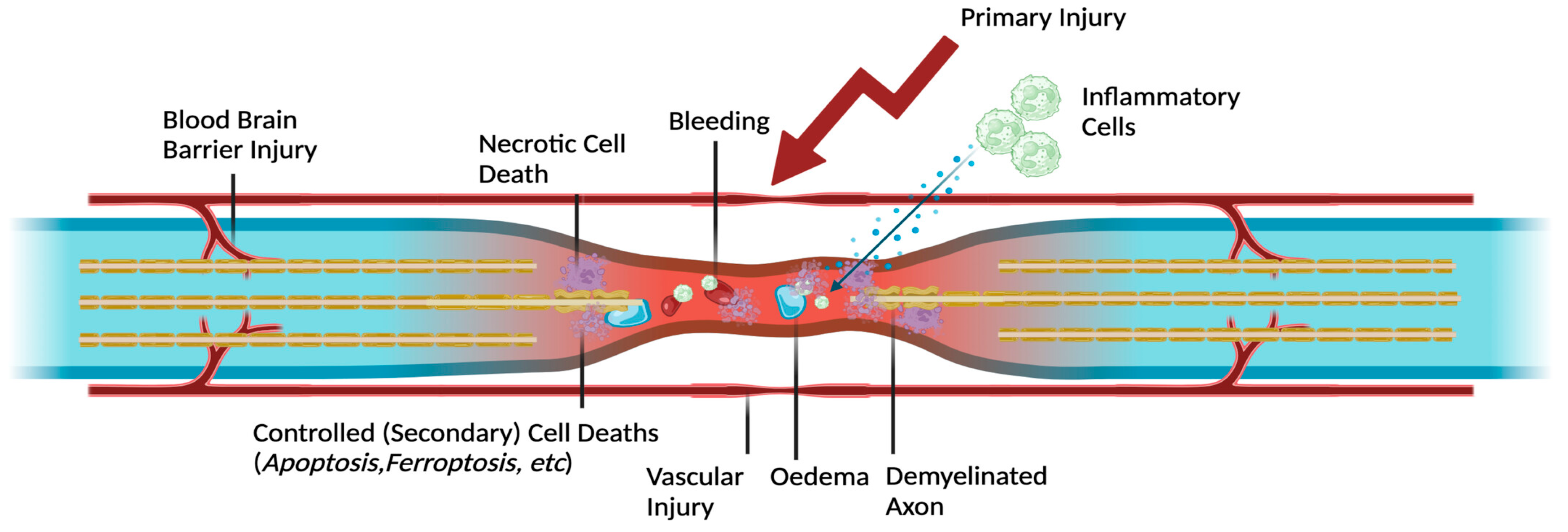
Figure 2.
Role of extracellular vesicles (EVs)-derived miRNAs in regulating ferroptosis. EVs are nanosized extracellular vesicles that mediate intercellular communication by transferring bioactive cargo, including miRNAs, mRNAs, proteins, lipids, and DNA. Following neural injury, EVs-derived miRNAs are internalized by recipient cells and regulate gene expression through binding to the 3′ untranslated region (3′UTR) of target mRNAs, resulting in translational repression or mRNA degradation. These miRNAs influence key ferroptosis-related processes, including iron uptake, lipid peroxidation, and antioxidant capacity. By modulating ferroptosis signaling pathways, EVs miRNAs contribute to the regulation of neuronal survival and may represent a promising therapeutic strategy for mitigating ferroptosis-mediated damage in spinal cord injury.
Figure 2.
Role of extracellular vesicles (EVs)-derived miRNAs in regulating ferroptosis. EVs are nanosized extracellular vesicles that mediate intercellular communication by transferring bioactive cargo, including miRNAs, mRNAs, proteins, lipids, and DNA. Following neural injury, EVs-derived miRNAs are internalized by recipient cells and regulate gene expression through binding to the 3′ untranslated region (3′UTR) of target mRNAs, resulting in translational repression or mRNA degradation. These miRNAs influence key ferroptosis-related processes, including iron uptake, lipid peroxidation, and antioxidant capacity. By modulating ferroptosis signaling pathways, EVs miRNAs contribute to the regulation of neuronal survival and may represent a promising therapeutic strategy for mitigating ferroptosis-mediated damage in spinal cord injury.
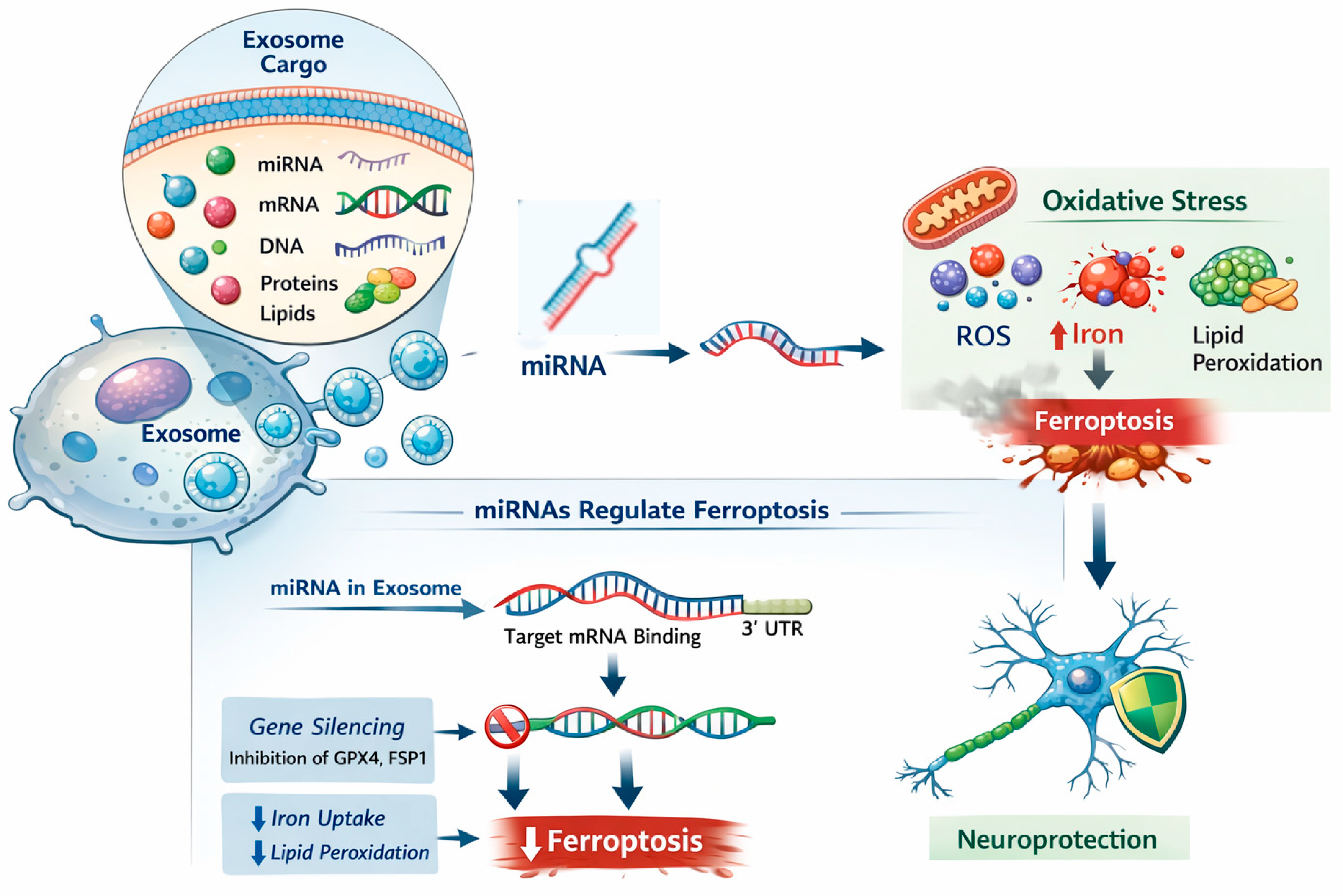
Figure 3.
The microRNA-ferroptosis axis in spinal cord injury and its therapeutic targeting. Primary spinal cord injury (SCI) triggers secondary injury cascades, including oxidative stress, inflammation, and iron dysregulation. microRNAs (miRNAs) regulate key ferroptosis-related pathways by modulating genes involved in iron metabolism, lipid peroxidation, and antioxidant defenses. Excess iron accumulation and impaired redox homeostasis promote ferroptotic cell death, contributing to neuronal loss and neurodegeneration. Targeting the miRNA-ferroptosis axis represents a promising therapeutic strategy to attenuate secondary injury. Potential approaches include miRNA mimic therapy to restore protective miRNAs, ferroptosis inhibitors to suppress iron-dependent lipid peroxidation, and miRNA-loaded exosomes for targeted delivery. These interventions collectively aim to enhance neuroprotection and promote neural regeneration after SCI.
Figure 3.
The microRNA-ferroptosis axis in spinal cord injury and its therapeutic targeting. Primary spinal cord injury (SCI) triggers secondary injury cascades, including oxidative stress, inflammation, and iron dysregulation. microRNAs (miRNAs) regulate key ferroptosis-related pathways by modulating genes involved in iron metabolism, lipid peroxidation, and antioxidant defenses. Excess iron accumulation and impaired redox homeostasis promote ferroptotic cell death, contributing to neuronal loss and neurodegeneration. Targeting the miRNA-ferroptosis axis represents a promising therapeutic strategy to attenuate secondary injury. Potential approaches include miRNA mimic therapy to restore protective miRNAs, ferroptosis inhibitors to suppress iron-dependent lipid peroxidation, and miRNA-loaded exosomes for targeted delivery. These interventions collectively aim to enhance neuroprotection and promote neural regeneration after SCI.
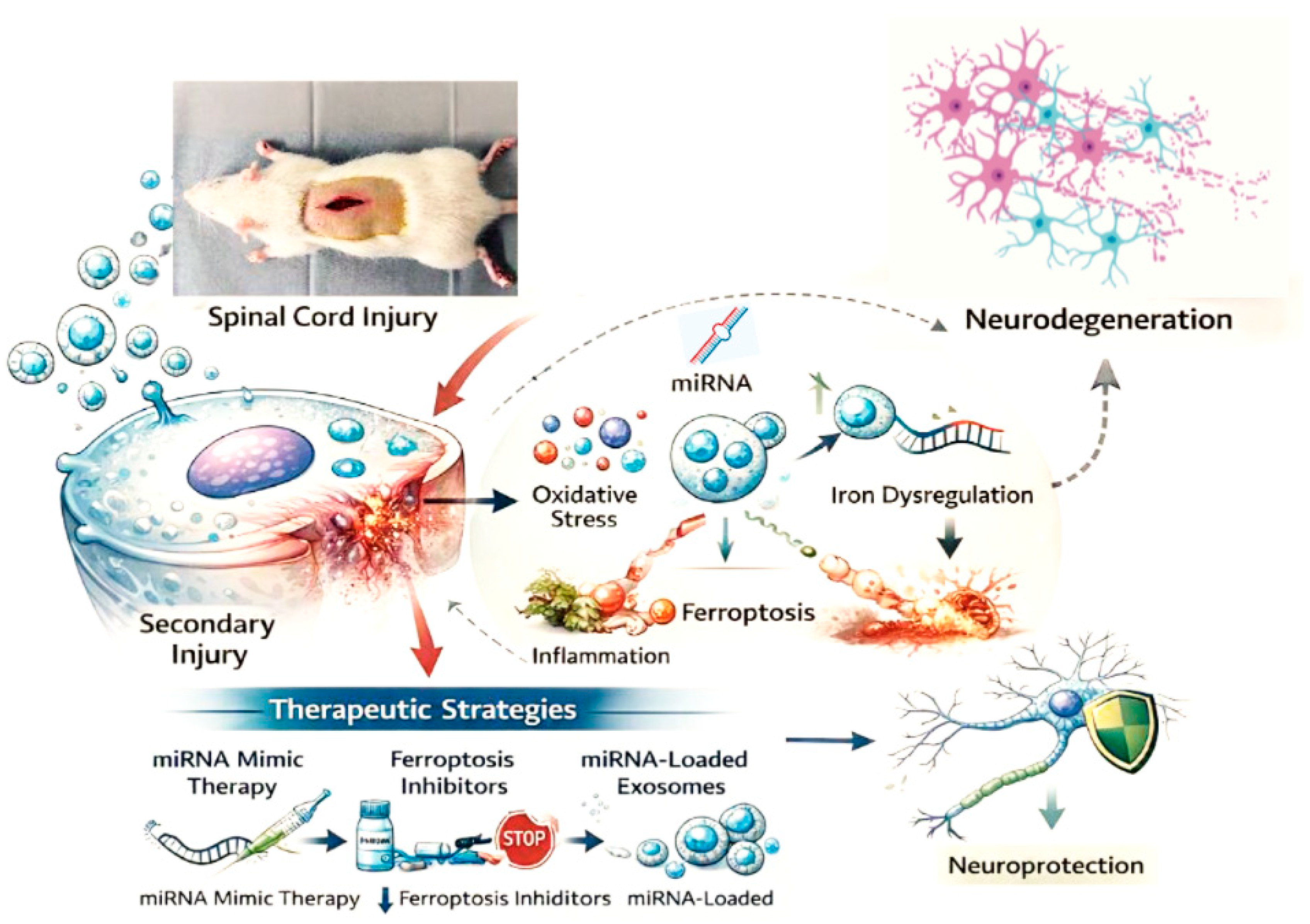
Figure 4.
Mechanistic pathways of ferritinophagy and ferroptosis regulation in spinal cord neurons after spinal cord injury (SCI). Extracellular stressors following SCI, including hemorrhage-induced red blood cell (RBC) lysis and microglial activation (via nitric oxide release and iron transfer), drive cellular iron influx. Within the neuronal cytoplasm, the nuclear receptor coactivator 4 (NCOA4) binds iron-storing ferritin, directing it to the autophagy machinery (Beclin-1/ATGs) for autophagosome encapsulation. Subsequent fusion with the lysosome leads to ferritin degradation and the release of free ferrous iron (Fe2+) into the labile iron pool (LIP). MicroRNA (miRNA) networks dynamically modulate this system by regulating iron levels and downstream pathways. Under homeostatic conditions, controlled LIP levels and robust antioxidant systems glutathione Peroxidase 4 (GPX4) and ferroptosis suppressor protein 1 (FSP1) inhibit lipid peroxidation, supporting neuroprotection and motor function recovery. Conversely, excessive or dysregulated ferritinophagy overwhelms cellular sequestration and ferroportin-mediated Fe2+ efflux. This accumulation of excess Fe2+ drives the Fenton reaction and lipid peroxidation, ultimately precipitating ferroptosis, axonal loss, and neuronal death.
Figure 4.
Mechanistic pathways of ferritinophagy and ferroptosis regulation in spinal cord neurons after spinal cord injury (SCI). Extracellular stressors following SCI, including hemorrhage-induced red blood cell (RBC) lysis and microglial activation (via nitric oxide release and iron transfer), drive cellular iron influx. Within the neuronal cytoplasm, the nuclear receptor coactivator 4 (NCOA4) binds iron-storing ferritin, directing it to the autophagy machinery (Beclin-1/ATGs) for autophagosome encapsulation. Subsequent fusion with the lysosome leads to ferritin degradation and the release of free ferrous iron (Fe2+) into the labile iron pool (LIP). MicroRNA (miRNA) networks dynamically modulate this system by regulating iron levels and downstream pathways. Under homeostatic conditions, controlled LIP levels and robust antioxidant systems glutathione Peroxidase 4 (GPX4) and ferroptosis suppressor protein 1 (FSP1) inhibit lipid peroxidation, supporting neuroprotection and motor function recovery. Conversely, excessive or dysregulated ferritinophagy overwhelms cellular sequestration and ferroportin-mediated Fe2+ efflux. This accumulation of excess Fe2+ drives the Fenton reaction and lipid peroxidation, ultimately precipitating ferroptosis, axonal loss, and neuronal death.
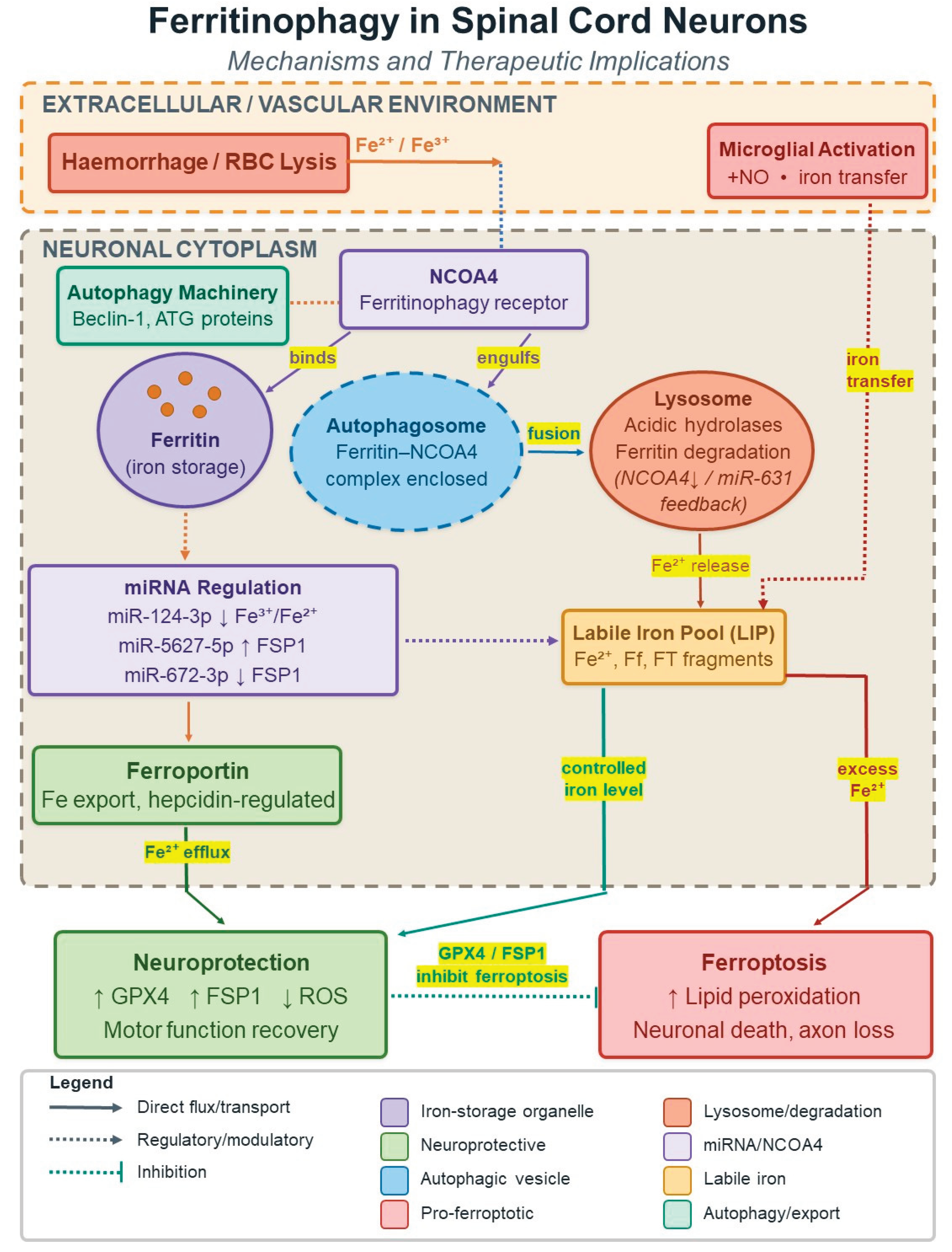

Table 1.
MicroRNAs involved in ferroptosis regulation following spinal cord injury (SCI).
| MicroRNAs | Expression change after SCI | Ferroptosis-related target(s) | Effect on ferroptosis | Functional implication in SCI | Reference |
| miR-672-3p | Upregulated | FSP1 | Promotes | Enhances neuronal ferroptosis and neurodegeneration | [37] |
| miR-6315 | Upregulated | Smo, xCT, GPX4, GSH | Promotes | Exacerbates ferroptosis-mediated neuronal injury | [38] |
| let-7b-5p | Network-associated | p53 (indirect) | Promotes | Context-dependent role in neurodegeneration and repair | [3] |
| miR-15b-5p | Network-associated | Not determined | Not determined | Not determined | [39] |
FSP1: Ferroptosis Suppressor Protein 1; Smo: Smoothened receptor; xCT: cystine/glutamate antiporter; GSH: Glutathione; GPX4: Glutathione Peroxidase 4; p53: protein 53.
Table 2.
Temporal dynamics of ferroptosis activation following spinal cord injury (SCI). The table summarizes the temporal profile of ferroptosis after SCI, including key molecular events and biomarkers observed at different post-injury stages.
Table 2.
Temporal dynamics of ferroptosis activation following spinal cord injury (SCI). The table summarizes the temporal profile of ferroptosis after SCI, including key molecular events and biomarkers observed at different post-injury stages.
| Time Post-Injury | Ferroptosis Activity | Key Features | Reference |
| 0–6 hours | Initiation | GSH depletion, early lipid ROS | [59] |
| 6–24 hours | Peak | GPX4 ↓, ACSL4 ↑, lipid peroxidation ↑ | [60] |
| 1–3 days | Sustained | Iron overload, mitochondrial damage | [60] |
| 3–7 days | Declining | Recovery of redox balance begins | [60] |
| >7 days | Minimal | Scar formation, long-term injury processes | [58] |
GSH: Glutathione; GPX4: Glutathione Peroxidase 4; ROS: Reactive oxygen species; Acyl-CoA synthetase long chain family member 4.
Table 3.
Multifunctional effects of extracellular vesicles in ferroptosis regulation and spinal cord injury repair.
Table 3.
Multifunctional effects of extracellular vesicles in ferroptosis regulation and spinal cord injury repair.
| Category | Mechanism / Function | Biological Effect in SCI | References |
| Therapeutic Advantages | Safer and more controllable than MSCs; easier handling | Improved clinical applicability | [71] |
| Neuroprotection | Delivery of protective biomolecules | Enhances neuronal survival | [72] |
| Neuro-regeneration | Promotes axonal growth and repair | Facilitates nerve tissue regeneration | [73,74] |
| Scar Reduction | Modulates fibrotic responses | Attenuates glial scar formation | [75,76] |
| Oxidative Stress Regulation | Reduces ROS and oxidative damage | Limits secondary injury | [77] |
| Angiogenesis Promotion | Enhances vascular remodeling | Improves blood supply to injury site | [77] |
| BBB Restoration | Strengthens blood–brain barrier integrity | Prevents further damage and inflammation | [78] |
| Anti-apoptotic Effects | Inhibits apoptosis pathways | Reduces neuronal cell death | [79] |
| Anti-ferroptotic Effects | Regulates iron metabolism and lipid peroxidation | Suppresses ferroptosis | [80] |
| Drug Delivery Capability | Small size, high penetration, immune evasion | Efficient therapeutic cargo delivery | [81] |
| Mechanistic Action | Suppresses pathological responses; activates regenerative signaling | Supports neural and vascular repair | [78] |
| Combination Therapy | EVs + biomaterial scaffolds | Synergistic repair and nutrient support | [79]; [82] |
Table 4.
MicroRNAs regulating ferroptosis-related mechanisms in spinal cord injury and related neural injury models. The table presents microRNAs experimentally linked to ferroptosis regulation in spinal cord injury or related neural injury models, highlighting their molecular targets, mechanisms of action, and cellular or tissue contexts.
Table 4.
MicroRNAs regulating ferroptosis-related mechanisms in spinal cord injury and related neural injury models. The table presents microRNAs experimentally linked to ferroptosis regulation in spinal cord injury or related neural injury models, highlighting their molecular targets, mechanisms of action, and cellular or tissue contexts.
| miRNAs | Injury model / tissue | Ferroptosis-related target(s) | Mechanism of action | Cellular outcome | Reference |
| miR-137 | Spinal cord neurons | SLC1A5 | Regulates glutamate metabolism and redox balance | Modulates neuronal susceptibility to ferroptosis | [89] |
| miR-214 | Neuronal cells | ATF4 | Suppresses stress-induced ferroptosis signaling | Enhances neuronal survival | [90,91] |
| miR-212-5p | CNS injury models | ACSL4 | Reduces lipid peroxidation | Attenuates ferroptotic cell death | [92] |
| miR-200c | Neuronal tissue | FSP1 | Impairs antioxidant defense | Promotes ferroptosis and neuronal damage | [93] |
| miR-27a-3p | Neural cells | SLC7A11 (xCT) | Inhibits cystine uptake and GSH synthesis | Increases ferroptosis sensitivity | [94] |
SLC1A5: Solute carrier family 1 member 5; ATF4: Activating transcription factor 4; ACSL4: Acyl-CoA synthetase long-chain family member 4; FSP1: Ferroptosis suppressor protein 1; SLC7A11: Solute carrier family 7 member 11; xCT: Cystine/glutamate antiporter.
Table 5.
Therapeutic strategies targeting the microRNA–ferroptosis axis in spinal cord injury. The table outlines experimental therapeutic approaches targeting microRNAs or ferroptosis-related pathways in spinal cord injury models, including molecular targets, delivery strategies, and reported neuroprotective outcomes in preclinical studies.
Table 5.
Therapeutic strategies targeting the microRNA–ferroptosis axis in spinal cord injury. The table outlines experimental therapeutic approaches targeting microRNAs or ferroptosis-related pathways in spinal cord injury models, including molecular targets, delivery strategies, and reported neuroprotective outcomes in preclinical studies.
|
Therapeutic strategy |
Molecular target(s) | Delivery method | Experimental model | Effect on ferroptosis | Functional outcome | Reference |
| miR-19b-3p–enriched ADSC-derived exosomes | GPX4-related pathways | Exosome-based delivery | Intracerebral hemorrhage/SCI | Inhibits | Reduces neuronal ferroptosis and tissue damage | [100] |
| Ferrostatin-1 | Lipid peroxidation | Intraperitoneal injection | SCI | Inhibits | Attenuates neuronal death and improves functional recovery | [101] |
| Liproxstatin-1 | Lipid peroxidation | Systemic administration | SCI | Inhibits | Reduces oxidative damage and neuronal loss | [102] |
| Iron chelators (e.g., deferoxamine) | Labile iron pool | Systemic administration | SCI | Inhibits | Decreases ferroptosis and secondary injury | [103] |
| miRNA modulation (mimics/inhibitors) | Ferroptosis-related genes | Viral or nanoparticle delivery | SCI | Context-dependent | Modulates neuronal survival and regeneration | [37] |
SCI: Spinal cord injury.
Disclaimer/Publisher’s Note: The statements, opinions and data contained in all publications are solely those of the individual author(s) and contributor(s) and not of MDPI and/or the editor(s). MDPI and/or the editor(s) disclaim responsibility for any injury to people or property resulting from any ideas, methods, instructions or products referred to in the content. |
© 2026 by the authors. Licensee MDPI, Basel, Switzerland. This article is an open access article distributed under the terms and conditions of the Creative Commons Attribution (CC BY) license (http://creativecommons.org/licenses/by/4.0/).
Copyright: This open access article is published under a Creative Commons CC BY 4.0 license, which permit the free download, distribution, and reuse, provided that the author and preprint are cited in any reuse.


