

Submitted:
30 June 2026
Posted:
02 July 2026
You are already at the latest version
Abstract
Levofloxacin (LVX) is a broad-spectrum antibiotic whose systemic administration can be limited by adverse effects and suboptimal drug levels at infection sites. Implantable drug-loaded meshes offer a promising strategy for localized, prolonged delivery. This study developed and validated a simple, rapid, and reliable high-performance liquid chromatography with ultraviolet detection (HPLC-UV) method for quantifying LVX in rat blood to support the preclinical evaluation of a prolonged-release LVX-loaded polypropylene mesh. The method utilized a Zorbax SB-C18 column with an isocratic mobile phase of acetonitrile:0.1% orthophosphoric acid (80:20, v/v) at a flow rate of 1.4 mL/min, with detection at 296 nm. Validation according to ICH Q2(R2) guidelines demonstrated excellent linearity (R² = 0.999) over 1–8 µg/mL, accuracy (mean recovery 99.68%), and precision (intra- and inter-day CV < 0.3%). The limits of detection and quantification were 0.3 µg/mL and 1.0 µg/mL, respectively. The validated method was successfully applied to quantify LVX in rat plasma samples collected over 10 days post-implantation, confirming its suitability for pharmacokinetic studies of implant-based antibiotic delivery systems. This method provides a practical and robust analytical tool for routine preclinical assessment of LVX release and systemic exposure from localized delivery devices.
Keywords:
validation
; levofloxacin
; blood
; high-performance liquid chromatography
1. Introduction
The creation of novel analytical approaches for detecting medicinal compounds in biological matrices represents a key area of pharmaceutical analysis. Precise measurement of active pharmaceutical ingredients depends on the establishment and validation of robust, dependable methods. Such methodologies are essential to guarantee the quality, safety, and therapeutic effectiveness of pharmaceutical products [1,2]. Levofloxacin (LVX) is a third-generation fluoroquinolone antibiotic with broad-spectrum activity against Gram-positive and Gram-negative bacteria. It is widely used in the treatment of respiratory, urinary tract, skin, and soft tissue infections due to its favorable pharmacokinetic properties, high bioavailability, and strong tissue penetration. Despite these advantages, systemic administration of LVX can be associated with adverse effects and suboptimal drug concentrations at the site of infection, particularly in chronic or localized infections that require sustained antimicrobial exposure [3,4].
Localized drug delivery systems have emerged as an effective strategy to overcome the limitations of conventional systemic therapy. Among these systems, implantable drug-loaded meshes have attracted increasing attention because they allow prolonged and controlled drug release directly at the target site, thereby reducing systemic exposure and improving therapeutic efficacy. Prolonged-release antibiotic implants are especially valuable in preventing and treating post-surgical infections and in managing chronic infections where long-term antimicrobial presence is required [5,6]. However, accurate evaluation of drug release behavior and systemic absorption from such implants necessitates sensitive and reliable analytical methods for drug quantification in biological matrices.
Several analytical methods have been reported for the determination of LVX in human plasma or serum using high-performance liquid chromatography (HPLC) with ultraviolet (UV) [7,8,9,10,11,12,13,14] or fluorescence detection (FLD) [15,16,17,18], as well as LC–MS/MS [19,20,21,22,23] techniques. HPLC coupled with UV detection remains one of the most widely used techniques for the quantitative determination of fluoroquinolone antibiotics in biological samples. HPLC-UV offers advantages such as robustness, cost-effectiveness, ease of operation, and adequate sensitivity for routine pharmacokinetic and bioavailability studies [24,25]. While LC–MS/MS methods provide high sensitivity, their high operational costs and limited accessibility restrict their routine use in many laboratories. Moreover, all of the reported HPLC-UV methods are optimized for conventional dosage forms or systemic administration and may not be directly applicable to studies involving prolonged-release implantable systems.
To date, limited attention has been given to the development and validation of analytical methods specifically tailored for the determination of LVX released from implantable mesh systems in small laboratory animals [26,27]. Rat models are commonly employed in preclinical studies to evaluate the pharmacokinetics, biocompatibility, and release characteristics of implantable drug delivery systems. Therefore, a validated, simple, and reliable HPLC-UV method for the quantification of LVX in rat blood is essential to support such investigations and ensure accurate interpretation of pharmacokinetic data.
The present study aimed to develop and validate a rapid, accurate, and precise HPLC-UV method for the determination of LVX in rat blood following administration of a prolonged-release LVX mesh implant. The method was validated in accordance with International Council for Harmonisation (ICH) Q2 (R2) guidelines, assessing parameters such as linearity, accuracy, precision, and robustness. The proposed method is intended to provide a practical analytical tool for routine analysis and preclinical evaluation of implant-based LVX delivery systems.
2. Materials and Methods
2.1. Chemicals and Reagents
LVX reference standard was obtained from KRKA (Slovenia; batch No. UG1456). Acetonitrile and methanol of gradient grade suitable for HPLC were purchased from Merck (Germany; batch Nos. I1167530 135 and I1154907 124, respectively). Orthophosphoric acid (85%, HPLC gradient grade) was also supplied by Merck (Germany; batch No. K52625773 029). Polypropylene hernia mesh implants loaded with LVX were used for in vivo experiments. A standard stock solution of LVX was prepared by accurately weighing 10 mg of LVX reference standard and transferring it into a 100 mL volumetric flask, 30 mL of methanol was added and shaken, then the volume of the solution was made up to the mark with solvent. Quality control (QC) solutions were then prepared by diluting the stock solution with mobile phase to the desired concentrations. The gelatin used in the research was obtained from Sigma-Aldrich) Germany), chitosan from Merck) Germany), citric acid from Sigma-Aldrich) Germany) and glycerin from Sigma-Aldrich) Germany). Ethyl alcohol meeting the requirements of the pharmacopoeia and having analytical purity was also used in the experimental studies. All reagents and materials used in the research process had high-quality indicators and were considered suitable for analytical purposes.
2.2. Animal Experiments and İmplantation Procedure
A prospective laboratory trial has been conducted in our research. The applicability of the proposed method was tested on rat plasma samples by using a HPLC method. The macroscopic structure of the implant utilized in this study, as well as the critical steps of the surgical implantation procedure in the rat model, are illustrated in Figure 1. All stages of the research were considered and approved by the Ethical Committee Board of Azerbaijan Medical University (Protocol No. 28; 05.07.2023) as a research conducted in the Helsinki Declaration principles. The UNIFLEX surgical endoprosthetic mesh is a non-absorbable implant manufactured from biocompatible polypropylene and exhibits high mechanical strength. Its mesh-like, porous structure promotes effective integration with surrounding tissues by facilitating fibroblast migration and collagen deposition, thereby enabling reliable repair of tissue defects. The UF-2010 model is produced in a standard white color and features blue orientation marking lines that assist in accurate intraoperative placement (Figure 1a). The implant is sterilized using ethylene oxide and is intended for single use only.
To 10 g of gelatin, 100 mL of water at a temperature of 90–95 °C is added, and the mixture is stirred until complete dissolution is achieved. The resulting hot solution is then filtered through a cotton filter. After cooling the solution to 40–45 °C, 50 mL of 95% ethyl alcohol is added. As a result, a cloudy, milky suspension forms as a precipitate. The mixture is thoroughly stirred, the container is covered with cellophane, and the solution is left at room temperature for 12 hours.
The formed precipitate is separated from the liquid phase by filtration through a paper filter. To the obtained precipitate, 95% ethyl alcohol (approximately 50–75 mL) is added until complete precipitation of gelatin from the solution occurs. The container with the mixture is then placed in a refrigerator for 12–24 hours. After this period, the liquid phase is decanted, and the precipitate is dissolved in 50 mL of water by heating at 90–95 °C. The resulting hot solution is evaporated on a water bath until a thickened dry residue is obtained. The residue is dried in a thermostat at 40–45 °C, yielding approximately 5.25 g of dry gelatin.
To 5 g of purified gelatin, 20 mL of purified water is added, and complete dissolution is carried out in a water bath with heating. The solution is then cooled to 40–45 °C, after which 0.05–0.1 g of citric acid is added. Subsequently, 2 g of chitosan is introduced into the solution and stirred until complete dissolution is achieved. Thereafter, the following components are added sequentially: 0.5 g of LVX, 10 mL of glycerin, and 5 mL of propolis tincture. The mixture is thoroughly stirred until a homogeneous mass is obtained. Purified water is then added to adjust the final volume of the solution to 50 mL.
This volume is intended for application onto five mesh prostheses measuring 20 × 10 cm; thus, 10 mL of the prepared solution is used for each mesh to form a film. The mesh endoprosthesis is placed on a glass surface, and the prepared solution, preheated to 40–45 °C, is evenly applied in a thin layer using a spatula. The coated endoprosthesis is then placed in a thermostat and dried at 35–40 °C for approximately 12 hours. Upon solidification, an elastic film with a smooth surface and prolonged antibacterial activity is formed on the mesh endoprosthesis. Following the coating process, the sterile implant was surgically placed onto the abdominal wall defect in the rat model (Figure 1b) and secured in the final anatomical position (Figure 1c).
Prior to use, the endoprosthesis is packaged and sterilized with ethylene oxide. The components of the claimed composition were selected experimentally to achieve a positive effect corresponding to the stated objective. Since LVX is the principal therapeutic component of the proposed formulation, all specific implementation examples were carried out using a base amount of the active substance, namely 0.5 g of LVX. The quantitative composition limits enabling the achievement of the stated objective were determined.
2.3. Biological Samples
Blood samples were collected from experimental rats following surgical implantation of a polypropylene mesh loaded with LVX. To evaluate the long-term release profile of LVX from the implanted mesh, blood samples were obtained on days 1, 3, 5, 9, and 14 after implantation. All samples were handled and stored under appropriate conditions until analysis. Rat blood samples used for analysis were provided by the laboratory of the Teaching Surgery Clinic of Azerbaijan Medical University. For preparation of the biological samples, 2.0 mL of whole blood was transferred into a centrifuge tube and centrifuged at 4000 rpm for 5 minutes at 4 °C. Following centrifugation, 1.0 mL of the separated plasma was collected and diluted to 2.0 mL with methanol for protein precipitation. The mixture was vortex-mixed and centrifuged again under the same conditions. Subsequently, 1.0 mL of the clear supernatant was transferred to a clean tube, diluted to 2.0 mL with the mobile phase, thoroughly mixed, and filtered through a 0.45 μm PTFE (polytetrafluoroethylene) membrane filter prior to HPLC analysis.
2.4. Chromatographic Conditions
Chromatographic analysis was performed using an Agilent 1260 HPLC (Agilent Technologies, USA) equipped with a UV detector. Data acquisition and peak integration were performed by OpenLab CDS software (version: 2.5.0.842). Separation was achieved on a Zorbax SB-C18 column (4.6 × 250 mm, 5 μm particle size). The mobile phase consisted of acetonitrile and 0.1% (v/v) orthophosphoric acid in water in an 80:20 ratio and was delivered at a flow rate of 1.4 mL/min. The mobile phase was prepared by mixing 800 mL of acetonitrile with 200 mL of deionized water adjusted to pH 3.5 in a 1000 mL volumetric flask. The mixture was degassed using an ultrasonic bath prior to use. The column temperature was maintained at 25 °C. The injection volume was 20 μL, and detection was carried out at a wavelength of 296 nm. The total chromatographic run time was 6 minutes. All solvents used were of HPLC-grade purity. Statistical evaluation of the analytical results was performed using Student’s t-test.
3. Results and Discussion
In accordance with the study objective, a reliable HPLC–UV method was developed and optimized for the determination of LVX in rat blood samples following implantation of a prolonged-release mesh formulation. Method development focused on achieving adequate chromatographic resolution, sensitivity, and reproducibility while maintaining a simple and rapid analytical procedure suitable for biological samples.
3.1. Optimization of Chromatographic Conditions
Key chromatographic parameters, including stationary phase, mobile phase composition, flow rate, injection volume, column temperature, and detection wavelength, were systematically investigated. Optimal separation and peak symmetry for LVX were achieved using a Zorbax SB-C18 column (4.6 × 250 mm), with a mobile phase consisting of acetonitrile and water (80:20, v/v) adjusted to pH 3.5 with orthophosphoric acid. The optimized conditions included a flow rate of 1.4 mL/min, column temperature of 25 °C, injection volume of 20 μL, and UV detection at 296 nm. Under these conditions, LVX was well resolved from endogenous matrix components with a total run time of 6 minutes. Figure 2 shows chromatograms of drug-free plasma of a rat (a), the LVX standard solution (b), and biological samples obtained after implantation of the prolonged-release formulation (c). No significant interfering peaks were observed at the retention time of LVX, confirming the selectivity of the method.
3.2. Method Validation
Validation of the developed HPLC–UV method was carried out in accordance with applicable analytical regulatory guidelines, especially the ICH Q2(R2) guidelines, including assessment of accuracy, linearity, precision, sensitivity, selectivity and stability.
Selectivity: Six blank plasma samples were analyzed to evaluate potential interferences by comparing the retention time values with those obtained from plasma samples spiked with LVX. As shown in Figure 2a, there is no interfering peak from the endogenous compounds.
Stability: The stability of LVX was investigated throughout different storage conditions. The stability results of LVX are summarized in Table 1. As shown, after 30 days of storage at room temperature (25 °C), the assay value of LVX decreased from 100.0% to 98.9%, corresponding to a 1.1% change from day 0. These minor variations are considered within the acceptable range for pharmaceutical stability.
Accuracy
Method accuracy was evaluated using QC samples prepared by spiking known amounts of LVX into the mobile phase. Working solutions at concentrations of 2, 4, and 8 μg/mL were analyzed in triplicate. As summarized in Table 2, recoveries ranged from 99.24% to 99.97%, with a mean recovery of 99.68%. The low standard deviation (0.26%) and narrow confidence interval (99.48–99.88%, p = 95%) demonstrate the high accuracy of the method across the studied concentration range. These results meet the acceptance criteria specified in the ICH Q2(R2) guidelines.
Linearity
Linearity was assessed by analyzing standard LVX solutions over the concentration range of 1–8 μg/mL. A calibration curve was constructed by plotting peak area versus concentration. As shown in Table 3 and Figure 3, the method exhibited excellent linearity with a correlation coefficient (R2) of 0.999. The regression equation was y = 72409.63x + 10.474, indicating a strong proportional relationship between detector response and analyte concentration within the tested range.
Precision
Method precision was evaluated in terms of both intra-day and inter-day repeatability using six replicate analyses of samples containing 2, 4 and 8 μg/mL of LVX. The results are presented in Table 4 and Table 5. Inter-day precision showed a mean recovery of 99.85% with a mean coefficient of variation (CV) of 0.24%, while intra-day precision yielded a mean recovery of 99.84% with a mean CV of 0.21%. These low variability values indicate excellent repeatability and robustness of the analytical method for routine application.
Sensitivity
The limits of detection (LOD) and quantification (LOQ) were calculated based on the standard deviation of the intercept and the slope of the calibration curve, in accordance with ICH recommendations. The LOD and LOQ were determined to be 0.3 μg/mL and 1.0 μg/mL, respectively, demonstrating adequate sensitivity for monitoring LVX concentrations in biological samples following prolonged release.
3.3. Application to Biological Samples
The validated method was successfully applied to the analysis of blood samples collected from six rats implanted with LVX-loaded mesh devices. The method enabled reliable quantification of LVX over the studied time points (Table 6), supporting its suitability for evaluating long-term drug release and systemic exposure from implantable formulations. The chromatograms obtained from the analysis of rat samples are shown in Figure 4.
Comparison of the Developed HPLC-UV Method with Previously Reported Methods for LVX Quantification
The HPLC-UV method developed in the present study demonstrates analytical performance characteristics well suited to its intended preclinical application, occupying a distinct niche within the extensive literature on levofloxacin quantification. As summarized in Table 7, while fluorescence and mass spectrometric methods achieve superior sensitivity with LOQs as low as 2.11 ng/mL in human milk and 100 ng/mL in serum, our method provides adequate sensitivity (LOQ 1.0 μg/mL) for monitoring therapeutic concentrations following prolonged-release mesh implantation in rats, where sustained drug levels above the MIC are anticipated. The linear range of 1–8 μg/mL is comparable to established HPLC-UV methods for human plasma (0.25–15 μg/mL), and the validation parameters, including mean recovery of 99.68% and intra-/inter-day CV <0.3%, compare favorably with published assays reporting recoveries of 87–99% and acceptable CV <5–15%. Unlike the majority of reported methods optimized for human plasma [28,29,30,31,32,33,34,35], serum, specialized clinical matrices [36], pharmaceutical formulations [37,38,39,40], or environmental samples [41], our method is specifically validated in rat blood, the most relevant preclinical model for evaluating implantable drug delivery systems. Furthermore, while Jiang et al. [42] employed HPLC for in vitro release testing from composite scaffolds, our study extends application to in vivo quantification over 14 days post-implantation, addressing a recognized methodological gap. The simple protein precipitation sample preparation and 6-minute run time confer practical advantages for routine preclinical analyses compared to more complex solid phase extraction, liquid-liquid extraction techniques. Thus, the developed method provides a fit-for-purpose, cost-effective, and accessible analytical tool that complements existing high-sensitivity clinical therapeutic drug monitoring platforms and pharmaceutical quality control assays by specifically addressing the requirements of preclinical implantable device research.
4. Conclusion
A selective, accurate, and precise HPLC-UV method has been successfully developed and validated for the determination of levofloxacin in rat blood. The method fulfills all critical validation parameters, including linearity, accuracy, precision, and sensitivity as per ICH guidelines, making it suitable for routine bioanalysis. Its successful application to plasma samples from rats implanted with a LVX-loaded polypropylene mesh demonstrates its practical utility in preclinical pharmacokinetic studies. This method effectively quantifies the sustained systemic exposure of LVX from a localized delivery system, addressing a gap in analytical approaches tailored for implantable drug delivery evaluations. The proposed HPLC-UV assay provides a cost-effective, accessible, and reliable analytical solution to support the development and optimization of prolonged-release antibiotic implants, facilitating accurate assessment of their in vivo release profiles and therapeutic potential.
References
- Suleymanov, T.A.; Balayeva, E.Z.; Ahmedov, E.Y. Development and determination of validation parameters for the HPLC method of thymol quantification in “Kalinol plus” syrup. News Pharm. Ukr. 2016, vol. 3(87), 22–27. [Google Scholar] [CrossRef]
- Suleymanov, Tahir; Aliyeva, Kubra; Balayeva, Emilya. Validation of the method for quantifying naringin in grapefruit (citrus paradisi) extract using high-performance liquid chromatography. Chem. Chem. Technol. 2024, vol. 18(1), 16–22. [Google Scholar] [CrossRef]
- Peng, Ke; Li, Mingshan; Himawan, Achmad; Domínguez-Robles, Juan. Amphotericin B- and Levofloxacin-Loaded Chitosan Films for Potential Use in Antimicrobial Wound Dressings: Analytical Method Development and Its Application. Pharmaceutics 2022, vol. 14, 1–16. [Google Scholar] [CrossRef] [PubMed]
- Notario, D.; Martono, S.; Ikawati, Z.; Rahman, A.H. / A Rapid and Simple High-Performance Liquid Chromatographic Method for Determination of Levofloxacin in Human Plasma. Indones. J. Chem. 2017, vol. 17(1), 54–62. [Google Scholar] [CrossRef]
- Taghiev, S.; Gasymov, E.; Dzhafarov, Ch. Method of manufacturing a mesh prosthesis for hernioplasty and having prolonged antibacterial properties. Eurasian patent N: 039154, 2021. [Google Scholar]
- Zahoor, Sana; Riaz, Tabinda; Shafiq, Muhammad; Gull, Iram; Abbas, Zaigham; Abdul Mannan, Hafiz; Ullah, Sami; Ali Assiri, Mohammed. An innovative ciprofloxacin-infused silk hernia mesh for abdominal wall repair and infection control. [CrossRef]
- Ebers, Andrew; Stroup, Suzanne; Mpagama, Stellah; Kisonga, Riziki. Determination of plasma concentrations of levofloxacin by high performance liquid chromatography for use at a multidrug-resistant tuberculosis hospital in Tanzania. Scott. HeysellPublished 2017, vol 12(1), 1–12. [Google Scholar] [CrossRef] [PubMed]
- Locatelli, Marcello; Ciavarella, Maria Teresa; Paolino, Donatella; Celia, Christian. Determination of ciprofloxacin and levofloxacin in human sputum collected from cystic fibrosis patients using microextraction by packed sorbent-high performance liquid chromatography photodiode array detector. J. Chromatogr. A 2015, vol. 1419, 58–66. [Google Scholar] [CrossRef] [PubMed]
- Sopyan; Dwiputri, D.; Muchtaridi, Muchtaridi. A novel validation of the levofloxacin determination method in urine (in vitro) using high-performance liquid chromatography uv/vis detector /. J. Chem. 2020, vol. 13(4), 2207–2214. [Google Scholar] [CrossRef]
- Gulfen, Mustafa; Janbaz, Yazgi; Özdemir, Abdil. Simultaneous Determination of Amoxicillin, Lansoprazole, and Levofloxacin in Pharmaceuticals by HPLC with UV–Vis Detector. J. Anal. Test. 2020, vol. 4, 45–53. [Google Scholar] [CrossRef]
- Dinchel, Aysun; Damla Gok Topak, E.; Onur, Feyyaz. THE NEW METHOD DEVELOPMENT FOR THE DETERMINATION OF FLUOROQUINOLONES BY HPLC-DAD /. J. Fac. Pharm. Ank. 2023, vol. 47(3), 987–993. [Google Scholar] [CrossRef]
- Yıldırım, Sercan; Nur Karakoç, Hanife; Yaşar, Ahmet; Köksal, Iftihar. Determination of levofloxacin, ciprofloxacin, moxifloxacin and gemifloxacin in urine and plasma by HPLC–FLD–DAD using pentafluorophenyl core–shell column: Application to drug monitoring. BioMed Chromatogr. 2020, vol. 34(10). [Google Scholar] [CrossRef] [PubMed]
- Zhou, Zhi-Ling; Yang, Min; Yu, Xi-Yong; Peng, Huai-Yan. A rapid and simple high-performance liquid chromatography method for the determination of human plasma levofloxacin concentration and its application to bioequivalence studies. BioMed Chromatog 2007, vol. 21(10), 1045–51. [Google Scholar] [CrossRef] [PubMed]
- Czyrski, A.; Szałek, E. An HPLC method for levofloxacin determination and its application in biomedical analysis. J. Anal. 2016, vol. 71(8), 840–843. [Google Scholar] [CrossRef]
- Toker, Sıdıka Ertürk; Kızılchay, Gamze Ergin; Sagirli, Olcay. Determination of levofloxacin by HPLC with fluorescence detection in human breast milk. Bioanalysis 2021, vol. 13(13), 1063–1070. [Google Scholar] [CrossRef] [PubMed]
- Kumar, H.; Sudha, V.; Ramachandran, G. A simple and rapid liquid chromatography method for determination of levofloxacin in plasma. SAARC J. Tuberc. Lung Dis. HIV/AIDS 2017, 14(1), 27–32. [Google Scholar] [CrossRef]
- Ebrahim, H.; Sonbol, H.; Malak, M.; Ali, A. / Green Automated Solid Phase Extraction to Measure Levofloxacin in Human Serum via Liquid Chromatography with Fluorescence Detection for Pharmacokinetic Study. Separations 2023, vol. 10(2), 136–149. [Google Scholar] [CrossRef]
- Schulte, S.; Ackermann, T.; Bertram, N.; Sauerbruch, T. Determination of the newer quinolones levofloxacin and moxifloxacin in plasma by high-performance liquid chromatography with fluorescence detection. J. Chromatogr. Sci. 2006, vol. 44(4), 205–8. [Google Scholar] [CrossRef] [PubMed]
- Zheng, Xubin; Jongedijk, Erwin M; Hu, Yi; Kuhlin, Johanna. Development and validation of a simple LC-MS/MS method for simultaneous determination of moxifloxacin, levofloxacin, prothionamide, pyrazinamide and ethambutol in human plasma. J. Chromatogr. B Anal. Technol. BioMed Life Sci. 2020, vol. 1158, 122397–122407. [Google Scholar] [CrossRef] [PubMed]
- Cairoli, Sara; Simeoli, Raffaele; Tarchi, Marco; Dionisi, Marco. A new HPLC-DAD method for contemporary quantification of 10 antibiotics for therapeutic drug monitoring of critically ill pediatric patients. BioMed Chromatogr. 2020, 34(10), 4880. [Google Scholar] [CrossRef]
- Fan, Xudong; Guo, Suhang; Zhang, Ruoying; Cai, Qingshan. Development, Validation, and Clinical Application of an Ultra High Performance Liquid Chromatography Coupled with Tandem Mass Spectrometry Method for the Determination of 10 Antituberculosis Drugs in Human Serum // Ther Drug Monit. 2024, vol. 46(4), 477–484. [Google Scholar] [CrossRef] [PubMed]
- Ghimire, Samiksha; Jongedijk, Erwin M.; van den Elsen, Simone H.J. Cross-validation of Liquid Chromatography-Tandem Mass Spectrometry Method for Quantification of Levofloxacin in Saliva. J. Appl. Bioanal. 2020, vol. 6(2), 56–58. [Google Scholar] [CrossRef]
- Dolaksız, Yasemin Ekin; Kaynak, Mustafa Sinan; Kabir, Abuzar; Furton, Kenneth G. Adapting Fabric Phase Sorptive Extraction as an Innovative Multitool for Sample Transfer and Extraction in Pharmacokinetic Analysis Followed by LC-MS Determination of Levofloxacin in Plasma Samples // ACS Omega. 2024, vol. 9(17), 18995–19002. [Google Scholar]
- Czyrski, Andrzej. Analytical Methods for Determining Third and Fourth Generation Fluoroquinolones: A Review. Chromatographia 2017, vol 80, 181–200. [Google Scholar] [CrossRef] [PubMed]
- LLopis, Benoit; Funck-Brentano, Christian; Tissot, Nadine; Bleibtreu, Alexandre. Development and validation of a UPLC-MS/MS method for simultaneous quantification of levofloxacin, ciprofloxacin, moxifloxacin and rifampicin in human plasma: Application to the therapeutic drug monitoring in osteoarticular infections. J. Pharm. BioMed Anal. 2020, vol.183, 1–27. [Google Scholar] [CrossRef] [PubMed]
- Jiang, Z.; Wang, T.; Pan, B.; Xie, Z. / Evaluation of the levofloxacin release characters from a rabbit foldable capsular vitreous body. Int. J. Nanomed. 2012, vol. 7, 1–10. [Google Scholar] [CrossRef] [PubMed]
- Wang, T.; Huang, X.; Gao, Q.; Feng, L. / A preliminary study to treat severe endophthalmitis via a foldable capsular vitreous body with sustained levofloxacin release in rabbits. Invest Ophthalmol. Vis. Sci. 2013, vol. 54(1), 804–12. [Google Scholar] [CrossRef]
- Siewert, S. Validation of a levofloxacin HPLC assay in plasma and dialysate for pharmacokinetic studies. J. Pharm. BioMed Anal. 2006, vol. 41(4), 1360–1362. [Google Scholar]
- Srinivas, Nimmagadda; Narasu, Lakshmi; Prabha Shankar, B.; Mullangi, Ramesh. Development and validation of a HPLC method for simultaneous quantitation of gatifloxacin, sparfloxacin and moxifloxacin using levofloxacin as internal standard in human plasma: application to a clinical pharmacokinetic study. BioMed Chromatogr. 2008, vol. 22(11), 1288–95. [Google Scholar] [CrossRef] [PubMed]
- Aragon-Martinez, Othoniel Hugo; Isiordia-Espinoza, Mario Alberto; Galicia, Othir; Romo, Saray Aranda. Measurement of levofloxacin in human plasma samples for a reliable and accessible drug monitoring. Clin. Biochem. 2017, vol. 50(1-2), 73–79. [Google Scholar] [CrossRef] [PubMed]
- Zheng, Yi; Wang, Ziqing; Lui, Gabrielle; Hirt, Déborah. Simultaneous quantification of levofloxacin, pefloxacin, ciprofloxacin and moxifloxacin in microvolumes of human plasma using high-performance liquid chromatography with ultraviolet detection. BioMed Chromatogr. 2019, vol. 33(5), 1–26. [Google Scholar] [CrossRef] [PubMed]
- Polachek, Hana; Holzberg, Gershon; Sapir, Gadi; Tsadkin -Tamir, Marina. Transfer of ciprofloxacin, ofloxacin and levofloxacin across the perfused human placenta in vitro. Eur. J. Obstet. Gynecol. Reprod. Biol. 2005, vol. 122(1), 61–65. [Google Scholar] [CrossRef] [PubMed]
- Ferrone, Vincenzo; Cotellese, Roberto; Di Marco, Lorenzo; Bacchi, Simona. Meropenem, levofloxacin and linezolid in human plasma of critical care patients: A fast semi-automated micro-extraction by packed sorbent UHPLC-PDA method for their simultaneous determination. J. Pharm. BioMed Anal. 2017, vol. 5(140), 266–273. [Google Scholar] [CrossRef] [PubMed]
- Van Toi, Pham; Pouplin, Thomas; Duc Khanh Tho, Nguyen; Nguyen Phuong, Pham. High-performance liquid chromatography with time-programmed fluorescence detection for the quantification of Levofloxacin in human plasma and cerebrospinal fluid in adults with tuberculous meningitis. J. Chromatogr. B Anal. Technol. BioMed Life Sci. 2017, vol. 1(1061-1062), 256–262. [Google Scholar] [CrossRef] [PubMed]
- Ghimire, Samiksha; van Hateren, Kai; Vrubleuskaya, Natasha; Koster, Remco. Determination of levofloxacin in human serum using liquid chromatography tandem mass spectrometry. J. Appl. Bioanal. 2018, vol. 4(1), 16–25. [Google Scholar] [CrossRef]
- Szerkus; Jacyna, J.; Wiczling, P.; Gibas, A. Ultra-high performance liquid chromatographic determination of levofloxacin in human plasma and prostate tissue with use of experimental design optimization procedures. J. Chromatogr. B 2016, vol. 1029–1030, 48–59. [Google Scholar] [CrossRef] [PubMed]
- Maharini, Indri; Martien, Ronny; Kharis Nugroho, Akhmad; Adhyatmika, Supanji. RP-HPLC-UV validation method for levofloxacin hemihydrate estimation in the nano polymeric ocular preparation. Arab. J. Chem. 2022, vol. 15(2), 1–7. [Google Scholar] [CrossRef]
- Indri, Maharini; Ronny, Martien; Akhmad, Nugroho. Validation UV Spectrophotometric Method to Determine Entrapment Efficiency of Ocular Polymeric Nanoparticle Levofloxacin Hemihydrate. Res. J. Pharm. Technol. 2021, vol. 14(10), 5479–5482. [Google Scholar] [CrossRef]
- Gholse, Yogesh N.; Chaple, Dinesh R.; Rahul, H. Kasliwal Development and Validation of Novel Analytical Simultaneous Estimation Based UV Spectrophotometric Method for Doxycycline and Levofloxacin Determination / Biointerface Research in Applied Chemistry. 2022, vol. 12(4), 5458–5478. [Google Scholar] [CrossRef]
- Babu, N. Prasad; Ramachandran, D.; Bhavani, K.Geetha. Development and validation of stability indicating RP-HPLC method for quantitative estimation of levofloxacin injection 5mg/ml dosage form. Curr. Trends Biotechnol. Pharm. 2021, vol. 15(1), 51–61. [Google Scholar] [CrossRef]
- Liu, Ziwei; Ni, Lanxiu; Hou, Zuojun; Wang, Yu; Shi, Zhenjia. The quantitative detection of levofloxacin in treated medical wastewater based on solid-phase extraction assisted dual wavelength absorption. J. Water Process Eng. 2025, vol.73, 1–13. [Google Scholar] [CrossRef]
- Wang, Q.; Wang, G.; Xie, S.; Zhao, X. Comparison of high-performance liquid chromatography and ultraviolet-visible spectrophotometry to determine the best method to assess Levofloxacin released from mesoporous silica microspheres/nano-hydroxyapatite composite scaffolds. Exp. Ther. Med. 2019, vol. 17(4), 2694–2702. [Google Scholar] [CrossRef] [PubMed]
- Mula, Jacopo; Chiara, Francesco; Manca, Alessandra; Palermiti, Alice. Analytical validation of a novel UHPLC-MS/MS method for 19 antibiotics quantification in plasma: Implementation in a LC-MS/MS Kit // Biomed Pharmacother. 2023, vol.163, 1–14. [Google Scholar] [CrossRef] [PubMed]
- Sartini, I.; Łebkowska-Wieruszewska, B.; Sitovs, A.; Lisowski, A. Levofloxacin pharmacokinetics and tissue residue concentrations after oral administration in Bilgorajska geese. Br. Poult. Sci. 2021, vol. 62(2), 193–198. [Google Scholar] [CrossRef] [PubMed]
Figure 1.
Surgical mesh implant and in vivo rat model implantation procedure. (a) Macroscopic appearance of the UNIFLEX UF-2010 polypropylene mesh, showing the standard white structure and blue orientation marking lines. (b) Intraoperative photograph of the mesh placement on the rat abdominal wall defect. (c) Final position of the coated mesh implant following fixation.
Figure 1.
Surgical mesh implant and in vivo rat model implantation procedure. (a) Macroscopic appearance of the UNIFLEX UF-2010 polypropylene mesh, showing the standard white structure and blue orientation marking lines. (b) Intraoperative photograph of the mesh placement on the rat abdominal wall defect. (c) Final position of the coated mesh implant following fixation.
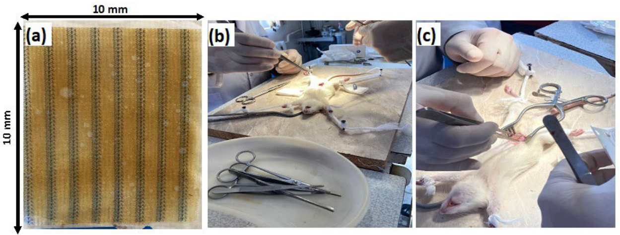
Figure 2.
Chromatograms of drug-free plasma of a rat (a), the LVX standard solution (b), and biological samples obtained after implantation of the prolonged-release formulation (c) obtained by the developed HPLC-UV method under optimal condition.
Figure 2.
Chromatograms of drug-free plasma of a rat (a), the LVX standard solution (b), and biological samples obtained after implantation of the prolonged-release formulation (c) obtained by the developed HPLC-UV method under optimal condition.
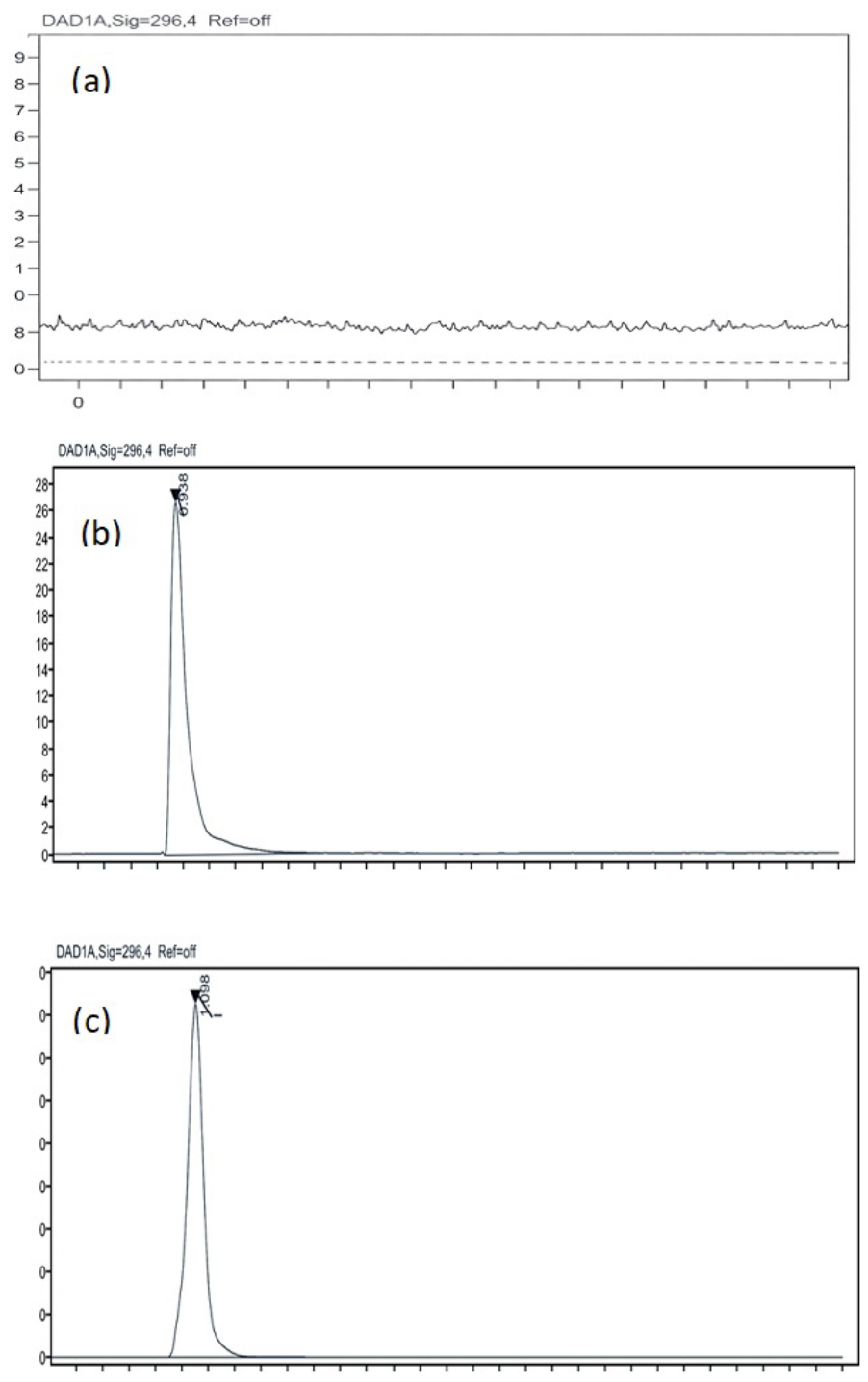
Figure 3.
Calibration curve of LVX showing linear detector response over the concentration range of 1–8 μg/mL.
Figure 3.
Calibration curve of LVX showing linear detector response over the concentration range of 1–8 μg/mL.
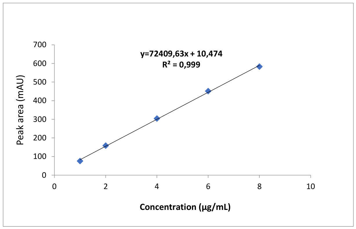
Figure 4.
The chromatograms of LVX in six plasma samples from different rats (a-f) obtained by the developed HPLC-UV method.
Figure 4.
The chromatograms of LVX in six plasma samples from different rats (a-f) obtained by the developed HPLC-UV method.
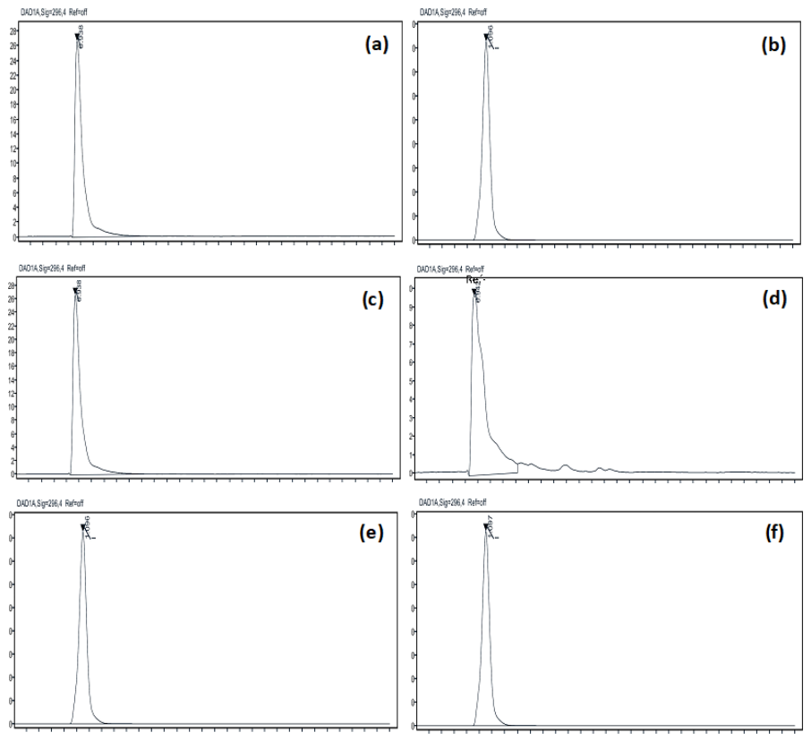

Table 1.
Stability behavior of LVX across different storage conditions over one month.
| Sample | Condition | Time (days) | Assay (%) | Change (%) |
|---|---|---|---|---|
| LVX | Room temperature (25 °C, 60% RH) | 0 | 100.0 | 0.0 |
| LVX | Room temperature (25 °C, 60% RH) | 7 | 99.5 | -0.5 |
| LVX | Room temperature (25 °C, 60% RH) | 14 | 99.2 | 0.8 |
| LVX | Room temperature (25 °C, 60% RH) | 30 | 98.9 | 1.1 |
| LVX | Refrigerated (4 °C) | 30 | 99.3 | 0.7 |
| RH: Relative humidity | ||||
Table 2.
Accuracy (recovery) assessment of LVX using QC samples at different concentrations.
| Spiked concentration (µg/mL) | Peak area (mAU) | Found concentration (µg/mL) | Recovery (%) |
|---|---|---|---|
| 2 |
152.88 | 1.996 | 99.81 |
| 151.87 | 1.992 | 99.61 | |
| 151.62 | 1.985 | 99.24 | |
| 4 |
304.25 | 3.972 | 99.29 |
| 302.37 | 3.986 | 99.64 | |
| 299.28 | 3.996 | 99.91 | |
| 8 |
582.71 | 7.986 | 99.83 |
| 577.08 | 7.988 | 99.85 | |
| 577.78 | 7.998 | 99.97 | |
| Standard deviation (%) | 0.26 | ||
| Coefficient of variation (%) | 0.26 | ||
| Average recovery (%) | 99.68 | ||
| Lower limit of confidence interval (%, p=95%) | 99.48 | ||
| Upper limit of confidence interval (%, p=95%) | 99.88 | ||
| Standard error | 0.08 | ||
Table 3.
Calibration data and regression parameters for LVX over the concentration range of 1–8 μg/mL.
Table 3.
Calibration data and regression parameters for LVX over the concentration range of 1–8 μg/mL.
| No. | LVX standard concentration (µg/mL) | Peak area (AU) | |
|---|---|---|---|
| 1 | 1 | 75,750 | |
| 2 | 2 | 158.62 | |
| 3 | 4 | 304.25 | |
| 4 | 6 | 451.64 | |
| 5 | 8 | 582.71 | |
| Correlation coefficient | 0.999 | ||
| Slope | 72409.63 | ||
| Standard error (SE) | 7.6 | ||
| Y-intercept | 10,474 | ||
| Determination coefficient (R2) | 0.999 | ||
| Linear function | y=72409.63x+10.474 | ||
Table 4.
Inter-day precision of the developed HPLC-UV method for the determination of LVX.
| Spiked concentration (µg/mL) | Found concentration (µg/mL) | Recovery (%) | Mean (%) | CV | SE |
|---|---|---|---|---|---|
| 2 |
1.998 | 99.91 | 99.80 |
0.28 |
0.11 |
| 1.994 | 99.68 | ||||
| 1.989 | 99.44 | ||||
| 1.992 | 99.59 | ||||
| 2.003 | 100.14 | ||||
| 2.001 | 100.06 | ||||
| 4 | 3,997 | 99,92 | 99,78 | 0.29 | 0.12 |
| 3,991 | 99,78 | ||||
| 3,978 | 99,46 | ||||
| 3,976 | 99,39 | ||||
| 4,002 | 100,04 | ||||
| 4,004 | 100,09 | ||||
| 8 |
7.985 | 99,81 | 99,99 |
0.15 |
0.06 |
| 7.990 | 99,88 | ||||
| 7.979 | 99,74 | ||||
| 7.983 | 99,79 | ||||
| 8.008 | 100,1 | ||||
| 8.007 | 100,09 |
Table 5.
Intra-day precision of the developed HPLC-UV method for the determination of LVX.
| Spiked concentration (µg/mL) | Found concentration (µg/mL) | Recovery (%) | Mean (%) | CV | SE |
|---|---|---|---|---|---|
| 2 |
1.997 | 99.83 | 99.89 |
0.19 |
0.07 |
| 1.991 | 99.55 | ||||
| 1.998 | 99.90 | ||||
| 2.002 | 100.08 | ||||
| 2.001 | 100.05 | ||||
| 1.999 | 99.95 | ||||
| 4 |
3,997 | 99,93 | 99,81 |
0,26 |
0,1 |
| 3,974 | 99,35 | ||||
| 3,995 | 99,89 | ||||
| 4,001 | 100,03 | ||||
| 4,000 | 100,01 | ||||
| 3,986 | 99,65 | ||||
| 8 |
7,990 | 99,87 | 99,83 |
0,18 |
0,07 |
| 7,980 | 99,75 | ||||
| 7,970 | 99,62 | ||||
| 8,003 | 100,04 | ||||
| 8,004 | 100,05 | ||||
| 7,972 | 99,65 |
Table 6.
Concentration of LVX in plasma samples obtained from rats (n = 6) after being implanted with LVX-loaded mesh.
Table 6.
Concentration of LVX in plasma samples obtained from rats (n = 6) after being implanted with LVX-loaded mesh.
| Rat | Day 1 (µg/mL) | Day 3 (µg/mL) | Day 5 (µg/mL) | Day 9 (µg/mL) | Day 14 (µg/mL) |
|---|---|---|---|---|---|
| 1 | 12.9 | 10.9 | 8.0 | 5.6 | 4.04 |
| 2 | 13.1 | 11.1 | 8.0 | 5.4 | 3.96 |
| 3 | 13.0 | 11.0 | 8.1 | 5.5 | 4.00 |
| 4 | 12.9 | 10.9 | 7.9 | 5.6 | 4.03 |
| 5 | 13.1 | 11.1 | 8.0 | 5.5 | 3.97 |
| 6 | 13.0 | 11.0 | 8.0 | 5.4 | 4.00 |
| Mean ± SD | 13.00 ± 0.08 | 11.00 ± 0.08 | 8.00 ± 0.07 | 5.50 ± 0.08 | 4.00 ± 0.04 |
| RSD (%) | 0.62 | 0.73 | 0.88 | 1.45 | 1.00 |
Table 7.
Comparison of the developed HPLC-UV method with previously reported techniques for LVX quantification in various matrices.
Table 7.
Comparison of the developed HPLC-UV method with previously reported techniques for LVX quantification in various matrices.
| Ref. | Matrix | Analytical Technique | Linear Range | LOD/LOQ | Recovery (%) | Application |
|---|---|---|---|---|---|---|
| Present study | Rat blood | HPLC-UV | 1–8 µg/mL | 0.3/1.0 µg/mL | 99.68 | Prolonged-release mesh implant in rats |
| [4] | Human plasma | HPLC-UV | 1.8-28.3 µg/mL | 0.6/1.8 µg/mL | NR | Pharmacokinetic/bioequivalence studies |
| [16] | Plasma | HPLC-FLD | 0.25–10 µg/mL | 0.04/0.12 μg/ml | 96-104 | Pharmacokinetic studies |
| [18] | Human plasma | HPLC-FLD | 0.1-15, 0.2/7.0 µg/mL |
0.05/0.2 µg/mL | 106 | Pharmacokinetic monitoring |
| [17] | Human serum | SPE-HPLC-FLD | 10–10,000 ng/mL | NR/10 ng/mL | 87–98 | TDM, pharmacokinetic studies |
| [7] | Human plasma | HPLC-UV | 0.25–15 µg/mL | NR/0.25 µg/mL | NR | TDM in MDR-TB patients |
| [13] | Human plasma | HPLC-UV | 0.0521-5.213 µg/mL | NR/0.0521 µg/mL | 86-89 | Bioequivalence studies |
| [42] | Aqueous humor of rabbits | HPLC-MS/MS | ??? | NR | NR | In vitro release from scaffolds |
| [15] | Human milk | HPLC-FLD | 2.5–500 ng/mL | 0.63/2.11 ng/mL | 96.18 | TDM in lactating women |
| [36] | Plasma/dialysate | HPLC-UV/FLD | 0.1-6 µg/mL | NR/0.1 µg/mL | 97.2-104.7 | TDM, pharmacokinetic studies |
| [43] | Human plasma | HPLC-UV | 100-10000 ng/mL | NR/100 ng/mL | 63.47 | Multi-fluoroquinolone assay |
| [19] | Plasma/prostate tissue | UHPLC | 0.030-10 µg/mL; 0.3-30 µg/mL | 0.01/0.03 µg/mL; 0.1-0.3 µg/mL |
93-108; 99-106 | Prostate biopsy prophylaxis |
| [27] | Plasma | UHPLC-MS/MS | 2500-80000 µg/mL | 0.750-2.380 µg/mL | 96 | Multi-antibiotic TDM (19 drugs) |
| [18] | Goose tissues | HPLC-FLD | 0.005-1.5 µg/mL | 0.001/0.005 µg/mL | 96 | Pharmacokinetics in geese |
| [44] | Plasma | HPLC-UV | 0.15-2.7 µg/mL | NR/0.05 µg/mL | 86.3 | TDM, specificity assessment |
| [3] | Human plasma | LC-MS/MS | 0.1–5, 0.4–40, 0.2–10, 2–100, 0.2–10 mg/L | NR/0.1, 0.2, 0.4, 2 mg/L | 91.4-109.7 | Multi-TB drug TDM |
| [30] | Human plasma | HPLC-UV | 0.125-25, 0.1-20, 0.05-10 mg/L | NR/0.125, 0.1, 0.05 µg/mL | 79 | Multi-fluoroquinolone TDM |
| [37] | Ocular nano-formulations | HPLC-UV | 4.8-29.04 µg/mL | 0.66686/2.22286 mg/mL | 80-110 | Pharmaceutical analysis |
| [10] | Medical wastewater | SPE-HPLC-UV | 10-1000 ng/mL |
2.95 −1 ng/mL | 76.8 -87.8 | Environmental monitoring |
| [32] | Pharmaceutical formulations | HPLC-UV | 15–40, 2.5–15.0, 7.5-20.0 mg/L |
2.14/7.15 mg/mL 0.24/0.81 mg/mL 0.29/0.95 mg/mL |
98.57, 105.5, 102.5 | Multi-drug analysis |
| [33] | Pharmaceutical formulations | HPLC-DAD | 0.5-10 mg/mL | 0.15/0.5 µg/mL | NR | Multi-fluoroquinolone assay |
| [41] | Injectable dosage forms | HPLC-UV | 100.07-300.21 g/mL | NR | 101-101.8 | Pharmaceutical QC |
| [31] | Ocular nano-formulations | UV spectrophotometry | 4–12 µg/mL | 0.3933/1.1919 µg/mL | 99.92–100.80 | Nanoparticle formulation analysis |
| [34] | Human urine/plasma | HPLC | 100-10000 µg/L (plasma) | 30.40/100 26.93/100 34.84/100 35.63/100 µg/L (plasma) | 85.79-111.07 (plasma) | TDM, pharmacokinetic/toxicology |
| [38] | Pharmaceutical formulations | UV spectrophotometry | 2-20 μg/mL | 1.41/4.3 μg/mL 0.63/1.92 μg/mL |
96.66-99.7, 99.58-99.66 |
Simultaneous DOXH/LVXH assay |
| NR: not reported HPLC: high-performance liquid chromatography UV: ultraviolet SPE: solid phase extraction LC-MS/MS: liquid chromatography- mass spectrometry FLD: fluorescence detector DAD: diode array detector TDM: therapeutic drug monitoring | ||||||
Disclaimer/Publisher’s Note: The statements, opinions and data contained in all publications are solely those of the individual author(s) and contributor(s) and not of MDPI and/or the editor(s). MDPI and/or the editor(s) disclaim responsibility for any injury to people or property resulting from any ideas, methods, instructions or products referred to in the content. |
© 2026 by the authors. Licensee MDPI, Basel, Switzerland. This article is an open access article distributed under the terms and conditions of the Creative Commons Attribution (CC BY) license (http://creativecommons.org/licenses/by/4.0/).
Copyright: This open access article is published under a Creative Commons CC BY 4.0 license, which permit the free download, distribution, and reuse, provided that the author and preprint are cited in any reuse.


