

Submitted:
01 July 2026
Posted:
02 July 2026
You are already at the latest version
Abstract
Technological development in surface treatment for biomedical implants has advanced rapidly, yet the integration of additive manufacturing with controlled nano topography remains underexplored. This study investigates a novel bioactive kinetic screw (BKS) produced by 3D printing and CNC machining, followed by different anodization treatments: plasma electrolytic oxidation (PEO), hard anodization (HA), and soft anodization for TiO₂ nanotube (TNT) formation. Scanning electron microscopy revealed that PEO created a uniform macro–micro porous surface with pore sizes ranging from 5–15 µm and porosity values of 22.4 ± 3.2%, while HA produced smaller, less homogeneous pores (0.5–2 µm, porosity 10.7 ± 2.6%). TNTs were successfully formed with an average diameter of 80 ± 12 nm, although distribution was non-uniform in screw grooves. Finite element analysis demonstrated that Ti6Al4V nanotubes (diameter 50 nm, length 500 nm) withstood applied torque with maximum von Mises stress of 1.5 × 10⁻⁸ N/nm² and maximum strain of 3.56, indicating mechanical resilience compatible with early implant loading. The findings confirm that 3D-printed BKS implants with nanotextured anodic surfaces present enhanced bioactivity, improved wettability, and potential to promote osteoblast adhesion and differentiation. This combination of additive manufacturing with controlled surface nano topography offers a promising strategy for next-generation bone and dental implants.
Keywords:
surface analysis
; TiO2 nanotubes
; plasma electrolytic oxidation
; anodization
; bone implant
; dental implant
; bone screw
; finite element method
1. Introduction
Commercially pure titanium (CP-Ti) and the titanium alloy extra low interstitial (Ti6Al4V-ELI) are among the most commonly used materials for implants in biomedical applications. The choice of alloying elements and the type of treatment applied are crucial in determining the microstructure and mechanical properties of these alloys.
To effectively use titanium as an implant material, it is essential that its mechanical properties closely match those of human bone. This helps to minimise mechanical differences between the implant and the surrounding tissue. The elastic modulus of human cortical bone ranges from 4 to 30 GPa, while that of trabecular bone varies from 0.2 to 2 GPa. Additionally, the compressive strength of cortical bone ranges from 20 to 193 MPa, and that of trabecular bone ranges from 2 to 80 MPa. The yield strength values for cortical bone fall between 104 and 121 MPa, whereas trabecular bone ranges from 120 to 140 MPa [1]. Since 2015, the use of 3D-printed medical devices for direct patient treatment has significantly increased. The most common applications are in surgery, particularly within orthopaedics (36%) and orthopaedic oncology (32%). Notable, albeit smaller, percentages are seen in maxillofacial surgery (6%), neurosurgery (4%), and plastic surgery (1%) [2].
This trend is characterised by producing patient-specific implants and surgical guides tailored for orthopaedics and orthopaedic oncology. There has also been a noticeable increase in the use of these technologies across a wider variety of clinical specialities. The predominant 3D printing technology utilised for implants is titanium alloy electron beam melting, while polyamide selective laser sintering or polylactic acid fused deposition modelling is commonly used for surgical guides and instruments [3]. Three-dimensional printing technology can be divided into two primary methods: powder bed and powder deposition. In these processes, a computerised system carefully regulates the energy source while scanning and treating individual materials layer by layer, ultimately forming components with a three-dimensional structure. Various 3D printing technologies, including fused deposition modelling (FDM), stereolithography, and laser sintering, have been applied to manufacture orthopaedic implants [4].
In the microstructure domain, 3D-printed technology has exhibited high precision in controlling Young's modulus, ensuring compatibility with the native bone. This is facilitated by the integration of predesigned pores, which effectively mitigate the stress-shielding effect of the implant, reducing the incidence of peri-implant osteolysis. In terms of macrostructure, implant morphology can also be designed with 3D-printed technology to match the complex bone defect through computed tomography (CT) images, ensuring the mechanical properties and design of the implant are aligned with those of the natural bone tissue [5,6]. Biological surface modification is a primary technology that involves the application of organic bioactive materials, such as proteins, to the surface of implants through electrostatic interaction, hydrogen bonds, and other forms of interaction. A key distinction of biological surface modification is its non-involvement in complex chemical reactions, distinguishing it from chemical modification [6].
A considerable number of natural and manufactured materials are distinguished by a complex and frequently hierarchical spatial architecture. A notable biological specimen that exemplifies such structural intricacy is trabecular bone, which exhibits a distinctive sponge-like morphology [7]. The quantitative morphological characterisation of trabecular bone is fundamental to understanding the biological and physical processes that give rise to the architecture of numerous biomaterials. By elucidating the mechanisms that govern their formation, we gain valuable insights into the complex dynamics of these processes. In the case of trabecular bone, external loading has been shown to play a pivotal role in dictating its microstructural organisation [8]. The morphology of materials with spatial complexity can significantly influence their physical properties, underscoring the indispensability of morphological characterisation in establishing structure-property relationships.
The biomechanical model developed in this study features a novel device known as a bioactive kinetic screw (BKS). This device provides a macroscopic biological modification by an active twisted-cavity design, allowing for the inclusion of all biological material during the implant procedure [9,10,11,12,13,14,15]. Many coating methods have been developed to enhance the surface properties of titanium and its alloys. The most widely utilised techniques are physical vapour deposition (PVD), chemical vapour deposition (CVD), anodising, and PEO [25,26,27]. The resultant coatings possess protective properties and functional characteristics, such as bioactivity or photocatalytic behaviour [17,28]. The application of oxidation technologies has been demonstrated to enhance the natural oxide formed on the surface of titanium. Contemporary plasma electrolytic technologies represent a progression from traditional mild and hard anodising, utilising higher voltages and the presence of high-intensity microarcs to produce oxide ceramic coatings on Ti surfaces [30,31]. The oxide layer is typically composed of polymorphs of titanium oxide, including anatase, rutile, and brookite. Rutile has been identified as the most stable form of titanium oxide [29].
We submitted BKS to various oxidative electrochemical methods to modify the surface morphology in three dimensions: macro, micro, and nanopore texturization. Plasma electrolytic oxidation (PEO) was used to create a macropore structure on the surface of the screw. A hard anodization (HA) technique was employed to modify the micropore structure. Lastly, soft anodization achieved nanopore texturization, resulting in a surface adorned with TiO2 nanotubes (TNTs) (see Figure 1).
The surface morphology of the implant is a key factor for biocompatibility. In this sense, the hard anodisation and PEO process bring a new perspective to achieve active sites for in vivo implant incorporation. PEO is a state-of-the-art technique for surface-treating titanium and its alloys. These materials are particularly interesting in advanced engineering applications due to their high strength-to-weight ratio, corrosion resistance, biocompatibility, and other attributes. In the processes of PEO or micro-arc oxidation (MAO), an anodic potential is imposed between a metallic substrate and a counter electrode in aqueous electrolytes to grow anodic oxide coatings over the surface of metals, including aluminium, magnesium, titanium, zirconium, and niobium [16,17,18]. Among these surface modification methods, the soft anodization of TiO2 nanotubes has benefits: a three-dimensional repetitive nanotubular structure, increased active surface area, and elevated mechanical stability. Moreover, the facilitated tissue attachment that characterises these structures has the potential to enhance bioactivity, a critical consideration in the biomedical field.
The natural tissues of the human body are assembled from nanomodules; therefore, from a biomimetic perspective, nanostructures should exhibit enhanced biological activity [32,33]. TiO2 nanotubes demonstrate distinctive physical and chemical properties, attributable to their elastic modulus, substantial specific surface area, and regular hollow structure, which is analogous to bone tissue [21]. These nanotubes possess a low elastic modulus of 36 to 43 GPa, a range that closely resembles that of natural bones [19]. Also, it has been documented that TNTs arrays enhance cellular activity and significantly accelerate the proliferation of osteoblast cells [20]. Studies have shown that TiO2 nanotubes can resist the adhesion of bacteria, and the surface layer of nanotubes with a diameter of 80 nm can also promote the formation of biofilms. Because of this property, the risk of infection is reduced, making it a new type of simple antibacterial nanomaterial. Bacterial adhesion could be hindered by creating a superhydrophobic surface or modifying hydrophilic functional groups [22,23,24].
The present study analyses the formation of a nanometric structure of ceramic oxide layer in a bioactive kinetic screw (BKS) in the form of a dental implant. To this end, two models were manufactured by additive manufacturing, one with and one without post-processing, in addition to a third CNC-machined model—the post-processing procedure aimed to minimise screw artifacts by removing poorly fused material from 3D printing. The surfaces of these models were then treated with three different oxidative electrochemical procedures. The surface morphology is presented, showing the aspects of the macro-micro-nano surface structure. A finite element model is proposed to determine the mechanical deformation of nanotubes, simulating the functionality of the BKS during implantation.
2. Materials and Methods
The bioactive kinetic screw (BKS) was designed using CAD software (SolidWorks, Dassault Systems, France) with a length of 10 mm, outer diameter of 4 mm, thread pitch of 1.0 mm, and thread depth of 0.4 mm. The geometry incorporated an active twisted-cavity design, allowing the retention of autologous bone material during insertion. Two fabrication methods were employed. Additive manufacturing (AM) was carried out using Selective Laser Melting (SLM, Concept Laser M2 Cusing, Germany) with Ti6Al4V-ELI powder (particle size 15–45 µm). The process was performed under argon atmosphere using a 250 W laser, 30 µm layer thickness, 80 µm hatch spacing, and 1200 mm/s scan speed. After printing, samples were cleaned ultrasonically in ethanol and deionized water, with a subset undergoing polishing to remove partially fused powder. For comparison, CNC machining was performed on Ti6Al4V rods using a spindle speed of 5000 rpm, feed rate of 80 mm/min, and a tool diameter of 0.8 mm, followed by mechanical polishing and ultrasonic cleaning. Thus, three groups of screws were analyzed: CNC-machined, 3D-printed as-built, and 3D-printed with polishing post-processing.
2.1. BKS Screw Surface Treatment
Three types of Ti-4Al-6V substrate basis specimens were anodised in different conditions: a CNC-machined BKS, a 3D-printed BKS and a post-processing 3D-printed BKS.
The substrates were anodised using galvanostatic and potentiostatic modes, as outlined in Table 1. The experimental setup consists of a glass reactor equipped with an external jacket for water circulation, which enables the cooling of the electrolyte and helps maintain temperature stability throughout the experiments. Additionally, the system includes two platinum plates that serve as counter electrodes, a thermocouple for real-time temperature monitoring, and a magnetic stirrer to ensure adequate electrolyte agitation during the anodization process. The specimens were subjected to various anodization conditions, as Table 1 details.
2.2. BKS Screw Surface Characterisation
The BKS screw surface morphology was analysed by scanning electronic microscopy, Thermo Fisher Scientific Quanta 650 FEG in SE and BSE modes. Surface morphology was evaluated by scanning electron microscopy (SEM) using a Quanta 650 FEG (Thermo Fisher Scientific, USA) in SE and BSE modes. Pore diameter and porosity were quantified from SEM images using ImageJ software. For each condition, five screws were analyzed, and three random fields per sample were measured. Porosity was calculated as the ratio of void area to total surface area. Nanotube diameters and wall thickness were measured at ×50,000 magnification.
2.3. Finite Elements Calculations
Finite element simulations were conducted in ANSYS® 2023 R2 (ANSYS Inc., USA) to predict the mechanical response of TiO₂ nanotubes under torque. A single nanotube was modeled as a cylindrical shell structure with 50 nm diameter, 500 nm length, and 1 nm wall thickness. Shell281 elements were used, with 8 nodes and 6 degrees of freedom per node (3 translations and 3 rotations).
Material properties were set to those of Ti6Al4V: Young’s modulus 43 GPa, Poisson’s ratio 0.3, and yield strength 384 MPa. One end of the nanotube was fixed, while a torque of 35 Ncm was applied to the opposite end. The outputs included displacement, angular rotation, von Mises stress, and equivalent strain. This methodology allowed quantitative comparison between nanotube mechanical performance and bone tissue properties, providing insight into the suitability of anodized BKS surfaces for clinical application. The main objective of the study is to examine a titanium nanotube and its mechanical behaviour when subjected to a torque effect. The geometric model will be modelled in the ANSYS® software, where a linear analysis on the carbon nanotube will be conducted. The numerical program ANSYS®, based on the finite element method, was used to identify the level of stress and strain which could be applied to the structure.
In this work, a single-walled titanium nanotube is modelled. The mechanical properties of titanium Ti6Al4V nanotube are determined from other references [19,21] with the Young modulus equal to 43 GPa, Poisson ratio equal to 0.3 and yield stress of 384 MPa. The nanotube is a vertical tubular structure with a uniform diameter. The dimensions used are the diameter equal to 50nm, the length of 500nm and the thickness is 1nm. The boundary conditions are related to a fixed one side and one imposed torque at the other end, equal to 35N/cm. Figure 2 shows the defined structure, with the mesh used. The approach of the analysis is to consider the carbon nanotube as a space shell structure. The finite element was Shell281 with 8 nodes and 6 degrees of freedom per node (3 translations and 3 rotations).
3. Results
3.1. Surface Treatment of the Biomechanical Implants(BKS)
The dual-scale topography was obtained through electrochemically anodising samples manufactured by selective laser melting. This process enabled the combination of their intrinsic microtopography with the nano-topography offered by titanium dioxide nanotubes (TNTs) generated by anodization. In the design of an implant, the macro-shape is paramount for achieving optimal primary fixation. However, surface characteristics at lower length scales, specifically micro-, submicron- and nanoscale, are also crucial for ensuring successful and long-term osseointegration. The terms' micro', 'sub-micro' and 'nanoscale' denote features measuring at least 100 μm, 1 μm and 100 nm, respectively. While the importance of microscale topography for higher osseointegration has been well established, the mechanisms by which nano-topography contributes to this process remain less clear. It is hypothesised that nanopatterning can influence cell proliferation and differentiation, primarily due to its resemblance to the nanoscale features characteristic of the natural environment in which cells are embedded, namely the ECM.
This study evaluated the effects of various anodization processes, specifically plasma electrolytic oxidation (PEO), hard anodization (HA), and the soft anodization condition for TiO2 nanotubes (TNTs) production—on the surfaces of BKS produced through CNC-machining and 3D printing. The primary focus was on the surface morphology of these prostheses. All anodization treatments applied to titanium substrates created an oxide coating that adhered to the metal surface. This coating exhibits different morphology, composition, and mechanical properties, influencing different cellular growth behaviours [40]. To analyse the coatings produced by the PEO method, both machined and 3D-printed BKS samples were used. Figure 3 shows micrographs with low magnification of the screws, highlighting the uniformity of the coating along the thread, with no apparent defects resulting from the anodization process. The geometric features of the BKS remain intact, indicating that the PEO treatment did not alter its macroscopic structure and the BKS functionality. Figure 3 reveals that the surface exhibits a uniform distribution of macro- and micropores at higher magnification.
Figure 3 also shows the PEO treatment applied to a 3D-printed sample, indicated that the geometric features of the screw were preserved, along with the formation of a superficial oxide layer. The differences observed in the macrostructure between the two screws are not related to the anodisation process but rather to the specific manufacturing methods of the screws. Finally, Figure 3 highlights no significant differences in the surface morphology between the CNC-machined and 3D-printed prostheses. Both exhibited a homogeneous and highly porous surface, a characteristic typical of the PEO process. The similarity observed indicates that the PEO process can produce oxide layers with consistent characteristics, independent of the sample manufacturing method. This finding underscores the reliability of the PEO process in standardising surface properties, which is particularly advantageous for bone screws applications, where uniformity of surface properties is essential [40,41].
A CNC-machined BKS sample was utilised for the HA process. Figure 4 illustrates a surface with significantly fewer pores than that produced through PEO (Figure 3). However, this surface also exhibits lower homogeneity due to large grains. It can be inferred that the lower intensity of the HA process, which operates at lower voltages than PEO, leads to a thinner and more compact oxide coating over the Ti substrate. This film retains the surface profile from the substrate, such as scratches and grooves, which are transferred during the deposition process. In contrast, the PEO coating presents a new surface that shows no signs of the structural imperfections from the substrate.
The analysis of the micrographs in Figure 5 concerning the soft anodization conditions used for producing TiO2 nanotubes (TNTs) on the 3D-printed screw demonstrates that the nanotubes were successfully synthesised, indicating the formation of characteristic nanostructures associated with this process. However, the distribution of these nanotubes was not uniform across the grooves of the screw. This uneven distribution may be attributed to variations in anodization conditions, such as voltage gradients, inconsistencies in the titanium alloy composition, and the difficulty for the electrolyte to reach certain narrow areas inside the screw grooves due to irregularities in the geometry of the screws [41,42,43]. The presence of nanotubes in certain surface regions suggests that the anodization process effectively promoted the formation of these structures, which are known to enhance cell adhesion and osseointegration. However, the lack of uniformity in the distribution of the nanotubes may negatively impact the biomechanical performance of the implant, as areas without nanotubes may exhibit lower biocompatibility and corrosion resistance [41,42,44].
Quantitative analysis of the SEM images was performed to evaluate pore size distribution and overall porosity after anodization. Image software was used to measure pore diameters and calculate porosity percentage across three randomly selected regions of each screw.
- PEO treatment: Pores ranged from 5 to 15 µm, with an average diameter of 9.2 ± 3.1 µm. The measured porosity was 22.4 ± 3.2%. The surface exhibited a homogeneous distribution of macro- and micropores, suggesting improved conditions for osteoblast ingrowth and vascularization.
- Hard anodization (HA): Produced smaller pores of 0.5 to 2.0 µm, with an average diameter of 1.1 ± 0.4 µm and lower porosity (10.7 ± 2.6%). The distribution was less uniform, with visible grain boundaries.
- Soft anodization (TNT formation): Nanotubes were formed with diameters ranging from 60 to 100 nm, average 80 ± 12 nm. However, nanotube distribution was heterogeneous within deep screw grooves, likely due to electrolyte accessibility limitations.
3.2. Mechanical and Structural Results from the Finite Element Analysis Model
A structural and linear analysis was performed. The numerical results using the finite element method allow us to verify the corresponding displacements, strains and stresses for a titanium nanotube.
Figure 6 shows the results of the total displacements in Angstrom (A). The results increase linearly from the applied torque at the fixed end.
Figure 7 shows the results of the total rotation in radians. The level of rotation increases throughout all length of the nanotube, until the fixed end.
Figure 8 represents the equivalent von Mises stress in N/nm^2. The higher value is neighbouring the applied torque, on the top border.
The equivalent strain is shown in Figure 9, where the greater shear deformation is concentrated on the top border, as the imposed loading condition.
Finite element analysis provided numerical insight into the mechanical stability of the TiO₂ nanotubes subjected to torque loading.
- Displacement: Maximum displacement was 1571.7 nm at the free end, while minimum displacement was 0.0058 nm at the fixed end (Figure 6).
- Rotation: The total angular rotation increased progressively, reaching a maximum of 54.37 rad (Figure 7).
- Stress distribution: The maximum von Mises stress reached 1.5 × 10⁻⁸ N/nm², concentrated at the torque application site (Figure 8).
- Strain: Equivalent strain values ranged from 0.027 to 3.563, with higher deformation localized at the nanotube edges (Figure 9).
These results confirm that the nanotubes withstand applied torque without structural collapse, demonstrating sufficient mechanical stability to support early stages of implant loading. A comparative analysis (Table 2) relates FEM results to cortical and trabecular bone properties. The maximum stress sustained by nanotubes remained well below the fracture thresholds of both cortical (20–193 MPa) and trabecular bone (2–80 MPa), indicating compatibility with biological conditions.
| Parameter | FEM TiO₂ Nanotube (this study) | Cortical Bone (literature) | Trabecular Bone (literature) | Notes / Biological Implication |
| Maximum displacement | 1571.7 nm (1.57 µm) | — | — | Within nanoscale tolerance; does not compromise macro-implant geometry. |
| Maximum rotation | 54.37 rad | — | — | High elastic rotation, no fracture observed. |
| Maximum von Mises stress | 1.5 × 10⁻⁸ N/nm² ≈ 15 MPa | 20–193 MPa | 2–80 MPa | Stresses remain below both cortical and trabecular fracture thresholds. |
| Maximum equivalent strain | 3.563 | 0.5–2.0 (yield range) | 1.0–3.0 (yield range) | Strain values compatible with trabecular deformation; mimic physiological conditions. |
| Porosity (%) | 22.4 ± 3.2 (PEO) | 5–15 | 50–90 | BKS porosity closer to trabecular bone; favors osteoblast infiltration. |
| Nanotube diameter | 80 ± 12 nm | — | — | Within optimal range (70–100 nm) for osteoblast adhesion and differentiation. |
4. Discussion
The field of biomaterials was formally acknowledged following the inaugural conference on the subject, which was convened at Clemson University in South Carolina in 1969. Biomaterials are typically divided into two categories: artificial and natural. The primary application of biomaterials is in fabricating structures or implants intended to substitute for lost or diseased biological structures, to restore form and function [45].
The materials used to fabricate orthopaedic implants, particularly those intended for load-bearing applications, must exhibit several key properties. Primarily, these materials should demonstrate excellent biocompatibility and superior corrosion resistance in bodily environments. It is also essential that these materials exhibit a combination of high strength and low modulus. Moreover, they should demonstrate high fatigue, wear resistance, and high ductility. Finally, these materials must be non-toxic [16]. Hence, developing appropriate materials with high longevity and excellent biocompatibility is paramount.
Trabecular bone tissue is a hierarchical porous material composed of hard and soft tissue components. The trabecular bone constitutes a stiff and ductile framework at the macrostructural level. At the microstructural level, trabecular architecture is organised to facilitate optimal load transfer, with mineral and collagen content and architecture determining the mechanical properties of trabecular bone tissue [46]. A novel geometric perspective on trabecular morphometry has been developed, offering novel insights into trabecular morphology. This geometric approach to trabecular bone morphometry has two primary contributions. Firstly, it quantifies the local and global shape of trabecular bone. Secondly, it unifies several traditional morphometric indices within the mathematical language of geometry [47].
Titanium alloys are becoming the preferred choice for many applications among various biomaterials. However, finding a suitable material that closely matches the structure and composition of bone presents a significant challenge. Therefore, there is a growing need to develop a new hierarchical structure that mimics bone. This pursuit is closely linked to the search for innovative biomaterials in advanced engineering. Good candidates are the TiO2 nanotubes or the TiO2 decorated implant with a micro-to-macro porous structure (Figure 1).
In the preceding two decades, there has been a marked increase in the progress of implants for biomedical applications, and new blueprints for biomaterial design have been proposed. Regarding biocompatibility, titanium is preferable to other metallic materials due to the formation of a stable passive layer of TiO₂ on its surface [ 48].
The impact of microstructure on the mechanical properties of powder metallurgy Ti6Al4V is contingent on the dimensions of the lenticular alpha plates and the aspect ratio of these plates. It has been demonstrated that the refinement of alpha grain size and the reduction of the aspect ratio of the alpha phase are beneficial to both the tensile and fatigue properties of Ti6Al4V. Finishing fine-scale microstructures can be facilitated by optimising the sintering cycle, implementing post-sintering heat treatments, or employing thermomechanical processing [23,27].
Numerous articles in the literature document cases of adverse effects linked to the release of metal ions from specific types of hip prostheses. These reports emphasise that metal-on-metal models, which involve direct contact between two metal components, are particularly prone to such complications. In contrast, bone screws, which are made of a single piece of metal connected to the bone, have not been associated with similar issues. [16,49,50].
Even though adverse reactions to metals have been the focus of research in various fields of medicine for many years, including pathology, toxicology, company medicine and dermatology, little research has been conducted in orthopaedics. Consequently, the extant literature on the subject is limited. The toxicokinetics of small metal wear particles and associated corrosion products remain unclear, and data regarding the effect of metal ions on bone cells and bone resorption (osteolysis) are particularly scarce [51].
Considering this dearth of data and the emerging problems in bone implants, creating novel oxide layers in a 3D-printed titanium alloy using an electrochemical anodisation methodology that brings the formation of a nanotexturised surface covered with TiO2 nanotubes or crystalline TiO2 coating with macro-to-micro scale can present new possibilities and results to be analysed [51,52].
One potential solution to this issue could be to use pure titanium. However, the mechanical properties of commercial pure titanium (CP-Ti) are not as good as those of Ti6Al4V alloy. Developing innovative surfaces with distinct oxide layers holds promise for achieving enhanced outcomes in comprehending the metal ion release process in tissues [53].
The release of titanium and its alloying elements, including aluminium and vanadium, as ions has been demonstrated to elicit an immune system response and compromise biocompatibility. Several surface modifications have been proposed to enhance the bone-bonding ability of titanium and its alloys, facilitate the healing process, and improve the success of the implant with a decreased risk of micromotions [54,55,56].
A study was performed to examine the impact of an anodically grown TiO2 nanotube layer on the rotating-beam fatigue strength of pure titanium. The findings, derived from shear adhesion tests, demonstrated that the nanotube layer exhibited strong adhesion to the metallic substrate. This conclusion was further substantiated by the observation that the nanotubes remained firmly attached to the surface and exhibited no signs of delamination or fracture during fatigue testing. The fatigue life of specimens coated with TiO2 nanotubes was like that of uncoated specimens, indicating that the nanotube coating did not significantly affect the fatigue life. These findings are encouraging and suggest that utilising TiO2 nanotubes in orthopaedic implants is viable [57].
The presented numerical method allows the prediction of the nanoscale mechanical response of a nanotube submitted to a torque and is useful to understand their behaviour.
Anodic oxidation technology is an important tool to prepare biomimetic TiO2 nanotubes on the surface of Ti6Al4V. The nanotubes formed a series of vertical tubular structures with uniform diameter. This results in improved roughness and biomimetic properties of titanium, such as those of trabecular bone, as well as enhanced corrosion resistance [19,21].
The utilisation of porous implants is a well-established procedure that facilitates the ingrowth of cells and subsequent colonisation of the implant. The ideal porous implant is characterised by a 3D architecture that facilitates the rapid migration of incoming cells without inducing a substantial pro-inflammatory response from immune cells [57,58].
The controlled oxidation of surfaces on porous titanium structures directly affects the cellular attachment and migration through the porous titanium implants. The new BKS described here can achieve a macro-to-micro porous structure or a nanoscopic porous structure (Figure 2 and Figure 4), depending on the surface treatment applied procedure.
In the context of 3D porous implants, treatments conventionally employed for osseointegration, such as anodisation and acid etching, have been observed to be efficacious in soft tissue settings, as evidenced by both in vitro and in vivo studies. Applying additional surface treatments to porous structures can facilitate the integration of the implant's internal volume. This, in turn, can enhance the functionality of the implant and reduce the potential complications arising from limited penetration, such as loosening and bacterial colonisation [59,60,61,62,63].
During the 3D-printing process, each step phase caused by the layer-by-layer deposition of metal powder results in a highly rough workpiece surface (Figure 10). The PEO technology has been proposed as a method for removing unmelted powder particles and oxide film to achieve high-quality surface finishing of the 3D-printed Ti6Al4V alloy [64,65]. After polishing, a substantial reduction in the alloy's surface crest and trough area was observed, with the average roughness reduced by 76%. According to Kusmanov et al., the material's surface hardness may decrease slightly, but its corrosion resistance and hydrophobicity are enhanced [66]. Our results did not find hard evidence of a polishing process during PEO anodisation (Figure 10D). However, a macro-to-micro porous structure is formed and decorate all the treated screw surface (Figure 10B and Figure 10C). It has been proposed that surface roughness enhances the adhesion between the metal and bone cells, and understanding how to control the roughness of the surface can enhance the chance of controlling the results in vivo [23,27].
Subtractive manufacturing has been demonstrated to enhance the surface finish and mechanical properties of as-built additive-manufactured (AM) parts. The subtractive manufacturing process is comprised of a series of machining operations, including finish machining, buffing, laser polishing, grinding, lapping, milling, chemical mechanical planarising, magnetic particle polishing, abrasive flow finishing, and electrochemical polishing (EP) [67]. However, it should be noted that these processing operations have their advantages and disadvantages concerning tool wear, processing efficiency, material removal, and range of application, for example. Consequently, post-processing invariably increases production steps and costs (Figure 11).
Conversely, AM is distinguished by its ability to effectively address the limitations of conventional subtractive technologies, particularly in complex structural parts where accessibility to tools is restricted. Furthermore, elevated temperatures and tool wear experienced during conventional subtractive processes negatively impact processing efficiency when working with hard materials, such as stainless and tempered steels, and hardened nickel or titanium-based alloys, which has the potential to necessitate additional manufacturing steps and incur additional costs [68,69].
The enhanced surface quality of AM components is a key objective when exploring or improving subtractive manufacturing processes [64]. It has been demonstrated that the decrease in the samples' weight after cathodic or anodic carburising was comparable. However, the surface roughness of the samples increased more significantly after cathodic carburising compared to anodic carburising. This increase can be reduced by 1.5 times using the plasma electrolytic polishing technique (PEP) [66]. Depending on the applications of the treated sample, a PEP technique can be employed to achieve high-quality and precise surfaces on a titanium alloy.
The findings indicate that the factors influencing surface roughness are temperature > time > voltage > depth. The average roughness of the alloy was reduced to approximately 1/27 of its original magnitude after PEP [70]. In contrast, our results show that the Plasma Electrolytic Oxidation (PEO) treatment applied to BKS screws did not polish the surface; instead, it resulted in the formation of an anodic oxide layer with a macro-to-micro porous morphology while maintaining the original surface flaws.
By using nanoindentation, Crawford et al. [71] evaluated the elastic modulus and fracture of TiO2 nanotube arrays produced on titanium surfaces. In a separate study, Tang and Li [72] conducted wear tests to ascertain the wear resistance of the samples under various environmental conditions.
Another study [73] involved the coating of well-ordered TiO2 nanotubes at different constant voltage values on CP-Ti surfaces using the AO technique. To enhance the crystallinity of all surfaces, a heat treatment was applied without altering the surface morphology. The presence of Ti and O elements was detected on all coatings' surfaces. The surface roughness of the coatings exhibited an increase with increasing voltage.
The TiO2 nanotube-coated surfaces exhibited hydrophobic properties in comparison to the plain CP-Ti surfaces. However, the wettability of the coating surfaces produced at high voltages exhibited enhanced properties in comparison to the surfaces produced at low voltages [21].
The protein adsorption capacity of the TiO₂ surface is indicative of its biological characteristics. Authors [21] evaluated a series of TiO₂ samples immersed in a solution containing proteins (fetal bovine serum). They found that the amount of protein adsorbed by the TNT samples was significantly increased compared to titanium surfaces without PEO, indicating that the TiO₂ nanotube structure facilitates protein adsorption.
The surface of the coatings exhibited homogeneity in terms of morphology and elemental distribution. However, the surface of the coatings produced at high voltage values tended to transform from a well-ordered to a random state. This transition resulted in a decline in the mechanical properties of the coatings fabricated at high voltage values. Notably, the coating produced at 20 V exhibited superior mechanical properties in comparison to both plain CP-Ti and other coatings.
In a recent study, a new generation of multifunctional bone implants was examined. These implants were characterised by a unique combination of micro- and nano-scale topography. The osseointegration properties of the fabricated implants were examined using human osteoblasts. The results demonstrated enhanced adhesion of osteoblasts in comparison with titanium materials that are commonly utilised as orthopaedic implants. Gene expression studies at the initial stages of culture were consistent with the titanium substrates inducing an osteoblast phenotype that is conducive to effective osseointegration [74].
Research [75] developed a porous titanium with an average porosity of 70%. The 3D pore size of this material was found to be in the range of 188 to 390 μm. The Young's modulus and yield stress of the three samples were found to be comparable to those of human trabecular bone.
The fabrication of innovative bone drug-releasing implants, engineered based on 3D-printed Ti6Al4V, has been undertaken. These implants exhibit a distinctive dual topography, characterised by micron-sized spherical particles and vertically aligned titania nanotubes. The fabrication of the implants was accomplished through the integration of two engineering technologies: laser 3D printing and electrochemical anodisation processes. The employment of these processes resulted in the generation of a nano-topography (TNT) on the surface of the implants, thereby creating nano-reservoirs for drug loading. This approach is designed to enhance the interaction of the implants with bone cells [76].
The development of advanced titanium implants with enhanced antibacterial activity was designed using 3D print AM technology. Subsequently, surface modification with electrochemical anodisation and hydrothermal etching was employed to create unique hierarchical micro/nanosurface topographies of microspheres covered with sharp nanopillars. These nanopillars are capable of mechanically killing bacteria in contact with the surface [23,27,76,77,78].
The investigation revealed that the varying voltages and times on the TiO2 oxide layer of the 3D-printed porous titanium alloy result in distinct changes. This observation offers insights into the growth mechanism of the TiO2 oxide layer on a 3D-printed unique titanium alloy. Furthermore, the study demonstrated that the surface hydrophilic and antibacterial properties of the 3D-printed porous titanium alloy underwent a substantial enhancement following modification by anodic oxidation [79].
A body of research has indicated that the surface regularity of the implants is a more significant factor in the cell adhesion process than coatings. A regular, nanostructured, hydrophilic, and rough topography has been demonstrated to generate a higher protein adsorption capacity and thus promote more efficient cell adhesion [80].
An increase in the levels of total and phospho-FAK (focal adhesion kinase) protein was observed in human gingival fibroblasts cultivated on roughened titanium. The material type and surface processing technique exert a significant influence on the interaction of gingival fibroblasts with dental implant abutment materials [81,82].
Oxidised surfaces have been demonstrated to promote the adhesion, proliferation, and extracellular matrix deposition of human gingival fibroblasts. This phenomenon can be attributed to the distinct structural characteristics present at micro- and nanoscales [83,84,85].
Surface morphology plays a pivotal role in determining the biological performance of titanium-based implants. The current findings show that plasma electrolytic oxidation (PEO) generated a macro–micro porous structure with pore diameters of 5–15 µm and porosity values above 20%, while soft anodization produced TiO₂ nanotubes (TNTs) with diameters close to 80 nm. These structural modifications are not only topographical but also biomimetic, resembling trabecular bone morphology and extracellular matrix (ECM) nanoscale features. From a biological standpoint, pore size and porosity strongly influence osteointegration. Micropores above 5 µm allow bone ingrowth and vascularization, while nanostructures enhance protein adsorption and osteoblast adhesion. The quantitative analysis of pore size distribution in this study supports literature reporting that hierarchical surfaces combining micro- and nano-topographies promote osteoblast differentiation and ECM mineralization more effectively than smooth surfaces [46,47].
TiO₂ nanotubes, in particular, have been shown to enhance osteoblast adhesion, proliferation, and differentiation due to their hydrophilic surface and high surface energy [32,86]. Bjursten et al. demonstrated that implants decorated with nanotubes increase bone bonding in vivo [87], while Jafari et al. highlighted their ability to stimulate osteoblast activity and accelerate osseointegration [24]. The nanotube diameter also plays a decisive role: TNTs around 70–100 nm induce higher alkaline phosphatase activity and osteogenic gene expression compared to smaller diameters. Our finding of 80 ± 12 nm average TNT diameter falls within this biologically favorable range, reinforcing the potential of the surface treatment applied to BKS. Another key advantage of nanotubular morphology is the antibacterial effect. Studies indicate that nanotube-modified surfaces resist bacterial adhesion and biofilm formation, thereby reducing peri-implant infection risk [22,88]. Yang et al. reported that surface patterning and wettability control bacterial colonization, with TNT arrays showing dual benefits of promoting osteoblast growth while inhibiting bacterial adhesion [89]. This dual behavior is particularly relevant in clinical dentistry and orthopedics, where infection is a leading cause of implant failure.
The finite element simulations in this work further demonstrate that TNTs withstand torque loading with minimal deformation, suggesting that mechanical stability is not compromised by the nano topography. This mechanical resilience, combined with improved wettability and protein adsorption capacity, strengthens the case for TNT-functionalized BKS implants. Taken together, the results of this study and the cited literature support the conclusion that hierarchical surface modification (macro–micro–nano) synergistically enhances implant performance. While macro-porosity favors primary fixation, nanotube arrays promote cell adhesion, differentiation, and long-term osseointegration. By uniting additive manufacturing with anodization, the bioactive kinetic screw introduces a clinically relevant innovation capable of improving both early and late-stage biological integration.
5. Conclusions
This study demonstrated that the integration of 3D printing and anodization technologies enables the development of hierarchical surface morphologies in the bioactive kinetic screw (BKS). Plasma electrolytic oxidation produced a consistent macro–micro porous structure with porosity above 20%, while soft anodization generated TiO₂ nanotubes with diameters close to 80 nm. These structures are highly relevant because pore size and nanotube arrangement directly influence cell adhesion, osteoblast differentiation, and bone–implant contact. Finite element simulations confirmed that TiO₂ nanotubes possess sufficient mechanical stability under torque conditions, supporting their application in load-bearing implants.
The quantitative evaluation of pore size, porosity, and mechanical resilience underscores the innovation of combining additive manufacturing with controlled anodization to produce surfaces that are not only mechanically robust but also biologically favorable. This dual-scale modification enhances bioactivity, reduces bacterial adhesion, and mimics trabecular bone morphology, thereby improving the potential for rapid osseointegration. The bioactive kinetic screw concept thus represents a promising advance toward next-generation dental and orthopedic implants that unite structural, mechanical, and biological optimization.
Acknowledgments
This research used facilities of the Brazilian Nanotechnology National Laboratory (LNNano), part of the Brazilian Centre for Research in Energy and Materials (CNPEM), a private non-profit organisation under the supervision of the Brazilian Ministry for Science, Technology, and Innovations (MCTI). The electronic microscopy staff from LNNano is acknowledged for the assistance during the experiments (research proposal 20233365). Some of the results depicted here are supported by the FAPESP grant (22/05195-3).
Author Contributions
Conceptualization, C.A.A. and F.T.S.; methodology, C.A.A.; software, E.M.M.F.; validation, C.A.A., F.T.S. and E.M.M.F.; formal analysis, C.A.A.; investigation, F.T.S.; resources, M.S.S.; data curation, J.R.D.B.; writing—original draft preparation, J.R.D.B.; writing—review and editing, C.A.A.; visualization, M.S.S.; supervision, C.A.A.; project administration, F.T.S.; funding acquisition, C.A.A. All authors have read and agreed to the published version of the manuscript.
Funding
This research received no external funding.
Informed Consent Statement
Not applicable.
Data Availability Statement
Data Availability Statements are available in the "MDPI Research Data Policies" section at https://www.mdpi.com/ethics.
Conflicts of Interest
The authors declare no conflicts of interest.
Abbreviations
The following abbreviations are used in this manuscript:
| CP-Ti | Pure titanium |
| BKS | Bioactive Kinetic Screw |
| PEO | Plasma Electrolytic Oxidation |
| SEM | Scanning Electron Microscope |
| FEG | Field Emission Gun |
| PEP | Plasma Electrolytic Polishing |
| AM | Additive Manufacturing |
References
- Li, Y.; Yang, C.; Zhao, H.; Qu, S.; Li, X.; Li, Y. New developments of Ti-based alloys for biomedical applications. Materials 2014, 7(3), 1709–1800. [CrossRef]
- Sheng, X.; Wang, A.; Wang, Z.; Liu, H.; Wang, J.; Li, C. Advanced Surface Modification for 3D-Printed Titanium Alloy Implant Interface Functionalization. Front. Bioeng. Biotechnol. 2022, 10, 850110. [CrossRef]
- Kermavnar, T.; Shannon, A.; O'Sullivan, K.J.; McCarthy, C.; Dunne, C.P.; O'Sullivan, L.W. Three-Dimensional Printing of Medical Devices Used Directly to Treat Patients: A Systematic Review. 3D Print. Addit. Manuf. 2021, 8(6), 366–408. [CrossRef]
- Liaw, C.-Y.; Guvendiren, M. Current and Emerging Applications of 3D Printing in Medicine. Biofabrication 2017, 9(2), 024102. [CrossRef]
- Minto, J.; Zhou, X.; Osborn, J.; Zhang, L.G.; Sarkar, K.; Rao, R.D. Three-Dimensional Printing: A Catalyst for a Changing Orthopaedic Landscape. JBJS Rev. 2020, 8(2), e0076. [CrossRef]
- Wong, K.C. 3D-printed Patient-specific Applications in Orthopedics. ORR 2016, 8, 57–66. [CrossRef]
- Wegst, U.G.; Bai, H.; Saiz, E.; Tomsia, A.P.; Ritchie, R.O. Bioinspired structural materials. Nat. Mater. 2015, 14(1), 23–36.
- Schröder-Turk, G.E.; Mickel, W.; Kapfer, S.C.; Klatt, M.A.; Schaller, F.M.; Hoffmann, M.J.; Kleppmann, N.; Armstrong, P.; Inayat, A.; Hug, D.; Reichelsdorfer, M. Minkowski tensor shape analysis of cellular, granular and porous structures. Adv. Mater. 2011, 23(22–23), 2535–2553. [CrossRef]
- Andreucci, C.A.; Fonseca, E.M.M.; Jorge, R.N. A New Simplified Autogenous Sinus Lift Technique. Bioengineering 2023, 10, 505. [CrossRef]
- Andreucci, C.A.; Fonseca, E.M.M.; Natal, R.M.J. Structural analysis of the new Bioactive Kinetic Screw in titanium alloy vs. commercially pure titanium. J. Comp. Art. Int. Mec. Biomec. 2022, 2, 35–43.
- Andreucci, C.A.; Alshaya, A.; Fonseca, E.M.M.; Jorge, R.N. Proposal for a New Bioactive Kinetic Screw in an Implant, Using a Numerical Model. Appl. Sci. 2022, 12, 779. [CrossRef]
- Andreucci, C.A.; Fonseca, E.M.M.; Jorge, R.N. 3D Printing as an Efficient Way to Prototype and Develop Dental Implants. BioMedInformatics 2022, 2, 44. [CrossRef]
- Andreucci, C.A.; Fonseca, E.M.M.; Jorge, R.N. Increased Material Density within a New Biomechanism. Math. Comput. Appl. 2022, 27, 90. [CrossRef]
- Andreucci, C.A.; Fonseca, E.M.M.; Jorge, R.N. Bio-lubricant Properties Analysis of Drilling an Innovative Design of Bioactive Kinetic Screw into Bone. Designs 2023, 7, 21. [CrossRef]
- Andreucci, C.A.; Fonseca, E.M.M.; Jorge, R.N. Immediate Autogenous Bone Transplantation Using a Novel Kinetic Bioactive Screw 3D Design as a Dental Implant. BioMedInformatics 2023, 3, 299–305. [CrossRef]
- Geetha, M.; Singh, A.K.; Asokamani, R.; Gogia, A.K. Ti based biomaterials, the ultimate choice for orthopedic implants—a review. Prog. Mater. Sci. 2009, 54(3), 397–425. [CrossRef]
- Nabavi, H.F.; Aliofkhazraei, M. Morphology, composition, and electrochemical properties of bioactive-TiO2/HA on CP-Ti and Ti6Al4V substrates fabricated by alkali treatment of hybrid plasma electrolytic oxidation process (estimation of porosity from EIS results). Surf. Coat. Technol. 2019, 375, 266–291. [CrossRef]
- Trivinho-Strixino, F.; Menck, M.A.C.; Pissolitto, Y.B.; Araújo, P.S.; Santos, J.S.; de Mendonça, V.R. Alumina coatings containing niobium pentoxide polymorphs prepared by plasma electrolytic oxidation of aluminum. Adv. Eng. Mater. 2023, 25, 2201284. [CrossRef]
- Macák JM, Tsuchiya H, Schmuki P. High-aspect-ratio TiO2 nanotubes by anodization of titanium. Angew Chem Int Ed Engl. 2005;44(14):2100-2102. [CrossRef]
- Lu, H.F.; Li, F.; Liu, G.; Chen, Z.G.; Wang, D.W.; Fang, H.T.; Lu, G.Q.; Jiang, Z.H.; Cheng, H.M. Amorphous TiO2 nanotube arrays for low-temperature oxygen sensors. Nanotechnology 2008, 19(40), 405504. [CrossRef]
- Zheng, H.; Xu, L.; Jiao, Y.; Xia, Y.; Wu, X.; Lu, K.; Zhang, P.; Zhao, Q.; Zhang, L.; Ni, X. Fabrication and surface characterization of titanium dioxide nanotubes on titanium implants. Front. Mater. 2023, 10, 1325997. [CrossRef]
- Yang, K.; Shi, J.; Wang, L.; Chen, Y.; Liang, C.; Yang, L.; Wang, L.-N. Bacterial anti-adhesion surface design: Surface patterning, roughness and wettability: A review. J. Mater. Sci. Technol. 2022, 99, 82–100. [CrossRef]
- Wang, G.; Weng, D.; Chen, C.; Chen, L.; Wang, J. Influence of TiO₂ nanostructure size and surface modification on surface wettability and bacterial adhesion. Colloid Interface Sci. Commun. 2019, 34, 100220. [CrossRef]
- Jafari, S.; Mahyad, B.; Hashemzadeh, H.; Janfaza, S.; Gholikhani, T.; Tayebi, L. Biomedical applications of TiO₂ nanostructures: Recent advances. Int. J. Nanomedicine 2020, 15, 3447–3470. [CrossRef]
- Sarakinos, K.; Alami, J.; Konstantinidis, S. High power pulsed magnetron sputtering: A review on scientific and engineering state of the art. Surf. Coat. Technol. 2010, 204(11), 1661–1684. [CrossRef]
- Choy, K.L. Chemical vapour deposition of coatings. Prog. Mater. Sci. 2003, 48(2), 57–170.
- Wang, J.; Fan, H.; Zhang, H.; Chen, Q.; Liu, Y.; Ma, W. Anodizing process of titanium and formation mechanism of anodic TiO2 nanotubes. Prog. Chem. 2016, 28(2/3), 284.
- Xiang, N.; Song, R.G.; Xiang, B.; Li, H.; Wang, Z.X.; Wang, C. A study on photocatalytic activity of micro-arc oxidation TiO2 films and Ag+/MAO-TiO2 composite films. Appl. Surf. Sci. 2015, 347, 454–460. [CrossRef]
- Diebold, U. The surface science of titanium dioxide. Surf. Sci. Rep. 2003, 48(5-8), 53–229. [CrossRef]
- Pesode, P.; Barve, S. Surface modification of titanium and titanium alloy by plasma electrolytic oxidation process for biomedical applications: A review. Mater. Today: Proc. 2021, 46, 594–602. [CrossRef]
- Tardelli, J.D.C.; Bolfarini, C.; Dos Reis, A.C. Comparative analysis of corrosion resistance between beta titanium and Ti-6Al-4V alloys: A systematic review. J. Trace Elem. Med. Biol. 2020, 62, 126618. [CrossRef]
- Regonini, D.; Bowen, C.R.; Jaroenworaluck, A.; Stevens, R. A review of growth mechanism, structure and crystallinity of anodized TiO2 nanotubes. Mater. Sci. Eng. R: Reports 2013, 74(12), 377–406. [CrossRef]
- Dhaliwal, J.S.; Rahman, N.A.; Knights, J.; Ghani, H.; de Albuquerque Junior, R.F. The effect of different surface topographies of titanium implants on bacterial biofilm: a systematic review. SN Appl. Sci. 2019, 1, 1–16. [CrossRef]
- Rafieerad, A.R.; Ashra, M.R.; Mahmoodian, R.; Bushroa, A.R. Surface characterization and corrosion behavior of calcium phosphate-base composite layer on titanium and its alloys via plasma electrolytic oxidation: A review paper. Mater. Sci. Eng. C 2015, 57, 397–413. [CrossRef]
- Costa, R.C.; Souza, J.G.; Cordeiro, J.M.; Bertolini, M.; de Avila, E.D.; Landers, R.; Rangel, E.C.; Fortulan, C.A.; Retamal-Valdes, B.; da Cruz, N.C.; Feres, M. Synthesis of bioactive glass-based coating by plasma electrolytic oxidation: Untangling a new deposition pathway toward titanium implant surfaces. J. Colloid Interface Sci. 2020, 579, 680–698. [CrossRef]
- Kazek-Kęsik, A.; Nosol, A.; Płonka, J.; Śmiga-Matuszowicz, M.; Student, S.; Brzychczy-Włoch, M.; Krok-Borkowicz, M.; Pamuła, E.; Simka, W. Physico-chemical and biological evaluation of doxycycline loaded into hybrid oxide-polymer layer on Ti–Mo alloy. Bioact. Mater. 2020, 5(3), 553–563. [CrossRef]
- Quan, U.; Drescher, P.; Zhang, F.; Burkel, E.; Seitz, H. Cellular Ti6Al4V with Carbon Nanotube-Like Structures Fabricated by Selective Electron Beam Melting. Rapid Prototyping J. 2014, 20(6), 541–550. [CrossRef]
- Cha, M.; Ma, J.; Kim, J.-Y.; Emre, E.S.T.; Kotov, N.A. Graph-Theoretical Chirality Measure and Chirality–Property Relations for Chemical Structures with Multiscale Mirror Asymmetries. Chirality 2024, 36(6), e23678. [CrossRef]
- Melchor, S.; Dobado, J.A. CoNTub: An Algorithm for Connecting Two Arbitrary Carbon Nanotubes. J. Chem. Inf. Comput. Sci. 2004, 44(5), 1639–1646. [CrossRef]
- Ozkan, D. Structural characteristics and wear, oxidation, hot corrosion behaviors of HVOF sprayed Cr₃C₂-NiCr hardmetal coatings. Surf. Coat. Technol. 2023, 457, 129319. [CrossRef]
- Zhang, Z.G.; Li, Z.H.; Mao, X.Z.; Wang, W.C. Advances in bone repair with nanobiomaterials: mini-review. Cytotechnology 2011, 63(5), 437–443. [CrossRef]
- Zakir, O.; Ait-Karra, A.; Idouhli, R.; et al. A review on TiO2 nanotubes: synthesis strategies, modifications, and applications. J. Solid State Electrochem. 2023, 27, 2289–2307. [CrossRef]
- Roy, P.; Berger, S.; Schmuki, P. TiO2 Nanotubes: Synthesis and Applications. Angew. Chem. Int. Ed. 2011, 50, 2904–2939. [CrossRef]
- Bjursten, L.M.; Rasmusson, L.; Oh, S.; Smith, G.C.; Brammer, K.S.; Jin, S. Titanium dioxide nanotubes enhance bone bonding in vivo. J. Biomed. Mater. Res. 2010, 92A, 1218–1224. [CrossRef]
- Rehman, M.; Madni, A.; Webster, T.J. The era of biofunctional biomaterials in orthopedics: What does the future hold? Expert Rev. Med. Devices 2018, 15(3), 193–204. [CrossRef]
- Oftadeh, R.; Perez-Viloria, M.; Villa-Camacho, J.C.; Vaziri, A.; Nazarian, A. Biomechanics and mechanobiology of trabecular bone: a review. J. Biomech. Eng. 2015, 137(1), 0108021–01080215. [CrossRef]
- Callens, S.; Betts, D.; Müller, R.; Zadpoor, A. The local and global geometry of trabecular bone. Acta Biomater. 2021, 130, 13–23. [CrossRef]
- Khorasani, A.M.; Goldberg, M.; Doeven, E.H.; Littlefair, G. Titanium in biomedical applications—properties and fabrication: A review. J. Biomater. Tissue Eng. 2015, 5(8), 593–619. [CrossRef]
- Valiev, R.Z.; Zhilyaev, A.P.; Langdon, T.G. Bulk nanostructured materials: Fundamentals and applications. John Wiley & Sons: Hoboken, NJ, USA, 2013.
- Sansone, V.; Pagani, D.; Melato, M. The effects on bone cells of metal ions released from orthopedic implants: A review. Clin. Cases Miner. Bone Metab. 2013, 10(1), 34–40. [CrossRef]
- Ji, H.; Xie, X.; Jiang, Z.; et al. Wear and corrosion of titanium alloy spinal implants in vivo. Sci. Rep. 2024, 14, 16847. [CrossRef]
- Skjöldebrand, C.; et al. Current status and future potential of wear-resistant coatings and articulating surfaces for hip and knee implants. Mater. Today Biol. 2022, 15, 100270. [CrossRef]
- Asri, R.I.M.; et al. Corrosion and surface modification on biocompatible metals: A review. Mater. Sci. Eng. C Mater. Biol. Appl. 2017, 77, 1261–1274. [CrossRef]
- Barthes, J.; Cazzola, M.; Muller, C.; Dollinger, C.; Debry, C.; Ferraris, S.; Spriano, S.; Vrana, N.E. Controlling porous titanium/soft tissue interactions with an innovative surface chemical treatment: Responses of macrophages and fibroblasts. Mater. Sci. Eng. C 2020, 112, 110845. [CrossRef]
- Chan, E.; Cadosch, D.; Gautschi, O.P.; Sprengel, K.; Filgueira, L. Influence of metal ions on human lymphocytes and the generation of titanium-specific T-lymphocytes. J. Appl. Biomater. Biomech. 2011, 9(2), 137–143. [CrossRef]
- Chen, L.; Wang, D.; Qiu, J.; Zhang, X.; Liu, X.; Qiao, Y.; Liu, X. Synergistic effects of immunoregulation and osteoinduction of ds-block elements on titanium surface. Bioact. Mater. 2021, 6, 191–207. [CrossRef]
- Chaves, R.R.; Fanton, L.; Figueiredo, R.S.; Caram, R.; Cremasco, A. Influence of anodically grown TiO2 nanotubes on shear strength and fatigue behavior of pure titanium. Surf. Coat. Technol. 2024, 494, 131358. [CrossRef]
- Schwarz, F.; Ferrari, D.; Herten, M.; Mihatovic, I.; Wieland, M.; Sager, M.; Becker, J. Effects of surface hydrophilicity and microtopography on early stages of soft and hard tissue integration at non-submerged titanium implants: An immunohistochemical study in dogs. J. Periodontol. 2007, 78(11), 2171–2184. [CrossRef]
- Webb, K.; Hlady, V.; Tresco, P.A. Relative importance of surface wettability and charged functional groups on NIH 3T3 fibroblast attachment, spreading, and cytoskeletal organization. J. Biomed. Mater. Res. 1998, 41(3), 422–430. [CrossRef]
- Mathis, A.; Rocca, E.; Veys-Renaux, D.; Tardelli, J. Electrochemical behaviour of titanium in KOH at high potential. Electrochim. Acta 2016, 202, 253–261. [CrossRef]
- Zhang, X.; Cai, G.; Lv, Y.; Wu, Y.; Dong, Z. Growth mechanism of titania on titanium substrate during the early stage of plasma electrolytic oxidation. Surf. Coat. Technol. 2020, 400, 126202. [CrossRef]
- Simon, A.P.; et al. Nanotechnology. Nanotechnology 2022, 33, 025706. [CrossRef]
- Batista, J.R.D.; Araújo, P.S.; Santos, J.S.; Mendonça, V.R.; Trivinho-Strixino, F. Large amplitude voltage oscillations during plasma electrolytic oxidation (PEO) of niobium and their correlation with the porous morphology. J. Electroanal. Chem. 2024, 963, 118292. [CrossRef]
- Smyslova, M.K.; Tamindarov, D.R.; Plotnikov, N.V.; Modina, I.M.; Semenova, I.P. Surface electrolytic-plasma polishing of Ti-6Al-4V alloy with ultrafine-grained structure produced by severe plastic deformation. IOP Conf. Ser. Mater. Sci. Eng. 2018, 461(1), 012079.
- Aliakseyeu, Y.; Bubulis, A.; Minchenya, V.; Korolyov, A.; Niss, V.; Janutienė, R.K. Plasma electrolyte polishing of titanium and niobium alloys in low concentrated salt solution based electrolyte. Mechanika 2021, 27(1), 88–93. [CrossRef]
- Kusmanov, S.A.; Tambovskiy, I.V.; Silkin, S.A.; Nikiforov, R.V.; Belkin, P.N. The effect of plasma electrolytic polishing on the surface properties of titanium alloy after plasma electrolytic chemical-thermal treatment. IOP Conf. Ser. Mater. Sci. Eng. 2020, 919(2), 022028.
- Mu, J.; Sun, T.; Leung, C.L.A.; Oliveira, J.P.; Wu, Y.; Wang, H.; Wang, H. Application of electrochemical polishing in surface treatment of additively manufactured structures: A review. Prog. Mater. Sci. 2023, 136, 101109. [CrossRef]
- Lizzul, L.; Sorgato, M.; Bertolini, R.; Ghiotti, A.; Bruschi, S. Anisotropy effect of additively manufactured Ti6Al4V titanium alloy on surface quality after milling. Precision Eng. 2021, 67, 301–310. [CrossRef]
- Bandyopadhyay, A.; Traxel, K.D.; Lang, M.; Juhasz, M.; Eliaz, N.; Bose, S. Alloy design via additive manufacturing: Advantages, challenges, applications and perspectives. Mater. Today 2022, 52, 207–224. [CrossRef]
- Zou, Y.; Wang, S.; Chen, G.; Wang, Y.; Zhang, K.; Zhang, C.; Wei, D.; Ouyang, J.; Jia, D.; Zhou, Y. Optimization and mechanism of precise finishing of TC4 alloy by plasma electrolytic polishing. Surf. Coat. Technol. 2023, 467, 129696. [CrossRef]
- Crawford, G.A.; Chawla, N.; Das, K.; Bose, S.; Bandyopadhyay, A. Microstructure and deformation behavior of biocompatible TiO2 nanotubes on titanium substrate. Acta Biomater. 2007, 3(3), 359–367. [CrossRef]
- Tang, X.; Li, D. Fabrication, geometry, and mechanical properties of highly ordered TiO2 nanotubular arrays. J. Phys. Chem. C 2009, 113(17), 7107–7113. [CrossRef]
- Durdu, S.; Cihan, G.; Yalcin, E.; Altinkok, A. Characterization and mechanical properties of TiO2 nanotubes formed on titanium by anodic oxidation. Ceram. Int. 2021, 47(8), 10972–10979. [CrossRef]
- Gulati, K.; Prideaux, M.; Kogawa, M.; Lima-Marques, L.; Atkins, G.J.; Findlay, D.M.; Losic, D. Anodized 3D–printed titanium implants with dual micro- and nano-scale topography promote interaction with human osteoblasts.
- Chang, B.; Song, W.; Han, T.; Yan, J.; Li, F.; Zhao, L.; Kou, H.; Zhang, Y. Influence of pore size of porous titanium fabricated by vacuum diffusion bonding of titanium meshes on cell penetration and bone ingrowth. Acta Biomater. 2016, 33, 311–321. [CrossRef]
- Maher, S.; Linklater, D.; Rastin, H.; Liao, S. T.-Y.; de Sousa, K. M.; Lima-Marques, L.; Kingshott, P.; Thissen, H.; Ivanova, E. P.; Losic, D. Advancing of 3D-Printed Titanium Implants with Combined Antibacterial Protection Using Ultrasharp Nanostructured Surface and Gallium-Releasing Agents. ACS Biomater. Sci. Eng. 2022, 8 (1), 314–327. [CrossRef]
- Hizal, F.; Zhuk, I.; Sukhishvili, S.; Busscher, H.J.; van der Mei, H.C.; Choi, C.H. Impact of 3D Hierarchical Nanostructures on the Antibacterial Efficacy of a Bacteria-Triggered Self-Defensive Antibiotic Coating. ACS Appl. Mater. Interfaces 2015, 7(36), 20304–20313. [CrossRef]
- Ma, Z.; Zhao, Y.; Xu, Z.; et al. 3D-Printed Porous Titanium Rods Equipped with Vancomycin-Loaded Hydrogels and Polycaprolactone Membranes for Intelligent Antibacterial Drug Release. Sci. Rep. 2024, 14, 21749. [CrossRef]
- Yang, G.; Liu, H.; Li, A.; Liu, T.; Lu, Q.; He, F. Antibacterial Structure Design of Porous Ti6Al4V by 3D Printing and Anodic Oxidation. Materials (Basel) 2023, 16(15), 5206. [CrossRef]
- Campos-Bijit, V.; Inostroza, N.C.; Orellana, R.; Rivera, A.; Von Marttens, A.; Cortez, C.; Covarrubias, C. Influence of Topography and Composition of Commercial Titanium Dental Implants on Cell Adhesion of Human Gingiva-Derived Mesenchymal Stem Cells: An In Vitro Study. Int. J. Mol. Sci. 2023, 24(23), 16686. [CrossRef]
- Rutkunas, V.; Bukelskiene, V.; Sabaliauskas, V.; Balciunas, E.; Malinauskas, M.; Baltriukiene, D. Assessment of Human Gingival Fibroblast Interaction with Dental Implant Abutment Materials. J. Mater. Sci. Mater. Med. 2015, 26(4), 169. [CrossRef]
- Rausch, M.A.; Shokoohi-Tabrizi, H.; Wehner, C.; et al. Impact of Implant Surface Material and Microscale Roughness on the Initial Attachment and Proliferation of Primary Human Gingival Fibroblasts. Biology (Basel) 2021, 10(5), 356. [CrossRef]
- Guida, L.; Oliva, A.; Basile, M.A.; Giordano, M.; Nastri, L.; Annunziata, M. Human Gingival Fibroblast Functions Are Stimulated by Oxidized Nano-Structured Titanium Surfaces. J. Dent. 2013, 41(10), 900–907. [CrossRef]
- Roy, M.; Corti, A.; Dominici, S.; et al. Biocompatibility of Subperiosteal Dental Implants: Effects of Differently Treated Titanium Surfaces on the Expression of ECM-Related Genes in Gingival Fibroblasts. J. Funct. Biomater. 2023, 14(2), 59. [CrossRef]
- Annunziata, M.; Oliva, A.; Buosciolo, A.; Giordano, M.; Guida, A.; Guida, L. Bone Marrow Mesenchymal Stem Cell Response to Nano-Structured Oxidized and Turned Titanium Surfaces. Clin. Oral Implants Res. 2012, 23(6), 733–740. [CrossRef]
- Lu, H.F.; Li, F.; Liu, G.; Chen, Z.G.; Wang, D.W.; Fang, H.T.; Lu, G.Q.; Jiang, Z.H.; Cheng, H.M. Amorphous TiO₂ nanotube arrays for low-temperature oxygen sensors. Nanotechnology 2008, 19(40), 405504. [CrossRef]
- Bjursten, L.M.; Rasmusson, L.; Oh, S.; Smith, G.C.; Brammer, K.S.; Jin, S. Titanium dioxide nanotubes enhance bone bonding in vivo. J. Biomed. Mater. Res. A 2010, 92(3), 1218–1224. [CrossRef]
- Jafari, S.; Mahyad, B.; Hashemzadeh, H.; Janfaza, S.; Gholikhani, T.; Tayebi, L. Biomedical applications of TiO₂ nanostructures: Recent advances. Int. J. Nanomed. 2020, 15, 3447–3470. [CrossRef]
- Yang, K.; Shi, J.; Wang, L.; Chen, Y.; Liang, C.; Yang, L.; Wang, L.-N. Bacterial anti-adhesion surface design: Surface patterning, roughness and wettability: A review. J. Mater. Sci. Technol. 2022, 99, 82–100. [CrossRef]
Figure 1.
BKS illustration depicting the active twisted-cavity design and the CNC-machined and 3D printed screw used in this essay. Below there are three different texturisate modification of the screw's surface by PEO (macro), HA (micro) and soft anodisation (nano).
Figure 1.
BKS illustration depicting the active twisted-cavity design and the CNC-machined and 3D printed screw used in this essay. Below there are three different texturisate modification of the screw's surface by PEO (macro), HA (micro) and soft anodisation (nano).
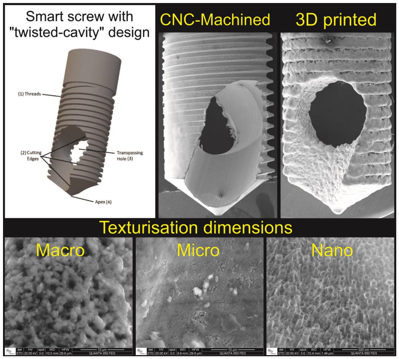
Figure 2.
Mesh of the nanotube structure.
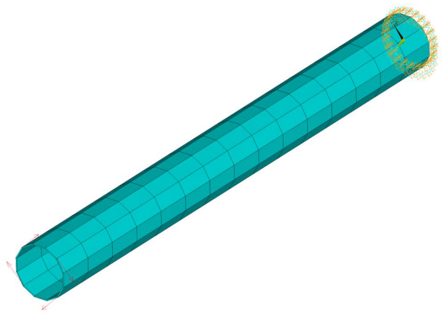
Figure 3.
Micrographs of CNC-machined and 3D-printed screws modified by PEO process.
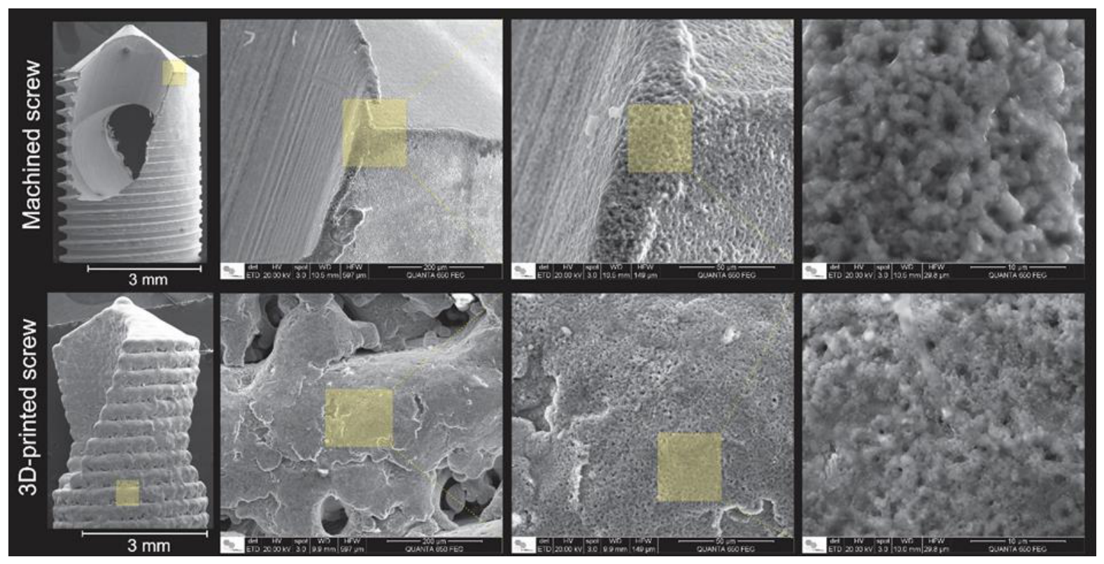
Figure 4.
Micrographs of CNC-machined BKS modified by the hard anodization process.
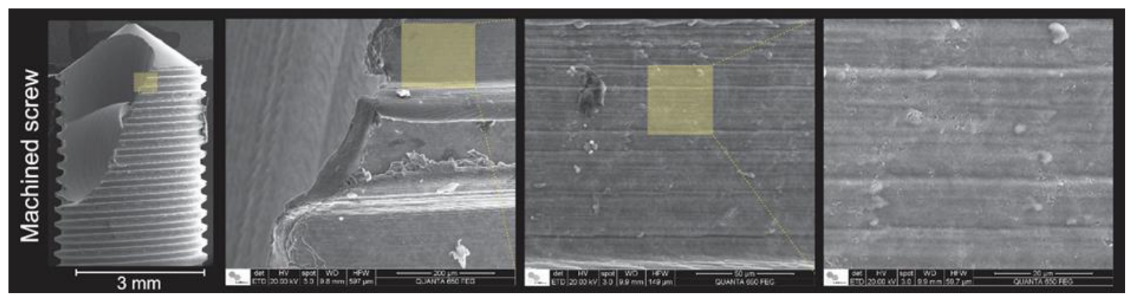
Figure 5.
Micrographs of 3D-printed with post-processing BKS modified by the soft anodization condition for TiO2 nanotubes production.
Figure 5.
Micrographs of 3D-printed with post-processing BKS modified by the soft anodization condition for TiO2 nanotubes production.
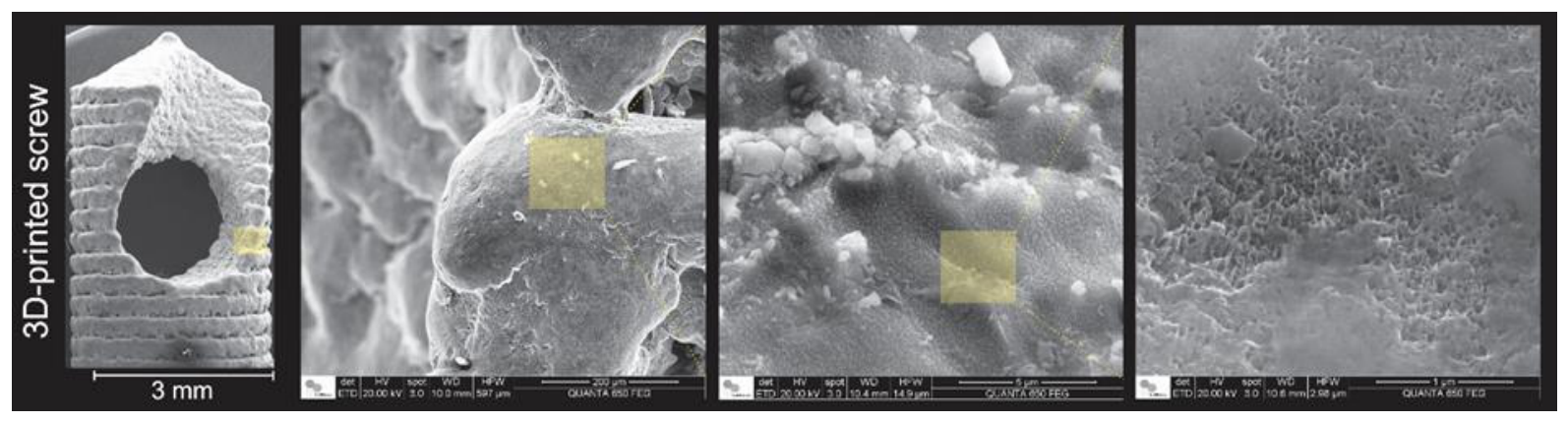
Figure 6.
Results of total displacements in the deformed shape (between min 0.005778 and max 1571.7, nm).
Figure 6.
Results of total displacements in the deformed shape (between min 0.005778 and max 1571.7, nm).
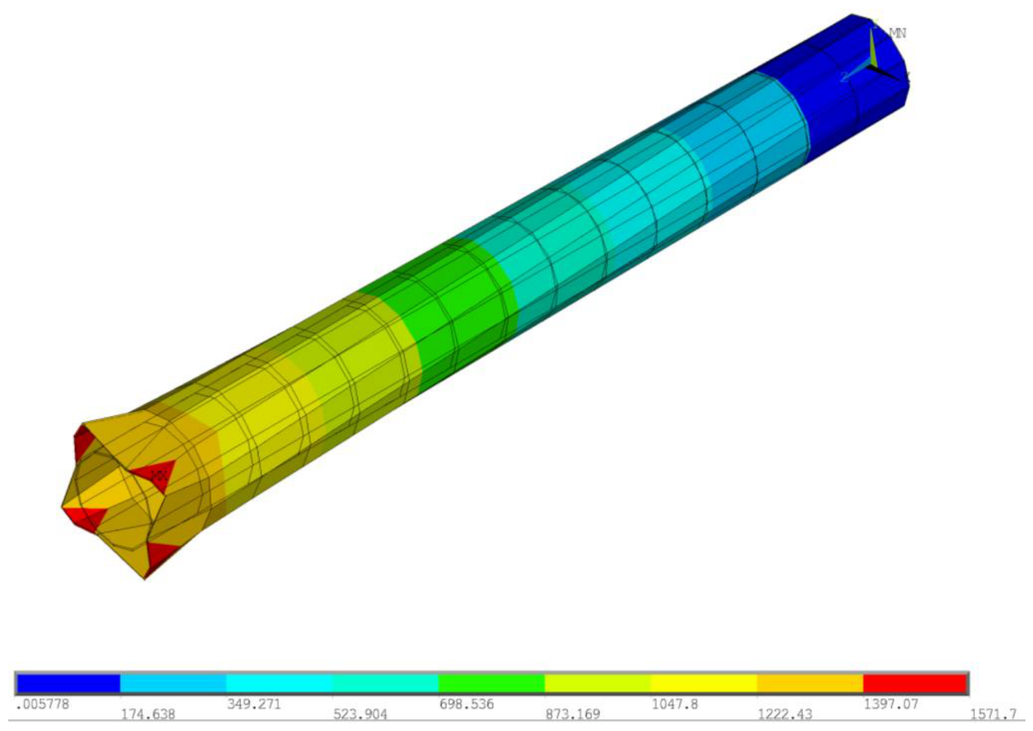
Figure 7.
Results of total rotations in the deformed shape (between min 0.0 and max 54.37, rad).
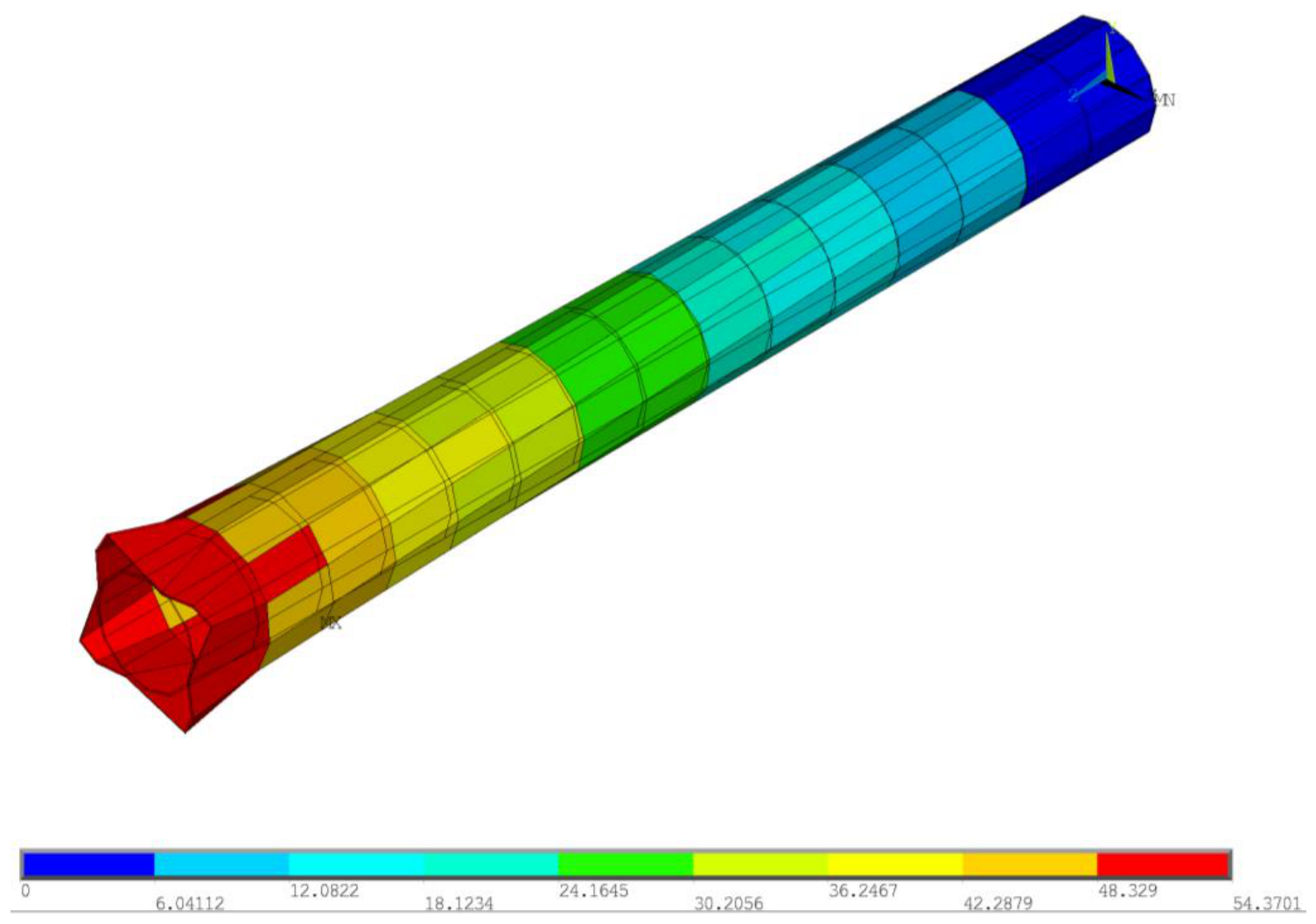
Figure 8.
Results of equivalent stresses in the deformed shape (between min 0.12E-10 and max 0.15E-8, N/nm^2).
Figure 8.
Results of equivalent stresses in the deformed shape (between min 0.12E-10 and max 0.15E-8, N/nm^2).
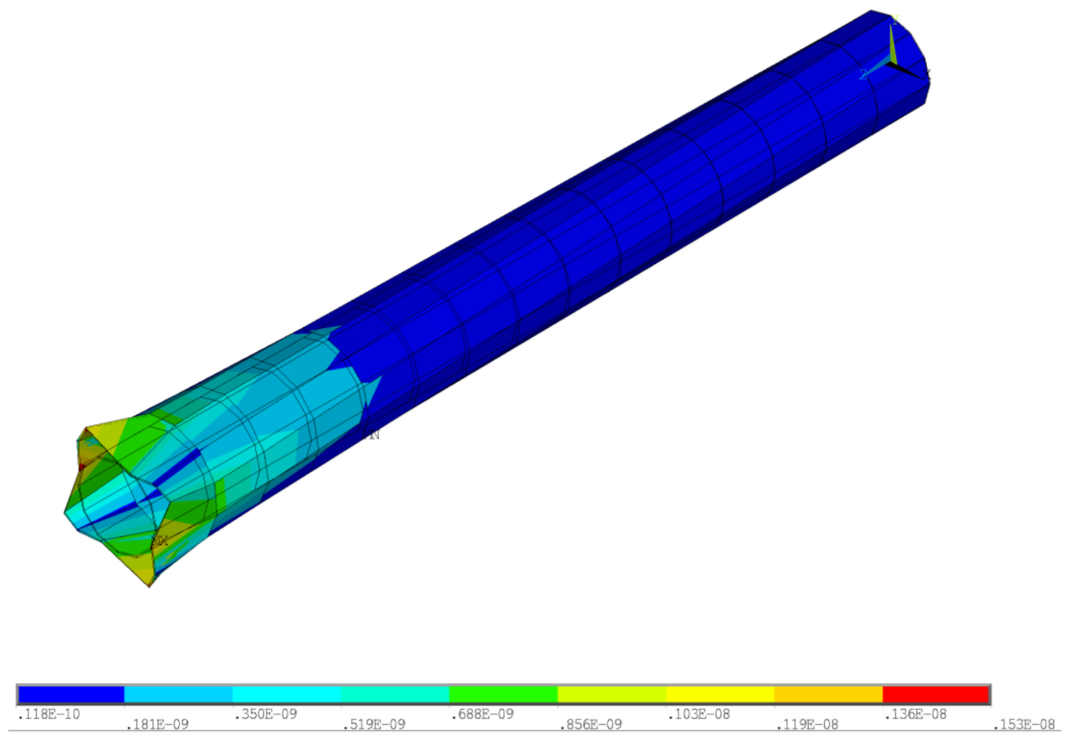
Figure 9.
Results of equivalent strains in the deformed shape (between min 0.027 and max 3.563).
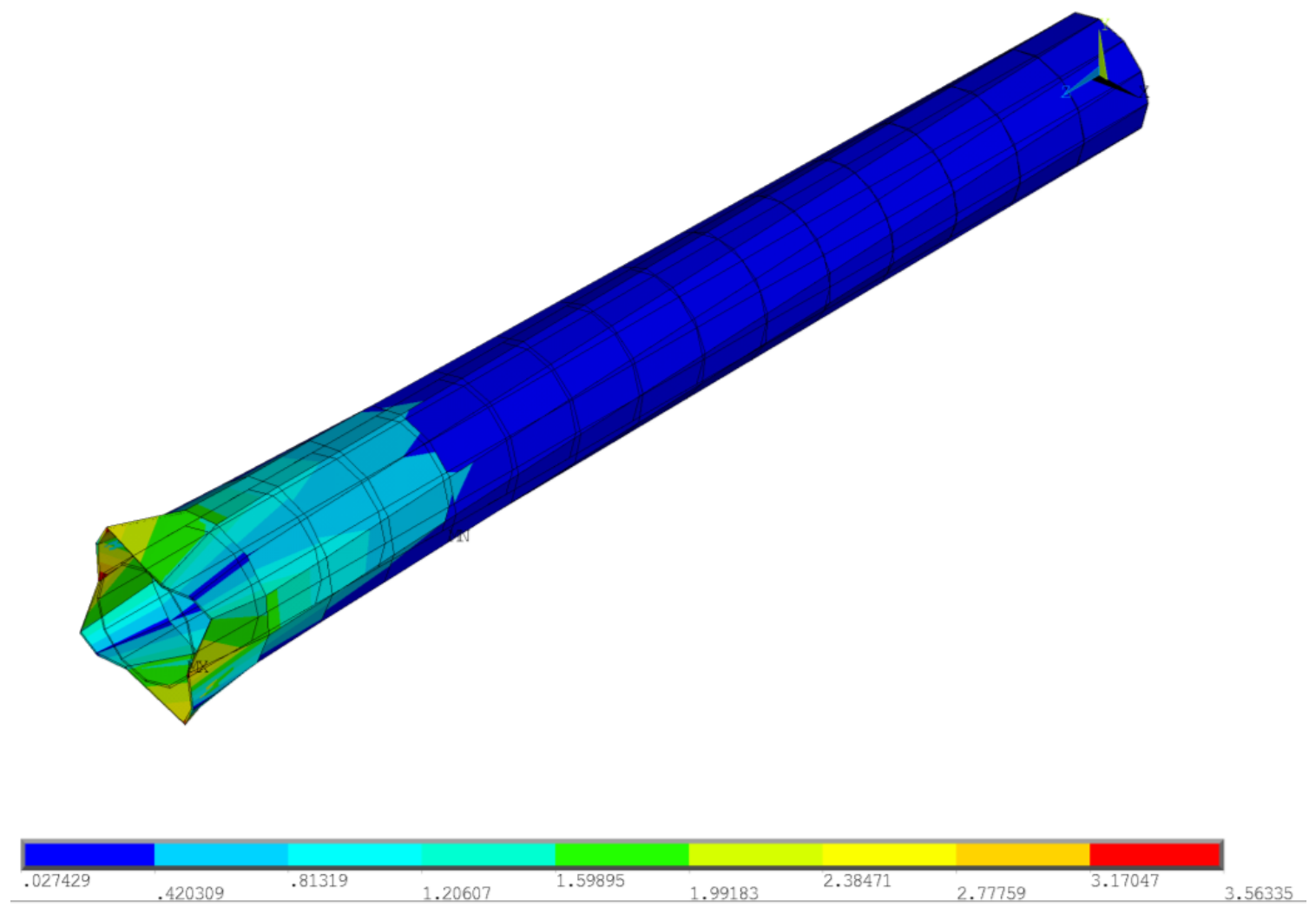
Figure 10.
(A) BKS screw depicting the layer-by-layer structure during powder metal deposition in 3D printing process. (B) and (C ) a macro-to-micro porous structure formations decorates the treated screw surface. (D) Comparison of the frontier region of a treated screw area and a non-treated screw area.
Figure 10.
(A) BKS screw depicting the layer-by-layer structure during powder metal deposition in 3D printing process. (B) and (C ) a macro-to-micro porous structure formations decorates the treated screw surface. (D) Comparison of the frontier region of a treated screw area and a non-treated screw area.
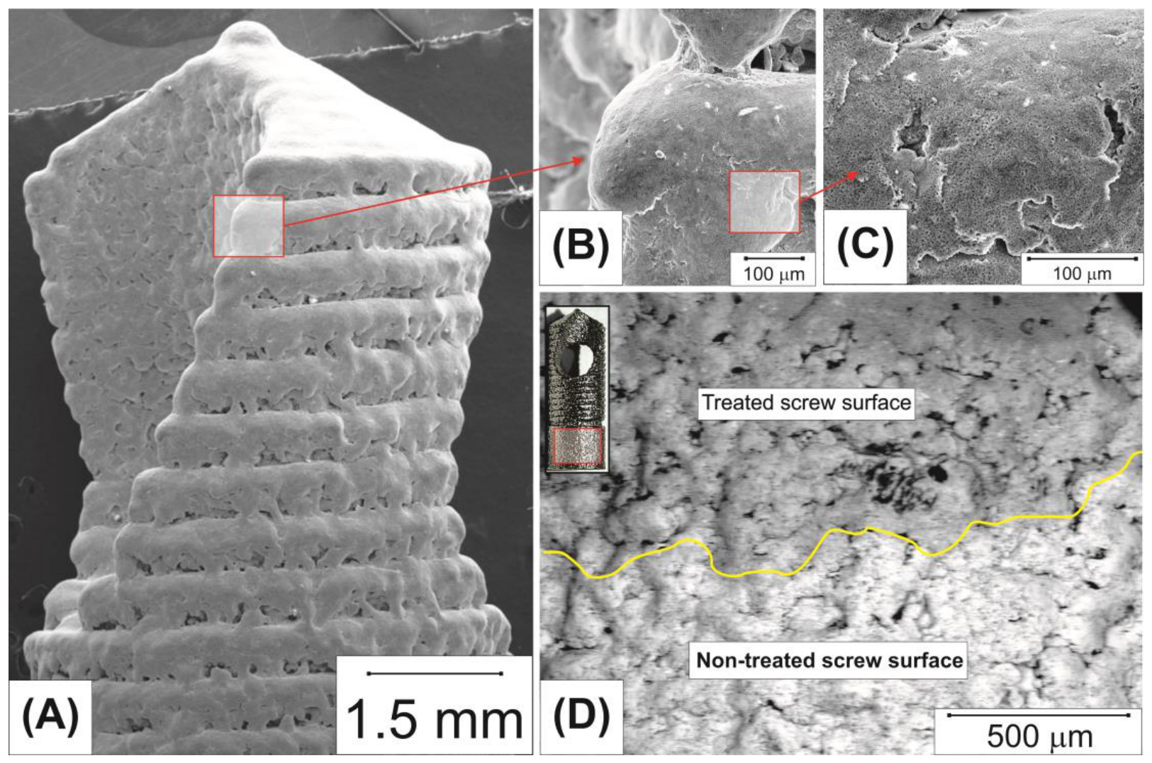
Figure 11.
CNC-machined BKS without finishing in subtractive manufacturing.
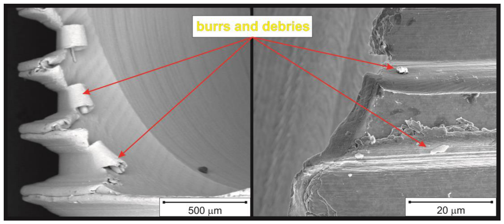

Table 1.
Oxidative electrochemical procedure conditions for different produced screws (BKS).
| Specimen | Screw type | Anodisation regime | Electrolyte composition | Electrolyte temperature | Applied Current/Voltage |
| Screw 1 | Machined | Hard anodisation (HA) | 0.1 Mol L-1 H2C2O4 | 15 °C | 10 mA cm-2 |
| Screw 2 | Plasma electrolytic oxidation (PEO) | 0.5 Mol L-1 H3PO4 | |||
| Screw 3 | 3D-printed | ||||
| Screw 4 | Soft anodisation (TiO2NTs) | 0.222 mol L-1 NH4F in Etileneglycol (10% H2O) | 50 °C | 20 V |
Disclaimer/Publisher’s Note: The statements, opinions and data contained in all publications are solely those of the individual author(s) and contributor(s) and not of MDPI and/or the editor(s). MDPI and/or the editor(s) disclaim responsibility for any injury to people or property resulting from any ideas, methods, instructions or products referred to in the content. |
© 2026 by the authors. Licensee MDPI, Basel, Switzerland. This article is an open access article distributed under the terms and conditions of the Creative Commons Attribution (CC BY) license (http://creativecommons.org/licenses/by/4.0/).
Copyright: This open access article is published under a Creative Commons CC BY 4.0 license, which permit the free download, distribution, and reuse, provided that the author and preprint are cited in any reuse.


