
Submitted:
01 July 2026
Posted:
02 July 2026
You are already at the latest version
Abstract
Autophagy is an evolutionarily conserved intracellular degradation pathway that has emerged as an important regulator of plant immunity and a recurrent target of pathogen virulence strategies. In this review, we examine how Candidatus Liberibacter (CL) species and their hosts provide a useful informative system for understanding convergent pathogen manipulation of the ATG6-ATG8 autophagic hub in agricultural and model plants. We synthesize recent evidence showing that CL-effectors reprogram autophagy through mechanistically distinct but functionally convergent strategies, including interference with ATG6- and ATG8-associated processes and exploitation of host metabolic regulators linked to autophagic flux. By comparing these mechanisms with autophagy targeting strategies reported for other plant-associated pathogens, we highlight recurring vulnerability nodes within the host autophagic machinery and suggest that these nodes may represent promising targets for future resistance engineering. We further discuss how structure guided molecular design and programmable genome editing could, over time, inform the development of more precise and durable approaches to crop protection.
Keywords:
autophagy
; ATG6-ATG8 Hub
; Candidatus Liberibacter asiaticus (CLas) effectors
; huanglongbing (HLB)
; metabolic hijacking
; synthetic immunity
1. Roles of Autophagy in Host-plant Immunity as a Defense System and a Pathogen Target
1.1. Autophagy and the ATG6-ATG8 Regulatory Hub in Plant Immunity
Intracellular bacteria of the genus Candidatus Liberibacter (CL) threaten several economically important crop systems and provide a useful pathosystem for examining how obligate pathogens manipulate host cellular processes (Saberi et al., 2024; Wang et al., 2017). Among these pathogens, CL asiaticus (CLas) is the principle causal agent of Citrus Huanglongbing (HLB), while related species broaden the relevance of this group across additional crops hosts, including potato, tomato, pepper, and tobacco (Haapalainen, 2014). A central theme emerging from recent work is that these pathogens can reshape host cellular homeostasis by targeting autophagy associated processes linked to immunity and stress adaptation (Lal et al., 2020; Leong et al., 2022b; Liu et al., 2005; Shi et al., 2023a). The ATG6-ATG8 autophagic hub is especially relevant in this context because it links autophagosome biogenesis, cargo recruitment, and immune associated cell fate control within a single regulatory space (Liu et al., 2024; Yuen et al., 2024; Zou et al., 2025).
Autophagy is a conserved intracellular degradation pathway in which cytoplasmic material is enclosed within autophagosomes and delivered to the vacuole for recycling. In plants, this process is controlled by core autophagy-related (ATG) proteins whose coordinated actions regulate intitiation, membrane expansion, cargo recognition, and vesicle maturation (Zhuang et al., 2024). Recent work continues to refine how these steps are organized in plant cells and how they respond to both abiotic and biotic signals (Li et al., 2024; Yuen et al., 2024). Under normal conditions, this process is tightly regulated by Target of Rapamycin (TOR) kinase that inhibits the formation of ATG1-ATG13 complex, often by phosphorylation of ATG13 preventing the interaction of ATG13 with ATG1 in a nutrient-rich environment. Under nutrient-scarce conditions, TOR is inactivated, which leads to de-phosphorylation of ATG13 and the formation of an active ATG1-ATG13 complex, thereby initiating the process of autophagy (Liu et al., 2021b; Qi et al., 2021; Suttangkakul et al., 2011; Wang et al., 2021; Yoshimoto and Ohsumi, 2018). Additionally, in response to carbon deficiency, KIN10, a catalytic subunit of Sucrose-nonfermentation1-Related Protein Kinase 1 (SnRK1), phosphorylates ATG6 to induce autophagy independently of TOR (Huang et al., 2019). Collectively, this core ATG machinery orchestrates autophagic flux to maintain cellular homeostasis, mediate stress responses, and regulate Programmed Cell Death (PCD). Recent evidence in A. thaliana demonstrates that, in addition to ATG6, a suite of proteins including ATG1, ATG8, ATG11, ATG13 and ATG14A are integral components of autophagosomes and associated vesicular structures. Further, in ConA-treated, nutrient-limited plants, all of the above ATGs, except ATG8, are transported to and accumulate in the vacuole. Additionally, they indicated that in nutrient-deprived cells, ATG6 and ATG8 tagged with fusion proteins Cyan Fluorescent Protein (CFP) and Yellow Fluorescent Protein (YFP), respectively accumulate in punctate-structures, as evidenced by their co-localized fluorescent signals in bright foci in A. thaliana (Qi et al., 2023). Overall, these findings place ATG6 and ATG8 within a closely connected regulatory axis is central to autophagosome formation and function in plant cells (Liu et al., 2024; Zou et al., 2025).
ATG6 and ATG8 are key regulators of the autophagy machinery with diverse roles in plant growth and development (PG&D) and cellular homeostasis (Ismayil et al., 2020; Leary et al., 2019; Yue et al., 2022). In A. thaliana, ATG6 has four splice variants and functions at the phagophore assembly site (Lamesch et al., 2012). It is essential for autophagosome assembly, nitrogen starvation responses, mitophagy, protein targeting to the vacuole, and late endosome-to-vacuole transport (Bu et al., 2020; Cao and Klionsky, 2007; Forde, 2002). Additionally, ATG6 plays a crucial role in plant adaptation to nutrient-scarce conditions and pathogen attack (Zhang et al., 2025). Loss-of-function mutations in ATG6 result in a complete blockage of autophagy, highlighting its critical role (Liu et al., 2021a). ATG8, a ubiquitin-like protein, is essential for autophagosome structure, cargo recognition, and recruitment of specific receptors such as NEIGHBOR OF BRCA1 (NBR1) and ATG8-interacting protein 1 (ATI1) through its ATG8-interacting motif (AIM), as well as recognition of ubiquitinated cargo via its ubiquitin-interacting motif (UIM) (Bu et al., 2020). In A. thaliana, nine isoforms of ATG8 (ATG8a–i) are expressed, and function at the autophagosomal membrane to mediate autophagosome formation and maturation (Bu et al., 2020). Simultaneous mutations in ATG8s isoforms can impair autophagosome formation, suggest their conserved and partially redundant roles in selective and non-selective autophagy (Bu et al., 2020). However, this phenotype is not yet confirmed in plants, only in yeast and Botrytis cinerea, where a single copy of ATG8 is present (Kirisako et al., 1999; Ren et al., 2018). Conversely, over-expression of ATG8 significantly enhances the autophagosome formation in A. thaliana (Chen et al., 2019) and O. sativa (Yu et al., 2019). Invading pathogens, in turn, manipulate autophagy to evade host defense, highlighting ATG8’s role in host-plant immunity (Bu et al., 2020). In a nutshell, ATG6 and ATG8 support cellular homeostasis while also shaping the immune functions of autophagy during plant stress and pathogen challenge (Jeon et al., 2025). Recent work in citrus further supports the immune relevance of this autophagic hub by showing that ATG8 mediated selective autophagy can directly limit CLas through effector targeting and degradation (Cui et al., 2025) (Figure 1, a).
1.2. The Evolutionary “Tug-of-War”: Autophagy as a Conserved Defense Strategy
During pathogen and host-plant interactions, immunity is initiated through Pattern-Triggered Immunity (PTI) and Effector-Triggered Immunity (ETI) responses together shape the downstream defense responses (Bjornson et al., 2021; Laflamme et al., 2020; Remick et al., 2023; Thordal-Christensen, 2020; Yuan et al., 2021). Autophagy acts alongside these immune pathways as a highly regulated process that influences defense at infection sites as well as in surrounding tissues (Kim et al., 2012; Lal et al., 2020; Zhang et al., 2025). Rather than functioning only as a bulk recycling pathway, plant autophagy can selectively influence immune signaling, pathogen restriction, and the spatial control of cell death (Jeon et al., 2025; Üstün et al., 2018). In the evolutionary Tug-of-War, autophagy restricts invading pathogens through various mechanisms, including degrading viruses and limiting bacterial and fungal infections (Leary et al., 2019). Autophagy shapes host plant immunity by regulating cell death and defense responses. A key contribution of autophagy to immunity is its ability to modulate Hypersensitive Response of PCD (HR-PCD). It can support local defense associated cell death at the infection site (Hofius et al., 2017) while also preventing its uncontrolled spread to adjacent cells and distal unaffected tissues (Liu et al., 2005). Autophagy also modulates plant resistance to biotrophic and necrotrophic pathogens via SA and JA pathways and plays a role in preventing virus-induced RNA silencing (Zhou et al., 2014). Autophagy also interact with broader defense signaling network, including pathways that influence cell wall reinforcement (e.g., lignin modification), hormone balance, and redox homeostasis during infection. In atg mutants, lignification and innate immune responses are impaired. Autophagy facilitates lignin transportation, thereby enhancing disease resistance (Jeon et al., 2023). In some cases, host selective autophagy machinery targets pathogens for degradation. For example, selective autophagy receptor such as NBR1 can binds with pathogen associated cargo to ATG8 labelled membranes for vacuolar degradation, demonstrating how autophagy can directly counter effector action (Leary et al., 2018; Leong et al., 2022b). These observations highlight autophagy as an active interface between host defense and pathogen counter defense rather than a passive recycling route (Jeon et al., 2025; Üstün et al., 2018) (Figure 1, b).
1.3. Strategic Hijacking of Autophagy during Plant Pathogen Interactions
Autophagy has context dependent roles in plant immunity and can either restrict or facilitate disease progression depending on the pathogen and stage of infection. Because autophagy influences both cellular survival and defense associated cell death, its perturbation can have beneficial or detrimental consequences for the host. Across bacteria, fungi, oomycetes, and nematodes, diverse pathogens have evolved effectors that redirect autophagy toward either suppression or selective activation in ways that favor infection (Lal et al., 2020; Sertsuvalkul et al., 2022; Testi et al., 2024; Zou et al., 2023). Autophagy can be induced or suppressed during infection depending on the pathogen strategy and stage of host colonization. In parallel, HR-PCD in response is typically restricted to the infection site as part of the host defense response. Interestinglly, pathogen success often depends not on autophagy as a whole, but on access to specific vulnerability hubs that control cargo selection, membrane dynamics, or immune signaling response (Cui et al., 2025; Liu et al., 2024; Yuen et al., 2024).
The strategic suppression of autophagy is a conserved hallmark of successful infection across diverse bacterial species. For instance, Xanthonomonas campestris pv. Vesicatoria (Xcv) utilizes its type III effector XopL to interact with and degrade SH3P2, a protein essential for autophagosome formation, thereby effectively silencing the host’s autophagic defense (Leong et al., 2022b; Zhuang et al., 2013). Similarly, Pseudonomonas syringae pv. tomato DC3000 (Pst) employs a multi-pronged attack on the autophagy core. Pst effector HopF3 directly targets ATG8, while AvrPtoB inhibits ATG1 kinase activity to prevent the initiation of autophagic flux (Lal et al., 2020). Considered together, these system show that distinct effectors can converge on core autophagy associated processes to reshape immunity in plants (Lal et al., 2020; Testi et al., 2024; Üstün et al., 2018).
ATG6 appears to be especially important for linking autophagy to the spatial control of HR-PCD and pathogen responsive stress signaling. Perturbation of ATG6-dependent autophagy can uncouple defense activation from cell death containment, thereby altering disease outcomes (Liu et al., 2024). ATG6-deficient plants experienced uncontrolled PCD in distal tissues despite normal HR initiation at the infection site, with reduced autophagy (Liu et al., 2005). Both, disrupted and excessive autophagy lead to cell death, highlighting the importance of regulating autophagy in immunity responses. ATG6 expression increases during early infection of Pst in A. thaliana, indicating its role in pathogen response. In ATG6-antisense plants, HR is induced, but Pst spreads, misregulating cell death (Patel and Dinesh-Kumar, 2008). In Arabidopsis, ATG6 abundance is subject to regulatory proteolysis, reinforcing the view that control of ATG6 stability is itself an important layer of autophagy regulation (Liu et al., 2024).
ATG8 is a recurrent effector target across multiple pathosystems, and recent work continues to support its role as both a cargo recruitment factor and a node of pathogen interference. Pathogen effectors can bind ATG8 family members either to suppress immune associated atutophagy or to exploit selective autophagy toward pathogen benefit (Lal et al., 2020; Testi et al., 2024; Zou et al., 2025). Interestingly, oomycete pathogens such as Phytophthora infestans release the PexRD54 effector protein, which directly binds to ATG8CL to induce autophagosome formation (Dagdas et al., 2016; Lal et al., 2020; Shi et al., 2023b). In N. benthamiana, autophagosome formation increases after Citrus leaf blotch virus (CLBV) infection. In ATG5 and ATG7 mutants, CLBV spreads more easily, as autophagy targets CLBV’s movement proteins for degradation. ATG8 isoforms bind with CLBV movement proteins, suppressing the virus (Niu et al., 2021). Pathogens can evade or hijack autophagy for their benefit (Hofius et al., 2017; Leary et al., 2018). Root-knot nematode (RKN) infection in tomatoes induces negative regulators like JAM1/2/3, interfering with jasmonic acid (JA)-mediated defense genes. Autophagy-inducing ATGs such as ATG13b, ATG8a, and ATG8d repress JAMs, increasing defense gene expression and resistance to RKN (Zou et al., 2023).
Cytosolic Glyceraldehyde-3-Phosphate (G3P) dehydrogenase (GAPDH), designated GAPC, interacts with ATG3 to regulate autophagy and influence plant immunity (Han et al., 2015). The E. coli and C. rodentium effector NleB O-GlcNAcylates GAPDH and disrupts the activation of GAPDH-mediated transcription factors (TFs) involved in the regulation of host-plant innate immunity (Gao et al., 2013). Meloidogyne incognita, a nematode pathogen, secretes the MiEFF1 effector, which interacts with GAPC to promote parasitism in A. thaliana (Shi et al., 2023b). Arabidopsis autophagy mutants (e.g., atg5, atg7, atg10, and atg18a) show compromised resistance to necrotrophic fungi such as Alternaria brassicicola and B. cinerea in line with a positive role of autophagy in antifungal defense. This succeptibility is associated with disrupted defense regulation and likely reflects, at least in part, altered crosstalk between salicylic acid and JA signaling pathways (Davière et al., 2025; Kim et al., 2012; Lai et al., 2011; Lenz et al., 2011) (Figure 1, c).
Taken together, the above findings support the view that the ATG6-ATG8 hub functions as a recurrent site of pathogen interference during plant intracellular pathogenesis. In this review, we use the Liberibacter host intercation as the promising case study and place it in the broader context of autophagy targeting strategies reported across plant-associated pathogens. Our aim is to highlight recurring mechanistic themes, clarify key vulnerability nodes within the autophagy network, and consider how these insights may inform future resistance engineering.
2. Mechanistic Convergence: How Liberibacter Effector Reprogram the Autophagic Hub?
The reliance on the Sec-translocon and T1SS for effector delivery is a hallmark of the genus CL, shared by CLas, CLam, and CLaf (Duan et al., 2009; Lin et al., 2015; Wulff et al., 2014). These reduced-genome pathogens have evolved a streamlined suite of Sec-delivered effectors (SDEs) that prioritize the manipulation of core host survival pathways over the complex multi-effector arsenals seen in T3SS-bearing bacteria (Lovelace et al., 2025; Prasad et al., 2016). By targeting the ATG6-ATG8 complex, these effectors achieve a high degree of “functional leverage”, allowing a single protein to modulate multiple layers of host immunity and metabolism simultaneously (Shi et al., 2023a; Shi et al., 2023b; Shi et al., 2025; Zhang et al., 2019). Specifically, the mature effectors m3875 (Zhang et al., 2019), SDE4405 (Shi et al., 2023a), and SDE3 have emerged as master regulators of the autophagic flux (Shi et al., 2023b). By either inducing or suppressing autophagy, these CEPs create a bimodal ‘Tug-of-War’ that results in pro-survival or pro-death outcomes, ultimately impairing the host’s HR-PCD (Shi et al., 2023a; Shi et al., 2023b; Zhang et al., 2020; Zhang et al., 2019). This manipulation of the autophagic machinery has been reported across diverse plant species, including, A. thaliana, N. benthamiana, and C. sinensis, highlighting the universal nature of these pathogen strategies. The following sections detail the specific molecular mechanisms by which these effectors reprogram host immunity.
2.1. m3875 and BI-2 Intercation: Hijacking the Intracellular Death Switch
Intracellular pathogens must delicately navigate the host’s PCD pathways to ensures a stable niche for replication. In both plants and animals, Bax Inhibitor-1 (BI-1)/ATG6 axis serves as a conserved ‘Intracellular Death Switch’, regulating the transition between pro-survival autophagy and pro-death apoptosis (Castillo et al., 2011; Zhang et al., 2019). This switch is a primary target for the CLas effector m3875, which hijacks the host’s regulatory machinery to stall the HR-PCD (Zhang et al., 2019). This strategy mirrors how viral BCL2 proteins or human pathogens like Salmonella manipulate BECN1 (the mammalian ATG6 homolog) to evade autophagic clearance while scavenging host resources (Jiao et al., 2020; Pattingre et al., 2005; Sinha et al., 2008). By targeting this universal checkpoint, CL species achieve high-level ‘functional leverage’ over host cellular homeostasis (Fu et al., 2025; Huang et al., 2024; Shi et al., 2023a; Zhang et al., 2020; Zhang et al., 2019). Similarly, in plants, BI-1 modulates HR-PCD by influencing ROS levels, Ca²⁺ flux, and lipid dynamics while responding to various abiotic and biotic stresses (Babaeizad et al., 2009; Duan et al., 2010; Kawai-Yamada et al., 2001; Watanabe and Lam, 2006; Watanabe and Lam, 2009). Loss of BI-1 function leads to hypersensitivity to stresses, whereas its overexpression suppresses cell death. In A. thaliana, BI-1 specifically regulates ER stress-mediated HR-PCD and plays a complex role in autophagy regulation (Ihara-Ohori et al., 2007; Ishikawa et al., 2011; Kawai-Yamada et al., 2009; Lisbona et al., 2009; Watanabe and Lam, 2008). Xu et al. (2017) revealed that BI-1 localizes within ER structures, where it interact with ATG6, core autophagy-related protein and is mainly found in ER-associated puncta (Xu et al., 2017). In N. benthamiana, NbBI-1 interacts with NbATG6 via its C-terminal 14 amino acids and the N-terminal 192 amino acids of ATG6 (Xu et al., 2017). Overexpression of NbBI-1 and AtBI-1 induces cell death phenotypes in N. benthamiana, suggesting a pro-death role under certain conditions. Additionally, overexpression of AtBI-1 increased autophagic structures, as indicated by CFP-ATG8F labeling, suggesting a positive effect on autophagy. Conversely, silencing of ATG6, PI3K, and ATG7 significantly reduced BI-1-induced HR-PCD, highlighting the essential role of autophagy in BI-1-mediated cell death (Xu et al., 2017). In N. benthamiana, silencing of BI-1, reduces autophagy activity triggered by N gene-mediated resistance to Tobacco mosaic virus (TMV) and methyl viologen, while enhancing N gene-mediated cell death. These findings suggest that plant BI-1 positively regulates autophagy machinery under specific conditions, and its modulation could be exploited by pathogens.
Mechanistic modeling demonstrates that m3875 functions as a transcriptional reprogrammer, specifically upregulating the expression of NbBI-2 (the N. benthamiana homolog of A. thaliana BI-1) to induce a state of ‘defensive autophagy’ (Zhang et al., 2019). By driving NbBI-2 expression, CLas exerts precise control over host cellular homeostasis, acting as a molecular brake on Ca2+-mediated HR-PCD through the modulation of cyclic nucleotide-gated channels (NbCNGC23-26) (Zhang et al., 2019). Consequently, the pathogen hijacks this conserved ‘death switch’ to create a pro-bacterial survival platform, inducing autophagic flux while strategically blocking the recruitment of the selective cargo receptor NBR1, a definitive hallmark of evasive hyper-autophagy (Zhang et al., 2019). Overexpression of m3875 resulted in dwarfing and leaf deformation phenotypes, indicating a significant role in plant growth and development (Zhang et al., 2019). Transcriptome analysis further revealed that transient overexpression of m3875 modulated the transcription of defense-related genes, including NbCNGC23-26 (homologs of AtCNGC2/4), NbWRKY9 (a homolog of AtWRKY33 TF involved in defense signaling), and NbBI-2. Since NbCNGC23-26 are positive regulators of HR-PCD, their inhibition likely suppresses Ca²⁺-mediated cell death, representing a pathway exploited by these CEPs (Balagué et al., 2003; Clough et al., 2000). Additionally, while AtWRKY33 is known for its roles in ROS accumulation and defense against necrotrophic fungi, it is less effective biotrophic bacteria like CLas (Adachi et al., 2015; Huang et al., 2012). Although BI-1 is a known HR-PCD suppressor, its transient overexpression paradoxically induces cell death (Xu et al., 2017). The m3875-induced upregulation of NbWRKY9 and NbBI-2 may contribute to the suppression of HR-PCD, thereby aiding CLas infection. Notably, m3875 did not alter BI-1 transcription, suggesting distinct and specialized roles for NbBI-1 and NbBI-2 in susceptibility to biotrophic pathogens (Zhang et al., 2019).
2.2. SDE4405 and ATG8c: Induction of Evasive Hyper-Autophagy
ATG8, a core autophagy protein, is frequently targeted by pathogen effectors, with 88 out of 174 effectors shown to interact directly with ATG8 (Lal et al., 2020). Shi et al. (2023) demonstrated that SDE4405 interacts with CsATG8c to enhance autophagy in Citrus, with CsATG8c sharing 97% similarity with NbATG8c, suggesting a similar interaction in N. benthamiana (Shi et al., 2023a). To monitor autophagy activity, using autophagy markers such as CFP-ATG8f and GFP-CsATG8c, co-expression of SDE4405 with CsATG8c significantly increased autophagic structures. Further accumulation upon treatment with the autophagy inhibitor E-64d, confirms that the interaction promotes autophagic flux (Moriyasu and Inoue, 2008; Shi et al., 2023a). Additionally, reduced levels of NBR1, an autophagy cargo receptor was inversely correlated with autophagy activity, further supporting this interaction (Shi et al., 2023a; Svenning et al., 2011). Cell structural observations using transmission electron microscopy of SDE4405-transgenic Citrus leaves treated with E-64d showed increased autophagosomes and autophagic bodies, providing morphological evidence of upregulated autophagy activity. Functional analysis uncovered that NbATG8c-silenced plants exhibit reduced proliferation of Pto DC3000ΔhopQ1-1 in N. benthamiana, while transiently overexpressed Citrus plants of CsATG8c and SDE4405 showed the enhanced growth of Xanthomonas citri subsp. citri (Xcc). In a nutshell, the above findings suggest that the interaction of SDE4405-ATG8c induces autophagy, negatively regulates plant immunity and promotes bacterial infection (Figure 2, a).
3. The Metabolic Hijacking Paradigm: Disruption of Autophagy Machinery
3.1. GAPC1/2: Metabolic Sensors as Negative Regulators of Autophagic Flux
Cytosolic Glyceraldehyde-3-phosphate dehydrogenases (GAPCs) function as essential metabolic sensors that ‘moonlight’ as master negative regulators of the autophagic flux functions by bridging primary metabolism and innate immunity, GAPCs act as a regulatory swicth (Garcin, 2019). Their physical interaction with the core protein ATG3 serves to repress constitutive autophagy under normal conditions. This ‘moonlighting’ role makes GAPCs a prime vulnerability hub for pathogens seeking to reprogram host homeostasis for their own survival (Han et al., 2015).
Han et al. (2015) were the first to report the intricate link between GAPCs and autophagy in N. Benthamiana (Han et al., 2015). Their results showed that GAPCs interact with ATG3 to negatively regulate the autophagy machinery. Furthermore, they demonstrated that ROS stress inhibits the interaction between GAPCs and ATG3. Notably, N. benthamiana plants silenced for GAPC1/2 exhibited an increased autophagosome formation, indicating upregulated ATG3-mediated autophagy. Conversely, overexpression of GAPCs led to reduced autophagy activity. These findings suggest that GAPCs function as suppressors of autophagy, likely through their physical interaction with the ATG3 protein (Han et al., 2015). In addition, Shi et al. (2023) demonstrated that in GAPC-silenced plants, the insertion of CFP-NbATG8f, an autophagy marker, resulted in a significant accumulation of CFP-positive puncta, indicative of increased autophagosome formation (Shi et al., 2023b). They observed similar results in plants where multiple GAPC isoforms were silenced simultaneously, suggesting a degree of functional redundancy among GAPC isoforms in negatively regulating autophagy. A similar finding was previously reported in A. thaliana, where GAPC1/2 isoforms were shown to regulate autophagy (Henry et al., 2015). Under nitrogen starvation conditions, a known inducer of autophagy, GAPC knockout plants exhibited constitutive autophagy, indicating that GAPCs suppress basal autophagy levels (Henry et al., 2015). Moreover, overexpression of CsGAPCs in A. thaliana resulted in the accumulation of MDC-labeled autophagic bodies, further confirming reduced autophagy (Shi et al., 2023b). In the same transgenic lines, increased levels of NBR1 (a cargo receptor for selective autophagy) were detected in the vacuole, a typical marker of suppressed autophagy when accumulated. Overall, these findings across different plant species highlight a conserved role of cytosolic GAPCs in the negative regulation of autophagy.
3.2. SDE3-GAPC Intercation: Dismantling the Autophagy Machinery through Metabolic Co-option
Shi et al. (2023) demonstrated that SDE3, a virulence-associated CEP, is responsible for promoting HLB disease in Citrus (Shi et al., 2023b). Their findings revealed that SDE3-overexpressing sweet orange plants exhibited increased susceptibility to both HLB and Citrus canker. To further explore its impact, their study of transgenic A. thaliana and N. benthamiana plants expressing SDE3 also showed enhanced susceptibility to viral and Phytophthora infections.
Autophagy is a crucial component of the host immune system, playing a pivotal role in antimicrobial defense mechanisms against various pathogens (Kim et al., 2012; Shi et al., 2023b). Consequently, plant pathogen-secreted effector proteins frequently target the host autophagy machinery, leading to the evolution of diverse strategies to manipulate this vital process, either by suppression or by co-opting it for their advantage (Lal et al., 2020). Specifically, SDE3 inhibits autophagic activity, disrupting its normal function, which results in increased susceptibility of SDE3-transgenic plants to CLas and other pathogens (Shi et al., 2023b). The diverse and sophisticated molecular strategies by which effectors from CL species and a broad spectrum of model intracellular pathogens, ranging from agricultural threats such as X. capestris and P. syringae, and P. infestans to vector-associated CLso and mammalian pathogens like E. coli and Salmonella, manipulate the conserved ATG6-ATG8 autophagic hub are summarized in Table 1.
3.2.1. Interaction of SDE3 and GAPCs Impacts on Relocation and Degradation of Autophagy-mediated Host-plant Immunity
Mechanistic modeling reveals that the CLas effector SDE3 executes a precise ‘Metabolic Hijacking’ by physically co-opting host CsGAPCs to dismantle the autophagy machinery. This strategic association does not merely inhibit flux. It creates a synergistic platform that targets ATG8 isoforms for selective degradation. By subverting a central glycolytic enzyme, SDE3 accelerates the breakdown of the ATG8-PE complex, thereby disrupting autophagosome maturation and effectively silencing the host’s primary line of antimicrobial defense. This hijacking of a metabolic hub provides functional leverage for the pathogen, allowing it to evade autophagic clearance while potentially redirecting metabolic resources toward its own proliferation (Shi et al., 2023b). This suggests a synergistic effect between SDE3 and CsGAPCs, stabilizing a molecular mechanism that disrupts key components of the autophagy machinery. Furthermore, they discovered that CsATG8 degradation was largely rescued by treatment with E64d, an inhibitor of proteases involved in the late autophagic pathway, indicating that ATG8 degradation is dependent on autophagic turnover itself. This finding points to a potential feedback loop in which GAPC enzymatic activity, possibly modulated by SDE3, promotes the selective breakdown of ATG8 proteins, thereby reducing autophagy. Additionally, the physical interaction between CsGAPC1 and NbATG8f altered NbATG8f subcellular localization, potentially contributing to further disruption of autophagy (Shi et al., 2023b). In summary, SDE3-mediated manipulation of GAPC activity effectively disrupts critical components of the autophagy machinery, leading to the inhibition of autophagy-mediated host-plant immunity (Figure 2, b).
3.2.2. Implications Following Disruption of Autophagy-balance for Pathogen-survival and Host-plant Immunity
Maintaining the autophagy balance is crucial for proper functioning of host-plant innate immune system; disturbance to this homeostasis, either through its inhibition or induction, can significantly impact on host-plant defense and influence the outcome of pathogen and host-plant interactions (Hofius et al., 2017; Leary et al., 2018; Leong et al., 2022b). These consequences can occur in both autophagy-deficient plants and those with hyper-active autophagy.
A reduced autophagy pathway, often resulting from alterations in ATG genes or pathogen effector-induced suppression, can lead to HR-PCD associated with ETI (Leary et al., 2018; Liu et al., 2005). The literature provides evidence that in autophagy-deficient plants, HR-PCD can expand beyond the site of infection, affecting adjacent uninfected tissues and, in some cases, even distal tissues (Liu et al., 2005). For example, in ATG6-silenced N. benthamiana plants infected with TMV, expanded HR-PCD was observed (Liu et al., 2005). Similar results were reported in plants with suppressed expression of ATG3, ATG7, and VPS34 (Liu et al., 2005). These findings indicate that autophagy is crucial for restricting HR-PCD to the site of infection during the host-plant’s innate immune response. It is possible that in autophagy-deficient cells, the signals required to initiate or limit HR-PCD are not properly perceived (Liu et al., 2005). However, our understanding of the role of autophagy in HR-PCD remains complex, as additional factors, such as the plant’s developmental stage and age, as well as the specific pathogen involved, may influence outcomes. A few studies have reported either impaired or delayed cell-death responses in plants with reduced autophagy activity upon infection with avirulent pathogens (Hofius et al., 2009; Yoshimoto et al., 2009).
CLas, through the SDE3-GAPC interaction, promotes the degradation of ATG8 proteins, and leads to autophagy suppression (Shi et al., 2023b). This suppression benefits CLas by increasing its survival and proliferation within the host plant, allowing it to evade autophagy-mediated clearance and potentially overcome HR-PCD responses (Shi et al., 2023b). Conversely, the host plant defense system relies on an activated and functional autophagy machinery to counter pathogen invasion (Cao and Klionsky, 2007; Han et al., 2015; Kim et al., 2012; Lal et al., 2020; Shi et al., 2023a; Shi et al., 2023b; Xu et al., 2017). As mentioned in the previous section, GAPC-silenced plants (where GAPCs, negative regulators of autophagy, are suppressed) exhibited enhanced autophagy levels, resulting in improved resistance against pathogens and an increased HR-driven cell death response (Han et al., 2015; Henry et al., 2015). These findings highlight the importance of maintaining an appropriate autophagy balance for robust defense mechanisms. On the other hand, CEP-induced modulation of this balance underscores a constant tug-of-war between pathogens and host plants (Lal et al., 2020; Shi et al., 2023b; Zhang et al., 2020; Zhang et al., 2019). Since ROS are known inducers of autophagy in some contexts, the role of GAPCs in ROS signaling further complicates this crosstalk (GAPCs-Autophagy, ROS-Autophagy, and GAPCs-ROS) (Baek et al., 2008; Han et al., 2015; Henry et al., 2015; Shi et al., 2023b). This raises the possibility that SDE3-mediated hijacking of GAPC function could also impact ROS levels, indirectly influencing the process of autophagy (Baek et al., 2008; Han et al., 2015; Henry et al., 2015; Shi et al., 2023b). As noted earlier, the enhanced resistance of GAPC-silenced plants, even against compatible pathogens, suggests that GAPCs play a critical role in autophagy-driven basal defense mechanisms (Han et al., 2015; Henry et al., 2015; Shi et al., 2023b).
Another important consequence of reduced autophagy is its interference with host-pants innate or basal immunity, affecting resistance to a variety of pathogens (Hofius et al., 2017; Lal et al., 2020; Leary et al., 2018; Lenz et al., 2011; Leong et al., 2022b; Levine and Kroemer, 2019). Generally, autophagy contributes positively to resistance against nectrotrophs by enhancing defense responses (Leary et al., 2018). Since, nectrotrophs feed on dead host tissue, plants with deficient autophagy, lack autophagy-associated defense mechanisms, often exhibit enhanced sensitivity to HR-PCD and increased susceptibility (Leary et al., 2018; Liu et al., 2005; Yoshimoto et al., 2009). For instance, in A. thaliana infected with B. cinerea, the cleavage of BCL2-associated athanogene 6 (BAG6, involved in various cellular processes, including protein quality control and cellular homeostasis) protein promotes autophagy and helps limit disease progression (Hofius et al., 2017; Li et al., 2016). In contrast, the role of autophagy in biotrophs, is more complex and can be contextually negative. As biotrophs rely on living host tissue, autophagy-induced pre-mature or excessive cell death may actually facilitate their growth and spread (Haxim et al., 2017). For example, in A. thaliana, ATG2 has been shown to negatively affect resistance against powdery mildew, suggesting that autophagy may, in some cases, compromises biotrophic pathogen resistance (Wang et al., 2011).
It is noteworthy that SDE3 represents one of the mechanisms used by pathogens to suppress the host-plant immune system (Shi et al., 2023b); however, pathogens may employ a variety of different mechanisms to alter autophagy for their benefit (Adachi et al., 2015; Förderer et al., 2022; Henry et al., 2015; Lal et al., 2020; Zhang et al., 2024; Zhang et al., 2020; Zhang et al., 2019). For example, Legionella pneumophila, an intracellular bacterial pathogen, secretes the effector RavZ through its TIVSS system. RavZ interacts with ATG8 to disrupt the autophagy process. Similarly, the effector proteins AvrPtoB, HrpZ1, HopF3, and HopM1, secreted by Pto, manipulate autophagy by targeting core autophagy machinery related proteins and process, such as ATG1, ATG4, ATG8, and autophagic flux, respectively, to facilitate bacterial infection (Lal et al., 2020; Shi et al., 2023a). Conversely, NBR1-driven selective autophagy counteracts this bacterial infection reducing the formation of water-soaked lesions induced by HopM1 through the unknown mechanisms (Üstün and Hofius, 2018). Interestingly, several other bacterial pathogens, such as C. rodentium, S. typhimurium, Xcv, and X. oryzae also releases their effecors i.e., NleB, SopF, XopL, and harpins (similar to HrpZ1) to modulate autophagy-driven immune responses through disrupting GAPDH-dependent TF activation, preventing the interaction between V-ATPase and ATG16L1, and other influences on the autophagy processes to facilitate bacterial spread and propagation (Henry et al., 2015; Leong et al., 2022b; Shi et al., 2023a; Üstün and Hofius, 2018; Zwack et al., 2015). The CEP, SDE3 has been shown to suppress autophagy by inducing the degradation of Citrus ATG8s, and to impact negatively host-plant innate immunity (Shi et al., 2023b). Likewise, CaMV protein P6 (encoded by gene VI) suppresses autophagy to shield the viral replication machinery from autophagic degradation (Hafrén et al., 2017). Collectively, these findings highlight that pathogen effector-mediated disruption of autophagy contributes to increased pathogen infection and enhanced disease susceptibility in the host-plant.
Conversely, some pathogens promote their infection by activating or manipulating the host’s autophagy machinery (Hofius et al., 2017; Leary et al., 2019). For example, PexRD54, an effector released from P. infestans, induces autophagosome formation in host-plants by interacting with ATG8CL (Dagdas et al., 2016). It also depletes NBR1 from ATG8CL complex, interferes with the cargo-delivery system that is crucial for pathogen defense (Dagdas et al., 2016). This finding suggests the above effector re-route components of autophagy machinery to the pathogen interface for yet-undiscovered functions, that ultimately benefit the pathogen (Leary et al., 2018). Similarly, P. syringae effector HopM1 induces autophagy and promotes the autophagic degradation of proteasomes to facilitate bacterial proliferation (Dagdas et al., 2016; Üstün et al., 2018).
Moreover, selective autophagy can target components of the host’s innate immunity for degradation, potentially increases susceptibility (Zhang et al., 2022). For instance, in A. thaliana, selectivey autophagy regulates the degradation of the FLS2 receptors (of selective autophagy), Orosomucoid 1 (ORM1) and Orosomucoid 2 (ORM2), when FLS2 is bound to flagellin, helps to maintain homeostasis (Zhang et al., 2022). However, excessive or uncontrolled degradation may reduce FLS2 levels, compromises innate immunity (Zhang et al., 2022). Interestingly, several viral proteins also promote the autophagic-degradation of RNAi-related components, which are essential for antiviral defense (Leary et al., 2018). For example, the RNA-silencing suppressor “P0” of Polerovirus regulates autophagic degradation of ARGONAUTE 1, an important component of RNA-mediated silencing complex (Leary et al., 2018). Similarly, genome-linked protein of Turnip mosaic virus (VPg, involved in translation and replication during viral lifecycle) regulates the degradation of host-plant SUPRESSOR OF GENE SILENCING 3 (SGS3)-RNA-Dependent RNA Polymerase 6 (RDR6) complex (Leary et al., 2018). In addition, in N. benthamiana, NbCaM, a host susceptibility factor is induced by a geminivirus-associated βC1 protein, which lead to the autophagic-degradation of SGS3-RDR6 complex (Leary et al., 2018). The above findings indicated that pathogen either induced or modulate host-plant autophagy that can result in disruption or re-direction of components of host plant innate immunity, lead to enhance pathogen infection.
Overall, complex cross-talk between host-plant autophagy and pathogen effectors suggested a dual (pro-survival or pro-death), context-dependent role of autophagy in plant-innate immunity, where its balanced regulation is crucial for useful defense-responses, hence, adds another complex layer to understand this interaction (Cao and Klionsky, 2007; Levine et al., 2008; Lindqvist et al., 2014). Moreover, to regulate host immune response, autophagy also interacts with other degradation pathways, including ubiquitin protease system, suggesting that pathogen-released effectors might disrupt this interconnected system as well (Kim et al., 2012; Zhang et al., 2024). Overall, the discovery that the SDE3-GAPCs interaction alters ATG8 stability provides noteworthy insight into how bacterial pathogens hijack the autophagy system of host-plants to promote infection, highlighting a critical vulnerability in host-plant innate immune systems (Shi et al., 2023b).
4. Engineering Resilience: A Roadmap for Synthetic Immunity and Programmable Resistance
4.1. Molecular Interception: Structure-Based Design of LIR-Mimetics and Decoys
Emerging structural and mechanistic insights into effector engagement of the autophagy machinery suggest that the ATG6-ATG8 hub may be amenable to molecular interception. In particular, the conserved recognition of AIM/LIR-like motif by ATG8-family proteins provides a plausible basis for the design of competitive peptides, engineered decoys, or other structure-guided binders that could interfere with pathogen effector docking. This approach allows for the chemical restoration of host cellular homeostasis, utilizing ATP-competitive inhibitors to prevent the “metabolic hijacking” of GAPCs of PI3P-binding inhibitors to protect ATG6 function from pathogen-mediated suppression. Following this roadmap, molecular docking and fragment-based drug discovery approaches can be utilized to identify inhibitors that outcompete CEPs for binding to host proteins at their precise interaction sites (Chen and Shoichet, 2009). For example, since multiple CEPs interact with ATG8 through a conserved LIR motif (Shi et al., 2023a), the development of effectively disrupt ATG8-CEP LIR-mimetic peptides represents a high-potential strategy to effectively disrupt ATG8-CEP interactions and restore autophagic flux. Similarly, ATP-competitive inhibitors (Olivieri et al., 2022) can be employed to alter GAPC conformation by binding to the GAPDH NAD-binding site, while PI3P-binding inhibitors (Wu et al., 2010) can be used to disrupt the specific ATG6-effector interfaces that lead to the suppression of host immunity. Systematic screening of these ATP-competitive and PI3P-binding compounds offers a viable path to effectively disrupt the GAPC1-ATG3 and ATG6-CEP complexes, respectively, thereby safeguarding the host’s innate immune response (Figure 2c).
4.2. Programmable Resistance Platforms: Overcoming Regulatory Barriers via Mobile-CRISPR
CRISPR-based genome engineering provides a flexible mechanism for strengthenic disease resistance through modification of host susceptible (S) targets or host components exploited by pathogen effectors (Oliva et al., 2019; Zaidi et al., 2018). In citrus, this concept is best exemplified by editing of CsLOB1, which has been shown to enhance resistance to citrus canker and demonstrates the practical value of targeting host determinants required for pathogen success (Peng et al., 2017). More broadly, transcriptomic and mechanistic studies suggest that additional defense-associated regulatory nodes, including PTI-linked transcriptional networks and jasmonate signaling components, may represent candidate targets for precision engineering. However, for targets such as WRKY22 (Pang et al., 2020) or MYC2 (Zhao et al., 2025), resistance-oriented editing strategies remain prospective and will require detailed functional validations to ensure that reduced pathogen responsiveness can be achieved without compromising endogenous immune or developmenal functions.
For woody perennials like Citrus, the transgene-minimized editing approaches are particularly attractive (Gill et al., 2024; Zaman et al., 2025). Graft-mobile CRISPR systems have demonstrated that editing of mobile components can generate heritable, transgene-free edits in model and crop plants (Yang et al., 2023b), suggesting a conceptual route by which rootstock-enabled editing strategies might eventually be adapted for perennial breeding (Gill et al., 2024; Zaman et al., 2025; Zaman et al., 2023). At present, however, their application to citrus disease resistance should be regarded as an emerging possibility rather that an established platform. Accodingly, programmable resistance in this context is best viewed as a medium-term translational strategy that links mechanistic effector biology with precision breeding of durable host resistance.(Figure 2, c).
4.3. Enhancing Selective Autophagy for Targeted Pathogen Clearance
The induction of selective autophagy in plants is crucial for maintaining the beneficial aspects (i.e., cellular homeostasis, development, and stress tolerance etc.) of autophagy while blocking key cellular pathways exploited by pathogen effectors (Anding and Baehrecke, 2017; Tong et al., 2023). Engineered plants with enhanced selective degradation capabilities, while preventing the suppression of selective autophagy by CEPs, could strengthen host-plant immunity. Furthermore, during selective autophagy, different ATG8 isoforms (ATG8a–ATG8i) play a critical role as adapters in cargo recognition for degradation and recycling. For example, in A. thaliana, AtNBR1 strongly binds with ATG8a, ATG8c, ATG8d and ATG8f, weakly with ATG8i and ATG8g but not make bonding with ATG8h (Svenning et al., 2011). Investigating the specific role of each isoform in targeting pathogen components could provide insights into engineering plants with enhanced autophagy-mediated defense mechanisms.
4.4. Genomic Engineering and Allelic Variation: Sustaining Long-term Disease Resistance
Beyond immediate molecular interventions, both transgenic (Dong and Ronald, 2019) and precision breeding techniques (Hamdan and Tan, 2024) offer essential long-term solutions for crop resilience. By introducing genomic variations that prevent effector modulation, such as developing SDE4405-resistant WRKY22 variants, we can significantly strengthen host immunity against HLB disease. A primary goal of this strategy is the engineering ATG8 alleles that possess altered recognition domains to evade effector binding while retaining their essential physiological functions in the autophagic process. Such targeted allelic modifications (i.e., disruption of the SDE4405–NbATG8c) have already demonstrated potential in reducing CLas bacterial proliferation (Shi et al., 2023a), providing a sustainable path for improving resistance in difficult-to-propagate perennial crops.
4.5. Establishing a Pan-Pathogen Resistance Framework: Leveraging Conserved Vulnerability Hubs
The strategic targeting of the autophagic hub is not a specialized adaptation of CL but a universal signature of intracellular pathogenesis (Keller et al., 2020; Lal et al., 2020). Insights gained from CLas-host interactions and the suppression of host proteins involved in core cellular processes, particularly autophagy, can be applied on a broader scale to combat a diverse range of pathogens. For instance, like many persistent pathogens, P. syringae secretes a suite of effector proteins (such as HopF3 and AvrPtoB) that similarly alter host immunity through the precise modulation of autophagy regulation (Lal et al., 2020; Sertsuvalkul et al., 2022). Through a comparative genomicss and proteomics framework, we can identify common host targets, defined here as ‘vulnerability hubs’ (Szymczak et al., 2019), such as the ATG6-ATG8 complex and GAPC-mediated metabolic sensors that are manipulated by a vast array of pathogens (Lal et al., 2020). Mapping these interactions across diverse pathogen classes, including bacteria, oomycetes (such as Phytophthora), and viruses, allows for the development of broad-spectrum, pan-pathogen resistance strategies (Dagdas et al., 2016; Lal et al., 2020; Li et al., 2018; Oliva et al., 2019; Weßling et al., 2014). In this paradigm, a single engineered modification to a core autophagic protein or its regulatory S-TF could provide robust, long-term protection against multiple intracellular threats, offering a unified bioengineering roadmap for global food security (Ceulemans et al., 2021; Han et al., 2025; Rani et al., 2024).
5. Conclusions and Future Perspectives: Toward Synthetic Immunity
Agricultural biotechnology may increasingly be shaped by a transition from descriptive pathology toward predictive and programmable intervention. As mechanistic understanding of the ATG6-ATG8 autophagic hub advances, the CLas-citrus system provides an informative platform for understanding how intracellular pathogens converge on host vulnerability nodes to suppress immunity. Recent advances in AI-assisted structural modeling, together with the maturation of fragment-based discovery platforms, provides a plausible foundation for the future development of LIR-mimetic peptides and small-moleculae interceptors that target autophagy-associated intercation hubs (AlKharboush et al., 2025; Rogov et al., 2023; Yoo et al., 2025). These candidates may be designed to target conserved host interaction surfaces within the autophagy network, including vulnerability nodes such as the GAPC1-ATG3 regulatory axis and effector-accessible ATG8 docking surfaces, thereby reducing effector-mediated subversion by proteins such as SDE3 and SDE4405 (Shi et al., 2023b; Yang et al., 2023a).
Over the longer term, programmable resistance platforms may provide useful complementary strategies for improving disease resilience in prennial crops. In particular, graft-mobile CRISPR systems have shown that mobile editing components can generate heritable, transgene-free edits in grafted plants, raising the possibility that related approaches could eventually help address some breeding and deployment constraints in woody crop improvement (Gill et al., 2024; Hu and Gao, 2023; Yang et al., 2023b; Zaman et al., 2025; Zaman et al., 2023). Although CsLOB1 promoter editing provides a strong precedent for susceptibility-gene editing in citrus canker (Peng et al., 2017), analogous HLB-targets must still be functionally validated in the CLas-citrus system. More directly relevant to HLB is the PUB21-MYC2 regulatory axis, in which CLas effector activity can exploit PUB21-mediated degration of MYC2. Therefore, stabilization of MYC2 represents an emerging but mechanistically grounded direction for host-directed resistance engineering (You et al., 2025; Zhao et al., 2025). Rather than an immediately deployable solution, these approaches should presently be viewed as prospective translational strategies that connect mechanistic effector biology with precision breeding and targeted immune reinforcement.
A key unresolved question is whether genome editing should target the autophagy pathway itself or the pathogen effectors that exploit it. The more feasible near-term strategy is host-directed editing rather than direct editing of CLas effector genes, because CLas remains phloem-limited, unculturable, and difficult to manipulate genetically. Therefore, future genome-editing efforts should focus on citrus alleles that control effector-accessible host vulnerability nodes. Rather than disrupting core autophagy genes such as ATG6 or ATG8, which have broad roles in plant development, stress adaptation, and cellular homeostasis, a more precise strategy would be to modify regulatory regions, isoform-specific interaction surfaces, or selective-autophagy receptor/adaptor interaction sites that are exploited by CEPs. For example, the interaction between SDE4405 and citrus ATG8 proteins (Shi et al., 2023a), together with evidence that CsATG8c can mediate autophagy degradation of the virulence effector SDE4040 (Cui et al., 2025), supports the idea that effector-autophagy binding interfaces could be engineered to reduce pathogen manipulation while preserving basal autophagy. In this context, pathogen effectors are best viewed not as direct editing targets, but as molecular probes that reveal which host interfaces should be edited, stabilized, or protected.
Ultimately, the molecular insights emerging from the CLas-Citrus model may provide a useful conceptual framework for resistance engineering beyond a single pathosystem. Because autophagy and selectively autophagy are recurrent targets of pathogen effector interactions involving bacteria, viruses, and oomycetes, selective reinforcement of key autophagy-associated vulnerability hubs may help inform broader strategies for improving crop resilience (Cui et al., 2025; Leong et al., 2022a). Instead of promising universal resistance, this perspective suggests that mechanistic dissection of effector-driven autophagy manipulation can support the rational design of more durable and adaptable approaches to crop protection.
CRedit Authorship Contribution Statement
RAG and RMX conceptualize the study, RAG wrote the first draft, and FN and RAG prepared one graphical abstract and two main figures. RMX, RJH, and XW critically revised and supervised the manuscript.
Funding Source
The author(s) declare financial support was received for the research, authorship, and/or publication of this article. This work was kindly funded by the National Natural Science Foundation of China (project nos. 32460150, 31771602, and 31640054), Jiangxi Province Recruitment Program of Foreign Experts (project no jxsq2023104003), Jiangxi Province Lushan Botanical Garden, CAS, National Demonstration Platform for Attracting Talents and Intelligence (project no YZJD2023020), Jiangxi Province Natural Science Foundation (project no S2023ZRYCL0074), and Starting Grants of the Lushan Botanical Garden, Chinese Academy of Sciences, Jiujiang (project nos 2021ZWZX24, and 2023ZWZX13) to RAG, and RM-X.
Compliance with Ethics Requirements
Ethics Statement: This review does not involve new experiments involving human participants or animals, nor did it involve new field sampling or collection of biological material. Therefore, institutional ethics approval and informed consent were not required.
Acknowledgments
We thank Biorender (https://BioRender.com) for providing the platfaorm to creat one graphical abstract and two main figures.
Declaration of Competing Interest
The authors declare that they have no known competing financial and personal relationships that could have appeared to influence the work reported in this paper.
Use of Artificial Intellegence
Generative AI assistance (ChatGPT, OpenAI) were used only to improve grammar, clarity, and readability. No scientific claims, data, analyses, citations, or conclusions were generated by AI. The authors reviewed and verified all content and take full responsibility for the accuracy, integrity, and originality of the manuscript.
Abbreviations
ACP, Asian citrus psyllid; AIM, ATG8-interacting motif; ATG, autophagy-related; ATG8–PE, phosphatidylethanolamine-conjugated ATG8; BI-1/2, Bax inhibitor 1/2; CEPs, CLas effector proteins; CLas, Candidatus Liberibacter asiaticus; CLso, Candidatus Liberibacter solanacearum; CNGCs, cyclic nucleotide-gated channels; ETI, effector-triggered immunity; G3P, glyceraldehyde-3-phosphate; GAPC, cytosolic glyceraldehyde-3-phosphate dehydrogenase; GAPDH, glyceraldehyde-3-phosphate dehydrogenase; HLB, huanglongbing; HR-PCD, hypersensitive response-associated programmed cell death; LIR, LC3-interacting region; NBR1, NEIGHBOR OF BRCA1 gene 1; PI3K, phosphatidylinositol 3-kinase; PI3P, phosphatidylinositol 3-phosphate; PRRs, pattern-recognition receptors; Pst, Pseudomonas syringae pv. tomato DC3000; PTI, pattern-triggered immunity; SDEs, Sec-delivered effectors; SERCA, sarco/endoplasmic reticulum Ca²⁺-ATPase; T1SS, type I secretion system; T3SS, type III secretion system; TOR, target of rapamycin; Xcc, Xanthomonas citri subsp. citri; Xcv, Xanthomonas campestris pv. vesicatoria.
References
- Adachi, H.; Nakano, T.; Miyagawa, N.; Ishihama, N.; Yoshioka, M.; Katou, Y.; Yaeno, T.; Shirasu, K.; Yoshioka, H. WRKY transcription factors phosphorylated by MAPK regulate a plant immune NADPH oxidase in Nicotiana benthamiana. The Plant Cell 2015, 27, 2645–2663. [Google Scholar] [CrossRef] [PubMed]
- AlKharboush, D.F.; Kozielski, F.; Wells, G.; Porta, E.O.J. Fragment-based drug discovery: A graphical review. Curr Res Pharmacol Drug Discov 2025, 9, 100233. [Google Scholar] [CrossRef] [PubMed]
- Anding, A.L.; Baehrecke, E.H. Cleaning house: selective autophagy of organelles. Developmental cell 2017, 41, 10–22. [Google Scholar] [CrossRef] [PubMed]
- Babaeizad, V.; Imani, J.; Kogel, K.-H.; Eichmann, R.; Hückelhoven, R. Over-expression of the cell death regulator BAX inhibitor-1 in barley confers reduced or enhanced susceptibility to distinct fungal pathogens. Theoretical and Applied Genetics 2009, 118, 455–463. [Google Scholar] [PubMed]
- Baek, D.; Jin, Y.; Jeong, J.C.; Lee, H.-J.; Moon, H.; Lee, J.; Shin, D.; Kang, C.H.; Kim, D.H.; Nam, J. Suppression of reactive oxygen species by glyceraldehyde-3-phosphate dehydrogenase. Phytochemistry 2008, 69, 333–338. [Google Scholar] [CrossRef] [PubMed]
- Balagué, C.; Lin, B.; Alcon, C.; Flottes, G.; Malmström, S.; Köhler, C.; Neuhaus, G.; Pelletier, G.; Gaymard, F.; Roby, D. HLM1, an essential signaling component in the hypersensitive response, is a member of the cyclic nucleotide–gated channel ion channel family. The Plant Cell 2003, 15, 365–379. [Google Scholar] [CrossRef] [PubMed]
- Bjornson, M.; Pimprikar, P.; Nürnberger, T.; Zipfel, C. The transcriptional landscape of Arabidopsis thaliana pattern-triggered immunity. Nature plants 2021, 7, 579–586. [Google Scholar] [CrossRef] [PubMed]
- Bu, F.; Yang, M.; Guo, X.; Huang, W.; Chen, L. Multiple functions of ATG8 family proteins in plant autophagy. Frontiers in Cell and Developmental Biology 2020, 8, 466. [Google Scholar] [CrossRef] [PubMed]
- Cao, Y.; Klionsky, D.J. Physiological functions of Atg6/Beclin 1: a unique autophagy-related protein. Cell research 2007, 17, 839–849. [Google Scholar] [PubMed]
- Castillo, K.; Rojas-Rivera, D.; Lisbona, F.; Caballero, B.; Nassif, M.; Schuck, S.; Ibar, C.; Walter, P.; Sierralta, J.; Glavic, A. BAX inhibitor-1 regulates autophagy by controlling the IRE1α branch of the unfolded protein response. The EMBO journal 2011, 30, 4465–4478. [Google Scholar] [PubMed]
- Ceulemans, E.; Ibrahim, H.M.; De Coninck, B.; Goossens, A. Pathogen effectors: exploiting the promiscuity of plant signaling hubs. Trends in plant science 2021, 26, 780–795. [Google Scholar] [PubMed]
- Chen, Q.; Soulay, F.; Saudemont, B.; Elmayan, T.; Marmagne, A.; Masclaux-Daubresse, C. Overexpression of ATG8 in Arabidopsis stimulates autophagic activity and increases nitrogen remobilization efficiency and grain filling. Plant and Cell Physiology 2019, 60, 343–352. [Google Scholar] [PubMed]
- Chen, Y.; Shoichet, B.K. Molecular docking and ligand specificity in fragment-based inhibitor discovery. Nature chemical biology 2009, 5, 358–364. [Google Scholar] [CrossRef] [PubMed]
- Clough, S.J.; Fengler, K.A.; Yu, I.-c.; Lippok, B.; Smith, R.K., Jr.; Bent, A.F. The Arabidopsis dnd1 “defense, no death” gene encodes a mutated cyclic nucleotide-gated ion channel. Proceedings of the National Academy of Sciences 2000, 97, 9323–9328. [Google Scholar]
- Cui, X.; Ren, L.; Yuan, Y.; Wang, Y.; Li, X.; Ma, S.; Fu, S.; Zhou, C.; Wang, X. Selective autophagy limits ‘Candidatus Liberibacter asiaticus’ infection by ATG8 mediated targeting of a virulence effector. Phytopathology Research 2025, 7, 68. [Google Scholar] [CrossRef]
- Dagdas, Y.F.; Belhaj, K.; Maqbool, A.; Chaparro-Garcia, A.; Pandey, P.; Petre, B.; Tabassum, N.; Cruz-Mireles, N.; Hughes, R.K.; Sklenar, J. An effector of the Irish potato famine pathogen antagonizes a host autophagy cargo receptor. Elife 2016, 5, e10856. [Google Scholar] [CrossRef] [PubMed]
- Davière, A.; Voxeur, A.; Jolivet, S.; Lelas, L.; Vernhettes, S.; Soulié, M.-C.; Fagard, M. Unravelling the interplay of nitrogen nutrition and the Botrytis cinerea pectin lyase BcPNL1 in modulating Arabidopsis thaliana susceptibility. Communications Biology 2025, 8, 204. [Google Scholar] [CrossRef] [PubMed]
- Dong, O.X.; Ronald, P.C. Genetic engineering for disease resistance in plants: recent progress and future perspectives. Plant physiology 2019, 180, 26–38. [Google Scholar] [CrossRef] [PubMed]
- Duan, Y.; Zhang, W.; Li, B.; Wang, Y.; Li, K.; Sodmergen; Han, C.; Zhang, Y.; Li, X. An endoplasmic reticulum response pathway mediates programmed cell death of root tip induced by water stress in Arabidopsis. New Phytologist 2010, 186, 681–695. [Google Scholar] [CrossRef] [PubMed]
- Duan, Y.; Zhou, L.; Hall, D.G.; Li, W.; Doddapaneni, H.; Lin, H.; Liu, L.; Vahling, C.M.; Gabriel, D.W.; Williams, K.P. Complete genome sequence of citrus huanglongbing bacterium,‘Candidatus Liberibacter asiaticus’ obtained through metagenomics. Molecular plant-microbe interactions 2009, 22, 1011–1020. [Google Scholar] [PubMed]
- Forde, B.G. Local and long-range signaling pathways regulating plant responses to nitrate. Annual Review of Plant Biology 2002, 53, 203–224. [Google Scholar] [CrossRef] [PubMed]
- Förderer, A.; Li, E.; Lawson, A.W.; Deng, Y.-n.; Sun, Y.; Logemann, E.; Zhang, X.; Wen, J.; Han, Z.; Chang, J. A wheat resistosome defines common principles of immune receptor channels. Nature 2022, 610, 532–539. [Google Scholar] [CrossRef] [PubMed]
- Fu, S.; Yang, X.; Zhao, H.; Yang, Z.; Wang, J.; Qin, M.; Zhou, C.; Wang, X. A ‘Candidatus Liberibacter asiaticus’ effector SDE2470 facilitates citrus transcription factor CsVOZ2 degradation via BRUTUS E3 ligases. PLoS Pathogens 2025, 21, e1013797. [Google Scholar] [CrossRef] [PubMed]
- Gao, X.; Wang, X.; Pham, T.H.; Feuerbacher, L.A.; Lubos, M.-L.; Huang, M.; Olsen, R.; Mushegian, A.; Slawson, C.; Hardwidge, P.R. NleB, a bacterial effector with glycosyltransferase activity, targets GAPDH function to inhibit NF-κB activation. Cell host & microbe 2013, 13, 87–99. [Google Scholar]
- Garcin, E.D. GAPDH as a model non-canonical AU-rich RNA binding protein. In Seminars in Cell & Developmental Biology; Elsevier, 2019; pp. 162–173. [Google Scholar]
- Gill, R.A.; Li, X.; Duan, S.; Xing, Q.; Müller-Xing, R. Citrus threat huanglongbing (HLB)-Could the rootstock provide the cure? Frontiers in Plant Science 2024, 15, 1330846. [Google Scholar] [PubMed]
- Haapalainen, M. Biology and epidemics of Candidatus Liberibacter species, psyllid-transmitted plant-pathogenic bacteria. Annals of applied biology 2014, 165, 172–198. [Google Scholar]
- Hafrén, A.; Macia, J.-L.; Love, A.J.; Milner, J.J.; Drucker, M.; Hofius, D. Selective autophagy limits cauliflower mosaic virus infection by NBR1-mediated targeting of viral capsid protein and particles. Proceedings of the National Academy of Sciences 2017, 114, E2026–E2035. [Google Scholar] [CrossRef]
- Hamdan, M.F.; Tan, B.C. Genetic modification techniques in plant breeding: A comparative review of CRISPR/Cas and GM technologies. Horticultural Plant Journal, 2024.
- Han, S.; Wang, Y.; Zheng, X.; Jia, Q.; Zhao, J.; Bai, F.; Hong, Y.; Liu, Y. Cytoplastic glyceraldehyde-3-phosphate dehydrogenases interact with ATG3 to negatively regulate autophagy and immunity in Nicotiana benthamiana. The Plant Cell 2015, 27, 1316–1331. [Google Scholar] [CrossRef] [PubMed]
- Han, X.; Li, S.; Zeng, Q.; Sun, P.; Wu, D.; Wu, J.; Yu, X.; Lai, Z.; Milne, R.J.; Kang, Z. Genetic engineering, including genome editing, for enhancing broad-spectrum disease resistance in crops. Plant Communications 2025, 6. [Google Scholar] [PubMed]
- Haxim, Y.; Ismayil, A.; Jia, Q.; Wang, Y.; Zheng, X.; Chen, T.; Qian, L.; Liu, N.; Wang, Y.; Han, S. Autophagy functions as an antiviral mechanism against geminiviruses in plants. Elife 2017, 6, e23897. [Google Scholar] [CrossRef] [PubMed]
- Henry, E.; Fung, N.; Liu, J.; Drakakaki, G.; Coaker, G. Beyond glycolysis: GAPDHs are multi-functional enzymes involved in regulation of ROS, autophagy, and plant immune responses. PLoS genetics 2015, 11, e1005199. [Google Scholar] [CrossRef] [PubMed]
- Hofius, D.; Li, L.; Hafrén, A.; Coll, N.S. Autophagy as an emerging arena for plant–pathogen interactions. Current opinion in plant biology 2017, 38, 117–123. [Google Scholar] [CrossRef] [PubMed]
- Hofius, D.; Schultz-Larsen, T.; Joensen, J.; Tsitsigiannis, D.I.; Petersen, N.H.; Mattsson, O.; Jørgensen, L.B.; Jones, J.D.; Mundy, J.; Petersen, M. Autophagic components contribute to hypersensitive cell death in Arabidopsis. Cell 2009, 137, 773–783. [Google Scholar] [CrossRef] [PubMed]
- Hu, J.; Gao, C. CRISPR-edited plants by grafting. Nature Biotechnology 2023, 41, 909–910. [Google Scholar] [CrossRef] [PubMed]
- Huang, G.; Chang, X.; Hu, Y.; Li, F.; Wang, N.; Li, R. SDE19, a SEC-dependent effector from ‘Candidatus Liberibacter asiaticus’ suppresses plant immunity and targets Citrus sinensis Sec12 to interfere with vesicle trafficking. PLoS pathogens 2024, 20, e1012542. [Google Scholar] [CrossRef] [PubMed]
- Huang, W.; Choi, W.; Hu, W.; Mi, N.; Guo, Q.; Ma, M.; Liu, M.; Tian, Y.; Lu, P.; Wang, F.-L. Crystal structure and biochemical analyses reveal Beclin 1 as a novel membrane binding protein. Cell research 2012, 22, 473–489. [Google Scholar] [CrossRef] [PubMed]
- Huang, X.; Zheng, C.; Liu, F.; Yang, C.; Zheng, P.; Lu, X.; Tian, J.; Chung, T.; Otegui, M.S.; Xiao, S. Genetic analyses of the Arabidopsis ATG1 kinase complex reveal both kinase-dependent and independent autophagic routes during fixed-carbon starvation. The Plant Cell 2019, 31, 2973–2995. [Google Scholar] [PubMed]
- Ihara-Ohori, Y.; Nagano, M.; Muto, S.; Uchimiya, H.; Kawai-Yamada, M. Cell death suppressor Arabidopsis bax inhibitor-1 is associated with calmodulin binding and ion homeostasis. Plant physiology 2007, 143, 650–660. [Google Scholar] [PubMed]
- Ishikawa, T.; Watanabe, N.; Nagano, M.; Kawai-Yamada, M.; Lam, E. Bax inhibitor-1: a highly conserved endoplasmic reticulum-resident cell death suppressor. Cell Death & Differentiation 2011, 18, 1271–1278. [Google Scholar] [CrossRef]
- Ismayil, A.; Yang, M.; Liu, Y. Role of autophagy during plant-virus interactions. In Seminars in cell & developmental biology; Elsevier, 2020; pp. 36–40. [Google Scholar]
- Jeon, H.S.; Jang, E.; Kim, J.; Kim, S.H.; Lee, M.-H.; Nam, M.H.; Tobimatsu, Y.; Park, O.K. Pathogen-induced autophagy regulates monolignol transport and lignin formation in plant immunity. Autophagy 2023, 19, 597–615. [Google Scholar] [PubMed]
- Jeon, H.S.; Jang, E.; Park, O.K. The multifaceted roles of autophagy in plant immunity. Current Opinion in Plant Biology 2025, 87, 102781. [Google Scholar] [CrossRef] [PubMed]
- Jiao, Y.; Zhang, Y.-g.; Lin, Z.; Lu, R.; Xia, Y.; Meng, C.; Pan, Z.; Xu, X.; Jiao, X.; Sun, J. Salmonella Enteritidis effector AvrA suppresses autophagy by reducing Beclin-1 protein. Frontiers in Immunology 2020, 11, 686. [Google Scholar] [CrossRef] [PubMed]
- Kawai-Yamada, M.; Hori, Z.; Ogawa, T.; Ihara-Ohori, Y.; Tamura, K.; Nagano, M.; Ishikawa, T.; Uchimiya, H. Loss of calmodulin binding to Bax inhibitor-1 affects Pseudomonas-mediated hypersensitive response-associated cell death in Arabidopsis thaliana. Journal of Biological Chemistry 2009, 284, 27998–28003. [Google Scholar] [PubMed]
- Kawai-Yamada, M.; Jin, L.; Yoshinaga, K.; Hirata, A.; Uchimiya, H. Mammalian Bax-induced plant cell death can be down-regulated by overexpression of Arabidopsis Bax Inhibitor-1 (AtBI-1). Proceedings of the National Academy of Sciences 2001, 98, 12295–12300. [Google Scholar]
- Keller, M.D.; Torres, V.J.; Cadwell, K. Autophagy and microbial pathogenesis. Cell Death & Differentiation 2020, 27, 872–886. [Google Scholar] [CrossRef]
- Kim, S.-H.; Kwon, C.; Lee, J.-H.; Chung, T. Genes for plant autophagy: functions and interactions. Molecules and Cells 2012, 34, 413–424. [Google Scholar] [CrossRef] [PubMed]
- Kirisako, T.; Baba, M.; Ishihara, N.; Miyazawa, K.; Ohsumi, M.; Yoshimori, T.; Noda, T.; Ohsumi, Y. Formation process of autophagosome is traced with Apg8/Aut7p in yeast. Journal of Cell Biology 1999, 147, 435–446. [Google Scholar] [PubMed]
- Laflamme, B.; Dillon, M.M.; Martel, A.; Almeida, R.N.; Desveaux, D.; Guttman, D.S. The pan-genome effector-triggered immunity landscape of a host-pathogen interaction. Science 2020, 367, 763–768. [Google Scholar] [PubMed]
- Lai, Z.; Wang, F.; Zheng, Z.; Fan, B.; Chen, Z. A critical role of autophagy in plant resistance to necrotrophic fungal pathogens. The Plant Journal 2011, 66, 953–968. [Google Scholar] [CrossRef] [PubMed]
- Lal, N.K.; Thanasuwat, B.; Huang, P.-j.; Cavanaugh, K.A.; Carter, A.; Michelmore, R.W.; Dinesh-Kumar, S.P. Phytopathogen effectors use multiple mechanisms to manipulate plant autophagy. Cell host & microbe 2020, 28, 558–571. [Google Scholar] [CrossRef]
- Lamesch, P.; Berardini, T.Z.; Li, D.; Swarbreck, D.; Wilks, C.; Sasidharan, R.; Muller, R.; Dreher, K.; Alexander, D.L.; Garcia-Hernandez, M. The Arabidopsis Information Resource (TAIR): improved gene annotation and new tools. Nucleic acids research 2012, 40, D1202–D1210. [Google Scholar] [PubMed]
- Leary, A.Y.; Sanguankiattichai, N.; Duggan, C.; Tumtas, Y.; Pandey, P.; Segretin, M.E.; Salguero Linares, J.; Savage, Z.D.; Yow, R.J.; Bozkurt, T.O. Modulation of plant autophagy during pathogen attack. Journal of experimental Botany 2018, 69, 1325–1333. [Google Scholar] [PubMed]
- Leary, A.Y.; Savage, Z.; Tumtas, Y.; Bozkurt, T.O. Contrasting and emerging roles of autophagy in plant immunity. Current opinion in plant biology 2019, 52, 46–53. [Google Scholar] [CrossRef] [PubMed]
- Lenz, H.D.; Haller, E.; Melzer, E.; Kober, K.; Wurster, K.; Stahl, M.; Bassham, D.C.; Vierstra, R.D.; Parker, J.E.; Bautor, J. Autophagy differentially controls plant basal immunity to biotrophic and necrotrophic pathogens. The Plant Journal 2011, 66, 818–830. [Google Scholar] [CrossRef] [PubMed]
- Leong, J.X.; Langin, G.; Üstün, S. Selective autophagy: adding precision in plant immunity. Essays Biochem 2022a, 66, 189–206. [Google Scholar]
- Leong, J.X.; Raffeiner, M.; Spinti, D.; Langin, G.; Franz-Wachtel, M.; Guzman, A.R.; Kim, J.G.; Pandey, P.; Minina, A.E.; Macek, B. A bacterial effector counteracts host autophagy by promoting degradation of an autophagy component. The EMBO journal 2022b, 41, e110352. [Google Scholar]
- Levine, B.; Kroemer, G. Biological functions of autophagy genes: a disease perspective. Cell 2019, 176, 11–42. [Google Scholar] [CrossRef] [PubMed]
- Levine, B.; Sinha, S.C.; Kroemer, G. Bcl-2 family members: dual regulators of apoptosis and autophagy. Autophagy 2008, 4, 600–606. [Google Scholar] [CrossRef] [PubMed]
- Li, F.; Zhang, C.; Li, Y.; Wu, G.; Hou, X.; Zhou, X.; Wang, A. Beclin1 restricts RNA virus infection in plants through suppression and degradation of the viral polymerase. Nature communications 2018, 9, 1268. [Google Scholar] [CrossRef] [PubMed]
- Li, X.; Liao, J.; Chung, K.K.; Feng, L.; Liao, Y.; Yang, Z.; Liu, C.; Zhou, J.; Shen, W.; Li, H.; Yang, C.; Zhuang, X.; Gao, C. Stress granules sequester autophagy proteins to facilitate plant recovery from heat stress. Nature Communications 2024, 15, 10910. [Google Scholar] [CrossRef] [PubMed]
- Li, Y.; Kabbage, M.; Liu, W.; Dickman, M.B. Aspartyl protease-mediated cleavage of BAG6 is necessary for autophagy and fungal resistance in plants. The Plant Cell 2016, 28, 233–247. [Google Scholar] [CrossRef] [PubMed]
- Lin, H.; Pietersen, G.; Han, C.; Read, D.A.; Lou, B.; Gupta, G.; Civerolo, E.L. Complete genome sequence of “Candidatus Liberibacter africanus,” a bacterium associated with citrus huanglongbing. Genome announcements 2015, 3. [Google Scholar] [CrossRef]
- Lindqvist, L.M.; Heinlein, M.; Huang, D.C.; Vaux, D.L. Prosurvival Bcl-2 family members affect autophagy only indirectly, by inhibiting Bax and Bak. Proceedings of the National Academy of Sciences 2014, 111, 8512–8517. [Google Scholar] [CrossRef]
- Lisbona, F.; Rojas-Rivera, D.; Thielen, P.; Zamorano, S.; Todd, D.; Martinon, F.; Glavic, A.; Kress, C.; Lin, J.H.; Walter, P. BAX inhibitor-1 is a negative regulator of the ER stress sensor IRE1α. Molecular cell 2009, 33, 679–691. [Google Scholar] [CrossRef] [PubMed]
- Liu, N.; Zhou, S.; Li, B.; Ren, W. Involvement of the autophagy protein Atg6 in development and virulence in the gray mold fungus Botrytis cinerea. Frontiers in Microbiology 2021a, 12, 798363. [Google Scholar]
- Liu, T.; Zheng, Y.; Zhou, S.; Wang, Y.; Lei, X.; Xie, L.; Lin, Q.; Chang, C.; Xiao, S.; Qiu, R.; Qi, H. 14-3-3 proteins inhibit autophagy by regulating SINAT-mediated proteolysis of ATG6 in Arabidopsis. BMC Plant Biology 2024, 24, 1148. [Google Scholar] [CrossRef] [PubMed]
- Liu, W.; Liu, Z.; Mo, Z.; Guo, S.; Liu, Y.; Xie, Q. ATG8-interacting motif: evolution and function in selective autophagy of targeting biological processes. Frontiers in Plant Science 2021b, 12, 783881. [Google Scholar]
- Liu, Y.; Schiff, M.; Czymmek, K.; Tallóczy, Z.; Levine, B.; Dinesh-Kumar, S. Autophagy regulates programmed cell death during the plant innate immune response. Cell 2005, 121, 567–577. [Google Scholar] [CrossRef] [PubMed]
- Lovelace, A.H.; Wang, C.; Levy, A.; Ma, W. Transcriptomic profiling of ‘Candidatus Liberibacter asiaticus’ in different citrus tissues reveals novel insights into Huanglongbing pathogenesis. Molecular Plant-Microbe Interactions 2025, 38, 56–71. [Google Scholar] [CrossRef] [PubMed]
- Moriyasu, Y.; Inoue, Y. Chapter Thirty-Two Use of Protease Inhibitors for Detecting Autophagy in Plants. Methods in enzymology 2008, 451, 557–580. [Google Scholar] [PubMed]
- Niu, E.; Liu, H.; Zhou, H.; Luo, L.; Wu, Y.; Andika, I.B.; Sun, L. Autophagy inhibits intercellular transport of citrus leaf blotch virus by targeting viral movement protein. Viruses 2021, 13, 2189. [Google Scholar] [CrossRef] [PubMed]
- Oliva, R.; Ji, C.; Atienza-Grande, G.; Huguet-Tapia, J.C.; Perez-Quintero, A.; Li, T.; Eom, J.-S.; Li, C.; Nguyen, H.; Liu, B. Broad-spectrum resistance to bacterial blight in rice using genome editing. Nature biotechnology 2019, 37, 1344–1350. [Google Scholar] [CrossRef] [PubMed]
- Olivieri, C.; Li, G.C.; Wang, Y.; VS, M.; Walker, C.; Kim, J.; Camilloni, C.; De Simone, A.; Vendruscolo, M.; Bernlohr, D.A. ATP-competitive inhibitors modulate the substrate binding cooperativity of a kinase by altering its conformational entropy. Science Advances 2022, 8, eabo0696. [Google Scholar] [CrossRef] [PubMed]
- Pang, Z.; Zhang, L.; Coaker, G.; Ma, W.; He, S.-Y.; Wang, N. Citrus CsACD2 is a target of Candidatus Liberibacter asiaticus in Huanglongbing disease. Plant physiology 2020, 184, 792–805. [Google Scholar] [CrossRef] [PubMed]
- Patel, S.; Dinesh-Kumar, S.P. Arabidopsis ATG6 is required to limit the pathogen-associated cell death response. Autophagy 2008, 4, 20–27. [Google Scholar] [CrossRef] [PubMed]
- Pattingre, S.; Tassa, A.; Qu, X.; Garuti, R.; Liang, X.H.; Mizushima, N.; Packer, M.; Schneider, M.D.; Levine, B. Bcl-2 antiapoptotic proteins inhibit Beclin 1-dependent autophagy. Cell 2005, 122, 927–939. [Google Scholar] [CrossRef] [PubMed]
- Peng, A.; Chen, S.; Lei, T.; Xu, L.; He, Y.; Wu, L.; Yao, L.; Zou, X. Engineering canker-resistant plants through CRISPR/Cas9-targeted editing of the susceptibility gene Cs LOB 1 promoter in citrus. Plant biotechnology journal 2017, 15, 1509–1519. [Google Scholar] [PubMed]
- Prasad, S.; Xu, J.; Zhang, Y.; Wang, N. SEC-translocon dependent extracytoplasmic proteins of Candidatus Liberibacter asiaticus. Frontiers in microbiology 2016, 7. [Google Scholar] [CrossRef] [PubMed]
- Qi, H.; Wang, Y.; Bao, Y.; Bassham, D.C.; Chen, L.; Chen, Q.-F.; Hou, S.; Hwang, I.; Huang, L.; Lai, Z. Studying plant autophagy: challenges and recommended methodologies. Advanced Biotechnology 2023, 1, 2. [Google Scholar] [CrossRef] [PubMed]
- Qi, H.; Xia, F.N.; Xiao, S. Autophagy in plants: Physiological roles and post-translational regulation. Journal of Integrative Plant Biology 2021, 63, 161–179. [Google Scholar] [PubMed]
- Rani, L.U.; Shelke, M.; Sandhya, M.; Senthilraja, G. Targeted editing of susceptibility genes for plant disease resistance: Current state and future hopes. Plant Gene 2024, 40, 100471. [Google Scholar] [CrossRef]
- Remick, B.C.; Gaidt, M.M.; Vance, R.E. Effector-triggered immunity. Annual Review of Immunology 2023, 41, 453–481. [Google Scholar] [CrossRef] [PubMed]
- Ren, W.; Liu, N.; Sang, C.; Shi, D.; Zhou, M.; Chen, C.; Qin, Q.; Chen, W. The autophagy gene BcATG8 regulates the vegetative differentiation and pathogenicity of Botrytis cinerea. Applied and environmental microbiology 2018, 84, e02455-02417. [Google Scholar] [CrossRef]
- Rogov, V.V.; Nezis, I.P.; Tsapras, P.; Zhang, H.; Dagdas, Y.; Noda, N.N.; Nakatogawa, H.; Wirth, M.; Mouilleron, S.; McEwan, D.G.; Behrends, C.; Deretic, V.; Elazar, Z.; Tooze, S.A.; Dikic, I.; Lamark, T.; Johansen, T. Atg8 family proteins, LIR/AIM motifs and other interaction modes. In Autophagy Rep; 2023; Volume 2. [Google Scholar]
- Saberi, E.; Qureshi, J.A.; Brown, J.K. Differential expression of “Candidatus Liberibacter solanacearum” genes and prophage loci in different life stages of potato psyllid. Sci Rep 2024, 14, 16248. [Google Scholar] [CrossRef]
- Sarkar, P.; Jassar, O.; Ghanim, M. The plant pathogenic bacterium Candidatus Liberibacter solanacearum induces calcium-regulated autophagy in midgut cells of its insect vector Bactericera trigonica. Microbiology Spectrum 2023, 11, e01301-01323. [Google Scholar] [CrossRef]
- Sertsuvalkul, N.; DeMell, A.; Dinesh-Kumar, S.P. The complex roles of autophagy in plant immunity. FEBS letters 2022, 596, 2163–2171. [Google Scholar] [CrossRef] [PubMed]
- Shi, H.; Yang, Z.; Huang, J.; Wu, H.; Fu, S.; Li, W.; Zou, X.; Zhou, C.; Wang, X. An effector of ‘Candidatus Liberibacter asiaticus’ manipulates autophagy to promote bacterial infection. Journal of Experimental Botany 2023a, 74, 4670–4684. [Google Scholar]
- Shi, J.; Gong, Y.; Shi, H.; Ma, X.; Zhu, Y.; Yang, F.; Wang, D.; Fu, Y.; Lin, Y.; Yang, N. ‘Candidatus Liberibacter asiaticus’ secretory protein SDE3 inhibits host autophagy to promote Huanglongbing disease in citrus. Autophagy 2023b, 19, 2558–2574. [Google Scholar]
- Shi, Y.; Fang, F.; Cui, X.; Shi, H.; Yang, Z.; Li, X.; Zhou, C.; Wang, X. A Bacterial Effector Hijacks NBR1 to Modulate Both Autophagy and Ubiquitination-Mediated Degradation That Promotes Bacterial Infection. Plant Biotechnology Journal, 2025.
- Sinha, S.C.; Colbert, C.L.; Becker, N.; Wei, Y.; Levine, B. Molecular basis of the regulation of Beclin 1-dependent autophagy by the γ-herpesvirus 68 Bcl-2 homolog M11. Autophagy 2008, 4, 989–997. [Google Scholar] [CrossRef] [PubMed]
- Suttangkakul, A.; Li, F.; Chung, T.; Vierstra, R.D. The ATG1/ATG13 protein kinase complex is both a regulator and a target of autophagic recycling in Arabidopsis. The Plant Cell 2011, 23, 3761–3779. [Google Scholar] [CrossRef] [PubMed]
- Svenning, S.; Lamark, T.; Krause, K.; Johansen, T. Plant NBR1 is a selective autophagy substrate and a functional hybrid of the mammalian autophagic adapters NBR1 and p62/SQSTM1. Autophagy 2011, 7, 993–1010. [Google Scholar] [CrossRef] [PubMed]
- Szymczak, P.; Rau, M.H.; Monteiro, J.M.; Pinho, M.G.; Filipe, S.R.; Vogensen, F.K.; Zeidan, A.A.; Janzen, T. A comparative genomics approach for identifying host-range determinants in Streptococcus thermophilus bacteriophages. Scientific Reports 2019, 9, 7991. [Google Scholar] [CrossRef] [PubMed]
- Testi, S.; Kuhn, M.-L.; Allasia, V.; Auroy, P.; Kong, F.; Peltier, G.; Pagnotta, S.; Cazareth, J.; Keller, H.; Panabières, F. The Phytophthora parasitica effector AVH195 interacts with ATG8, attenuates host autophagy, and promotes biotrophic infection. BMC Biology 2024, 22, 100. [Google Scholar] [CrossRef] [PubMed]
- Thordal-Christensen, H. A holistic view on plant effector-triggered immunity presented as an iceberg model. Cellular and Molecular Life Sciences 2020, 77, 3963–3976. [Google Scholar] [CrossRef] [PubMed]
- Tong, X.; Zhao, J.-J.; Feng, Y.-L.; Zou, J.-Z.; Ye, J.; Liu, J.; Han, C.; Li, D.; Wang, X.-B. A selective autophagy receptor VISP1 induces symptom recovery by targeting viral silencing suppressors. Nature communications 2023, 14, 3852. [Google Scholar] [CrossRef] [PubMed]
- Üstün, S.; Hafrén, A.; Liu, Q.; Marshall, R.S.; Minina, E.A.; Bozhkov, P.V.; Vierstra, R.D.; Hofius, D. Bacteria Exploit Autophagy for Proteasome Degradation and Enhanced Virulence in Plants. The Plant Cell 2018, 30, 668–685. [Google Scholar] [CrossRef] [PubMed]
- Üstün, S.; Hofius, D. Anti-and pro-microbial roles of autophagy in plant-bacteria interactions. Autophagy 2018, 14, 1465–1466. [Google Scholar] [PubMed]
- Wang, N.; Pierson, E.A.; Setubal, J.C.; Xu, J.; Levy, J.G.; Zhang, Y.; Li, J.; Rangel, L.T.; Martins, J., Jr. The Candidatus Liberibacter–host interface: insights into pathogenesis mechanisms and disease control. Annual review of phytopathology 2017, 55, 451–482. [Google Scholar] [CrossRef] [PubMed]
- Wang, P.; Wang, T.; Han, J.; Li, M.; Zhao, Y.; Su, T.; Ma, C. Plant autophagy: an intricate process controlled by various signaling pathways. Frontiers in Plant Science 2021, 12, 754982. [Google Scholar] [CrossRef] [PubMed]
- Wang, Y.; Nishimura, M.T.; Zhao, T.; Tang, D. ATG2, an autophagy-related protein, negatively affects powdery mildew resistance and mildew-induced cell death in Arabidopsis. The Plant Journal 2011, 68, 74–87. [Google Scholar] [PubMed]
- Watanabe, N.; Lam, E. Arabidopsis Bax inhibitor-1 functions as an attenuator of biotic and abiotic types of cell death. The plant journal 2006, 45, 884–894. [Google Scholar] [PubMed]
- Watanabe, N.; Lam, E. BAX inhibitor-1 modulates endoplasmic reticulum stress-mediated programmed cell death in Arabidopsis. Journal of Biological Chemistry 2008, 283, 3200–3210. [Google Scholar] [CrossRef] [PubMed]
- Watanabe, N.; Lam, E. Bax inhibitor-1, a conserved cell death suppressor, is a key molecular switch downstream from a variety of biotic and abiotic stress signals in plants. International Journal of Molecular Sciences 2009, 10, 3149–3167. [Google Scholar] [CrossRef] [PubMed]
- Weßling, R.; Epple, P.; Altmann, S.; He, Y.; Yang, L.; Henz, S.R.; McDonald, N.; Wiley, K.; Bader, K.C.; Gläßer, C. Convergent targeting of a common host protein-network by pathogen effectors from three kingdoms of life. Cell host & microbe 2014, 16, 364–375. [Google Scholar] [CrossRef]
- Wu, Y.-T.; Tan, H.-L.; Shui, G.; Bauvy, C.; Huang, Q.; Wenk, M.R.; Ong, C.-N.; Codogno, P.; Shen, H.-M. Dual role of 3-methyladenine in modulation of autophagy via different temporal patterns of inhibition on class I and III phosphoinositide 3-kinase. Journal of Biological Chemistry 2010, 285, 10850–10861. [Google Scholar] [CrossRef] [PubMed]
- Wulff, N.A.; Zhang, S.; Setubal, J.C.; Almeida, N.F.; Martins, E.C.; Harakava, R.; Kumar, D.; Rangel, L.T.; Foissac, X.; Bové, J.M. The complete genome sequence of ‘Candidatus Liberibacter americanus’, associated with citrus huanglongbing. Molecular Plant-Microbe Interactions 2014, 27, 163–176. [Google Scholar] [CrossRef] [PubMed]
- Xu, G.; Wang, S.; Han, S.; Xie, K.; Wang, Y.; Li, J.; Liu, Y. Plant Bax Inhibitor-1 interacts with ATG6 to regulate autophagy and programmed cell death. Autophagy 2017, 13, 1161–1175. [Google Scholar] [CrossRef] [PubMed]
- Xu, Y.; Zhou, P.; Cheng, S.; Lu, Q.; Nowak, K.; Hopp, A.-K.; Li, L.; Shi, X.; Zhou, Z.; Gao, W. A bacterial effector reveals the V-ATPase-ATG16L1 axis that initiates xenophagy. Cell 2019, 178, 552–566. e520. [Google Scholar] [PubMed]
- Yang, C.; Li, X.; Yang, L.; Chen, S.; Liao, J.; Li, K.; Zhou, J.; Shen, W.; Zhuang, X.; Bai, M.; Bassham, D.C.; Gao, C. A positive feedback regulation of SnRK1 signaling by autophagy in plants. Mol Plant 2023a, 16, 1192–1211. [Google Scholar]
- Yang, L.; Machin, F.; Wang, S.; Saplaoura, E.; Kragler, F. Heritable transgene-free genome editing in plants by grafting of wild-type shoots to transgenic donor rootstocks. Nat Biotechnol 2023b, 41, 958–967. [Google Scholar]
- Yoo, J.; Jang, W.; Shin, W.H. From part to whole: AI-driven progress in fragment-based drug discovery. Curr Opin Struct Biol 2025, 91, 102995. [Google Scholar] [PubMed]
- Yoshimoto, K.; Jikumaru, Y.; Kamiya, Y.; Kusano, M.; Consonni, C.; Panstruga, R.; Ohsumi, Y.; Shirasu, K. Autophagy negatively regulates cell death by controlling NPR1-dependent salicylic acid signaling during senescence and the innate immune response in Arabidopsis. The Plant Cell 2009, 21, 2914–2927. [Google Scholar] [CrossRef] [PubMed]
- Yoshimoto, K.; Ohsumi, Y. Unveiling the molecular mechanisms of plant autophagy—from autophagosomes to vacuoles in plants. Plant and Cell Physiology 2018, 59, 1337–1344. [Google Scholar] [PubMed]
- You, X.; Wang, J.; Wang, G.L.; Ning, Y. Disruption of E3 ubiquitin ligase confers disease resistance. Trends Plant Sci 2025, 30, 1183–1185. [Google Scholar] [CrossRef] [PubMed]
- Yu, J.; Zhen, X.; Li, X.; Li, N.; Xu, F. Increased autophagy of rice can increase yield and nitrogen use efficiency (NUE). Frontiers in Plant Science 2019, 10, 584. [Google Scholar] [CrossRef] [PubMed]
- Yuan, M.; Ngou, B.P.M.; Ding, P.; Xin, X.-F. PTI-ETI crosstalk: an integrative view of plant immunity. Current opinion in plant biology 2021, 62, 102030. [Google Scholar] [CrossRef] [PubMed]
- Yue, W.; Zhang, H.; Sun, X.; Su, N.; Zhao, Q.; Yan, Z.; Weining, S.; Yue, H. The landscape of autophagy-related (ATG) genes and functional characterization of TaVAMP727 to autophagy in wheat. International Journal of Molecular Sciences 2022, 23, 891. [Google Scholar] [PubMed]
- Yuen, E.L.H.; Leary, A.Y.; Clavel, M.; Tumtas, Y.; Mohseni, A.; Zhao, J.; Picchianti, L.; Jamshidiha, M.; Pandey, P.; Duggan, C.; Cota, E.; Dagdas, Y.; Bozkurt, T.O. A RabGAP negatively regulates plant autophagy and immune trafficking. Current Biology 2024, 34, 2049–2065.e2046. [Google Scholar] [CrossRef] [PubMed]
- Zaidi, S.S.-e.-A.; Mukhtar, M.S.; Mansoor, S. Genome editing: targeting susceptibility genes for plant disease resistance. Trends in biotechnology 2018, 36, 898–906. [Google Scholar] [CrossRef] [PubMed]
- Zaman, Q.U.; Khan, D.; Wang, H.F.; Hu, Q.; Gill, R.A. Plant Genome Editing. In Gene-Edited Crops; CRC Press, 2025; pp. 162–169. [Google Scholar]
- Zaman, Q.U.; Raza, A.; Gill, R.A.; Hussain, M.A.; Wang, H.F.; Varshney, R.K. New possibilities for trait improvement via mobile CRISPR-RNA. Trends in Biotechnology 2023, 41, 1335–1338. [Google Scholar] [CrossRef] [PubMed]
- Zhang, B.; Huang, S.; Guo, S.; Meng, Y.; Tian, Y.; Zhou, Y.; Chen, H.; Li, X.; Zhou, J.; Chen, W. ATG6 interacting with NPR1 increases Arabidopsis thaliana resistance to Pst DC3000/avrRps4 by increasing its nuclear accumulation and stability. eLife 2024, 13, RP97206. [Google Scholar]
- Zhang, B.; Huang, S.; Guo, S.; Meng, Y.; Tian, Y.; Zhou, Y.; Chen, H.; Li, X.; Zhou, J.; Chen, W. ATG6 interacting with NPR1 increases Arabidopsis thaliana resistance to Pst DC3000/avrRps4 by increasing its nuclear accumulation and stability. eLife 2025, 13, RP97206. [Google Scholar] [PubMed]
- Zhang, C.; Du, P.; Yan, H.; Zhu, Z.; Wang, X.; Li, W. A Sec-dependent secretory protein of the Huanglongbing-associated pathogen suppresses hypersensitive cell death in Nicotiana benthamiana. Frontiers in microbiology 2020, 11, 594669. [Google Scholar] [PubMed]
- Zhang, C.; Wang, X.; Liu, X.; Fan, Y.; Zhang, Y.; Zhou, X.; Li, W. A novel ‘Candidatus Liberibacter asiaticus’-encoded Sec-dependent secretory protein suppresses programmed cell death in Nicotiana benthamiana. International journal of molecular sciences 2019, 20, 5802. [Google Scholar] [PubMed]
- Zhang, Y.; Xia, G.; Sheng, L.; Chen, M.; Hu, C.; Ye, Y.; Yue, X.; Chen, S.; OuYang, W.; Xia, Z. Regulatory roles of selective autophagy through targeting of native proteins in plant adaptive responses. Plant Cell Reports 2022, 41, 2125–2138. [Google Scholar] [CrossRef] [PubMed]
- Zhao, P.; Yang, H.; Sun, Y.; Zhang, J.; Gao, K.; Wu, J.; Zhu, C.; Yin, C.; Chen, X.; Liu, Q. Targeted MYC2 stabilization confers citrus Huanglongbing resistance. Science 2025, 388, 191–198. [Google Scholar] [CrossRef] [PubMed]
- Zhou, J.; Yu, J.Q.; Chen, Z. The perplexing role of autophagy in plant innate immune responses. Molecular plant pathology 2014, 15, 637–645. [Google Scholar] [CrossRef] [PubMed]
- Zhuang, X.; Li, B.; Jiang, L. Autophagosome biogenesis and organelle homeostasis in plant cells. The Plant Cell 2024, 36, 3009–3024. [Google Scholar] [CrossRef] [PubMed]
- Zhuang, X.; Wang, H.; Lam, S.K.; Gao, C.; Wang, X.; Cai, Y.; Jiang, L. A BAR-domain protein SH3P2, which binds to phosphatidylinositol 3-phosphate and ATG8, regulates autophagosome formation in Arabidopsis. The Plant Cell 2013, 25, 4596–4615. [Google Scholar] [PubMed]
- Zou, J.; Chen, X.; Liu, C.; Guo, M.; Kanwar, M.K.; Qi, Z.; Yang, P.; Wang, G.; Bao, Y.; Bassham, D.C. Autophagy promotes jasmonate-mediated defense against nematodes. Nature Communications 2023, 14, 4769. [Google Scholar] [CrossRef] [PubMed]
- Zou, Y.; Ohlsson, J.A.; Holla, S.; Sabljić, I.; Leong, J.X.; Ballhaus, F.; Krebs, M.; Schumacher, K.; Moschou, P.N.; Stael, S.; Üstün, S.; Dagdas, Y.; Bozhkov, P.V.; Minina, E.A. ATG8 delipidation is not universally critical for autophagy in plants. Nature Communications 2025, 16, 403. [Google Scholar] [CrossRef] [PubMed]
- Zwack, E.E.; Snyder, A.G.; Wynosky-Dolfi, M.A.; Ruthel, G.; Philip, N.H.; Marketon, M.M.; Francis, M.S.; Bliska, J.B.; Brodsky, I.E. Inflammasome activation in response to the Yersinia type III secretion system requires hyperinjection of translocon proteins YopB and YopD. MBio 2015, 6. [Google Scholar] [CrossRef]
Figure 1.
The Eukaryotic Autophagic Hub: Mechanism of Innate Defense and Strategic Pathogen Hijacking. a) Initiation of Autophagy and Core Regulatory Machinery. Autophagy is activated by nutrient starvation or environmental sensors, incluidng pathogen-derived cues. Pathogen-induced stress inhibits the Target of Rapamycin (TOR) kinase, a central growth regulator that normally represses autophagy by phosphorylating ATG13 to prevent its interaction with ATG1. Inactivation of TOR allows the formation of ATG1-ATG13 complex, initiating the phagophore assembly site (PAS). The PAS is supplied by the ATG9 complex (comprised of ATG2, ATG9 and ATG18), which recruites lipid membranes primarily from the Golgi appratus. During phagophore expansion, the Class III PI3K complex (including ATG6, ATG14, VPS15, VPS34, and VPS38) delivers critical lipid signals. Simultaneously, the cystein protease ATG4 cleaves precursor ATG8 to expose a C-terminal glycine, enabling its conjugation with phosphatidylethanolamine (PE). This ATG8-PE complex anchors to the membrane to facilitate autophagosome maturation. Within the mature vesicles, ATG8 interacts with the PI3K complex and binds the selective cargo receptor NBR1 to target pathogen-derived components for vacuolar degradation and nutrient recycling. b) Selective Autophagy in Pathogen Containment. Host plants deploys selective autophagy as high-precision defense mechanism to target and eliminate pathogen virulence factors. For example, the effector XoPL from the pathogen Xanthomonas compestris is recognized by the host’s selective machinery and and targeted for NBR1-mediated degradation in the vacuole, effectively neutralizing the pathogen’s primary line of attack. c) Paradigm of Pathogen-Mediated Hijacking. Evolutionarily advanced pathogens have developed diverse strategies to subvert the autophagic hub. Induction as an Evasive Strategy: The P. syringae effector HrpZ1 interacts with ATG4b to accelerate ATG8 maturation and promote phagophore formation, potentially creating “evasive” autophagic flux. Selective Cargo Blockage: The oomycete effector PexRD54 binds to ATG8CL, physically displacing the NBR1 binding and preventing the delivery of defensive cargo for degradation. Direct Suppression of the Hub: Pathogens also execute direct inhibition, for instance, AvrPtoB interacts with ATG4 to block phagophore initiation, while HopF3 binds ATG8a to repress maturation. The resulting accumulation of NBR1 in the vacuole serves as a definitive molecular marker of autophagic suppression. Both induction and inhibition of this hub disrupt host cellular homeostasis, facilitating systemic pathogen colonization and infection spread.
Figure 1.
The Eukaryotic Autophagic Hub: Mechanism of Innate Defense and Strategic Pathogen Hijacking. a) Initiation of Autophagy and Core Regulatory Machinery. Autophagy is activated by nutrient starvation or environmental sensors, incluidng pathogen-derived cues. Pathogen-induced stress inhibits the Target of Rapamycin (TOR) kinase, a central growth regulator that normally represses autophagy by phosphorylating ATG13 to prevent its interaction with ATG1. Inactivation of TOR allows the formation of ATG1-ATG13 complex, initiating the phagophore assembly site (PAS). The PAS is supplied by the ATG9 complex (comprised of ATG2, ATG9 and ATG18), which recruites lipid membranes primarily from the Golgi appratus. During phagophore expansion, the Class III PI3K complex (including ATG6, ATG14, VPS15, VPS34, and VPS38) delivers critical lipid signals. Simultaneously, the cystein protease ATG4 cleaves precursor ATG8 to expose a C-terminal glycine, enabling its conjugation with phosphatidylethanolamine (PE). This ATG8-PE complex anchors to the membrane to facilitate autophagosome maturation. Within the mature vesicles, ATG8 interacts with the PI3K complex and binds the selective cargo receptor NBR1 to target pathogen-derived components for vacuolar degradation and nutrient recycling. b) Selective Autophagy in Pathogen Containment. Host plants deploys selective autophagy as high-precision defense mechanism to target and eliminate pathogen virulence factors. For example, the effector XoPL from the pathogen Xanthomonas compestris is recognized by the host’s selective machinery and and targeted for NBR1-mediated degradation in the vacuole, effectively neutralizing the pathogen’s primary line of attack. c) Paradigm of Pathogen-Mediated Hijacking. Evolutionarily advanced pathogens have developed diverse strategies to subvert the autophagic hub. Induction as an Evasive Strategy: The P. syringae effector HrpZ1 interacts with ATG4b to accelerate ATG8 maturation and promote phagophore formation, potentially creating “evasive” autophagic flux. Selective Cargo Blockage: The oomycete effector PexRD54 binds to ATG8CL, physically displacing the NBR1 binding and preventing the delivery of defensive cargo for degradation. Direct Suppression of the Hub: Pathogens also execute direct inhibition, for instance, AvrPtoB interacts with ATG4 to block phagophore initiation, while HopF3 binds ATG8a to repress maturation. The resulting accumulation of NBR1 in the vacuole serves as a definitive molecular marker of autophagic suppression. Both induction and inhibition of this hub disrupt host cellular homeostasis, facilitating systemic pathogen colonization and infection spread.
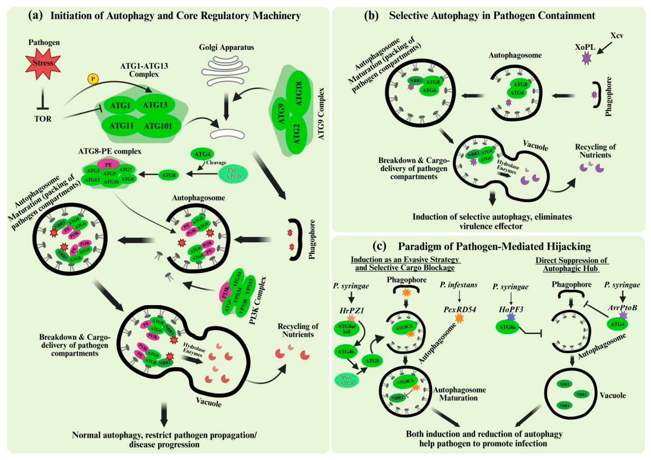
Figure 2.
Strategic Hijacking of the Autophagic Hub and Programmable Resistance Solutions. a) Induction of Evasive Hyper-autophagy by m3875 and SDE4405. The CLas effector protein (CEP) m3875 functions as a transcriptional reprogrammer to upregulate host NbBI-2 (a conserved death suppressor), which activate ATG6 to initiate autophagic flux. Simultaneously, SDE4405 interacts directly with the ATG8c hub to enhance autophagosome formation. While these effectors induce state of “hyper-autophagy”, they strategically block the recruitment of the selective cargo receptor NBR1, preventing the vacuolar degradation of pathogen components and creating a pro-survival niche across various plant hosts. b) Metabolic Hijacking and Autophagy Inhibition by SDE3. In a contrasting stratgey, the effector SDE3 executes a metabolic hijacking by co-opting host GAPC1. This physical association facilitates the targeted degradation of ATG8 isoforms (a/d/g), dismantling the autophagic machinery and blocking autophagosome maturation. The resulting suppression of autophagic flux facilitates systemic bacterial colonization by disabling the host’s primary line of antimicrobial defense. c) A Programmable Roadmap for Synthetic Immunity: CRISPR-mediated Editing and Engineered Alleles. This integrated strategy utilizes CRISPR/Cas9 to precisely modify the susceptibility (S) transcription factor (TF) binding sites located at upstream regions of host targets (e.g., BI-2, GAPC1, ATG8c) targeted by CEPs. By introducing genomic variations that render these host components ‘non-recognizable’ to pathogen effectors while preserving their essential physiological functions, the host can effectively bypass pathogen-mediated suppression. These resistance-conferring modifications can be deployed through Mobile-CRISPR technology, where signals are transferred from a transgenic rootstock to a non-transgenic scion. This multi-tiered approach prevents the hijacking of the ATG6-ATG8 hub, thereby restoring autophagosome maturation and NBR1-mediated cargo delivery for vacuolar degradation. The ultimate restoration of autophagic homeostasis restricts CLas proliferation and halts HLB progression.
Figure 2.
Strategic Hijacking of the Autophagic Hub and Programmable Resistance Solutions. a) Induction of Evasive Hyper-autophagy by m3875 and SDE4405. The CLas effector protein (CEP) m3875 functions as a transcriptional reprogrammer to upregulate host NbBI-2 (a conserved death suppressor), which activate ATG6 to initiate autophagic flux. Simultaneously, SDE4405 interacts directly with the ATG8c hub to enhance autophagosome formation. While these effectors induce state of “hyper-autophagy”, they strategically block the recruitment of the selective cargo receptor NBR1, preventing the vacuolar degradation of pathogen components and creating a pro-survival niche across various plant hosts. b) Metabolic Hijacking and Autophagy Inhibition by SDE3. In a contrasting stratgey, the effector SDE3 executes a metabolic hijacking by co-opting host GAPC1. This physical association facilitates the targeted degradation of ATG8 isoforms (a/d/g), dismantling the autophagic machinery and blocking autophagosome maturation. The resulting suppression of autophagic flux facilitates systemic bacterial colonization by disabling the host’s primary line of antimicrobial defense. c) A Programmable Roadmap for Synthetic Immunity: CRISPR-mediated Editing and Engineered Alleles. This integrated strategy utilizes CRISPR/Cas9 to precisely modify the susceptibility (S) transcription factor (TF) binding sites located at upstream regions of host targets (e.g., BI-2, GAPC1, ATG8c) targeted by CEPs. By introducing genomic variations that render these host components ‘non-recognizable’ to pathogen effectors while preserving their essential physiological functions, the host can effectively bypass pathogen-mediated suppression. These resistance-conferring modifications can be deployed through Mobile-CRISPR technology, where signals are transferred from a transgenic rootstock to a non-transgenic scion. This multi-tiered approach prevents the hijacking of the ATG6-ATG8 hub, thereby restoring autophagosome maturation and NBR1-mediated cargo delivery for vacuolar degradation. The ultimate restoration of autophagic homeostasis restricts CLas proliferation and halts HLB progression.
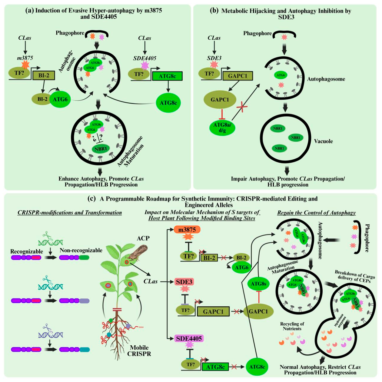

Table 1.
Strategic modulation of the core autophagy machinery by diverse pathogen effectors.
| Pathogen/ Target Species | Effector | Target (Host Protein) | Mode of Action | Outcome | Citation |
|---|---|---|---|---|---|
| CLas/ N. benthamiana | m3875 | NbBI-2, NbWRKY9, NbCNGCs | Transcriptional upregulation of NbBI-2 to induce “evasive” hyper autophagy; inhibits NbCNGCs. | Suppresses HR-PCD; promote bacterial growth. | (Zhang et al., 2019) |
| CLas; CLam; CLaf/ C. sinensis; N. benthamiana; A. thaliana | SDE3 | GAPC1/2 | Physically co-opts GAPCs to promote the specific degradation opf ATG8 isoforms (a/d/g). | Dismantles autophagy; promote HLB progression. | (Shi et al., 2023b) |
| Xcv/ N. benthamiana; N. tabacum; S. lycopersicum | XopL | SH3P2 | Execute proeasomal degradation of autophagy component SH3P2 via E3 ligase activity. | Dampens autophagic flux; promotes infection. | (Leong et al., 2022b) |
| Pst/ A. thaliana | AvrPtoB | ATG1 kinase | Target MIT domain of ATG1 to inhibit kinase phosphorylation and initiation. | Suppresses autophagy; enhances bacterial virulence. | (Lal et al., 2020) |
| CLas/ C. sinensis; N. benthamiana | SDE4405 | CsATG8c | Directly interacts with ATG8s to activate flux while blocking selective cargo delivery. | Negative immunity regulation; promotes proliferation. | (Shi et al., 2023a) |
| Pst/ A. thaliana | HrpZ1 | ATG4b, ATG8 | Enhances ATG4b-mediated cleavage of ATG8 to accelerate phagophore formation. | Induces hyper-autophagy; promotes infection. | (Lal et al., 2020) |
| Pst/ A. thaliana | HopF3 | ATG8 | Directly binds multiple ATG8 isoforms to inhibit their conjugation and maturation. | Represses autophagy; promotes bacterial virulence. | (Lal et al., 2020) |
| Pst/ N. benthamiana | unknown | NBR1-mechanism | NBR1-driven selective autophagy counteracts water-soaked lesions induced by HopM1. | Limits pathogen growth and contains infection. | (Üstün et al., 2018) |
| CLso/ Psyllid midgut | unknown | Beclin-1 (ATG6 ortholog); SERCA, ITPR | Pathogen-associated changes in SERCA/ITPR and Beclin-1 phosphorylation via Ca2+ signaling induce autophagy. | Induction of autophagy; facilitates bacterial persistance and transmission | (Sarkar et al., 2023) |
| CLas/ C. sinensis | SDE4040 | CsATG8c | Pathogen effector is targeted and degraded by CsATG8c-mediated selective autophagy. | Enhances host host defense; limits bacterial proliferation. | (Cui et al., 2025) |
| Phytophthora infestans/ S. tuberosum | PexRD54 | ATG8CL | Physically displaces NBR1 (Joka2) from ATG8CL to reprogram vesicle trafficking. | Blocks defensive cargo delivery; enhances virulence. | (Dagdas et al., 2016) |
| Pst/ A. thaliana | HopM1 | Proteasome | Targets proteasomes for autophagic degradation, a process termed “proteaphagy”. | Disables host UPS defense; facilitates proliferation | (Üstün et al., 2018) |
|
NleB | GAPDH | Modifies GAPDH via O-GlcNAcylation to disrupt GAPDH-mediated transcriptional defense | Facilitates bacterial spread and parasitism | (Gao et al., 2013) |
|
SopF | V-ATPase | Prevents V-ATPase intercation with ATG16L1 to inhibit the initiation of xenophagy | Promotes bacterial survival and proliferation | (Xu et al., 2019) |
Disclaimer/Publisher’s Note: The statements, opinions and data contained in all publications are solely those of the individual author(s) and contributor(s) and not of MDPI and/or the editor(s). MDPI and/or the editor(s) disclaim responsibility for any injury to people or property resulting from any ideas, methods, instructions or products referred to in the content. |
© 2026 by the authors. Licensee MDPI, Basel, Switzerland. This article is an open access article distributed under the terms and conditions of the Creative Commons Attribution (CC BY) license.
Copyright: This open access article is published under a Creative Commons CC BY 4.0 license, which permit the free download, distribution, and reuse, provided that the author and preprint are cited in any reuse.


