

Submitted:
30 June 2026
Posted:
02 July 2026
You are already at the latest version
Abstract
HMGB-1 is an alarmin representative of DAMP playing a central role in the immuno-genic cell death (ICD), a necessary condition in the dialogue stablished between dying tumor cells and immune system during some anticancer therapies. Therefore, early screening for ICD inducers represents a major priority in drug development today. In this work, we investigated the effect elicited by the clinical-grade CIGB-300 peptide, which impairs the Protein Kinase CK2-mediated phosphorylation and other CK2 signaling connected kinases. Here, the HMGB-1 extracellular release was investigated in an 18-cell line panel from blood malignancies, uterine-cervical cancer and NSCLC treated with CIGB-300 at equipotent doses (IC50) during 24 hours. Interestingly, CIGB-300 treatment upregulated the HMGB-1 protein levels at the culture supernatant in most of the cell lines (p=0.01) and fold-change increase ≥2 trended to be associated with intrinsic cell line sen-sitivity towards CIGB-300 cytotoxic effect. However, the HMGB-1 release by CIGB-300 was context-specific with clear induction on blood and uterine-cervical cancer cells and a diffused response pattern in NSCLC. Importantly, CIGB-300 treatment of blood cancer patients enrolled in a Phase I study induced plasma HMGB-1 alarmin in 4 out of 7 subject who received the entire treatment plan. Altogether, our data reveal for the first time that CIGB-300 treatment is able to induce extracellular HMGB-1 release in vitro and in vivo which could be indicative of ICD induction in some kind of tumors, also the induction of extracellular HMGB-1 alarmin as putative CIGB-300 response biomarker merits further investigation.
Keywords:
immunogenic cell death
; protein kinase CK2
; HMGB-1
; CIGB-300
1. Introduction
High mobility group box 1 (HMGB1) is a highly conserved nuclear protein that plays essential roles in chromatic condensation and transcriptional regulation [1]. In addition to its nuclear function, HMGB1 has a well-characterized function in the extracellular environment where it acts as a damage-associated molecular pattern (DAMP) to mediate inflammation and immune responses [2]. HMGB1 release can occur passively, as consequence of cell necrosis, or actively through regulated secretion in different cell types, including, activated immune cells, necrotic cells, and certain types of tumor cells. This process is tightly controlled by post-translational modifications, including phosphorylation, acetylation, and oxidation, which determine its translocation from nucleus to cytoplasm and its subsequent release [3]. On the other hand, high levels of HMGB-1 alarmin have been reported in patients with solid tumors and blood cancer [4,5].
Casein kinase 2 (CK2) is a constitutively active serine/threonine kinase that is frequently overexpressed in multiple types of cancer [6]. CK2 regulates a wide range of key cellular processes, including proliferation, apoptosis, stress response, and intracellular signaling making it an important therapeutic target [7]. Recent evidence indicates that CK2 is also involved in the regulation of nuclear proteins and in the modulation of inflammatory pathways [8,9], suggesting a connection with the mechanisms that control the release of HMGB1. In this regard, different studies have demonstrated the effect of CK2-mediated phosphorylation of HMGB1 on modulating its stability, DNA binding activity [10,11] and the subsequent shuttles between nucleus and cytoplasm [12]. Despite these advances, the effect of CK2 inhibition on HMGB1 release in tumor cells has not been fully elucidated.
Several strategies for CK2 enzyme inhibition have been evaluated in pre-clinical studies. Of these, only two have emerged as promising tools for both research and therapeutic applications, the synthetic-peptide CIGB-300 and the ATP-competitive inhibitor CX-4945 (Silmitasertib). CIGB-300, currently under clinical evaluation, is a chimeric peptide containing a cell-penetrating moiety that impairs CK2-mediated phosphorylation through targeting the phosphoacceptor domain in the substrates [13] or direct binding to the enzyme [14]. By the other hand, CX-4945 is a selective ATP-competitive CK2 inhibitor orally bioavailable, indicated by the FDA as an “orphan drug” for cholangiocarcinoma in 2017 [15]. Both inhibitors have demonstrated anti-proliferative and antineoplastic effect in solid tumor and hematological malignancies [16,17,18,19]
Despite these advances, the specific role of CK2 inhibition in regulating HMGB1 alarmin release and subsequent ICD induction remain insufficiently characterized. In particular, the effect that the clinical grade CIGB-300 and CX-4945 inhibitors might elicit and consequent inflammatory response, is unclear. Thus, this study aims to investigate “a priori” the effect of pharmacological inhibition of CK2 using the CIGB-300 peptide on the induction of HMGB1 release in leukemia, non-small cell lung cancer (NSCLC) and cervical cancer cell lines as a previous step to subsequent investigation of ICD induction.
2. Results
2.1. CIGB-300 Treatment Induces HMGB1 Release
To investigate the putative effect of CIGB-300 on the extracellular HMGB-1 alarmin release, a panel containing 18 tumor cell lines from different origins were assayed in presence of two CK2 inhibitors, the CIGB-300 peptide and CX-4945 during 24 hours of treatment. Interestingly, CIGB-300 but not CX-4945 significantly induced the extracellular levels of the HMGB-1 alarmin in most of the tumor cell lines panel (p=0,01 versus p=0.34) (Figure 1A).
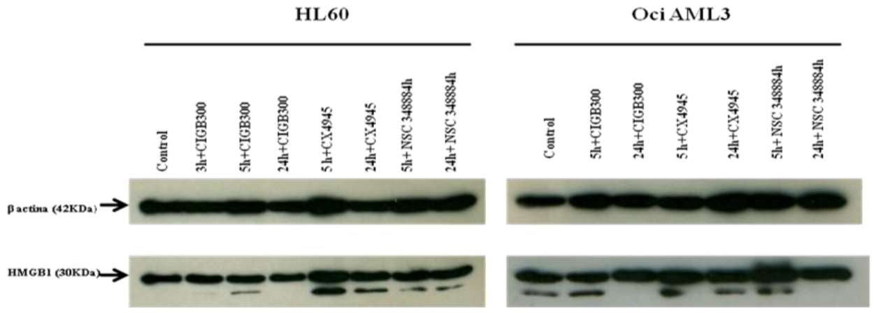
The segregated analysis showed that on blood cancer cell lines (n=5), HMGB1 was detected in culture supernatant of all cell lines after treating with CIGB-300 IC50. and levels were at least twice as high as those found in untreated condition (p=0.043) (Figure 1B). Otherwise, non-significant effect was observed for CX-4945 with just a mild increase of extracellular HMGB-1on HL-60 cells. Thus, equipotent dose levels of both CK2 inhibitors which elicits 50% of cell death, differ in their ability to increase the HMGB-1 extracellular levels on these leukemia cell lines. To investigate whether induction of extracellular HMGB-1 levels was correlated with putative regulation of the alarmin intracellular levels by CIGB-300, total cell extracts from HL60 and OCI-AML3 cell lines treated with CIGB-300 or CX-4945 were prepared and analyzed by Western Blot. Of note, neither CIGB-300 nor CX-4945 equipotent doses (IC50) seemed to affect significantly the HMGB-1 intracellular levels at these experimental conditions (Figure2).
Figure 2.
CIGB-300 treatment does not modify the HMGB-1 intracellular levels. HL60 and OCI-AML cell lines were treated with CIGB-300 and CX-4945 IC50 at indicated times. Cell extracted was prepared and analyzed by western blot as indicated in Materials and Methods.
Figure 2.
CIGB-300 treatment does not modify the HMGB-1 intracellular levels. HL60 and OCI-AML cell lines were treated with CIGB-300 and CX-4945 IC50 at indicated times. Cell extracted was prepared and analyzed by western blot as indicated in Materials and Methods.
We next investigated whether the induction of HMGB-1 release is also observed by treating epithelial tumor cells with CIGB-300. To do that, cell lines from uterine-cervix and lung cancer were treated with IC50 equipotent doses of the CIGB-300 peptide and CX4945 for 24 hours.
Similarly to leukemia, extracellular HMGB-1 levels were detected in the supernatant of all cervical cancer cell lines treated with CIGB-300 (p=0.034). The highest levels were detected in the supernatant of the HeLa and Hep2C cell lines (Figure 1B). Conversely, the addition of CX4945 to the culture of cervical cancer cells did not induce HMGB-1 release, instead a very modest inhibition in CaSki, SiHa and HeLa cells was observed.
Contrary to what was observed for leukemia and uterine-cervical cancer cell lines, an heterogenous response profile was observed for NSCLC cells treated with CIGB-300 (Figure 1B). CIGB-300 treatment increased the extracellular levels of HMGB-1 by more than 50% on NCI-H460 and NCI-H125 cell lines (Figure 1B) and no effect was elicited by CX-4945. Interestingly, neither CIGB-300 nor CX-4945 did modify the extracellular HMGB-1 levels on A549 and H226 cells whose baseline levels were notably high. Otherwise, both CIGB-300 and CX-4945 inhibited HMGB1 release in H522 and SK-MES-1 cell lines. This heterogenous response profile contributed to the non-significant increase of the HMGB-1 levels after CIGB-300 in NSCLC cell lines compared to blood cancer and uterine cervical cancer.
To look for putative associations between the HMGB-1 release induced by CIGB-300 and other parameters like intrinsic cell sensitivity towards CIGB-300, HMGB-1 baseline levels and cancer type, Spearman’s rank correlation analysis was used in the 18-cell line panel. A moderated association was observed for HMGB-1 release fold change after 24 hours of CIGB-300 treatment with a negative correlation between the HMGB1 increase and IC50 values (p=0.043, corr = -0.482) (Table 1). Otherwise, association between the CIGB-300 intrinsic cell sensitivity and the baseline or drug-induced HMGB-1 levels was not observed (Table 1).
Once we evidenced that CIGB-300 treatment in vitro induced extracellular levels of the HMGB-1 alarmin, we subsequently investigated whether in vivo treatment is able to elicit similar effect. For that purpose, we analyzed the plasma HMGB-1 levels from blood cancer patients enrolled in a Phase I clinical trial who received an in-patient CIGB-300 dose-escalation treatment consisting in 1.6, 3.2, 5.4, 8.6 and 12.8 mg/kg of weight. Interestingly, after completing the CIGB-300 regime during 6 weeks, 4 of out 7 patients who received the entire treatment plan, did experience at least 2-fold increase of the plasma HMGB-1 levels compared to baseline (Table 2). Analysis was not performed in three patients who did not complete all the CIGB-300 dose-levels because of disease progression or abandoned the study.
3. Discussion
Some anticancer strategies today focus on the transformation of cold- into hot-tumors by creating an immune-inflamed phenotype favored by drug-induced ICD, which can be optimal for efficient immunotherapy [20]. Likewise, the early screening for anticancer drugs, which induce immunogenic instead of tolerogenic cell death, represents a major priority during drug development today. The exploration of extracellular ICD key biomarkers on anticancer drug-treated cells and in plasma from patients treated with anticancer therapy, is the first step towards confirmation of ICD induction by anticancer drugs. Particularly, HMGB1 is a key DAMP released during early stages of ICD, crucial for stimulating an effective anticancer immune response [21,22].
The induction of apoptosis and cell cycle arrest by inhibiting the CK2-meadiated phosphoryltation have been experimentally validated by using different kinds of CK2 inhibitors. However, the knowledge of the type of cell death produced by the different CK2 inhibitors and their possible connection with the immune system has been remained to be elucidated. In this work we explored the effect of CIGB-300 anticancer peptide, which impinge the CK2-mediated phosphorylation by different mechanisms, on the HMGB-1 alarmin release by tumor cells. Of note, treatment with CIGB-300 IC50 significantly induced the HMGB-1 alarmin extracellular levels in most of the cell lines from the panel (p=0.01), with significant effect on blood and uterine-cervical cancer cell lines and heterogenous response on NSCLC. Interestingly, blood cancer cells which showed the greatest sensitivity towards CIGB-300 cytotoxic effect (lowest IC50 values), exhibited at least 2-fold induction of the extracellular alarmin release regardless the leukemia type. Likewise, integration of results in the entire 18-cell line panel indicated a moderated negative correlation between intrinsic high cell sensitivity towards cytotoxic effect of CIGB-300 (lowest IC50 values) and extracellular HMGB-1 release fold-change after 24 hours of CIGB-300 treatment. These results merit further confirmation in a broader cell panel.
Although HMGB-1 is a substrate of CK2 [10,12], it was not found among the CK2 substrates inhibited when the CIGB-300-regulated phosphoproteome was interrogated in AML cells such as HL-60 and OCI-AML3 [14]. In addition, the HMGB-1 intracellular levels were not visibly changed by CIGB-300 treatment in both cell lines according to Western Blot analysis. These evidences suggest that the phosphorylation of HMGB-1 by CK2 does not play a causal role during its extracellular release induced by CIGB-300, instead, such release seems rather be a consequence of the cellular reprogramming mechanisms driving cell death where the inhibition of other CK2 substrates by CIGB-300 could play a central role. Besides, in the above-mentioned phosphoproteomic analysis, a significant number of phosphosites attributed to other CK2-sequentialy activated kinases such as to glycogen synthase kinase-3beta (GSK3B), members of mitogen-activated protein kinases (MAPKs) and cyclin-dependent kinases (CDKs) families also appeared modulated in HL-60 and OCI-AML3 by CIGB-300 [14], which could also be connected with the molecular and cellular events driving cell death and HMGB-1 release.
Additionally, previous studies using microarray, qPCR, and ELISA confirmed that treatment with the CIGB-300 peptide increases TNFα at both transcriptional and protein synthesis levels in HL-60 and OCI-AML3 cells. TNFα was detected in the culture supernatant of both cell lines, with the highest levels found in HL-60 [23]. TNFα appears to be involved in HMGB1 release into the extracellular environment. In assays performed on monocytes and macrophages stimulated with LPS, the simultaneous release of TNFα and HMGB1 was detected. In that work, the researchers demonstrated that direct TNFα suppression by genetic TNFα knockout or TNFα-neutralizing antibodies partially inhibits LPS-induced HMGB1 secretion in macrophages, suggesting that HMGB1 secretion could be partially mediated through a TNFα-dependent mechanism [24]. In line with this, some genes involved in the NF-κB pathway were also upregulated by CIGB-300 in the same study [23]. Therefore, we cannot rule out the putative role of the NF-κB/TNF-α in the HMGB-1 alarmin release induced by CIGB-300 at least in both acute myeloid leukemia cells.
Considering that blood cancer cells responded efficiently to the effect of CIGB-300, we verified whether the in vivo treatment was capable of inducing levels of the plasma HMGB-1 alarmin in blood cancer patients who had participated in a Phase 1 clinical study. Despite the limited population size, 4 of 7 patients who completed the five-planned CIGB-300 intravenous infusion cycles, showed an increase in HMGB-1 alarmin levels ≥ 2-fold. Although very preliminary, these data indicate that CIGB-300 treatment is able to induce extracellular HMGB-1 alarmin levels both in blood cancer cell lines and patients. However, this effect seems to be context-specific as some patients remained refractory, something that also happened in the analysis of the cell panel in vitro.
Contrary to what was observed for blood and uterine-cervical cancer, NSCLC cell lines showed a diffuse response pattern towards the CIGB-300 effect where induction of extracellular HMGB-1 alarmin levels was not clearly documented. In fact, only two cell lines (NCI-H125 and NCI-H460), the most sensitive ones to cytotoxic effect of CIGB-300, exhibited increase of the HMGB-1 levels at the supernatant by treating with equipotent CIGB-300 doses, two cell lines (A549 and NCI-H226) showed no CIGB-300 effect on the HMGB-1 levels and instead, and the two other cell lines (H552 and SK-MES-1) experienced a reduction of the extracellular HMGB-1. Interestingly, the cell lines which exhibited high extracellular HMGB-1 baseline levels (A549 and NCI-H226), CIGB-300 failed to modify the alarmin release. Similar to AML cells, in the phosphoproteomic analysis on NCI-H125 cells, the CK2 phosphosite on the HMGB-1 protein was not found to be inhibited by CIGB-300 treatment among the CK2 substrate pool [25], which rules out a putative instrumental role for the CK2-mediated phosphorylation on HMGB-1 in the subsequent release to extracellular compartment.
In addition to the mechanisms involved in the active secretion of HMGB1, it is known that it can be secreted passively by the induction of cell death and increased permeability of the cytoplasmic membrane [3]. Accumulated evidence has shown the induction of cell death by both CK2 inhibitors evaluated in this work [26,27] which would support the passive release of HMGB1 after treatment with these molecules. However, here the treatment with CX-4945 at IC50 dose (5 µM) only induced modest release of HMGB1 in the HL60 and OCI-AML3 leukemia cell lines. In the other cell lines studied, CX-4945 did not promote the release of HMGB1 or, as observed in some cases, inhibited its release, since extracellular HMGB1 levels on CIGB-300 treated-cells were lower than those belong to baseline. Previous studies reported that high CX-4945 doses (100 µM) induced other ICD biomarkers like ATP release as monotherapy, and calreticulin exposure only in combination with Temozolomide on glioblastoma cells [28]. We found no previous reports investigating the effect of CX-4945 on HMGB-1 release on tumor cells. Whether CX-4945 induces HMGB-1 release at dose levels over IC50, it merits further clarification.
Despite its dual role in cancer, extracellular HMGB1 constitutes a paradigmatic biomarker of ICD which plays a central role in the dialogue established between dying cancer cells and the immune system in cancer therapy due to its well-characterized function as a DAMP [22]. The results obtained in this work show for the first time that CIGB-300 doses killing the 50% of the tumor cells induce HMGB-1 alarmin release in context-specific way and treatment of blood cancer patients with CIGB-300 can also increase the plasma HMGB-1 alarmin levels. However, further studies are required to determine the redox status of HMGB1 secreted after treatment with the CIGB-300 peptide, since its chemoattractant [29], cytokine [30], or tolerogenic [31] activity depends largely on the oxidation state of the molecule. Also, our results do not rule out that in addition to the inhibition of signaling the CK2, other kinases inhibited by CIGB-300 as part of the in vivo signal amplification, contribute to the process of cellular reprogramming entailing o cell death and HMGB-1 release. A comprehensive experimental setting will be required for the final assessment of ICD induction by CIGB-300.
4. Materials and Methods
4.1. Cell Lines and Cultures
The human (HL-60, OCI-AML3, THP-1, K562) and mouse (L1210) leukemia cell lines were cultured in RPMI 1640 Medium (Gibco, Waltham, MA, USA) supplemented with 10% (v/v) fetal bovine serum (FBS) (Invitrogen, Carlsbad, CA, USA) and 50 μg/mL gentamicin (Sigma, St. Louis, MO, USA). The human epithelial lung (NCI-H460, NCI-H125, A549, NCI-H522, NCI-H226, SK-MES-1) and cervical (C4, CaSki, SiHa, HeLa, Hep2C) cancer cell lines were cultured in Dulbecco´s Modified Eagle medium (DMEM) (Gibco, Waltham, MA, USA) supplemented with 10% (v/v) FBS and 50 μg/mL gentamicin. All cell lines were originally obtained from American Type Culture Collection (ATCC, Manassas, VA, USA).
4.2. Cell Viability Assay
Cytotoxic effect was determined using AlamarBlue assay (Life Technologies, Carlsbad, CA, USA). Cells were seeded in flat-bottom 96-well plates (leukemia cells at 2x10⁵ cells/mL, 100 uL/well and epithelial cells at 6x10⁴ cells/mL, 100 uL/well), and 24 hours later, serial dilutions 1:2 ranging from 200 to 12.5 μM of CIGB-300 and serial dilutions 1:2 ranging from 50 to 3.12 μM of CX4945 were added. After 48 hours of incubation, AlamarBlue was added at 10% (v/v) and cells were further incubated for 4 hours. Fluorescence was measured in a CLARIOstar microplate reader (BMG LABTECH, Ortenberg, Germany) and half-inhibitory concentrations (IC50) values were estimated using CalcuSyn software (v2.1) (Biosoft, Cambridge, UK).
4.3. Cuantification of HMGB1 Release
The human epithelial cells were planted in 24-well plates with 0.5 mL full medium, which was changed 24 hours later and treatment added onto the cells. In the case of leukemia cell lines, these were seeded at 0.5x10⁶ cells/mL in 12-well plates with 1 mL full medium. The cells were treated with CIGB-300 peptide and CX4945 during 24 hours at a dose equivalent to the IC50, determined as indicated above. Supernatants were collected 24 hours later, dying cells removed by centrifugation and supernatants isolated and frozen immediately. Quantification of HMGB1 in the cell culture supernatant or plasma from blood cancer patients who had been enrolled in a CIGB-300 Phase I clinical study, was assessed by commercial ELISA kits obtained from IBL and Shanghai Mlbio Technology Co., LTD according to the manufacturer´s instructions.
4.4. Intracellular Levels of HMGB1 on Tumor Cells
HL-60 and OCI-AML 3 cell lines were incubated with CIGB-300 and CX-4945 IC50 levels during 3, 5 and 24 hours or during 5 and 24 hours respectively. Cells were lysed in RIPA buffer containing protease/phosphatase inhibitor (Thermo Fisher Scientific, Waltham, MA, USA), and equal amounts of protein were resolved in 12.5% SDS-PAGE. Next, proteins were transferred to a nitrocellulose membrane and immunoblotted with the following antibodies according to instructions from the manufacturer: NPM1 and β-actin (Abcam, Sigma, St. Louis, MO, USA). Detection was performed with peroxidase conjugated anti-mouse or anti-rabbit IgG (Sigma, St. Louis, MO, USA), and signal was developed using SuperSignal West Pico Chemiluminescent Substrate (Thermo Fisher Scientific, Waltham, MA, USA).
4.5. Soluble HMGB1 Levels in Plasma from the EHPMA Clinical Study
An open, adaptative, and multicenter Phase I CIGB-300 in-patient dose-escalation clinical trial had been previously performed in ten patients with blood malignancies like Acute Myeloid Leukemia (AML), Acute Lymphocytic Leukemia (ALL) or Myelodysplastic Syndrome (MDS). CIGB-300 regimen consisted in five consecutive 3 days cycles with escalated dose at 1.6. 3.2, 5.3, 8.0, 12.8 mg/kg and 4 day-resting period between cycles. Plasma samples were taken after every single cycle however, the HMGB-1 levels were investigated at baseline and after concluding the entire treatment plan at week 6. The EHPMA clinical study was conducted in accordance with the Declaration of Helsinki, and approved by the Institutional Review Board of Center for State Control of Medicines, Equipment and Medical Devices (CECMED)(protocol code IG/CIGB-300I/LAR/1101 and date of approval: November 5, 2014).
4.6. Statistical Analysis
The HMGB-1 soluble values of untreated and drug-treated cell lines was compared using the Wilcoxon matched pairs test. Spearman’s rank correlation was used to estimate the association between CIGB-300 response and HMGB-1 expression levels. Statistical analyses were performed with Statistica version 8.0 and SPSS version 13.0. The level of significance used was α = 0.05. Prism GraphPad Version 6.0 Software was used for data visualization.
Author Contributions
D.A.N (Investigation, methodology, written original draft), Y.Y. (investigation, methodology), J.M. and R.B (data analysis), D.M.V. and L.Y. (methodology and data analysis), L.W. and Y.P. (conceptualization and supervision), S.E.P. (conceptualization, supervision, writing review, project administration and funding acquisition). All the authors have read and agreed to the published version of the manuscript.
Funding
The present research was supported by a grant from the Science and Technology Innovation Program of Hunan Province”, China, (2024RC4027). Also, this work was supported in part by a CITMA-Cuba grant: PN385LH007-013.
Institutional Review Board Statement
The EHPMA clinical study was conducted in accordance with the Declaration of Helsinki, and approved by the Institutional Review Board of Center for State Control of Medicines, Equipment and Medical Devices (CECMED)(protocol code IG/CIGB-300I/LAR/1101 and date of approval: November 5, 2014).
Informed Consent Statement
Oral and Written informed consent was obtained from patients to be enrolled at the EHPMA clinical study. Patient’s names were blinded by internal codes of the study as a rule of the Good Clinical Practices.
Data Availability Statement
The original contributions presented in this study are included in the article/supplementary material. Further inquiries can be directed to the corresponding author(s).
Acknowledgments
Authors want to acknowledge Evelin Caballero for her technical support in this work.
Conflicts of Interest
The authors declare no conflict of interest.
Abbreviations
The following abbreviations are used in this manuscript:
| HMGB1 | High Mobility Group Box 1 |
| DAMP | Damage Associated Molecular Patterns |
| ICD | Immunogenic cell death |
| CK2 | Casein Kinase 2 |
| NSCLC | Non-Small Cell Lung Cancer |
| IC50 | half-inhibitory concentrations |
| AML | Acute Myeoloide Leukemia |
| MDS | Myelodisplasic Syndrome |
| ALL | Acute Lymphocytic Leukemia |
| GSK3B | glycogen synthase kinase-3beta |
| MAPKs | mitogen-activated protein kinases |
| CDKs | cyclin-dependent kinases |
References
- Lv, G.; Yang, G.; Gai, K.; et al. Multiple functions of HMGB1 in cancer. Front. Oncol. 2024, 14, 1384109. [Google Scholar] [CrossRef] [PubMed]
- Yuan, J.Y.; Guo, L.; Ma, J.T.; et al. HMGB1 as an extracellular pro-inflammatory cytokine: Implications for drug-induced organic damage. Cell Biol. Toxicol. 2024, 40, 55. [Google Scholar] [CrossRef] [PubMed]
- Chen, R.; Kang, R.; Tang, D. The mechanism of HMGB1 secretion and release. Exp. Mol. Med. 2022, 54, 91–102. [Google Scholar] [CrossRef] [PubMed]
- Yang, H.; Zhao, X.; Li, X.; et al. A pan-cancer analysis of the oncogenic function of HMGB1 in human tumors. Biochem. Biophys. Rep. 2024, 40, 101851. [Google Scholar] [CrossRef] [PubMed]
- Rapoport, B.L.; Steel, H.C.; Theron, A.J.; et al. High Mobility Group Box 1 in Human Cancer. Cells 2020, 9(7), 1664. [Google Scholar] [CrossRef] [PubMed]
- Trembley, J.H.; Kren, B.T.; Afzal, M.; et al. Protein kinase CK2 - diverse roles in cancer cell biology and therapeutic promise. Mol. Cell. Biochem. 2022, 478, 899–926. [Google Scholar] [CrossRef] [PubMed]
- Nuñez de Villavicencio-Diaz, T.; Rabalski, A.J.; Litchfield, D.W. Protein kinase CK2: intricate relationships within regulatory cellular networks. Pharmaceuticals. 2017, 10(1), 27. [Google Scholar] [CrossRef] [PubMed]
- Singh, N.N.; Ramji, D.P. Protein kinase CK2, an important regulator of the inflammatory response? J. Mol. Med. 2008, 86, 887–897. [Google Scholar] [CrossRef] [PubMed]
- Liu, D.; Marie, J.C.; Pelletier, A.L.; et al. Protein kinase CK2 acts as a molecular brake to control NADPH oxidase 1 activation and colon inflammation. Cell. Mol. Gastroenterol. Hepatol. 2022, 13(4), 1073–1093. [Google Scholar] [CrossRef] [PubMed]
- Stemmer, C.; Leeming, D.J.; Franssen, L.; et al. Phosphorylation of Maize and Arabidopsis HMGB Proteins by Protein Kinase CK2α. Biochemistry 2003, 42(12), 3503–3508. [Google Scholar] [CrossRef] [PubMed]
- Taira, J.; Higashimoto, Y. Evaluation of in vitro properties of predicted kinases that phosphorylate serine residues within nuclear localization signal 1 of high mobility group box 1. J. Pept. Sci. 2014, 20, 613–617. [Google Scholar] [CrossRef] [PubMed]
- De Abreu da Silva, I.C.; Carneiro, V.C.; de Moraes Maciel, R.; et al. CK2 phosphorylation of Schistosoma mansoni HMGB1 protein regulates its cellular traffic and secretion but not its DNA transactions. PLoS ONE 2011, 6(8), e23572. [Google Scholar] [CrossRef]
- Perea, S.E.; Baladrón, I.; Valenzuela, C.; et al. CIGB-300: A peptide-based drug that impairs the protein kinase CK2-mediated phosphorylation. Semin. Oncol. 2018, 45(1–2), 58–67. [Google Scholar] [CrossRef] [PubMed]
- Rosales, M.; Pérez, G.V.; Ramón, A.C.; et al. Targeting of protein kinase CK2 in acute myeloid leukemia cells using the clinical-grade synthetic-peptide CIGB-300. Biomedicines 2021, 9(7), 766. [Google Scholar] [CrossRef] [PubMed]
- Siddiqui-Jain, A.; Drygin, D.; Streiner, N.; et al. CX-4945, an orally bioavailable selective inhibitor of protein kinase CK2, inhibits prosurvival and angiogenic signaling and exhibits antitumor efficacy. Cancer Res. 2010, 70(24), 10288–10298. [Google Scholar] [CrossRef] [PubMed]
- Perea, S.E.; Reyes, O.; Baladrón, I.; et al. CIGB-300, a novel proapoptotic peptide that impairs the CK2 phosphorylation and exhibits anticancer properties both in vitro and in vivo. Mol. Cell. Biochem. 2008, 316, 163–167. [Google Scholar] [CrossRef] [PubMed]
- Perea, S.E.; Perera, Y.; Baladrón, I.; et al. CIGB-300: A promising anti-casein kinase 2 (CK2) Peptide for Cancer Targeted Therapy. In Protein Kinase CK2 Cellular Function in Normal and Disease States. Advances in Biochemistry in Health and Disease; Ahmed, K., Issinger, O.G., Szyszka, R., Eds.; Springer: Cham, 2015; vol 12. [Google Scholar] [CrossRef]
- Prins, R.; Burke, R.; Tyner, J.; et al. CX-4945, a selective inhibitor of casein kinase-2 (CK2), exhibits anti-tumor activity in hematologic malignancies including enhanced activity in chronic lymphocytic leukemia when combined with fludarabine and inhibitors of the B-cell receptor pathway. Leukemia 2013, 27, 2094–2096. [Google Scholar] [CrossRef] [PubMed]
- D´Amore, C.; Borgo, C.; Sarno, S.; Salvi, M. Role of CK2 inhibitor CX-4945 in anti-cancer combination therapy – potential clinical relevance. Cell. Oncol. 2020, 43, 1003–1016. [Google Scholar] [CrossRef] [PubMed]
- Wu, B.; Zhang, B.; Li, B.; et al. Cold and hot tumors: from molecular mechanisms to targeted therapy. Signal Transduct. Target. Ther. 2024, 9, 274. [Google Scholar] [CrossRef] [PubMed]
- Decraene, B.; Yang, Y.; De Smet, F.; et al. Immunogenic cell death and its therapeutic or prognostic potential in high-grade glioma. Genes Immun. 2022, 23, 1–11. [Google Scholar] [CrossRef] [PubMed]
- Galluzzi, L.; Vitale, I.; Warren, S.; et al. Consensus guidelines for the definition, detection and interpretation of immunogenic cell death. J. Immunother. Cancer. 2020, 8, e000337. [Google Scholar] [CrossRef] [PubMed]
- Vázquez-Blomquist, D.; Ramón, A.C.; Rosales, M.; et al. Gene expression profiling unveils the temporal dynamics of CIGB-300-regulated transcriptome in AML cell lines. BMC Genom. 2023, 24, 373. [Google Scholar] [CrossRef] [PubMed]
- Chen, G.; Li, J.; Ochani, M.; et al. Bacterial endotoxin stimulates macrophages to release HMGB1 partly through CD14- and TNF-dependent mechanisms. J. Leukoc. Biol. 2004, 76(5), 994–1001. [Google Scholar] [CrossRef] [PubMed]
- Rodríguez-Ulloa, A.; Ramos, Y.; Sánchez-Puente, A.; et al. The combination of the CIGB-300 anticancer peptide and cisplatin modulates proteins related to cell survival, DNA repair and metastasis in a lung cancer cell line model. Curr. Proteomics. 2019, 16(4), 338–349. [Google Scholar] [CrossRef]
- Perera, Y.; Costales, H.C.; Diaz, Y.; et al. Sensitivity of tumor cell towards CIGB-300 anticancer peptide relies on its nucleolar localization. J. Pept. Sci. 2012, 18, 215–223. [Google Scholar] [CrossRef] [PubMed]
- Pierre, F.; Chua, P.C.; O’Brien, S.E.; et al. Pre-clinical characterization of CX-4945, a potent and selective small molecule inhibitor of CK2 for the treatment of cancer. Mol. Cell Biochem. 2011, 356, 37–43. [Google Scholar] [CrossRef] [PubMed]
- Villamaña, L.; Martínez-Escardó, L.; Arús, C.; et al. Successful Partnerships: Exploring the Potential of Immunogenic Signals Triggered by TMZ, CX-4945, and Combined Treatment in GL261 Glioblastoma Cells. Int. J. Mol. Sci. 2021, 22(7), 3453. [Google Scholar] [CrossRef] [PubMed]
- Venereau, E.; Casalgrandi, M.; Schiraldi, M.; et al. Mutually exclusive redox forms of HMGB1 promote cell recruitment or proinflammatory cytokine release. J. Exp. Med. 2012, 209(9), 1519–1528. [Google Scholar] [CrossRef] [PubMed]
- Yang, H.; Lundbäck, P.; Ottosson, L.; et al. Redox modifications of cysteine residues regulate the cytokine activity of HMGB1. Mol. Med. 2021, 27, 58. [Google Scholar] [CrossRef] [PubMed]
- Kazama, H.; Ricci, J.E.; Herndon, J.M.; et al. Induction of immunological tolerance by apoptotic cells requires caspase-dependent oxidation of High-Mobility Group Box-1 protein. Immunity 2008, 29(1), 21–32. [Google Scholar] [CrossRef] [PubMed]
Figure 1.
CIGB-300 treatment induces HMGB1 release in different tumor cell lines. (a) AML, uterine cervical cancer and NSCLC cell lines were treated with equipotent doses (IC50) of CIGB-300 and CX-4945 during 24 hours. (b) Individual effect according to the tumor cell line origin. Extracellular HMGB-1 levels were measured by ELISA as indicated in Material and Methods.
Figure 1.
CIGB-300 treatment induces HMGB1 release in different tumor cell lines. (a) AML, uterine cervical cancer and NSCLC cell lines were treated with equipotent doses (IC50) of CIGB-300 and CX-4945 during 24 hours. (b) Individual effect according to the tumor cell line origin. Extracellular HMGB-1 levels were measured by ELISA as indicated in Material and Methods.
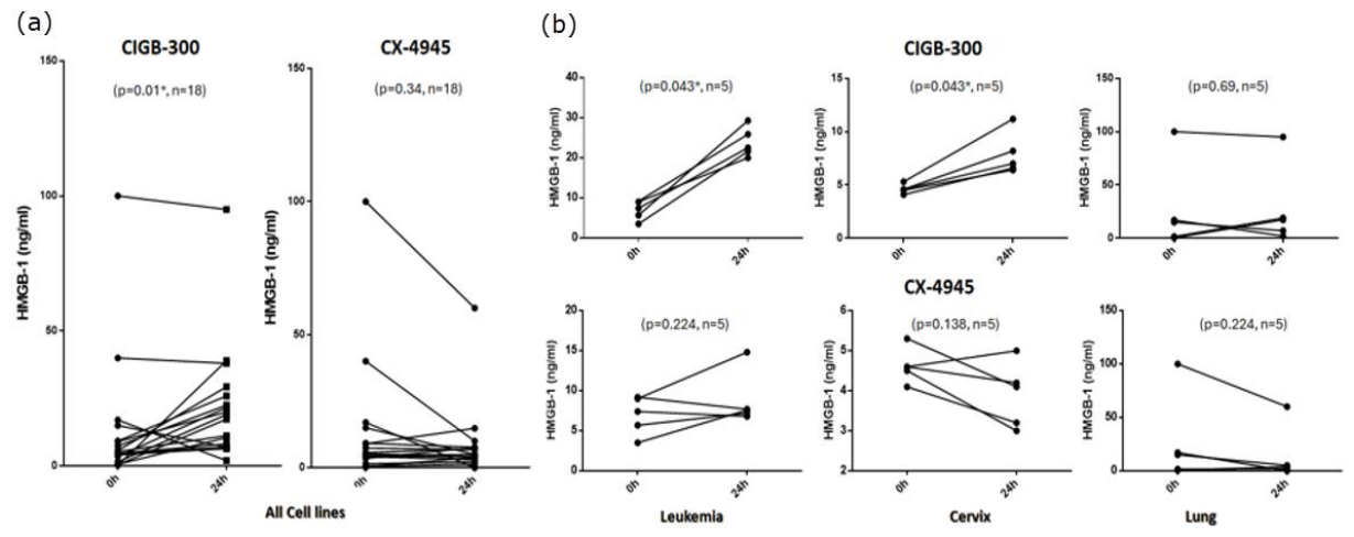

Table 1.
Non-parametric correlation between the CIGB-300 response variable (IC50- CIGB-300) and the expression of HMGB-1 at 0 and 24 hours.
Table 1.
Non-parametric correlation between the CIGB-300 response variable (IC50- CIGB-300) and the expression of HMGB-1 at 0 and 24 hours.
| HMGB-1_0h | HMGB-1_24h | FC_HMGB-1_24vs0h | |||
|---|---|---|---|---|---|
| Spearman's rho | IC50_300 | Correlation Coefficient | .110 | -.197 | -.482* |
| Sig. (2-tailed) | .664 | .432 | 0.043 | ||
| N | 18 | 18 | 18 |
Table 2.
Plasma HMGB-1 levels in blood cancer patients treated with CIGB-300.
| Patient Code | Diagnosis | Baseline (ng/mL) | After CIGB-300 treatment | Fold-change |
|---|---|---|---|---|
| AMC-01 | Elderly AML | 4.04 | 13.05 | 3.23 |
| HHA-02 | MDS | 0.10 | 7.12 | 71.2 |
| HHA-03 | ALL | 2.44 | 1.71 | 0.70 |
| GAL-01 | Elderly AML | 3.60 | 2.24 | 0.62 |
| HHA-01 | Relapsed AML | 1.87 | 2.38 | 1.27 |
| HHA-04 | MDS | 2.44 | 14.4 | 5.9 |
| HHA-05 | ALL | 7.02 | 20.23 | 2.88 |
| AMC-02 | Refractory AML | 11.0 | * | NA |
| AMC-03 | Refractory AML | 4.10 | * | NA |
| AMC-04 | Elderly AML | * | * | NA |
Notes: AML (Acute Myeoloide Leukemia), MDS (Myelodisplasic Syndrome), ALL (Acute Lymphocytic Leukemia. *Patients who did not complete all the CIGB-300 dose cycles. NA (Non-Applicable).
Disclaimer/Publisher’s Note: The statements, opinions and data contained in all publications are solely those of the individual author(s) and contributor(s) and not of MDPI and/or the editor(s). MDPI and/or the editor(s) disclaim responsibility for any injury to people or property resulting from any ideas, methods, instructions or products referred to in the content. |
© 2026 by the authors. Licensee MDPI, Basel, Switzerland. This article is an open access article distributed under the terms and conditions of the Creative Commons Attribution (CC BY) license (http://creativecommons.org/licenses/by/4.0/).
Copyright: This open access article is published under a Creative Commons CC BY 4.0 license, which permit the free download, distribution, and reuse, provided that the author and preprint are cited in any reuse.


