

Submitted:
01 July 2026
Posted:
01 July 2026
You are already at the latest version
Abstract
Parkinson’s disease is marked by the progressive loss of dopaminergic neurons in the substantia nigra, and current treatments can reduce symptoms but do not stop neurodegeneration. Cell replacement using induced pluripotent stem cell-derived dopaminergic neurons is a promising approach because it directly aims to restore the damaged nigrostriatal pathway. This review focuses on autologous iPSC-derived dopaminergic therapy while also comparing it with allogeneic approaches, since the main translational question is whether the immune advantages of patient-derived cells outweigh the greater scalability and standardization of donor-derived cell products. We discuss the molecular steps required to generate authentic midbrain A9 dopaminergic neurons, including SHH, FGF8, WNT signaling, and key transcription factors such as FOXA2, LMX1A, NURR1, and PITX3. We also review the role of gene correction in patient-derived iPSC lines, the potential immune benefits and remaining immunogenic risks of autologous grafts, and early clinical evidence showing feasibility, graft survival, and tolerability in small cohorts. Although autologous iPSC therapy has strong biological potential, major challenges remain, including manufacturing reproducibility, quality control, safety, cost, and the need to prove durable clinical benefit. Overall, this review argues that autologous iPSC-derived dopaminergic therapy is a feasible but still developing strategy, and that future progress will depend on stronger comparative evidence against allogeneic approaches, validated potency assays, and long-term clinical outcomes.
Keywords:
induced pluripotent stem cells
; Parkinson’s disease
; dopaminergic neurons
; autologous cell therapy
; allogeneic transplantation
; cell replacement therapy
Introduction
With over 10 million cases worldwide, Parkinson’s disease (PD) is the second most common neurodegenerative disorder [1]. It is now one of the main causes of neurological disability, with a prevalence that has increased by 2.5 times over the previous generation [2]. Its etiology involves a combination of environmental factors and genetic variants, ranging from rare, highly penetrant mutations to common alleles that modestly increase risk [3]. While PD is not immediately lethal, it markedly elevates morbidity due to complications including falls, fractures, pneumonia, and aspiration, and correlates with an increased risk of premature mortality [4,5,6]. The disease causes a significant socioeconomic cost, impacting patients, caregivers, and healthcare systems owing to prolonged care requirements, diminished productivity, and emotional and financial stress [2,7,8]. Notwithstanding symptomatic interventions, there exists no cure or approved therapy that halts or reverses disease development, underscoring the pressing need for disease-modifying approaches.
Traditionally, therapies for PD have primarily focused on symptom management rather than modifying disease progression. Pharmaceuticals such as L-dopa, MAO inhibitors, and dopamine agonists have enhanced quality of life, although other medications have had inconsistent outcomes, and deep brain stimulation can address motor symptoms without slowing the underlying disease [9,10]. To date, no therapeutic agents have conclusively demonstrated disease-modifying effects in Phase III clinical trials, although several candidates remain under investigation [11,12]. Promising approaches include disease-modifying drugs, gene therapy, dopaminergic cell transplantation, and autologous induced pluripotent stem cell (iPSC) therapy.
Among these, dopaminergic cell replacement is the only strategy that directly targets the lost neurons, aiming to restore the nigrostriatal pathway rather than compensate for its loss. Autologous iPSC therapy for PD has rapidly progressed from concept to clinic in the last few years. Preliminary evidence from compassionate-use cases and ongoing clinical trials suggests that patient-derived DA neurons can survive long-term and integrate functionally, with encouraging early safety in a small number of patients [13]. Its main advantage over allogeneic cell sources is immune compatibility and personalization [14]; however, autologous manufacture is also slower, more expensive, and harder to standardize, so whether its immune advantage justifies these costs relative to allogeneic, donor-derived products remains unresolved [15].
In this review, we focus on autologous iPSC-derived dopaminergic therapy for PD and use allogeneic therapy as a comparator throughout. We argue that the approach is biologically feasible but still developing, and that its clinical value will depend on whether immune compatibility outweighs the challenges of reproducibility, quality control, cost, and manufacturing. We discuss the molecular steps required to generate authentic midbrain A9 dopaminergic neurons, the role of gene correction in patient-derived lines, the immune benefits and remaining risks of autologous grafts, the early clinical evidence, and the manufacturing and translational hurdles that will determine whether this therapy reaches patients (Figure 1).
Parkinson’s Disease Overview
PD is a chronic, progressive neurodegenerative disorder marked by the gradual loss of dopaminergic neurons in the substantia nigra pars compacta (SNc) in the midbrain [17]. This loss lowers dopamine levels in the dorsal striatum (caudate and putamen), a key node of the basal ganglia–thalamocortical circuit that governs voluntary movement (Figure 2) [18,19]. Dopamine often enhances movement by regulating the activity between the direct (facilitatory) and indirect (inhibitory) pathways in the basal ganglia [18,20]. In PD, dopamine deficiency diminishes activation of D1 receptors in the direct pathway and enhances activity at D2 receptors in the indirect pathway, resulting in excessive inhibition of the thalamus [18]. Furthermore, dopamine deficiency leads to augmented glutamatergic activity from the subthalamic nucleus (intensifying motor impairment), increased GABAergic inhibition in the internal segment of the globus pallidus (which diminishes thalamocortical output), and formation of Lewy bodies (intracellular clumps of misfolded alpha-synuclein contributing to neuronal dysfunction and mortality) [21,22,23].
The pathophysiological alterations present as the primary motor symptoms and are frequently associated with non-motor symptoms resulting from the engagement of extranigral circuits, encompassing cholinergic, serotonergic, and noradrenergic systems [22,23]. Bradykinesia, resting tremor, rigidity, and changes in posture and gait are key motor symptoms of PD. These early signs lead to progressive disability, with later complications like postural instability, gait disturbances, dysphagia, and dysarthria further reducing quality of life [24]. Importantly, because this motor syndrome arises from the loss of a specific, well-defined neuronal population in a defined target, PD is, in principle, well suited to cell replacement, the rationale developed in the following sections.
Conventional and Current Treatments for Parkinson’s Disease
Although there is no cure for PD, several approved treatments reduce symptoms, spanning pharmacological agents, rehabilitative therapy, and surgical or device-based options [28]. Historically, these have aimed to relieve symptoms or offset medication side effects rather than alter the disease itself. Levodopa (with carbidopa or benserazide) was the first specific medication for PD and remains the most effective symptomatic agent [29,30]. It is supplemented by MAO-B inhibitors (selegiline, rasagiline, safinamide), which slow dopamine breakdown; directly acting dopamine agonists (pramipexole, ropinirole, rotigotine, and injectable apomorphine), which stimulate dopamine receptors; and COMT inhibitors (entacapone, tolcapone, opicapone), which prolong levodopa’s effect [31,32,33]. Anticholinergics such as trihexyphenidyl and benztropine provide limited relief of tremor and rigidity by restoring neurotransmitter balance in the basal ganglia [34,35], and the adenosine A2A receptor antagonist istradefylline offers a non-dopaminergic option that improves motor function by reducing indirect-pathway overactivity [36,37]. Amantadine is another agent used for symptom control. Over the years, several others have also been tried with limited or inconsistent success, including metatyrosine, melatonin, thyrotrophin-releasing hormone, lithium, baclofen, marijuana, and vitamin E [38,39,40,41,42,43,44,45]. More recently, the PPARγ agonist pioglitazone has shown neuroprotective effects in preclinical PD models [46,47].
Non-pharmacological care is also important for maintaining daily function: physiotherapy and regular aerobic exercise improve mobility, balance, and strength and may confer neuroprotective benefit [48,49]; occupational therapy supports independence through adaptive strategies [50]; speech therapy (e.g., LSVT “LOUD”) addresses soft speech and swallowing difficulties [51,52]; and dietary and psychosocial support aid medication absorption, constipation, and emotional well-being [53,54,55,56]. For advanced disease, deep brain stimulation reduces motor symptoms without halting progression [57], while ablative surgeries such as pallidotomy and thalamotomy are now used less often given the adjustability of DBS [58,59]. Continuous infusion therapies such as levodopa–carbidopa intestinal gel and apomorphine pumps provide steadier dopaminergic stimulation and reduce “off” episodes when oral drugs are inadequate, at the cost of surgical or infusion-site maintenance [60,61,62].
Despite these advances, current therapies provide symptomatic relief but often lead to long-term complications, including motor fluctuations, dyskinesias, cognitive impairment, and device-related problems. Because they do not impede disease progression, their benefit diminishes over time while adverse effects accumulate, underscoring the need for disease-modifying treatments. Over the past five years, research has increasingly focused on strategies that aim to regenerate lost neurons or slow degeneration. These include stem-cell therapies (particularly autologous iPSC-derived cells), gene therapy, neuroprotective and anti-inflammatory approaches (e.g., the GLP-1 agonist exenatide), and disease-modifying drugs targeting alpha-synuclein aggregation (e.g., prasinezumab) or enhancing its clearance (e.g., ambroxol) [63,64,65,66,67,68].
Rationale for Stem Cell Therapy
Dopaminergic cell replacement therapy for PD is based on the rationale of directly reversing the primary pathology (loss of DA neurons) by reintroducing new DA neurons into the brain. The aim is to restore regulated dopamine release and normalize circuit function by reinnervating the dopamine-depleted striatum with healthy dopaminergic neurons, offering an alternative to the lifelong drug intervention or device upkeep that current treatments require [69,70,71]. Preliminary clinical achievements with stem cell–derived dopamine neurons suggest that replacing missing midbrain neurons is a viable way to address the underlying cause of PD motor symptoms rather than only its manifestations [70].
Historically, fetal mesencephalic transplants provided the proof of principle for this approach: grafted dopaminergic neurons survived and, in some patients, produced sustained motor benefit for years [72,73,74]. However, double-blind controlled trials also revealed important limitations, including inconsistent efficacy across patients and, in a subset, graft-induced dyskinesias, as well as the practical and ethical constraints of relying on fetal tissue [73,74]. These lessons shifted the field toward renewable, quality-controlled cell sources. Mesenchymal stem cells (MSCs) have been investigated in PD, in both animal models and early patient studies [75,76,77]; however, they do not differentiate efficiently into functional dopaminergic neurons, and their benefit appears to derive mainly from paracrine and immunomodulatory effects, with transplanted cells often showing poor survival and engraftment in the target tissue [78,79]. MSCs are therefore better regarded as a supportive or neuroprotective strategy than as true dopaminergic cell replacement. Pluripotent stem cells, including human embryonic stem cells (hESCs) and induced pluripotent stem cells (iPSCs), can instead be expanded and differentiated into authentic midbrain dopaminergic neurons in large, standardized quantities (Table 1), providing a renewable supply without the availability and ethical limitations of fetal tissue [13,80,107]. Notably, iPSCs can be derived either from the patient (autologous) or from banked donors (allogeneic).
Background of Autologous Induced Pluripotent Stem Cell Therapy
Induced pluripotent stem cells (iPSCs) are lab-generated stem cells produced by “reprogramming” adult somatic cells back into an embryonic-like pluripotent state. In 2006, Shinya Yamanaka’s team initially transformed mouse fibroblasts into iPSCs by introducing a quartet of transcription factors (Oct4, Sox2, Klf4, c-Myc, referred to as the Yamanaka factors) [81,82]. These factors induce a reprogramming of the cell’s gene expression towards a pluripotent state, thereby enabling the cells to undergo indefinite self-renewal and to differentiate into any cell type across the three germ layers, akin to embryonic stem cells (Figure 3) [83,84]. Reprogramming occurs in two stages: early and late. In the initial phase, somatic genes are repressed while early pluripotency-associated genes are expressed; in the subsequent phase, late pluripotency-associated genes are activated. The initial stages of reprogramming are predominantly stochastic, likely due to the limited accessibility of closed chromatin by OSKM and other transcription factors, while the latter stages seem to be more deterministic [85]. Reprogramming factors must be administered to somatic cells to reset the cell’s identity and produce iPSCs. Numerous techniques, including integrating and non-integrating viral vectors, plasmid and episomal vectors, direct mRNA/protein delivery, microRNA-mediated reprogramming, and small-molecule chemical reprogramming, have been developed to induce pluripotency [86,87,88,89,90,91]. For clinical applications, integration-free methods such as episomal vectors or synthetic mRNA are generally preferred to avoid insertional mutagenesis and support a safer, clinical-grade product [90]. This significant finding validated the ability of mature cells to revert to a pluripotent state, transforming the field of stem cell research and currently providing a basis for various applications in research and regenerative medicine [84,92].
“Autologous” iPSC therapy denotes the utilization of a patient’s own cells to produce iPSCs and subsequently transplanting derivatives of those iPSCs back into the same individual. The primary advantage of this method is to avert immunological rejection of the transplanted cells [93]. In practice, a tiny tissue biopsy (e.g., skin or blood) is collected from the patient, the cells are reprogrammed into iPSCs in the lab, and then the iPSCs can be differentiated into whatever cell type is needed for therapy. The iPSC-derived cells are tested extensively before implantation to ensure their safety and function. If the patient has a genetic condition, CRISPR can be used to repair the gene in iPSCs to remove the disease-causing mutation. Following differentiation and genetic correction, the cells are implanted back into the patient. The autologous cycle (patient → iPSC → therapeutic cells → same patient) ensures individualized therapy based on individual biology [93,94]. The iPSC-derived graft possesses the patient’s distinct genetic identity, including identical HLA markers and other antigens, enabling the immune system to identify it as autologous tissue, so significantly diminishing the likelihood of host-versus-graft immune rejection [13,95]. By contrast, allogeneic grafts are more scalable and standardized but are subject to immune recognition unless donors are HLA-matched or the cells are engineered to be hypoimmunogenic, and they generally require immunosuppression [84,96]. Whether the immune advantage of autologous cells outweighs the greater scalability, lower cost, and quality control of allogeneic products remains unresolved.
Autologous iPSC-derived therapies have demonstrated encouraging outcomes in both preclinical and clinical environments. A pivotal study in Japan utilized patient-derived iPSCs to address macular degeneration, showcasing prolonged transplant life without immune rejection [97]. Clinical efforts in Parkinson’s disease are now evaluating autologous dopaminergic neuron transplantation, with the aim of restoring function without the need for immunosuppression [13,104,105,106]. Preclinical models have demonstrated efficacy in rectifying genetic disorders such as antithrombin deficiency and Wolfram syndrome [94,99]. These advancements underscore the promise of individualized, immune-compatible therapies utilizing a patient’s own reprogrammed and genetically modified cells.
Current Clinical Applications of iPSC-Derived Dopaminergic Cell Therapy
Advancements in iPSC technology and genomics have motivated researchers to develop clinical studies employing iPSC-derived dopamine neuron precursors as a cell replacement therapy for Parkinson’s disease [100]. In a single-patient, compassionate-use case, Jeffrey S. Schweitzer et al. [13] reported a tailored cell-therapy approach using autologous, iPSC-derived dopaminergic progenitor cells in a patient with Parkinson’s disease. Imaging indicated that the two iPSC-derived grafts were administered to the designated putaminal locations and persisted for 24 months (left side) and 18 months (right side). Enhancements in the 18F-DOPA PET signal were modest but most pronounced around the graft locations in the posterior putamen [101,102]. While encouraging, this single case demonstrates feasibility rather than efficacy. Two formal autologous trials have since opened, both designed to avoid immunosuppression by using the patient’s own cells. A Phase 1 study at McLean Hospital and Mass General Brigham is transplanting dopaminergic progenitors from patients’ blood-derived iPSCs, and as of the March 2025 announcement three of a planned six patients had been treated, with the first implanted in September 2024 [104]. Aspen Neuroscience’s multicenter Phase 1/2a ASPIRO trial (ANPD001/sasineprocel; NCT06344026) reported 12-month data in March 2026 for its first eight patients: numerical gains in MDS-UPDRS Part III OFF scores (−15.5 and −13.5 points across the low- and high-dose cohorts), good ON time (+2.1 and +2.4 hours), and patient-reported outcomes, although the larger motor change in the low-dose group means no clear dose-response was shown, as expected in a small open-label cohort; the product holds FDA Fast Track designation and is slated to advance to Phase 3 in 2026 [106,135].
In several respects the allogeneic programs have advanced further and carry the peer-reviewed efficacy data the autologous trials still lack. The Kyoto University trial transplanted allogeneic iPSC-derived dopaminergic progenitors into seven patients under tacrolimus for fifteen months, with no serious adverse events, no graft-induced dyskinesias, and no overgrowth; among the six patients evaluable for efficacy, MDS-UPDRS Part III improved by 9.5 points (20.4%) OFF and 4.3 points (35.7%) ON at 24 months and putaminal 18F-DOPA rose by 44.7%, and a companion study showed the grafts were tolerated regardless of HLA matching, reflecting the relative immune privilege of the central nervous system [16,98,143]. BlueRock and Bayer’s allogeneic candidate bemdaneprocel (BRT-DA01), derived from human embryonic stem cells, was well tolerated in its 12-patient Phase 1 trial under one year of multi-agent immunosuppression, with no serious adverse events related to the cell product and a roughly 23-point reduction in the high-dose OFF score at 18 months, and it has since entered exPDite-2, the first Phase 3 sham-surgery-controlled registrational trial of an allogeneic pluripotent-stem-cell-derived therapy for Parkinson’s disease, enrolling approximately 102 participants with a primary endpoint of the change to week 78 in ON time without troublesome dyskinesia [136,137]. Most consequentially, in March 2026 the allogeneic iPSC-derived product developed from the Kyoto program received conditional and time-limited approval in Japan as AMCHEPRY (INN raguneprocel), the world’s first approved iPSC-derived regenerative medicine, with full approval contingent on post-marketing studies [142].
That the more scalable and standardized allogeneic route is the first to reach a pivotal trial and the first to gain approval sharpens the central question of this review: the immunological and personalization advantages of autologous therapy must be weighed against an allogeneic approach that is both more reproducible and, at present, further along in clinical development. Across both strategies, the clinical evidence to date supports feasibility, graft survival, and tolerability in small cohorts rather than proven efficacy or disease modification, and the ongoing studies, including the first randomized Phase 3, will be the real tests of benefit.
Molecular Mechanisms
PD is characterized by the degeneration of dopamine-secreting neurons in the midbrain. Autologous iPSC therapy aims to address this loss by reprogramming a patient’s somatic cells into iPSCs utilizing transcription factors (OCT4, SOX2, KLF4, c-MYC), subsequently differentiating them into dopaminergic neurons through Sonic Hedgehog (SHH), Fibroblast Growth Factor 8 (FGF8), and Wingless/Integrated (WNT) signaling pathways. Gene editing, such as Clustered Regularly Interspaced Short Palindromic Repeats/CRISPR-associated protein 9 (CRISPR/Cas9), rectifies genetic mutations as necessary. The neurons are subsequently implanted into the brain, where they assimilate, synthesize dopamine, and, in preclinical models, rehabilitate motor function [103,108]. The following are the major steps in iPSC therapy for PD (Figure 4), and the fidelity of each step shapes the identity, purity, and safety of the final cell product.
- Reprogramming Somatic Cells into iPSCs (Transcription Factors and Epigenetic Resetting): The induction of pluripotency in somatic cells is achieved by introducing the Yamanaka transcription factors (OCT4, SOX2, KLF4, and c-MYC), which reprogram the cells into iPSCs [109,110]. OCT4, SOX2, and KLF4 act as pioneer factors that bind closed chromatin, open it up, and recruit epigenetic modifiers [109,111]. They work against repressive complexes such as Polycomb Repressive Complex 1/2 (PRC1/2) and the Nucleosome Remodeling and Deacetylase (NuRD) complex and promote activation of pluripotency genes by adding active histone marks (e.g., H3K4me3) and removing repressive ones (e.g., H3K27me3) [112,113]. Ten-Eleven Translocation (TET) enzymes further assist by demethylating DNA and erasing somatic epigenetic signatures, leading to activation of stemness genes (e.g., OCT4, NANOG) and silencing of lineage-specific genes [111]. This reprogramming is inefficient and variable between cell lines; while it effectively resets cell identity to a pluripotent, embryonic-stem-cell-like state, that variability between patient-derived lines is one reason autologous therapy is harder to standardize than a single banked allogeneic product.
- Directed Differentiation to Midbrain Dopaminergic Neurons (Signaling Pathways and Markers): Upon establishment of iPSCs, they are guided to differentiate into midbrain dopaminergic (mDA) neurons by emulating embryonic neural development, following the floor-plate protocols that first showed iPSC- and ESC-derived dopaminergic progenitors could engraft and reverse motor deficits in animal models [138]. The procedure initiates with dual SMAD inhibition, using inhibitors such as Noggin and SB431542 to block BMP and TGF-β signaling and thereby drive early neuroectodermal precursors characterized by SOX1 and Nestin expression [114]. Ventral midbrain patterning is then accomplished with SHH and FGF8, which ventralize the progenitors toward a midbrain floor-plate identity; these cells begin to express the transcription factors FOXA2 and LMX1A, followed by NURR1, that are essential for dopaminergic specification [115,116]. WNT signaling, typically initiated with CHIR99021, is applied in a controlled fashion to reinforce midbrain identity and support progenitor proliferation, and at this stage the cells co-express EN1, OTX2, FOXA2, and LMX1A, confirming their midbrain regional identity [117,118,119]. As they mature, the neurons express TH, AADC, VMAT2, DAT, and PITX3, the machinery required for dopamine synthesis, storage, and reuptake, together with synaptic proteins such as synaptophysin and synapsin that support integration into host circuitry [120].
A central quality-control issue at this stage, and one with direct bearing on autologous therapy, is the identity and composition of the resulting population. The neurons most relevant to motor recovery are the A9 subtype of the substantia nigra pars compacta, distinguished by GIRK2 and SOX6 expression, rather than the calbindin-expressing A10 neurons of the ventral tegmental area; graft potency and functional outcome track with the proportion of correctly specified A9-type cells and with the absence of contaminating non-dopaminergic or proliferative cells [120,139]. Reliably enriching this A9 identity, rather than simply generating tyrosine-hydroxylase-positive cells, remains a recognized challenge, so the earlier expectation that protocols routinely yield a high A9 fraction should be treated as a target rather than a guaranteed result. This challenge is amplified in the autologous setting: differentiation efficiency and final composition vary appreciably from one iPSC line to another, reflecting donor genetic background, clonal differences, and residual epigenetic memory, so that a single master cell line validated once for an allogeneic product must instead be re-established and re-qualified for every individual patient, a reproducibility and potency burden examined further in the Challenges section [91,130,133].
- Gene Correction of Patient iPSCs (CRISPR/Cas9 and Related Gene-Editing Tools): Autologous iPSC therapy provides an opportunity to correct mutations in genes such as LRRK2, SNCA, PRKN, PINK1, DJ-1, or GBA1 that are associated with familial PD prior to transplantation [121]. CRISPR/Cas9 is frequently employed for precise gene modification, either by correcting pathogenic point mutations (e.g., SNCA A30P or LRRK2 G2019S) or, where relevant, by resolving copy-number variants such as SNCA duplication or triplication [122]. Earlier gene-editing tools, such as zinc-finger nucleases (ZFNs), were used to correct mutations like LRRK2 G2019S and, in doing so, to reverse associated mitochondrial dysfunction [121]. These corrections generate isogenic iPSC lines (genetically identical except for the corrected mutation), which support improved disease modeling and safer, mutation-corrected transplants [121,123,124]. Base editors and prime editors offer newer, higher-precision options, although their use in clinical-grade cell manufacture remains largely preclinical. All edited iPSCs undergo stringent screening to confirm on-target precision and exclude off-target changes [124], after which they are differentiated into midbrain dopaminergic neurons for transplantation. Because gene correction applies only when treating genetically defined PD with the patient’s own mutation-carrying cells, it is an autologous-specific step that banked allogeneic lines from unaffected donors do not require; most PD, however, is idiopathic, so this step is relevant to a minority of patients.
Functional Integration of Transplanted iPSC-Derived Neurons: Preclinical models and, more recently, early clinical studies suggest that transplanted autologous iPSC-derived dopaminergic neurons survive, mature, and integrate into host neural circuits. They develop into TH⁺ neurons that synthesize and release dopamine and display the autonomous pacemaker activity characteristic of A9 neurons [125]. In animal models, grafted neurons project axons along the nigrostriatal pathway, form synapses with host neurons (as shown by synaptic markers and electrophysiological recordings), and, through dopamine-handling enzymes such as TH, AADC, and VMAT2, improve motor function [108,126].
An important limitation on durability, however, comes from the fetal-graft experience. Postmortem studies of patients who received fetal mesencephalic grafts found that, over 11 to 16 years, a rising proportion of grafted dopaminergic neurons accumulated α-synuclein, reaching roughly 40% of neurons at 12 years and 80% at 16 years, with classic Lewy-body-like inclusions appearing in a subset and resembling host pathology, which indicates that the disease process can propagate from host to graft [140]. Most grafted cells nonetheless remained functional, and patients continued to benefit across this period, so the phenomenon represents a slow, long-term concern rather than early graft failure [140]. Preclinical work indicates that iPSC-derived dopaminergic neurons are similarly susceptible to uptake of host-derived α-synuclein [141]. Because this applies to grafts of any origin, autologous or allogeneic, it bounds the extent to which cell replacement can be considered disease-modifying: replacing lost neurons does not by itself halt the underlying synucleinopathy, which may, over time, affect the graft.
Immune Compatibility and Avoidance of Rejection: Because autologous iPSC-derived cells carry the patient’s own HLA and genetic identity, they are largely recognized as self and provoke substantially less immune response than allogeneic grafts, reducing or removing the need for chronic immunosuppression. Direct comparisons in non-human primates support this: autologous iPSC-derived neural grafts elicited only minimal immune responses, whereas allogeneic grafts provoked stronger reactions [128]. The brain also offers a degree of immune privilege, which further limits graft rejection [127].
This advantage is real but not absolute. The brain is immune-privileged rather than immune-absent, retaining microglia and a capacity for immune surveillance, and autologous cells are not inherently non-immunogenic. In particular, de novo mutations in mitochondrial DNA, which has weaker repair than nuclear DNA, can arise and accumulate during reprogramming, expansion, and differentiation; the resulting neoantigens can be recognized by the recipient’s immune system in an MHC-dependent manner and have been shown to trigger rejection of otherwise autologous iPSC-derived cells in mice and humans [144]. For this reason, clinical-grade autologous products should be screened for acquired mitochondrial and nuclear mutations, and integration-free, xeno-free manufacturing is used to limit additional sources of immunogenicity [127]. Autologous transplantation therefore offers a meaningful immunological advantage and likely freedom from long-term immunosuppression but not guaranteed immune silence. This residual immunogenicity, alongside the cost and complexity of patient-specific manufacture, is what must be weighed against the scalability and standardization of a well-matched or hypoimmunogenic allogeneic product.
Challenges and Future Outlook
Autologous iPSC therapy for PD presents the possibility to replace lost dopaminergic neurons with a patient’s own reprogrammed cells, so potentially circumventing immunological rejection and ethical concerns associated with embryonic sources. Several scientific and safety challenges remain. The most important near-term concerns are tumorigenicity and the elimination of residual undifferentiated or proliferative cells, which require sensitive release assays and long-term imaging and clinical monitoring [130,131]. Reproducibility is a second challenge: differentiation into authentic A9 dopaminergic neurons is variable, and diversity among patient-derived iPSC lines complicates standardization, making validated potency assays essential [130,131]. A further, longer-term limitation is durability, since the underlying synucleinopathy can propagate into grafted neurons over years and is not addressed by cell replacement alone [140]. To date, preclinical primate studies and early clinical trials, including the Kyoto trial, have not shown serious adverse effects or graft overgrowth, but follow-up remains short relative to the disease course, so long-term monitoring is essential [98].
Autologous iPSC therapy encounters significant challenges in production and regulation. Producing a patient-specific product is expensive (estimated at up to hundreds of thousands of dollars per patient), slow (taking months), and difficult to scale, because each line must be generated, differentiated, and qualified individually. Automation, improved reprogramming, and machine-learning-based genomic quality control are being developed to reduce cost and shorten timelines [130,132,133], and some groups are exploring direct reprogramming or hospital-based biomanufacturing to make autologous therapy faster and cheaper [134]. These constraints are the principal counterweight to the immunological advantages of autologous cells, since a banked allogeneic product can be manufactured once, characterized thoroughly, and supplied at scale.
On the regulatory front, the field has moved quickly. In March 2026, an allogeneic iPSC-derived dopaminergic progenitor product developed from the Kyoto program received manufacturing and marketing approval in Japan, making it the first iPSC-derived dopaminergic cell product to reach approval anywhere [142], while the leading allogeneic candidate in the United States and Europe, bemdaneprocel, has progressed to a Phase 3 registrational trial [136,137]. Autologous programs, by contrast, remain in Phase 1/2a. Regulators generally require evidence of safety, product consistency, and long-term monitoring before approval, requirements that are harder to satisfy for an individualized product than for a standardized banked one. The current sequence, in which the more scalable allogeneic route has reached approval and pivotal trials first, underscores that autologous therapy must justify its immunological advantage against a faster and more scalable alternative.
What would establish the value of autologous therapy is now reasonably clear: beyond continued safety data, the field needs validated potency assays, confirmation of authentic A9 identity, and longer-term follow-up on objective endpoints such as standardized motor scales and 18F-DOPA PET, ideally in controlled comparisons against allogeneic products rather than in isolation, and cost and scalability must improve if it is not to risk inequitable access. The trade-off itself is straightforward (Table 2): autologous therapy buys immune compatibility and freedom from immunosuppression at the price of a bespoke, per-patient cell line, while allogeneic therapy accepts immunosuppression in return for one standardized, scalable product. On demonstrated results the allogeneic route currently leads, holding the peer-reviewed efficacy, the first sham-controlled Phase 3, and the first regulatory approval [136,137,142], while the autologous programs remain early-phase [135]. Rather than fixing an approval date, the accurate conclusion is that autologous iPSC therapy is feasible and genuinely advantageous but still unproven, with its eventual place in Parkinson’s disease care depending on whether it can demonstrate durable, cost-effective benefit against an allogeneic alternative that has so far moved faster.
Conclusion:
Autologous iPSC therapy offers a mechanistically direct approach to Parkinson’s disease, replacing lost dopaminergic neurons with the patient’s own reprogrammed cells and thereby reducing immunological rejection and avoiding the ethical concerns associated with fetal or embryonic tissue. Early clinical studies, together with longer experience from fetal grafting, indicate that transplanted dopaminergic neurons can survive, integrate, and be delivered safely, with grafts persisting for years in some cases and no graft overgrowth or rejection reported in the small numbers of patients treated so far. These results establish feasibility and tolerability rather than proven efficacy or disease modification. The approach also faces substantial challenges, most of them specific to its individualized nature: high production cost, long preparation times, variability among patient-derived lines, and complex regulatory requirements. Automated manufacturing, machine-learning-based quality control, and direct reprogramming may help address these, but remain to be demonstrated at scale.
The decisive question is therefore not whether patient-derived dopaminergic neurons can work, but whether the immunological and personalization advantages of the autologous route justify its cost, slower manufacture, and reproducibility burden relative to allogeneic products that are more scalable, more standardized, and at present further along in clinical development. Settling that comparison will require validated potency assays, confirmation of authentic A9 identity, and long-term, ideally controlled, outcome data, alongside recognition that cell replacement of either type may not halt the underlying synucleinopathy, which can propagate into grafted neurons over time [140,141]. Autologous iPSC therapy is thus a feasible and advancing but still developing strategy, and its eventual place in Parkinson’s disease care will depend on whether it can demonstrate durable, cost-effective benefit against an allogeneic alternative that has so far moved faster.
Ethics approval
Not applicable. This article is a review of previously published literature and did not involve any new studies of human or animal subjects performed by any of the authors.
Consent to participate
Not applicable.
Consent for publication
Not applicable.
Availability of data and material
Not applicable. No datasets were generated or analyzed during this study; all data discussed are available in the cited published sources.
Code availability
Not applicable.
Funding
No funding was received for conducting this study.
Author contributions
Hassan Tariq conceived the review and performed the literature search and analysis. Hassan Tariq and Amina Rao drafted the manuscript. Muhammad R. Younis supervised the work and critically revised the manuscript. All authors approved the final version.
Conflicts of Interest
The authors declare no conflicts of interest.
References
- Ps, Foundation. Statistics: Parkinson.org. 2024. Available online: https://www.parkinson.org/understanding-parkinsons/statistics.
- Global, regional, and national burden of neurological disorders, 1990-2016: a systematic analysis for the Global Burden of Disease Study 2016. Lancet Neurol. 2019, 18(5), 459–80. [PubMed]
- Vázquez-Vélez, G.E.; Zoghbi, H.Y. Parkinson’s Disease Genetics and Pathophysiology. Annu Rev. Neurosci. 2021, 44, 87–108. [Google Scholar] [PubMed]
- Kaczyńska, K.; Orłowska, M.E.; Andrzejewski, K. Respiratory Abnormalities in Parkinson’s Disease: What Do We Know from Studies in Humans and Animal Models? Int. J. Mol. Sci. 2022, 23(7). [Google Scholar] [CrossRef] [PubMed]
- Won, J.H.; Byun, S.J.; Oh, B.-M.; Park, S.J.; Seo, H.G. Risk and mortality of aspiration pneumonia in Parkinson’s disease: a nationwide database study. Sci. Rep. 2021, 11(1), 6597. [Google Scholar] [CrossRef] [PubMed]
- Dommershuijsen, L.J.; Darweesh, S.K.L.; Ben-Shlomo, Y.; Kluger, B.M.; Bloem, B.R. The elephant in the room: critical reflections on mortality rates among individuals with Parkinson’s disease. npj Park. Dis. 2023, 9(1), 145. [Google Scholar] [CrossRef]
- Albarmawi, H.; Zhou, S.; Shulman, L.M.; Gandhi, A.B.; Johnson, A.; Myers, D.E.; et al. The economic burden of Parkinson disease among Medicare beneficiaries. J. Manag. Care Spec. Pharm. 2022, 28(4), 405–14. [Google Scholar] [CrossRef] [PubMed]
- Geerlings, A.D.; Kapelle, W.M.; Sederel, C.J.; Tenison, E.; Wijngaards-Berenbroek, H.; Meinders, M.J.; et al. Caregiver burden in Parkinson’s disease: a mixed-methods study. BMC Med. 2023, 21(1), 247. [Google Scholar] [PubMed]
- Church, F.C. Treatment Options for Motor and Non-Motor Symptoms of Parkinson’s Disease. Biomolecules 2021, 11(4). [Google Scholar] [PubMed]
- Tanner, C.M.; Ostrem, J.L. Parkinson’s Disease. N Engl. J. Med. 2024, 391(5), 442–52. [Google Scholar] [CrossRef] [PubMed]
- Kalia, L.V.; Asis, A.; Arbour, N.; Bar-Or, A.; Bove, R.; Di Luca, D.G.; et al. Disease-modifying therapies for Parkinson disease: lessons from multiple sclerosis. Nat. Rev. Neurol. 2024, 20(12), 724–37. [Google Scholar] [CrossRef] [PubMed]
- Vijiaratnam, N.; Simuni, T.; Bandmann, O.; Morris, H.R.; Foltynie, T. Progress towards therapies for disease modification in Parkinson’s disease. Lancet Neurol. 2021, 20(7), 559–72. [Google Scholar] [PubMed]
- Schweitzer, J.S.; Song, B.; Herrington, T.M.; Park, T.Y.; Lee, N.; Ko, S.; et al. Personalized iPSC-Derived Dopamine Progenitor Cells for Parkinson’s Disease. N Engl. J. Med. 2020, 382(20), 1926–32. [Google Scholar] [PubMed]
- Tao, Y.; Vermilyea, S.C.; Zammit, M.; Lu, J.; Olsen, M.; Metzger, J.M.; et al. Autologous transplant therapy alleviates motor and depressive behaviors in parkinsonian monkeys. Nat. Med. 2021, 27(4), 632–9. [Google Scholar] [CrossRef] [PubMed]
- Skidmore, S.; Barker, R.A. Challenges in the clinical advancement of cell therapies for Parkinson’s disease. Nat. BioMed Eng. 2023, 7(4), 370–86. [Google Scholar] [CrossRef] [PubMed]
- Sawamoto, N.; Doi, D.; Nakanishi, E.; Sawamura, M.; Kikuchi, T.; Yamakado, H.; et al. Phase I/II trial of iPS-cell-derived dopaminergic cells for Parkinson’s disease; Nature, 2025. [Google Scholar]
- Poewe, W.; Seppi, K.; Tanner, C.M.; Halliday, G.M.; Brundin, P.; Volkmann, J.; et al. Parkinson disease. Nat. Rev. Dis. Prim. 2017, 3, 17013. [Google Scholar] [CrossRef] [PubMed]
- Rocha, G.S.; Freire, M.A.M.; Britto, A.M.; Paiva, K.M.; Oliveira, R.F.; Fonseca, I.A.T.; et al. Basal ganglia for beginners: the basic concepts you need to know and their role in movement control. Front Syst. Neurosci. 2023, 17, 1242929. [Google Scholar] [CrossRef] [PubMed]
- Bloem, B.R.; Okun, M.S.; Klein, C. Parkinson’s disease. Lancet 2021, 397(10291), 2284–303. [Google Scholar] [PubMed]
- Zhou, Z.D.; Yi, L.X.; Wang, D.Q.; Lim, T.M.; Tan, E.K. Role of dopamine in the pathophysiology of Parkinson’s disease. Transl. Neurodegener. 2023, 12(1), 44. [Google Scholar] [CrossRef] [PubMed]
- Calabresi, P.; Mechelli, A.; Natale, G.; Volpicelli-Daley, L.; Di Lazzaro, G.; Ghiglieri, V. Alpha-synuclein in Parkinson’s disease and other synucleinopathies: from overt neurodegeneration back to early synaptic dysfunction. Cell Death Dis. 2023, 14(3), 176. [Google Scholar] [PubMed]
- Morris, H.R.; Spillantini, M.G.; Sue, C.M.; Williams-Gray, C.H. The pathogenesis of Parkinson’s disease. Lancet 2024, 403(10423), 293–304. [Google Scholar] [PubMed]
- McGregor, M.M.; Nelson, A.B. Circuit Mechanisms of Parkinson’s Disease. Neuron 2019, 101(6), 1042–56. [Google Scholar] [CrossRef] [PubMed]
- Tolosa, E.; Garrido, A.; Scholz, S.W.; Poewe, W. Challenges in the diagnosis of Parkinson’s disease. Lancet Neurol. 2021, 20(5), 385–97. [Google Scholar] [PubMed]
- Benes, F.M. Carlsson and the discovery of dopamine. Trends Pharmacol. Sci. 2001, 22(1), 46–7. [Google Scholar] [CrossRef] [PubMed]
- Costa, K.M.; Schoenbaum, G. Dopamine. Curr. Biol. 2022, 32(15), R817–r24. [Google Scholar] [CrossRef] [PubMed]
- Eschbach, C.; Fushiki, A.; Winding, M.; Schneider-Mizell, C.M.; Shao, M.; Arruda, R.; et al. Recurrent architecture for adaptive regulation of learning in the insect brain. Nat. Neurosci. 2020, 23(4), 544–55. [Google Scholar] [CrossRef] [PubMed]
- Foltynie, T.; Bruno, V.; Fox, S.; Kühn, A.A.; Lindop, F.; Lees, A.J. Medical, surgical, and physical treatments for Parkinson’s disease. Lancet 2024, 403(10423), 305–24. [Google Scholar] [PubMed]
- Elsworth, J.D. Parkinson’s disease treatment: past, present, and future. J. Neural Transm. 2020, 127(5), 785–91. [Google Scholar] [CrossRef] [PubMed]
- Zahoor, I.; Shafi, A.; Haq, E. Pharmacological Treatment of Parkinson’s Disease. In Parkinson’s Disease: Pathogenesis and Clinical Aspects; Stoker, T.B., Greenland, J.C., Eds.; Codon Publications. Copyright: The Authors.: Brisbane (AU), 2018. [Google Scholar]
- Elsworth, J.D.; Glover, V.; Reynolds, G.P.; Sandler, M.; Lees, A.J.; Phuapradit, P.; et al. Deprenyl administration in man: a selective monoamine oxidase B inhibitor without the ‘cheese effect’. Psychopharmacology (Berl) 1978, 57(1), 33–8. [Google Scholar] [CrossRef] [PubMed]
- Stibe, C.M.; Lees, A.J.; Kempster, P.A.; Stern, G.M. Subcutaneous apomorphine in parkinsonian on-off oscillations. Lancet 1988, 1(8582), 403–6. [Google Scholar] [CrossRef] [PubMed]
- Fabbri, M.; Ferreira, J.J.; Rascol, O. COMT Inhibitors in the Management of Parkinson’s Disease. CNS Drugs 2022, 36(3), 261–82. [Google Scholar] [CrossRef] [PubMed]
- LeWitt, P.A.; Hong, L.; Moehle, M.S. Anticholinergic drugs for parkinsonism and other movement disorders. J. Neural Transm. 2024, 131(12), 1481–94. [Google Scholar] [CrossRef] [PubMed]
- Pirker, W.; Katzenschlager, R.; Hallett, M.; Poewe, W. Pharmacological Treatment of Tremor in Parkinson’s Disease Revisited. J. Park. Dis. 2023, 13(2), 127–44. [Google Scholar] [CrossRef]
- Müller, T. The role of istradefylline in the Parkinson’s disease armamentarium. Expert Opin. Pharmacother. 2023, 24(7), 863–71. [Google Scholar] [CrossRef] [PubMed]
- Isaacson, S.H.; Betté, S.; Pahwa, R. Istradefylline for OFF Episodes in Parkinson’s Disease: A US Perspective of Common Clinical Scenarios. Degener. Neurol. Neuromuscul. Dis. 2022, 12, 97–109. [Google Scholar] [CrossRef] [PubMed]
- Hunter, K.R.; Stern, G.M.; Laurence, D.R.; Armitage, P. Combined treatment of parkinsonism with L-dopa and amantadine. Lancet 1970, 2(7672), 566. [Google Scholar] [CrossRef] [PubMed]
- Sandler, M.; Corne, S.J.; Stephens, R.; Shaw, D.M.; Hunter, D.R.; Stern, G.M. Metatyrosine in the treatment of parkinsonism. Lancet 1972, 2(7777), 605. [Google Scholar] [CrossRef] [PubMed]
- Shaw, K.M.; Stern, G.M.; Sandler, M. Melatonin and parkinsonism. Lancet 1973, 1(7797), 271. [Google Scholar] [CrossRef] [PubMed]
- McCaul, J.A.; Cassell, K.J.; Stern, G.M. Letter: Intravenous thyrotrophin-releasing hormone in Parkinson’s disease. Lancet 1974, 1(7860), 735. [Google Scholar] [PubMed]
- McCaul, J.A.; Stern, G.M. Letter: Lithium in Parkinson’s disease. Lancet 1974, 1(7866), 1117. [Google Scholar] [PubMed]
- Lees, A.J.; Shaw, K.M.; Stern, G.M. Baclofen in Parkinson’s disease. J. Neurol. Neurosurg. Psychiatry 1978, 41(8), 707–8. [Google Scholar] [PubMed]
- Frankel, J.P.; Hughes, A.; Lees, A.J.; Stern, G.M. Marijuana for parkinsonian tremor. J. Neurol. Neurosurg. Psychiatry 1990, 53(5), 436. [Google Scholar] [CrossRef] [PubMed]
- Stern, G.M. Vitamin E and Parkinson’s disease. Lancet 1987, 1(8531), 508. [Google Scholar] [PubMed]
- Blackburn, J.K.; Curry, D.W.; Thomsen, A.N.; Roth, R.H.; Elsworth, J.D. Pioglitazone activates paraoxonase-2 in the brain: A novel neuroprotective mechanism. Exp. Neurol. 2020, 327, 113234. [Google Scholar] [CrossRef] [PubMed]
- Machado, M.M.F.; Bassani, T.B.; Cóppola-Segovia, V.; Moura, E.L.R.; Zanata, S.M.; Andreatini, R.; et al. PPAR-γ agonist pioglitazone reduces microglial proliferation and NF-κB activation in the substantia nigra in the 6-hydroxydopamine model of Parkinson’s disease. Pharmacol. Rep. 2019, 71(4), 556–64. [Google Scholar] [PubMed]
- Lee, D.H.; Woo, B.S.; Park, Y.H.; Lee, J.H. General Treatments Promoting Independent Living in Parkinson’s Patients and Physical Therapy Approaches for Improving Gait-A Comprehensive Review. Medicina 2024, 60(5). [Google Scholar] [PubMed]
- Gaßner, H.; Trutt, E.; Seifferth, S.; Friedrich, J.; Zucker, D.; Salhani, Z.; et al. Treadmill training and physiotherapy similarly improve dual task gait performance: a randomized-controlled trial in Parkinson’s disease. J. Neural Transm. 2022, 129(9), 1189–200. [Google Scholar] [PubMed]
- Wood, J.; Henderson, W.; Foster, E.R. Occupational Therapy Practice Guidelines for People With Parkinson’s Disease. Am. J. Occup. Ther. 2022, 76(3). [Google Scholar] [CrossRef] [PubMed]
- Theodoros, D.; Anderson, A. Speech therapy for people with Parkinson’s disease. BMJ 2024, 386, q1254. [Google Scholar] [CrossRef] [PubMed]
- Pu, T.; Huang, M.; Kong, X.; Wang, M.; Chen, X.; Feng, X.; et al. Lee Silverman Voice Treatment to Improve Speech in Parkinson’s Disease: A Systematic Review and Meta-Analysis. Park. Dis. 2021, 2021, 3366870. [Google Scholar]
- Dyńka, D.; Kowalcze, K.; Paziewska, A. The Role of Ketogenic Diet in the Treatment of Neurological Diseases. Nutrients 2022, 14(23). [Google Scholar] [CrossRef] [PubMed]
- Boelens Keun, J.T.; Arnoldussen, I.A.; Vriend, C.; van de Rest, O. Dietary Approaches to Improve Efficacy and Control Side Effects of Levodopa Therapy in Parkinson’s Disease: A Systematic Review. Adv. Nutr. 2021, 12(6), 2265–87. [Google Scholar] [PubMed]
- Yang, M.; Xue, J.; Kong, X.; Liu, W.; Wang, Y.; Zou, Y.; et al. Correlation of psychological resilience with social support and coping style in Parkinson’s disease: A cross-sectional study. J. Adv. Nurs. 2024. [Google Scholar] [PubMed]
- Weintraub, D.; Aarsland, D.; Biundo, R.; Dobkin, R.; Goldman, J.; Lewis, S. Management of psychiatric and cognitive complications in Parkinson’s disease. BMJ 2022, 379, e068718. [Google Scholar] [CrossRef] [PubMed]
- Faggiani, E.; Benazzouz, A. Deep brain stimulation of the subthalamic nucleus in Parkinson’s disease: From history to the interaction with the monoaminergic systems. Prog. Neurobiol. 2017, 151, 139–56. [Google Scholar] [CrossRef] [PubMed]
- Krauss, J.K.; Lipsman, N.; Aziz, T.; Boutet, A.; Brown, P.; Chang, J.W.; et al. Technology of deep brain stimulation: current status and future directions. Nat. Rev. Neurol. 2021, 17(2), 75–87. [Google Scholar] [PubMed]
- Sharma, V.D.; Patel, M.; Miocinovic, S. Surgical Treatment of Parkinson’s Disease: Devices and Lesion Approaches. Neurotherapeutics 2020, 17(4), 1525–38. [Google Scholar] [PubMed]
- Fung, V.S.C.; Aldred, J.; Arroyo, M.P.; Bergquist, F.; Boon, A.J.W.; Bouchard, M.; et al. Continuous subcutaneous foslevodopa/foscarbidopa infusion for the treatment of motor fluctuations in Parkinson’s disease: Considerations for initiation and maintenance. Clin. Park Relat. Disord. 2024, 10, 100239. [Google Scholar] [PubMed]
- Katzenschlager, R.; Laar, T.V. 3.2 Apomorphine injections and infusion in patients with Parkinson’s disease. In Where are We? Continuous Dopaminergic Stimulation for Parkinson’s Disease; Odin, P., Chaudhuri, K.R., Aquilonious, S.M., Mundt-Petersen, U., Eds.; Empowering Strategic Performance Ltd © 2023 Empowering Strategic Performance Ltd.: Berkshire (UK), 2023. [Google Scholar]
- Wang, S.; Jiang, Y.; Yang, A.; Meng, F.; Zhang, J. Continuous subcutaneous levodopa-carbidopa infusion for Parkinson’s disease. Lancet Neurol. 2024, 23(9), 856. [Google Scholar] [PubMed]
- Temple, S. Advancing cell therapy for neurodegenerative diseases. Cell Stem Cell. 2023, 30(5), 512–29. [Google Scholar] [CrossRef] [PubMed]
- Park, T.Y.; Jeon, J.; Lee, N.; Kim, J.; Song, B.; Kim, J.H.; et al. Co-transplantation of autologous T(reg) cells in a cell therapy for Parkinson’s disease. Nature 2023, 619(7970), 606–15. [Google Scholar] [PubMed]
- Christine, C.W.; Richardson, R.M.; Van Laar, A.D.; Thompson, M.E.; Fine, E.M.; Khwaja, O.S.; et al. Safety of AADC Gene Therapy for Moderately Advanced Parkinson Disease: Three-Year Outcomes From the PD-1101 Trial. Neurology 2022, 98(1), e40–e50. [Google Scholar] [PubMed]
- Vijiaratnam, N.; Girges, C.; Auld, G.; McComish, R.; King, A.; Skene, S.S.; et al. Exenatide once a week versus placebo as a potential disease-modifying treatment for people with Parkinson’s disease in the UK: a phase 3, multicentre, double-blind, parallel-group, randomised, placebo-controlled trial. Lancet 2025, 405(10479), 627–36. [Google Scholar] [PubMed]
- Cole, T.A.; Zhao, H.; Collier, T.J.; Sandoval, I.; Sortwell, C.E.; Steece-Collier, K.; et al. α-Synuclein antisense oligonucleotides as a disease-modifying therapy for Parkinson’s disease. JCI Insight 2021, 6(5). [Google Scholar] [PubMed]
- Yang, S.Y.; Taanman, J.W.; Gegg, M.; Schapira, A.H.V. Ambroxol reverses tau and α-synuclein accumulation in a cholinergic N370S GBA1 mutation model. Hum. Mol. Genet. 2022, 31(14), 2396–405. [Google Scholar] [PubMed]
- Maheshwari, S.; Akram, H.; Bulstrode, H.; Kalia, S.K.; Morizane, A.; Takahashi, J.; et al. Dopaminergic Cell Replacement for Parkinson’s Disease: Addressing the Intracranial Delivery Hurdle. J. Park. Dis. 2024, 14(3), 415–35. [Google Scholar] [CrossRef]
- Liu, Z.; Cheung, H.H. Stem Cell-Based Therapies for Parkinson Disease. Int. J. Mol. Sci. 2020, 21(21). [Google Scholar] [CrossRef] [PubMed]
- Fan, Y.; Winanto; Ng, S.Y. Replacing what’s lost: a new era of stem cell therapy for Parkinson’s disease. Transl. Neurodegener. 2020, 9, 2. [Google Scholar] [PubMed]
- Liu, J.; Huang, H.Y. How to improve the survival of the fetal ventral mesencephalic cell transplanted in Parkinson’s disease? Neurosci. Bull. 2007, 23(6), 377–82. [Google Scholar] [CrossRef] [PubMed]
- Olanow, C.W.; Goetz, C.G.; Kordower, J.H.; Stoessl, A.J.; Sossi, V.; Brin, M.F.; et al. A double-blind controlled trial of bilateral fetal nigral transplantation in Parkinson’s disease. Ann. Neurol. 2003, 54(3), 403–14. [Google Scholar] [PubMed]
- Kefalopoulou, Z.; Politis, M.; Piccini, P.; Mencacci, N.; Bhatia, K.; Jahanshahi, M.; et al. Long-term clinical outcome of fetal cell transplantation for Parkinson disease: two case reports. JAMA Neurol. 2014, 71(1), 83–7. [Google Scholar] [PubMed]
- Gugliandolo, A.; Bramanti, P.; Mazzon, E. Mesenchymal stem cell therapy in Parkinson’s disease animal models. Curr. Res. Transl. Med. 2017, 65(2), 51–60. [Google Scholar] [PubMed]
- Chung, T.H.; Hsu, S.C.; Wu, S.H.; Hsiao, J.K.; Lin, C.P.; Yao, M.; et al. Dextran-coated iron oxide nanoparticle-improved therapeutic effects of human mesenchymal stem cells in a mouse model of Parkinson’s disease. Nanoscale 2018, 10(6), 2998–3007. [Google Scholar] [PubMed]
- Venkataramana, N.K.; Kumar, S.K.; Balaraju, S.; Radhakrishnan, R.C.; Bansal, A.; Dixit, A.; et al. Open-labeled study of unilateral autologous bone-marrow-derived mesenchymal stem cell transplantation in Parkinson’s disease. Transl. Res. 2010, 155(2), 62–70. [Google Scholar] [PubMed]
- Phinney, D.G.; Prockop, D.J. Concise review: mesenchymal stem/multipotent stromal cells: the state of transdifferentiation and modes of tissue repair--current views. Stem Cells 2007, 25(11), 2896–902. [Google Scholar] [PubMed]
- Li, Q.; Wang, Y.; Deng, Z. Pre-conditioned mesenchymal stem cells: a better way for cell-based therapy. Stem Cell Res. Ther. 2013, 4(3), 63. [Google Scholar] [PubMed]
- Sonntag, K.C.; Song, B.; Lee, N.; Jung, J.H.; Cha, Y.; Leblanc, P.; et al. Pluripotent stem cell-based therapy for Parkinson’s disease: Current status and future prospects. Prog. Neurobiol. 2018, 168, 1–20. [Google Scholar] [PubMed]
- Takahashi, K.; Yamanaka, S. Induction of pluripotent stem cells from mouse embryonic and adult fibroblast cultures by defined factors. Cell. 2006, 126(4), 663–76. [Google Scholar] [CrossRef] [PubMed]
- Yu, J.; Vodyanik, M.A.; Smuga-Otto, K.; Antosiewicz-Bourget, J.; Frane, J.L.; Tian, S.; et al. Induced pluripotent stem cell lines derived from human somatic cells. Science 2007, 318(5858), 1917–20. [Google Scholar] [CrossRef] [PubMed]
- Huangfu, D.; Osafune, K.; Maehr, R.; Guo, W.; Eijkelenboom, A.; Chen, S.; et al. Induction of pluripotent stem cells from primary human fibroblasts with only Oct4 and Sox2. Nat. Biotechnol. 2008, 26(11), 1269–75. [Google Scholar] [CrossRef] [PubMed]
- Cerneckis, J.; Cai, H.; Shi, Y. Induced pluripotent stem cells (iPSCs): molecular mechanisms of induction and applications. Signal Transduct. Target. Ther. 2024, 9(1), 112. [Google Scholar] [CrossRef] [PubMed]
- Takahashi, K.; Yamanaka, S. A decade of transcription factor-mediated reprogramming to pluripotency. Nat. Rev. Mol. Cell Biol. 2016, 17(3), 183–93. [Google Scholar] [CrossRef] [PubMed]
- Zhou, W.; Freed, C.R. Adenoviral gene delivery can reprogram human fibroblasts to induced pluripotent stem cells. Stem Cells 2009, 27(11), 2667–74. [Google Scholar] [CrossRef] [PubMed]
- Kaji, K.; Norrby, K.; Paca, A.; Mileikovsky, M.; Mohseni, P.; Woltjen, K. Virus-free induction of pluripotency and subsequent excision of reprogramming factors. Nature 2009, 458(7239), 771–5. [Google Scholar] [CrossRef] [PubMed]
- Chao, J.; Feng, L.; Ye, P.; Chen, X.; Cui, Q.; Sun, G.; et al. Therapeutic development for Canavan disease using patient iPSCs introduced with the wild-type ASPA gene. iScience 2022, 25(6), 104391. [Google Scholar] [PubMed]
- Warren, L.; Manos, P.D.; Ahfeldt, T.; Loh, Y.H.; Li, H.; Lau, F.; et al. Highly efficient reprogramming to pluripotency and directed differentiation of human cells with synthetic modified mRNA. Cell Stem Cell. 2010, 7(5), 618–30. [Google Scholar] [CrossRef] [PubMed]
- Wen, W.; Zhang, J.-P.; Xu, J.; Su, R.J.; Neises, A.; Ji, G.-Z.; et al. Enhanced generation of integration-free iPSCs from human adult peripheral blood mononuclear cells with an optimal combination of episomal vectors. Stem Cell Rep. 2016, 6(6), 873–84. [Google Scholar]
- Scesa, G.; Adami, R.; Bottai, D. iPSC Preparation and Epigenetic Memory: Does the Tissue Origin Matter? Cells 2021, 10(6). [Google Scholar] [CrossRef] [PubMed]
- Shi, Y. Induced pluripotent stem cells, new tools for drug discovery and new hope for stem cell therapies. Curr. Mol. Pharmacol. 2009, 2(1), 15–8. [Google Scholar] [CrossRef] [PubMed]
- Madrid, M.; Sumen, C.; Aivio, S.; Saklayen, N. Autologous Induced Pluripotent Stem Cell-Based Cell Therapies: Promise, Progress, and Challenges. Curr. Protoc. 2021, 1(3), e88. [Google Scholar] [CrossRef] [PubMed]
- Tang, L.V.; Tao, Y.; Feng, Y.; Ma, J.; Lin, W.; Zhang, Y.; et al. Gene editing of human iPSCs rescues thrombophilia in hereditary antithrombin deficiency in mice. Sci. Transl. Med. 2022, 14(673), eabq3202. [Google Scholar] [CrossRef] [PubMed]
- Schweitzer, J.S.; Song, B.; Kim, K.S. A step closer to autologous cell therapy for Parkinson’s disease. Cell Stem Cell. 2021, 28(4), 595–7. [Google Scholar] [CrossRef] [PubMed]
- Lanza, R.; Russell, D.W.; Nagy, A. Engineering universal cells that evade immune detection. Nat. Rev. Immunol. 2019, 19(12), 723–33. [Google Scholar] [CrossRef] [PubMed]
- Takagi, S.; Mandai, M.; Gocho, K.; Hirami, Y.; Yamamoto, M.; Fujihara, M.; et al. Evaluation of Transplanted Autologous Induced Pluripotent Stem Cell-Derived Retinal Pigment Epithelium in Exudative Age-Related Macular Degeneration. Ophthalmol. Retin. 2019, 3(10), 850–9. [Google Scholar]
- Sawamoto, N.; Doi, D.; Nakanishi, E.; Sawamura, M.; Kikuchi, T.; Yamakado, H.; et al. Phase I/II trial of iPS-cell-derived dopaminergic cells for Parkinson’s disease. Nature 2025, 641(8064), 971–7. [Google Scholar] [PubMed]
- Maxwell, K.G.; Augsornworawat, P.; Velazco-Cruz, L.; Kim, M.H.; Asada, R.; Hogrebe, N.J.; et al. Gene-edited human stem cell-derived β cells from a patient with monogenic diabetes reverse preexisting diabetes in mice. Sci. Transl. Med. 2020, 12(540). [Google Scholar] [PubMed]
- Parmar, M.; Falk, A. Autologous cells, no longer lost in translation. Cell Stem Cell. 2025, 32(3), 341–2. [Google Scholar] [CrossRef] [PubMed]
- Brooks, D.J. Neuroimaging in Parkinson’s disease. NeuroRx 2004, 1(2), 243–54. [Google Scholar] [CrossRef] [PubMed]
- Ma, Y.; Tang, C.; Chaly, T.; Greene, P.; Breeze, R.; Fahn, S.; et al. Dopamine cell implantation in Parkinson’s disease: long-term clinical and (18)F-FDOPA PET outcomes. J. Nucl. Med. 2010, 51(1), 7–15. [Google Scholar] [PubMed]
- Hargus, G.; Cooper, O.; Deleidi, M.; Levy, A.; Lee, K.; Marlow, E.; et al. Differentiated Parkinson patient-derived induced pluripotent stem cells grow in the adult rodent brain and reduce motor asymmetry in Parkinsonian rats. Proc. Natl. Acad. Sci. U S A 2010, 107(36), 15921–6. [Google Scholar] [CrossRef] [PubMed]
- Brigham, M.G. Clinical Trial Tests Novel Stem-Cell Treatment for Parkinson’s Disease. Available online: https://www.massgeneralbrigham.org/en/about/newsroom/press-releases/clinical-trial-novel-stem-cell-treatment-for-parkinsons.
- Brigham, M.G. McLean Hospital Receives Coveted NIH Grant to Clinically Study Autologous Stem Cell Therapy for Parkinson’s Disease. Available online: https://www.mcleanhospital.org/news/mclean-hospital-receives-coveted-nih-grant-clinically-study-autologous-stem-cell-therapy.
- Neuroscience, A. FDA Grants Fast Track Designation to ANPD001, Autologous Investigational Cell Therapy for the Treatment of Parkinson’s Disease. Available online: https://aspenneuroscience.com/fda-grants-fast-track-designation-to-anpd001-autologous-investigational-cell-therapy-for-the-treatment-of-parkinsons-disease/.
- Clark, B.J.; Lelos, M.J.; Loring, J.F. Advancing Parkinson’s disease treatment: cell replacement therapy with neurons derived from pluripotent stem cells. Stem Cells 2024, 42(9), 781–90. [Google Scholar] [CrossRef] [PubMed]
- Song, B.; Cha, Y.; Ko, S.; Jeon, J.; Lee, N.; Seo, H.; et al. Human autologous iPSC-derived dopaminergic progenitors restore motor function in Parkinson’s disease models. J. Clin. Invest. 2020, 130(2), 904–20. [Google Scholar] [PubMed]
- Iwafuchi-Doi, M.; Zaret, K.S. Pioneer transcription factors in cell reprogramming. Genes Dev. 2014, 28(24), 2679–92. [Google Scholar] [CrossRef] [PubMed]
- Takahashi, K.; Tanabe, K.; Ohnuki, M.; Narita, M.; Ichisaka, T.; Tomoda, K.; et al. Induction of pluripotent stem cells from adult human fibroblasts by defined factors. Cell. 2007, 131(5), 861–72. [Google Scholar] [CrossRef] [PubMed]
- Mirizio, G.; Sampson, S.; Iwafuchi, M. Interplay between pioneer transcription factors and epigenetic modifiers in cell reprogramming. Regen. Ther. 2025, 28, 246–52. [Google Scholar] [PubMed]
- Bernstein, B.E.; Mikkelsen, T.S.; Xie, X.; Kamal, M.; Huebert, D.J.; Cuff, J.; et al. A bivalent chromatin structure marks key developmental genes in embryonic stem cells. Cell. 2006, 125(2), 315–26. [Google Scholar] [CrossRef] [PubMed]
- Piunti, A.; Shilatifard, A. Epigenetic balance of gene expression by Polycomb and COMPASS families. Science 2016, 352(6290), aad9780. [Google Scholar] [CrossRef] [PubMed]
- Chen, Y.; Kuang, J.; Niu, Y.; Zhu, H.; Chen, X.; So, K.F.; et al. Multiple factors to assist human-derived induced pluripotent stem cells to efficiently differentiate into midbrain dopaminergic neurons. Neural Regen. Res. 2024, 19(4), 908–14. [Google Scholar] [PubMed]
- Jovanovic, V.M.; Salti, A.; Tilleman, H.; Zega, K.; Jukic, M.M.; Zou, H.; et al. BMP/SMAD Pathway Promotes Neurogenesis of Midbrain Dopaminergic Neurons In Vivo and in Human Induced Pluripotent and Neural Stem Cells. J. Neurosci. 2018, 38(7), 1662–76. [Google Scholar] [PubMed]
- Yeap, Y.J.; Teddy, T.J.W.; Lee, M.J.; Goh, M.; Lim, K.L. From 2D to 3D: Development of Monolayer Dopaminergic Neuronal and Midbrain Organoid Cultures for Parkinson’s Disease Modeling and Regenerative Therapy. Int. J. Mol. Sci. 2023, 24(3). [Google Scholar] [CrossRef] [PubMed]
- Tian, L.; Al-Nusaif, M.; Chen, X.; Li, S.; Le, W. Roles of Transcription Factors in the Development and Reprogramming of the Dopaminergic Neurons. Int. J. Mol. Sci. 2022, 23(2). [Google Scholar] [CrossRef] [PubMed]
- Fuccillo, M.; Joyner, A.L.; Fishell, G. Morphogen to mitogen: the multiple roles of hedgehog signalling in vertebrate neural development. Nat. Rev. Neurosci. 2006, 7(10), 772–83. [Google Scholar] [CrossRef] [PubMed]
- Rekler, D.; Kalcheim, C. From Neural Crest to Definitive Roof Plate: The Dynamic Behavior of the Dorsal Neural Tube. Int. J. Mol. Sci. 2021, 22(8). [Google Scholar] [CrossRef] [PubMed]
- Xu, P.; He, H.; Gao, Q.; Zhou, Y.; Wu, Z.; Zhang, X.; et al. Human midbrain dopaminergic neuronal differentiation markers predict cell therapy outcomes in a Parkinson’s disease model. J. Clin. Invest. 2022, 132(14). [Google Scholar] [CrossRef] [PubMed]
- Sanders, L.H.; Laganière, J.; Cooper, O.; Mak, S.K.; Vu, B.J.; Huang, Y.A.; et al. LRRK2 mutations cause mitochondrial DNA damage in iPSC-derived neural cells from Parkinson’s disease patients: reversal by gene correction. Neurobiol. Dis. 2014, 62, 381–6. [Google Scholar] [PubMed]
- Barbuti, P.; Antony, P.; Santos, B.; Massart, F.; Cruciani, G.; Dording, C.; et al. Using High-Content Screening to Generate Single-Cell Gene-Corrected Patient-Derived iPS Clones Reveals Excess Alpha-Synuclein with Familial Parkinson’s Disease Point Mutation A30P. Cells 2020, 9(9). [Google Scholar] [PubMed]
- Barbuti, P.A.; Ohnmacht, J.; Santos, B.F.R.; Antony, P.M.; Massart, F.; Cruciani, G.; et al. Gene-corrected p.A30P SNCA patient-derived isogenic neurons rescue neuronal branching and function. Sci. Rep. 2021, 11(1), 21946. [Google Scholar] [PubMed]
- Steyer, B.; Bu, Q.; Cory, E.; Jiang, K.; Duong, S.; Sinha, D.; et al. Scarless genome editing of human pluripotent stem cells via transient puromycin selection. Stem Cell Rep. 2018, 10(2), 642–54. [Google Scholar] [CrossRef]
- Swistowski, A.; Peng, J.; Liu, Q.; Mali, P.; Rao, M.S.; Cheng, L.; et al. Efficient generation of functional dopaminergic neurons from human induced pluripotent stem cells under defined conditions. Stem Cells 2010, 28(10), 1893–904. [Google Scholar] [CrossRef] [PubMed]
- Barker, R.A.; Parmar, M.; Studer, L.; Takahashi, J. Human Trials of Stem Cell-Derived Dopamine Neurons for Parkinson’s Disease: Dawn of a New Era. Cell Stem Cell. 2017, 21(5), 569–73. [Google Scholar] [PubMed]
- Scheiner, Z.S.; Talib, S.; Feigal, E.G. The potential for immunogenicity of autologous induced pluripotent stem cell-derived therapies. J. Biol. Chem. 2014, 289(8), 4571–7. [Google Scholar] [CrossRef] [PubMed]
- Morizane, A.; Doi, D.; Kikuchi, T.; Okita, K.; Hotta, A.; Kawasaki, T.; et al. Direct comparison of autologous and allogeneic transplantation of iPSC-derived neural cells in the brain of a non-human primate. Stem Cell Rep. 2013, 1(4), 283–92. [Google Scholar]
- Philippidis, A. Aspen Neuroscience Pursues Autologous Cell Therapies for Parkinson’s with $147.5M Series B. GEN Edge 2022, 4(1), 359–65. [Google Scholar]
- Moy, A.B.; Kamath, A.; Ternes, S.; Kamath, J. The Challenges to Advancing Induced Pluripotent Stem Cell-Dependent Cell Replacement Therapy. Med. Res. Arch. 2023, 11(11). [Google Scholar] [CrossRef] [PubMed]
- Wang, F.; Sun, Z.; Peng, D.; Gianchandani, S.; Le, W.; Boltze, J.; et al. Cell-therapy for Parkinson’s disease: a systematic review and meta-analysis. J. Transl. Med. 2023, 21(1), 601. [Google Scholar] [PubMed]
- Lopes, G.; Adriana, A.S.; Frohlich, Bert. Cost Analysis of Cell Therapy Manufacture: Autologous Cell Therapies 2018. Available online: https://www.bioprocessintl.com/cell-therapies/cost-analysis-of-cell-therapy-manufacture-autologous-cell-therapies-part-1.
- Madrid, M.; Sumen, C.; Aivio, S.; Saklayen, N. Autologous Induced Pluripotent Stem Cell–Based Cell Therapies: Promise, Progress, and Challenges. Current Protocols, 2021; 1. [Google Scholar]
- Kirkeby, A. M.H.; Carpenter, M. Pluripotent stem-cell-derived therapies in clinical trial: A 2025 update. Cell Stem Cell. 2025, 32(1), 10–37. [Google Scholar] [PubMed]
- Neuroscience, Aspen. Aspen Neuroscience Announces Positive 12-Month Data from its ASPIRO Clinical Trial (ANPD001/sasineprocel; NCT06344026). Press release. 2026. [Google Scholar] [PubMed]
- Tabar, V.; Sarva, H.; Lozano, A.M.; Fasano, A.; Kalia, S.K.; Yu, K.K.H.; et al. Phase I trial of hES cell-derived dopaminergic neurons for Parkinson’s disease. Nature 2025, 641(8064), 978–983. [Google Scholar] [CrossRef] [PubMed]
- BlueRock Therapeutics. First Parkinson’s disease patient treated in BlueRock’s pivotal Phase III trial of investigational cell therapy bemdaneprocel (exPDite-2). Press release. 2025. [Google Scholar] [PubMed]
- Kriks, S.; Shim, J.W.; Piao, J.; Ganat, Y.M.; Wakeman, D.R.; Xie, Z.; et al. Dopamine neurons derived from human ES cells efficiently engraft in animal models of Parkinson’s disease. Nature 2011, 480(7378), 547–551. [Google Scholar] [CrossRef] [PubMed]
- Kirkeby, A.; Nolbrant, S.; Tiklova, K.; Heuer, A.; Kee, N.; Cardoso, T.; et al. Predictive markers guide differentiation to improve graft outcome in clinical translation of hESC-based therapy for Parkinson’s disease. Cell Stem Cell. 2017, 20(1), 135–148. [Google Scholar] [PubMed]
- Li, J.Y.; Englund, E.; Holton, J.L.; Soulet, D.; Hagell, P.; Lees, A.J.; et al. Lewy bodies in grafted neurons in subjects with Parkinson’s disease suggest host-to-graft disease propagation. Nat. Med. 2008, 14(5), 501–503. [Google Scholar] [PubMed]
- Gima, S.; Oe, K.; Nishimura, K.; Ohgita, T.; Ito, H.; Kimura, H.; et al. Host-to-graft propagation of inoculated α-synuclein into transplanted human induced pluripotent stem cell-derived midbrain dopaminergic neurons. Regen. Ther. 2024, 25, 229–237. [Google Scholar] [PubMed]
- Sumitomo Pharma Co.; Ltd.; RACTHERA Co.; Ltd. Announcement on the Approval for Manufacturing and Marketing Authorization of the Allogeneic iPS Cell-Derived Dopaminergic Neural Progenitor Cell Product “AMCHEPRY” (INN: raguneprocel) in Japan. Press release. 6 March 2026.
- Morizane, A.; Yamasaki, E.; Shindo, T.; Anazawa, T.; Sawamoto, N.; Shima, A.; et al. Control of immune response in an iPSC-based allogeneic cell therapy clinical trial for Parkinson’s disease. Cell Stem Cell. 2025, 32(9), 1346–1355.e3. [Google Scholar] [PubMed]
- Deuse, T.; Hu, X.; Agbor-Enoh, S.; Koch, M.; Spitzer, M.H.; Gravina, A.; et al. De novo mutations in mitochondrial DNA of iPSCs produce immunogenic neoepitopes in mice and humans. Nat. Biotechnol. 2019, 37(10), 1137–1144. [Google Scholar] [CrossRef] [PubMed]
Figure 1.
Graphical summary of autologous iPSC-derived dopaminergic neuron therapy for Parkinson’s disease. The therapy reprograms a patient’s own somatic cells into induced pluripotent stem cells, differentiates them into midbrain dopaminergic neurons, and transplants them into the striatum to replace those lost in the disease; this review weighs that approach’s immune-compatibility and personalization advantages against its cost and scalability relative to allogeneic alternatives. Created with BioRender.com.
Figure 1.
Graphical summary of autologous iPSC-derived dopaminergic neuron therapy for Parkinson’s disease. The therapy reprograms a patient’s own somatic cells into induced pluripotent stem cells, differentiates them into midbrain dopaminergic neurons, and transplants them into the striatum to replace those lost in the disease; this review weighs that approach’s immune-compatibility and personalization advantages against its cost and scalability relative to allogeneic alternatives. Created with BioRender.com.
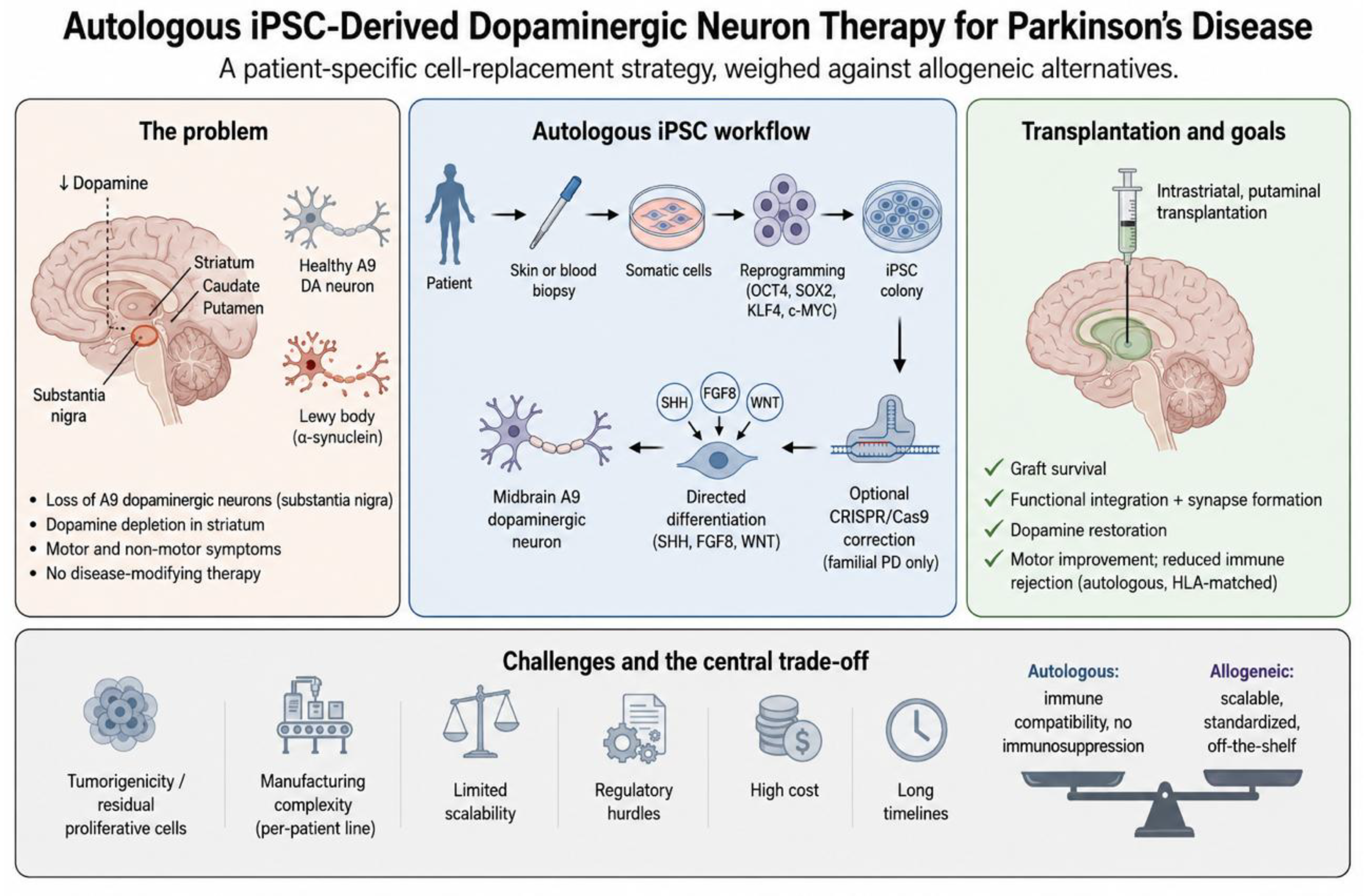
Figure 2.
Loss of nigral dopaminergic neurons unbalances the basal ganglia motor circuit in Parkinson’s disease. Degeneration of dopaminergic neurons in the substantia nigra pars compacta depletes dopamine in the dorsal striatum, shifting the balance between the direct (D1, facilitatory) and indirect (D2, inhibitory) pathways toward excessive inhibition of the thalamus and the motor signs of the disease. Cell replacement aims to restore striatal dopamine at this point in the circuit. Created with BioRender.com.
Figure 2.
Loss of nigral dopaminergic neurons unbalances the basal ganglia motor circuit in Parkinson’s disease. Degeneration of dopaminergic neurons in the substantia nigra pars compacta depletes dopamine in the dorsal striatum, shifting the balance between the direct (D1, facilitatory) and indirect (D2, inhibitory) pathways toward excessive inhibition of the thalamus and the motor signs of the disease. Cell replacement aims to restore striatal dopamine at this point in the circuit. Created with BioRender.com.
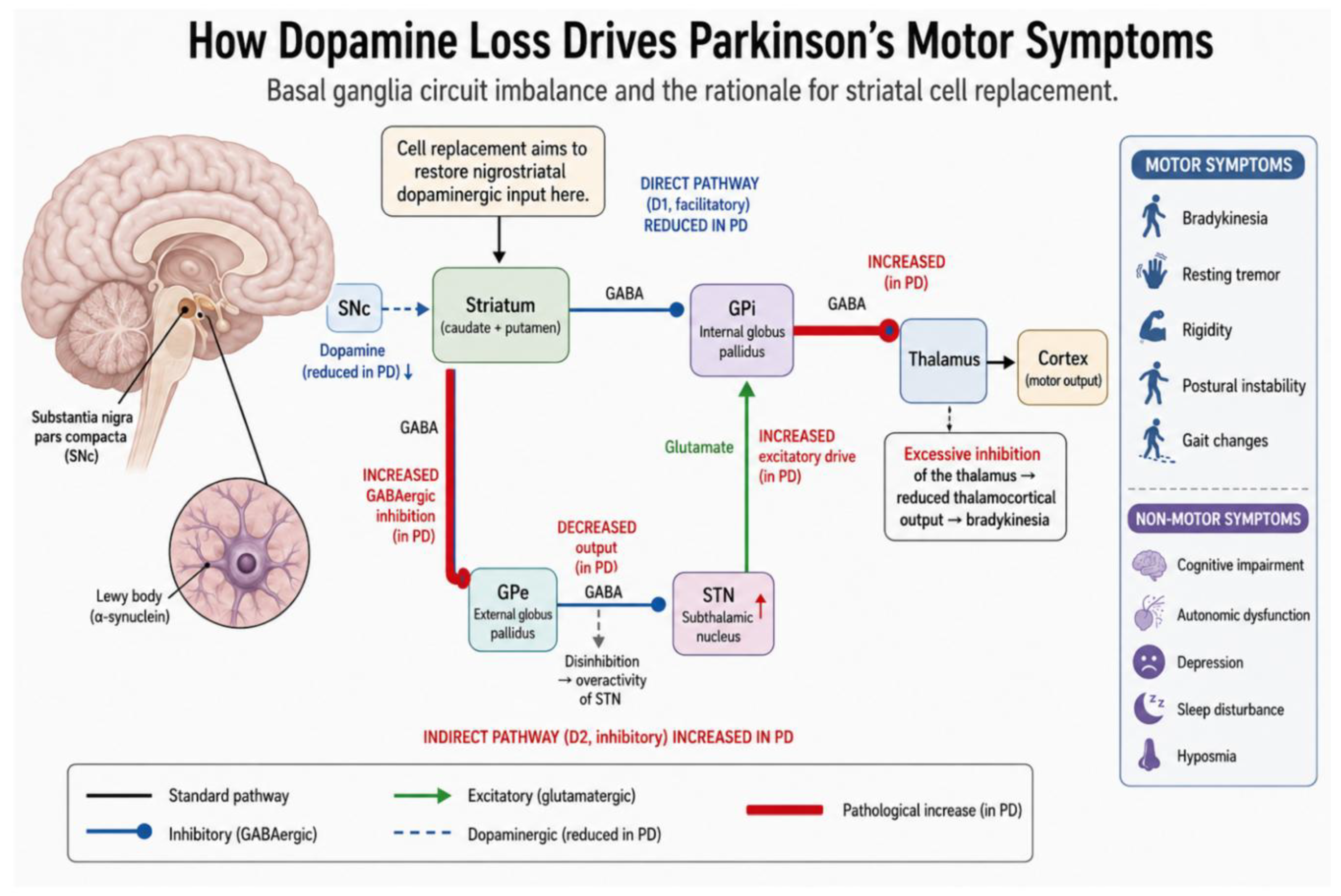
Figure 3.
Autologous versus allogeneic routes to iPSC-derived cell therapy. In the autologous route, induced pluripotent stem cells are generated from the patient’s own cells, so the derived neurons are immunologically matched; in the allogeneic route, a single banked donor line supplies a standardized product for many recipients but requires immunosuppression. The contrast in immune compatibility, scalability, and manufacturing that follows from this choice is the trade-off this review evaluates. Adapted from Cerneckis et al. [84], licensed under Creative Commons Attribution 4.0 (CC BY 4.0).
Figure 3.
Autologous versus allogeneic routes to iPSC-derived cell therapy. In the autologous route, induced pluripotent stem cells are generated from the patient’s own cells, so the derived neurons are immunologically matched; in the allogeneic route, a single banked donor line supplies a standardized product for many recipients but requires immunosuppression. The contrast in immune compatibility, scalability, and manufacturing that follows from this choice is the trade-off this review evaluates. Adapted from Cerneckis et al. [84], licensed under Creative Commons Attribution 4.0 (CC BY 4.0).
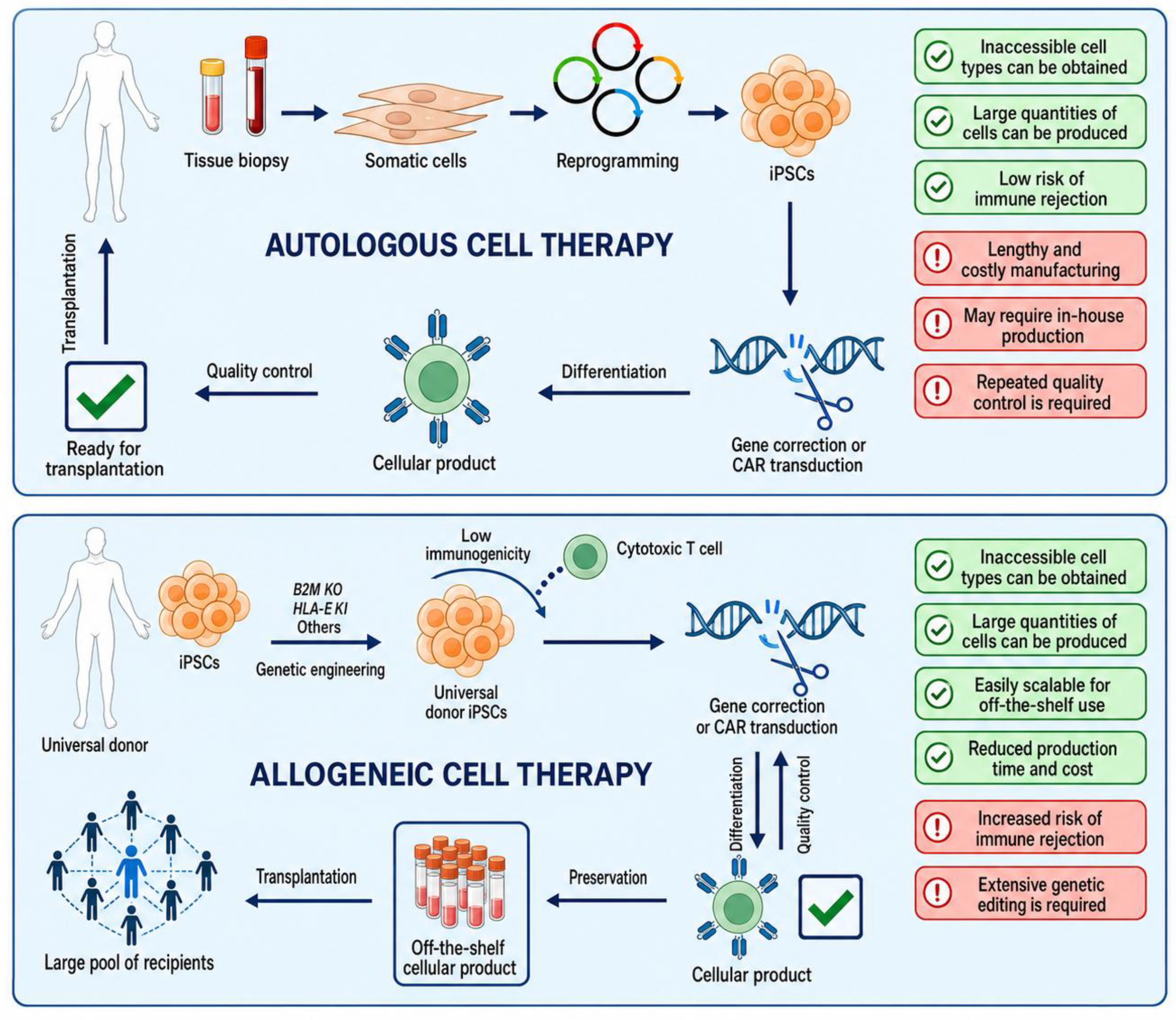
Figure 4.
The autologous iPSC therapy workflow, from patient cells to intrastriatal graft. Somatic cells from the patient are reprogrammed to induced pluripotent stem cells with the Yamanaka factors (OCT4, SOX2, KLF4, c-MYC), with the inset showing the epigenetic reset: pioneer-factor binding, TET-mediated DNA demethylation, and the shift from repressive H3K27me3 to active H3K4me3 marks. Disease-causing mutations may be corrected by CRISPR/Cas9 where applicable; the cells are then differentiated into midbrain dopaminergic neurons under SHH, FGF8, and WNT signaling; and the resulting neurons are transplanted into the striatum, where they release dopamine and support motor recovery. As the figure notes, variable reprogramming efficiency and the need to maximize authentic neuron identity and purity remain key limits on standardization. Created with BioRender.com.
Figure 4.
The autologous iPSC therapy workflow, from patient cells to intrastriatal graft. Somatic cells from the patient are reprogrammed to induced pluripotent stem cells with the Yamanaka factors (OCT4, SOX2, KLF4, c-MYC), with the inset showing the epigenetic reset: pioneer-factor binding, TET-mediated DNA demethylation, and the shift from repressive H3K27me3 to active H3K4me3 marks. Disease-causing mutations may be corrected by CRISPR/Cas9 where applicable; the cells are then differentiated into midbrain dopaminergic neurons under SHH, FGF8, and WNT signaling; and the resulting neurons are transplanted into the striatum, where they release dopamine and support motor recovery. As the figure notes, variable reprogramming efficiency and the need to maximize authentic neuron identity and purity remain key limits on standardization. Created with BioRender.com.
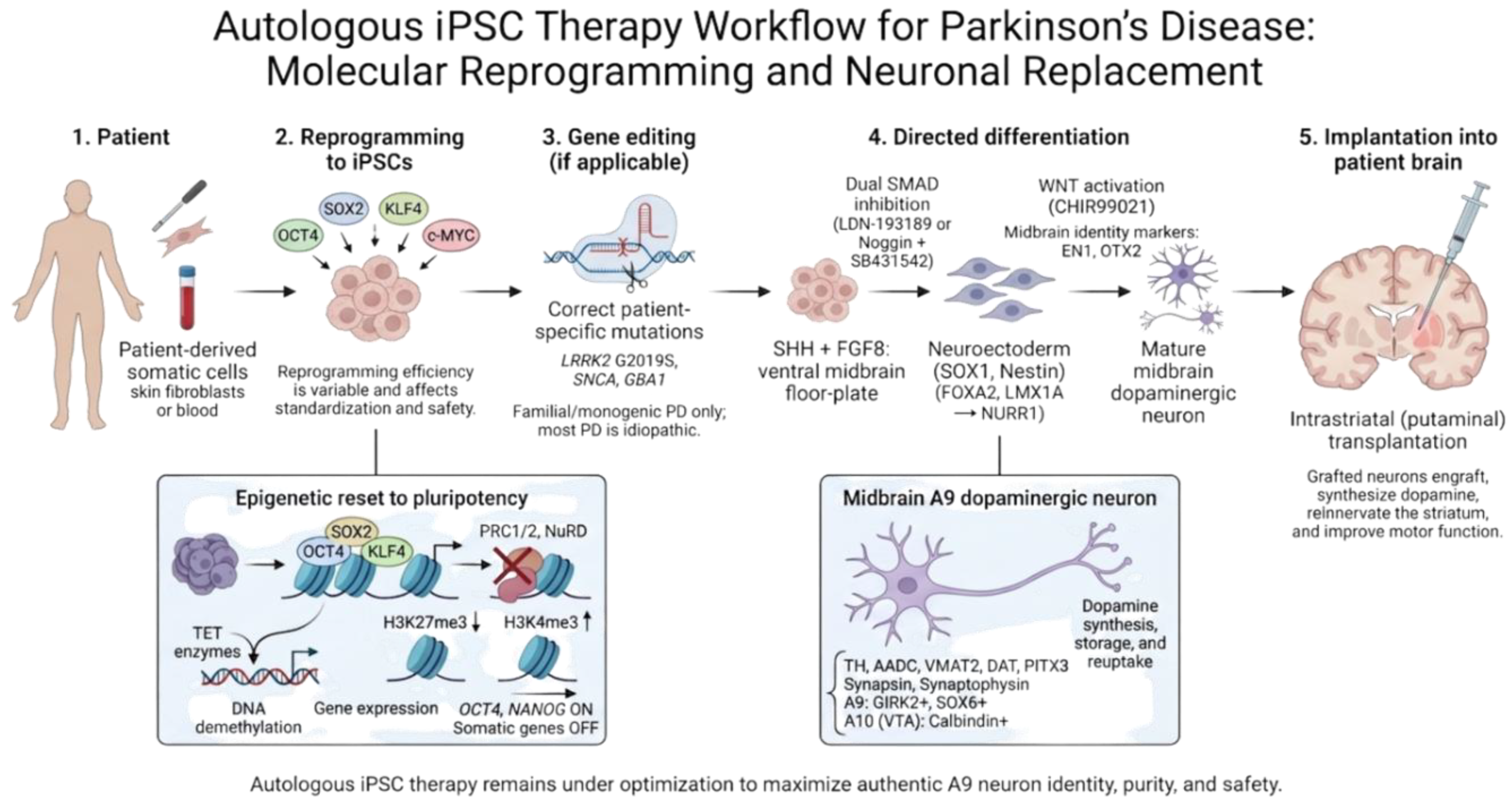

Table 1.
Comparison between Conventional Treatments and Stem Cell Therapy for Parkinson’s Disease.
| Aspects | Conventional Treatments for PD | Stem Cell Therapy for PD |
| Therapeutic Approach | Pharmacological Drugs (Levodopa, COMT inhibitors, Dopamine Agonists, etc); Physiotherapy; Surgical Treatments, DBS | Implantation of stem cell-derived midbrain dopaminergic progenitors, either autologous (iPSC) or allogeneic in origin |
| Underlying Mechanism | Augmentation of dopaminergic neurotransmission or modulation of basal ganglia circuitry via exogenous agents | Anatomical and functional restitution of dopaminergic pathways through integration of transplanted neurons |
| Primary Objective | Symptomatic relief of motor symptoms | Functional recovery with potential disease modification |
| Duration of Action | Temporary benefits with diminishing efficacy over time |
Sustained effects |
| Personalization | Standardized therapeutic protocols with limited patient-specific customization |
Personalized cellular therapies possible with autologous iPSC-derived neurons, minimizing immune rejection |
| Adverse Effects | Common adverse events include motor fluctuations, dyskinesias, hallucinations, and orthostatic hypotension | Potential complications include graft overgrowth, dysregulated dopamine release, and immunogenicity |
| Regulatory Status | Approved for widespread clinical application and integrated into treatment guidelines |
Currently under investigation in early-phase human clinical trials; not yet approved for routine clinical use |
| Disease Modification | No disease-modifying properties; interventions address downstream effects rather than etiological factors | Aims to modify disease progression by targeting primary neurodegenerative mechanisms |
Table 2.
Autologous versus allogeneic iPSC/PSC-derived dopaminergic cell therapy for Parkinson’s disease.
Table 2.
Autologous versus allogeneic iPSC/PSC-derived dopaminergic cell therapy for Parkinson’s disease.
| Dimension | Autologous | Allogeneic |
| Cell source | Patient’s own cells; one line per patient | Banked or master PSC line; one product for many |
| Immune and immunosuppression | HLA-matched, run without immunosuppression, but reprogramming-acquired mtDNA neoantigens can break tolerance [13,135,144] | Mismatched, needs immunosuppression, yet tolerated under moderate regimens regardless of HLA [16,98,143] |
| Manufacturing, scalability, cost | Bespoke; per-patient derivation and QC; line-to-line variability; high cost and delay [91,130,132,133] | Standardized, scalable, off-the-shelf; quality set once; commercial scale [142] |
| Genetic correction | Patient mutations correctable before transplant; mainly monogenic PD; base/prime editing preclinical [122,123,124] | Fixed line; engineerable (e.g., hypoimmune) but not personalized |
| Clinical maturity and regulation | Early-phase, mostly press-release data; Fast Track [13,135] | Peer-reviewed efficacy, first Phase 3, first approval; RMAT and Fast Track [16,98,136,137,142] |
| Shared caveat | Host α-synuclein can seed grafts of any origin over time [140,141]; tumorigenicity excluded by QC in trials so far | Same |
Disclaimer/Publisher’s Note: The statements, opinions and data contained in all publications are solely those of the individual author(s) and contributor(s) and not of MDPI and/or the editor(s). MDPI and/or the editor(s) disclaim responsibility for any injury to people or property resulting from any ideas, methods, instructions or products referred to in the content. |
© 2026 by the authors. Licensee MDPI, Basel, Switzerland. This article is an open access article distributed under the terms and conditions of the Creative Commons Attribution (CC BY) license (http://creativecommons.org/licenses/by/4.0/).
Copyright: This open access article is published under a Creative Commons CC BY 4.0 license, which permit the free download, distribution, and reuse, provided that the author and preprint are cited in any reuse.


