
Preprint
Article

This version is not peer-reviewed.

Baseline Investigation of Non-Wild-Type Bacterial Indicators and Antibiotic Resistance Genes in Urban Wastewater from Patras, Greece

Submitted:

29 June 2026

Posted:

01 July 2026

You are already at the latest version

Abstract

Antimicrobial resistance (AMR) is a growing public health concern that requires surveillance approaches capable of capturing resistance circulation at the community level. Wastewater-based monitoring provides an opportunity to assess antibiotic-resistant bacteria and antimicrobial resistance genes (ARGs) beyond clinical settings. This study investigated selected bacterial indicators and ARGs in untreated urban wastewater from the municipal wastewater treatment plant of Patras, Greece. Influent wastewater samples were analysed for Escherichia coli, Pseudomonas aeruginosa and Enterococcus spp. using culture-based methods. Isolates were tested for antimicrobial susceptibility by disk diffusion and Etest, and results were interpreted according to EUCAST epidemiological cut-off values. Molecular detection of ARGs was performed by real-time PCR targeting intl1, sul1, qnrS1, blaTEM, blaVIM, vanA and ermB. A total of 16 E. coli, 13 P. aeruginosa and 17 Enterococcus spp. isolates were included in the phenotypic analysis. Among E. coli, non-wild-type profiles were detected in 14/16 isolates for meropenem and 15/16 isolates for ciprofloxacin, while only 1/16 isolates showed a non-wild-type profile for ampicillin. In contrast, all P. aeruginosa isolates were classified as wild type for meropenem but non-wild type for ciprofloxacin, while all Enterococcus spp. isolates were classified as wild type for vancomycin and ampicillin. Molecular screening showed that blaTEM was the most frequently detected gene in E. coli isolates, followed by lower detection frequencies of qnrS1, intl1 and sul1. In P. aeruginosa, intl1 and sul1 were detected in a subset of isolates, whereas blaVIM was not detected. None of the targeted ARGs were detected in Enterococcus spp. These findings highlight the potential of untreated urban wastewater as a complementary matrix for community-level AMR surveillance and support the combined use of phenotypic and molecular approaches to better characterize resistance patterns in environmental settings.

Keywords: 
;  ;  ;  ;  ;  ;  ;  ;  

1. Introduction

Epidemiological surveillance is a fundamental function of Public Health, involving continuous and systematic collection, integration, analysis, interpretation, and timely dissemination of health-related data to support evidence-based public health action. Effective surveillance systems are essential for identifying priorities, assessing risks, monitoring epidemiological trends, and evaluating the impact of prevention and control measures at the population level. In this context, environmental monitoring has become increasingly important, as it provides insight into long-term ecological and microbial trends, supports policy evaluation, and contributes to the generation of new research hypotheses [1].
Wastewater-based epidemiology (WBE) has emerged as an innovative and complementary approach to conventional clinical surveillance, offering population-level information on the circulation of pathogens and other health-related biomarkers within a community. Through the analysis of raw wastewater samples, WBE enables the detection of biological markers, including DNA and RNA from microorganisms shed by infected or colonized individuals. Unlike clinical surveillance, which relies on individual testing, wastewater surveillance provides aggregated data from the population served by a sewerage network, without identifying individuals [2]. Initially applied in 2005 for monitoring illicit drug use, WBE has since evolved into a valuable public health tool capable of tracking infectious diseases, pharmaceuticals, antibiotics, and antimicrobial resistance indicators [3]. Its relevance was strongly demonstrated during the COVID-19 pandemic, when wastewater monitoring provided early information on viral circulation at the community level, often before increases in clinically reported cases [4].
Urban wastewater is a complex matrix composed of domestic, commercial, industrial, agricultural, and stormwater inputs. Domestic wastewater, in particular, contains human excreta and represents a major source of microorganisms entering sewerage systems [5]. Wastewater may contain a broad range of bacteria, viruses, protozoa, and parasites, making it an important substrate for studying microbial circulation and environmental dissemination. Among these microorganisms, bacteria represent one of the most diverse groups of human pathogens detected in wastewater. Several bacterial species colonize the human intestine and are excreted in faeces, while many are commensal or beneficial, others are pathogenic and may contribute to the microbial burden of wastewater. Important bacterial pathogens or opportunistic pathogens associated with wastewater include Salmonella spp., Escherichia spp., Shigella spp., Yersinia spp., Klebsiella spp., Leptospira spp., Vibrio cholerae, Aeromonas hydrophila, Legionella pneumophila, and Pseudomonas spp. Some of these organisms, such as L. pneumophila, Mycobacterium avium, Pseudomonas aeruginosa, and A. hydrophila, are primarily environmental opportunistic pathogens that may cause disease in individuals [6]
Τreatment plants are of particular interest for Antimicrobial Resistance (AMR) surveillance, as they receive antibiotics, Antibiotic-Resistant Bacteria (ARBs), and antimicrobial resistance genes (ARGs) from human, animal, healthcare, and environmental sources. A substantial proportion of antibiotics may be excreted in active forms and enter wastewater systems, where they may exert selective pressure and contribute to the persistence and dissemination of resistant bacteria and ARGs [7]. Through the discharge of treated effluents, resistant microorganisms and resistance determinants may re-enter aquatic and terrestrial ecosystems, reinforcing the environmental dimension of AMR transmission [8]. Recent research conducted in Patras further supports the value of WBE beyond viral monitoring, demonstrating its applicability for detecting pharmaceuticals and antibiotics and for generating information relevant to public health and the environmental dissemination of AMR [9].
AMR occurs when microorganisms, including bacteria, viruses, fungi, and parasites, adapt and grow in the presence of antimicrobial agents that were previously effective against them. Infections caused by resistant microorganisms are associated with increased morbidity, prolonged hospitalization, higher healthcare costs, use of second-line or more toxic drugs, and increased treatment failure [10]. AMR is driven by several interconnected factors, including inappropriate and excessive antimicrobial use, limited access to clean water, sanitation and hygiene, inadequate infection prevention and control measures, insufficient awareness, and weaknesses in legislation and implementation [7]. At the molecular level, AMR is associated with the presence and expression of ARGs, which may be maintained and disseminated through selection pressure, genetic mutations, and horizontal gene transfer [11,12] Mobile genetic elements, including plasmids, integrons, and transposons, further facilitate the spread of resistance among bacterial species [8]. Key resistance mechanisms include enzymatic inactivation of antibiotics, modification of target sites, reduced intracellular drug accumulation through efflux pumps, and biofilm formation, which can reduce antimicrobial efficacy [13].
Within wastewater-based AMR surveillance, Escherichia coli, Pseudomonas aeruginosa, and Enterococcus spp. are of relevance. E. coli is a Gram-negative bacillus associated with diarrhoeal disease, urinary tract infections, bacteraemia, pneumonia, and intra-abdominal infections [14]. Although intrinsically susceptible to many clinically relevant antimicrobial agents, E. coli has a high capacity to acquire and accumulate resistance genes, mainly through horizontal gene transfer [15]. It is also widely used as an indicator of faecal contamination in environmental waters and as a useful organism for AMR monitoring, given its role as both donor and recipient of ARGs [16].
Pseudomonas aeruginosa is a Gram-negative, aerobic, non-spore-forming opportunistic pathogen capable of causing a wide range of infections, particularly in immunocompromised individuals and patients with cystic fibrosis [17]. Its clinical importance is strongly linked to its intrinsic and acquired resistance to multiple antibiotics, its ability to form biofilms, and its capacity to develop multidrug-resistant phenotypes. These characteristics limit therapeutic options and make P. aeruginosa infections difficult to treat [18].
Enterococci are Gram-positive facultative anaerobic cocci commonly found in human faeces and the environment. They are important causes of healthcare-associated infections, including urinary tract infections, bacteraemia, infective endocarditis, and, less frequently, intra-abdominal infections and meningitis [14]. Due to their persistence in the environment and association with faecal contamination, enterococci are widely used as indicators of water quality and faecal pollution [19,20]. Their clinical significance is closely related to their intrinsic resistance to several antimicrobial agents and their ability to acquire and transfer resistance determinants through mobile genetic elements [21].
The global public health importance of AMR is substantial. According to the World Health Organization, more than 1.27 million deaths globally are directly attributable to infections caused by antibiotic-resistant bacteria worldwide [22]. The spread of resistant microorganisms reduces available treatment options, compromises the effectiveness of essential medical procedures that depend on antimicrobial prophylaxis or therapy, and increases the burden on healthcare systems. Because AMR arises and circulates across humans, animals, and the environment, its monitoring requires a One Health approach that recognizes the interconnectedness of these sectors [7,22]
WBE provides a unique opportunity to integrate the surveillance of viral pathogens and AMR indicators within a single population-level monitoring framework. Wastewater analysis can be used to detect respiratory viruses, while simultaneously assessing the occurrence of ARBs and ARGs. Although viruses and bacteria differ in their biology and transmission pathways, their co-occurrence in wastewater allows the combined surveillance of infectious disease trends and resistance dynamics. Such an integrated approach can support early warning, improve understanding of community-level health risks, and strengthen evidence-based decision-making in public health [22,23]
The aim of the present study was to investigate non wild type bacterial indicators and ARGs from the inlet wastewater using microbiological and molecular techniques. Through this approach, the study seeks to highlight the value of WBE as a tool for public health and integrated surveillance of infectious diseases and antimicrobial resistance within a One Health framework.

2. Materials and Methods

2.1. Study Design and Sampling Site

The study was conducted to investigate the occurrence of non-wild type indicators and selected antimicrobial resistance genes in urban wastewater for that reason wastewater samples were collected from the municipal wastewater treatment plant of Patras, Greece, which serves an estimated population of approximately 168,000 inhabitants. Sampling was performed at the inlet of the treatment plant, prior to wastewater treatment, in order to capture the microbial load entering the facility and to obtain a representative overview of community-level bacterial contamination. Samples were transported to the laboratory by authorized personnel and processed within a few hours after collection.

2.2. Bacterial Isolation and Identification

For bacterial analysis, 10 mL of diluted wastewater samples (10-3 and 10-4 dilutions) were filtered through 45-mm membrane filters, which were subsequently placed on selective culture media for the isolation of target bacterial groups. Escherichia coli was cultured on Chromocult Coliform Agar and incubated at 37 °C for 24 h, in accordance with EN ISO 9308-1. Enterococcus spp. was cultured on Slanetz and Bartley Agar and incubated at 36 ± 2 °C for 48 h, in accordance with EN ISO 7899-2, while Pseudomonas aeruginosa was cultured on Pseudomonas Agar Base (CN-supplemented) and incubated at 36 ± 2 °C for 48 h, in accordance with EN ISO 16266.
Identification of E. coli was based on characteristic colony morphology and coloration on the respective selective medium, with blue colonies considered indicative of E. coli., P. aeruginosa colonies were identified presumptively on the basis of green fluorescence under UV light (360 ± 20 nm), without further biochemical confirmation. Presumptive Enterococcus spp. colonies were confirmed by transferring the membrane filter onto pre-warmed (44 °C) Bile Aesculin Azide Agar and incubating for 2 h, following the rapid confirmation protocol of EN ISO 7899-2, the appearance of a black/brown discoloration in the surrounding medium was recorded as a positive reaction.
One representative colony per bacterial species per sample was selected using a sterile microbiological loop and subcultured in Tryptone Soy Broth (TSB) for 24 h. From each culture, a bacterial suspension was prepared and adjusted spectrophotometrically to a turbidity of 0.5 McFarland (corresponding to approximately 1-2 × 10⁸ CFU/mL), in accordance with the EUCAST guidelines (v.15.0). Within 15 min of preparation, each suspension was inoculated onto the entire surface of Mueller-Hinton Agar plates using a sterile cotton swab to obtain confluent (lawn) growth. Antimicrobial susceptibility was assessed by both the disk diffusion method and gradient strip testing (Etest), antimicrobial disks and Etest strips were applied within 15 min of inoculation, and plates were incubated at 35 ± 1 °C in air for 18 ± 2 h (24 h for vancomycin testing of Enterococcus spp.). Inhibition zone diameters and minimum inhibitory concentrations (MICs) were interpreted on the basis of the EUCAST epidemiological cut-off values (ECOFFs, v.15.0), and isolates were classified as wild-type or non-wild-type accordingly.

2.3. Antimicrobial Susceptibility Testing

Antimicrobial susceptibility testing was performed using the disk diffusion and Etest methods, with results interpreted on the basis of the EUCAST epidemiological cut-off values (T)ECOFFs), classifying isolates as wild-type or non-wild-type. The panel of antimicrobial agents was tailored to each bacterial species. For E. coli, meropenem (ΤECOFF 0.06 mg/L) and ciprofloxacin (T)ECOFF 0.06 mg/L were tested by Etest, while ampicillin was tested by disk diffusion. For P. aeruginosa, meropenem (Τ)ECOFF 2 mg/L and ciprofloxacin (ΤECOFF 0.5 mg/L) were tested by Etest. For Enterococcus spp., vancomycin (Τ)ECOFF 4 mg/L was tested by Etest, while ampicillin was tested by disk diffusion. The Etest method was used to determine the minimum inhibitory concentration (MIC) of the selected antibiotics, and the results were interpreted against the corresponding MIC-based (Τ)ECOFFs (mg/L), whereas disk diffusion was used to measure inhibition zone diameters, which were interpreted against the corresponding zone-diameter (Τ)ECOFFs (mm). Non-wild-type profiles were considered indicative of reduced susceptibility and the possible presence of acquired resistance mechanisms. Bacterial culture assays were performed in duplicate for each sample.
Two points regarding the Enterococcus spp. analysis should be noted. The isolates were not identified to the species level (e.g., E. faecalis versus E. faecium) as all Enterococcus spp. isolates in this study were classified as wild-type, the classification was unambiguous regardless of species, and species-level identification was therefore not required for the interpretation of the results and was not pursued. Moreover, the vancomycin (Τ)ECOFF 4 mg/L is identical for both species and was thus unaffected by this consideration.
Table 1. Antibiotics and methods used for the antimicrobial susceptibility testing of the isolates and EUCAST epidemiological cut-off values (Τ)ECOFFs for the tested antibiotics.
Bacteria Antibiotic Method EUCAST ΤECOFF Unit
E. coli Meropenem Etest (MIC) 0.06 mg/L
E. coli Ciprofloxacin Etest (MIC) 0.06 mg/L
E. coli Ampicillin (10 μg) Disk diffusion 13 mm
P. aeruginosa Meropenem Etest (MIC) 2 mg/L
P. aeruginosa Ciprofloxacin Etest (MIC) 0.5 mg/L
Enterococcus spp. Vancomycin Etest (MIC) 4 mg/L
Enterococcus spp. Ampicillin (2 μg) Disk diffusion 12 mm

2.4. Molecular Detection of Antimicrobial Resistance Genes

For molecular analysis, well-isolated single colonies from each bacterial culture were inoculated into Tryptic Soy Broth (TSB) and incubated for 24 h under agitation to obtain sufficient biomass. Total genomic DNA was extracted using the NucleoSpin Microbial DNA kit (Macherey-Nagel, Düren, Germany) according to the manufacturer's instructions. The presence of antimicrobial resistance genes was assessed by real-time PCR. The targets investigated were intl1, sul1, qnrS1, blaTEM, blaVIM, ermB and vanA, selected to represent genetic determinants associated with mobile genetic elements (intl1), sulfonamide resistance (sul1), quinolone resistance (qnrS1), β-lactam resistance (blaTEM), carbapenem resistance (blaVIM), macrolide resistance (ermB) and vancomycin resistance (vanA). In addition, a housekeeping reference gene was included for each organism to confirm successful DNA extraction, template integrity and bacterial identity: the gyrA gene for P. aeruginosa and the 16S rRNA gene (16S rDNA) for E. coli and Enterococcus spp. These targets served as internal amplification and process controls and were not used for the assessment of antimicrobial resistance.
Following determination of the amount of extracted DNA using Qubit Fluorometer, and detection of ARGs using real time-PCR, Aria MX (Agilent Technologies, USA) and Luna Universal Master Mix SYBR green (New England Biolabs, USA) using a specific set of primers for each gene. The primer sequences and annealing temperatures used in the real-time PCR assays are listed in Table 2 and real time PCR conditions are presented in Supplementary Table S1. PCR results were interpreted qualitatively as presence or absence of each target gene. A reaction was considered positive when amplification occurred above the defined threshold and was supported by an appropriate melting curve profile. Reactions with no Ct value, non-specific amplification, or values below the established limit of detection were considered negative. Borderline reactions were reported separately and were not included in the calculation of positive detection frequencies. The 16S rRNA gene assay was used as a reference marker to assess DNA amplifiability and assay sensitivity, with the lowest reliably detected standard corresponding to 10 copies/reaction. Because the study focused on qualitative screening of selected ARGs, gene-specific quantitative limits of detection were not established for all targets. The limit of detection (LOD) of the 16S rRNA gene qPCR assay was defined as the lowest quantified standard reliably detected, corresponding to 10 copies/reaction, with a mean Ct value of approximately 31.03.

3. Results

3.1. Antimicrobial Susceptibility Profiles of Bacterial Isolates

A total of 16 E. coli, 13 P. aeruginosa, and 17 Enterococcus spp. isolates were included in the phenotypic antimicrobial susceptibility analysis. Among the E. coli isolates, non-wild-type profiles were detected in 14/16 isolates for meropenem and in 15/16 isolates for ciprofloxacin, corresponding to 87.5% and 93.8% of the tested isolates, respectively. In contrast, ampicillin non-wild-type profiles were observed in only 1/16 E. coli isolate, corresponding to 6.3%, while the remaining isolates were classified as wild type. For P. aeruginosa, all tested isolates were classified as wild type for meropenem and non-wild type for ciprofloxacin. Among Enterococcus spp., all isolates were classified as wild type for both vancomycin and ampicillin, with no non-wild-type profiles observed among the 17 tested isolates. The antimicrobial susceptibility testing results are summarized in Table 3, and the frequency of non-wild-type (non-WT) phenotypes among bacterial isolates is shown in Figure 1.
Antimicrobial susceptibility results were interpreted using EUCAST (T)ECOFFs rather than clinical breakpoints, because the isolates originated from untreated urban wastewater and were analysed for environmental AMR surveillance purposes rather than clinical decision-making. Therefore, the terms wild type and non-wild type were used to describe whether isolates belonged to, or deviated from, the expected wild-type MIC distribution. This classification indicates possible acquired resistance mechanisms or other shifts in susceptibility but should not be interpreted as clinical resistance. For example, meropenem non-wild-type classification in E. coli and ciprofloxacin non-wild-type classification in P. aeruginosa reflect interpretation against the corresponding EUCAST (T)ECOFFs, not against clinical resistance breakpoints.

3.2. Detection of Antimicrobial Resistance Genes in E. coli

Molecular analysis of the 16 E. coli isolates showed that blaTEM was the most frequently detected resistance gene, identified in 9/16 isolates (56.3%), with two additional isolates showing borderline results. The plasmid-mediated quinolone resistance gene qnrS1 was detected in 3/16 isolates (18.8%), with one further borderline result. The class 1 integron-associated gene intl1 was detected in 2/16 isolates (12.5%), and the sulfonamide resistance gene sul1 in 1/16 isolates (6.3%). The 16S rRNA gene, included as a process and PCR control, was successfully amplified in all isolates, confirming the adequacy of the extracted DNA. These findings indicate that β-lactam resistance determinants were present in a substantial proportion of the E. coli isolates, accompanied by a lower frequency of plasmid-mediated quinolone, class 1 integron and sulfonamide resistance determinants (Supplementary Table S1).

3.3. Detection of Antimicrobial Resistance Genes in P. aeruginosa

Among the 13 P. aeruginosa isolates analysed, class 1 integron-associated gene intl1 was the most frequently detected target, identified in 6/13 isolates (46.2%), with one additional borderline result. The sulfonamide resistance gene sul1 was detected in 4/13 isolates (30.8%). The carbapenemase-associated gene blaVIM was not detected in any of the tested isolates. The gyrA gene, included as a process and PCR control, was successfully amplified in all isolates, confirming successful DNA extraction and template integrity. The concurrent detection of intl1 and sul1 in a subset of isolates suggests the presence of genetic elements associated with the dissemination of antimicrobial resistance in part of the wastewater-derived population. The absence of blaVIM was consistent with the wild-type meropenem profiles observed among the tested P. aeruginosa isolates, suggesting that this carbapenemase gene was not present in the analysed collection (Supplementary Table S2).

3.4. Detection of Antimicrobial Resistance Genes in Enterococcus spp.
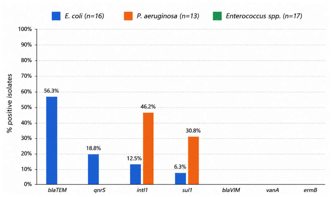
Among the 17 Enterococcus spp. isolates examined, none of the targeted antimicrobial resistance genes were detected, all isolates were negative for intl1, sul1, vanA and ermB (0/17). The 16S rRNA gene, included as a process and PCR control, was successfully amplified in all isolates, confirming the adequacy of the extracted DNA. These findings were consistent with the phenotypic results, as all tested Enterococcus spp. isolates were classified as wild type for vancomycin and ampicillin (Supplementary Table S3).
Figure 2. Frequency of antimicrobial resistance gene detection among bacterial isolates recovered from untreated urban wastewater, expressed as the percentage of isolates positive for each target gene by species. E. coli (n = 16) isolates were screened for blaTEM, qnrS1, intl1 and sul1, P. aeruginosa (n = 13) for intl1, sul1 and blaVIM, and Enterococcus spp. (n = 17) for intl1, sul1, vanA and ermB.
Preprints 220730 g002

4. Discussion

The present study highlights the potential of untreated urban wastewater as an informative matrix for monitoring antimicrobial resistance (AMR) indicators at the community level. By combining culture-based antimicrobial susceptibility testing with molecular detection of selected antimicrobial resistance genes (ARGs), the study provides complementary information on both phenotypic susceptibility profiles and resistance-associated genetic determinants in bacterial isolates recovered from influent wastewater. This approach is consistent with the concept of wastewater-based epidemiology as a complementary public health surveillance tool, since municipal wastewater aggregates biological material from large populations and may provide information on AMR indicators circulating beyond clinical settings [5,23,30,31,32].
Wastewater treatment plants are important interfaces between human activity, microbial pollution, antimicrobial residues and the receiving environment. Urban wastewater receives domestic, healthcare-associated and environmental inputs, creating conditions in which antibiotic-resistant bacteria, ARGs and mobile genetic elements may co-occur [23,30]. From a One Health perspective, untreated wastewater is therefore particularly relevant for monitoring AMR dissemination, as it can reflect signals originating from the served community and from environmental sources connected to the sewerage network. The detection of non-wild-type bacterial phenotypes and ARGs in untreated wastewater can thus provide useful information for community-level AMR surveillance and environmental risk assessment.
In the present study, non-wild-type profiles were mainly observed among Gram-negative isolates, particularly E. coli and P. aeruginosa. Among E. coli, 14/16 isolates were classified as non-wild-type for meropenem and 15/16 for ciprofloxacin, whereas only one isolate showed a non-wild-type profile for ampicillin. These findings suggest that reduced susceptibility to clinically relevant antibiotics was present in a substantial proportion of wastewater-derived E. coli isolates. E. coli is widely used as an indicator of faecal contamination and is also an important organism for AMR monitoring, due to its ability to acquire and disseminate resistance determinants through horizontal gene transfer [15]. Its detection in wastewater therefore provides relevant information on the possible circulation of resistant Enterobacteriaceae within the served community.
Τhe meropenem non-wild-type profiles observed among E. coli isolates and the ciprofloxacin non-wild-type profiles observed among P. aeruginosa isolates should not be interpreted as clinical resistance. In this study, isolates were classified as wild type or non-wild type using EUCAST (T)ECOFFs, as they originated from untreated urban wastewater and were analysed for environmental AMR surveillance purposes. Non-wild-type classification indicates deviation from the expected wild-type MIC distribution and possible acquired resistance mechanisms, whereas clinical resistance requires interpretation according to clinical breakpoints used for therapeutic decision-making.
Molecular analysis of the E. coli isolates showed that blaTEM was the most frequently detected resistance gene, identified in 9/16 isolates, with two additional isolates showing borderline results. This finding is relevant because blaTEM is associated with β-lactam resistance and has been frequently reported among E. coli and other Enterobacteriaceae in environmental and wastewater-associated settings [5,31]. The plasmid-mediated quinolone resistance gene qnrS1 was detected in 3/16 isolates, indicating that plasmid-mediated quinolone resistance determinants were present in part of the E. coli population. However, the frequency of qnrS1 detection was lower than the frequency of ciprofloxacin non-wild-type phenotypes, suggesting that additional mechanisms may contribute to reduced ciprofloxacin susceptibility, including chromosomal mutations in quinolone resistance-determining regions, altered membrane permeability or efflux pump activity [33,34].
The class 1 integron-associated gene intl1 and the sulfonamide resistance gene sul1 were detected at lower frequencies in E. coli. Although these targets were not the dominant findings in this bacterial group, their detection remains important because class 1 integrons and associated resistance genes are linked to anthropogenic pollution and the mobilization of resistance determinants [35]. The presence of these genes in wastewater-derived isolates supports the role of untreated wastewater as a matrix where mobile genetic elements and ARGs can be detected, even when they are present only in a subset of bacterial isolates.
For P. aeruginosa, all isolates were classified as wild type for meropenem, whereas all isolates were classified as non-wild type for ciprofloxacin. This pattern indicates that, within the tested isolate collection, reduced susceptibility was consistently observed for ciprofloxacin but not for meropenem. P. aeruginosa is an opportunistic pathogen of environmental importance, with well-recognized intrinsic and acquired mechanisms. Reduced susceptibility to fluoroquinolones such as ciprofloxacin may involve several mechanisms, including mutations in quinolone resistance-determining regions, efflux pump overexpression and reduced membrane permeability [33,34].
Molecular screening of P. aeruginosa detected the class 1 integron-associated gene intl1 in 6/13 isolates and the sulfonamide resistance gene sul1 in 4/13 isolates. The detection of these genes is important from an environmental AMR perspective, as intl1 has been proposed as a proxy marker for anthropogenic pollution and is frequently associated with genes conferring resistance to antibiotics, disinfectants and heavy metals [35]. Similarly, sul1 is commonly detected in wastewater environments and is often associated with class 1 integrons and anthropogenic resistance gene pollution [5,35]. The presence of intl1 and sul1 in P. aeruginosa isolates indicates the occurrence of genetic elements associated with AMR dissemination in part of the wastewater-derived bacterial population.
The carbapenemase-associated gene blaVIM was not detected in any of the P. aeruginosa isolates. This finding is consistent with the phenotypic classification of all P. aeruginosa isolates as wild type for meropenem. However, the absence of blaVIM does not exclude the presence of other resistance determinants or mechanisms not included in the molecular panel. Broader molecular panels or sequencing-based approaches would be required to characterize the full resistome and to identify additional genetic determinants that may be present in wastewater-derived bacterial populations [31,32]
Among Enterococcus spp., all tested isolates were classified as wild type for both vancomycin and ampicillin. Accordingly, no phenotypic evidence of reduced susceptibility to these antibiotics was observed in the tested Enterococcus spp. isolates. Molecular analysis also showed that none of the targeted resistance genes, including vanA, ermB, intl1 and sul1, were detected in this bacterial group. The absence of vanA is consistent with the wild-type vancomycin susceptibility profiles observed phenotypically. Similarly, the absence of ermB indicates that this specific macrolide resistance determinant was not detected in the tested isolates. However, because the isolates were not identified to species level and the molecular panel included only selected genes, further characterization would be required to fully assess the resistance potential of wastewater-derived enterococci.
In summary, the findings demonstrate that phenotypic and molecular AMR indicators were not uniformly distributed across the bacterial groups examined. E. coli showed frequent non-wild-type profiles for both meropenem and ciprofloxacin and carried several ARGs, especially blaTEM. P. aeruginosa showed a consistent ciprofloxacin non-wild-type phenotype while remaining wild type for meropenem and carried intl1 and sul1 in a subset of isolates. In contrast, Enterococcus spp. isolates were phenotypically wild type for the tested antibiotics and were negative for the targeted ARGs. These results highlight that culture-based phenotypic testing and targeted molecular detection provide different but complementary information. Phenotypic testing reflects the expressed susceptibility profile under the conditions tested, whereas PCR-based detection identifies selected genetic determinants that may be present but not necessarily expressed, or may not fully explain the observed phenotype.
The incomplete concordance between phenotypic and molecular findings, particularly for ciprofloxacin non-wild-type profiles in E. coli and P. aeruginosa, highlights the complexity of AMR characterization in environmental matrices. In several cases, the observed non-wild-type phenotypes were not fully explained by the specific resistance genes included in the targeted PCR panel. These may include genes not targeted by the assay, chromosomal mutations, efflux-mediated resistance, permeability changes, differential gene expression or limitations related to detection thresholds [31,33,34]. No quinolone-resistance-specific determinant or mutation analysis was included for P. aeruginosa in the present targeted PCR panel. Further investigation of mechanisms such as gyrA/parC mutations or efflux pump-related resistance was beyond the technical scope of this study, as it would have required additional validated assays or sequencing-based approaches. Furthermore, targeted PCR should not be interpreted as a complete substitute for phenotypic antimicrobial susceptibility testing. Instead, the two approaches should be used together to provide a broader understanding of resistance patterns in wastewater-derived bacterial isolates.
From a public health perspective, the detection of non-wild-type E. coli isolates, ciprofloxacin non-wild-type P. aeruginosa isolates and selected ARGs in untreated urban wastewater supports the value of wastewater-based monitoring as a complementary tool for AMR surveillance. Untreated wastewater can provide aggregated information on bacterial populations and resistance determinants circulating within the served community, including signals that may not be captured through routine clinical surveillance alone [23,32]. These findings are particularly relevant within a One Health framework, as wastewater treatment plants represent interfaces between human activity, microbial contamination and the receiving aquatic environment.
To our knowledge, this is the first investigation of antibiotic-resistant bacteria and selected antimicrobial resistance genes in untreated urban wastewater from the city of Patras using microbiological and molecular techniques. These findings provide a first local baseline for future wastewater-based AMR surveillance in the region. Future studies should include larger numbers of isolates, longitudinal sampling, species-level confirmation of bacterial isolates and expanded molecular panels covering additional resistance genes and mechanisms. Sequencing-based approaches would also be useful to clarify the genetic basis of phenotypic resistance, investigate mobile genetic elements and explore the diversity of resistance determinants present in wastewater-derived bacteria [31,32]. Such integrated approaches could improve the interpretation of wastewater-based AMR data and strengthen its application as part of community-level and environmental AMR surveillance.

5. Conclusions

This study highlights the potential of untreated urban wastewater as a useful matrix for monitoring antimicrobial resistance at the community level. The detection of non-wild-type antimicrobial susceptibility profiles mainly among E. coli isolates, and for ciprofloxacin among P. aeruginosa isolates, indicates that wastewater can reflect the circulation of bacterial populations with reduced antimicrobial susceptibility in the served population. In contrast, Enterococcus spp. isolates were classified as wild type for the tested antimicrobial agents, suggesting that reduced susceptibility was not observed in this bacterial group within the analysed isolate collection. Moreover, molecular analysis further confirmed the presence of selected antimicrobial resistance genes, particularly blaTEM in E. coli and intl1 and sul1 in P. aeruginosa, supporting the role of wastewater as a reservoir of resistance-associated genetic determinants. However, the partial discrepancies between phenotypic non-wild-type profiles and the targeted genes detected underline the complexity of antimicrobial resistance in environmental matrices. These findings suggest that targeted PCR approaches should be interpreted alongside phenotypic testing and, where possible, complemented by broader molecular methods such as expanded ARG panels or sequencing-based analyses. Ιn conclusion, wastewater-based monitoring can provide complementary information to conventional clinical surveillance and may contribute to the early detection of AMR trends within a One Health framework. Future studies should include longitudinal sampling, larger numbers of isolates, species-level confirmation, and more comprehensive molecular characterization to better understand the persistence, dissemination and public health relevance of antimicrobial resistance in urban wastewater systems.

Supplementary Materials

The following supporting information can be downloaded at the website of this paper posted on Preprints.org.

Author Contributions

Conceptualization, A.V. and Z.A.; methodology, A.V, Z.A., R.F. N. G., and M.A.; validation, A.V., Z.A. R.F. and M.A.; formal analysis, K.C., A.P. K.A.K.; investigation, K.C., Z.A., A.P. K.A.K; resources, A.V.; data curation, K.C. and Z.A.; writing-original draft preparation, K.C. and Z.A.; writing-review and editing, A.V, R.F., N.G.; visualization, K.C.; supervision, A.V.; project administration, A.V. and Z.A.; funding acquisition, A.V. All authors have read and agreed to the published version of the manuscript.

Funding

This research was co-funded by the EU4HEALTH, One Bridge project under Grant Agreement Nr 101233407 and the National Public Health Organization (NPHO) of Greece.

Data Availability Statement

All data is available.

Acknowledgments

We acknowledge the support received by the Wastewater-based Surveillance Network of Greece, the WWTP of Municipality of Patras and the National Public Health Organization (NPHO) of Greece.

Declaration of AI-Assisted Technologies in the Writing Process

During the preparation of this manuscript, the authors used OpenAI for basic language support, limited to checks of grammar, spelling, syntax, and punctuation. The AI tool was not used for study design, data collection, data analysis, data interpretation, generation of scientific conclusions, or preparation of original research content. All AI-assisted edits were reviewed, verified, and approved by the authors, who take full responsibility for the content of the manuscript.

Conflicts of Interest

The authors declare no conflicts of interest.

References

  1. Mazzotti, F. J.; Hughes, N.; Harvey, R. G. Why Do We Need Environmental Monitoring for Everglades Restoration? EDIS 2008, 2008(2). [Google Scholar] [CrossRef]
  2. Polo, D.; Quintela-Baluja, M.; Corbishley, A.; Jones, D. L.; Singer, A. C.; Graham, D. W.; Romalde, J. L. Making waves: Wastewater-based epidemiology for COVID-19 – approaches and challenges for surveillance and prediction. Water Res. 2020, 186. [Google Scholar] [CrossRef] [PubMed]
  3. Zuccato, E.; Chiabrando, C.; Castiglioni, S.; Bagnati, R.; Fanelli, R. Estimating community drug abuse by wastewater analysis. Environ. Health Perspect. 2008, 116(8), 1027–1032. [Google Scholar] [CrossRef] [PubMed]
  4. Markt, R.; Stillebacher, F.; Nägele, F.; Kammerer, A.; Peer, N.; Payr, M.; Scheffknecht, C.; Dria, S.; Draxl-Weiskopf, S.; Mayr, M.; Rauch, W.; Kreuzinger, N.; Rainer, L.; Bachner, F.; Zuba, M.; Ostermann, H.; Lackner, N.; Insam, H.; Wagner, A. O. Expanding the Pathogen Panel in Wastewater Epidemiology to Influenza and Norovirus. Viruses 2023, 15(2). [Google Scholar] [CrossRef] [PubMed]
  5. Cacace, D.; Fatta-Kassinos, D.; Manaia, C.M.; Cytryn, E.; Kreuzinger, N.; Rizzo, L.; Karaolia, P.; Schwartz, T.; Alexander, J.; Merlin, C.; Garelick, H.; Schmitt, H.; de Vries, D.; Schwermer, C.U.; Meric, S.; Tandoi, V.; Henriques, I.; Michael, I.; Pärnänen, K.M.M.; et al. Antibiotic resistance genes in treated wastewater and in the receiving water bodies: A pan-European survey of urban settings. Water Res. 2019, 162, 320–330. [Google Scholar] [CrossRef] [PubMed]
  6. Chahal, C.; van den Akker, B.; Young, F.; Franco, C.; Blackbeard, J.; Monis, P. Pathogen and Particle Associations in Wastewater: Significance and Implications for Treatment and Disinfection Processes. Adv. Appl. Microbiol. 2016, 97, 63–119. [Google Scholar] [CrossRef] [PubMed]
  7. Velazquez-Meza, M. E.; Galarde-López, M.; Carrillo-Quiróz, B.; Alpuche-Aranda, C. M. Antimicrobial resistance: One Health approach. In Veterinary World; Veterinary World, 2022; Vol. 15, Number 3, pp. 743–749. [Google Scholar] [CrossRef] [PubMed]
  8. Verraes, C.; Van Boxstael, S.; Van Meervenne, E.; Van Coillie, E.; Butaye, P.; Catry, B.; de Schaetzen, M. A.; Van Huffel, X.; Imberechts, H.; Dierick, K.; Daube, G.; Saegerman, C.; De Block, J.; Dewulf, J.; Herman, L. Antimicrobial resistance in the food chain: A review. In International Journal of Environmental Research and Public Health; MDPI, 2013; Vol. 10, Number 7, pp. 2643–2669. [Google Scholar] [CrossRef] [PubMed]
  9. Anastopoulou, Z.; Sazakli, E.; Bouzoukas, C.; Kostakis, M. G.; Leotsinidis, M.; Thomaidis, N. S.; Vantarakis, A. Tracking Pharmaceuticals and Illicit Drugs in Urban Wastewater: Drug Consumption Trends in Patras, Greece. Int. J. Environ. Res. 2026, 20(2), 80. [Google Scholar] [CrossRef]
  10. Dadgostar, P. Antimicrobial resistance: implications and costs. In Infection and Drug Resistance; Dove Medical Press Ltd, 2019; Vol. 12, pp. 3903–3910. [Google Scholar] [CrossRef] [PubMed]
  11. Zankari, E.; Hasman, H.; Cosentino, S.; Vestergaard, M.; Rasmussen, S.; Lund, O.; Aarestrup, F. M.; Larsen, M. V. Identification of acquired antimicrobial resistance genes. J. Antimicrob. Chemother. 2012, 67(11), 2640–2644. [Google Scholar] [CrossRef] [PubMed]
  12. Singh, A. K.; Kaur, R.; Verma, S.; Singh, S. Antimicrobials and Antibiotic Resistance Genes in Water Bodies: Pollution, Risk, and Control. Front. Environ. Sci. 2022, 10. [Google Scholar] [CrossRef]
  13. Pulingam, T.; Parumasivam, T.; Gazzali, A. M.; Sulaiman, A. M.; Chee, J. Y.; Lakshmanan, M.; Chin, C. F.; Sudesh, K. Antimicrobial resistance: Prevalence, economic burden, mechanisms of resistance and strategies to overcome. Eur. J. Pharm. Sci. 2022, 170, 106103. [Google Scholar] [CrossRef] [PubMed]
  14. Mueller, M.; Tainter, C. R. Escherichia coli infection. In StatPearls; StatPearls Publishing, 2023; Available online: https://www.ncbi.nlm.nih.gov/books/NBK564298/.
  15. Poirel, L.; Madec, J. Y.; Lupo, A.; Schink, A. K.; Kieffer, N.; Nordmann, P.; Schwarz, S. Antimicrobial resistance in Escherichia coli. Microbiol. Spectr. 2018, 6(4), ARBA-0026-2017. [Google Scholar] [CrossRef] [PubMed]
  16. Bojar, B.; Sheridan, J.; Beattie, R.; Cahak, C.; Liedhegner, E.; Munoz-Price, L. S.; Hristova, K. R.; Skwor, T. Antibiotic resistance patterns of Escherichia coli isolates from the clinic through the wastewater pathway. Int. J. Hyg. Environ. Health 2021, 238, 113863. [Google Scholar] [CrossRef] [PubMed]
  17. Pang, Z.; Raudonis, R.; Glick, B. R.; Lin, T.-J.; Cheng, Z. Antibiotic resistance in Pseudomonas aeruginosa: mechanisms and alternative therapeutic strategies. Biotechnol. Adv. 2019, 37(1), 177–192. [Google Scholar] [CrossRef] [PubMed]
  18. Roulová, N.; Mot’ková, P.; Brožková, I.; Pejchalová, M. Antibiotic resistance of Pseudomonas aeruginosa isolated from hospital wastewater in the Czech Republic. J. Water Health 2022, 20(4), 692–701. [Google Scholar] [CrossRef] [PubMed]
  19. Boehm, A. B.; Sassoubre, L. M. Enterococci as indicators of environmental fecal contamination. In Enterococci: From commensals to leading causes of drug resistant infection. Massachusetts Eye and Ear Infirmary.; Gilmore, M. S., Clewell, D. B., Ike, Y., Shankar, N., Eds.; 2014; Available online: https://www.ncbi.nlm.nih.gov/books/NBK190421/.
  20. Byappanahalli, M. N.; Nevers, M. B.; Korajkic, A.; Staley, Z. R.; Harwood, V. J. Enterococci in the environment. Microbiol. Mol. Biol. Rev. 2012, 76(4), 685–706. [Google Scholar] [CrossRef] [PubMed]
  21. Kristich, C. J.; Rice, L. B.; Arias, C. A. Enterococcal infection—treatment and antibiotic resistance. In Enterococci: From commensals to leading causes of drug resistant infection. Massachusetts Eye and Ear Infirmary.; Gilmore, M. S., Clewell, D. B., Ike, Y., Shankar, N., Eds.; 2014; Available online: https://www.ncbi.nlm.nih.gov/books/NBK190420/.
  22. World Health Organization. Antimicrobial resistance. 2023. Available online: https://www.who.int/news-room/fact-sheets/detail/antimicrobial-resistance.
  23. Rizzo, L.; Manaia, C.; Merlin, C.; Schwartz, T.; Dagot, C.; Ploy, M. C.; Michael, I.; Fatta-Kassinos, D. Urban wastewater treatment plants as hotspots for antibiotic resistant bacteria and genes spread into the environment: A review. Sci. Total Environ. 2013, 447, 345–360. [Google Scholar] [CrossRef] [PubMed]
  24. Sucato, A.; Nazzaro, F.; Scaloni, A.; De Angelis, M.; Mauriello, G. A comparative analysis of aquatic and polyethylene-associated antibiotic-resistant microbiota in the Mediterranean Sea. Biology 2021, 10, 168. [Google Scholar] [CrossRef]
  25. Jayavarsha, V.; Smiline Girija, A.S.; Gunasekaran, S.; Priyadharsini, J.V. Characterization of vancomycin resistant enterococci and drug ligand interaction between vanA of E. faecalis with the bio-compounds from Aegle marmelos. J. Pharmacopunct. 2023, 26, 247–256. [Google Scholar] [CrossRef] [PubMed]
  26. Fallah, F.; Borhan, R. S.; Hashemi, A. Detection of blaIMP and blaVIM metallo-β-lactamases genes among Pseudomonas aeruginosa strains. Int. J. Burn. Trauma 2013, 3(2), 122–124. Available online: https://pmc.ncbi.nlm.nih.gov/articles/PMC3636667/.
  27. Akasaka, T.; Tanaka, M.; Yamaguchi, A.; Sato, K. Type II topoisomerase mutations in fluoroquinolone-resistant clinical strains of Pseudomonas aeruginosa isolated in 1998 and 1999: Role of target enzyme in mechanism of fluoroquinolone resistance. Antimicrob. Agents Chemother. 2001, 45(8), 2263–2268. [Google Scholar] [CrossRef] [PubMed]
  28. Chau, K. K.; Barker, L.; Budgell, E. P.; Vihta, K. D.; Sims, N.; Kasprzyk-Hordern, B.; Harriss, E.; Crook, D. W.; Read, D. S.; Walker, A. S.; Stoesser, N. Systematic review of wastewater surveillance of antimicrobial resistance in human populations. Environ. Int. 2022, 162, 107171. [Google Scholar] [CrossRef] [PubMed]
  29. Milligan, E. G.; Calarco, J.; Davis, B. C.; Harwood, V. J.; Pruden, A. A systematic review of culture-based methods for monitoring antibiotic-resistant Acinetobacter, Aeromonas, and Pseudomonas as environmentally relevant pathogens in wastewater and surface water. Curr. Environ. Health Rep. 2023, 10(2), 132–149. [Google Scholar] [CrossRef]
  30. Berendonk, T. U.; Manaia, C. M.; Merlin, C.; Fatta-Kassinos, D.; Cytryn, E.; Walsh, F.; Bürgmann, H.; Sørum, H.; Norström, M.; Pons, M. N.; Kreuzinger, N.; Huovinen, P.; Stefani, S.; Schwartz, T.; Kisand, V.; Baquero, F.; Martinez, J. L. Tackling antibiotic resistance: The environmental framework. Nat. Rev. Microbiol. 2015, 13(5), 310–317. [Google Scholar] [CrossRef] [PubMed]
  31. Karkman, A.; Do, T. T.; Walsh, F.; Virta, M. P. J. Antibiotic-resistance genes in waste water. Trends Microbiol. 2018, 26(3), 220–228. [Google Scholar] [CrossRef] [PubMed]
  32. Hendriksen, R. S.; Munk, P.; Njage, P.; van Bunnik, B.; McNally, L.; Lukjancenko, O.; Röder, T.; Nieuwenhuijse, D.; Pedersen, S. K.; Kjeldgaard, J.; Kaas, R. S.; Clausen, P. T. L. C.; Vogt, J. K.; Leekitcharoenphon, P.; van de Schans, M. G. M.; Zuidema, T.; de Roda Husman, A. M.; Rasmussen, S.; Petersen, B.; Global Sewage Surveillance Project Consortium. Global monitoring of antimicrobial resistance based on metagenomics analyses of urban sewage. Nat. Commun. 2019, 10, 1124. [Google Scholar] [CrossRef] [PubMed]
  33. Robicsek, A.; Jacoby, G. A.; Hooper, D. C. The worldwide emergence of plasmid-mediated quinolone resistance. Lancet Infect. Dis. 2006, 6(10), 629–640. [Google Scholar] [CrossRef] [PubMed]
  34. Marti, E.; Variatza, E.; Balcázar, J. L. The role of aquatic ecosystems as reservoirs of antibiotic resistance. Trends Microbiol. 2014, 22(1), 36–41. [Google Scholar] [CrossRef] [PubMed]
  35. Gillings, M. R.; Gaze, W. H.; Pruden, A.; Smalla, K.; Tiedje, J. M.; Zhu, Y. G. Using the class 1 integron-integrase gene as a proxy for anthropogenic pollution. ISME J. 2015, 9(6), 1269–1279. [Google Scholar] [CrossRef] [PubMed]
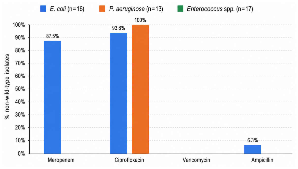
Figure 1. Frequency of non-wild-type (non-WT) phenotypes among bacterial isolates recovered from untreated urban wastewater, expressed as the percentage of non-WT isolates per antibiotic for each bacterial group.
Preprints 220730 g001
Table 2. Primer sequences and annealing temperatures used for real-time PCR assays.
Genes Primer sequence Annealing temperature(°C) Reference
intl1 5’-GATCGGTCGAATGCGTGT-3’
5’-GCCTTGATGTTACCCGAGAG-3’
59 [5]
sul1 5’-CGCACCGGAAACATCGCTGCAC-3’
5’- TGAAGTTCCGCCGCAAGGCTCG-3’
60 [5]
qnrS1 5’-GACGTGCTAACTTGCGTGAT-3’
5’-TGGCATTGTTGGAAACTTG-3’
62 [24]
blaTEM 5’-TTCCTGTTTTTGCTCACCCAG-3’
5’-CTCAAGGATCTTACCGCTGTTG-3’
60 [5]
ermB 5’-CCGAACACTAGGGTTGCTC-3’
5’-ATCTGGAACATCTGTGGTATG-3’
55 [5]
vanA 5’-TCTGCAATAGAGATAGCCGC -3’
5’-GGAGTAGCTATCCCAGCATT -3’
62 [25]
blaVIM 5’-GTTTGGTCGCATATCGCAAC-3’
5’-AATGCGCAGCACAGGATAG-3’
58 [26]
gyrA 5’-AGTCCTATCTCGACTACGCGAT-3’
5’-AGTCGACGGTTTCCTTTTCCAG-3’
60 [27]
16S rRNA gene 5’-TCCTACGGGAGGCAGCAGT-3’
5’-ATTACCGCGGCTGCTGG-3’
60 [5]
Table 3. Antimicrobial susceptibility testing results and classification as wild-type (WT) or non-wild-type (non-WT) for bacterial isolates recovered from urban wastewater. Etest results were interpreted using MIC-based EUCAST (T)ECOFFs, while disk diffusion results were interpreted using the corresponding zone-diameter (T)ECOFFs. “—” indicates that the specific antimicrobial agent was not tested or was not applicable for the respective bacterial group.
Sampling Date Microorganism MER. Etest CIP. Etest VAN. Etest Amp. 10 μg disk Amp. 2 μg disk
12/05/2026 E. coli non-WT non-WT WT
13/05/2026 E. coli WT WT WT
18/05/2026 E. coli WT non-WT WT
09/06/2025 E. coli non-WT non-WT WT
17/06/2025 E. coli non-WT non-WT WT
25/08/2025 E. coli non-WT non-WT WT
26/08/2025 E. coli non-WT non-WT WT
27/08/2025 E. coli non-WT non-WT WT
15/09/2025 E. coli non-WT non-WT WT
17/09/2025 E. coli non-WT non-WT WT
29/10/2025 E. coli non-WT non-WT WT
03/11/2025 E. coli non-WT non-WT WT
24/11/2025 E. coli non-WT non-WT non-WT
25/11/2025 E. coli non-WT non-WT WT
26/11/2025 E. coli non-WT non-WT WT
01/12/2025 E. coli non-WT non-WT WT
12/05/2026 P. aeruginosa WT non-WT
13/05/2026 P. aeruginosa WT non-WT
01/09/2025 P. aeruginosa WT non-WT
03/09/2025 P. aeruginosa WT non-WT
08/09/2025 P. aeruginosa WT non-WT
10/09/2025 P. aeruginosa WT non-WT
15/09/2025 P. aeruginosa WT non-WT
17/09/2025 P. aeruginosa WT non-WT
22/10/2025 P. aeruginosa WT non-WT
27/10/2025 P. aeruginosa WT non-WT
29/10/2025 P. aeruginosa WT non-WT
03/11/2025 P. aeruginosa WT non-WT
24/11/2025 P. aeruginosa WT non-WT
12/05/2026 Enterococcus spp. WT WT
13/05/2026 Enterococcus spp. WT WT
18/05/2026 Enterococcus spp. WT WT
08/09/2025 Enterococcus spp. WT WT
10/09/2025 Enterococcus spp. WT WT
15/09/2025 Enterococcus spp. WT WT
17/09/2025 Enterococcus spp. WT WT
22/10/2025 Enterococcus spp. WT WT
27/10/2025 Enterococcus spp. WT WT
29/10/2025 Enterococcus spp. WT WT
03/11/2025 Enterococcus spp. WT WT
24/11/2025 Enterococcus spp. WT WT
25/11/2025 Enterococcus spp. WT WT
01/12/2025 Enterococcus spp. WT WT
09/02/2026 Enterococcus spp. WT WT
10/02/2026 Enterococcus spp. WT WT
11/02/2026 Enterococcus spp. WT WT
Disclaimer/Publisher’s Note: The statements, opinions and data contained in all publications are solely those of the individual author(s) and contributor(s) and not of MDPI and/or the editor(s). MDPI and/or the editor(s) disclaim responsibility for any injury to people or property resulting from any ideas, methods, instructions or products referred to in the content.
Copyright: This open access article is published under a Creative Commons CC BY 4.0 license, which permit the free download, distribution, and reuse, provided that the author and preprint are cited in any reuse.
Prerpints.org logo

Preprints.org is a free preprint server supported by MDPI in Basel, Switzerland.

Subscribe

© 2026 MDPI (Basel, Switzerland) unless otherwise stated

Accessibility

Disclaimer

Terms of Use

Privacy Policy

Privacy Settings